
Postherpetic Pseudolymphomatous Angiosarcoma Concealed Within Milia en Plaque: Expanding the Spectrum of Wolf Isotopic Response with a Literature Review


Abstract
1. Introduction
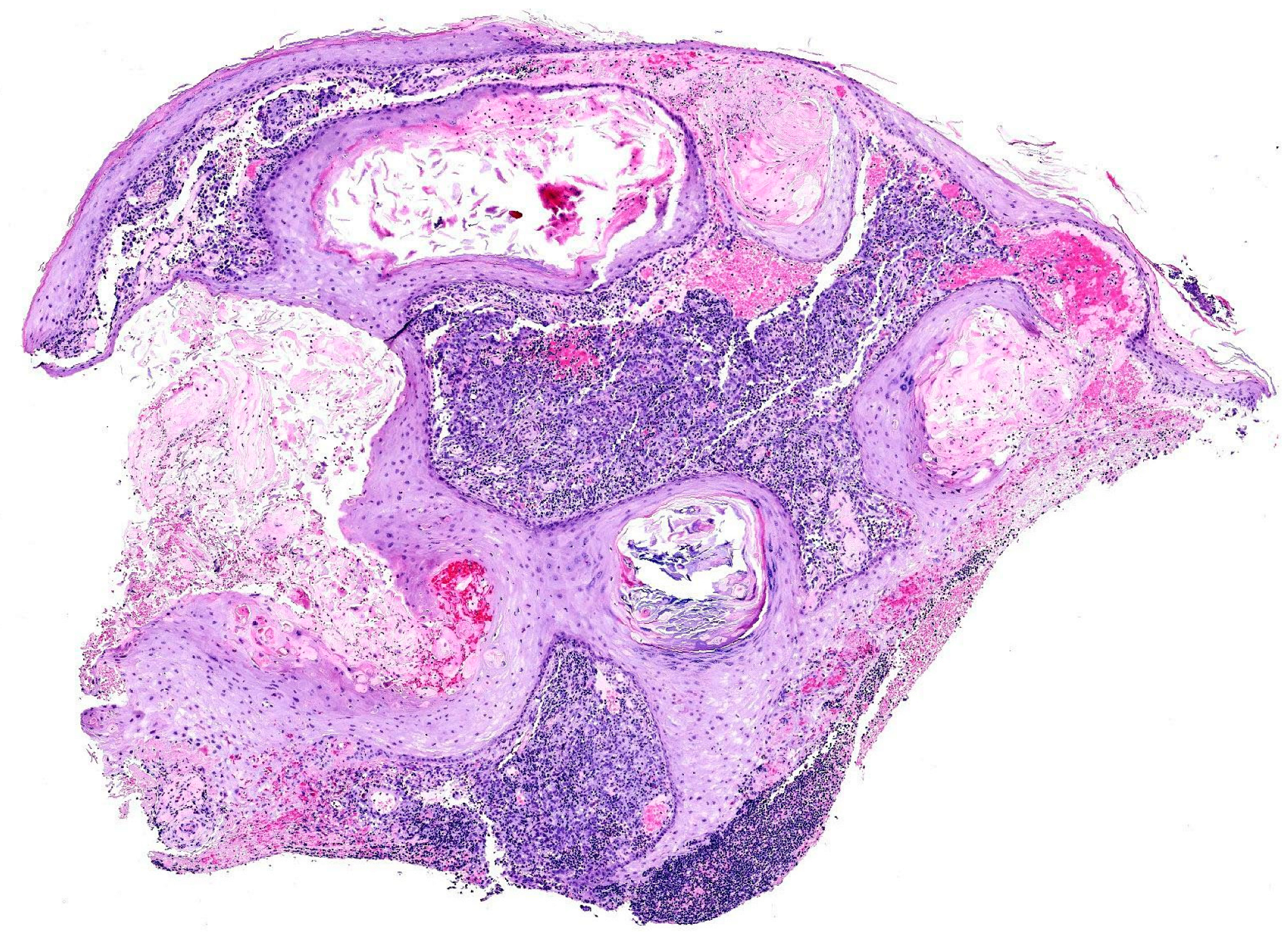
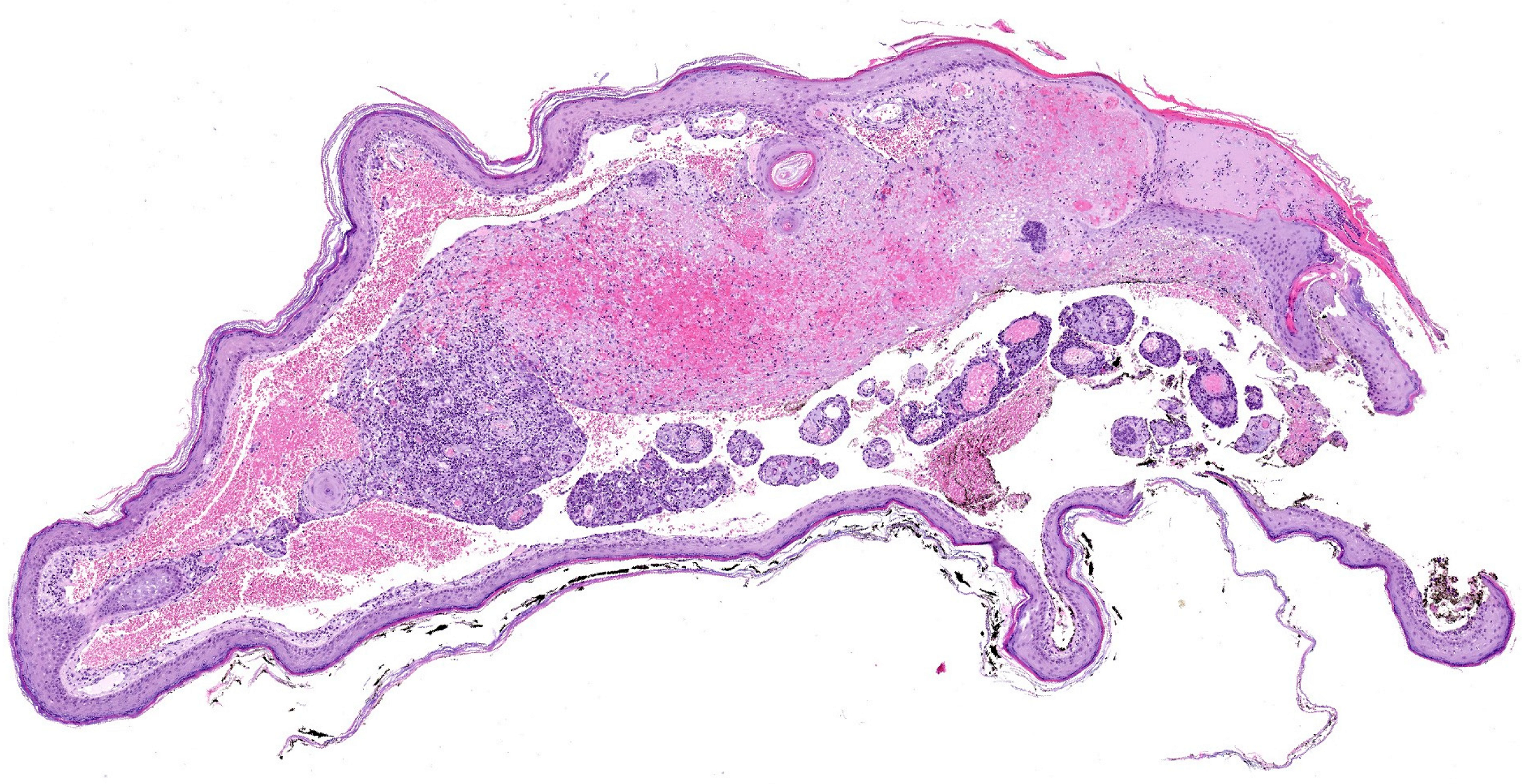
2. Case Report
3. Discussion
- Viral persistence: This may explain some postherpetic reactions since viral DNA has been isolated in certain cases. However, after herpetic infections resolve, viral DNA is no longer detected. This mechanism likely applies only to reactions occurring within a short interval (less than four weeks) after the herpes infection [6,15].
- Lymphovascular microvasculature alteration: Damage to the microcirculation can render an area unable to respond adequately to subsequent insults, leading to localized inflammation at the same site [6].
- Neural injury: Damaged dermal nerve fibers can contribute to disease pathogenesis, either directly through neuropeptide release or indirectly via immune system activation [6]. Specifically, neuropeptides such as nerve growth factor (NGF), which regulates skin epithelization, angiogenesis, and the formation of an extracellular matrix [17], as well as substance P from damaged nerve endings, which might play a crucial role in inducing the development of epidermal changes [18].

| Inflammatory Diseases | Infections |
|---|---|
| Acne | Molluscum contagiosum |
| Acneiform lesions | Warts and papillomata |
| Actinic granuloma (O’Brien) [21] | Candidiasis |
| Bullous pemphigoid [18] | Dermatophytosis |
| Chronic cutaneous graft-versus-host disease | |
| Chronic small vessel vasculitis (Extrafacial eosinophilic granuloma or Lever granuloma) [22] | |
| Chronic urticaria [23] | |
| Comedones | |
| Comedonic-microcystic reactions | |
| Contact dermatitis | |
| “Dysimmune” reactions | |
| Eosinophilic dermatosis | |
| Erythema annulare centrifugum | |
| Fibroelastolytic papulosis | |
| Folliculitis (granulomatous or eosinophilic [24]) | Tumors and pseudotumors |
| Follicular mucinosis [25] | Angiosarcoma |
| Furunculosis | Basal cell carcinoma |
| Granuloma annulare [26] | Basosquamous carcinoma |
| Granulomatous folliculitis | Benign lymphangioendothelioma [19] |
| Granulomatous reactions (necrotizing or non-necrotizing) | Bowen disease |
| Granulomatous vasculitis | Breast carcinoma |
| Grover disease (personal case) | Kaposi sarcoma |
| Infections | Leukemia |
| Keloid | Lymphangiogenesis (pseudotumoral) [19] |
| Lichen planus | Lymphomas |
| Lichen sclerosus et atrophicus-morphea | Mastocytosis [27] |
| Lichen simplex | Metastases (from breast carcinoma and others) |
| Lichenoid dermatitis | Pseudolymphoma |
| Linear IgA dermatosis | Rosai-Dorfman disease |
| Lupus erythematosus [28] | Squamous cell carcinoma |
| Milia | Syringoid eccrine carcinoma [18] |
| Mucinosis | Tufted Angioma [29] |
| Nodular solar degeneration | |
| Palpable purpura | |
| Pityriasis rosea (atypical) [30] | |
| Prurigo-like eruption | |
| Psoriasis | |
| Reactive perforating collagenosis | |
| Rosacea | |
| Sarcoidosis | |
| Unilateral nevoid telangiectasia [31] | |
| Vitiligo [32] | |
| Xanthoma [31] |
4. Conclusions
5. Future Directions
Funding
Acknowledgments
Conflicts of Interest
References
- Wolf, R.; Wolf, D.; Ruocco, E.; Brunetti, G.; Ruocco, V. Wolf’s isotopic response. Clin. Dermatol. 2011, 29, 237–240. [Google Scholar]
- Wyburn-Mason, R. Malignant change arising in tissues affected by herpes. Br. Med. J. 1955, 2, 1106–1109. [Google Scholar] [PubMed]
- Wolf, R.; Brenner, S.; Ruocco, V.; Filioli, F.G. Isotopic response. Int. J. Dermatol. 1995, 34, 341–348. [Google Scholar]
- Happle, R.; Kluger, N. Koebner’s sheep in Wolf’s clothing: Does the isotopic response exist as a distinct phenomenon? J. Eur. Acad. Dermatol. Venereol. 2018, 32, 542–543. [Google Scholar] [PubMed]
- Sanchez, D.P.; Sonthalia, S. Koebner Phenomenon. In StatPearls; StatPearls Publishing: Treasure Island, FL, USA, 2025. Available online: https://www.ncbi.nlm.nih.gov/books/NBK553108/ (accessed on 8 February 2025).
- Ruocco, V.; Ruocco, E.; Piccolo, V.; Brunetti, G.; Guerrera, L.P.; Wolf, R. The immunocompromised district in dermatology: A unifying pathogenic view of the regional immune dysregulation. Clin. Dermatol. 2014, 32, 569–576. [Google Scholar]
- Jenkins, A.M.; Skinner, D.; North, J. Postherpetic isotopic responses with 3 simultaneously occurring reactions following herpes zoster. Cutis 2018, 101, 195–197. [Google Scholar]
- Schmidt, A.P.; Tjarks, B.J.; Lynch, D.W. Gone fishing: A unique histologic pattern in cutaneous angiosarcoma. Cutis 2018, 101, 270–272. [Google Scholar] [PubMed]
- Hudson, C.P.; Hanno, R.; Callen, J.P. Cutaneous angiosarcoma in a site of healed herpes zoster. Int. J. Dermatol. 1984, 23, 404–407. [Google Scholar]
- Kwon, C.W.; Stephens, D.M.; Gilmore, E.S.; Tausk, F.A.; Scott, G.A. Cutaneous Pseudolymphoma Arising as Wolf’s Post-Herpetic Isotopic Response. JAMA Dermatol. 2017, 153, 1198–1200. [Google Scholar]
- Sanchez-Salas, M.P. Appearance of comedones at the site of healed herpes zoster: Wolf’s isotopic response. Int. J. Dermatol. 2011, 50, 633–634. [Google Scholar]
- Elgoweini, M.; Blessing, K.; Jackson, R.; Duthie, F.; Burden, A.D. Coexistent granulomatous vasculitis and leukaemia cutis in a patient with resolving herpes zoster. Clin. Exp. Dermatol. 2011, 36, 749–751. [Google Scholar] [PubMed]
- Requena, L.; Santonja, C.; Stutz, N.; Kaddu, S.; Weenig, R.H.; Kutzner, H.; Menzel, T.; Cerroni, L. Pseudolymphomatous cutaneous angiosarcoma: A rare variant of cutaneous angiosarcoma readily mistaken for cutaneous lymphoma. Am. J. Dermatopathol. 2007, 29, 342–350. [Google Scholar] [PubMed]
- Diaz-Cascajo, C.; Weyers, W.; Borghi, S.; Reichel, M. Verrucous angiosarcoma of the skin: A distinct variant of cutaneous angiosarcoma. Histopathology 1998, 32, 556–561. [Google Scholar]
- Kuet, K.; Tiffin, N.; McDonagh, A. An unusual eruption following herpes zoster infection. Clin. Exp. Dermatol. 2019, 44, 197–199. [Google Scholar]
- Ise, M.; Tanese, K.; Adachi, T.; Du, W.; Amagai, M.; Ohyama, M. Postherpetic Wolf’s isotopic response: Possible contribution of resident memory T cells to the pathogenesis of lichenoid reaction. Br. J. Dermatol. 2015, 173, 1331–1334. [Google Scholar]
- El Baassiri, M.G.; Dosh, L.; Haidar, H.; Gerges, A.; Baassiri, S.; Leone, A.; Rappa, F.; Jurjus, A. Nerve growth factor and burn wound healing: Update of molecular interactions with skin cells. Burns 2023, 49, 989–1002. [Google Scholar]
- Wang, T.; Zhang, M.; Zhang, Y.; Zhang, S.; Qu, T.; Liu, Y.; Jin, H. Wolf’s Isotopic Response after Herpes Zoster Infection: A Study of 24 New Cases and Literature Review. Acta Derm. Venereol. 2019, 99, 953–959. [Google Scholar]
- Schnebelen, A.M.; Page, J.; Gardner, J.M.; Shalin, S.C. Benign lymphangioendothelioma presenting as a giant flank mass. J. Cutan. Pathol. 2015, 42, 217–221. [Google Scholar] [PubMed]
- Cai, Y.-T.; Xu, H.; Guo, Y.; Guo, N.-N.; Li, Y.-M. A Case Report on Acquired Tufted Angioma with Severe Pain after Healed Herpes Zoster. Chin. Med. J. 2018, 131, 2378–2379. [Google Scholar]
- Kim, Y.J.; Kim, S.K.; Kim, Y.C.; Kang, H.Y. Actinic Granuloma Developed in a Herpes Zoster Scar. Ann. Dermatol. 2007, 19, 35–37. [Google Scholar]
- Melgar, E.; Henry, J.; Valois, A.; Dubois-Lacour, M.B.; Truchetet, F.; Cribier, B.; Cuny, J.F. Extra-facial Lever granuloma on a herpes zoster scar: Wolf’s isotopic response. Ann. Dermatol. Venereol. 2018, 145, 354–358. [Google Scholar]
- Lee, H.J.; Ahn, W.K.; Chae, K.S.; Ha, S.J.; Kim, J.W. Localized chronic urticaria at the site of healed herpes zoster. Acta Derm. Venereol. 1999, 79, 168. [Google Scholar] [PubMed]
- Xv, L.; Wang, B.; Zhu, Q.; Zhang, G. A Case of Isotopic Response Presented with Eosinophilic Pustular Folliculitis. Clin. Cosmet. Investig. Dermatol. 2023, 16, 1749–1752. [Google Scholar] [CrossRef]
- Blakey, B.L.; Gratrix, M.L. Reactive benign follicular mucinosis: A report of 2 cases. Cutis 2012, 89, 266–268. [Google Scholar]
- Rademaker, R.; van Dalen, E.; Ossenkoppele, P.M.; Knuiman, G.J.; Kemme, S.M. Granuloma annulare after herpes zoster: Wolf’s isotopic response. Ned. Tijdschr. Voor Geneeskd. 2021, 165, D5344. [Google Scholar]
- Sarsik, S.; Soliman, S.H.; Elhalaby, R.E. Unusual presentation of telangiectasia macularis eruptiva perstans at the site of healed herpes zoster; Wolf’s isotopic response. Australas. J. Dermatol. 2023, 64, e237–e2440. [Google Scholar]
- Darsha, A.; Oldenburg, R.; Hinds, B.; Paravar, T. A crack in the armor: Wolf isotopic response manifesting as cutaneous lupus. Dermatol. Online J. 2022, 28, 8. [Google Scholar] [CrossRef]
- Rahman, S.; Daveluy, S. Pathergy Test. In StatPearls; StatPearls Publishing: Treasure Island, FL, USA, 2025. Available online: https://www.ncbi.nlm.nih.gov/books/NBK558909/ (accessed on 9 February 2025).
- Ustuner, P.; Balevi, A.; Ozdemir, M.; Türkmen, İ. Atypical pityriasis rosea presenting with a herald patch lesion on the healed site of herpes zoster: Wolf’s isotopic response. Eur. J. Dermatol. 2016, 26, 102–103. [Google Scholar]
- Jaka-Moreno, A.; López-Pestaña, A.; López-Núñez, M.; Ormaechea-Pérez, N.; Vildosola-Esturo, S.; Tuneu-Valls, A.; Lobo-Morán, C. Wolf’s Isotopic Response: A Series of 9 Cases. Actas Dermo-Sifiliogr. 2012, 103, 798–805. [Google Scholar]
- Cheng, J.-R.; Mao, H.; Wang, Y.-J.; Hui, H.-Z.; Zheng, J.; Shi, B.-J. Postherpetic Vitiligo: A Wolf’s Isotopic Response. Indian J. Dermatol. 2024, 69, 195–196. [Google Scholar] [PubMed]
- Ji, Y.-Z.; Liu, S.-R. Koebner phenomenon leading to the formation of new psoriatic lesions: Evidences and mechanisms. Biosci. Rep. 2019, 39, BSR20193266. [Google Scholar] [CrossRef]
- Lo Schiavo, A.; Ruocco, E.; Russo, T.; Brancaccio, G. Locus minoris resistentiae: An old but still valid way of thinking in medicine. Clin. Dermatol. 2014, 32, 553–556. [Google Scholar] [CrossRef]
- Zuehlke, R.L.; Rapini, R.P.; Puhl, S.C.; Ray, T.L. Dermatitis in loco minoris resistentiae. J. Am. Acad. Dermatol. 1982, 6, 1010–1013. [Google Scholar] [CrossRef]
- Sagher, F.; Liban, E.; Kocsard, E. Specific Tissue Alteration in Leprous Skin: VI. “Isopathic Phenomenon” Following BCG Vaccination in Leprous Patients. AMA Arch. Dermatol. Syphilol. 1954, 70, 631–639. [Google Scholar] [CrossRef]
- Lewandowsky, F.; Lutz, W. Ein Fall einer bisher nicht beschriebenen Hauterkrankung (Epidermodysplasia verruciformis). Arch. Für Dermatol. Und Syph. 1922, 141, 193–203. [Google Scholar] [CrossRef]
- Ghosh, S.; Jain, V.K. “Pseudo” Nomenclature in Dermatology: What’s in a Name? Indian J. Dermatol. 2013, 58, 369–376. [Google Scholar] [CrossRef]
- Berthelot, C.; Dickerson, M.C.; Rady, P.; He, Q.; Niroomand, F.; Tyring, S.K.; Pandya, A.G. Treatment of a patient with epidermodysplasia verruciformis carrying a novel EVER2 mutation with imiquimod. J. Am. Acad. Dermatol. 2007, 56, 882–886. [Google Scholar] [CrossRef]
- Happle, R. The Renbok phenomenon: An inverse Kobner reaction observed in alopecia areata. Eur. J. Dermatol. 1991, 2, 39–40. [Google Scholar]
- Ovcharenko, Y.; Litus, O.; Khobzey, K.; Serbina, I.; Zlotogorski, A. Inverse Koebner reaction observed in alopecia areata: The Renboek phenomenon. J. Investig. Dermatol. 2013, 133, 1406. [Google Scholar]

| Term | First Described | Definition and * Interesting Facts | Example |
|---|---|---|---|
| Koebner isomorphic phenomenon [4,5] | H. Koebner, 1872 | Development of lesions from an existing cutaneous disease in previously healthy skin that has undergone non-specific injury, which can be mild. This disease may appear in locations where it typically does not occur. | Psoriasis lesions developing on surgical scars in a patient with underlying psoriasis. |
| Wolf isotopic response [2,3] | R. Wyburn-Mason, 1955; refined by R. Wolf, 1995 | Appearance of new skin lesions at the site of a previously healed, unrelated skin disease. | Comedones appearing exactly at the site of a previously healed herpes zoster. |
| Locus minoris resistentiae [34,35] | R.L. Zuehlke, 1982 | Areas of the body that are more vulnerable than others to suffer some pathologic conditions. * Its origins trace back to ancient myths, such as Achilles’ heel or Siegfried’s shoulder. | Eczema appearing in skin previously damaged by surgery or burns. |
| Isopathic phenomenon [36] | F. Sagher, 1954 | The injection of specific or non-specific proteins triggers the development of a disease already present in the patient. | In leprous patients, the cutaneous injection of several substances provokes changes typical of lepromatous leprosy. Conversely, in healthy individuals, the same injections cause only nonspecific inflammatory reaction. Similar reactions can be triggered by insect bites. |
| Pseudoisomorphic response (pseudo-Koebner) [37,38,39] | Unknown. First examples in the literature from F. Lewandowsky, W. Lutz, 1922 | Spread of a cutaneous infection along a line of previously damaged skin. | Lineal lesions in epidermodysplasia verruciformis. Commonly seen in warts and molluscum contagiosum. |
| Renboek/Renbök phenomenon (inverse or reverse Koebner phenomenon) [40,41] | R. Happle, 1991 | Disappearance of an existing skin condition following the onset of a new dermatosis at the same site. * The term “Renboek” or “Renbök” is not an eponym, but “Koebner” spelled backwards. | A plaque of scalp psoriasis disappears as a patch of alopecia areata develops in the same location. |
| Pathergy [29] | Blobner, 1937 | A minor trauma on healthy skin induces non-specific cutaneous lesions, often rich in neutrophils. | Frequently seen in pyoderma gangrenosum, Sweet’s syndrome or Beçet’s disease. |
Disclaimer/Publisher’s Note: The statements, opinions and data contained in all publications are solely those of the individual author(s) and contributor(s) and not of MDPI and/or the editor(s). MDPI and/or the editor(s) disclaim responsibility for any injury to people or property resulting from any ideas, methods, instructions or products referred to in the content. |
© 2025 by the authors. Published by MDPI on behalf of the European Society of Dermatopathology. Licensee MDPI, Basel, Switzerland. This article is an open access article distributed under the terms and conditions of the Creative Commons Attribution (CC BY) license (https://creativecommons.org/licenses/by/4.0/).
Share and Cite
Corral-Forteza, M.; Pérez-Muñoz, N.; Fernández-Figueras, M.-T. Postherpetic Pseudolymphomatous Angiosarcoma Concealed Within Milia en Plaque: Expanding the Spectrum of Wolf Isotopic Response with a Literature Review. Dermatopathology 2025, 12, 9. https://doi.org/10.3390/dermatopathology12020009
Corral-Forteza M, Pérez-Muñoz N, Fernández-Figueras M-T. Postherpetic Pseudolymphomatous Angiosarcoma Concealed Within Milia en Plaque: Expanding the Spectrum of Wolf Isotopic Response with a Literature Review. Dermatopathology. 2025; 12(2):9. https://doi.org/10.3390/dermatopathology12020009
Chicago/Turabian StyleCorral-Forteza, Marina, Noelia Pérez-Muñoz, and Maria-Teresa Fernández-Figueras. 2025. "Postherpetic Pseudolymphomatous Angiosarcoma Concealed Within Milia en Plaque: Expanding the Spectrum of Wolf Isotopic Response with a Literature Review" Dermatopathology 12, no. 2: 9. https://doi.org/10.3390/dermatopathology12020009
APA StyleCorral-Forteza, M., Pérez-Muñoz, N., & Fernández-Figueras, M.-T. (2025). Postherpetic Pseudolymphomatous Angiosarcoma Concealed Within Milia en Plaque: Expanding the Spectrum of Wolf Isotopic Response with a Literature Review. Dermatopathology, 12(2), 9. https://doi.org/10.3390/dermatopathology12020009