Article

Influence of Process Design on the Preparation of Solid Lipid Nanoparticles by an Ultrasonic-Nanoemulsification Method

by
Agata Pucek-Kaczmarek
Department of Physical and Quantum Chemistry, Faculty of Chemistry, Wrocław University of Science and Technology, 53-370 Wrocław, Poland
Processes 2021, 9(8), 1265; https://doi.org/10.3390/pr9081265
Submission received: 28 June 2021 / Revised: 19 July 2021 / Accepted: 21 July 2021 / Published: 22 July 2021
(This article belongs to the Special Issue Nanoemulsion Processes Design and Applications)

Abstract

:
In recent years, lipid-based nanosystems have emerged as a promising class of nanocarriers for encapsulating many active agents. Solid lipid nanoparticles (SLNs) provide good stability (colloidal as well as physical) and high biocompatibility. Appropriate design of the carrier structure through a selection of components and preparation methods allows us to obtain formulations with desired physicochemical parameters and biological properties. The present contribution has been carried out to investigate SLNs containing biocompatible phosphatidylcholine mixed with non-ionic surfactant Tween 60 as stabilizing agents. The internal lipid phase consisted of glyceryl monostearate was confirmed as safe for drug delivery by the Food and Drug Administration. The SLNs were fabricated by ultrasonic-nanoemulsification method. The preparation process was optimized in regard to variable parameters such as ultrasonication time and used amplitude and number of cycles. The sizes of the studied nanoparticles along with the size distribution were determined by dynamic light scattering (DLS), while shape and morphology were determined by atomic force microscopy (AFM) and transmission electron microscopy (TEM). The colloidal stability was measured by a turbidimetric method. The physical state of SLNs was characterized using differential scanning calorimetry (DSC). The obtained results indicate that the proposed SLNs may provide great potential for design and preparation of novel delivery nanosystems with a variety of possible applications.

Graphical Abstract

1. Introduction

In the last few decades, lipid-based formulations have emerged as an attractive platform for encapsulating a number of active compounds that can be difficult to deliver on their own. Solid lipid nanoparticles (SLNs) can be considered as one of the most promising carriers due to their multiple properties. SLNs are nano-sized water-dispersible colloidal system, similar to oil-in-water nanoemulsions, but their cores are composed of a biodegradable and physiologically tolerated lipid-based matrix that, unlike nanoemulsions, remains solid both at room and human body temperature [1]. To achieve the reduction of the interfacial tension between the aqueous medium and the lipid core, surfactants, or a mixture of surfactants, are used to stabilize the formulation [2]. The main benefits of SLNs include high biocompatibility and low toxicity, good stability, protection of encapsulated cargo against degradation, as well as improvement of the bioavailability of active components and the possibility of controlled/prolonged drug release. SLNs require inexpensive, organic solvent-free fabrication methods that are usually easily scalable [3,4].
Solid lipid nanoparticles are able to entrap both lipophilic and hydrophilic active ingredients [5,6]. Despite the lipid core being hydrophobic in nature, SLNs can also incorporate hydrophilic components by techniques of preparation based on double-emulsion and microemulsion. Another strategy for entrapping hydrophilic drugs is to modify them, for example by conjugating drug molecules with polymers or pairing them with surfactants that carry a charge opposite to that of the drug [6]. They are most often used as carriers for the encapsulation of anticancer drugs [4], antibiotics [7], photosensitive agents [8], antimicrobials [9], and peptides [10] as well as essential oils [11] or food ingredients [12]. SLN formulations are used not only in the treatment [13], but also in the diagnosis of diseases [14,15]. In terms of application, next to the most common intravenous application [16], SLNs can be also applied in oral [17], dermal/transdermal [18], ocular [19], nasal [20] or pulmonary [21] delivery of active compounds. Thanks to these reports, SLNs are considered to be versatile carriers with great potential and a variety of possible applications.
The efficiency of SLNs in drug delivery mainly depends on physicochemical parameters of carriers. The size of nanoparticles and the stability of the system are essential to reach the target site of action and achieve the desired therapeutic effect. These properties depend not only on the formulation composition but also on the method of its synthesis. The preparation of nanoparticles by the ultrasound-based method is considered to be an efficient, competitive and highly effective technique. This technique has shown promising results on minimizing the droplet size of nanoparticles and in reducing the polydispersity of systems. The use of ultrasonic-emulsification for nanoformulation is well-established on a laboratory scale, as well as in industrial applications, in particular in chemistry, cosmetics, food, paints, coatings and pharmaceuticals [22,23]. Optimization of the SLNs synthesis technique allows us to obtain particles with a size in the range of 100–200 nm, which is suitable for both intravenous and oral delivery [24,25].
The purpose of this work was to develop and optimize the ultrasonic-nanoemulsification method to obtain stable SLNs based on components, which are safe for drug delivery. The classic ultrasonic-nanoemulsification method used to obtain nanoparticles has been enriched with the step of dispersion in cold water, which allows an increase in the homogeneity of the systems. To stabilize the formulations, a new combination of Lipoid S 75 with a non-ionic surfactant Tween 60 was used (usually phosphatidylcholine-based surfactants are combined with Tween 80). Evaluation of the influence of the preparation process on the final product properties was carried out in order to expand the possibilities of their practical application and to increase their therapeutic usefulness. The impact of various preparation parameters, such as ultrasonication time, amplitude and number of cycles, on the characteristics of the obtained nanocarriers (i.e., particle size, polydispersity index and zeta potential) was also investigated.

2. Materials and Methods

2.1. Materials

Capmul GMS-50K (chemical name: glyceryl monostearate), used as a lipid matrix material, was provided by Abitec Corporation (Peterborough, UK). Lipoid S75 (fat-free soybean phospholipids with 70% phosphatidylcholine) and Tween 60 (polyoxyethylene sorbitan monostearate) were supplied by Lipoid GmbH (Ludwigshafen, Germany) and Sigma-Aldrich (Poznań, Poland), respectively. For all experiments, distilled water was used.

2.2. Preparation of Solid Lipid Nanoparticles

SLNs were prepared by an ultrasonic-nanoemulsification method described in the literature with slight modifications [26]. The lipid phase consisting of glyceryl monostearate (75 mg) was heated up above the melting point of the lipid. Surfactants (Lipoid S75, 100 mg and Tween 60, 50 mg) were dispersed in 5 mL distilled water at the same temperature. The water phase was added to the melted lipid drop by drop at 50 °C, followed by magnetic stirring for 10 min. Then, the formed coarse emulsion was subjected to 100 W of ultrasonic treatment using an ultrasonic Lab Homogenizer UP100H (Hielscher Ultrasonics GmbH, Teltow, Germany). The process was performed for various time periods (3 or 5 min), with different power outputs (amplitude between 50 and 100%) in a continuous mode or with pulse control (cycles). The process details for subsequent samples are presented in Table 1. The pre-nanoemulsions were immediately dispersed in cold distilled water (75 mL) during magnetic stirring. SLN formulations were filtered through a 0.45 μm membrane in order to remove any impurities, poured into glass vials and stored at 4 °C for further analysis.

2.3. Characterization Methods

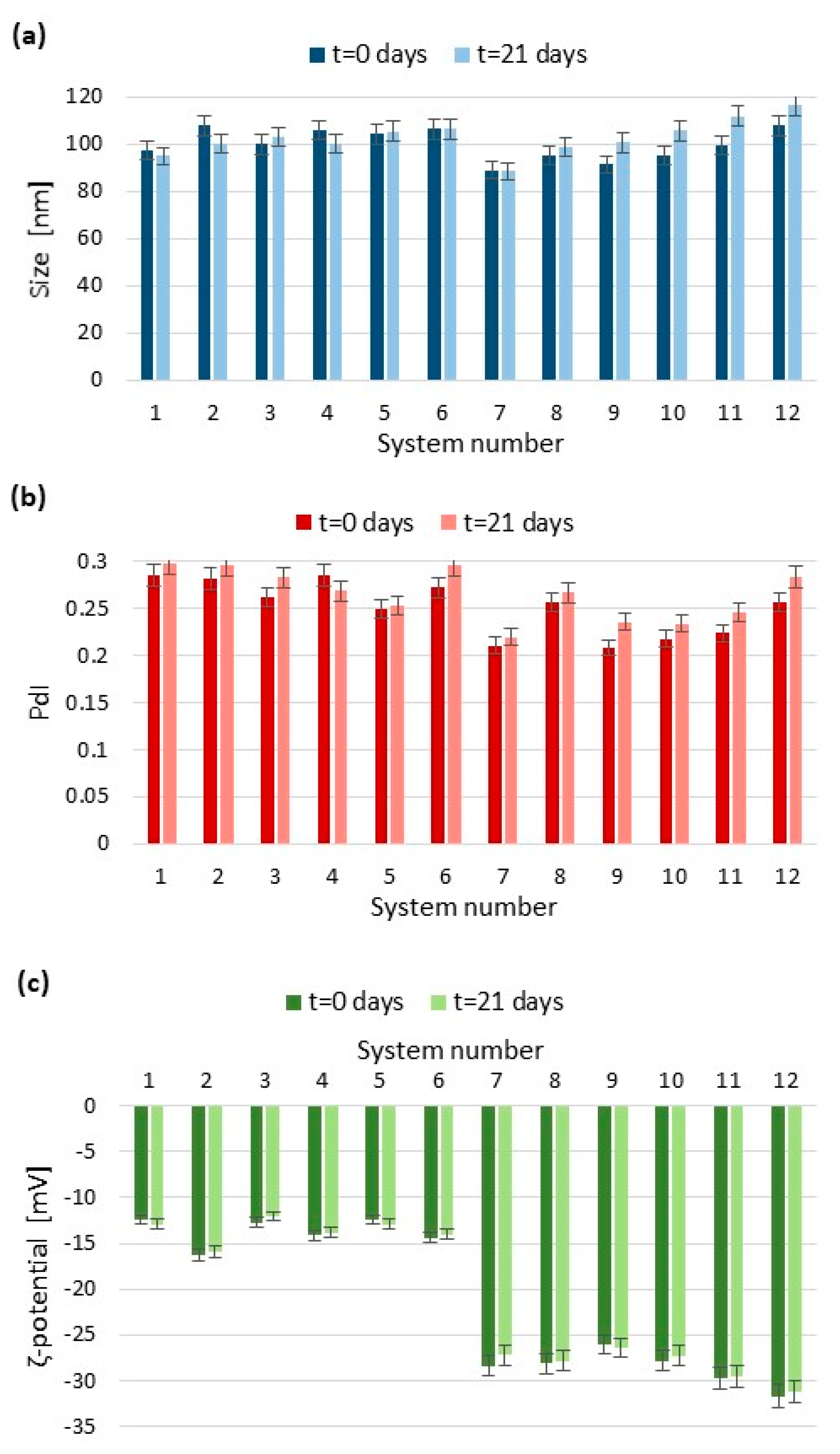
2.3.1. Particle Size and Polydispersity Index by Dynamic Light Scattering

The measurements of the obtained SLN dispersions were performed by dynamic light scattering (DLS) using a Malvern Zetasizer Nano ZS (Malvern Instruments, UK) with the detection angle of 173° in optically homogeneous square polystyrene cells. DLS yields the hydrodynamic diameter (DH) of nanoparticles, which is an intensity-weighted mean diameter of the bulk population and the polydispersity index (PdI) as a measure of the width of the particle size distribution. Each value was obtained as an average of 3 subsequent runs of the instrument, with at least 10 measurements. All the measurements were performed at 25 °C.

2.3.2. Surface Charge by Electrophoretic Light Scattering

The particle charge (i.e., ζ-potential) of the studied lipid nanoparticles was determined by the electrophoretic method using a Malvern Zetasizer Nano ZS apparatus (Malvern Instruments, UK). The applied field strength was 20 V/cm. The results are given as an average of 3 measurements, each with at least 20 runs. All the measurements were determined at 25 °C.

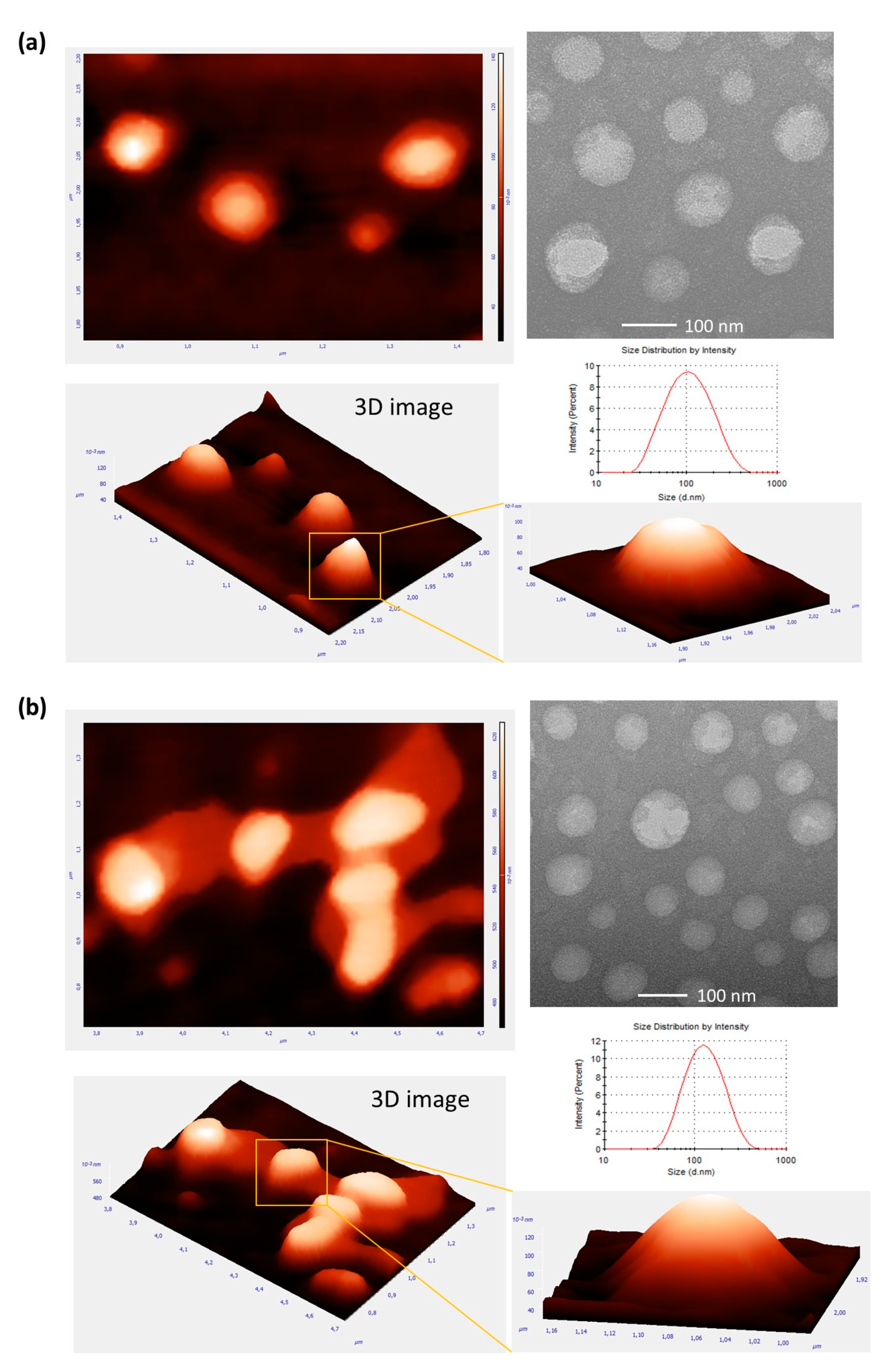
2.3.3. Morphological Characterization by Atomic Force Microscopy and Transmission Electron Microscopy

The morphology of the SLNs was visualized by atomic force microscopy (AFM) using a Veeco NanoScope Dimension V AFM (Plainview, New York, NY, USA) equipped with an RT ESP Veeco tube scanner. The scanning speed was 0.5 Hz and a low-resonance-frequency pyramidal silicon cantilever resonating at 250–331 kHz was employed at a constant force of 20–80 N/m. Before observations, the lipid nanoparticles were allowed to adsorb on a fresh mica surface for 24 h by dipping it in the suspension. Then, the surfaces were rinsed in double-distilled water and dried at room temperature.
The morphology of the obtained nanoparticles was also studied by transmission electron microscopy (TEM) using an FEI Tecnai G2 20 X-TWIN microscope. A drop of the dispersion sample was placed on a perforated, carbon-film-coated copper grid and was left to dry at room temperature for 1 h before the examination.

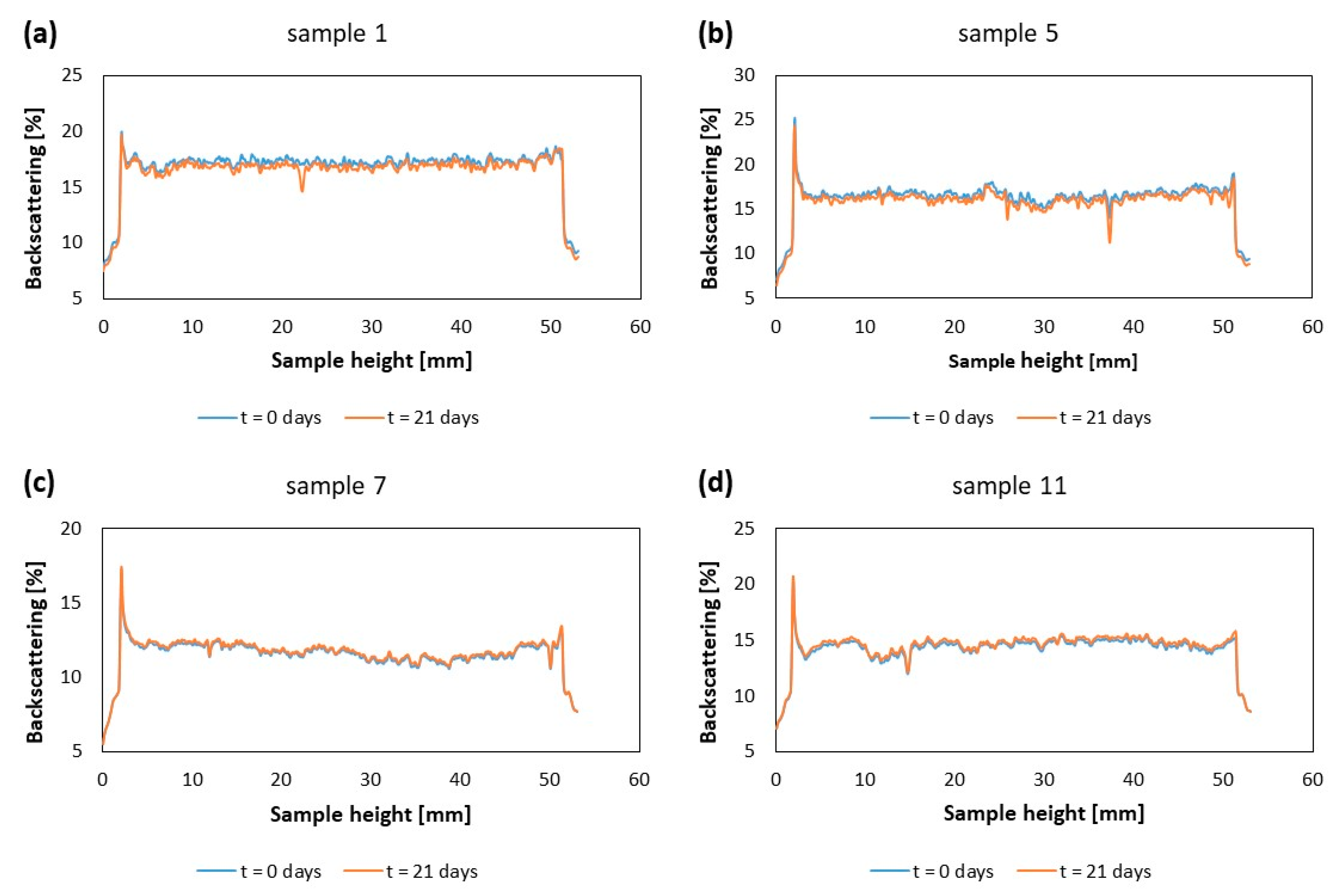
2.3.4. Kinetic Stability by Multiple Light Scattering

The prediction of the physical stability of the obtained formulations was performed using the TurbiScanLab Expert (Formulaction SA, Toulouse, France). The analysis was performed in a cylindrical glass cell at 25 °C by measuring the backscattering (BS) of pulsed near infrared light (λ = 880 nm). BS profiles as a function of sample height were collected and analyzed by the instrument software (Turbisoft, version 2.2.0.82).

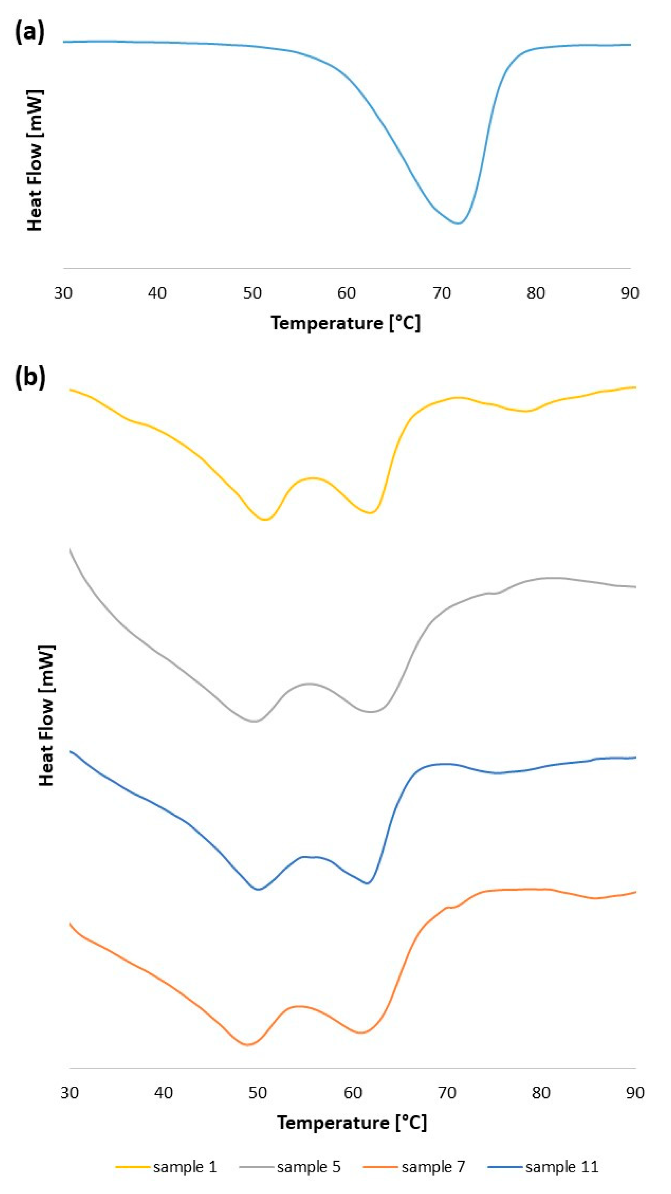
2.3.5. Thermal Analysis by Differential Scanning Calorimetry

The melting behavior analysis of the lyophilized lipid nanoparticles was performed by a Setaram 32 CS (SETARAM, France) calorimeter. The measurements were carried out in standard aluminum pans (40 μL). An empty pan was used as a reference. The study was taken in a temperature range between 20 and 100 °C, with a heating rate of 5 °C/min.

3. Results and Discussion

In the present study, solid lipid nanoparticles (SLNs) were fabricated by the ultrasonic-nanoemulsification method. The studied technique is low cost, highly efficient and does not require the use of harmful organic solvents. Glyceryl monostearate (GMS)—biocompatible, non-toxic and Generally Recognized as Safe (GRAS) [27]—was chosen as the solid lipid-matrix material. Two different surfactants: amphoteric phosphatidylcholine-based Lipoid S75 and non-ionic polyoxyethylete-origin Tween 60 were used. The use of such a mixture of surfactants allows for the more effective prevention of agglomeration as a result of a combination of electrostatic and steric stabilization [28]. The amount of lipid phase was kept constant (1.5%, w/w), as well as surfactant content in the water phase (2% w/w for phosphatidylcholine-based surfactant and 1% w/w for non-ionic surfactant). The greater amount of glyceryl monostearate (lipid material) in the formulation caused the precipitation of large amounts of lipid, making it impossible to reliably analyze the data using the DLS method (data not shown). Based on my previous reports [29], a higher concentration of phosphatidylcholine-type surfactant than non-ionic surfactant has a positive effect on the parameters of nanoparticles. The impact of production parameters (ultrasonication time, used amplitude and number of cycles) on the SLN properties was determined. Table 1 summarizes the details of the carried-out process for subsequent samples. Figure 1 shows the influence of the variables used for the formulation production on size (hydrodynamic diameter, DH), polydispersity index (PdI) and zeta potential (ζ-potential) of the nanocarriers, for which the assessment was performed using dynamic light scattering (DLS) and electrophoretic light scattering (ELS). The parameters for all formulations have been evaluated at production time (t = 0 days) and during a storage period of 21 days.
The results indicate that the produced solid lipid nanoparticles have a size which is suitable for both intravenous and oral delivery [24,25], since almost all of the samples presented diameter values in the range of 95–110 nm at the time of preparation (i.e., t = 0 days). The similarity of the obtained results can be explained by the identical compositions of the formulations [26]. The size results are in good agreement with those reported by Luo et al. [30] about glyceryl monostearate-based solid lipid nanoparticles stabilized by soya lecithin and Tween 80 (an analogous composition with similar proportions results in nanoparticles with size between 70 and 149 nm). After 21 days, the SLN formulations were again analyzed by the DLS technique. As shown in Figure 1, the size, polydispersity index, and zeta potential of formulations are only slightly changed after storage. These results indicated good colloidal stability of the obtained nanosystems.
The influence of the nanoparticle preparation process on their physicochemical properties can be noted. The increase in homogenization duration decreases the particle size and the size distribution. The continuous operation of the homogenizer results in smaller particles than those after the pulsed operation. During continuous homogenization (no pulses, number of cycles = 1) at the same homogenization duration and the same amplitude, the obtained particles are smaller (by 2–10 nm) with a lower PdI. However, the changes are not very large; for example, for formulation ultrasonicated through 3 min (75% amplitude) with continuous operation, the size and PdI are 99.7 nm and 0.262 (sample 3), while using pulses (number of cycles = 0.5)—105.7 nm and 0.285 (sample 4), respectively. The size reduction can be explained by the greater amount of energy supplied to the system, which makes it more monodisperse. The amplitude also affects the size of the nanoparticles—increasing the amplitude (from 50 to 100%) causes a decrease in the size of the carriers. A decrease in particle size with an increasing amplitude value was observed in all formulations. The PdI values between 0.208–0.285 indicate a rather narrow size distribution of the system, which can be considered as monodisperse [31]. The obtained results are correlated to other data reported in the literature [32] and indicate that the application of an increase in sonication time results in a decrease in the size of nanoparticles and PdI values.
Zeta potential (ζ-potential) of the formulation was determined in the original dispersion media [33]. The absolute value of ζ-potential for suspensions considered highly stable should be about 30 mV. However, in the case of a combined electrostatic and steric stabilization (resulting from the use of ionic and non-ionic surfactants), this value may be lower [34]. The comparison of the obtained formulations directly after production revealed a zeta potential between −12.4 mV (samples 1 and 3) and −31.7 mV (sample 12). Nanoparticles with a higher zeta potential were obtained using a longer homogenization time. The observed phenomenon can be explained by better mixing of surfactants in dispersion, resulting in an increase in the values of the zeta potential, and consequently enhanced formulation stability, which was proved also in the described below backscattering profiles.
After 21 days (see Figure 1), no significant changes in size and polydispersity of formulations were observed. Furthermore, no significant differences in the zeta potential of nano-particles were observed during storage time (t = 21 days), which proves the good colloidal stability of the obtained systems. The obtained results show that the synthesized nanocarriers were characterized by an appropriate particle size with a narrow size distribution and a negative surface charge (ζ-potential), which makes them promising candidates as carriers for many active substances. After additional in vivo studies, it will be possible to confirm their suitability for drug delivery.
Appropriate morphology of nanocarriers is crucial in designing drug delivery systems. The morphology was evaluated by atomic force microscopy (AFM) and transmission electron microscopy (TEM). AFM 2D and 3D images (in terms of height) with (TEM) images are shown in Figure 2. For morphological studies, two systems—sample 1 and sample 7—with the smallest and the largest size distribution (PdI parameters determined by the DLS technique) were chosen. The obtained SLNs were observed to have a spherical shape without roughness. The analyzed samples did not show any enhanced aggregation. The particle size range obtained with AFM was similar to data predicted from DLS (the example size distribution spectra are also shown in Figure 2). The values obtained by TEM were slightly lower than from DLS. This could be related to the possible deformation of nanocarriers during the drying procedure before microscopic observation.
Backscattering profiles (BS) enable the detection of various types of instability of a formulation, and thus the determination, of the kinetic stability of a sample. The SLN dispersions were assessed using the Turbiscan Lab Expert optical analyzer, based on multiple light scattering technology. The dynamics of the processes occurring in the dispersions were determined at time t = 0 (freshly prepared SLNs) and after 21 days of storage in 4 °C (Figure 3). The X-axis shows the level of the colloidal sample in the measurement vial (the height of the sample, expressed in mm). Deviations above 10% in backscattering profiles indicate sample instability [35]. The slight changes in BS are only observed for systems 1 and 5 (Figure 3a,b). These carriers may be less stable, which was also confirmed by electrophoretic light scattering (lower zeta potentials than samples 7 and 11). The presented graphs (Figure 3c,d) do not show the separation between the curves on the day of preparation and after 21 days, indicating no visible particle growth nor migration in the dispersion. Rapid destabilization is usually characterized by large gaps between the curves, while overlapping of individual curves indicates a slow destabilization process, and thus a high stability of the analyzed sample [36].
Development of physically stable formulation is a crucial aspect in the design of an effective drug delivery system. The thermal behavior of bulk glyceryl monostearate (lipid-matrix material) and optimized solid lipid nanoparticles was assessed by DSC (Figure 4a,b, respectively). The single peak of pure lipid, which corresponds to the melting point of glyceryl monostearate, is more intense than those observed for the nanoparticles. This phenomenon could be associated with the carbon chains in lipids, which are more packaged and organized than those in the obtained SLNs [37].
The signals of the lipid nanocarriers are broader in contrast to the lipid in bulk state due to the Thomson effect caused by the smaller particle size [33]. All SLN formulations showed two thermal transitions with a melting temperature lower than pure lipid (the maximum of peaks appears near 50 °C and about 61 °C). These irregularities might be due to the large quantities of phosphatidylcholine used in the formulations. According to the literature, the peak at the temperature of 48 °C corresponds to the α-modification, while the peak at 57 °C corresponds to the β-modification of phosphatidylcholine, of which the content is high in the tested formulations [38]. The slight shifts of the peaks to the values of 50 °C and 61 °C are related to the combination of the phospholipid with the lipid-matrix material during the SLN preparation process. The obtained results are related to the data reported in the literature [33] about lipid nanoparticles based on hydrogenated sunflower oil, the main ingredient of which is also phosphatidylcholine.

4. Conclusions

Solid lipid nanoparticles were successfully obtained by the ultrasonic-nanoemulsification method, which is low cost and highly efficient. The process was optimized in relation to ultrasonication time, amplitude and the number of cycles. The main targets of the optimization, i.e., the appropriate particle size with a minimized polydispersity index, have been achieved. The developed formulations exhibited a size of about 100 nm with uniform size distribution (PdI < 0.3). Atomic force microscopy and transmission electron microscopy proved a near spherical shape of nanoparticles and confirmed the diameter obtained by the dynamic light-scattering technique. The stability of the studied SLNs was proven by high negative zeta potential values and no significant changes in the backscattering profiles. The thermal behavior of lyophilized solid lipid nanoparticles confirmed the effective formation of nanoparticles based on glyceryl monostearate and phosphatidylcholine. The presented results confirm that the suitably designed ultrasonic-nanoemulsification process for the solid lipid nanoparticle engineering may constitute an attractive and versatile platform for drug delivery in a variety of possible applications.

Funding

This research was funded by Wroclaw University of Science and Technology.

Data Availability Statement

Not applicable.

Conflicts of Interest

The author declares no conflict of interest.

References

  1. Ganesan, P.; Narayanasamy, D. Lipid nanoparticles: Different preparation techniques, characterization, hurdles, and strategies for the production of solid lipid nanoparticles and nanostructured lipid carriers for oral drug delivery. Sustain. Chem. Pharm. 2017, 6, 37–56. [Google Scholar] [CrossRef]
  2. Mishra, V.; Bansal, K.K.; Verma, A.; Yadav, N.; Thakur, S.; Sudhakar, K.; Rosenholm, J.M. Solid lipid nanoparticles: Emerging colloidal nano drug delivery systems. Pharmaceutics 2018, 10, 191. [Google Scholar] [CrossRef] [PubMed] [Green Version]
  3. Gordillo-Galeano, A.; Mora-Huertas, C.E. Solid lipid nanoparticles and nanostructured lipid carriers: A review emphasizing on particle structure and drug release. Eur. J. Pharm. Biopharm. 2018, 133, 285–308. [Google Scholar] [CrossRef]
  4. Bayón-Cordero, L.; Alkorta, I.; Arana, L. Application of solid lipid nanoparticles to improve the efficiency of anticancer drugs. Nanomaterials 2019, 9, 474. [Google Scholar] [CrossRef] [PubMed] [Green Version]
  5. da Silva Santos, V.; Badan Ribeiro, A.P.; Andrade Santana, M.H. Solid lipid nanoparticles as carriers for lipophilic compounds for applications in foods. Food Res. Int. 2019, 122, 610–626. [Google Scholar] [CrossRef]
  6. Mirchandani, Y.; Patravale, V.B.; Brijesh, S. Solid lipid nanoparticles for hydrophilic drugs. J. Control. Release 2021, 335, 457–464. [Google Scholar] [CrossRef]
  7. Arana, L.; Gallego, L.; Alkorta, I. Incorporation of antibiotics into solid lipid nanoparticles: A promising approach to reduce antibiotic resistance emergence. Nanomaterials 2021, 11, 1251. [Google Scholar] [CrossRef]
  8. Pucek, A.; Tokarek, B.; Waglewska, E.; Bazylińska, U. Recent advances in the structural design of photosensitive agent formulations using “soft” colloidal nanocarriers. Pharmaceutics 2020, 12, 587. [Google Scholar] [CrossRef]
  9. Mauricio, C.; Pinilla, B.; Lopes, N.A.; Brandelli, A. Lipid-Based Nanostructures for the Delivery of Natural Antimicrobials. Molecules 2021, 26, 3587. [Google Scholar] [CrossRef]
  10. Dumont, C.; Bourgeois, S.; Fessi, H.; Jannin, V. Lipid-based nanosuspensions for oral delivery of peptides, a critical review. Int. J. Pharm. 2018, 541, 117–135. [Google Scholar] [CrossRef] [PubMed]
  11. Cimino, C.; Maurel, O.M.; Musumeci, T.; Bonaccorso, A.; Drago, F.; Souto, E.M.B.; Pignatello, R.; Carbone, C. Essential oils: Pharmaceutical applications and encapsulation strategies into lipid-based delivery systems. Pharmaceutics 2021, 13, 327. [Google Scholar] [CrossRef]
  12. Barroso, L.; Viegas, C.; Vieira, J.; Ferreira-Pêgo, C.; Costa, J.; Fonte, P. Lipid-based carriers for food ingredients delivery. J. Food Eng. 2021, 295. [Google Scholar] [CrossRef]
  13. García-Pinel, B.; Porras-Alcalá, C.; Ortega-Rodríguez, A.; Sarabia, F.; Prados, J.; Melguizo, C.; López-Romero, J.M. Lipid-based nanoparticles: Application and recent advances in cancer treatment. Nanomaterials 2019, 9, 638. [Google Scholar] [CrossRef] [PubMed] [Green Version]
  14. Smith, B.R.; Gambhir, S.S. Nanomaterials for in Vivo Imaging. Chem. Rev. 2017, 117, 901–986. [Google Scholar] [CrossRef] [PubMed]
  15. Chen, G.; Roy, I.; Yang, C.; Prasad, P.N. Nanochemistry and Nanomedicine for Nanoparticle-based Diagnostics and Therapy. Chem. Rev. 2016, 116, 2826–2885. [Google Scholar] [CrossRef] [PubMed]
  16. Wacker, M. Nanocarriers for intravenous injection—The long hard road to the market. Int. J. Pharm. 2013, 457, 50–62. [Google Scholar] [CrossRef]
  17. Salah, E.; Abouelfetouh, M.M.; Pan, Y.; Chen, D.; Xie, S. Solid lipid nanoparticles for enhanced oral absorption: A review. Colloids Surf. B Biointerfaces 2020, 196, 111305. [Google Scholar] [CrossRef]
  18. Zoabi, A.; Touitou, E.; Margulis, K. Recent advances in nanomaterials for dermal and transdermal applications. Colloids Interfaces 2021, 5, 18. [Google Scholar] [CrossRef]
  19. de Oliveira, I.F.; Barbosa, E.J.; Peters, M.C.C.; Henostroza, M.A.B.; Yukuyama, M.N.; dos Santos Neto, E.; Löbenberg, R.; Bou-Chacra, N. Cutting-edge advances in therapy for the posterior segment of the eye: Solid lipid nanoparticles and nanostructured lipid carriers. Int. J. Pharm. 2020, 589, 119831. [Google Scholar] [CrossRef]
  20. Costa, C.P.; Moreira, J.N.; Sousa Lobo, J.M.; Silva, A.C. Intranasal delivery of nanostructured lipid carriers, solid lipid nanoparticles and nanoemulsions: A current overview of in vivo studies. Acta Pharm. Sin. B 2021, 11. [Google Scholar] [CrossRef]
  21. Weber, S.; Zimmer, A.; Pardeike, J. Solid Lipid Nanoparticles (SLN) and Nanostructured Lipid Carriers (NLC) for pulmonary application: A review of the state of the art. Eur. J. Pharm. Biopharm. 2014, 86, 7–22. [Google Scholar] [CrossRef]
  22. Modarres-Gheisari, S.M.M.; Gavagsaz-Ghoachani, R.; Malaki, M.; Safarpour, P.; Zandi, M. Ultrasonic nano-emulsification—A review. Ultrason. Sonochem. 2019, 52, 88–105. [Google Scholar] [CrossRef]
  23. Kentish, S.; Wooster, T.J.; Ashokkumar, M.; Balachandran, S.; Mawson, R.; Simons, L. The use of ultrasonics for nanoemulsion preparation. Innov. Food Sci. Emerg. Technol. 2008, 9, 170–175. [Google Scholar] [CrossRef]
  24. Murugan, K.; Choonara, Y.E.; Kumar, P.; Bijukumar, D.; du Toit, L.C.; Pillay, V. Parameters and characteristics governing cellular internalization and trans-barrier trafficking of nanostructures. Int. J. Nanomed. 2015, 10, 2191–2206. [Google Scholar] [CrossRef] [Green Version]
  25. Roger, E.; Lagarce, F.; Garcion, E.; Benoit, J.P. Biopharmaceutical parameters to consider in order to alter the fate of nanocarriers after oral delivery. Nanomedicine 2010, 5, 287–306. [Google Scholar] [CrossRef] [Green Version]
  26. Firdaus, S.; Hassan, N.; Mirza, M.A.; Ara, T.; El-Serehy, H.A.; Al-Misned, F.A.; Iqbal, Z.; de Sousa, M.; Pessine, F.B.T.; Wang, H.; et al. Improving the oral bioavailability of an anti-glioma prodrug cat3 using novel solid lipid nanoparticles containing oleic acid-cat3 conjugates. Saudi J. Biol. Sci. 2020, 298, 242–254. [Google Scholar] [CrossRef]
  27. Sampaio de Sousa, A.R.; Simplício, A.L.; de Sousa, H.C.; Duarte, C.M.M. Preparation of glyceryl monostearate-based particles by PGSS®-Application to caffeine. J. Supercrit. Fluids 2007, 43, 120–125. [Google Scholar] [CrossRef]
  28. Aditya, N.P.; Macedo, A.S.; Doktorovova, S.; Souto, E.B.; Kim, S.; Chang, P.S.; Ko, S. Development and evaluation of lipid nanocarriers for quercetin delivery: A comparative study of solid lipid nanoparticles (SLN), nanostructured lipid carriers (NLC), and lipid nanoemulsions (LNE). LWT-Food Sci. Technol. 2014, 59, 115–121. [Google Scholar] [CrossRef]
  29. Pucek, A.; Niezgoda, N.; Kulbacka, J.; Wawrzeńczyk, C.; Wilk, K.A. Phosphatidylcholine with conjugated linoleic acid in fabrication of novel lipid nanocarriers. Colloids Surfaces A Physicochem. Eng. Asp. 2017, 532, 377–388. [Google Scholar] [CrossRef]
  30. Luo, Y.F.; Chen, D.W.; Ren, L.X.; Zhao, X.L.; Qin, J. Solid lipid nanoparticles for enhancing vinpocetine’s oral bioavailability. J. Control. Release 2006, 114, 53–59. [Google Scholar] [CrossRef]
  31. Danaei, M.; Dehghankhold, M.; Ataei, S.; Hasanzadeh Davarani, F.; Javanmard, R.; Dokhani, A.; Khorasani, S.; Mozafari, M.R. Impact of particle size and polydispersity index on the clinical applications of lipidic nanocarrier systems. Pharmaceutics 2018, 10, 57. [Google Scholar] [CrossRef] [PubMed] [Green Version]
  32. Firdaus, S.; Hassan, N.; Mirza, M.A.; Ara, T.; El-Serehy, H.A.; Al-Misned, F.A.; Iqbal, Z. FbD directed fabrication and investigation of luliconazole based SLN gel for the amelioration of candidal vulvovaginitis: A 2 T (thermosensitive & transvaginal) approach. Saudi J. Biol. Sci. 2021, 28, 317–326. [Google Scholar] [CrossRef]
  33. Ding, Y.; Nielsen, K.A.; Nielsen, B.P.; Bøje, N.W.; Müller, R.H.; Pyo, S.M. Lipid-drug-conjugate (LDC) solid lipid nanoparticles (SLN) for the delivery of nicotine to the oral cavity—Optimization of nicotine loading efficiency. Eur. J. Pharm. Biopharm. 2018, 128, 10–17. [Google Scholar] [CrossRef] [PubMed]
  34. Müller, R.H.; Jacobs, C.; Kayser, O. Nanosuspensions as particulate drug formulations in therapy: Rationale for development and what we can expect for the future. Adv. Drug Deliv. Rev. 2001, 47, 3–19. [Google Scholar] [CrossRef]
  35. Fangueiro, J.F.; Andreani, T.; Egea, M.A.; Garcia, M.L.; Souto, S.B.; Silva, A.M.; Souto, E.B. Design of cationic lipid nanoparticles for ocular delivery: Development, characterization and cytotoxicity. Int. J. Pharm. 2014, 461, 64–73. [Google Scholar] [CrossRef]
  36. Bazylińska, U. Rationally designed double emulsion process for co-encapsulation of hybrid cargo in stealth nanocarriers. Colloids Surf. A Physicochem. Eng. Asp. 2017, 532, 476–482. [Google Scholar] [CrossRef]
  37. de Sousa, M.; Pessine, F.B.T. Production of Mannosylated Solid Lipid Nanoparticles by Using Experimental Design: Application to Saquinavir. J. Pharm. Sci. Pharmacol. 2015, 2, 64–72. [Google Scholar] [CrossRef]
  38. Schubert, M.A.; Schicke, B.C.; Müller-Goymann, C.C. Thermal analysis of the crystallization and melting behavior of lipid matrices and lipid nanoparticles containing high amounts of lecithin. Int. J. Pharm. 2005, 298, 242–254. [Google Scholar] [CrossRef]
Figure 1. Influence of the process parameters on (a) size and (b) polydispersity index (c) ζ-potential of the studied lipid nanoparticles. Details of the system number are included in Table 1.
Processes 09 01265 g001
Figure 2. AFM and TEM images compared to DLS graphs of solid lipid nanoparticles: (a) system 1; and (b) system 7.
Processes 09 01265 g002
Figure 3. Backscattering (%) profiles of the obtained SLN formulations as a function of sample height (mm) analyzed over 21 days of storage: (a) system 1; (b) system 5; (c) system 7; (d) system 11.
Processes 09 01265 g003
Figure 4. DSC thermograms of: (a) pure lipid material (glyceryl monostearate) and (b) optimized solid lipid nanoparticles (systems: 1, 5, 7, 11).
Processes 09 01265 g004
Table 1. Optimization parameters for solid lipid nanoparticles prepared by the ultrasonic-nanoemulsification method.
System NumberUltrasonication Time (min)Amplitude (%)Number of Cycles
131001
231000.5
33751
43750.5
53501
63500.5
751001
851000.5
95751
105750.5
115501
125500.5
Publisher’s Note: MDPI stays neutral with regard to jurisdictional claims in published maps and institutional affiliations.

