Article

Moringa oleifera—Storage Stability, In Vitro-Simulated Digestion and Cytotoxicity Assessment of Microencapsulated Extract

by
Cecilia Castro-López
1,2,
Catarina Gonçalves
3,
Janeth M. Ventura-Sobrevilla
4,
Lorenzo M. Pastrana
3,
Cristóbal N. Aguilar-González
2 and
Guillermo C. G. Martínez-Ávila
1,*
1
School of Agronomy, Autonomous University of Nuevo Leon, Nuevo León 66050, Mexico
2
School of Chemistry, Autonomous University of Coahuila, Coahuila 25280, Mexico
3
Food Processing Group, International Iberian Nanotechnology Laboratory, Av. Mestre José Veiga S/N, 4715-330 Braga, Portugal
4
School of Health Sciences, Autonomous University of Coahuila, Coahuila 26090, Mexico
*
Author to whom correspondence should be addressed.
Processes 2020, 8(7), 770; https://doi.org/10.3390/pr8070770
Submission received: 1 June 2020 / Revised: 23 June 2020 / Accepted: 28 June 2020 / Published: 1 July 2020

Abstract

:
Moringa extract was microencapsulated for the first time by spray-drying technique using tragacanth gum (MorTG) to improve its stability under gastrointestinal and storage conditions, assessing total polyphenolic content (TPC) and antioxidant activity. Additionally, cytotoxicity of the microencapsulated components was evaluated after contact with Caco-2 cells. Results showed that TPC was released as follows—oral (9.7%) < gastric (35.2%) < intestinal (57.6%). In addition, the antioxidant activity in in vitro digestion reached up to 16.76 ±0.15 mg GAE g−1, which was 300% higher than the initial value. Furthermore, microencapsulated moringa extract presented a half-life up to 45 days of storage, where the noticeably change was observed at 35 °C and 52.9% relative humidity. Finally, direct treatment with 0.125 mg mL−1 MorTG on Caco-2 cells showed a slight antiproliferative effect, with a cell viability of approx. 87%. Caco-2 cells’ viability demonstrated non-cytotoxicity, supporting the safety of the proposed formulation and potential use within the food field.

Graphical Abstract

1. Introduction

The increasing demand for healthy foods has led current research to the development of new and natural additives or ingredients that can provide a benefit beyond nutrition [1]. In this sense, Moringa oleifera Lam (Moringaceae) has been documented as a rich plant of bioactive compounds (e.g., polyphenols, carbohydrates, fatty acids and biofunctional peptides) with several advantages for human health and food applications [2]. Earlier studies focused mainly in the polyphenol content since it has been reported that these compounds have antioxidant abilities that may be used for human consumption [3,4,5]. However, they are sensitive to several factors used in food processing operations (pH, water activity, light conditions, oxygen and temperature). Thus, it is necessary to prevent their degradation and improve their stability in those conditions. Within these, encapsulation technology is a method that can provide a good physical barrier against the above-mentioned factors [6,7]. And even, some authors report that encapsulation may also decrease their unpleasant taste and improve sensory properties [8,9]. Additionally, from a technological point of view, it would be most applicable to benefit from Moringa phenolics in powder form due to the easiest handling in food and pharmaceutical industries [10].
In this respect, in a previous study we reported a comprehensive characterization of the polyphenols present in M. oleifera extract, as well as its antioxidant properties [11]. Hence, in a later study, three different, suitable, novel, natural and generally recognized as safe (GRAS) wall materials (tragacanth gum, locust bean gum and carboxymethyl-cellulose) were explored for Moringa spray-drying microencapsulation followed by several physicochemical analyses (data under submission). Among the three materials, microencapsulates produced with Tragacanth gum (TG) showed better performance in antioxidant properties retention, aside from good particle size distribution and morphological, thermal and crystallinity characteristics. Nevertheless, there is no available information of either bioaccessibility and storage stability, controlling release and/or enhancing solubility. Thus, the aim of this paper was to determine the stability of microencapsulated Moringa’s activity in in vitro-simulated digestion and at different storage conditions. Additionally, Caco-2 cells’ viability after incubation with the microencapsulated Moringa or with the individual components was also evaluated, as a first safety assessment of the proposed formulation envisaging its potential use in the food industry.

2. Materials and Methods

2.1. Chemicals and Reagents

All the chemicals used were analytical grade and purchased from Sigma-Aldrich (Toluca, México). Tragacanth gum was obtained from a domestic supplier (Deiman S.A. de C.V., Puebla, México); while minimum essential medium (MEM) was obtained from Thermo-Scientific (Portugal) and penicillin/streptomycin, fetal bovine serum (FBS) and non-essential amino-acids were acquired from Millipore (Oeiras, Portugal).

2.2. Plant Material and Extraction of Polyphenolic Compounds

Moringa leaves (Moringa oleifera Lam.) were provided from a producer from General Escobedo, Nuevo León, México. They were leafed off manually, washed in water, dried in an air-forced oven (60 °C for 24 h), grounded using an electric grinder until having a particle size between 3–5 mm and finally stored in a dry place. Then, the polyphenolic compounds were extracted by microwave-assisted extraction with a solid—liquid ratio of 1:50 w/v, using water as solvent. The conditions used for the extraction method were reported in a previous study [11]. Briefly, the extraction conditions were microwave power, 550 W; extraction time, 90 s; and controlled temperature, 70 °C.

2.3. Microencapsulation Process

Moringa polyphenol-rich extract was microencapsulated with tragacanth gum (TG), as encapsulating agent. The dispersion was prepared using the raw extract (200 mL) and adding 1% w/v of TG. The resulting mixture was stirred by a constant speed stirrer at 300 rpm for 90 min at 30 °C until a homogenized system was obtained. The liquid feed was spray dried in a Büchi B-290 Mini Spray Dryer (Büchi Laboratoriums-Technik AG, Flawil, Switzerland) under the following experimental conditions—drying air inlet temperature, 120 °C; outlet temperature, 68–71 °C; atomization air volumetric flow rate, 601 L h−1; feed volumetric flow rate, 2 mL min−1; nozzle diameter, 0.7 mm; and aspirator, 100% [12].

2.4. Stability During Storage

For stability tests, the microencapsulates (200 mg) were put in plastic cups, uniformly spreaded and stored in airtight plastic containers filled with saturated MgCl2 and Mg(NO3)2 solutions to produce a relative humidity (RH) with values of 32.8% and 52.9%, respectively. These containers were stored at three different temperatures—(a) 5 °C (refrigeration), (b) 25 °C (room temperature) and (c) 35 °C (temperature recommended for accelerated shelf life studies) [13]. The samples were analyzed after the following storage times—0, 5, 10, 15, 20, 25, 30 and 35 days, where the total polyphenol content and the antioxidant capacity were the parameters evaluated to determine stability. The first-order reaction rate constants (k) and half-lives (t1/2) were calculated as follows:
−Ɩn (Ct/C0) = kt
t1⁄2 = (Ɩn (2))/k
where C0 is the initial content of the microencapsulated compound and Ct is the content of the microencapsulated compound in the time-point (t).
Also, the values of Q10 (meaning that this coefficient reflects the number of times the deterioration of the particles accelerates or decreases depending on whether the temperature increases or decreases 10 degrees Celsius) were determined at a specific temperature (Tx) by Equations (2) and (3), where kT is the constant reaction rate at a temperature Tx and kT-10 is the constant reaction rate at a temperature 10 °C lower than the temperatures tested. For purpose of this analysis values of 25 and 35 °C were used in order to determine Q10 [14].
Q10 = kT/kT − 10.

2.5. Release of Compounds during In Vitro Digestion

The simulated in vitro digestion was prepared according to the method of Ahmad et al. [15]. First, the simulated gastric fluid (SGF) was prepared from a NaCl solution (0.2%) to pH ≈ 3 (adjusted by the addition of HCl 1 M). Later, the simulated intestinal fluid (SIF) was prepared by dissolving KH2PO4 (0.68 g) in deionized water (75 mL) and then by adjusting the pH to 7.1 (with KOH 0.2 M) and topping up the final volume to 100 mL. Finally, the saliva fluid was prepared by dissolving NaCl (4.68 mg), KCl (5.96 mg), NaHCO3 (0.084 g) and α-amylase (7.0 mg) in deionized water (40 mL). The pH of this solution was adjusted to 6.8. Afterwards, samples (200 mg) were placed in a flask, incubated at 37 °C under constant agitation and digested sequentially as follows—(a) mouth: 10 mL of salivary juice was added and mixed for 5 minutes and an aliquot (2 mL) was collected; (b) stomach: 10 mL of SGF were added and mixed for 1 h, aliquots (2 mL) were collected after 30 minutes and 1 h of incubation; and (c) intestine: 10 mL of SIF were added and mixed for 3 h. Aliquots (2 mL) were collected after 2 h, 3 h and 4 h.

2.6. Activities Assessment

2.6.1. Preparation of Samples

Powder from storage stability was dissolved in 2 mL ethanol: water solution (50:50 v/v). This mixture was agitated for 3 min and centrifuged at 10,000 rpm for 10 min at 4 °C. Finally, the supernatant containing the Moringa extract was collected and stored at −20 °C until measured. Aliquots from simulated in vitro digestion received the same treatment of centrifugation and supernatant recollection.

2.6.2. Determination of Total Polyphenol Content (TPC)

The total polyphenol content (TPC) was measured to evaluate the release during in vitro digestion or degradation under storage conditions and was determined according to the method proposed by Georgé et al. [16]. Briefly, 250 μL of supernatant was mixed with 250 μL of Folin-Ciocalteu’s reagent for 1 min, after 250 μL of sodium carbonate (75 g L−1) were added. The final solution was mixed and incubated at 40 °C for 30 minutes. Subsequently, 2 mL of distilled water were added and the absorbance at 750 nm was recorded. TPC was calculated using a calibration curve performed with gallic acid in the concentration range 20 to 150 ppm.

2.6.3. DPPH radical Scavenging Activity

The antioxidant capacity of the supernatant after in vitro digestion or after being subjected to different storage conditions, was evaluated by the methodology proposed by Brand-Williams et al. [17]. Briefly, 50 µL of each supernatant was added to 2950 µL DPPH radical methanol (60 µM) solution. After 30 minutes of incubation in the dark, the absorbance of the samples was recorded at a wavelength of 517 nm. The capability of inhibition was calculated with the following equation and expressed as percentage of inhibition of DPPH radical comparing to the control (distilled water):
Inhibition (%) = [(Acontrol - Asample)/Acontrol] × 100.
The antioxidant capacity was calculated using a calibration curve performed with gallic acid in the concentration range 20 to 150 ppm.

2.7. Cell Toxicity

2.7.1. Cell Culture

Caco-2 human colon epithelial cancer cells (ATCC, HTB-37, LGC Standards S.L.U., Barcelona, Spain) were routinely cultured in minimum essential medium (MEM), supplemented with 20% fetal bovine serum (FBS), 1% non-essential amino acids, 1 mM sodium pyruvate and 1% penicillin/streptomycin. These cells were maintained in T75 cell culture flasks, at 37 °C, in a humidified 5% CO2 atmosphere and harvested at 80% of confluency using Trypsin/EDTA solution 0.25%/0.02% (w/v). Cells were used in passage 20–40.

2.7.2. Cell Viability Assay

The resazurin salt was used to assess the cellular compatibility of encapsulating agent, raw extract and microencapsulates diluted in the culture medium, at different concentrations: 0.125, 0.075, 0.050 and 0.025 mg mL−1. Caco-2 cells were seeded onto 96-well plates at a density of 10,000 cells per well and left adhering overnight. After that, the culture medium was eliminated and replaced by the samples. Then, the cells were incubated for 24 and 48 h with resazurin (0.01 mg mL−1). The fluorescence intensity, was measured at an excitation wavelength of 530 nm and an emission wavelength of 590 nm. The percentage of cell viability was expressed as the percentage of fluorescence in treated cells compared with the fluorescence of cells growing in the culture medium (considered as 100% cell viability). A positive control with 30% of DMSO was performed [18]. For each condition, the fluorescence intensity of samples without cells (background) was measured to assess any interference of samples with resazurin and subtracted to the fluorescence obtained with cells, in each time-point.

2.8. Statistical Analysis

The Minitab 17 Statistical Software (Minitab, Inc., State College, PA, USA) was used to analyze all results. Data are representative of two independent experiments, each containing three biological replicates and expressed as mean ± standard deviation (SD).

3. Results and Discussion

3.1. Influence of Storage Conditions on the Polyphenolic Content

The stability of the total polyphenolic content (TPC) in the microencapsulated Moringa was estimated under different storage conditions and the half-life values of the reactions were calculated. In addition, the degradation kinetics of the polyphenols were monitored during the storage period and the rate constants and the degradation of the polyphenols were calculated. A first-order reaction model was adjusted under all the conditions evaluated (Table 1). Microencapsulated moringa microparticles stored at 5 and 25 °C at 32.8% RH showed the lowest total polyphenol loss, about 42.47%–45.28% of the initial total content (23.24 mg GAE g−1), suggesting that microencapsulation, combined with storage at refrigeration or room temperature, help to maintain part of the polyphenols contained in the particle (Figure 1a). As expected, increasing the storage temperature (35 °C) led to an increased degradation of the compounds with a loss of 53.58%, since they are sensitive to temperature. It was also observed that the effect of temperature on degradation rates was greater for samples stored at higher RH (52.9%). This indicates that the activity of water also has a significant role in the degradation of tested compounds, given that the higher the water content, the greater the molecular mobility within the microparticles, which facilitates the physicochemical degradation reactions [13]. In the same way, according to Desobry et al. [19], the interval that presents a higher degradation rate corresponds to the degradation of compounds present on the surface of the microcapsules or compounds within the microcapsules that are in contact with oxygen, which is present in the pores of the particle or trapped inside it in the form of bubbles, which allows their easy oxidation.
On the other hand, the parameters of the reaction rate constant (k), half-life time (t1/2) and factor Q10 are shown in Table 1. The Moringa microparticles have first-order kinetics, showing a linear degradation with respect to time. Storage at 35 °C showed a higher value of k (0.0218) in the degradation of the polyphenol content than the samples stored at 5 and 25 °C. The half-life data (t1/2) showed an inverse relation with the storage temperature. The longest half-life for microencapsulated MorTG was verified for storage at 25 °C (32.8% RH) presenting up to 44.47 days of stability. The values of Q10 calculated for 25 and 35 °C, were higher than 1, which means that the degradation rate of polyphenols increases with temperature due to the high sensitivity of these compounds. This negative effect of high temperature and humidity on the stability of such compounds has already been reported in many studies available in the literature [10,20,21,22].

3.2. Influence of Storage Conditions on Antioxidant Activity

As already mentioned, one of the most important criteria for evaluating polyphenol microencapsulation quality is the length of time during which the powder retains its bioactivity [23]. In this context, the antioxidant activity was monitored during the storage conditions as shown in Figure 1b. The data obtained showed that microparticles presented a moderate decrease of original antioxidant activity along storage time, which was enhanced by higher temperature and water activity.
At all temperatures tested, with 32.8% RH, the microparticles were stable up to 30 days of storage, while at 52.9% RH a notable tendency to decrement was found after 25 days; thus, it was observed that the retained activity was in a range between 39.7% and 75.32% regarding the initial activity (18.60 mg GAE g−1). These results suggested that the stability of microencapsulated extract is seriously compromised when exposed to higher temperature and humidity environments, which is in agreement with the results obtained by Wang et al. [24], Zheng et al. [25] and Bakowska-Barczak & Kolodziejczyk [7] who reported changes in the antioxidant activity of encapsulated tea polyphenols, bayberry polyphenols and polyphenols extracted from black currant pomace, when the storage temperature was increased.
Nevertheless, the antioxidant activity presents less variation during storage compared with the reduction of TPC. Regarding this, several studies have tried to correlate the antioxidant potential and the polyphenolic content of microparticles submitted to different storage conditions. Fracassetti et al. [26] and Moser et al. [22] studied the influence of storage on antioxidant activity of freeze-dried wild blueberry powder and violeta grape juice microencapsulates. They reported a decreased of the antioxidant activity with increasing temperature but the reduction does not seem directly correlated with the observed decrease in the total polyphenol content, which showed linear degradation. In accordance with these authors, such behavior may be attributed to many factors which can influence hydrolysis, oxidation and condensation reactions that take place during storage of phenolic compounds which bring out formation of new antioxidant polymers.

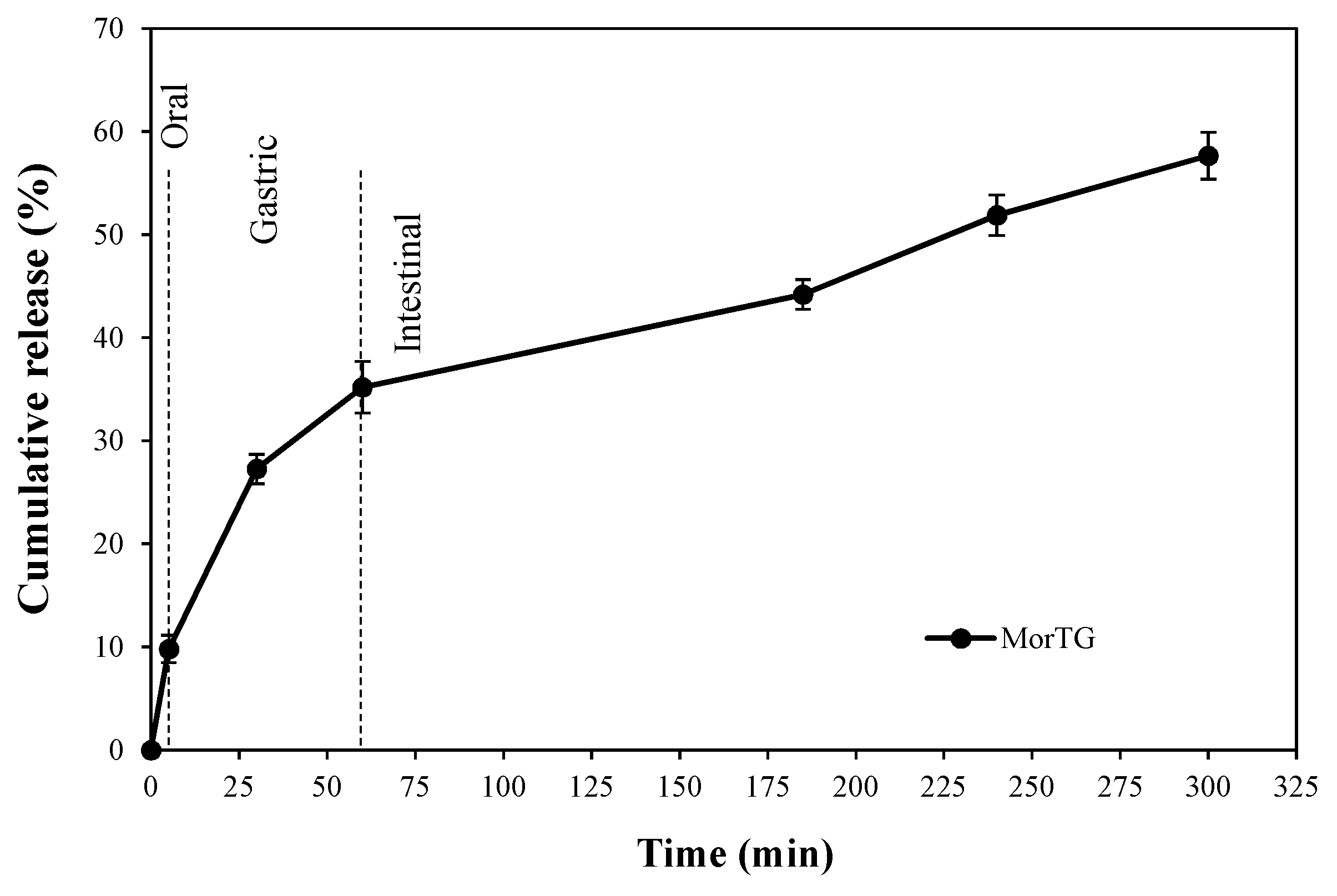
3.3. Release of Polyphenolic Compounds during In Vitro Digestion

As previously reported by our group, extracts of Moringa oleifera are a potential source of antioxidant compounds that must be protected by microencapsulation to preserve their bioactivity after biological processes such as digestion [11]. Nevertheless, it has been reported that the stability and bioaccessibility (fraction of the compound that is released from its matrix after digestion and become available for absorption) of these compounds greatly affect their possible benefits (bioactivity) [15]. So, it is important to study and understand how these compounds are released from the encapsulated structures during the digestion process, in the gastrointestinal conditions [15].
The impact of gastrointestinal digestion on the release of polyphenolic compounds from the microencapsulated structures (MorTG) to the supernatant (micellar phase, potentially absorbable) obtained, after low-speed centrifugation, is shown in Figure 2. The results revealed that the TPC in the supernatant increased during the digestion phases (oral < gastric < intestinal) and the highest value was recorded in the intestinal phase. As can be seen, the release of polyphenols under salivary conditions was found to be 9.7%; while, after 30 and 60 minutes under gastric conditions 27.2 and 35.2% were released, respectively. Finally, afterwards 2, 3 and 4 h in intestinal conditions, 44.1, 51.8 and 57.6% were released into the micellar phase. Hence, this behavior indicate that the encapsulated compounds were protected against the condition changes of in vitro digestion such as the pH variations and the presence of enzymes (e.g., α-amylase in saliva fluid).
Regarding the low values of TPC observed in the oral and gastric phases, it has been reported that this behavior may be associated to the low solubility of these compounds in the aforementioned fluids, their molecular binding mechanisms with polymer or also because they were released only from the microcapsules surface pores [27]. Notably, the more complete release of TPC in intestinal phase may be attributed to the enhanced swelling of the microcapsules in the simulated intestinal fluid, that might have increased the diffusion path length of polyphenols within the particles [28]. Furthermore, it has been described that the release properties depend on the type of polymer used for encapsulation. Since the higher solubility of wall materials in solvent, the higher the release of encapsulated compounds [29]. In this sense, a characteristic of tragacanth gum is that it is composed of water-soluble and insoluble fractions which provide it swelling properties that have an impact on the disintegration rate of the powder. So, the water-soluble fraction might cause the fast release of surface polyphenols (during the first minutes with solvent contact); while the water-insoluble fraction might form, gel leading to slow and gradual release (remaining polyphenols with a more possible strong molecular binding) [29,30,31]. Finally, it has also been described that tragacanth structural relaxation can influences compound diffusivity within microcapsules and thus modulate polyphenol release [32]. One of the main factors affecting this relaxation is the environmental pH since both under or above the isoelectric point of tragacanth, this gum may either take or give protons (protonation or ionization of carboxyl groups) and lead to a decreased (lower pH) or increased (higher pH) polyphenol diffusion from the matrix [33,34].

3.4. Antioxidant Activity During in Vitro Digestion

The antioxidant potential of plant extracts is mainly attributed to the phenolic contents. However, the antioxidant properties of phenolic compounds might change due to the chemical transformations resulting from different mechanisms during the gastrointestinal digestion [27]. Therefore, to evaluate the effect of in vitro digestion on the antioxidant potential of microencapsulated Moringa, DPPH assay was performed.
The obtained results, which are shown in Table 2, agree with the measurements of TPC, being the highest antioxidant activity associated to the intestinal phase. It can be seen that in the first two phases there was an antioxidant cumulative activity of 5.23 ± 0.36 mg GAE g−1 (oral) and 11.07 ± 0.46 mg GAE g−1 (gastric). Whereas in the intestinal phase 16.76 ± 0.15 mg GAE g−1 was obtained. Clearly, the results found in this study indicated that antioxidant activity increased gradually (up to >300% more activity) during and at the end of in vitro digestion (initial activity of 5.12 mg GAE g−1). In this regard, Flores et al. [35] and Ahmad et al. [36], mentioned that DPPH scavenging values increased significantly after the gastric phase of digestion for encapsulated blueberry (Vaccinium ashei) extracts and saffron anthocyanins, respectively. Furthermore, according to You et al. [37] this increment in antioxidant activity could be attributed to the formation of some components with stronger antioxidant activities during the simulated gastrointestinal digestion. Besides, pH of fluid may affect the racemization of molecules, which probably creates enantiomers with different reactivities [38]. Additionally, during the in vitro digestion process, antioxidant compounds could be more reactive depending on the acidic pH (gastric medium) or neutral pH (intestinal medium) since it was confirmed that the susceptibility of these compounds to pH strongly depends on the phenol’s structure [39]. Interestingly, this hypothesis can be associated with previous work [11] in which we characterized the polyphenolic compounds present in Moringa extracts, determining a main presence of flavonoids, which may be more resistant to pH degradation than the monocyclic polyphenolic compounds (e.g., phenolic acids).
Finally, it is necessary to add that the increase antioxidant activity can also be associated with the phenolics released from the microcapsules after the digestive process. The weak activity recorded in oral and gastric phases may be related to the low phenolic compounds content, while the higher activity in the intestinal phase confirms the maximum release of compounds in intestinal conditions.

3.5. Effect in Caco-2 Cells Viability

Usually, the biocompatibility of materials is normally evaluated using in vitro methods. Cell lines are frequently cultivated in contact with test materials and after a variable period, the proliferation and death rates are measured. Thus, viability of Caco-2 cells under exposure to different concentrations of non-encapsulated and microencapsulated Moringa extract was determined, as an in vitro model to assess safety of the proposed formulation. Furthermore, in order to exclude the possible cytotoxicity of the wall material, tragacanth gum was also evaluated.
As shown in Figure 3, cell viability did not decrease after incubation with the wall material, at all concentrations tested, showing approximately 99.5% (48 h) of cell viability compared to the control (cells growing in the culture medium). Regarding to the non-encapsulated Moringa extract and microcapsules (MorTG), after 48 h of incubation, they presented above 82 and 87% of cell viability, at the highest concentration (0.125 mg mL−1), respectively. Based on these data, it can be established that, the effect on Caco-2 viability was dose-and time-dependent. Comparing wall material, raw extract and Moringa microencapsulated results it seems that the effect on cell viability should be exert by raw extract. According to the ISO 10993-5:2009 standard [40], if the relative cell viability for the highest concentration of the test sample is >70% of the control group, then the material shall be considered non-cytotoxic. Consequently, further studies may be performed if Moringa microencapsulated in higher concentrations are needed for nutraceutical and/or therapeutic applications.
Our findings are consistent with reports showing antiproliferative and/or toxic effects of polyphenols or polyphenol rich extracts in Caco-2 cells and other cell lines, such as Courtney et al. [41], Sánchez-Vioque et al. [42] and Szewczyk et al. [43] with Terminalia carpentariae and Terminalia grandiflora extracts, Oenothera paradoxa seed extract and Crocus sativus L. leaf extract, respectively. However, it is important to note that the authors reported concentrations between 0.5 to 1 mg mL−1 to exert a more potent inhibition of Caco-2 cell proliferation, which are 4–8 times higher compared to the highest concentration (0.125 mg mL−1) proved in this study. This could be explained by the potential presence of mixtures of polyphenol compounds in Moringa oleifera, as previously reported by our group [11]. Since it has been associated that high doses of polyphenolic compounds (>100 mg) such as hydroxycinnamic acids, hydroxybenzoic acids and flavonoids mostly (maybe due to the synergistic action), can act as pro-oxidants in cell culture systems and induce selective cytotoxicity [44,45]. Finally, to the best of our knowledge, this is the first report on Caco-2 viability after incubation with Moringa polyphenol extract (free or microencapsulated form).

4. Conclusions

This study proposed the microencapsulation of Moringa polyphenols with the utilization of tragacanth gum as wall material. The Moringa microencapsulates were found to be stable in salivary and gastric digestion phases (up to 35.2%); while most of the compounds resulted in higher percent release in intestinal phase, reaching a maximum value of 57.6%. Besides, under a relative humidity of 32.8% and storage temperatures of 5 and 25 °C, the TPC and DPPH radical scavenging activity showed the lowest total polyphenol losses (between 42.47%–45.28%, respectively). Additionally, Caco-2 cells treated with MorTG presented a cell viability of 87% at the highest concentration (0.125 mg mL−1), thereby confirming the lack of toxicity of microencapsulates. These promising results suggest that MorTG may be an interesting polyphenol source for incorporation into other products. Nevertheless, further studies must be performed since the possible inclusion of other adjuvant materials could improve their stabilization and antioxidant properties when subjected to adverse conditions.

Author Contributions

Conceptualization, investigation and writing—original draft preparation, C.C.-L.; conceptualization, methodology and resources, C.G. and L.M.P.; resources, writing—review and editing, J.M.V.-S.; visualization and supervision, C.N.A.-G.; resources, supervision and writing—review and editing, G.C.G.M.-Á. All authors have read and agreed to the published version of the manuscript.

Funding

This work was funded by MICRODIGEST project (grant agreement 037716) co-funded by FCT and ERDF through COMPETE2020.

Acknowledgments

Cecilia Castro-López thanks to Mexican Council for Science and Technology (CONACYT) for the postgraduate scholarship.

Conflicts of Interest

The authors declare no conflict of interest.

References

  1. Onwulata, C.I. Encapsulation of new active ingredients. Annu. Rev. Food Sci. Technol. 2011, 3, 183–202. [Google Scholar] [CrossRef]
  2. Saucedo-Pompa, S.; Torres-Castillo, J.A.; Castro-López, C.; Rojas, R.; Sánchez-Alejo, E.J.; Ngangyo-Heya, M.; Martínez-Ávila, G.C.G. Moringa plants: Bioactive compounds and promising applications in food products. Food Res. Int. 2018, 111, 438–450. [Google Scholar] [CrossRef] [PubMed]
  3. Bholah, K.; Ramful-Baboolall, D.; Neergheen-Bhujun, V.S. Antioxidant activity of polyphenolic rich Moringa oleifera Lam. Extracts in food Systems. J. Food Biochem. 2015, 39, 733–741. [Google Scholar] [CrossRef]
  4. Nadeem, M.; Abdullah, M.; Hussain, I.; Inayat, S.; Javid, A.; Zahoor, Y. Antioxidant potential of Moringa oleifera leaf extract for the stabilisation of butter at refrigeration temperature. Czech J. Food Sci. 2013, 31, 332–339. [Google Scholar] [CrossRef] [Green Version]
  5. Sreelatha, S.; Padma, P.R. Antioxidant activity and total phenolic content of Moringa oleifera leaves in two stages of maturity. Plant Foods Hum. Nutr. 2009, 64, 303–311. [Google Scholar] [CrossRef]
  6. Sun-Waterhouse, D.; Wadhwa, S.S.; Waterhouse, G.I.N. Spray-drying microencapsulation of polyphenol bioactives: A comparative study using different natural fibre polymers as encapsulants. Food Bioprocess Technol. 2013, 6, 2376–2388. [Google Scholar] [CrossRef]
  7. Bakowska-Barczak, A.M.; Kolodziejczyk, P.P. Black currant polyphenols: Their storage stability and microencapsulation. Ind. Crop. Prod. 2011, 34, 1301–1309. [Google Scholar] [CrossRef]
  8. Fang, Z.; Bhandari, B. Encapsulation of polyphenols-A review. Trends Food Sci. Technol. 2010, 21, 510–523. [Google Scholar] [CrossRef]
  9. Munin, A.; Edwards-Lévy, F. Encapsulation of natural polyphenolic compounds: A review. J. Pharm. 2011, 4, 793–829. [Google Scholar] [CrossRef] [Green Version]
  10. Laine, P.; Kylli, P.; Heinonen, M.; Jouppila, K. Storage stability of microencapsulated cloudberry (Rubus chamaemorus) phenolics. J. Agric. Food Chem. 2008, 56, 11251–11261. [Google Scholar] [CrossRef]
  11. Castro-López, C.; Ventura-Sobrevilla, J.M.; González-Hernández, M.D.; Rojas, R.; Ascacio-Valdés, J.A.; Aguilar, C.N.; Martínez-Ávila, G.C.G. Impact of extraction techniques on antioxidant capacities and phytochemical composition of polyphenol-rich extracts. Food Chem. 2017, 237, 1139–1148. [Google Scholar] [CrossRef] [PubMed]
  12. Medina-Torres, L.; Santiago-Adame, R.; Calderas, F.; Gallegos-Infante, J.A.; González-Laredo, R.F.; Rocha-Guzmán, N.E.; Núñez-Ramírez, D.M.; Bernad-Bernad, M.J.; Manero, O. Microencapsulation by spray-drying of laurel infusions (Litsea glaucescens) with maltodextrin. Ind. Crop. Prod. 2016, 90, 1–8. [Google Scholar] [CrossRef]
  13. Tonon, R.V.; Brabet, C.; Hubinger, M.D. Anthocyanin stability and antioxidant activity of spray-dried acai (Euterpe oleracea Mart.) juice produced with different carrier agents. Food Res. Int. 2010, 43, 907–914. [Google Scholar] [CrossRef]
  14. Ferrari, C.C.; Germer, S.P.M.; Alvim, I.D.; de Aguirre, J.M. Storage stability of spray-dried blackberry powder produced with maltodextrin or gum arabic. Dry. Technol. 2013, 31, 470–478. [Google Scholar] [CrossRef]
  15. Ahmad, M.; Qureshi, S.; Maqsood, S.; Gani, A.; Masoodi, F.A. Micro-encapsulation of folic acid using horse chestnut starch and β-cyclodextrin: Microcapsule characterization, release behavior & antioxidant potential during GI tract conditions. Food Hydrocoll. 2017, 66, 154–160. [Google Scholar]
  16. Georgé, S.; Brat, P.; Alter, P.; Amiot, M.J. Rapid determination of polyphenols and vitamin C in plant-derived products. J. Agric. Food Chem. 2005, 53, 1370–1373. [Google Scholar] [CrossRef]
  17. Brand-Williams, W.; Cuvelier, M.E.; Berset, C. Use of a free radical method to evaluate antioxidant activity. LWT Food Sci. Technol. 1995, 28, 25–30. [Google Scholar] [CrossRef]
  18. Emter, R.; Natsch, A. A fast Resazurin-based live viability assay is equivalent to the MTT-test in the keratino sens assay. Toxicol. In Vitro 2015, 29, 688–693. [Google Scholar] [CrossRef] [PubMed]
  19. Desobry, S.A.; Netto, F.M.; Labuza, T.P. Comparison of spray-drying, drum-drying and freeze-drying for β-carotene encapsulation and preservation. J. Food Sci. 1997, 62, 1158–1162. [Google Scholar] [CrossRef]
  20. Díaz, D.I.; Beristain, C.I.; Azuara, E.; Luna, G.; Jimenez, M. Effect of wall material on the antioxidant activity and physicochemical properties of Rubus fruticosus juice microcapsules. J. Microencapsul. 2015, 32, 247–254. [Google Scholar] [CrossRef] [PubMed]
  21. Mahdavi, S.A.; Jafari, S.M.; Assadpour, E.; Ghorbani, M. Storage stability of encapsulated barberry’s anthocyanin and its application in jelly formulation. J. Food Eng. 2016, 181, 59–66. [Google Scholar] [CrossRef]
  22. Moser, P.; Telis, V.R.N.; de Andrade Neves, N.; García-Romero, E.; Gómez-Alonso, S.; Hermosín-Gutiérrez, I. Storage stability of phenolic compounds in powdered BRS Violeta grape juice microencapsulated with protein and maltodextrin blends. Food Chem. 2017, 214, 308–318. [Google Scholar] [CrossRef] [PubMed]
  23. Wilkowska, A.; Ambroziak, W.; Adamiec, J.; Czyżowska, A. Preservation of antioxidant activity and polyphenols in chokeberry juice and wine with the use of microencapsulation. J. Food Process. Preserv. 2016, 41, 1–9. [Google Scholar] [CrossRef]
  24. Wang, J.; Li, H.; Chen, Z.; Liu, W.; Chen, H. Characterization and storage properties of a new microencapsulation of tea polyphenols. Ind. Crop. Prod. 2016, 89, 152–156. [Google Scholar] [CrossRef]
  25. Zheng, L.; Ding, Z.; Zhang, M.; Sun, J. Microencapsulation of bayberry polyphenols by ethyl cellulose: Preparation and characterization. J. Food Eng. 2011, 104, 89–95. [Google Scholar] [CrossRef]
  26. Fracassetti, D.; Del Bo, C.; Simonetti, P.; Gardana, C.; Klimis-Zacas, D.; Ciappellano, S. Effect of time and storage temperature on anthocyanin decay and antioxidant activity in wild blueberry (Vaccinium angustifolium) powder. J. Agric. Food Chem. 2013, 61, 2999–3005. [Google Scholar] [CrossRef]
  27. Ydjedd, S.; Bouriche, S.; López-Nicolás, R.; Sánchez-Moya, T.; Frontela-Saseta, C.; Ros-Berruezo, G.; Rezgui, F.; Louaileche, H.; Kati, D.E. Effect of in vitro gastrointestinal digestion on encapsulated and no encapsulated phenolic compounds of Carob (Ceratonia siliqua L.) pulp extracts and their antioxidant capacity. J. Agric. Food Chem. 2017, 65, 827–835. [Google Scholar] [CrossRef]
  28. Wongsasulak, S.; Pathumban, S.; Yoovidhya, T. Effect of entrapped α-tocopherol on mucoadhesivity and evaluation of the release, degradation, and swelling characteristics of zein-chitosan composite electrospun fibers. J. Food Eng. 2014, 120, 110–117. [Google Scholar] [CrossRef]
  29. Asghari-Varzaneh, E.; Shahedi, M.; Shekarchizadeh, H. Iron microencapsulation in gum tragacanth using solvent evaporation method. Int. J. Biol. Macromol. 2017, 103, 640–647. [Google Scholar] [CrossRef]
  30. Hostler, A.C. Hydrocolloids: Practical guides for the food industry. In Proceedings of the International Conference on Harmonisation of Technical Requirements for Registration of Pharmaceuticals for Human Use. Stability Testing of New Drug Substances and Products Q 1A (R2), Geneva, Switzerland; Eagan Press Handbook Series; Eagan Press: St. Paul, MI, USA, 2004. [Google Scholar]
  31. Sansone, F.; Picerno, P.; Mencherini, T.; Russo, P.; Gasparri, F.; Giannini, V.; Lauro, M.R.; Puglisi, G.; Aquino, R.P. Enhanced technological and permeation properties of a microencapsulated soy isoflavones extract. J. Food Eng. 2013, 115, 298–305. [Google Scholar] [CrossRef]
  32. Allison, S.D. Effect of structural relaxation on the preparation and drug release behavior of poly (lactic-co-glycolic) acid microparticle drug delivery systems. J. Pharm. Sci. 2008, 97, 2022–2035. [Google Scholar] [CrossRef]
  33. Nur, M.; Vasiljevic, T. Insulin inclusion into a tragacanth hydrogel: An oral delivery system for insulin. Materials 2018, 11, 79. [Google Scholar] [CrossRef] [PubMed] [Green Version]
  34. Verma, C.; Pathania, D.; Anjum, S.; Gupta, B. Smart designing of tragacanth gum by graft functionalization for advanced materials. Macromol. Mater. Eng. 2020, 1900762. [Google Scholar] [CrossRef]
  35. Flores, F.P.; Singh, R.K.; Kerr, W.L.; Phillips, D.R.; Kong, F. In vitro release properties of encapsulated blueberry (Vaccinium ashei) extracts. Food Chem. 2015, 168, 225–232. [Google Scholar] [CrossRef] [PubMed]
  36. Ahmad, M.; Ashraf, B.; Gani, A.; Gani, A. Microencapsulation of saffron anthocyanins using β-glucan and β-cyclodextrin: Microcapsule characterization, release behaviour & antioxidant potential during in vitro digestion. Int. J. Biol. Macromol. 2018, 109, 435–442. [Google Scholar] [PubMed]
  37. You, L.J.; Zhao, M.M.; Regenstein, J.M.; Ren, J.Y. Changes in the antioxidant activity of loach (Misgurnus anguillicaudatus) protein hydrolysates during a simulated gastrointestinal digestion. Food Chem. 2010, 120, 810–816. [Google Scholar] [CrossRef]
  38. Wootton-Beard, P.C.; Moran, A.; Ryan, L. Stability of the total antioxidant capacity and total polyphenol content of 23 commercially available vegetable juices before and after in vitro digestion measured by FRAP, DPPH, ABTS and Folin-Ciocalteu methods. Food Res. Int. 2011, 44, 217–224. [Google Scholar] [CrossRef]
  39. Friedman, M.; Jürgens, H.S. Effect of pH on the stability of plant phenolic compounds. J. Agric. Food Chem. 2000, 48, 2101–2110. [Google Scholar] [CrossRef] [PubMed]
  40. ISO (International Organization for Standardization). ISO 10993–5:2009 Standard “Biological Evaluation of Medical Devices—Part 5: Tests for In Vitro Cytotoxicity”. Available online: https://www.iso.org/standard/36406.html (accessed on 15 June 2020).
  41. Courtney, R.; Sirdaarta, J.; White, A.; Cock, I.E. Inhibition of Caco-2 and HeLa proliferation by Terminalia carpentariae C. T. white and Terminalia grandiflora benth. extracts: Identification of triterpenoid components. Pharmacogn. J. 2017, 9, 441–451. [Google Scholar] [CrossRef] [Green Version]
  42. Sánchez-Vioque, R.; Santana-Méridas, O.; Polissiou, M.; Vioque, J.; Astraka, K.; Alaiz, M.; Herraiz-Peñalver, D.; Tarantilis, P.A.; Girón-Calle, J. Polyphenol composition and in vitro antiproliferative effect of corm, tepal and leaf from Crocus sativus L. on human colon adenocarcinoma cells (Caco-2). J. Funct. Foods 2016, 24, 18–25. [Google Scholar] [CrossRef] [Green Version]
  43. Szewczyk, K.; Lewandowska, U.; Owczarek, K.; Sosnowska, D.; Gorlach, S.; Koziołkiewicz, M.; Hrabec, Z.; Hrabec, E. Influence of polyphenol extract from evening primrose (Oenothera paradoxa) seeds on proliferation of Caco-2 cells and on expression, synthesis and activity of matrix metalloproteinases and their inhibitors. Pol. J. Food Nutr. Sci. 2014, 64, 181–191. [Google Scholar] [CrossRef] [Green Version]
  44. Baeza, G.; Amigo-Benavent, M.; Sarriá, B.; Goya, L.; Mateos, R.; Bravo, L. Green coffee hydroxycinnamic acids but not caffeine protects human HepG2 cells against oxidative stress. Food Res. Int. 2014, 62, 1038–1046. [Google Scholar] [CrossRef] [Green Version]
  45. Wang, S.; Mateos, R.; Goya, L.; Amigo-Benavent, M.; Sarriá, B.; Bravo, L. A phenolic extract from grape by-products and its main hydroxybenzoic acids protect Caco-2 cells against pro-oxidant induced toxicity. Food Chem. Toxicol. 2016, 88, 65–74. [Google Scholar] [CrossRef] [PubMed] [Green Version]
Figure 1. Total polyphenol content (a) and antioxidant activity (b) of Moringa microencapsulates (MorTG) subject to different storage temperatures and relative humidities for 35 days.
Processes 08 00770 g001
Figure 2. Polyphenol cumulative release during in vitro digestion of Moringa microencapsulates (MorTG) determined by Folin-Ciocalteu’s method.
Processes 08 00770 g002
Figure 3. Cell viability (%) of Caco-2 cells incubated with bioactive (raw extract), wall material (tragacanth) and Moringa microencapsulates (MorTG) for 24 and 48 h. Positive control (cell death) was performed with cells incubated with 30% of DMSO.
Processes 08 00770 g003
Table 1. Degradation kinetic parameters of Moringa microencapsulates (MorTG) stored at different conditions.
Storage Conditionsk (days−1)t1/2
(days)
R2Q10
Temperature (°C)Water Activity (aw)
50.3280.017040.750.989
0.5290.017539.420.971
250.3280.015544.470.9781.26
0.5290.017539.420.986
350.3280.019735.040.9611.24
0.5290.021831.650.983
Table 2. Antioxidant activity of Moringa microencapsulates (MorTG) during in vitro digestion by DPPH assay.
Digestion PhaseDuration of DigestionDPPH 1
MorTG
Oral5 min5.23 (±0.36)
Gastric30 min8.54 (±0.51)
60 min11.07 (±0.46)
Intestinal2 h13.19 (±0.73)
3 h15.20 (±0.51)
4 h16.76 (±0.15)
1 Cumulative antioxidant activity expressed as mg GAE g−1.

