Article

Metabolomic Profiling and Antioxidant, Anticancer and Antimicrobial Activities of Hyphaene thebaica

by
Ghada A. Taha
1,†,
Ibrahim B. Abdel-Farid
1,2,†,
Hassan A. Elgebaly
2,
Usama A. Mahalel
1,2,
Mohamed G. Sheded
1,
May Bin-Jumah
3 and
Ayman M. Mahmoud
4,*
1
Botany Department, Faculty of Science, Aswan University, Aswan 81528, Egypt
2
Biology Department, College of Science, Jouf University, Sakaka 2014, Saudi Arabia
3
Biology Department, College of Science, Princess Nourah bint Abdulrahman University, Riyadh 84428, Saudi Arabia
4
Physiology Division, Zoology Department, Faculty of Science, Beni-Suef University, Beni-Suef 62514, Egypt
*
Author to whom correspondence should be addressed.
The two authors have the same contributions to the paper.
Processes 2020, 8(3), 266; https://doi.org/10.3390/pr8030266
Submission received: 30 January 2020 / Revised: 19 February 2020 / Accepted: 24 February 2020 / Published: 26 February 2020
(This article belongs to the Section Biological Processes and Systems)

Abstract

:
This study investigated the metabolic content and biological activities of Hyphaene thebaica leaves, male parts and fruits methanolic extracts. The phytochemical constituents were determined, and multivariate data analysis discriminated the evaluated three parts into three groups according to the content of phenolics, flavonoids, flavonols, saponins, anthocyanins and tannins. High-performance liquid chromatography (HPLC) profiling of polyphenols revealed the dominance of catechins, rutin and apigenin-7-glucosides in leaves, protocatechuin, p-hydroxybenzoic, syringic, vanillic, rosmarinic, p-coumaric acids and chrysin in male parts, and chlorogenic acid in fruits. Leaves and male parts showed stronger free radical scavenging activity than the fruits. Positive correlations between total antioxidant capacity and carbohydrates, phenolics and flavonols were observed. The three extracts exhibited potent anti-cancer activity against liver and lung carcinoma cell lines. All extracts exhibited antibacterial activity, while only fruits showed antifungal efficacy. In conclusion, H. thebaica leaves, male parts and fruits contain a variety of phytochemicals with antioxidant, anticancer and antimicrobial activities.

Graphical Abstract

1. Introduction

Oxidative stress has been implicated in the pathogenesis and progression of different diseases [1]. Increased generation of reactive oxygen species (ROS) and declined antioxidant defenses are the characteristic features of oxidative stress. Therefore, antioxidants and radical-scavenging agents can confer protection against the deleterious effects of oxidative stress. The use of synthetic antioxidants, such as butylated hydroxytoluene (BHT) has been associated with liver injury and other side effects [2]. Recently, the search for natural compounds as antioxidants from plant materials has become the main scientific interest to avoid the utilization of synthetic compounds. Plants produce several arrays of low-molecular weight secondary metabolites, such as phenolics, hydroxycinamic acids, terpenoids, alkaloids, saponins, glucosinolates, isothiocyanates, tannins and anthocyanins [3]. Although present at low concentrations in different parts of the plant, these compounds protect the plants against pathogens, herbivores and different biotic and abiotic stresses. Owing to their antioxidant and radical scavenging activities, these compounds represent an important source of medications for the treatment of diseases associated with oxidative stress [4]. All over the world, and particularly in Africa, the use of folk medicine has been increased over the orthodox medicine due to the high price of the synthetic available drugs and the less side effects, safety and cheapness of the herbs [5]. In addition, traditional Chinese medicine has recommended the consumption of foods rich in antioxidant compounds, such as polyphenols, anthocyanins, saponins and carotenoids due to their health benefits [6]. For instance, polyphenols are a group of natural plant compounds that showed very strong radical-scavenging activity and protected against oxidative damage [7].
Hyphaene thebaica (doum palm) is one of the most important desert medicinal plants in Egypt. It is distributed wildly in the Egyptian deserts particularly in the southern region, and is also found in sub-Saharan Africa and west India. H. thebaica has a high content of polyphenols, and its fruits contain sufficient amounts of essential minerals, including calcium, sodium, potassium, phosphorus and magnesium [8]. Carbohydrates, vitamins and fibers are present in good contents in doum palm fruits in addition to saponins, tannins, essential oils, linoleic acid, coumarins, flavonoids and hydroxycinnamic acids [8]. Studies have demonstrated the beneficial health effects of doum palm fruits and seeds. In this context, flavonoids isolated from the doum palm epicarp improved glucose and insulin tolerance in diabetic rats [9], and the ethanolic extract of fruits ameliorated diabetic nephropathy [10]. In addition, the suspension of doum fruits prevented chromosomal aberrations and protected the liver and pancreas against diabetes-induced damage in rats [11]. In female rats, the administration of different fractions of the doum palm exerted a hypocholesterolemic effect [12]. The methanolic extract of doum fruits exhibited antimicrobial and antioxidant properties as demonstrated by Aboshora et al. [13]. Although the biological activities of doum fruits have been investigated in different studies, other parts, such as leaves, have received less attention from researchers. In this context, the phytochemical constituents and biological activities of the male parts of the doum palm have not been investigated yet. Therefore, this study is the first to assess the metabolic content, and in vitro antimicrobial and anticancer efficacies of the leaves, male parts and fruits of H. thebaica.

2. Materials and Methods

2.1. Plant Material

H. thebaica leaves, male parts and fruits were collected from Sahary, the southern west part of Egypt. The collected plant materials were oven dried at 45 °C, separately powdered and stored in closed containers until use.

2.2. Phytochemical Analysis

2.2.1. Determination of Carbohydrates

Certain weights of the dried plant materials were hydrolyzed in a boiling water bath for 2 h using 4N hydrochloric acid (HCl). The hydrolyzed plant materials were cooled and filtered. 9 mL anthrone reagent was added to one mL of the carbohydrate containing filtrate. Different concentrations of glucose were prepared in HCl and treated as samples, and a blank was prepared using HCl and anthrone reagent without glucose. The mixtures were heated in a boiling water bath for 7 min, cooled under tap water and the formed blue green color was read at 620 nm on a Thermo Spectronic Genesys 5 Spectrophotometer. The content of carbohydrates in plant materials was calculated from the constructed glucose standard curve and was expressed as mg/g dry weight (DW) [14].

2.2.2. Determination of Anthocyanins

Acidified methanol was added to the plant materials in brown well-closed tubes and incubated for 24 h at 4 °C. After centrifugation, the absorbance of the filtrate was measured at 530 nm and 657 nm. The anthocyanins content was calculated as follows: anthocyanins (μmol/g) = ([A530 − 0.33 × A657]/31.6) × (volume (mL)/weight (g)).

2.2.3. Determination of Secondary Metabolites

Four mL 80% methanol was added to 100 mg of powdered dried plant materials in a test tube. Samples were incubated for 1h at 60 °C and then centrifuged at 1200 rpm for 10 min. The supernatant was collected for the determination of saponins, tannins, total phenolics, flavonoids and flavonols.

Determination of Saponins

2.5 mL of 2% vanillin reagent in sulfuric acid was mixed with one mL of each extract or saponin standard, and the mixture was vortexed and incubated at 60 °C for 1 h. After cooling in an ice bath for 10 min, the absorbance was read at 473 nm. The content of saponins was calculated as mg saponins equivalent/g DW (SE/g DW).

Determination of Total Phenolics Content

Folin-Ciocalteau method was used for estimation of the content of total phenolics as described previously [15], using gallic acid as a standard. Briefly, 1 mL of Folin-Ciocalteau reagent, 1 mL of sample and 1 mL of 10% Na2CO3 were incubated for 1 h at room temperature (RT) protected from light. The absorbance was read at 700 nm and the content of total phenolics was calculated and expressed as mg Gallic acid equivalent/g DW (GAE/g DW).

Determination of Flavonoids

The sample was mixed with 5% sodium nitrite (NaNO2) for 6 min, followed by the addition of 10% aluminum chloride (AlCl3) solution and incubation for 6 min. 1 M NaOH was added to the mixture, allowed to settle for 15 min and the absorbance was read at 510 nm. The content of flavonoids was calculated as mg quercetin equivalent/g DW (QE/g DW) [16].

Determination of Flavonols

The methanolic extract, 0.2% AlCl3 and 5% sodium acetate were mixed (1:1:6), incubated at RT for 2.5 h and the absorbance was read at 440 nm. Quercetin was used as a standard and flavonols content was expressed as mg quercetin equivalent/g DW (QE/g DW) [17].

Determination of Total Condensed Tannins

The vanillin assay was used to determine the content of total tannins [18]. Vanillin (4% in methanol) was mixed with 0.05 mL of methanolic extract, vortexed, received concentrated HCl and left for 20 min at RT. A series of catechol’s standard was treated similarly and the absorbance was read at 550 nm. Total tannins content was expressed as mg catechols equivalent/g DW (CE/g DW).

2.3. Determination of Total Antioxidant Capacity (TAC)

A reagent solution composed of sodium phosphate, sulfuric acid and ammonium molybdate was added to the extract, and the solution was incubated at 90 °C for 90 min [19]. A series of ascorbic acid was similarly treated, and the absorbance was read at 695 nm. TAC was calculated as mg ascorbic acid equivalent/g DW (ASE/g DW).

2.4. 2,2-Diphenyl-1-Picryl-Hydrazyl (DPPH) Free Radical Scavenging Activity Assay

The extract was vigorously shaken with a mixture of acetic acid solution (pH 5.5), 50% ethanol aqueous solution and DPPH in ethanol. The mixture was incubated for 30 min at RT protected from light. Different concentrations of ascorbic acid, positive and negative controls were similarly treated as above. The absorbance was read at 517 nm and inhibition percentage (%) and the half maximal inhibitory concentration (IC50) were calculated.

2.5. Analysis of Polyphenolics Using High-Performance Liquid Chromatography (HPLC)

The conditions of HPLC analysis of H. thebaica, including the analytical column, mobile phase, flow rate and the injection volume, were selected as previously described [20]. Retention times and UV spectra were used to identify peaks in comparison with those of the standards.

2.6. Assay of the In Vitro Anticancer Activity of H. thebaica Extracts

Human hepatocellular carcinoma (HCC) (HepG-2), lung carcinoma (A549) and Vero (normal kidney epithelial cells) cell lines (American Type Culture Collection), obtained from VACSERA (Giza, Egypt), were cultured in PRMI-1640 supplemented with 10% fetal bovine serum (FBS), 1% glutamine and 1% penicillin/streptomycin (100 U/mL) at 37 °C and 5% CO2. The cells were grown in 96-well micro-plates at a density of 4 × 10³ and incubated with different concentrations of doum palm extracts and doxorubicin dissolved in DMSO. Control wells were formed by culture media with the maximum concentration of DMSO (1%). The anti-proliferative activity of the extracts was assessed using the sulforhodamine B (SRB) colorimetric assay as previously described [21].

2.7. Assay of the Antimicrobial Activities of H. thebaica Extracts

Antimicrobial activity of the extracts was determined using the disc diffusion assay [22]. Discs were impregnated with 500 µg of the evaluated parts of the doum palm. Positive control was also prepared where discs were impregnated with 200 µg ampicillin or amphotericin B (antibacterial and antifungal agents, respectively). The inhibition zone was measured in millimeters and each experiment was repeated three times, and an average of three readings of the diameters of the inhibition zones were calculated.

2.8. Statistical Analysis

Data of spectrophotometer was subjected to multivariate data analysis (MVDA) such as principal component analysis (PCA) and hierarchical clustering analysis (HCA) using SIMCA-P software (version 12.0; Umetrics, Umeå, Sweden). The statistical differences among the content of secondary metabolites in different parts of the doum palm was evaluated using analysis of variance (ANOVA) from Minitab (version 12.21; Minitab, Coventry, UK). The correlation between the determined metabolites in different parts and TAC was assessed using Pearson’s correlation test. Data was presented as mean ± the standard deviation (SD).

3. Results

3.1. Phytochemical Composition and Antioxidant Activity of H. thebaica

Data of the phytochemical analysis of different parts (leaves, male parts and fruits) of the doum palm are shown in Table 1. Although fruits have the highest concentration of sugar (173.77 mg/g) when compared with the male parts (124.4 mg/g) and leaves (135.4 mg/g), the differences were non-significant (P > 0.05). Fruits and male parts had higher concentrations of saponins (18.65 and 18.6 mg/g, respectively) than the leaves (17.5 mg/g). The content of saponins in male parts and fruits was significantly different from the content of saponins of leaves (P < 0.05; Table 1). The phenolics content showed a significant (P < 0.05) variation among the evaluated parts. Fruits and male parts showed significantly higher content of phenolics (73.3 and 36.04 mg/g, respectively) than leaves (20.6 mg/g). In contrast, leaves showed a significantly (P < 0.01) high content of flavonoids (32.7 mg/g) when compared with the male parts (22.1 mg/g) and fruits (20.4 mg/g), as represented in Table 1. Flavonols content followed the same pattern, where the highest content was found in leaves and male parts (61.29 and 61.23 mg/g, respectively) and the lowest content was found in fruits (38.2 mg/g). A significant difference in the content of flavonols was found between leaves and fruits and between male parts and fruits (P < 0.05). The fruits and male parts showed a significantly (P < 0.05) higher content of anthocyanins (0.33 μmol/g) when compared with the male parts (0.12 μmol/g) and leaves (0.11 μmol/g) (Table 1). Tannins content of male parts was the highest, followed by fruits and leaves (3.85, 3.44 and 0.57 mg/g, respectively). Significant differences were recognized between male parts and leaves and between fruits and leaves (P < 0.05).
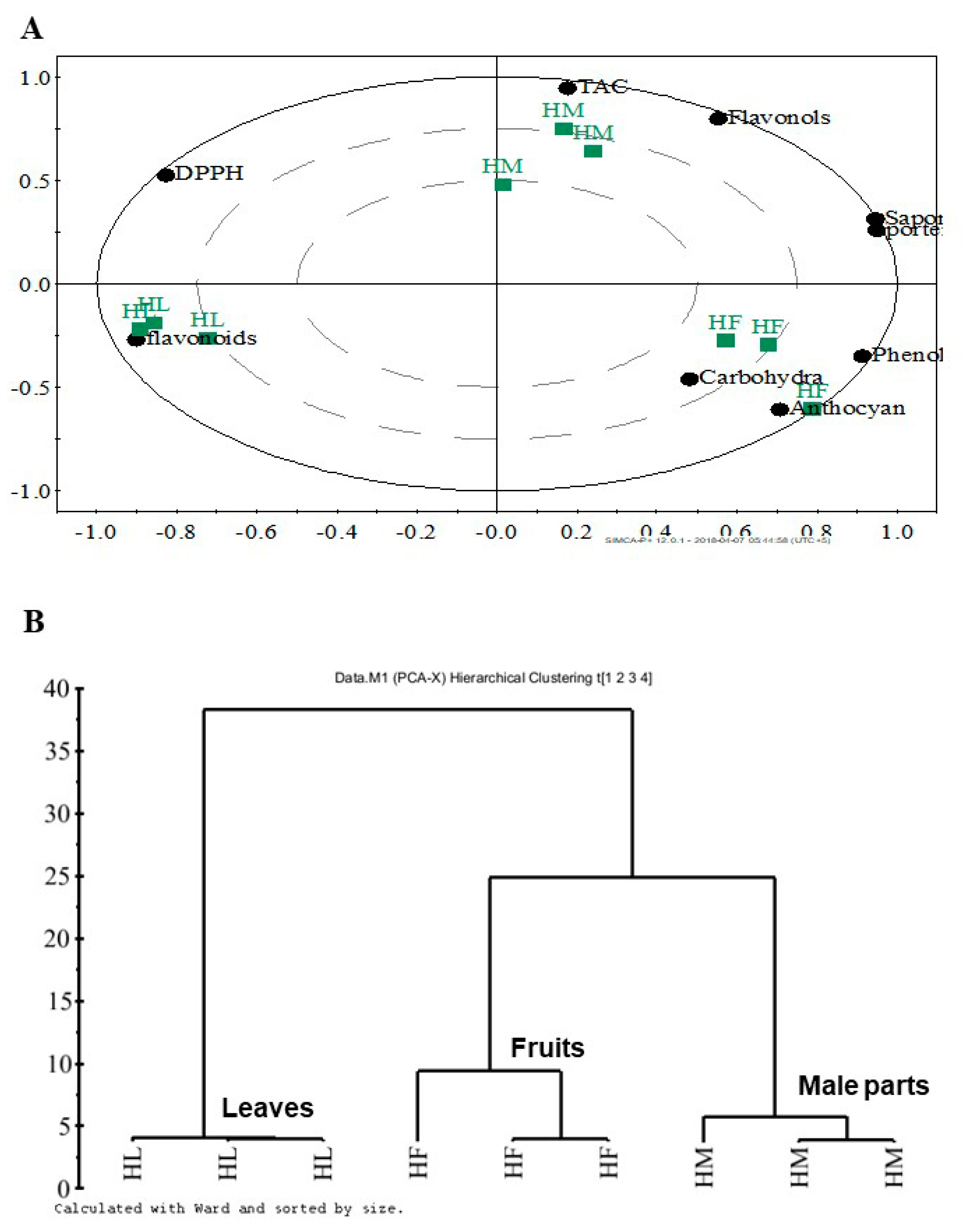
The data of phytochemical analysis were subjected to different tools of MVDA to assess the similarity and dissimilarity between different parts of the doum palm from the point of their metabolic profiling and phytochemical composition. The data of the three evaluated parts were subjected to PCA and followed by HCA. PCA discriminated the three parts of the doum palm into three separated groups and showed that there is a similarity between fruits and male parts resulted from the approximation of the two parts in the positive part of PC1, as shown in the score scatter plot (Figure 1A). The score loading plot (Figure 1B) showed the important metabolites responsible for the separation obtained in the score scatter plot. Male parts had higher contents of flavonols and saponins as well as TAC. Fruits had higher contents of anthocyanins, carbohydrates and phenolics. Leaves had higher contents of flavonoids and stronger radical scavenging activity. The score biplot confirmed the results of the score and loading plots of PCA (Figure 2A). HCA and dendrogram showed the classification of the three parts in three groups with a similarity between fruits and male parts (Figure 2B).
Male parts of the doum palm showed the highest TAC (308.8 mg ascorbic acid equivalent/g extract), and the leaves and fruits showed lower values (123.8 and 127.9 mg ascorbic acid equivalent/g extract, respectively) (Figure 3A). A significant difference was found between the TAC in leaves and male parts and that of male parts and fruits (P < 0.05). Leaves and male parts showed a significantly higher DPPH•-scavenging activity (94.6 and 92.1%, respectively) when compared with fruits (73.75%). A significant difference was found between DPPH of leaves and fruits and between fruits and male parts (P < 0.05; Figure 3B–D).
The relation between TAC and determined metabolites in the three doum palm parts was evaluated using Pearson’s correlation. Positive correlation between TAC and the content of total carbohydrates, phenolics and flavonols was found with the following probabilities: 0.013, 0.003 and 0.006, respectively and r values: 0.904, 0.957 and 0.935, respectively (Table 2).

3.2. Profiling of Polyphenols in H. thebaica Using HPLC

Leaves, male parts and fruits of the doum palm were assessed for the presence of polyphenols in their extracts. Twenty-four standards were injected, and their peaks are presented in Supplementary Figure S1. HPLC showed the presence of 14 polyphenols in the evaluated parts. Rutin, catechins, apigenin-7-glucoside and ferulic acid were the dominant polyphenols detected in leaves (Table 3). Protocatechuin, p-hydroxybenzoic, syringic, caffeic, vanillic, rosmarinic and p-coumaric acids and chrysin were the dominant polyphenols in male parts and chlorogenic acid was dominant in fruits (Table 3).

3.3. Anticancer Activity of H. thebaica

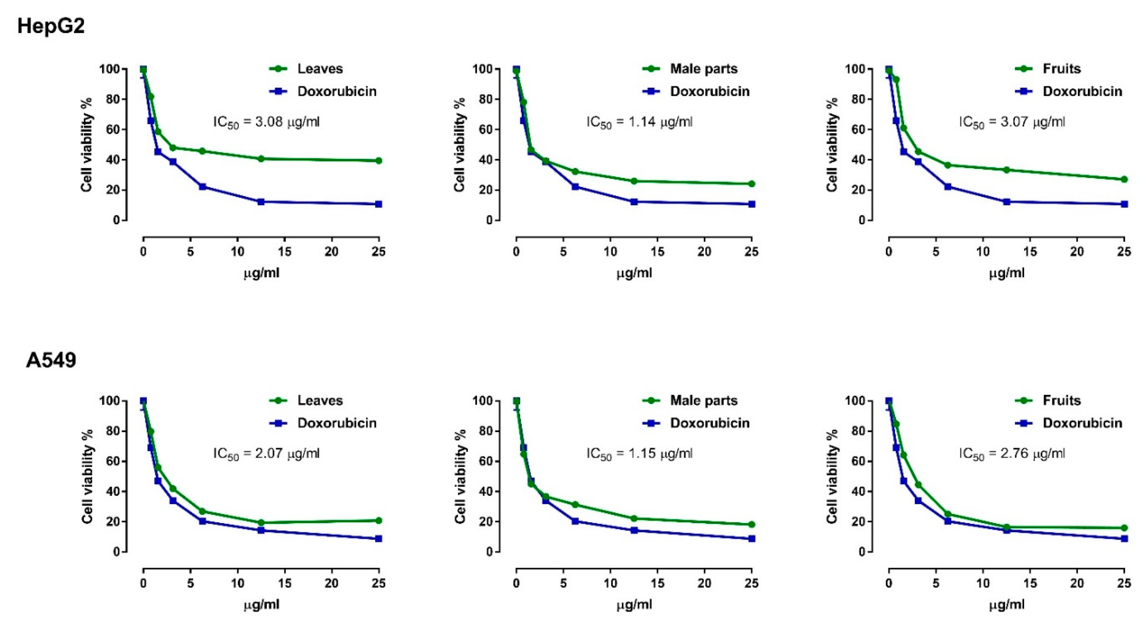
Leaves, male parts and fruits showed potent cytotoxic potentiality against HepG2 and A549 cancer cell lines. In contrast, all tested extracts exhibited non-significant effects on the viability of Vero cells (Supplementary Figure S2). When compared with the leaves and fruits, male parts showed stronger antiproliferative activity against HepG2 (IC50 = 1.14 µg/mL) and A549 (IC50 = 1.15 µg/mL) as represented in Figure 4.

3.4. Antimicrobial Activity of H. thebaica

The antimicrobial activity of different parts of the doum palm was evaluated using the paper diffusion disc assay. Although the three extracts showed a strong antibacterial activity against the evaluated microorganisms, the leaves extract showed more prominent effect followed by fruits and male parts extracts (Table 4). Moreover, fruit and male part extracts were more potent against Gram-negative than Gram-positive bacteria. Among the evaluated extracts, only fruits extract showed antifungal activity against Candida albicans. Bacterial strains were more sensitive than the fungal strains to the doum palm extracts as no extract showed antifungal activity against A. flavus (Table 4).

4. Discussion

The doum palm is a non-exploited important dietary source of active metabolites exerting potential effects on human health. This study investigated the metabolic content and in vitro antimicrobial and anticancer efficacies of the leaves, male parts and fruits extracts of H. thebaica. The metabolomic profiling by spectrophotometer and MVDA has not reflected the whole picture of the doum palm metabolome. The fruits showed higher content of some active secondary metabolites, but weak radical scavenging activity and TAC when compared to leaves and male parts. To unveil the whole picture and shed light on the dominant individual polyphenol metabolites present in each part, HPLC analysis was used. The variation of secondary metabolites among different parts of the doum palm is in consistence with many previous reports. Different parts of different species of Acacia showed significant differences in their metabolomic profiling [23].
The DPPH assay was used to assess the antioxidant potentilaity of plant extracts through evaluating their potentiality as free radical scavengers or hydrogen donors. Given their high content of polyphenols and saponins, the extracts of doum palm may possess hydrogen donating ability and act as antioxidants. The high content of total secondary metabolites or individual polyphenols in doum palm was reflected on the TAC and DPPH radical scavenging activity of its extracts. Previous studies have demonstrated a positive correlation between the content of phenolics and flavonoids and TAC [23,24]. The DPPH radical scavenging activity and TAC of Medicago lupolina aerial part, Carica papaya leaf, Erheropheleum suaveolens stem park and Terminalia schimperiana root extracts were attributed to their content of saponins, flavonoids and phenolics [25,26]. In addition, glycosides have been suggested to contribute to the antioxidant effects of plant extracts [27]. Here, a positive correlation between TAC and carbohydrates content was observed. This correlation might be attributed to the presence of glucosides, such as apigenin-7-glucoside as recognized in the HPLC profile.
The cytotoxic activity of doum palm extracts was attributed to the high content of the secondary metabolites contributing to anticancer activity, such as phenolics, flavonoid, flavonols, anthocyanins and saponins. The content of these metabolites was positively correlated with the anticancer activity of some Saudi desert plants against HCC and breast carcinoma cell lines [28]. Plant extracts rich in saponins and phenolics showed cytotoxic potentiality against several cell lines. In this context, Carica papaya leaf extract rich in phenolics and saponins showed anti-pancreatic cancer activity [29], and anthocyanins extracted from Zea mays exhibited antiproliferative activity against colon cancer cell lines [30]. Previous work from our lab has demonstrated the potent anti-carcinogenic effect of polyphenols as well as phenolics-rich extracts against HCC and colon carcinogenesis both in vivo and in vitro [31,32]. Here, HCC and lung cancer cell lines showed different sensitivity to each extract. Male parts showed a potent antiproliferative effect on HepG2 and A549 cells when compared with the fruits and leaves extracts as reflected by the lower IC50. The phytochemical analysis revealed the higher tannins content of male parts which may play a potent antiproliferative role. The broad cancer chemopreventive activity of various tannins in several animal models has been reviewed by Nepka et al. [33]. In addition, recent studies have shown the anticancer efficacy of tannins, particularly tannic acid, against prostate and lung cancers [34,35]. The anticancer potentiality of doum palm extracts could be attributed to the high content of individual polyphenols, such as protocatechuic, p-hydroxybenzoic, chlorogenic, rosmarinic, vanillic, p-coumaric acids, apigenin-7-glucoside, rutin and chrysin. The anticancer activity of these metabolites has been well-established [27,36,37]. In addition to the effect of individual metabolites, synergistic interactions between phenolics, flavonoids, saponins, anthocyanins and other phytochemicals might be suggested to mediate the anti-carcinogenic effects of plant extracts.
Besides their radical scavenging activity, doum palm extracts showed antibacterial efficacy. Bacterial strains differ in their responses to the plant extracts depending on the strain, nature of the cell wall, plant part, extraction method and extraction solvent [38]. In this study, E. coli (Gram-negative) was more sensitive to leaves and fruits methanol extracts followed by S. aureus and B. subtilis (Gram-positive), respectively. Accordingly, previous studies have demonstrated higher sensitivity of E. coli than S. aureus and B. subtilis towards methanolic flower extract of Capparis spinosa and root extract of C. decidua [38]. The antimicrobial activity of doum palm was attributted to its rich content of secondary metabolites. The correlation between the plant content of secondary metabolites, such as anthocyanins, saponins, phenolics, flavonoids and tannins, and the antimicrobial potentiality was reported [38,39]. The individual polyphenolic compounds profiled in HPLC were well-known as antimicrobial agents against wide spectrum of Gram-positive and negative bacteria [40,41]. The mechanism of polyphenols toxicity against microorganisms may be attributed to the suppression of proteases and/or inactivation of the microbial adhesions [42].

5. Conclusions

The high content of secondary metabolites in different parts of the doum palm was reflected on the TAC, and free radical scavenging, anticancer and antimicrobial activities of their extracts. Male parts and fruits extracts showed effective anticarcinogenic effects, whereas leaves extract showed more potent bactericidal efficacy. The cytotoxic and antimicrobial effects of doum palm were attributed to its high contents of active metabolites, such as, saponins, anthocyanins and polyphenols (apigenin-7-glucoside, rutin, vanilic acid, protocatechuic, p-hydroxybenzoic, catechins, chlorogenic acid, caffeic, p-coumaric, rosmarinic acid and chrysin). Given the strong in vitro anticancer and antimicrobial activities of doum palm, this study recommends the separation of its extracts and individual active components and evaluating their therapeutic effects using in vitro and in vivo studies.

Supplementary Materials

The following are available online at https://www.mdpi.com/2227-9717/8/3/266/s1, Figure S1: Supplementary Figure 1. HPLC chromatogram of 24 polyphenols standards at 280 nm. Figure S2. Effect of H. thebaica leaves, male parts and fruits on Verocells. Data are mean ±SD, N = 3.

Author Contributions

Conceptualization, I.B.A.-F., G.A.T. and U.A.M.; methodology, I.B.A.-F., M.G.S., A.M.M., G.A.T. and U.A.M.; validation, I.B.A-F., G.A.T., A.M.M. and U.A.M.; formal analysis, I.B.A.-F., G.A.T., M.G.S., A.M.M. and U.A.M.; investigation, I.B.A.-F., G.A.T., M.G.S., A.M.M., H.A.E. and U.A.M.; resources, M.B.-J.; data curation, I.B.A.-F., G.A.T., M.G.S., A.M.M., M.B.-J, H.A.E. and U.A.M.; writing—original draft preparation, I.B.A.-F., G.A.T., M.G.S., and U.A.M.; writing—review and editing, A.M.M.; supervision, I.B.A.-F., M.G.S. and U.A.M. All authors have read and agreed to the published version of the manuscript.

Funding

The Deanship of Scientific Research at Princess Nourah bint Abdulrahman University supported this research through the Fast-track Research Funding Program.

Acknowledgments

The authors thank Muhammad Jahangir, Haripur University for assisting in multivariate data analysis and providing valuable comments and suggestions. The authors thank the Deanship of Scientific Research at Princess Nourah bint Abdulrahman University for supporting this research through the Fast-track Research Funding Program.

Conflicts of Interest

The authors declare no conflict of interest.

References

  1. Mahmoud, A.M.; Alexander, M.Y.; Tutar, Y.; Wilkinson, F.L.; Venditti, A. Oxidative stress in metabolic disorders and drug-induced injury: The potential role of nrf2 and ppars activators. Oxidative Med. Cell. Longev. 2017, 2017, 2508909. [Google Scholar] [CrossRef] [Green Version]
  2. Lindenschmidt, R.C.; Tryka, A.F.; Goad, M.E.; Witschi, H.P. The effects of dietary butylated hydroxytoluene on liver and colon tumor development in mice. Toxicology 1986, 38, 151–160. [Google Scholar] [CrossRef]
  3. Kruse, M.; Strandberg, M.; Strandberg, B. Ecological Effects of Allelopathic Plants—A Review; Technical Report No. 31; National Environmental Research Institute: Silkeborg, Denmark, 2000; p. 66. [Google Scholar]
  4. Olajuyigbe, O.O.; Afolayan, A.J. Phytochemical assessment and antioxidant activities of alcoholic and aqueous extracts of acacia mearnsii de wild. Int. J. Pharmacol. 2011, 7, 856–861. [Google Scholar]
  5. Gurib-Fakim, A. Medicinal plants: Traditions of yesterday and drugs of tomorrow. Mol. Asp. Med. 2006, 27, 1–93. [Google Scholar] [CrossRef] [PubMed]
  6. Langley-Evans, S.C. Antioxidant potential of green and black tea determined using the ferric reducing power (frap) assay. Int. J. Food Sci. Nutr. 2000, 51, 181–188. [Google Scholar] [CrossRef] [PubMed]
  7. Kamel, E.M.; Mahmoud, A.M.; Ahmed, S.A.; Lamsabhi, A.M. A phytochemical and computational study on flavonoids isolated from trifolium resupinatum l. and their novel hepatoprotective activity. Food Funct. 2016, 7, 2094–2106. [Google Scholar] [CrossRef] [PubMed]
  8. Hsu, B.; Coupar, I.M.; Ng, K. Antioxidant activity of hot water extract from the fruit of the doum palm, hyphaene thebaica. Food Chem. 2006, 98, 317–328. [Google Scholar] [CrossRef]
  9. Salib, J.Y.; Michael, H.N.; Eskande, E.F. Anti-diabetic properties of flavonoid compounds isolated from hyphaene thebaica epicarp on alloxan induced diabetic rats. Pharmacogn. Res. 2013, 5, 22–29. [Google Scholar] [CrossRef] [Green Version]
  10. AbdEl-Moniem, M.; Mustafa, H.N.; Megahed, H.A.; Agaibyi, M.H.; Hegazy, G.A.; El-Dabaa, M.A. The ameliorative potential of hyphaene thebaica on streptozotocin-induced diabetic nephropathy. Folia Morphol. 2015, 74, 447–457. [Google Scholar] [CrossRef] [Green Version]
  11. Tohamy, A.A.; Mohammed, R.S.; Abdalla, M.S.; Ibrahim, A.K.; Mahran, K.F.; Ahmed, K.A. The effect of lupinus albus and hyphaene thebaica on chromosomal aberrations and histopathological changes of liver and pancreas in streptozotocin-induced diabetic rats. Egypt. J. Hosp. Med. 2013, 53, 763–769. [Google Scholar] [CrossRef]
  12. Hetta, M.H.; Yassin, N.Z. Comparative studies on hypocholesterolemic effect of different fractions of hyphaene thebaica (doum) in experimental animals. Die Pharm. 2006, 61, 230–232. [Google Scholar]
  13. Aboshora, W.; Lianfu, Z.; Dahir, M.; Qingran, M.; Qingrui, S.; Jing, L.; Al-Haj, N.Q.M.; Ammar, A.F.; Lianfu, Z.; Aboshora, W.; et al. Effect of extraction method and solvent power on polyphenol and flavonoid levels in hyphaene thebaica l mart (arecaceae) (doum) fruit, and its antioxidant and antibacterial activities. Trop. J. Pharm. Res. 2014, 13, 2057–2063. [Google Scholar] [CrossRef] [Green Version]
  14. Morris, D.L. Quantitative determination of carbohydrates with dreywood’s anthrone reagent. Science 1948, 107, 254–255. [Google Scholar] [CrossRef] [PubMed]
  15. Singleton, V.L.; Orthofer, R.; Lamuela-Raventós, R.M. Analysis of total phenols and other oxidation substrates and antioxidants by means of folin-ciocalteu reagent. In Methods in Enzymology; Academic Press: London, UK, 1999; Volume 299, pp. 152–178. [Google Scholar]
  16. Zhishen, J.; Mengcheng, T.; Jianming, W. The determination of flavonoid contents in mulberry and their scavenging effects on superoxide radicals. Food Chem. 1999, 64, 555–559. [Google Scholar] [CrossRef]
  17. Kumaran, A.; Joel Karunakaran, R. In vitro antioxidant activities of methanol extracts of five phyllanthus species from india. LWT Food Sci. Technol. 2007, 40, 344–352. [Google Scholar] [CrossRef]
  18. Julkunen-Tiitto, R. Phenolic constituents in the leaves of northern willows: Methods for the analysis of certain phenolics. J. Agric. Food Chem. 1985, 33, 213–217. [Google Scholar] [CrossRef]
  19. Prieto, P.; Pineda, M.; Aguilar, M. Spectrophotometric quantitation of antioxidant capacity through the formation of a phosphomolybdenum complex: Specific application to the determination of vitamin E. Anal. Biochem. 1999, 269, 337–341. [Google Scholar] [CrossRef]
  20. Kim, K.-H.; Tsao, R.; Yang, R.; Cui, S.W. Phenolic acid profiles and antioxidant activities of wheat bran extracts and the effect of hydrolysis conditions. Food Chem. 2006, 95, 466–473. [Google Scholar] [CrossRef]
  21. Vichai, V.; Kirtikara, K. Sulforhodamine b colorimetric assay for cytotoxicity screening. Nat. Protoc. 2006, 1, 1112–1116. [Google Scholar] [CrossRef]
  22. Bauer, A.W.; Kirby, W.M.M.; Sherris, J.C.; Turck, M. Antibiotic susceptibility testing by a standardized single disk method. Am. J. Clin. Pathol. 1966, 45, 493–496. [Google Scholar] [CrossRef]
  23. Abdel-Farid, I.B.; Sheded, M.G.; Mohamed, E.A. Metabolomic profiling and antioxidant activity of some acacia species. Saudi J. Biol. Sci. 2014, 21, 400–408. [Google Scholar] [CrossRef] [Green Version]
  24. Mansour, R.B.; Jilani, I.B.H.; Bouaziz, M.; Gargouri, B.; Elloumi, N.; Attia, H.; Ghrabi-Gammar, Z.; Lassoued, S. Phenolic contents and antioxidant activity of ethanolic extract of capparis spinosa. Cytotechnology 2016, 68, 135–142. [Google Scholar] [CrossRef] [Green Version]
  25. Kicel, A.; Olszewska, M.A. Evaluation of antioxidant activity, and quantitative estimation of flavonoids, saponins and phenols in crude extract and dry fractions of medicago lupulina aerial parts. Nat. Prod. Commun. 2015, 10, 483–486. [Google Scholar] [PubMed] [Green Version]
  26. Diaconeasa, Z.; Leopold, L.; Rugina, D.; Ayvaz, H.; Socaciu, C. Antiproliferative and antioxidant properties of anthocyanin rich extracts from blueberry and blackcurrant juice. Int. J. Mol. Sci. 2015, 16, 2352–2365. [Google Scholar] [CrossRef] [PubMed] [Green Version]
  27. Fuchs, J.; Milbradt, R. Skin anti-inflammatory activity of apigenin-7-glucoside in rats. Arzneim. Forsch. 1993, 43, 370–372. [Google Scholar]
  28. Abdel-Farid, I.B.; Mahalel, U.A.; Jahangir, M.; Elgebaly, H.A.; El-Naggar, S.A. Metabolomic profiling and antioxidant activity of opophytum forsskalii. Aljouf Sci. Eng. J. 2016, 286, 1–6. [Google Scholar] [CrossRef]
  29. Vuong, Q.V.; Hirun, S.; Chuen, T.L.K.; Goldsmith, C.D.; Murchie, S.; Bowyer, M.C.; Phillips, P.A.; Scarlett, C.J. Antioxidant and anticancer capacity of saponin-enriched carica papaya leaf extracts. Int. J. Food Sci. Technol. 2015, 50, 169–177. [Google Scholar] [CrossRef]
  30. Zhao, X.; Zhang, C.; Guigas, C.; Ma, Y.; Corrales, M.; Tauscher, B.; Hu, X. Composition, antimicrobial activity, and antiproliferative capacity of anthocyanin extracts of purple corn (Zea mays L.) from China. Eur. Food Res. Technol. 2009, 228, 759–765. [Google Scholar] [CrossRef]
  31. Mahmoud, A.M.; Abdella, E.M.; El-Derby, A.M. Protective effects of turbinaria ornata and padina pavonia against azoxymethane-induced colon carcinogenesis through modulation of ppar gamma, nf-kappab and oxidative stress. Phytother. Res. 2015, 29, 737–748. [Google Scholar] [CrossRef]
  32. Mahmoud, A.M.; Mohammed, H.M.; Khadrawy, S.M.; Galaly, S.R. Hesperidin protects against chemically induced hepatocarcinogenesis via modulation of nrf2/are/ho-1, ppargamma and tgf-beta1/smad3 signaling, and amelioration of oxidative stress and inflammation. Chem. Biol. Interact. 2017, 277, 146–158. [Google Scholar] [CrossRef]
  33. Nepka, C.; Asprodini, E.; Kouretas, D. Tannins, xenobiotic metabolism and cancer chemoprevention in experimental animals. Eur. J. Drug Metab. Pharmacokinet. 1999, 24, 183–189. [Google Scholar] [CrossRef] [PubMed]
  34. Nagesh, P.K.B.; Chowdhury, P.; Hatami, E.; Jain, S.; Dan, N.; Kashyap, V.K.; Chauhan, S.C.; Jaggi, M.; Yallapu, M.M. Tannic acid inhibits lipid metabolism and induce ros in prostate cancer cells. Sci. Rep. 2020, 10, 980. [Google Scholar] [CrossRef] [PubMed]
  35. Hatami, E.; Nagesh, P.K.B.; Chowdhury, P.; Shetty, A.B.; Tripathi, M.K.; Chauhan, S.; Jaggi, M.; Yallapu, M. Abstract 1871: Tannic acid: A natural anticancer agent for non-small cell lung cancer. Cancer Res. 2019, 79, 1871. [Google Scholar]
  36. Chandra, Y.P.; Viswanathswamy, A.H.M. Chemo preventive effect of rutin against n-nitrosodiethylamine-induced and phenobarbital-promoted hepatocellular carcinoma in wistar rats. Indian J. Pharm. Educ. Res. 2018, 52, 78–86. [Google Scholar] [CrossRef] [Green Version]
  37. Yamagata, K.; Izawa, Y.; Onodera, D.; Tagami, M. Chlorogenic acid regulates apoptosis and stem cell marker-related gene expression in a549 human lung cancer cells. Mol. Cell. Biochem. 2018, 441, 9–19. [Google Scholar] [CrossRef] [PubMed]
  38. Gull, T.; Sultana, B.; Bhatti, I.A.; Jamil, A. Antibacterial potential of capparis spinosa and capparis decidua extracts. Int. J. Agric. Biol. 2015, 17, 727–733. [Google Scholar] [CrossRef]
  39. Cisowska, A.; Wojnicz, D.; Hendrich, A.B. Anthocyanins as antimicrobial agents of natural plant origin. Nat. Prod. Commun. 2011, 6, 149–156. [Google Scholar] [CrossRef] [Green Version]
  40. Alves, M.J.; Ferreira, I.C.F.R.; Froufe, H.J.C.; Abreu, R.M.V.; Martins, A.; Pintado, M. Antimicrobial activity of phenolic compounds identified in wild mushrooms, sar analysis and docking studies. J. Appl. Microbiol. 2013, 115, 346–357. [Google Scholar] [CrossRef]
  41. Nayaka, H.B.; Londonkar, R.L.; Umesh, M.K.; Tukappa, A. Antibacterial attributes of apigenin, isolated from Portulaca oleracea L. Int. J. Bacteriol. Int. J. Bacteriol. 2014, 2014, 1–8. [Google Scholar] [CrossRef] [Green Version]
  42. Cowan, M.M. Plant products as antimicrobial agents. Clin. Microbiol. Rev. 1999, 12, 564–582. [Google Scholar] [CrossRef] [Green Version]
Figure 1. Score scatter plot of PC1 versus PC2 (A), and the score loading plot of PC1 versus PC2 of the metabolomic profiling of different parts of H. thebaica (B). HL = doum palm leaves, HM = doum palm male parts and HF = doum palm fruits.
Processes 08 00266 g001
Figure 2. Score biplot of PC1 versus PC2 (A), and hierarchal clustering analysis (HCA) (B) of the metabolomic profiling of different parts of H. thebaica. HL = doum palm leaves, HM = doum palm male parts and HF = doum palm fruits.
Processes 08 00266 g002
Figure 3. Total antioxidant capacity (A) and DPPH radical scavenging activity (BD) of H. thebaica leaves, male parts and fruits.
Processes 08 00266 g003
Figure 4. Antiproliferative activity of H. thebaica leaves, male parts and fruits against HepG2 and A549 cancer cell lines. Data are mean ± SD, N = 3.
Processes 08 00266 g004
Table 1. Phytochemical analysis of H. thebaica leaves, male parts and fruits.
LeavesMale partsFruits
Carbohydrates (mg/g DW)135.4 ± 0.18124.4 ± 0.5173.77 ± 0.4
Saponins (mg saponins equivalent/g)17.5 ± 0.1 a18.6 ± 0.1 b18.65 ± 0.04 b
Phenolics (mg/gallic acid equivalent/g)20.6 ± 2.9 a36.04 ± 0.06 b72.3 ± 2.9 c
Flavonoids (mg quercetin equivalent/g)32.7 ± 2.9 a22.1 ± 2.4 b20.4 ± 2.0 b
Flavonols (mg quercetin equivalent/g)61.29 ± 3.51 a61.23 ± 3.56 a38.2 ± 4.5 b
Anthocyanin (μmol/g)0.11 ± 0.05 a0.12 ± 0.09 a0.33 ± 0.06 b
Tannins (mg catechols equivalent/g)0.57 ± 0.03 a3.85 ± 0.3 b3.44 ± 0.05 b
a–c show significant difference at P < 0.05. Data are mean ± SD, N = 3.
Table 2. Correlation between the detected metabolites and TAC of H. thebaica.
Correlation Coefficient (r)Probability (p)
Carbohydrates0.9040.03
Saponins--
Phenolics0.9570.003
Flavonoids--
Flavonols0.9350.006
Anthocyanin--
Tannins--
Table 3. Individual polyphenolic compounds (mg/g) in H. thebaica leaves, male parts and fruits.
LeavesMale PartsFruits
Protocatechuic acid0.0720.463ND
p-hydroxybenzoic acid0.1260.275ND
Catechins0.56NDND
Chlorogenic acid0.0850.080.152
Caffeic acid0.0780.132ND
Syringic acid0.0370.049ND
Vanillic acid0.5551.081ND
Ferulic acid0.0340.03ND
Sinapic acid0.0520.065ND
Rutin2.6950.3240.073
p-coumaric acid0.1160.1370.062
Apigenin-7-glucoside5.428ND0.169
Rosmarinic acid0.0540.544ND
ChrysinND0.083ND
ND = non-detectable.
Table 4. Antimicrobial activity of H. thebaica leaves, male parts and fruits.
Zone of Inhibition (mm)
ControlLeavesMale PartsFruits
Gram +ve
bacteria
Bacillus subtilis28.0 ± 2.012.3 ± 1.110.0 ± 0.110.7 ± 0.5
Staphylococcus aureus31.0 ± 6.513.3 ± 1.512.3 ± 0.512.3 ± 1.1
Gram −ve
bacteria
Pseudomonas aeruginosa32.4 ± 1.113.3 ± 0.510.3 ± 0.511.3 ± 0.5
Escherichia coli29.0 ± 1.713.7 ± 0.510.0 ± 0.012.3 ± 1.0
FungiAspergillus flavus14.6 ± 1.50.0 ± 0.00.0 ± 0.00.0 ± 0.0
Candida albicans16.3 ± 1.10.0 ± 0.00.0 ± 0.09.0 ± 0.0
Data are mean ±SD, N = 3.