Review

The Concept of Sustainable Development of Modern Dentistry

by
Leszek A. Dobrzański
1,*,
Lech B. Dobrzański
1,
Anna D. Dobrzańska-Danikiewicz
2 and
Joanna Dobrzańska
1
1
Medical and Dental Engineering Centre for Research, Design and Production ASKLEPIOS, 13 D Królowej Bony St., 44-100 Gliwice, Poland
2
Department of Mechanical Engineering, University of Zielona Góra, 4 Prof. Z. Szafrana St., 65-516 Zielona Góra, Poland
*
Author to whom correspondence should be addressed.
Processes 2020, 8(12), 1605; https://doi.org/10.3390/pr8121605
Submission received: 8 September 2020 / Revised: 25 November 2020 / Accepted: 2 December 2020 / Published: 6 December 2020
(This article belongs to the Special Issue Synthesis and Characterization of Biomedical Materials)

Abstract

:
This paper concerns the assessment of the current state of dentistry in the world and the prospects of its sustainable development. A traditional Chinese censer was adopted as the pattern, with a strong and stable support on three legs. The dominant diseases of the oral cavity are caries and periodontal diseases, with the inevitable consequence of toothlessness. From the caries 3.5–5 billion people suffer. Moreover, each of these diseases has a wide influence on the development of systemic complications. The territorial range of these diseases and their significant differentiation in severity in different countries and their impact on disability-adjusted life years index are presented (DALY). Edentulousness has a significant impact on the oral health-related quality of life (OHRQoL). The etiology of these diseases is presented, as well as the preventive and therapeutic strategies undertaken as a result of modifying the Deming circle through the fives’ rules idea. The state of development of Dentistry 4.0 is an element of the current stage of the industrial revolution Industry 4.0 and the great achievements of modern dental engineering. Dental treatment examples from the authors’ own clinical practice are given. The systemic safety of a huge number of dentists in the world is discussed, in place of the passive strategy of using more and more advanced personal protective equipment (PPE), introducing our own strategy for the active prevention of the spread of pathogenic microorganisms, including SARS-CoV-2. The ethical aspects of dentists’ activity towards their own patients and the ethical obligations of the dentist community towards society are discussed in detail. This paper is a polemic arguing against the view presented by a group of eminent specialists in the middle of last year in The Lancet. It is impossible to disagree with these views when it comes to waiting for egalitarianism in dental care, increasing the scope of prevention and eliminating discrimination in this area on the basis of scarcity and poverty. The views on the discrimination of dentistry in relation to other branches of medicine are far more debatable. Therefore, relevant world statistics for other branches of medicine are presented. The authors of this paper do not agree with the thesis that interventional dental treatment can be replaced with properly implemented prophylaxis. The final remarks, therefore, present a discussion of the prospects for the development of dentistry based on three pillars, analogous to the traditional Chinese censer obtaining a stable balance thanks to its three legs. The Dentistry Sustainable Development (DSD) > 2020 model, consisting of Global Dental Prevention (GDP), Advanced Interventionist Dentistry 4.0 (AID 4.0), and Dentistry Safety System (DSS), is presented.

1. Introduction

In order to introduce the subject of this paper, several concepts are presented that seemingly do not combine into a whole, but are necessary in order to understand the view and content presented in this paper. It is obvious that the main content of this paper will discuss all issues that relate exclusively to dentistry, but the broad context presented in this introduction is necessary.
The first of these notions is balance. In the Dictionary of the Polish Language, we find a definition of balance as a stable system of opposing forces and values.
“Balance appears as the basic nature of all things, no matter what level we pay attention to. It applies to the same degree to elementary particles, the functioning of the organism, social phenomena, or the movement of galaxies. Opposites strive towards each other, they try to complement and complete each other because in themselves they do not actually exist. This process is constantly accompanied by movement, which is a tool of balance. It is because of him that everything that exists is constantly changing and tends to the perfect point that can never be achieved. Because balance is not so much a point, an ideal, but a constant balancing around that point. Balancing unity is the ever-pulsating dance of everything, it is the beating heart of the Cosmos, it is the very breath of God.” [1].
It has been known in statics for centuries that a stable, balanced position is provided by three points of support. Examples of this principle have been in existence for centuries. Figure 1 shows a Chinese censer dating back to a centuries-old tradition made at the turn of the 19th and 20th centuries as a bronze cast in the form of a spherical vase on three animal legs, with mascarons at the base, a belly decorated in relief with two pairs of dragons holding a fireball, ears with dragon heads on the sides, and a meander pattern strip around the top edge. The cover is strongly arched, openwork, with four handles in the form of elephant heads; at the top, there is a figure of a lying Chinese lion. This piece of Chinese art can be considered a symbol of balance.
Another defined term is sustainable development. It is necessary to distinguish between instantaneous equilibrium and a process based on equilibrium. One such notion is sustainable development.
The sources of this concept are ecological, because Hans Carl von Carlowitz in his book [2], published firstly in 1713, which was a comprehensive treatise on forestry, introduced the concept of sustainable yield. The view outlined by him assumed forest management in such a way that the forest could always rebuild itself, which required planting as many trees as had just been cut down. The concept spread in many European countries and gained the English term Sustained Yield Forestry, and in the 1980s it was spread by environmental movements to finally evolve into sustainable development in a very broad context.
Currently, the content of this notion is determined by the doctrine of economics, assuming the quality of life at the level allowed by the current civilization development provided in the preamble to the World Commission on Environment and Development (WCED) report: “At the current civilization level, sustainable development is possible (…) in which the needs of the present generation can be satisfied without reducing future opportunities generations to satisfy them” [3]. The achieved level of prosperity is possible to maintain, provided that the relationship between economic growth, care for the environment, including the man-made environment, and quality of life, including human health, is appropriately and consciously formed. This document draws on a study by the Club of Rome dating back to 1972 [4]. The basis of all activities in this area is included in the theory of public good [5].
One of the indisputable goods that sustainable development guarantees to people in the world is health, as a necessary condition of well-being, and the concern of all societies is to extend human life. Health, including oral health, is therefore a human right, and inclusive development reduces inequalities, provided by universal access to healthcare, including primary dental care. Improving oral health reduces development burdens and costs to society, health systems, and the economy in general. Oral disease affects approximately 3 to 5 billion people worldwide [6,7]. The proportion of the population receiving oral health care in Low and Middle-Income Countries (LMIC) is generally lower than in High-Income Countries (HIC), with median estimates ranging from 35% in Low-Income Countries (LIC) to 60% in lower-income low-middle-income countries, 75% in upper-income low-middle-income countries, and 82% in high-income countries [7]. Within each country, the poorest quintiles have the lowest service coverage. Oral diseases reduce life chances, diminish the sense of dignity, and reduce the productive contribution of individuals to their local communities and to society as a whole.
The aim of this paper is to analyze the state of modern dentistry and indicate the conditions for ensuring its current balance and sustainable development, which on the one hand must always involve an analysis of the needs, but on the other hand must take into account the possibility of indicating benefits but without exposing anyone to any loss or harm. The dominant diseases of the oral cavity are caries and periodontal diseases. The strategies for treating caries and periodontal diseases require detailed analysis, which is presented in this paper. The territorial range of these diseases and their significant differentiation in severity in different countries as well as their impact on the disability-adjusted life years (DALY) index are presented. Across the world, 3–5 billion people suffer from tooth decay. Caries very often in adults is accompanied by periodontal diseases, which are also very widespread across the world. Toothlessness, an inevitable consequence of both of these diseases, especially if not treated at all and in the case of bad habits, has a significant impact on the oral health-related quality of life (OHRQoL). Oral diseases have a wide impact on other organs, even distantly, and are a direct cause of systemic diseases, which not all people are aware of. This paper was inspired because it is a polemic arguing against the views presented by a group of eminent specialists in the middle of last year in a series of two articles [6,7]. It is impossible to disagree with these views when it comes to waiting for egalitarianism in dental care, increasing the scope of prevention, and eliminating social exclusion in this area due to scarcity and poverty. The views on the discrimination of dentistry as compared to other fields of medicine are much more debatable. Relevant statistical data concerning, inter alia, the mortality from heart disease, stroke, and cancer and human health expenditure, for example, can vary between OECD countries by more than 50 times. This situation is largely shaped by politicians in individual countries and the extent to which individual societies enforce their democratic rights, including the share of health care and social security costs in the gross domestic product (GDP). The most debatable view is that prevention in the field of dentistry is treated as an opposite approach to interventional dentistry and is mutually exclusive with the most advanced stage of Dentistry 4.0. There are many examples of the development of Dentistry 4.0 as great achievements, based on the development of computer imaging methods, computer-aided design, and manufacturing of implants and prosthetic restorations, including the use of cloud computing, automation, and robotization, as well as additive manufacturing technologies determining the advanced state of modern dental engineering. The cooperation between the dentist and the dental engineer plays an important role here, which is a prerequisite for success in this field. The level of modern dentistry is also determined by modern endodontic treatment methods and the possibility of using avant-garde implant scaffolds and implant scaffolds. In the field of periodontitis, an important role is played by the TIPPS strategy (Talk-Instruct-Practise-Plan-Support) [8], which draws on the traditional quality management solutions known as the Deming circle [9]. Therefore, this case requires a very broad and multifaceted analysis, which is also the purpose of this paper. Caries treatment, including endodontic and periodontal treatment, eliminates many diseases of distant organs of the body, as well as the implantological and prosthetic treatment of toothlessness. An analysis of the contexts of these systemic interactions is also one of the purposes of this paper. This significantly relieves hospital treatment and the social insurance system from eliminating long-term illnesses. The authors of this paper do not agree with the thesis that interventional dental treatment can be replaced with properly implemented prevention and, in broadly discussing this problem, indicate that sustainable development is essential in both of these areas. Both are important and require equal effort for the sustainability of dentistry. The third important development pillar of dentistry, and therefore another goal of the analyses presented in this paper, is the systemic epidemiological safety of a huge number of dentists in the world, an issue the current COVID-19 pandemic has highlighted. In place of the passive STOP strategy (system, technical, organizational, personal) used so far, which determines the introduction of more and more advanced personal protective equipment (PPE), assuming that the entire environment around the dentist and the patient are infected, the proprietary system, prevention, efficiency, cause (SPEC) strategy is introduced, consisting of the active prevention of the spread of pathogenic microorganisms, including SARS-CoV-2, in a dental practice environment. The point is to eliminate the threat directly at the source. An example of a proprietary accessory that meets these requirements is presented. In the course of this paper, an attempt is made to prove this thesis, supported by extensive literature studies, showing that the developmental balance of modern dentistry requires three equal elements. The association and the similarity to the case of the Chinese censer, which stands steadily on three legs, is clear. Hence, the aim of this paper is to support the thesis that the stability of dentistry development depends on three thematic areas that determine its even development, with numerous pieces of evidence from the literature. The thesis includes prevention and interventionist treatment in the Dentistry 4.0 system, including, among other things, endodontic and periodontal treatment, as well as an efficient system of epidemiological safety related to the prevention of the spread of pathogenic pathogens at the source. These analyses were preceded by a brief analysis of the condition of oral cavity diseases. The final remarks present a discussion of the prospects for the development of dentistry based on the three above-mentioned pillars, the prototype of which is the presented Chinese censer.

2. Analysis of the Contemporary State of Oral Cavity Diseases in the World

In order to draw any conclusions about the development of dentistry in the coming decades, it is necessary to analyze its current state.
Mature men and women have 32 teeth. Their condition in each case depends on many factors, including genetic conditions; developmental conditions; age; oral hygiene in relation to life history, diet, and nutritional preferences; interactions with other diseases; possible sports; transport’s and professional accidents; treatment history and possible errors in this regard; and many other factors (Figure 2) [6].
The stomatognathic system, especially the dentition, is also subject to numerous disease processes, under the influence both of internal and external factors. Oral diseases affect approximately 3 to 5 billion people worldwide [6,7]. Pulp diseases are common, mainly causing inflammation and pulp necrosis due to bacterial, thermal, mechanical, chemical, and electrical factors, as well as due to infection by blood vessels that penetrate the pulp through the apical foramen as well as due to cavities originated by caries and by non-carious factors and also by iatrogenic agents created during therapeutic procedures. The causes of oral disease, often leading to tooth loss, vary, although microbes are the most common cause of pulpitis. Hence, according to epidemiological data, the most common cause of tooth extraction is caries [10,11,12,13,14,15,16,17,18,19,20,21,22,23,24,25]. Carefully performed conservative treatment, especially endodontic treatment, allows for a relatively long time to keep the healed teeth in the mouth. Periodontal disease has also reached epidemic proportions [26,27,28,29,30,31,32,33,34], although this has only recently been recognized. This is the most common inflammation, affecting almost 50% of adults in the world, and can lead to complete toothlessness if left untreated. Up to 83% of the population develop some degree of bleeding from the gums during their lifetime. Careful hygiene and timely and carefully performed periodontal treatment also allow you to preserve your own teeth for a long time. Both caries and periodontal disease can lead to the extraction of single teeth and even complete toothlessness. Often, it is impossible to avoid tooth extraction, which, although allowing for immediate problem solving, creates further problems. The consequence of extracting even one tooth is an imbalance in the stomatognathic system, which ceases to function efficiently in the absence of any component. Oral neoplasms were excluded from consideration in this paper; although they occur quite frequently, they require separate analysis and usually fall within the scope of oncology or/and craniofacial surgery.
Oral diseases, both caries and periodontitis, as well as toothlessness, which after some time is an inevitable consequence of both of these diseases, are serious problems in modern medicine, not only because of local diseases of the oral cavity but above all because of the high risk of systemic complications.
Caries, as the primary pulp disease in the oral cavity, in the case of delayed, ineffective, or not-performed endodontic treatment is the direct cause of bacterial pneumonia, nephritis, rheumatoid arthritis, osteoporosis, abscess, stroke, endocarditis, ischemic heart disease, the decreased birth mass of infants, premature birth, and even the patient’s death [35,36,37,38,39,40]. Carefully performed endodontic treatment eliminates these risks and allows for a relatively long time to preserve healed teeth in the mouth.
Periodontal disease has also reached epidemic proportions [26,27,28,29,30], although this has only recently been recognized. It are the most common inflammation, affecting almost 50% of adults in the world, and can lead to complete toothlessness if left untreated. Up to 83% of the population develop some degree of bleeding from the gums during their lifetime. Periodontal disease, like tooth decay, is a direct cause of many long-term systemic diseases and complications, including diseases of the kidneys, lungs, cardiovascular system, pancreatic and oral cancer, as well as strokes and dementia [26,27,28,29,30,41]. The group of diseases commonly associated with periodontal diseases include cardiovascular diseases, heart failure, ischemic disease, and hypertension [42,43,44,45,46,47]. Numerous literature reports indicate erectile dysfunction in men and other complications of the reproductive system [48,49,50,51,52,53,54,55,56,57,58,59,60,61,62,63,64,65,66,67,68,69,70,71,72,73,74], which may also affect women [75] who are also pregnant [76].
Nevertheless, tooth extraction is often unavoidable. Both caries and periodontitis can even lead to complete toothlessness.
Lack of teeth causes not only a loss of aesthetic value, as many think, but is associated with some discomfort associated with eating difficulties and is also the cause of many diseases throughout the body due to chewing dysfunction or a lack of it [27,77,78,79,80,81,82,83,84,85,86,87,88,89,90]. Toothlessness can often be a direct cause of shortening the human life and is even a predictor of mortality, mainly from cardiovascular causes. Missing teeth cause coronary plaque formation and aortic hardening, increased susceptibility to electrocardiographic abnormalities, heart failure, ischemic heart disease, hypertension, and stroke. Diseases associated with edentulousness include certain cancers, diabetes mellitus, insulin independence, kidney disease, and rheumatoid arthritis. Often, there are diseases of the stomach; gastritis; duodenal ulcer; diseases of the pancreas, including tumors; as well as neoplastic changes in the esophagus and upper gastrointestinal tract. Chewing dysfunction, including it being impossible to grind and chew food, also causes unfavorable results in pregnancy. Mood and neurocognitive disorders, obstructive sleep apnea, cervical spine pain, migraine, and headaches, the direct cause of which are malocclusions due to tooth loss, determining the metabolism of the cerebrospinal fluid, may appear. Malocclusions and masticatory dysfunctions cause functional and morphological changes in the hippocampus in the temporal lobe of the forebrain. They negatively affect the hippocampus in the elderly [91,92,93,94,95,96,97] and cause associated spatial and episodic memory disturbances, as well as exacerbating symptoms of multiple sclerosis and increasing the risk of general dementia. Improvement in chewing conditions as a result of implantological and prosthetic treatment prevents hippocampus dysfunction.
It is also worth noting that the treatment of these pathologies requires high financial outlays and entails high costs. Undoubtedly, this makes it difficult to access both preventive and curative dental care in a lot of countries, mainly in LICs or LMICs. As a result, the scale of damage in the oral cavity in many patients throughout their life is catastrophically increased [98].
A measure of disease prevalence is the disability-adjusted life-years (DALY) indicator, often referring 100,000 inhabitants, which is used to determine the health condition of a given society. It expresses the total number of years of life lost as a result of premature death or damage to health as a result of an injury or disease (Figure 3) [99].
Caries is widespread practically all over the globe. It is considered the most common and costly chronic social disease, depending on the conditions of civilization. Figure 4 [100] shows the spread of caries in 2017 in different countries of the world and its diversity. The countries with the most widespread caries in the 5–14 age group are Kyrgyzstan, Uzbekistan, Romania, Bulgaria, Poland, and Ecuador. In addition, in the entire age range, these countries include France, Spain, Iceland, Greece, and Georgia. Figure 4 illustrates also the spread of edentulousness and severe tooth loss in different countries and the spread of periodontal disease. There seems to be a clear correlation between edentulousness and the prevalence of caries, with Ukraine, Russia, and some Balkan countries in Europe among the infamous leaders. In some cases, such as Australia and Brazil, edentulousness is relatively greater than the spread of caries. In almost all of the above-mentioned cases, as well as in others, where edentulousness is relatively greater than the prevalence of caries, endodontic treatment on an appropriate scale is most likely neglected or even not implemented. It seems that the situation is quite the opposite in countries such as France and Spain, where, despite a very high spread of caries, the share of edentulousness is relatively small. In countries such as Chile, Norway, Germany, and Denmark, periodontal diseases have a significant impact on the level of edentulousness. It should be noted that these observations are only correct to the extent that they are adequate to the actual states of the cited statistical studies. It seems, for example, in the case of Ukraine, Uzbekistan, and Kyrgyzstan, that these data do not correspond.
The incidence of severe tooth loss in the world in 1990 to 2010 is not gender-dependent, but increases gradually with age, showing a sharp increase around the seventh decade, with a peak incidence after the age of 65. On the other hand, there are clear geographical differences in the prevalence, incidence, and rate of improvement in the indicated period [101]. It is obvious that missing teeth, especially large ones, have a significant impact on the oral health-related quality of life (OHRQoL). People with shortened dental arches (SDAs) do not show OHRQoL worse than people with removable dentures. The number of closing pairs and the location of the remaining teeth have a great influence on the OHRQoL [102].
The incidence of severe tooth loss in the world in the period from 1990 to 2010 is not gender-dependent, but increases gradually with age, showing a sharp increase around the seventh decade, with a peak incidence after the age of 65.
On the other hand, there are clear geographical differences in the prevalence, incidence, and rate of improvement in the indicated period [101]. It is obvious that missing teeth, especially large ones, have a significant impact on the oral health-related quality of life (OHRQoL). People with shortened dental arches (SDAs) do not show a OHRQoL worse than people with removable dentures. The number of closing pairs and the location of the remaining teeth have a great influence on the OHRQoL [102]. Tooth loss causes malocclusion and, apart from aesthetic considerations, causes serious gastric diseases and has an adverse effect on the metabolism of the cerebrospinal fluid, which nourishes the brain. The consequence of this may be headaches, migraines, pains in the cervical spine, mood disorders, the intensification of the symptoms of multiple sclerosis, an unfavorable effect on the hippocampus in the temporal lobe of the cerebral cortex and other effects responsible among others on spatial memory. In a particularly unfavorable situation, caries and periodontal diseases may directly contribute to the patient’s death. Counteracting the consequences of caries and periodontal diseases requires replacing the missing teeth with implants and prosthetic restorations, including bridges and crowns. Dental prosthetics are used to rebuild the aesthetic value of the appearance, improve the psychological comfort of patients, and restore the appropriate functions of the digestive system related to taking and chewing food. This is a branch of dentistry dealing with the restoration of the original occlusal conditions after the loss of natural teeth or after their massive damage. For reconstruction, a prosthetic restoration is used, requiring the use of various biomaterials. Dental surgery is often associated with prosthetics, which deals with the surgical treatment of the oral cavity and adjacent areas, especially in the field of implantology. Dental implant prosthetics cover the reconstruction of teeth on implants and allow them to achieve a permanent aesthetic effect by improving the mechanics of the masticatory apparatus, disturbed as a result of tooth loss. The reconstruction of the structures and functions of lost periodontal tissues or of the alveolar bone itself often requires controlled tissue regeneration with the use of non-resorbable or resorbable materials, stimulating fibroblasts to grow and separating bone tissue from rapidly growing epithelial tissue. Modern dentistry allows us to perform advanced prosthetic restorations that rebuild all kinds of missing teeth, both in the case of complete tooth lessness and partially missing ones.

3. Dentistry 4.0 as an Avant-Garde Stage of Dental Treatment

Dental diseases have accompanied man since the dawn of time. As the human species developed and with the discovery of fire, humans began to cook at least some of their food, including fish and shellfish, nuts, fruit, and animal meat, which became the staple diet. Over time, crops such as rice, wheat, and barley began to be cultivated, which caused the proportion of sugars in food to increase, favoring humus. Such ailments can be seen, among others, in as member of the subspecies of Homo sapiens called the Cro-Magnon man from the Upper Paleolithic era, who lived in Europe between 43 thousand and 10 thousand BC and was found in the rock shelter of Abri de Cro-Magnon in Les Eyzies near the Dordogne in France. Traces of dental treatment were found in the Gaione cemetery in Italy in pre-Neolithic finds, and the removal of food debris with wooden or bone toothpicks has been confirmed as common among Neanderthals and Paleolithic people [103,104]. Skulls with traces of caries were found in the Egyptian pyramids of Giza, other places in Africa, America, and Asia, including in the village of Mehrgarh in a neolithic urban settlement from the 7th millennium BC belonging to the Indus Valley Civilization, located on the Kacchi plateau in present-day Balochistan in Pakistan [105,106]. It is reported that a tooth root abscess was removed through the skull in around 2500 BC in prehistoric Malta. However, confirmations of dental interventions from the neolithic period are rare [107]. Studies of the remains of ancient Egyptians, Greeks, and Romans also confirm early practices in the field of dental prosthetics, although perhaps not performed in life but after death—e.g., as part of the mummification of a corpse. At Gebel Ramlah cemetery in Egypt, an artificial tooth has been identified that can be used as a dental prosthesis [108].
Figure 5 shows [109] an infected molar with a large cavity, partially cleaned with flint tools from the skeleton of a ca. 25-years-old man, from the rocky shelter Ripari Villabruna in the Italian Dolomites of Veneto near Belluno [109]. This is the first-ever evidence of intentional dental intervention, although it is suggested [110] that research carried out in 2017 indicates that, as early as 130,000 years ago, Neanderthals used dental tools known to them [111]. The earliest beeswax tooth fillings dating back 6500 years were found in Slovenia [112]; however, a mixture of beeswax mixed with powdered minerals was previously used in ancient Egypt to repair loose teeth, as reported in the 16th century BC Ebers Papyrus [113]. There are many other written documents confirming various dental activities in the past, including the so-called Edwin Smith papyrus [110,114], and the Ebers, Kahun, Brugschu, and Hearst papyri from ancient Egypt [115,116,117]. Dental ailments were reported by the Chinese as long as 3000 years ago [110]. An ancient Sumerian text describes “dental worm” as the cause of caries [109,118], which Homer also writes about, and this is even repeated in late medieval documents.
Oral diseases, mainly caries and periodontitis, are diseases that are spread all over the world in the 21st century. Dental treatment includes several specialties, depending on whether the disease is of the hard or soft tissues of the mouth.
Tooth caries is the most common infectious disease in the world and includes interactions between many factors, including the structure of the tooth; the bacterial biofilm formed on its surface; the interaction of saliva and genes; dietary carbohydrates, especially sugars, but also starches, though to a lesser extent; as well as behavioral, social, and psychological factors [16,19,119].
Figure 6 illustrates the dynamics of interactions—e.g., factors, some of which are protective and some pathological, which can shift the balance in the mouth or individual teeth towards health or disease, respectively. Tooth decay is the result of an unfavorable disturbance of the dynamic balance between two opposing processes of demineralization and remineralization, which are influenced by these factors [23,120,121]. A healthy state corresponds to the dynamic balance of the tooth surface with the local environment of the oral cavity and the conditions ensuring the balance of demineralization and remineralization processes or the advantage of remineralization [122,123,124]. The lack of this balance favors the demineralization of the tooth surface, because then organic acids, mainly lactic, are formed as a result of sugar metabolism [125]. Under these conditions, the pH decreases, and hydroxyapatite (Ca10(PO4)6(OH)2), which is the mineral phase of the tooth, dissolves, diffusing outwards from the tooth [23]. The speed of demineralization is the greater the greater the degree of unsaturation and the lower the pH [126], which inevitably favors the development of caries. The average pH = 5.5 is considered the critical point when the tooth enamel begins to demineralize, however it depends on the concentration of fluoride, calcium, and phosphate ions [127]. For dentin, the critical pH = 6 is higher. The diffusion of mineral ions outside the tooth towards the surface determines the rate of demineralization [128]. Demineralization is greater in subsurface enamel than on the surface. Single acts of demineralization and remineralization are dynamic, so they can take place simultaneously in different places depending on the local pH values. In the presence of fluoride ions [129,130,131,132,133], remineralization processes are activated, consisting of the re-deposition of calcium [134,135], sodium [136], and phosphate ions [129]. Enamel crystallites containing fluorapatite have a lower solubility than hydroxyapatite, and the average critical pH = 4.5 is lowered when tooth enamel begins to demineralize; therefore, susceptibility to caries development is much lower [10]. The result of subsurface demineralization is the progression of humus in this zone and the formation of the so-called white spot with no cavitation on the enamel surface. It is considered to be a very early stage of the subclinical course of caries, and the unstable equilibrium of demineralization and remineralization phenomena will persist at this level over the period from several weeks to many years. Fluoride activity may counteract this, stopping, slowing down, or even reversing the course of caries at this early stage, while if the predominance of demineralization processes is maintained, more advanced symptoms of the disease, including cavitation, will appear [10,137].
The retention areas for biofilm formation and the deposition of food debris are occlusal cavities and molar grooves related to the morphology of the teeth, becoming the most vulnerable parts of the teeth, especially in children, as the solubility of the teeth decreases with post-eruptive age [23]. The clearance of cariogenic foods and drinks understood as the purification factor, which is the volume completely cleared of a given substance per unit time, depends on the flow rate and thickness of the salivary membrane, its bicarbonate buffering capacity, and the proximity of the teeth to the salivary glands [23] and the modification of the biofilm pH by saliva [138], which is responsible for maintaining a neutral pH of the biofilm, despite exposure to sugars in the diet. The course of the disease, as well as the increased susceptibility of some teeth and even places in them to it, depend on the environmental conditions of the postoperative age, ultrastructure, location, morphology, and composition of individual teeth [23,139]. The cause of successive damage to the hard tissues of the tooth; pain; abscesses; and, ultimately, the possibility of losing teeth is the influence of sugar-metabolizing biofilm bacteria, resulting in the formation of acids, which lower the pH of the biofilm. The resulting unsaturated conditions systematically demineralize the enamel and break down the dentin. The situation changes in the presence of fluoride, which delays cavitation, even if the sugar level in the diet is elevated [140], but this is often not a sufficient condition as, despite the extensive use of fluoride in many countries, the proportion of caries is still at an unacceptably high level [141]. This issue is described in more detail later in this paper.
In recent decades, the importance of bacterial biofilm on the tooth surface, which also includes saliva and protein glycoproteins, called the enamel epithelium, and its microorganisms, mainly Streptococcus mutans, and their important role in the etiology of caries have been explained. Biofilm is a set of endogenous microorganisms which, under unfavorable environmental conditions in the oral cavity resulting from a relatively high level of sugars in the diet with a simultaneous low flow of saliva, may shift the equilibrium from a healthy state to a state conducive to the development of caries [125,142,143]. Therefore, biofilm can be a binding site and a conditioning layer for primary bacterial colonizers, and then the formation of tooth biofilm is an important stage in the generation of caries. Dietary sugars are readily metabolized by biofilm microorganisms to produce organic acids (mainly lactic acid), which lower the biofilm pH, while the temporary reduction in biofilm pH cannot directly cause permanent demineralization. Biofilm plays a pathogenic role when there is a long-term decrease in the pH of the tooth biofilm—i.e., its acidification—and the endogenous microorganisms that are tolerant to the increasingly acidic environment determine the increased tendency of acid production, multiplying demineralization and causing the simultaneous development of caries [125,142,143]. Many of the microorganisms in the biofilm, such as S. mutans and lactobacilli, can therefore produce acids, unlike the others such as Veillonella, Lactobacillus, Bifidobacterium and Propionibacterium, streptococci nonmutans with a low pH, Actinomyces spp., and Atopobium spp. [144]. Biofilm can therefore play a twofold role depending on the composition and environmental conditions in the oral cavity [142,144]. Bacteria are able to form bases, especially in the presence of nitrogen (e.g., in peptides or proteins) [145], and for some bacteria, such as VeiIloneIla, lactic acid, once it is produced, is a source of energy, so they consume it [146]. In this way, a decrease in pH is prevented [145]. Thus, the presence of a biofilm on the tooth surface does not mean disease in essence, as it may even constitute a physical barrier to acid diffusion.
The indicated information clearly shows the necessary and synergistic interaction of both factors—i.e., tooth biofilm and the presence of sugars in the diet—for the development of caries [19,141,147,148], which occurs when the pH of the tooth biofilm is lowered. Thus, the modification of the composition and metabolic activity of the tooth biofilm indirectly depends on dietary factors. A protein-fat diet ensures a neutral pH of the biofilm, as it increases the proportion of urea in saliva, converted into ammonia by ureolytic bacteria, blocking the development of caries [149]. On the other hand, the pH value decreases when the diet consists mainly of carbohydrates, especially sugars, which, when present in the diet, are one of the most important factors causing caries [140,141]. It should be taken into account that the limit of the negative effect of sugars promoting caries is a share of sugars in the diet exceeding 5% of the daily energy intake [147], or slightly more in the presence of fluoride [150,151,152], which will also be discussed in more detail later in this paper. Apart from fluoride, protective factors in food also include calcium and phosphates [153].
Extending the time in the mouth of foods containing fermentable carbohydrates promotes the extension of the actual time of acid formation, making it an important cariogenic factor [154]. Cariogenic monosaccharides, such as glucose, fructose, and sucrose—which is the most cariogenic among them due to the synthesis of soluble and insoluble extracellular glucan by glucosyltransferases (GTF) from S. mutans—are more harmful [140,155]. The appearance of insoluble glucan increases the extent of demineralization [23,156], causing the deeper penetration of carbohydrates from food [23]. Complex carbohydrates, such as starches [141,153], are more difficult to dissolve in fluids in the oral cavity, and their penetration rate into dental biofilms is slow. The biofilm bacteria require the starch to be broken down by saliva amylase to maltose first, which is usually not the case when starch is removed from the mouth by swallowing, which is not the case with the combination of starch and sugar found in most modern processed foods [157]. The intensity of the flow and the composition of saliva also depend on the composition of the diet, because with a liquid or even a soft diet salivary glands weaken or atrophy, while in the case of a coarse-grain diet the activity of the salivary glands is favorably stimulated [158].
The classic but now archaic approach to caries treatment was to remove demineralized tissue. Currently, such exclusivity may raise legitimate objections, as one would expect the knowledge of this regulatory mechanism to be knowingly used in dental practice. It can be concluded with a high degree of probability that the essence of these processes is well known to practicing dentists. They can, of course, have an ethical duty to spread this knowledge to people who come to them for advice or to treat oral diseases. However, there is a huge group of people in many countries of the world, especially those with a low income and in high poverty spheres, especially in LIC and LMIC countries, where a large proportion of people never go to a dentist. Therefore, they can only go there when forced to do so by pain associated with a very advanced stage of the disease. It is difficult to imagine that in such conditions it would be possible to implement any policy of making patients aware of prevention and thus provide prophylaxis. The general, relatively high level of knowledge in this area among dentists cannot, therefore, constitute any guarantee for a preventive policy. Regardless of these objective difficulties, it is imperative that caries be detected as early as possible in the disease, preferably when cavitation is not present, and a risk assessment is highly desirable in order to determine the appropriate preventive intervention and the desired recall frequency [10,159,160,161]. Undoubtedly, in an ideal understanding of the problem, it would be extremely beneficial to move from caries treatment to its early detection and the implementation of appropriate preventive measures. This issue is described in detail later in this paper.
In the field of epidemiology and scientific research, three main grades of caries severity are distinguished, distinguishing visually changes in enamel, dentin, and pulp [162,163]. The caries classification system is derived from the works of G.V. Black from 1917 [164]. The next stages of caries development are well illustrated by the so-called caries continuum, shown in Figure 7, which also shows the International Caries Detection and Classification System (ICDAS), which was initially used in epidemiology, practice, research, and education [21,120,165] and is now available on the website [166]. In [167], the criteria according to which carries should be classified (Table 1) are given.
In turn, Table 1, drawing on the work [168], presents the International Dentist Federation (FDI) World Dental Federation Caries Matrix as a framework (not a new system), integrating as much as possible at least some of the previously used classification systems, incl. those of clinicians, researchers, teachers, healthcare professionals, and policymakers.
Works in this area are still being carried out, which has resulted in, among other thing, the International Caries Classification and Management System (ICCMS) [169,170,171,172]. While the ICDAS uses an evidence-based and prophylaxis-oriented approach to detect and evaluate caries steps by their histology and activity, supporting decision-making at both the individual and public health levels, ICCMS can help to improve the long-term results of caries treatment. ICCMS enables dentists to integrate and synthesize dental and patient information, including caries risk indicators, to plan, manage, and evaluate caries in clinical and public health practice. The principles of the ICCMS system are presented in Figure 8.
Over time, the “iceberg” model was developed [173,174,175], which was supposed to be very suggestive, but, disregarding the substantive aspects, it should be stated that the analogy cannot be considered accurate, despite the fact that it has survived for over a quarter of a century.
Figure 9 shows a schematic image of the iceberg for comparison, the greater part of which is always submerged below the water level, while the entire caries classification scheme should, in principle, concern the visible part above the water level. The D3 carious threshold was defined as large lesions with open cavities extending to the pulp of the tooth, together with more limited open cavities in the dentin visible using organoleptic methods “above the waterline” [176]. In order to account for clinically detectable enamel defects with the same methods, dentin lesions not visualized were assessed as the D1 threshold, including lesions inherent to the D3 threshold with enamel lesions, but this threshold was already largely below the water level in this model. Of course, the use of microscopic techniques and other modern diagnostic methods, including radiological methods, detects the number of subclinical carious lesions, which will turn out to be greater [16], and the classification becomes ambiguous; as a result, it is of little applicability in clinical practice. It is not true that changes above the water level in this iceberg diagram correspond to the development of caries when those below the water level are decay-free stages. This does not correspond to the separation between “obvious distribution” and “no clear distribution”, as discussed in [16]. In fact, all the changes shown in the diagram of the iceberg concern the part of the mountain visible above the water level, which clearly proves that this comparison is not accurate. It must certainly be recognized that this model has already fulfilled its historic role. Therefore, this paper presents the scheme of the caries development pyramid (CDP) from the most common (layer line) and the least advanced subclinical stages of lesions (height) to deep lesions extending into the pulp, relatively less frequent in relation to the developed hygienic activities of patients and the therapeutic activities undertaken by dentists in the previous, less advanced stages of the disease. This diagram also shows the caries development pyramid (CDP) individual methods of the methodological approach, ranging from background-level care (BLC) through to preventive treatment options (PTOs) to the operative treatment option (OTO), all distinguished in the works [174,175,177].
Assessing the risk of developing caries in a given patient is an essential element of good professional dental care. Of the many caries risk factors that can shift the balance towards health or disease and can be rated as low, moderate, or high, the most important are:
  • The frequent and/or long-term consumption of sugars, when they are a large part of the diet;
  • insufficient saliva flow rate;
  • poor oral hygiene;
  • suboptimal exposure to fluoride;
  • tooth deformities caused by poor nutrition due to socio-economic degradation;
  • a lack or low level of dental care.
This paper presents a few of the “Fives’ rules” several times, wherein this detailed case 5D Caries Management Cycle Rules (CMCRs) is the first such example. It should be noted that each time such a mnemonic approach to selected specific aspects is inspired that is not necessarily a repeated approach resulting from literature studies, it is usually a modification of the assumptions of the behavioral strategy based on the idea of Deming’s circle. Deming’s cycle of Plan-Do-Check-Act (PDCA) illustrates the basic principle of continual improvement [9]. In the case of evidence-based clinical caries management, especially in the initial stages, this cycle includes the essential cyclical steps of the International Caries Classification and Management System (ICCMS) and shows how this system can be implemented as the 5D Caries Management Cycle Rules 5D (CMCRs) (Figure 10). The essential 5Ds are implemented cyclically and concern the following assessment and therapeutic activities:
  • Detect lesions;
  • Determine lesions’ activity;
  • Dispense and assess the scale of lesions and their activity;
  • Decide on the patient’s personalized care plan;
  • Conduct the right intervention at the right time.
In general, the preventive and therapeutic strategies for the treatment of dental caries cover the three most important stages—i.e., primary, secondary, and tertiary. When lesions in the ICDAS 1–4 range are identified, the most commonly used approach is the 5D Caries Management Cycle Rules 5D (CMCRs), which were presented previously (Figure 10). Secondary prevention strategies are aimed at stopping or reversing the progression of caries at the stage of identified changes that are not manifested by cavitation, mainly referred to as the moderate state (Figure 7). An important goal of this procedure is to eliminate the need for caries surgical intervention by placing fillings, which may result in the long-term preservation of the patient’s own teeth. The accurate detection and evaluation of the early stages of non-cavitated caries require immediate intervention through fluoride therapy and the use of sealants to reverse or halt the progression of caries [178]. Due to the possibility of subclinical active caries lesions, it is impossible to establish a precise boundary between primary and secondary caries prevention, and many of the same interventions, such as biofilm control and topical fluoride and sealant application, apply to both prevention groups. Similarly, the transition between secondary and tertiary prophylaxis is blurred due to the possibility of the biological non-invasive treatment of cavitation changes in both cases, both in deciduous and permanent teeth [179]. Precision Caries Management (PCM), the assumptions of which are presented by analogy to the Deming circle in the form of the complex developed concentric circles, provides a wide range of possible therapeutic decisions (Figure 11). Each decision is based on evidence and the best decision from the point of view of the patient’s oral wellbeing and wellbeing in general, taking into account the severity and activity of the lesions, the assessed risk of caries, and the prediction of patient compliance and preferences [22].
The most popular and most frequently performed procedure is conservative treatment, which deals with the preparation of carious lesions and then the direct reconstruction of hard tooth tissues with the use of filling material. Progress in this regard is obvious. Currently, composite materials, glass-ionomer types of cement, compomers (a mixture of composite and glass-ionomer materials), silicon types of cement, and silicon-phosphate types of cement are used for this purpose. Composite dental restorations are used in direct fillings and are tooth-colored. Dental composite materials usually consist of a resin-based matrix, which contains a modified methacrylate or acrylate—for example, bisphenol A-glycidyl methacrylate (BISMA) and urethane dimethacrylate (UDMA), together with tri-ethylene glycol dimethacrylate (TEGMA). A coupling agent such as a silane is used to strengthen the bond between the resin matrix and the filler particles. The initiator pack starts the polymerization reaction of the resins when external energy (light/heat, etc.) is applied. For example, camphorquinone can be excited with visible blue light with a critical wavelength of 460–480 nm to obtain the free radicals necessary to initiate the process. After the teeth are prepared, a thin primer or bonding agent is used. Photo-polymerized composites are used as thin layers determined by their opacity. The final surface is shaped and polished after some curing. Glass ionomer cement (GIC) and resin-modified glass ionomer cement (RMGIC), which is a composite technology with the properties of glass ionomer cement, may be used for forming dental compomers by modifying dental composites with poly-acid.
The most doubts have been raised by the use of amalgam as a dental filling, to such an extent that there are so-called “amalgam wars” between the supporters and opponents of amalgams. Amalgams are metal alloys in which mercury is the primary component, forming solutions of other metals in mercury under ambient conditions. Most metals form amalgams, with the exception of iron. Dental amalgams have been used almost since the dawn of conservative dentistry, because they show good properties, despite their lack of aesthetic values, and due to their relatively low costs they are widely used and there is concern that they are the only ones in Low and Middle-Income Countries (LMIC), as well as among people with lower status in other countries, which can be considered a cause for concern and a sign of failure to follow development trends in these cases. The composition of dental amalgam is given in the ISO 1559 standard [180] of 18 July 2001, which, however, was finally withdrawn on 26 February 2008. With mercury, amalgam includes silver, tin, and copper, and, in low concentrations, zinc, palladium, platinum, and indium. In conventional grades of dental amalgams, there was at least 65% wt. Ag, while after 1986 the concentration was increased to 30% wt. Cu, and the concentration were reduced to at least 40% wt. Ag. Amalgam is recognized by the FDA as safe in the US for adults and children over six [181], while, in the European Union, based on the opinion of Scientific Committee on Emerging and Newly Identified Health Risks (SCENIHR) on “The safety of dental amalgam and alternative dental restoration materials for patients and users” from 29 April 2015 [182], the Minamata Convention on the Reduction of Mercury was adopted in 2017 [183], according to which it is necessary, inter alia, to gradually phase out the use of amalgam fillings so that they are not completely used in dentistry by 2030. The most susceptible to the harmful effects of unbound mercury are dentists, as they have the closest and longest contact with the vapors of this element. In order to reduce the possibility of negative consequences for the patient and dentist, encapsulated amalgams are currently used during filling, reducing the exposure to mercury. Considering the above-mentioned documents, the use of this material should be considered high risk, and in the case of the exclusive use in the aforementioned countries and towards the social groups mentioned above, as a probable element of exclusion or even unequal treatment, contrary to the Convention on Human Rights [184]. This problem seems to be important on a global scale.
An element of noticeable progress in this area, as well as an alternative to the traditional direct filling in dental conservative treatment, is the indirect method with the use of a system of cosmetic ceramic inlays and crowns prepared in the dental engineering laboratory on the basis of the impressions taken and used in the event of large cavities. Inlays and crown onlays enable the exact reconstruction of the anatomical shape of the teeth, including the cusps and fissures of the contact points between the teeth. Most often, they are made of porcelain, but they can also be made of gold alloys, composite materials, or gold alloys and ceramics. Inlays and onlays, made in a dental engineering laboratory, are glued to a properly prepared cavity in a tooth with the use of a bonding type of cement. Inlays and onlays are durable, aesthetic, and resistant to abrasion, and the reconstructed tooth, thanks to the precise reconstruction of the anatomical shape, can fulfill its functions and look very natural.
The attack of the dental pulp by bacteria of the carious cavity results in pain, which requires dental intervention in the form of endodontic treatment [185,186,187]. This treatment consists of the removal of the dental pulp and the preparation and filling of the entire root canal system with replacement material, thanks to which the tooth, devoid of the living intra-root structure, can continue to function in the oral cavity. The development of root canals consists of removing the contents inside the root canal, disinfecting it, and giving it a shape that will ensure the best hermitization of the root canal with filling material. Root canal filling material, filling the root canal space during endodontic treatment, replaces the living tissue. The most popular material for filling root canals at present is gutta-percha [188,189,190,191,192,193], which is a trans polymer with 1.4 polyisoprene in three isomeric forms, α, β, and γ, of natural origin, obtained from the milk juice of Palaquium gutta and Palaquium oblongifolia plants. In dentistry, only two types are used—β, which has the form of a solid and turns into the α form of gutta-percha under the influence of 48.6–55.7 °C. The melting point of gutta-percha is 64 °C. Gutta-percha used in endodontics is most often in the form of studs or pellets, in which pure β gutta-percha is only 18–22% and the rest is zinc oxide at 59–75%, metal compounds such as barium and strontium sulfate at 1.1–31.2%, as well as wax and other polymers at 1–4.1%. Gutta-percha is always used in combination with a sealant. Both in cold and thermoplastic condensation methods, sealants based on synthetic resins are recommended. There is also a liquid gutta-percha available on the market for filling cold canals, which is in a powdered form with a particle size of less than 30 μm with a sealant and nanosilver particles to prevent infections; it is packaged in capsules. An alternative to gutta-percha is a material based on polymeric polyester materials, composed of an organic part constituting a resin polymer matrix and inorganic fillers (Figure 12). The quality of endodontic treatment depends mainly on the tightness of the root canal filling and the tight fusion of the root canal dentin with the filling material. Polymer materials with a matrix of gutta-percha and polyester polymeric materials enable the 3D filling of the entire inner space of the root canal as a result of thermal plasticization. In this regard, they are far from the other materials used for tooth fillings during endodontic treatment. The material with a gutta-percha matrix together with the applied sealant ensures close bonding with the root canal wall. In the case of the matrix of materials, leaks at the boundary of the polyester-polymer material with the walls of the root canal were found much more often, and are caused, among other things, by polymerization shrinkage [192,193].
Carefully endodontic treated teeth remain in the patient’s mouth for a relatively long time, although in 3% of cases, 8 years after the end of treatment, periapical changes in the form of cysts or granulomas may occur [194]. However, in over 90% of cases, the causes of possible treatment failure, in this case, are errors and inaccuracies in the preparation and filing of root canals [195,196,197,198,199], as well as iatrogenic causes, mainly crown or root fractures [200]. In such cases, tooth extractions are most often unavoidable, which results in the need for prosthetic and/or implantology treatment.
Figure 13 shows an example of a circumferential bridge when the patient’s teeth were removed as a result of his neglect of caries treatment and subsequent local extractions in the last dozen or so years. The example concerns a fully digitized model in the Dentistry 4.0 standard and the manufacturing of dental bridges on this basis, alternatively using the technology of milling blocks from solid metals or selective laser sintering (SLS) from metal powders. Co25Cr5W5MoSi-type alloy was used. In the dentist’s clinic, the dentist takes an impression, which is then scanned together with the impression tray, (Figure 13a), which is the basis for the development of a model with digital dentition with an occlusal relationship (Figure 13b). After the tooth stumps are separated and the edge is marked by a dental engineer, the parallelism of the pillars is verified (Figure 13c) and a tooth design is generated using virtual ready-made libraries of shapes, and the required corrections of these shapes are made to adapt them to the situation in the oral cavity of the patient. The finished framework design (Figure 13d) is transferred from the computer-aided design (CAD) software to the computer-aided manufacturing (CAM) software, in which the dentist engineer virtually selects the position of the model on the disk in the case of the removal method by milling, or on the worktable in the case of making a bridge using the method of additive manufacturing. The bridge substructure can be produced by milling in a controlled numerically center (CNC) (Figure 13e) or, alternatively, in additive selective laser sintering technology, where, of course, it is also necessary to design brackets (Figure 13f). The framework of the prosthetic restoration, produced in one of the methods given above (Figure 13g), is veneered with ceramics (Figure 13h), after which the bridge can be installed in the patient’s mouth (Figure 13i).
A common cause of dental extractions is periodontal diseases, sometimes requiring even all teeth to be removed, sometimes when the patient is not more than 40 years old. Periodontology concerns the suspension apparatus of the teeth—i.e., the periodontium. As a field of dentistry, it deals with the prevention and treatment of periodontal and oral mucosa diseases. Despite great progress in this area, in many cases, patients cannot be prevented from the tooth extraction. Rarely, orthodontic considerations also require the extraction of certain teeth.
Dental extractions require prosthetic and/or implantological treatment. The causes of tooth extraction include not only caries and iatrogenic causes during treatment, but also periodontal disease. Sometimes it is even necessary to remove all teeth, and sometimes at the age of 40. Periodontics—i.e., the suspension of the teeth—is dealt with by periodontics. As a field of dentistry, it deals with the prevention and treatment of periodontal and oral mucosa diseases.
Periodontal disease is one of the major clinical problems in the field of oral cavity diseases worldwide. This group of acute and chronic diseases that manifest as inflammation of the periodontal tissues in the presence of plaque and are interrelated collectively include periodontal diseases. Risk factors for periodontal disease include those that depend on lifestyle, including smoking and alcohol consumption, as well as diseases and conditions such as diabetes, obesity, metabolic syndrome, osteoporosis, low dietary calcium and vitamin D, osteoporosis in postmenopausal women [201], as well as bruxism [202]. The listed risk factors are modifiable, which gives modern dentistry a chance to treat periodontal diseases. Genetic factors (i.e., specific genes) also play a role in aggressive periodontitis and most likely in chronic adult periodontitis. However, there is no clear evidence for this in the general population so far. The etiological microorganisms P. gingivalis, T. forsythia, and A. actinomycetemcomitans are risk indicators [203] for periodontal disease. Other factors contributing to or supporting symptoms of periodontal disease include age, oral hygiene [204], diet [205], and its supplementation [206].
The immediate cause of periodontal disease is the deposition of dental plaque and tartar as a result of the coalescence and biofilm accumulation of complex bacteria, combined with the immunoinflammatory mechanism, other risk factors, and the disruption of host–microbial interactions near the gingiva [207]. Tartar is a mineralized, hardened plaque that contains bacteria, calcium and phosphorus compounds, food debris, and substances contained in saliva. Advanced stone becomes darker in color, which is due to the consumption of large amounts of coffee, tea, red wine, and smoking. The supragingival calculus is deposited on the surface of the tooth crown, above the gum line, and is more visible and more comfortable to remove. Stone subgingival is deposited below the gum line, making it more difficult to notice, and, because it is harder than stone supragingival, its elimination is more time-consuming.
Untreated, periodontal disease results in the loss of alveolar attachment and bone destruction [208], and can also lead to tooth loss. Detailed studies have shown that periodontal disease is the cause of tooth loss in as many as 70.8% [209] of cases and that, after caries, it is the second most common cause of tooth loss [210]. The symptoms of periodontal disease include bleeding gums and gum recession, bad breath, as well as moving teeth and poorly fitting dentures. The subsequent stages and forms of the disease development can be defined as follows: parodonthopaties periodontitis, chronic periodontitis, aggressive periodontitis, periodontitis as a manifestation of systemic disease, necrotizing periodontal diseases, abscesses of the periodontium, periodontitis associated with endodontic lesions, development or acquired deformities and conditions, periodontitis, inflammation in deeper structures of the periodontium, periodontitis, gingivitis, pocketing, loss of alveolar bone, drifting and mobility, exposure to furcation, and recession. Furcation covers the anatomical inter-root space within multi-rooted teeth, located at the base of the root bifurcation. Pathological changes involving the periodontium reveal it, creating favorable conditions for pathology [211]. Furcations constitute a specific, separately classified group of intramedullary defects in the alveolar bone [212]. The exposure of the inter-root area and the formation of a defect in the furcation area, related to the loss of attachment, is an area where adequate therapeutic management is difficult and maintaining hygiene causes many problems for patients, worsening the long-term prognosis [213,214,215].
One of the common forms of periodontal disease is the inflammation of the gums or the mucosa tissue that surrounds the teeth. Although this disease does not cause permanent changes or damage to the periodontium, there is reddening of the mucosa, swelling, and generalized inflammation, and this is usually accompanied by pain that is burdensome for the patient and typically spontaneous bleeding during eating or brushing the teeth. Gingivitis requires the removal of the causative agents and, ultimately, treatment to prevent complications of other diseases, including periodontitis, of greater health risk [204,216]. Reversible gingivitis is easy to treat by the patients themselves. Without adherence to the dentist’s recommendations and the continued removal of plaque and tartar, gingivitis cannot be completely cured and may develop into irreversible periodontitis [217].
Periodontal diseases, but also growths, inflammations, improper flossing, aggressive tooth brushing, abnormal occlusal conditions, and dominant roots can cause local or generalized gingival recession. Gingival recession is a peak migration of gingival margins into the cement-enamel junction (CEJ), with or without loss of attached tissue, and affects almost all middle-aged and older people to some extent. Gingival recession causes the increased sensitivity of exposed dentin [218], including to temperature and the environment. Figure 14 shows a simplified longitudinal section through the periodontal pocket. The normal position of the gingival margin is at the cement-enamel junction (CEJ), but in healthy young patients, or in cases where the gingival margin is swollen, it may be coronal to the CEJ. If the gingival margin is at the apex of the CEJ, there is a gingival recession—i.e., “receding gums”—and the exposure of the root surfaces, which is usually accompanied by excessive sensitivity to various external stimuli, mainly temperature changes. However, Table 2 lists also several different basic symptoms of the various stages of periodontal disease [219].
Periodontology is the branch of dentistry dealing with the prevention and treatment of periodontal disease and also includes the treatment of periimplantitis [220], which is similar to periodontal disease. A schematic classification of diseases, conditions, and periodontal issues surrounding implants was presented by the American Academy of Periodontology (AAP) in 1989 and revised ten years later. In detail, it was analyzed during a workshop with the participation of the European Federation of Periodontology [221,222] and it is necessary for clinicians for the correct diagnosis and treatment of patients, and for scientists studying the etiology, pathogenesis, and treatments of periodontal disease. The latest findings are the result of a joint work by the American Academy of Periodontology and the European Federation of Periodontology conducted at the end of 2017 [222] (Table 3).
The most effective method for preventing periodontal diseases is the self-discipline of the patient and the proper maintenance of oral hygiene by the proper cleaning and brushing of teeth and implants using appropriate devices for cleaning interdental spaces, including dental brushes and dental floss, and fluoridating the teeth with the use of a proper paste. Periodic checkup with a dentist is necessary. In addition to these approaches, which are well-known and confirmed by scientific research results, we also rely on the topical application of chemotherapeutic agents, reducing risk factors, including smoking, and numerous new methods are introduced and investigated, such as antioxidants, probiotics, vaccines, and alternative chemotherapeutic agents. The new strategies also provide for the appropriate shaping of a new approach and previously unknown preventive behaviors of patients [223].
It turns out, however, that, for various reasons, including patient negligence and also objective difficulties in maintaining proper hygiene as the disease progresses, these activities become insufficient. Tartar that is not removed on an ongoing basis can be a severe threat to the health of the teeth, causing caries and cavities. It causes bleeding and gum disease over time and can also be one of the causes of bad breath. The subgingival calculus leads to the exposure of the necks of the teeth, which means that patients experience pain due to progressive hypersensitivity. The most severe consequence of unremoved tartar is periodontal disease, which promotes loosening in the sockets and the loss of teeth. A general dentist-assisted action should therefore be taken through routine scaling and cleaning using hand tools or an ultrasonic scaler, or a combination thereof. Individual techniques can be combined, depending on the type and position of the stone (supragingival or subgingival), and they are selected individually. Then, polishing is performed—i.e., the surface of the teeth is smoothed with a special paste, which additionally strengthens the teeth and prevents the development of caries. Many patients are concerned about pain during scaling, but local anesthesia may only be necessary when removing the subgingival calculus. The doctor may also use specialized methods of dental prophylaxis. Due to the possibility of releasing bacteria from deposits, the dentist may decide to use disinfectant rinses.
Diagnosis of periodontitis and associated conditions is made after a thorough examination of the patient’s medical, dental, and social history, combined with the results of a detailed internal and oral investigation. The periodontal screening record (PSR) and the Community Periodontal Treatment Requirement Index (CPITN) are used to determine the stage of the disease [224]. In turn, a complete periodontal analysis is performed, with measurements of pocket depth, clinical attachment loss and recession, plaque, bleeding, furcation and motility involvement, and often also radiographs, assessing the bone level and alveolar destruction [225].
Comprehensive diagnostics always precedes the treatment of periodontal diseases to identify the causes and adequately plan the procedure precisely. Conical beam computed tomography (CBCT) is useful in diagnosis and periodontal treatment [226,227]. CBCT results have been found to have a high accuracy (80–84%) vs. interoperation statements attachment furcations. Comparing the CBCT of interoperation measurements, the vertical or horizontal accuracy bone loss was found to be between 58% and 93%. The CBCT is, therefore, an accurate diagnostic tool in periodontology [228]. Three-dimensional CBCT images are necessary for the diagnosis of inter bone defects and damaged furcation class cheekbone/language, therefore, the use of CBCT provides obvious advantages in periodontology [229]. However, despite this, the use of CBCT in periodontology is limited, especially for the assessment of furcations and congenital malformations [230].
The treatment is divided into four stages [219], starting with the hygienization phase and the initial treatment, through to the proper non-surgical treatment phase, the surgical treatment phase in a very advanced disease stage when non-surgical treatment has not brought good results, to the last maintenance phase for the long-term maintenance of the periodontal tissue condition achieved in the previous steps of treatment. The hygienization phase aims to remove the cause of infection; eliminate or at least reduce inflammation; remove plaque and tartar; clean the teeth; correct iatrogenic factors, carious cavities, and occlusion; and undertake minor orthodontic procedures. Very loose teeth are removed in this phase, while damaged or incorrect fillings and prosthetic restorations in the remaining teeth are corrected.
During this phase, the cooperation of the periodontist with the patient is necessary and efforts to convince the patient to take the correct approach to the strategy of solving his/her health problem. Figure 15 presents the assumptions of a behavioral strategy based on the idea of Deming’s circle. Deming’s cycle of (PDCA) Plan-Do-Check-Act illustrates the basic principle of continuous improvement [9]. The TIPPS concept comprises several successive steps: Talk-Instruct-Practise-Plan-Support [8]. It is another example of the use of the fives’ rule concept in this paper.
This cycle begins with the dentist talking to the patient about the causes of periodontal disease and the advisability of plaque removal, then instructing how to remove it effectively. The next step is to practice the application of the recommended methods. Then, together with the patient, a plan is developed by the dentist that the patient incorporates into their daily oral hygiene. The last stage of this cycle consists of supporting the patient in this action and eliminating the mistakes made, which results in the continuous improvement of the quality of activities undertaken in this phase of treatment from cycle to cycle.
The dentist’s task is to continually control the indicators of oral hygiene and strive to obtain favorable values. It is necessary to determine several quantitative indicators based on a detailed analysis of each tooth and the space between them after appropriate measurements (Figure 16).
The condition of the periodontium of patients in everyday clinical practice is assessed by Basic Periodontal Examination (BPE) indicators when the World Health Organization Probe is used. BPE was developed by the British Society of Periodontology in 1986, and the latest changes come from 2011 and mainly consist of * marking the furcations occurring in a given sextans immediately after the BPE code. BPE is a simple and quick screening tool that is used to indicate the periodontium condition, provide basic guidance on treatment needs, and help motivate patients to improve their oral hygiene. The accurate evaluation of periodontal tissues is therefore an important element of patient management. The procedure includes the operations shown in Table 4, and scoring codes are shown in Table 5 [231].
In addition, the determinations or measurements of other indicators given in Table 6 are made respectively. Among other things, the Plaque Index (PI), Bleeding on Probing (BOP), Periodontal Probing Depths (PPDs), the Furcation Index (FUR), and the Gum Recession Index (REC) are determined to finally establish the Community Periodontal Index of Treatment Needs (CPITN).
The interpretation of the BPE score depends on many factors that are personalized for each patient. However, these results should be taken into account by the dentist among other factors when making decisions about the prophylaxis or therapy strategy for a given patient.
The activities related to the prevention and treatment of periodontal diseases in primary care are schematically presented in Figure 17. These sequential activities begin with the assessment and diagnosis of the patient’s oral cavity and soft and hard tissue condition, which is supported by a whole set of indicators, including BPE [219]. This enables the assessment and constitutes the basis for explaining to the patient about the state of his disease and the proposed, and indeed necessary, therapeutic measures. An important aspect of the entire therapy is the reliable and accurate keeping of medical records. As a result of the assessment, it is possible to use the TIPPS strategy and analyze all the risk factors for periodontal disease. In fact, every measure of assessing and diagnosing a patient’s oral condition is a screening test. There are different methods of proceeding when the BPE indexes do not exceed 2, while the procedure is different when BPE is equal to 3 or 4 and when furcations occur. Then, a full periodontal examination should be performed. The next phase, when BPE indicators are relatively low, comes down to influencing the change in the patient’s behavior, according to the TIPPS strategy. The next phase concerns treatment in the non-surgical periodontal therapy phase. Long-term maintenance consists of dental prophylaxis for patients without periodontal diseases and supporting therapies for people with a history of periodontal diseases. In the case of patients receiving dental prophylaxis, plaque, tartar, supragingival discoloration, and subgingival deposits are removed when necessary. For patients undergoing periodontal supportive therapy, the instrumentation of the root surface takes place in places with a probing depth of ≥4 mm, where subgingival deposits are present or which bleed during probing. Dental implants must also be maintained. In some cases, it may be advisable to refer the patient to a specialist or primary care physician, especially when there is a reasonable suspicion of systemic complications or a need for more extensive surgery. This is particularly true for patients with a BPE 4 score in any sextant with an additional modifying factor, such as a disease state affecting periodontal tissues or when diagnosed with aggressive periodontitis.
Table 7 shows the procedures that should be adopted depending on the results of activities carried out in the hygienization phase of the periodontal disease treatment.
The Approximal Plaque Index (API) is the ratio of the sum of positive findings/sum of investigated approximal spaces multiplied by ten, with one meaning that there is a change and 0 meaning that there is no change. If the API index is lower than 25%, the situation is correct; it is good if it does not exceed 39%; and it is insufficient if it exceeds 70%.
Very loose teeth are removed in this phase, while damaged or incorrect fillings and prosthetic restorations in the remaining teeth are corrected.
In the proper phase of treatment of periodontal diseases, defined as non-surgical, deep subgingival scaling, analysis and correction of the bite, and the permanent or temporary immobilization of teeth are usually performed. Mechanotherapy used in this phase is the most crucial method for the elimination of periodontal diseases or at least limiting their advancement. Mechanotherapy is both manual deep scaling and tooth-root polishing, related to the removal of subgingival calculus and contaminated cement from the root surface. It results in the elimination of pathogenic bacteria, preventing their re-colonization and the diminishing pockets of periodontal. Under these conditions, within six months to a year, new fibres of the gum attachment are formed, with time more and more resistant to the adverse effects of bacteria. Pharmacological treatment is also used, and targeted antibiotic therapy is preceded by non-invasive bacteriological tests in periodontal pockets.
In the phase referred to as surgery, appropriate surgical resection procedures are performed related to the removal of diseased and/or regenerative tissues to rebuild damaged bone defects to restore the bone support of the teeth and soft tissue reconstruction. They are usually deep cleansing treatments for the flap pockets of the periodontal.
Despite periodontal treatment, various complications can occur, including reversible gingivitis [217], necrotizing ulcerative gingivitis, acute [233] gingivitis, and the irreversible destruction of the alveolar bone and surrounding tooth structures [234], aggressive periodontitis [235], and periodontitis as a manifestation of systemic disease [236]. In this case, it is necessary to extend the treatment to the proper phase and apply appropriate therapeutic methods.
The last maintenance phase after proper periodontal treatment is aimed at the long-term maintenance of the periodontal tissues improved as a result of the treatment performed in the previous steps. The treatment of periodontal disease continues as long as the patient has his/her own teeth.
Periodontological treatment is usually associated with implant-prosthetic procedures to supplement missing teeth and prevent tooth loss. Of course, if it is necessary to extract teeth, it is necessary to continue implant-prosthetic treatment.
An example of such treatment from the authors of this paper’s own practice is given in Figure 18. An example is presented in which a 57-year-old female patient came to the dentist with a 3-degree unstable (MOB = 3) prosthetic restoration of the 12–24 segment. The pantomographic X-ray revealed significant bone loss in the area of all abutment teeth and the resorption of the root of tooth 21. The intraoral examination showed a 3-degree loosening of the teeth (MOB = 3) and periodontal disease. Due to irreversible inflammatory changes and the looseness of the teeth, the prosthetic restoration with the abutment teeth was qualified for removal and replacement with a restoration based on three implants located in the area of teeth 12, 22, and 24. Periodontal disease was also found in the area of the remaining teeth in the maxilla and all the mandibular teeth, requiring starting periodontal treatment and the constant monitoring of the progress of this disease according to the TIPPS strategy at a given stage of disease development. The remaining teeth were not loose. The procedure consisted of tooth extraction after the prior disassembly of the prosthetic restoration, the provision of a temporary restoration, and implantation performed after 10 weeks in a two-stage procedure—i.e., prosthetic loading after approx. 4 months after implantation with a cemented bridge based on individual abutments. In order to assemble the prosthetic bridge, a surgical template was previously made for the targeted drilling of holes for implants after a careful inventory of the remaining bone base. The remaining teeth in the mouth were subjected to periodontal procedures according to the TIPPS strategy.
The condition of the periodontium is a decisive factor in the prognosis in restorative treatment. A prerequisite is a stable gingival margin and no bleeding tissue [237]. Specific periodontal treatments are aimed at increasing the length of the teeth sufficient for retention. Failure to perform a restoration may lead to treatment failure—e.g., the inability to make an impression and prepare the restoration. Periodontal treatment should follow the restorative method; therefore, tooth repositioning as well as changes in the shape of soft tissues may occur.
Over the last several decades, huge progress has been made in the field of implantology and dental prosthetics. Undoubtedly, the breakthrough was the work of P.I. Brånemark [238,239,240,241,242] concerning cylindrical dental implants with a helical surface and secondary stabilization provided by osseointegration. Especially since then, modern dentistry has widely used engineering support. Most generally, the interdisciplinary branch of technology dealing with these issues is referred to as Dental Engineering. Engineering activities in this area fully correspond to contemporary trends in industrial development [108,243,244,245,246,247,248], and dental engineering has all the achievements of material engineering and material processing technologies, including additive technologies, manufacturing engineering with computer-aided design and computer-aided manufacturing (CAD/CAM), tissue engineering, as well as information technology and automation and robotics, taking into account the development of machines and technological devices used both by engineers producing elements of prosthetic restorations and implantable devices and by dentists directly during the implementation of medical procedures. An extremely important issue is the use of medical imaging methods, including intraoral and extraoral scanning, and mainly cone-beam computed tomography (CBCT), as the basis for treatment planning and designing implants and prosthetic restorations. A very large share of engineering works in modern dentistry determines that this area is also subject to general processes related to industrial development. In the previous stage, which can be referred to as Dentistry 3.0 and as noted previously, the most important attribute of real progress was the progress in conservative dentistry and the implementation of X-ray imaging of the patients’ dentition. The current stage of Dentistry 4.0, described along with this newly introduced concept in the original works of L.A. Dobrzanski and L.B. Dobrzanski [108,248], is characterized by advances in cloud computing, 3D imaging with the use of CBCT, data manipulation, personalized additive technologies, so-called 3D printing. Information on digital dentistry can also be found in other publications [249,250,251,252,253], although the problem posed today in one of them [188] could be surprising—is digital dentistry disruptive or destructive? It is important to realize that this is not an academic problem, but a normal practice in numerous centers for the manufacture of prosthetic restorations and in numerous dental clinics. It is therefore about real activities, and not about hypothetical studies. Fortunately, the conclusion of this overview is positive because it proves that digital dentistry has not been destructive. The publication [254] provides a detailed division of tasks of the center for the manufacture of prosthetic restorations and the dental clinic, as well as patients’ expectations regarding implantological treatment in accordance with the concept of Dentistry 4.0. The target diagram of horizontal and vertical integration in intelligent centers for the production of prosthetic restorations was also indicated, along with an example of a technological line. Dentistry 4.0 is accompanied by significant benefits and improvement in the care of the oral cavity, as well as minimizing the cost of manufacturing prosthetic restorations which are personalized and, extremely importantly, time saving for a dentist, medical staff, as well as the team of dental engineers designing and manufacturing prosthetic restorations. Finally, this is significantly and positively felt by the patient, who requires less time for visits and attempts to adjust prosthetic restorations. Thanks to digital technology, it is possible to reduce the production costs of some prosthetic restorations by over 70%. Systemic and computerized activities favor the integration of networks of suppliers and patients. The implementation of this concept, however, requires highly trained engineering staff and significant investments in technological machines, computer hardware, and specialized computer software, which is even several dozen times greater than in the case of previously used conventional methods and technologies.
The above-mentioned standards of the modern center for the manufacturing of prosthetic restorations fully correspond to the dental version of the smart factory, in accordance with the requirements of the modern stage of Industry 4.0 of the industrial revolution. The Industry 4.0 stage consists of systematically implemented cyber-physical systems, although the Industry 4.0 model included in the source reports [243,244,245] turned out to be incomplete, as it reduced the problem only to the important, albeit partial, issue of cyber-IT systems. The criticism of this model has led the authors to develop an augmented holistic Industry 4.0 model [108,246,247,255]. In the technological plane of this model, there is an appropriately developed current model, which is one of the four components of the technological plane (Figure 19).
This technological level also includes materials with the achieved material design stage Materials 4.0 [108,248,256] (Figure 20), technological machines and devices, as well as technological processes that cannot only take into account additive manufacturing.
Therefore, the material issues play an important role in the implementation of the humanistic mission and the tasks of the engineering community in general, including the implementation of the assumptions of Dentistry 4.0. The material engineering paradigm is defined by the 6xE principle [257] (Figure 20). The expected operational functions of the product, including the prosthetic restoration used in dentistry, are strictly dependent on the expected functional properties of the product, which can be achieved by designing the expected material, processed in the expected production process, which allows achieving both the expected shape and the expected geometric features of the product, as well as the expected structure of the material, which determines the set of the expected mechanical, physical, and/or chemical properties, on which the expected operational functions of the product are strictly dependent [257,258] (Figure 20). There are no starting preferences in the material design process, as a result of which all engineering materials, regardless of whether they are metals, ceramics, polymers, composites, or special materials—e.g., porous—are equal and are subject to multi-criteria optimization. Numerous criteria of varying weight are taken into account, starting with the determination of a set of requirements for functional properties in the procedure “on-demand” material: the possibilities and conditions of operation and maintenance, and the method of removing material waste in the post-consumer phase, through production conditions, price conditions, and cost related to the material acquisition and the entire material and product life cycle, through the structural and chemical composition and modeling of all processes and properties related to materials, inevitably computer-aided. With the passage of time, initially, the methodology of selecting and then more and more advanced methods of material design were developed (Figure 20) [108,246,248,257,258].
The initial trial and error method was replaced with a newer and newer approach, through the development and verification of the concept and prototyping and its evaluation in laboratory tests and testing in real working conditions, which is still often used today, Another method involves the use of advanced modeling methods physical and artificial intelligence and material data to the most advanced stage. Materials 4.0 widely uses cyber-physical systems, material data, artificial intelligence tools, smart machine learning algorithms, and cooperating human systems. It is no different when it comes to designing dental materials. It should be realized that, unfortunately, due to the poor substantive education of many people dealing with these issues in dentistry, in this professional group many stereotypes, simplifications bordering on untruth, and erroneous or anachronistic solutions in this area are spread. This results from reading numerous published papers and from reading the content of papers sent to various editorial offices of eminent scientific journals as part of the procedures for reviewing these proposals. These are the personal experiences of the authors of this paper. Material design is closely related to technological design, which is also related to the above-mentioned reservations about anachronisms, stereotypes, and unfortunately too-often-encountered untruths. There is no doubt that, in each such case, it is acting to the detriment and sometimes even to the harm of patients. Some of these glaring examples are given later in this paper, among the too many that have been identified through a literature study.
Numerous materials have been used in dental prosthetics, which results from many years of experience and multiple attempts over many years. Virtually all groups of biomaterials [259,260] are used in dentistry, including metal, ceramic, carbon, polymer, and composite materials. The metal biomaterials include [260] Cr-Ni-Mo steels with an austenitic structure; titanium and its alloys; cobalt-based alloys; tantalum, niobium, and their alloys; and noble metals. Among the noble metal alloys, mainly Au, Pd, and Ag, with other additives, including Pt, Cu, Zn, Sn, Ga, In, Re, Ir, and Ru, were used, although due to the high cost and high density of the alloys, they are used relatively less often or even rarely.
During the oxidation preceding the sintering of the applied dental covering porcelain, internal oxidation processes take place in the outer layer of the dental restoration made of these alloys, resulting in the formation of Pd, Cu, Ga, In, and Sn oxides, facilitating the diffusion connection of the substrate with the ceramic layer. Apart from noble metals and their alloys, alloys of other non-ferrous metals are of great practical importance, as they are used because of their satisfactory mechanical properties and appropriate biocompatibility, and above all, their price is much lower, especially than Au alloys. For many years, Ni-Cr alloys played a very important role in dental prosthetics, as well as steels and/or cast steels resistant to corrosion with austenitic structure type 18-8. Today these steels are completely withdrawn from use for prosthetic purposes, due to the relevant directive of the European Union, due to the harmful health effects of nickel. Although in some countries these materials are still used for prosthetic purposes, due to the very large proportion of people allergic to nickel in the general population, where the use of such materials has not been explicitly prohibited, they should be avoided all over the world. Co-Cr alloys, also containing various concentrations of W, Mo, and Si, play a fundamental role among the material used in dental prosthetics. These alloys are derivatives of the Vitalium group of alloys, known from American applications in aviation and energy since World War II. Grade 1 pure technical titanium has been used more and more often in these applications, especially for dental implants, although usually a higher metallurgical purity is required and hence most of the major manufacturers use Grade 4 technical titanium for this purpose. Titanium alloys with Al, V, Nb, and Ta, and, in particular, the Ti6Al4V alloy known as Grade 5, belong to the materials that meet the requirements regarding strength for use in dentistry as well as for the treatment of bone fractures very well. One of the essential advantages of titanium alloys is their relatively low density. It has been reported that V can cause aseptic abscesses and Al can cause scarring, while Ti, Zr, Nb, and Ta show excellent biocompatibility [261]. Some publications [262] contain even limited information on the toxic activity of V as an alloying element in the Ti6Al4V alloy and on the possible cytotoxicity of this alloy [261,263,264,265]. Therefore, some publications indicate that the unfavorable interaction of V in titanium alloys can be eliminated by replacing this element with Nb. The use of Ti24Nb4Zr8Sn, Ti7.5Mo, and Ti40Nb alloys with mechanical properties comparable to their traditionally produced counterparts [239,240,241,242] or other alloys above 7% Nb may be more advantageous because they can be used with selective laser sintering technologies, and their modulus elasticity is more similar to the bone than Ti6Al4V alloy. Comparative tests of the Ti6Al4V alloy and the Ti6Al7Nb alloy used as a replacement with allegedly better bioavailability and better corrosion resistance were carried out [266,267,268,269]. A direct comparison of these alloys under the same test conditions did not show significant differences [269], and even the Ti6Al4V alloy showed a higher antibacterial activity, resistance to Gram-positive bacteria, and thrombotic compatibility than the Ti6Al7Nb alloy [269,270,271,272], although the opposite is true for Gram-negative bacteria [269]. Titanium and Co-Cr alloys can be used as porous materials also [108,248,273,274].
Material design is closely related to technological design. It often happens that the decision regarding the choice of the material requires the correction of the decision regarding the correction or selection of the technological manufacturing process, and sometimes even the design assumptions. For example, after designing the shape and determining the type of material from which a given prosthetic restoration is to be made—e.g., Co-Cr or Ti6Al4V alloys—the selected material disc is machined on a CNC milling machine. In the case of using ceramic materials, coloring paints are applied to prosthetic restorations and then sintering in resistance or microwave ovens. The last stage of manufacturing prosthetic restorations is the superficial layering of veneering ceramics [108,248].
An alternative and increasingly used technology in dental prosthetics is additive manufacturing (AM) [275,276,277,278,279,280,281,282,283,284,285,286,287,288,289,290,291,292,293,294,295,296,297,298,299,300,301,302,303,304,305]. Among the many possible additive technologies, the most important in relation to metal prosthetic restorations are mainly selective laser sintering (SLS), also called selective laser melting (SLM) or DMLS (Direct Metal Laser Sintering), which it is not a separate method because, in both cases, there is the presence of sintering with liquid phase participation [306]. With regard to models and some movable and temporary restorations made of polymeric materials, stereolithography is most often used. In these applications, the use of additive technologies is unrivaled in relation to other technologies, as evidenced by the results obtained by the method of procedural benchmarking developed in the works [307,308,309,310] using the dendrological matrix of technology values [307,308,309,310]. The weighted scoring method was applied using a ten-point unipolar positive universal scale of relative states without zero, where 1 is the smallest rating and 10 is the largest possible rating [307,308,309,310]. The assumed criteria for assessing the attractiveness of subjective from the point of view of customers and an independent objective assessment of the technology potential were taken into account, taking into account the appropriate weights assigned to each criterion. The value of the additive technologies for the manufacturing of solid and microporous materials in medicine and dentistry was assessed compared to other technologies. The comparison of TAM additive manufacturing technologies is characterized by the coordinates given successively for potential and attractiveness (8.6; 6.6), the potential of which is much greater than that of other technologies, which indicates the purposefulness of their development. Other technologies do not show such favorable properties, including TPM powder metallurgy technologies (3.5; 5.0), TC casting technologies (4.9; 4.4), and TMF metal foams manufacturing technologies (6.5; 4.3).
Broadly following the general augmented holistic Industry 4.0 model and due to the very large share of digitization and computerized and robotic technology, the current stage of dentistry is rightly referred to as Dentistry 4.0 [256,311,312]. The role of computer-aided design/manufacturing (CAD/CAM) in dental engineering has grown steadily over the past decade [108,255,313,314,315,316,317,318]. It is worth noting that information about the possibility of using additive manufacturing technologies, mainly selective laser sintering/melting SLS/SLM, despite the fact that they are in fact the same processes, sometimes called direct metal laser sintering DMLS, which is slightly differentiated from the previously mentioned, has appeared in numerous publications [108,248,295,318,319,320,321,322,323,324,325,326,327,328].
Probably in the majority of these publications, the authors did not pay attention to the appropriate selection of the entire set of technological conditions, which include, among others, laser power, the diameter of the laser spot, the partial overlapping of stitches or not with subsequent laser passes, scanning speed, powder diameter, and its spread, powder layer thickness, etc., which may decide about the porosity variation e.g., from 0.03–10% [329,330], and this, in turn, determines the approx. 2.5-fold differentiation of the mechanical properties. Figure 21 shows, for example, the dependence of the bending strength on micro samples made of Ti6Al4V alloy on the laser power and on the diameter of the laser spot according to [319]. The top line of photos shows the structure of these materials produced by the SLS method in appropriately selected conditions, guaranteeing a bending strength of approx. 2464 MPa. The bottom line of photos presents a structure of materials ensuring a strength of only approx. 1099 MPa. The first column of photos corresponds to the illustration in red of the pores at a magnification of 100×, in the first case with a pore area fraction of 0.03% and in the second case a fraction of 10.53%. In both cases, the width of the laser dot is the same and amounts to 120 µm, while the laser power is 110 and 60 W, respectively. The presence of these pores in the second case in the second column at the bottom is visualized in a scanning electron microscope. The third column shows the fractures of the tested specimens after bending. In the first case, there is a fracture with a homogeneous structure, without pores and visible boundaries of the sintered powder particles. In the lower photo, the structure of the fracture is heterogeneous, with visible fragments where there are open pores and spherical particles, completely or partially unsintered, are visible. This is obvious proof that a too low sintering power does not guarantee its full and proper course, and therefore leads to a reduction in strength of almost 2.5 times. It is important that the flow rate of the inert gas and possible significant differences in this rate and the unexpectedness or lack of control of this flow also significantly affect the meaningful changes in porosity and thus the differences in the strength of the materials thus produced.
In order to obtain the appropriate properties, the technological conditions must be precisely optimized. For example, if the density of the Co-Cr alloy varies between 8.52 and 8.66 g/cm3 [331] or if it is stated that, regardless of whether the material is cast after making a die or laser sintered, the same properties are obtained [332], this most likely indicates the random selection of sintering conditions. It is possible to think, as the result of this, that firstly the conclusions are faulty, and secondly that the patients’ service is with bad and technologically underdeveloped prosthetic restorations. It is enough that there is an error in one of the sintered layers for the product thus manufactured to be completely disqualified. Importantly, that factory settings suggested by manufacturers of technological devices for additive manufacturing differ far from those actually necessary for use. The Authors of this paper have this knowledge from their own technological practice [329,330]. It is a very high probability, and practically certainty, that the produced materials for dental application usually achieve about 60% of the properties that could be obtained if the process was performed correctly. It is a conjecture that, in most cases, this is the case in dental laboratories. this has led to the circulation of erroneous information among dentists on the possibility of using additive manufacturing technologies in dentistry, significantly harming the implementation of this modern technology, as well as, in fact, violating the interests of patients.
Usually, the appropriate prosthetic restoration is individually designed [101,187] based on the diagnosis of the state of damage to the patients’ teeth using the cone-beam computed tomography (CBCT) method. The authors, together with the representatives of other centers, participate in the work and research on the dissemination of this diagnostic method and the development of the digitization of dental diagnostics [108,240,331,332,333,334,335,336,337,338,339,340,341,342,343,344,345]. Figure 22 schematically illustrates the synergistic interaction of the three pillars of the Dentistry 4.0 model, including dentistry, dental engineering, and materials engineering. The authors’ view on this issue is presented in detail in [329].
Fulfilling the tasks of modern dentistry requires the use of various engineering devices to replace teeth removed due to disease or loss due to other causes, as well as those resulting from malformations. Such medical devices, including dental implants and other prosthetic restorations, are manufactured artificially in order to be placed wholly or partially under the epithelial surface in order to fulfill their intended functions for a long time.
A new class of implantable devices, the so-called implant-scaffolds (Figure 23), allow living cells to grow into pores or a surface composed of characteristic protrusions, and not only on their surface. Implant-scaffolds, which not only concern the teeth but can also apply to bones in general for applications such as orthopedics or maxillofacial surgery, are devices that, if only in part, are intended to weaken the artificial character of the parts implanted into the body. They consist of a solid part, typical for all implants, but also have porous parts [346,347] or protrusions on the surface [348,349] as scaffolds that create spaces of 400–800 nm in which living cells can proliferate either after implantation into the body, or prior to implantation, as autologous cultured under laboratory conditions. As a result, after implantation, engineering-biological materials are produced after implantation, coexisting with the body.
Both implants and implant-scaffolds are made of biomaterials, and can also have biocompatible surface coatings when applied to substrates of other materials. These layers are used in the manufacture of implants and other prosthetic restorations using some surface engineering technologies [334,350,351,352]—e.g., Atomic Layers Deposition (ALD), and less preferably Physical Vapor Deposition (PVD). These layers can prevent the re-diffusion of the base metal atoms into the ceramic surfaces of crowns and bridges, which detracts from the aesthetic effects of prosthetic restorations, especially those made of titanium and its alloys, and prevents cracking of the face layer of porcelain. In the case of the manufacturing of a selectively sintered laser skeleton porous structure—e.g., from titanium powders and its alloys or Co-Cr alloys, as well as other alloys used in dentistry—thin layers can be applied using the ALD method—e.g., TiO2, ZrO2, or Al2O3—to improve the proliferation and growth of living cells inside the pores [273,274].
Figure 24 shows as an example the models of fully porous implant-scaffolds with dimensions of 10 × 10 × 10 mm, made of pure titanium, coated in the ALD process with TiO2 nanolayers in 500, 1000 and 1500 cycles. The next column shows the surface morphology of these samples observed in the Atomic Force Microscope AFM corresponding to these thicknesses. The next column contains Fast Fourier Transform FFT of pure titanium sample film coated with a TiO2 layer for 1500 cycles tested in a high-resolution transmission electron microscope confirming the crystalline Ti substrate structure and amorphous structure of TiO2 layer.
Among the dental specializations in the Dentistry 4.0 model, dental and maxillofacial surgery with implantology and periodontics as well as prosthetics and implant-prosthetics are of particular importance. Other specialties in general dentistry and orthodontics are slightly less related to this concept, although they cannot be excluded.
It is necessary to note the differences between the classical approach and the Dentistry 4.0 concept. In the first situation, the work of a dental engineer began when the patient no longer had any teeth, which needed to be restored. Currently, it is necessary for a dental engineer to participate in treatment planning together with the dentist before any actions by the dentist in the patient’s mouth, and for this purpose advanced computational methods are used for planning surgical and prosthetic procedures. In order to develop a treatment plan, the patient should first take impressions showing the condition of the dentition in the patient’s mouth and perform a CBCT tomogram [337,338,339]. Based on the results of these activities performed jointly by the dentist realizing the treatment together with the dental engineer and after the diagnosis of the patient’s dental condition, including fractures of the root or crown, irreversible pulp disease, and carious lesions or other important circumstances, the teeth are qualified for removal as a result of these diagnoses. The dental engineer and the dentist still make a plan for the type and method of installation of the intended prosthetic restoration, taking into account the existing teeth or dental implants that will need to be introduced. The dental engineer verifies the developed treatment plan, either with the digital twin technique in the virtual space or through experimental verification with the use of wax-up or mock-up restorations along with the production of PMMA models that faithfully imitate the final prosthetic restoration. It enables the dentist to make the necessary corrections before the complete manufacturing of the restoration, similar to assisting the dentist in assessing the parallelism of the ground pillars. The pillars teeth require graded preparation, and the verification of this occlusal position is therefore a guarantee of minimizing premature contacts, preventing porcelain deterioration after the final installation of the prosthetic restoration. In the case of work mounted on implants, very important support from the dental engineer is the design and production of surgical templates. After designing the placement of implants in the oral cavity, taking into account the reconstruction of the dental condition using medical imaging using CBCT methods, intraoral and extraoral scanning after making precise impressions made together with the dentist, the final design that is the basis for the creation of a surgical template is a specialized design made using CAD software. Oftenm these activities are also accompanied by the individual design and production of the necessary instruments. These activities are accompanied by manufacturing processes with the use of milling in a CNC machining center, and most often with the use of additive technologies, usually SLA for polymer materials or selective laser sintering SLS for metal alloys and composite materials. A successful design process requires close cooperation between the dentist and the dental engineer.
The widespread implementation of the Dentistry 4.0 assumptions still requires a continuous, extensive educational campaign among dentists to raise their level of awareness of the need, possibilities, scope, and benefits of joining these activities, as well as the education of new dental engineers with a hybrid knowledge of engineering and biomedicine, including the basics of dentistry and anatomy. Dental engineers are required to have proficiency in medical imaging techniques, including CBCT; very advanced IT tools, including programming skills; the use of cloud computing, Internet of Things, and augmented reality; as well as theoretical and practical knowledge of bioengineering materials and advanced manufacturing engineering, and using themed for technological machinery and equipment, including additive technologies. This knowledge must be supplemented by issues related to techniques and methods ensuring the aesthetic production of prosthetic restorations and methods of communicating with patients. The relationship between the dentist and the dental engineer undergoes qualitative changes. In fact, the entire program of designing and manufacturing a prosthetic restoration falls under the competence of a dental engineer who works with dentists to assemble the prosthetic restoration in the patient’s mouth. The dentist invariably supervises the entire treatment from the medical point of view. The dentist diagnoses and documents the condition of the patient and his teeth with the use of CBCT. The entire prosthetic restoration and its design and manufacturing are planned by a dental engineer with the approval of the dentist. The dental engineer is fully responsible for the design and manufacturing of this restoration, which, after approval by the dentist, is installed in the patient’s mouth. In addition to guaranteeing the high quality standards and tight dimensional tolerances of the manufactured prosthetic restorations, the dental engineer often designs and produces an implant template that allows the dentist to correctly make holes for mounting implants and/or implant-scaffolds, taking into account the condition of the maxillary and mandibular bone processes diagnosed by the dentist. Using the digital twin idea, virtual simulations are performed in order to optimize the design features of prosthetic restorations and dental templates, as well as experimental verification through rapid prototyping and the production of the model using lithographic methods and the assessment of the occlusal height with the use of an articulator.
Clinical activity in modern Dentistry 4.0 standards is synergistically dependent on highly advanced activities in the field of dental engineering. It applies equally to the design of prosthetic restorations and dental implants; material design, taking into account the advanced engineering materials used in these cases; the technological design of manufacturing processes using additive technologies; and structural design using computer-aided CAD/CAM engineering methods. There is no doubt that the Dentistry 4.0 stage is an important determinant of the current level of clinical possibilities offered by modern dentistry and in the logic of this paper; it is one of the legs (in the analogy with the Chinese censer) ensuring balance and sustainable development in the next few decades. Undoubtedly, it is also a reason for many dentists and dental engineers to be proud of the results achieved so far.

4. Development Strategy for the Prevention of Oral Cavity Diseases and Coordinated and Related Systemic and Political Actions

Reading two works by M.A. Peres et al. [6] and R. G. Watt et al. [7] that were developed by the same team of authors sheds a different light on the expected development of dentistry. Despite the fact that the content of these studies may seem controversial in many respects, it is impossible to disagree with the arguments discussed in them. In addition to the consistent implementation of the outlined trends resulting from the idea of Dentistry 4.0 following the modern stage of Industry 4.0 of the industrial revolution, in these works it is not enough that it is not considered a success, but it is even considered one of the symptoms of the crisis that has hit dentistry in the 21st century. The current dentistry is still unable to overcome the global challenge of oral diseases [353,354,355]. Despite significant advances in science in recent decades, among others in terms of explaining the pathogenesis and etiology of oral diseases, the global scope and scale of the problem are certainly not decreasing, and it can be said that it is even increasing. It is undeniable that tooth decay is the most serious social disease in the world. Oral diseases are therefore the most serious global public health problem due to their high prevalence and their detrimental effect on individuals but also on society as a whole.
Although in High-Income Countries (HIC) the incidence of caries in children has decreased, as age increases, the problem worsens, starting in childhood and progressing through adolescence, adulthood, and old age. The effects of the disease accumulate with age. In view of significant socio-economic inequalities, caries particularly affects poor and marginalized social groups in HIC countries and especially in Low and Middle-Income Countries (LMIC). Moreover, many countries have a significant predominance of dental services in major urban centers, where the private dental practice is more lucrative and services often partially or generally not reach patients living in more remote areas of the country [355,356]. The increase in the share of caries in these countries is favored by the growing consumption of free sugars, which also affects the morbidity of, among other things, obesity and diabetes. On the other hand, it should be noted that the distribution of caries (Figure 4) is much smaller in Africa and Southeast Asia. Therefore, it should be clarified that such generalizations apply to HIC countries, especially LMICs or LICs, where the severity of this disease is particularly high. This does not mean that in Africa or Southeast Asia the problem does not occur at all, but it is naturally less influential. In addition to the pain that affects many people in the world and a significant impact on reducing the quality of life, dealing with the effects of caries and the related costs of treatment have an impact on household budgets and the social security system, the costs of sickness absenteeism, and numerous systemic and related complications and hospital treatment costs.
In the cited studies, it was noted that the shortcomings of modern dentistry are caused by a defective general system approach, and often even its lack and the implemented largely imperfect models of dental care, and the study does not blame the individual dentists who care for individual patients. It was assessed that dentistry organized in the same way as before is not able to solve the problem of the spread of caries, let alone its elimination. This view is based on the statement that caries can be prevented and, if it occurs, even reversed with appropriate treatment [20,357]. An alternative is a minimal intervention (MI) and patient-centered care approach, based on an updated understanding of the histopathological process of caries, as well as the development of diagnostic and adhesive technologies and bioactive restorative materials and, finally, a revision consultations [357]. It was even written in [355] that, while tooth decay is the main reason for tooth extraction, it is dentists who actually contribute to tooth loss because they participate in the tooth repair cycle. It has been shown that up to 70% of cases there is an increase in the size of restored surfaces, and it has been suggested that dentists should try to avoid premature restoration, as this may speed up the repair cycle [358]. Therefore, it is necessary to abandon the cyclical and subsequent preparation of the cavity, starting with a very small one and placing more and more extensive fillings, which ultimately must result in failure and tooth extraction [359,360]. As an argument for the thesis put forward in the paper [355], it was shown that studies performed in populations with little or no access to dental care indicate that most people have most of their teeth throughout their lives, despite the lack of care for oral hygiene. It is important to record the early stages of disease symptoms—i.e., changes without cavitation. These changes result from the dynamic course of the processes of the net loss of minerals. Since the initiation and progression of lesions take place throughout life, the course of caries can be controlled in various ways, which, however, do not prevent the disease. This can only be prevented by controlling pathophysiological phenomena, preventing or at least limiting the net loss of minerals [19].
Therefore, it has been shown that topical fluoride application is different from the current repair approach to reduce caries [361,362,363]. The use of fluoride supplements prevents decay in deciduous teeth, although the clinical evidence in this regard is not entirely convincing. In contrast, there is strong evidence that fluoride supplements prevent caries in permanent teeth, although they may also cause mild to moderate dental fluorosis [362]. Therefore, low concentrations of fluoride have a beneficial effect on the counteraction of the removal and remineralization of enamel and dentin. When fluoride is applied topically, by rinsing or with dentifrice, the concentration of fluoride in saliva drops to very low levels within hours [363]. For treatments to be effective, fluoride must be deposited and slowly released. This is how tooth coatings with calcium fluoride or the like work. Undoubtedly, both the use of fluoride and the progress of conservative dentistry have a positive effect on reducing the extent of tooth decay at a given age and delaying the onset of the cavitation process. Such actions have been proven to be highly effective [358,364], and the topical application of fluoride is a proven clinical prophylactic method that should be promoted and improved [7]. This importance should not be underestimated, but the beneficial effects of fluoride as a fully effective means of caries prevention cannot be overstated. These actions, even systematically performed, cannot completely prevent tooth decay, which was confirmed in the WHO report [365]. Dental caries continues to be advanced in populations systematically using fluoride [364,365,366,367]. Tooth brushing and the use of fluoride in drinking water or toothpaste may only interfere with or delay the development of caries on a global scale [147].
The consumption of free and disaccharide sugars is essential and detrimental to the development of caries [368,369,370,371,372,373,374,375,376,377,378]. This group includes simple sugars added to food and beverages, regardless of whether they have been prepared by the producer, cook, or consumer personally, and natural sugars present in honey, syrups, fruit juices, and fruit juice concentrates [364]. Regardless of the topical application of fluoride, caries will continue to develop when sugar is consumed in excess of 10% of a person’s daily energy requirement [147,364]. Meticulous Japanese data on caries incidence, assessed over several years in Japan on two types of teeth, show a log-linear dependence on sugar consumption in the range of 0–10% in excess of the daily energy requirement, with a 10-fold increase in the incidence of dental caries [148]. It has been found that nearly half of all tooth surfaces are affected by caries in adults aged 65 and over, despite living in areas where water is fluoridated and a large number of people use fluoridated toothpaste. Such a threat was not identified when sugar consumption was reduced to less than 3% of the daily energy requirement [148]. Moreover, even then, the level of fluoride use is appropriate, while the consumption of free sugars is greater than 2–3% of the total daily energy requirement, there is a significant risk of caries [364]. There is therefore a clear relationship between the over-consumption of free sugars and the development of caries. The WHO strongly recommends limiting the consumption of free sugars to a maximum of 10% of the daily energy requirement [141,148,364,379] and recommends reducing the consumption of these sugars throughout one’s life [365]. It is noted that the reduction in free sugar consumption also results from coping with excessive body weight gain and obesity [379]. The limitation of excess body weight can occur by modifying consumption and is dependent on the change in energy intake, since the isoenergetic exchange of sugars for other carbohydrates does not change body weight [380]. Quantitative recommendations in this respect concern the consumption of sugars at below 5% of the daily energy requirement only due to the association with caries [381,382]. However, such limitations have been suggested [365], although there is no clear evidence of a correlation between the consumption of free sugars and the presence of caries [147,148], although such limitations are confirmed by the performed analyses [148,382]. It is indicated that immediate effects cannot be assessed because the negative effects of caries on the health of the teeth accumulate from childhood to adulthood [383,384]. For this reason, the consumption of free sugars should be minimized throughout one’s life [364].
In the paper [7], it was pointed out that the issue of sugar consumption in the world was treated on a peripheral basis, and now this it is gaining strategic importance due to its strategic role in the field of public health. The voluntary arrangements made so far with the sugar-containing production and distribution industry have failed [385,386,387]. It is therefore necessary to develop a global strategy to reduce sugar consumption [384]. This also applies to ready-made food for infants [388,389], which largely contributes to the formation of eating habits that result in a predisposition to caries in adulthood. Among the various necessary legal, political, and organizational measures, it is suggested that the labeling of sugar-containing products, especially in excess, contains similar warnings, as is done in many countries for tobacco and alcohol. Similar actions are also proposed to those which led to a reduction in salt consumption [7].
The paper [390] emphasizes that, in almost every country, a different preventive approach should be applied and that there is no one universal preventive program. This is closely related to cultural and behavioral habits. Fluoridation can be accomplished by a variety of methods, including mouth rinsing, fluoridated toothpaste, water fluoridation, supervised tooth brushing, and other methods. Moreover, these methods should be developed and refined as the level of prevention progresses by each patient. Not only that, but it has also been shown that there are different oral health trajectories [391]. Starting on one of the possible trajectories, even in childhood or adolescence, practically excludes its change in later age. The idea is to start caries prevention according to a favorable trajectory before birth and follow it in the life cycle. Of course, health depends on many factors, including social, environmental, and economic ones. This requires constant supervision and care by primary health care staff also in the field of oral health. This applies to oral health screening, preventive education, and the use of fluoride, brushing teeth twice a day with fluoridated toothpaste and maintaining oral hygiene and patient self-care techniques. This method stimulates patients to self-control and plan appropriate actions with the support of the dentist. The dentist’s activities and activity schedules are tailored to the goals of monitoring, motivating, and stimulating the patient [391].
Oral diseases are a neglected problem, mainly by governments, rarely seen in almost all countries (and perhaps even to a different degree in all countries without exception) as a health policy priority. Oral diseases are generally not mainstreamed in health policies and national health systems [392,393]. Similarly, in this sense, the profession and mission of the dentist have also been isolated, which should be changed [394,395]. As a result, dentistry does not currently meet the health needs of large social groups in many countries. It is assessed as unfavorable consumption motivations and the increasing concentration of dental treatment on the aesthetic context of the procedures performed, often combined with the motivation aimed at increasing the profits of people performing these works [355,396,397]. This thesis seems highly controversial due to the reliable performance of their tasks by many dentists, fully committed to their professional mission and in accordance with the Hippocratic Oath [398]. Consequently, this opinion appears to be unfair at least to this group of dentists, which may even include all medics.
Even assuming that, in fact, many millions of poor people in developing countries cannot afford basic dental treatment, to the point where they may never see a dentist, it does not seem justified that everyone else should give up proper care for this scope, consistent with the current state of knowledge and development at the stage of Dentistry 4.0. In the paper [397], it was written that wealthy members of society demand expensive, top-shelf treatment, mostly cosmetic, and not necessary to cure diseases. This opinion seems to be greatly exaggerated and not supported by sufficient evidence, as dental services mainly concern the treatment of diseases and the removal of their effects. It should be noted that such statements are also factually unreliable. In the case of objectively diagnosed caries, as presented in the second chapter of this article, there are numerous distant negative effects throughout the body caused directly by caries. In turn, many systemic diseases result directly from the fact that at least one tooth or rather a few teeth are missing in the stomatognathic system, which is also presented in chapter two. Removing the causes of all these diseases significantly reduces the social costs of health care and insurance for people who do not fall ill with these serious diseases. Therefore, it will not be necessary to bear the long-term costs of their hospital stays, as well as often the costs of sickness pensions and the elimination of these people from the labor market. These activities are obvious savings in countries’ budgets and are by all means justified on each country scale, although it is true that many patients are motivated to undertake them in relation to themselves and often for aesthetic reasons. It cannot be denied that dentists, to some extent, perform cosmetic services also, which, however, are not dominant and are in no way financed by public funds. Regardless of the arguments presented, it is surprising in the scientific text that too many governments and dentists insist on an expensive and destructive regime of “drilling and filling and billing” [397]. Such slogans concerning social egalitarianism, bordering on populism, may be received positively by many people, but it could be no consolation that nobody, as a result of such an approach, will receive proper dental care. Unfortunately, it sounds like a policy that scientists should avoid, and communist ideas cannot meet with universal acceptance, as demonstrated by the social changes made over the last 40 years in many countries—e.g., in Europe in countries previously dominated by the former Soviet Union and in some countries, such as Belarus, where it is ongoing today. A reasonable compromise must be found, which inter alia it is the basis for the partial or complete privatization of dental services in many countries and their submission to market laws, such as in Europe [356,395]. In a discourse at this level, even serious factual arguments are lost in the climate of populism. On the other hand, it should not exempt the government of any country from its obligation to provide egalitarian oral health care for all citizens, under publicly available social security and a publicly accessible basket of basic and tax-financed health services. Nevertheless, it is shown below that this postulate is not very realistic. It is impossible to argue with the statement [397] that the aim of such activities should be to help people maintain healthy, natural teeth throughout their lives, as an important element in improving their general health. The issue, however, is far from medical considerations. In each country with a democratic system, the scope and development of policies in general, including health policy with oral health policy as part of it, are decided by electors. Elected politicians carry out all tasks in such a way as to stay in power, which may mean that this aspect is largely or completely ignored. The direct influence of doctors, including dentists, on these issues is negligible. This seems obvious. International conventions can help if they are signed and ratified in a given country. However, this is also a decision of politicians.
This is accompanied by the criticism contained in the cited studies [6,7] of an approach to dental care that is dominated by treatment, which, as described, is interventionist, specialized, and increasingly technology-driven. It should be noted that, in the previous subsection of this paper, these are the achievements deciding about reaching the stage of Dentistry 4.0, which the Authors of this paper classified as the greatest, leading achievements of dentistry in general. There is therefore an absolute double-voice in the formulated opinions. The question of whether this is a contradiction or an antagonistic contradiction, or whether the stated regularities should be interpreted as a form of coexistence or even synergy of both views. The authors dedicate the rest of their considerations in this paper to this problem.
It is estimated that dental treatment alone, regardless of the level achieved, will not solve the problem of caries removal in the world [6,7]. It is impossible to disagree with this opinion. A new and completely different approach is required to meet this global challenge. Therefore, effectively dealing with the global problem of oral cavity diseases requires a fundamentally different approach, which is the main thesis presented and demonstrated in these studies [6,7]. A systemic change is necessary and a decisive departure from the further application of the current interventionist approach. Current public health and healthcare responses are costly, largely inadequate, unfair, and billions of people in the world lack even basic oral health care. As the world intensifies its efforts to meet the Sustainable Development Goals over the next decade, oral health cannot lag behind and requires urgent, determined action. The authors of the cited studies [6,7] believe that the interventionist approach to the issues of oral cavity diseases, and mainly caries, could not effectively deal with global problems, and indeed it did not. Therefore, this approach should now be radically changed [6,7]. The main focus should be on prevention and focus all efforts on population-wide outcomes [394,399]. Even the concept of the so-called Western Dental Care, related to treatment, is more and more technologically advanced, interventionist, and specialized, which, according to the authors of this paper, corresponds to the current stage of Dentistry 4.0. This approach is undoubtedly dominant in the HCI countries, and in the cited studies it is accused of a lack of interest in the root causes of oral diseases and ineffectiveness in eliminating inequalities in access to dental care. The result is that in the LMIC countries, exclusions affect the majority of these populations, especially the poor from rural regions, and dental care is not only often financially unavailable, but very often also because of inadequate organization. The main goal of dental care systems should be maintaining oral health and promoting on achieving greater equity in oral health in socialites. The global view of dentistry covers three highly contrasting but interconnected realities. In high-income countries, the current dental care system—dominated by treatment and increasingly technology-centered—is a victim of an interventionist cycle that neither addresses the root causes of disease nor meets the needs of large sections of the population [359,394]. In many middle-income countries, the burden of oral disease is significant, and dental care systems are often underdeveloped and financially inaccessible to the majority of the population. The situation is worst in low-income countries. Although in these countries the overall burden of oral diseases is often still relatively low, the cited studies estimate that their incidence is increasing. As there are other needs with insufficient resources, investment in oral health is very limited, making dentistry a physically and financially inaccessible luxury reserved for the wealthy. As a result, most oral diseases remain untreated in the majority of the population, but especially among rural poor, who have very limited access to dental care. This, therefore, requires a change in the current individualistic clinical paradigm, as its application has not led to a lasting improvement in oral health in the global population, nor has it proven effective in removing the existing inequalities and exclusions consisting in the lack of access to the required level of dental care and dedicated in the just-in-time formula. The suggestions for changing the dental care system also concern changes in the academic and postgraduate education of dentists [354,397,400], which, as in the case of ENT and ophthalmologists, should be more related to the general education of doctors. In addition to changing the education profile in these papers are also suggestions for limiting the number of educated dentists and even limiting the number of universities or faculties educating in this area in some countries, as well as limiting the number of dental specialties.
It is impossible to resist the impression that the authors of these quoted studies [6,7] overestimate the roles of the medical staff, especially doctors, medical personnel, and even medical administration, because these otherwise legitimate objections do not so much apply to dentistry as a part of medicine, but to politicians responsible for the legal status, financial streams of the economy, and social security policies in each country. Therefore, in these studies, there is a postulate to abandon isolation and support the integration of dentistry with the mainstream of primary health care in individual countries. The global drive to provide universal health insurance provides an excellent opportunity to do so.
Undoubtedly, the problem requires a broader view of dentistry as part of the overall health care system. The thesis about particular discrimination of dentistry, due to the exceptional diversity of access and financing in different countries, is not confirmed by the facts. Figure 25 shows, for example, the prevalence of cancer, stroke, and heart disease [304,401]. These diseases, in many countries, and practically all over the world, affect millions of people. Data concern the member countries of the Organization for Economic Co-operation and Development (OECD) that works to build better policies for better lives. It is impossible to find analogies with the prevalence of oral diseases, especially caries.
On the other hand, there are interesting examples concerning expenditures per person on health care and the share of expenditures in the costs of gross domestic income GDP (Figure 26) [395,401]. Data concern the member countries of the OECD, which brings together 36 highly developed and democratic countries. Despite this fact, the health care expenditure per person differs in these countries up to more than 50 times.
Figure 27, on the other hand, shows the variation in the number of doctors and auxiliary medical personnel referenced to 1000 inhabitants in different countries. The number of doctors varies from country to country by about 20 times. In fact, in [355], the current situation can be diagnosed as the creation of a two-tier health service: one for the rich, and the other, which is limited and often of worse quality, for the majority. This conclusion cannot be fully confirmed because, as stated above, the OECD represents a group of relatively highly developed countries but there are no general comparative statistics for all World. The indicated 50-fold and 20-fold differentiation undoubtedly confirm the thesis that access to medical care, including dental care, is very different in individual countries. It can be guessed that the lower the level of financing of this type of benefit within the budget of a given country, the worse the access to them, especially for the poor social classes. The situation is certainly much worse in the LIC and LMIC countries.
Figure 28 also shows the differences in the share of individual types of medical care, including dental care, covered by insurance paid by the Government. There is a significant variation in this regard depending of the country.
Figure 29 shows the statistics for visits to doctors and dentists. The information presented shows that the subsidies for health care and the possibility of using medical services, including dental services, are extremely diversified. In light of this information, the problem appears to be more general than that of dental care only. The authors of this article comment on the powers of politicians to make decisions in these matters. This applies in general to medical care and, therefore, to oral health issues. All postulates in this regard in each country should be addressed to the political circles that are shaped by democratic elections. Medical circles, including dentists, have the role of diagnosing the general condition of medical care and reporting postulates in this regard. To put it simply, not doctors, including dentists, are responsible for the organization of health care. However, they can only be a pressure or lobbying group.
Figure 30 illustrates exemplary changes in the way health care financed in some European countries for all habitants, adults, or children. There are visible tendencies on the one hand in the privatization of medical services—e.g., in Central East Europe (CEE) countries but also in Sweden. In countries where medical services have already been privatized, shifts are also often made towards financing provisions towards greater private funds, such as in the United Kingdom, Denmark, Sweden, Germany, and the Netherlands. In some countries, such as Denmark, Norway, and France, all children’s medical care is financed entirely from public funds. The figure is qualitative in nature, so no conclusions should be drawn from the position of the wheels represented on the figure for each country as to the level of funding, as it only concerns the principles [398].
Politically coordinated programs with support from patients and communities in each country are needed to integrate health systems priorities, public health in general including oral health, and relevant activities related to research, education, and training in this field [6,7]. It is estimated that the currently chosen wrong paths to reach the so-called good dental form, consisting of reactive interventions when the disease manifests itself, instead of proactive action, consisting of the prevention of oral cavity diseases. Undoubtedly, it is a long-known principle in medicine in general that it is better and cheaper to prevent than to cure, only that it is an ideal, a kind of asymptote that everyone knows about, but its achievement is not and will never be possible. This is a kind of Holy Grail of medicine never found like the original in the Arthurian legend cycle. It is hard to resist the impression that this is an idealistic and somewhat romantic approach. One can quote the words of Carl Christian Schurz, an American statesman, who was not a romantic but wrote: “Ideals are like stars; you will not succeed in touching them with your hands, but like the sea fearing man on the desert of waters, you choose them as your guides, and following them, you reach your destiny”. The criticism presented in these two cited studies [6,7] also concerns the biomedical and reductionist understanding of the causality of oral diseases and the belief that treatment and highly technical interventions will eventually restore oral health. These are the next arguments in this thoughts exchange, which accompanies the discussion of this problem in this subsection, but they do not remove the main controversies and analyses as to what extent such a view excludes the one considered to be the main one.
In the opinion of the authors of this paper, such a circumstance does not occur, as both views are not mutually exclusive. According to the authors of this paper, the views, opinions, and conclusions of both cited studies [6,7] should be considered as the second leg of the virtual Chinese censer, symbolizing the current balance of dentistry, and above all its sustainable development in the coming decades. However, it cannot be concluded that the view outlined in this way is an alternative or even antinomy to the current development reaching the stage of Dentistry 4.0. However, it must be remembered that the implementation of such an idea all over the world is a process that, once started, will take many years, or even decades. While the efforts in the field of systemic global prevention are fully justified, the interventionist and inherently individualistic removal of the effects of identified diseases is an indispensable synergistic action. Looking practically, it should be noted that even the most effective prevention is not able to effectively eliminate disease states in general.

5. Safety of Dentists and Dental Staff as a Development Determinant of Dentistry

The professional group of dentists is very large. However, there are no detailed statistical data. However, indirectly it can be established that there are at least over a million dentists in the world because this is the number of members affiliated with the FDI World Dental Federation as the largest membership-based dental organization in the world. The FDI’s membership comprises approximately 200 national member dental associations and specialist groups in some 130 countries [402]. The situation is different, e.g., in Europe, where EUROSTAT keeps accurate statistics and the number of dentists in 2017 (the last year for which full data was provided) was 429,202, taking into account the amendment introduced by the Polish government [403]. The detailed distribution of dentists by country is given in Figure 31 and Figure 32 show a map of Europe including the number of dentists per 100,000 inhabitants in 2017 and the rate of changes in this number in 2018 compared to the previous year, which is 1, that is, there are no significant changes in this respect [403]. Figure 33 shows the dynamics of changes in the number of dentists in the USA, which in 2017 reached 198,517 [404]. Such a large number of dentists in Europe and the USA seems to indicate that the number of dentists in the world certainly exceeds one million, which also indicates that many dentists in many countries in the world are not affiliated with any professional association.
The work of a dentist, just like an ENT and anesthesiologist, takes place in the patient’s respiratory tract. Therefore, the patient breathing in always causes the dentist to inhale most of the exhaled bioaerosol. The use of a turbine or even a high-speed motor in the preparation of the teeth, and the associated intense cooling, mixes the resulting clinical aerosol with the respiratory bioaerosol, but also with the patient’s saliva and blood, and the cooling stream is mixed with the infected saliva and the patient’s blood intensely sprayed, disproportionately increasing the risk of the infection of the dentist and dental staff. Performing any dental treatment or preventive treatment requires bringing the dentist’s face close to the patient’s mouth at a distance of good vision (up to 20 cm), of which up to 5–10 cm is in the patient’s mouth. This situation requires special care, because, in the event of any infection of the patient, there is a very high risk of infection of the dentist and the cooperating medical staff, because most procedures require assistance and so-called work for four hands. Therefore, the standard is to provide appropriate personal protective equipment, as well as protective gloves and masks with adequately effective filters, as well as highly demanding procedures for the disposal of biological waste, defined in each country by the relevant legal regulations.
The case gained special importance with the outbreak of the COVID-19 pandemic caused by the spread of the SARS-CoV-2 coronavirus. This is a virus with which the human population has not had any contact so far. For this reason, the virus has turned out to be particularly aggressive. The first information about its appearance comes from Wuhan, China, as of December 2019. On 20 March 2020, the World Health Organization declared COVID-19 a pandemic. By the time this paper was written, the disease has affected nearly 26 million people in 218 countries, the death toll was approaching 830,000 [405], and there were about 230,000 new infections per day. By the time the text of this article was revised, these numbers increased more than 2.3 times between 2 September 2020 and 25 November 2020. The disease has affected over 60.4 million people, and the death toll is approaching 1,306,500, and there are about 116,500 new infections per day and over 2750 deaths per day (Figure 34) [405].
The disease spreads quickly by droplets. It is obvious that the infection is transmitted along with the virus contained in the colloidal particles of the secretion sprayed by the patient when coughing or sneezing, which are the colloidal particles of bioaerosol. It turns out, however, that transmission of the SARS-CoV-2 coronavirus in the form of bioaerosol takes place even with normal breathing [406]. However, the virus is viable and can survive on the ground and, for example, on clothing, on which it will settle for several or even several dozen hours. It is therefore easy to transfer with the hands after touching it and then touching the conjunctiva or mucosa of the nose or mouth, and most likely also an exposed wound or scratch. The virus incubation time has not been clearly established and maybe five or six days after infection, but may be up to fourteen days or even longer [407]. There are more and more reports about the possibility of reinfection by cured patients. This information is treated with considerable reserve, for example, because diagnostic errors may have occurred, especially in the case of asymptomatic disease.
The official statistics disseminated by Amnesty International indicate that at least 3000 medical workers in 79 countries around the world have died from the COVID-19 disease [408]. On the other hand, the World Health Organization indicates that dentists are included in the group of the highest risk of contracting SARS-CoV-2 coronavirus. Dental procedures are performed in the patient’s open mouth, and the bioaerosol generated during the natural respiratory processes contains pathogenic viruses, bacteria, and fungi absorbed by them as a result of respiration, including the SARS-CoV-2 coronavirus. The dentist is therefore exposed to the direct transmission of these microorganisms multiplied in the upper and lower respiratory tract of the patient, especially since, as a result of the intensive cooling during tooth preparation, the pathogenic bioaerosol is mixed with the clinical aerosol, giving the intensive exposure. Therefore, it is necessary to strictly respect the rules of environmental and hand hygiene [409]. Any mistake made in this area results in exposure to the disease by dentists, as well as several hundred or even thousand patients as a result of the secondary infection of subsequent patients, who may be admitted by a dentist to several dozen or even over a hundred during the incubation of the disease or its asymptomatic course, while these patients, in turn, infect other people around them. A diagram of this process is shown in Figure 35.
The spreading COVID-19 pandemic has caused an enormous disturbance in health care in many countries, mainly in infectious diseases hospitals, and also the abandonment of medical services in many other specializations and health care areas. In the case of dentistry in many countries, the governments of the USA, Europe, and Brazil have recommended closing offices in line with the “stay at home” strategy, adding to it “do nothing” to avoid becoming infected [410,411,412,413,414]. Several dental associations around the world have also ordered dentists to postpone scheduled procedures. For example, in Poland, on 15 July 2020, the decision to work in the emergency mode and the prohibition of normal dental practice to the full extent was renewed. Of course, such an attitude is insulting to the Hippocratic oath and dentists [398], dental staff, dental engineers, and the employees of prosthetic restoration manufacturing centers, depriving them of a chance to earn money.
The development of the COVID-19 pandemic has made dentists aware of the importance of the safety of dentists and dental staff, not only to dentists but also to the governments in many countries of the world. In addition to a development determinant of dentistry, this is on a par with the development strategies of Dentistry 4.0 and Global Dental Prevention, the third leg of the virtual Chinese censer guaranteeing the stable development of dentistry. Already at the beginning of the pandemic, the necessity of introducing a surgical mask and eye protection for the dentist with solid side shields or a face shield to protect the mucous membranes of the eyes, nose, and mouth during procedures, personal protective equipment for dentists and the requirement of strict hand hygiene, as well as in relation to rinsing the mouth of the patient before dental procedures, isolation by a cofferdam, but mainly in relation to the entire dental clinic, disinfection and ventilation of the entire clinic, and proper management of medical waste [415]. The problem is that the diameter of the SARS-CoV-2 coronavirus varies from 40–150 nm, and the most effective filters used in surgical masks have a mesh size of 300 nm, which indicates that surgical masks are not an effective protection in this situation. This approach is in line with the defensive STOP strategy (system, technical, organizational, personal), which accepts the fact that the SARS-CoV-2 virus is present throughout the clinic, requires passive protection of the patient by sterilizing all possible surfaces and air exchange at an appropriate frequency through ventilation and isolating the dentist and medical staff from this environment as much as possible, possibly testing each patient for the presence of coronavirus or antibodies. As a result, personal protective equipment PPE is intensified and practically impossible for the dentist to perform the work. Glasses and shields cause parallax and make it completely impossible to perform precise activities, and protective clothing causes sweating, fatigue, immobility, and a lack of precision in performing procedures that require it. Recommendations “do nothing” because you will get infected or if you have to dress as a result that you can do nothing, this approach is offered by medical administration almost all over the world. As a result, dentists and their mainly private clinics were practically deprived of a chance to return to normal functioning. This forces dentists to disregard the Hippocratic oath [398] because many patients cannot be treated because of the absolute organizational failure of the system. The context of the case is multifaceted, as there are violations of ethical principles, legal provisions in the field of protection of employee interests, and the protection of patients’ interests, with violations in many countries of constitutional rights to protect the health, as well as a conflict with the European Union’s priorities in terms of improving the comfort and extension of human health and longitude life and aspect of human rights violations. The matter is really dangerous from the point of view of every dentist, when the patient unknowingly infects the environment, including the dentist, in the case of asymptomatic COVID-19 disease or during the five or even fourteen days of incubation of the disease.
In the previous work of the authors of this paper [35], a comparative analysis was performed and the potential was analyzed as an objective measure resulting from technical analysis, and attractiveness as a subjective measure based on the recipient’s impression, and the results of assessing the usefulness of all previous groups of methods of preventing the harmful effects of SARS-CoV-2 were presented. The results of these analyses were obtained using knowledge engineering tools [309,352]—i.e., procedural benchmarking with the use of ten detailed criteria and the corresponding weights—using the universal scale of relative states with positive values from 1 as the lowest value to 10 as the maximum possible value, and using the contextual matrix. The results are illustrated in the dendrological matrix presented in this paper. Table 8 summarizes the results of the analyses performed.
None of the currently used groups of methods obtained in any case a value greater than 7—that is, from quite low to at most moderate. Therefore, none of these groups of methods provides the expected effectiveness in removing the sources of the SARS-CoV-2 coronavirus threat. Moreover, the comparative analysis and its results indicate that the groups of methods known so far from reports on manufacturers’ websites and from the literature usually show less potential than attractiveness, which is usually greater. This indicates that their importance is usually overestimated in practice, and their effectiveness in preventing the effects of the coronavirus is lower than it is believed. The methods marked as (1,2) have a high potential, but they cannot meet all the requirements on their own. On the other hand, methods (3,4) are highly attractive, but they can only prove successful if they are significantly strengthened in terms of design and technology, otherwise, they will not be successful in terms of application. On the other hand, method (5) has little potential and little attractiveness and is being replaced by other, better solutions.
In the cited author’s work [35], a decidedly offensive SPEC (system, prevention, efficiency, cause) strategy was implemented and, as a result, a completely new breakthrough technical solution was proposed (Figure 36), consisting in the elimination of clinical aerosol mixed with the patient’s pathogenic respiratory bioaerosol at the source, right next to the mouth of an infected patient, from the space of the dental clinic [416,417,418,419]. This is done by collecting this bioaerosol directly into an epidemiologically safe container and its complete deactivation and decontamination, rather than indirect isolation of medical personnel from the pathogenic aerosol and tolerating its presence in the entire space of the dental clinic. The design details and numerous design variants are given in [35,416,417,418,419], the solution is protected by a patent [416] and is started the commencement of serial market production of the device [417]. The own solution, according to the criteria relating to all the previously discussed groups of methods of preventing harmful effects of SARS-CoV-2 coronavirus, receives very high scores, respectively, 9 (potential) and 8.5 (attractiveness—there are difficulties at work) (Table 8). This demonstrates the breakthrough and full competitiveness of the developed solution. This solution is a set of many complex devices (Figure 36). As a result of negative pressure given by pomp, the bioaerosol exhaled by the patient and contaminated with the SARS-CoV-2 virus, mixed with clinical aerosol from the patient’s respiratory tract is eliminated for to be completely deactivated and effectively decontaminated in a multi-stage procedure. An important part of the device is the face cap, which is available in several variants with an ever wider range of functions. The device also includes an extractor to aspirate the patient’s saliva and blood, and a nasal cannula to allow oxygen to be delivered directly to the patient when needed.
The presented analysis shows that for the development of dentistry in the following decades, apart from other factors, it is necessary to pay special attention to the safety issues of dentists and medical staff during the performance of a full range of dental procedures. If a dentist is to do his job well, in order to effectively help the patient, he must have the inner conviction that he is safe if all applicable rules are complied with, in accordance with common sense. They cannot feel threatened, because in many cases they will not undertake the procedure, or in the event of excessive nervousness resulting from the fear of infection, they will do the job poorly, without due diligence, hurriedly, and under pressure. Everyone who works in direct contact with this deadly coronavirus is aware that after returning home from work, they can infect their family, children, elderly people who are at high risk, and other people around themselves. This is an enormous psychological pressure that many people, including dentists, cannot overcome. It cannot be assumed that every dentist who, due to their work in the patient’s respiratory tract, is particularly exposed to droplet infection, will show heroism every day at their workplace, including the fact that they can literally die at this position. Currently, the world is struggling with a pandemic that has already claimed nearly 830,000 lives. Microbes, viruses, bacteria, and fungi have accompanied us for centuries, and many of them threaten with deadly droplet infection. The current SARS-CoV-2 coronavirus pandemic has made the entire medical community aware of the importance of ensuring safety and preventing a patient’s droplet infection as an important development factor in dentistry. One solution is presented as an example, which is the result of an active strategy of removing threats at the source. It should be expected that over time, new ones that are equally good or better will emerge. It must be a stable direction of development, as the “stay at home” and “do nothing” strategies do not exist in the field of dentistry, and most likely in a much wider context.
A separate, but equally important, is also the active strategy of protecting patients against infection during dental procedures. However, it should be noted that a healthy dentist and healthy uninfected medical staff in a dental clinic do not pose a threat to patients.

6. Ethical Context of the Treatment of Oral Diseases

The view that the so-called nice and above all healthy teeth is a private matter of each person is absolutely wrong. A nice appearance is of course important, but the matter has a much wider context and much, including social and economic issues. Numerous systemic diseases that are complications of caries, periodontitis and toothlessness significantly affect the burden on health care and social security systems in all countries. Therefore, it has a significant impact on the economic condition of states and health welfare of societies. Therefore, the dentists’ community bears a special responsibility in a professional and ethical context. Ethical aspects constitute one of the important determinants of the sustainable development of dentistry in the future, and therefore this chapter of this paper is dedicated to these issues.
Since antiquity, every doctor of every specialty, and therefore also a dentist, is required to take and obey the Hippocratic Oath [420], the words of which has changed throughout history, but the ethical meaning has remained unchanged. Every doctor is supposed to help people with illness and in details he/she should respect the patient’s autonomy [421,422] and show charity [423], harmlessness [424] and equity [425] towards him, defined as the four fundamental ethical principles [426,427,428,429] and the fifth general principle special for dentists, which additionally requires to be truthful in matters concerning the diagnosis and treatment of oral diseases [430,431,432,433] (Figure 37). These issues have been widely presented by the authors of this paper in two extensive literature studies [434,435]. Both parts of the figure show, in a form analogous to the Deming’s circle [9], the basic and subsequent features that characterize the moral directives that characterize the professional community of dentists and engineers. These are other examples of the fives’ rule idea contained in this paper.
An ethical requirement for doctors is the obligation to systematically expand their knowledge, and in particular to follow the latest scientific and clinical achievements, in order to quickly apply them to patients. The dentist’s ethical obligation is also the cooperation with the center for the design and manufacturing of prosthetic restorations run by dental engineers. The requirements for this cooperation, illustrated by two gears’ wheels, relate to advanced design and manufacturing methods using avant-garde methods of computer aided design and manufacturing, CAD/CAM, computerization, robotization and automation. The requirement, for obvious technical reasons, is to provide the most advanced materials and the most modern engineering technologies. This includes state-of-the-art additive manufacturing methods, as well as advanced surface treatment methods for implants and prosthetic restorations. Dental engineers working closely with dentists to meet the expectations of patients, similarly to engineers of all other specializations, including those who do not participate in medical procedures, also apply ethical principles [436,437,438,439] (Figure 37), which include improving specialist knowledge, achieving high competences and ensuring high standards, as well as serving society in accordance with the ideal of the profession of engineer and providing valuable products. It should be emphasized that the high standards of both dentists and dental engineers result from the internal moral imperatives of each of these people, but also from the entire their societies, and also they testify to the highest possible standards. It is also worth noting that among the three ethical orders, customary in a given community and legal in a given country, there are often analogous regulations, although they are undertaken for significantly different motivations. Therefore, various types of ethical codes usually belong to the second or even the third of the mentioned groups of regulations, which of course does not mean that they should not be respected, but on the contrary, the highest possible value is the inner conviction of both the dentist and the dental engineer about the need for proper fulfilling obligations. Therefore, one of the ethical requirements of dental engineers in such an approach to duties is moral responsibility towards patients. Both the dentist and, above all, the patients have the right to expect the implementation of advanced technologies in order to design and manufacture the necessary implants and prosthetic restorations. However, the implementation of such avant-garde solutions is morally acceptable only when the absolute risk of their application is lower than the total benefits, although new experimental technologies may be an exception [440]. A deliberate avoidance of the use of modern materials and available technological possibilities, and even the lack of sufficient knowledge in this field, should be treated as an ethical delict of a dental engineer and a dentist cooperating with him/her. It applies mainly to fillers, the achievements of nanodentistics, and above all, the conscious, but also unconscious underestimation of the achievements of the current stage of development of Dentistry 4.0, as well as the results of research obtained by tissue engineering, applicable in dentistry. It involves the need to implement new scaffolds designs for implants and biological-engineering materials [434,435].
Ethics has been considered paramount in all professional dental and engineering activities to provide each patient with the highest level of oral health care currently possible and in line with each country’s political and economic trends and in line with all available strategies, essentially adapting the Deming Circle [9] to clinical issues and engineering activities in the field of dental engineering at the stage of Dentistry 4.0. In the works [434,435] the Authors of this paper schematically present the relationship between dentistry at the level of Dentistry 4.0 in connection with nanoengineering with nanodentistry, and dental engineering (Figure 38), showing development perspectives in relation to the current requirements of the present day and ethical imperatives from the point of view of the health well-being of each patient. In [441] seven aspects of sustainable health personal dimensions SHPD are defined. They include the personal values of each patient, which is certainly significantly influenced by the condition of the dentition and the well-being of the oral cavity, as well as the elimination of all systemic complications of oral cavity diseases. This set includes the following factors:
  • physical (health and fitness, endurance, disease resistance and regenerative abilities);
  • emotional (the ability to realistically perceive the world, coping with stress, flexibility and a compromise attitude to conflict);
  • social (family and environmental relationships, social skills and cultural sensitivity);
  • intellectual (personal development, perceptual, analytical, decision-making and rational skills);
  • spiritual (religiosity and/or value system, relations to the living world, empathy and an active attitude towards society and other people);
  • professional (career, finances, quality of life, professional and non-professional interactions);
  • environmental (relations with the natural environment and with the material and metaphysical surroundings in general).
Thus far, the ethical responsibilities of dentists and dental engineers involved in the design and manufacturing of prosthetic restorations have been presented in relation to the individual expectations of each patient. The ethical aspects of the entire community of dentists and dental engineers include patients and societies in general, and in this context the social function of dentistry is determined. In the literature on the development prospects of dentistry on a global scale and in individual countries, two opposing views have emerged. In fact, the matter has an ethical aspect, because the supporters of one of these views contrast with the latter by acting to the detriment of the social mission of dentistry and even acting out of low motives to achieve unjustified material benefits. The authors of this paper have already been critical regarding the professional context of formulating the goal of dentists’ activities in many countries as “drilling, filling and billing”, as expressed in [397], treating this as an unethical and contemptuous act towards most dentist’s honestly and ethically performing their dentist tasks at the service of patients and for their well-being. It cannot be ruled out that, in order to meet the demands of wealthy patients trying to satisfy their vanity without the need for dental treatment, some dentists take such measures, but it is certainly an insignificant margin of the entire professional community. Even if medical procedures are accompanied by exaggerated cosmetic procedures, the active prevention of many systemic diseases is extremely important in the oral cavity. However, the works [6,7] present the radical view that a comprehensive change is needed in the approach to the treatment of oral diseases and a departure from the so-called Western model of dental care, the culmination of which is the Dentistry 4.0 stage, characterized by more and more technologically advanced specialist treatment, which, in the opinion of the authors of this paper, is the pinnacle of achievements in this field. The professional and ethical dilemma can be solved [6,7] by the proper organization and implementation of dental prophylactic procedures for the entire world’s population [403,404]. Such an approach to the problem bears the hallmarks of an ethical delict, as it is obvious that such a theoretical assumption is unfeasible in practice, for which one can easily find a large amount of evidence in clinical practice and scientific research studies. Paradoxically, the evidence for this seemingly modern approach to solving the problem is the archaeological clinical evidence from over 14,000 years ago presented in Figure 1. Therefore, it is difficult to agree with such an opinion, which may be considered unethical, as it contradicts the level of current knowledge on this subject, and the ethical obligations of both dentists and dental engineers include continuous training and current knowledge of all scientific achievements in the field of the practiced profession. This does not mean that prevention is unnecessary and is not supported by the authors of this paper. The aim of this paper is to present a sustainable model of dentistry development, in which prophylaxis is prominent as one of the three important elements, but not excluding the other two. It is not true that the treatment of the effects of the disease is unnecessary and therefore unethical; on the contrary, it is another element of this model. As long as any of these actions are not ruled out, the ethical problem is entirely unapparent and essentially non-existent. This has been proven throughout the content of this paper. It should be clearly emphasized that it is the moral duty of every physician, including dentists, to provide medical assistance to each affected patient. It is obvious that the best method of treatment is disease prevention, which fully justifies the widespread development of methods and a prophylactic system on a global scale, but this cannot be treated as an alternative to dental treatment. Should that happen, it would be a serious breach of ethical principles. This is because it is not possible to prevent the disease in a fully effective manner in every case, for every patient, and for every clinical situation. As presented in the previous chapters of this paper, ethics requires the implementation of subsequent stages of treatment strategies resulting from the previously presented adaptations of Deming’s circle [9], both in the field of caries treatment and periodontal treatment, which, unfortunately, in many cases even leads to implant-prosthetic treatment as the only available option to improve the patient’s health. It is important, also from the ethical point of view of the dentist community, that proper dental care covers all, including the poorest social strata in many countries. In many cases, as was shown earlier in this paper, this depends to a small extent on the professional community of dentists, because the decisive factor is the assessments and actions of politicians in this area, subject to social evaluation in the electoral procedure and the economic condition of individual countries.
It should be clearly emphasized that interventional treatment according to the so-called Western model of dental care, the culmination of which is the Dentistry 4.0 stage, cannot constitute an antinomy of preventive actions, as suggested in the works [6,7]. Since interventional treatment, as repeatedly shown in this paper, is a necessity, ethical obligation requires the best possible use of modern scientific and technical achievements in this field. This applies to the use of cone beam computed tomography (CBCT) with intraoral and extraoral scans by dentists in dental diagnostics and imaging, as well as the use of computer-aided design and manufacturing (CAD/CAM) by dental engineers, as well as additive manufacturing methods of prosthetic restorations and implants in computerized and robotic technological processes. It should also be noted that nanodentistry is becoming an important area of modern dentistry. Nanodentistry solutions are increasingly available in clinical practice and in dental engineering. Many avant-garde solutions, or maybe just ideas, are likely to become available over time, including the design, manufacture, and implementation of dental nanobots. While professional ethics requires that patients be presented with proven innovative engineering solutions, ethical requirements make it impossible to present overly attractive visions that are practically unrealistic or that have not been properly tested in the existing situation.
On the other hand, any omission or conscious avoidance of using these achievements in the treatment of a patient, in which case they could or should be used, should be treated as a serious ethical delict. This applies to the non-use of modern materials and technologies, as well as the use of improperly selected technological conditions in the process of making prosthetic restorations [435]. Such a strict ethical assessment is also deserved by unintentional or even unknowingly committed errors related to the lack of specialist engineering knowledge in this field. The ethical duties of both dental engineers and especially the dentists cooperating with them and directly applying given solutions to patients, include continuing education and practical application. The patient, in his confidence in the dentist, should be sure that, in accordance with the first ethical rule binding the dentist, he/she obtains the optimal treatment strategy and therapeutic solution, including engineering solutions with optimal properties due to the entire set of required performance properties. From an ethical point of view, it is unacceptable to use stereotypes or unverified information in such cases, as such an approach directly harms patients [435].
A separate ethical problem is the dissemination in the scientific and scientific-technical press of unverified or underdeveloped information about allegedly advantageous technological or material solutions. In the name of patients’ well-understood interests, it is necessary to point out substantive irregularities of such articles in order to eliminate them from conceptual circulation in the dentist community. Several such negative examples can be given, e.g., about doubts about the introduction of computerization in dentistry and dental engineering [254], as well as in the dissemination of the information that both casting and additive manufacturing result in identical product properties [332,442], which it is obviously contrary to the objective truth. This may eliminate or weaken dentists’ interest in modern technologies in defiance of patients’ health interests. Such actions of the authors of such articles must be treated as an ethical delict, because such incomplete and unverified information spreads among many dentists and harms many patients. It is not ethical and even not lawful to do this.
One more ethical aspect remains that is related to answering the question of under what circumstances some or all dental procedures may be discontinued—when and if the dentist may refuse to provide dental services at all, if even at all. This problem has gained particular practical importance with the announcement on March 20, 2020, by the Word Health Organization of the COVID-19 pandemic related to the transmission of the SARS-CoV-2 coronavirus, which is a direct threat to life. At the same time, the governments of many countries are following the recommendation of the International Dentist Federation (FDI) to stay at home and limit the provided services only to cases of so-called pain relief. There is no doubt that the dentist must be safe when doing their job. This is an obvious part of the ethical canon. On the other hand, the refusal to help patients, regardless of the reasons, must always be perceived pejoratively. In most cases, this must be treated as an ethical delict. The innovative accessory proposed by the authors of this paper to prevent the spread of the SARS-CoV-2 coronavirus (Figure 36) and the concept of the sustainable development model of dentistry presented in this paper meet the ethical requirements in this area and enable the uninterrupted work of dentists, even in the face of the special conditions of a pandemic.
The current SARS-CoV-2 coronavirus pandemic will force the stabilization of the systems to prevent droplet infections, similar to the system that once was established for the disease caused by the human immunodeficiency virus (HIV), which is transmitted through blood contamination. Both of these strategies will remain in the canon of pragmatic anti-epidemiological actions for good, which is also an obvious ethical imperative, due to the health wellbeing of dentists and patients.
These considerations, transferred from the works [434,435], indicate that contemporary activities related to dentistry and dental engineering—including the standards of Dentistry 4.0, and nanodentistry widely described in [435], and the systems protecting dentists and patients against contamination by harmful droplets—are strictly determined by the principles of ethics. The achievement of therapeutic goals and the actual improvement of the health of the oral cavity, manifested by the sustainable health personal dimensions (SHPD) in this regard, which constitute the individual feeling and assessment of each patient, are determined by the principles of general human ethics, especially medical and dental ethics, together with engineering ethics, if they are actually respected by dental clinics and dental restoration design and manufacturing centers. FollowingProf. Władysław Bartoszewski, the former Polish Minister of Foreign Affairs, it is worth repeating constantly and daily applying in clinical dental practice the phrase, “It’s worth being decent!” [443]. This is absolutely necessary, unless somebody does not want to take own job, profession, and duties seriously.

7. Final Remarks

This paper discusses the general disease state in the world and the scale of oral diseases, mainly caries and periodontitis, which currently affect 3–5 billion people.
The etiology; course; and, most importantly, strategies for the prevention and treatment of both of these diseases were analyzed in detail using appropriate modifications of the Deming circle and models based on the idea of the fives’ rule. This chapter presents also the development of the medical imaging state and treatment methodology and the achievement of the current computerized, and, in the most avant-garde cases, robotic stage of Dentistry 4.0, for which the use of computer-aided design and the production of CAD/CAM methods as well as additive methods of manufacturing prosthetic restorations and implants are also appropriate. Diagnostics at this stage is largely based on medical imaging using cone-beam computed tomography (CBCT) and intraoral and extraoral scanning. There are undoubtedly great achievements that make the production centers of prosthetic restorations as intelligent factories fit into the idea of Industry 4.0, the most advanced stage of development of the modern industry.
The considerations in this paper were inspired by two papers published in The Lancet [6,7], or rather a series of two papers or two parts of one long paper, the publishing of which was undoubtedly a highlighted event. It was impossible not to notice it, but also impossible to agree with it. The main theses of this joint paper are therefore discussed in this chapter. In general, the authors of these papers accuse the current developments in the field of dentistry of fundamental errors and demand a radical change in approach. They expect a move away from the interventionist treatment philosophy and the associated computerization and technical domination, which in fact contradicts all the achievements of dentistry. They believe that, instead, prevention should be developed very broadly, and that dental care should reach the poorest sections of society in many countries. While some of these postulates concern the dentist community and there is no doubt as to their validity, the widely discussed needs of including broadly understood aspects of dentistry into the mainstream of social security and the general development of medicine in general concern mainly politicians and national governments. For this reason, it is highly unlikely that such a demand could be implemented on a global scale. These theses, however, deserve full support and implementation in terms of an egalitarian approach to prophylaxis and dental care. However, this view cannot be an antinomy of the achievements of dentistry in terms of treatment and the achieved stage of Dentistry 4.0.
The ethical aspects of dentists’ work and their moral obligations towards patients were also considered. These are closely related to the cooperation with dental engineers, and the holistic approach also takes into account the ethical obligations of dental engineers. Particular attention was paid also to the obligation to proclaim the truth as a fifth ethical rule for dentists. This applies to the dentist’s intimate relations with each patient, but also to general issues of taking up challenges resulting from the avant-garde state of current technologies and the complex Industry 4.0 instrumentation in relation to dentistry defined as Dentistry 4.0. Of great ethical and practical importance is the publication of modern, but at the same time reliable and proven research results on modern materials and technologies used in dentistry, as well as methods of diagnosis, including medical imaging. The dissemination of stereotypes, the multiplication of unjustified doubts, and the dissemination of false and/or unverified news causes enormous damage and must be considered a serious ethical delict of the authors and publishers of such information. Some negative examples in this regard are given. An ordinary dentist reading such papers has the right to trust the author and publisher, and, without the slightest doubt, to apply them to his/her own patients, thinking that he/she is helping while he/she is actually causing harming without realizing. He/she cannot be burdened with ethical responsibility, because he/she acted in good faith, while the entire ethical responsibility rests with the author of a poorly prepared article that appears to be scientific which may harm thousands of patients in various places around the world. This is an extremely serious ethical aspect addressed in this paper.
An ethical delict towards the entire dentist community is the call presented at a world conference to discredit this environment and criticize its behavior as being dominated by “drilling, filling & billing”. This might sound impressive were it not for the fact that the authors seem to forget about the enormous amount of work conducted by dentists in preventing the worldwide development of the huge number of systemic complications of oral diseases and toothlessness resulting from these diseases. The trivialization of the role of dentistry is the formulation of its goals as cosmetic activities consisting of extorting money from millions of patients. This is clearly not true, even if it is possible to find such negative examples of dentists who also do or did this, because they are in the vast minority and in this sense constitute a small, negative margin of this community.
Both ethical and professional duties come down to preventing the development of caries and periodontal diseases, and, if this turns out to be impossible, then stopping these diseases at the existing stage. If these actions are not successful, one or more teeth will be extracted, or even all teeth will be lost, and then it is necessary to undertake implantological and prosthetic therapy in order to avoid the negative systemic complications of toothlessness. It is a completely ethical behavior. It should also be noted that despite the designation of care for the general health welfare of people in the world, as it belongs to the main Sustainable Development Goals designated by the United Nations, to what extent it will be implemented in individual countries depends on the economic condition and political situation of each country. The impact of the professional approach of the dentists’ community is much less possible. Therefore, false premises are based on the assumption that dentists are the community that should change the general approach to oral diseases, i.e. that instead of practicing interventionist dentistry according to the so-called Western approach should focus on preventing oral diseases, starting with changing the basics of education to achieve the desired effect.
Quoting the words of Pope Francis, although in a completely different context, “Who am I, to judge someone …”, it is not the task of the authors of this paper to assess the ethical aspect of the matter at hand, but it is difficult to agree substantially with the approach presented in the papers [6,7]. For this reason, in the name of honesty in reporting the truth, this paper was prepared.
We recall here a mechanical pendulum clock based on an idea by Christiaan Huygens in 1657 on the basis of the principles formulated by Galileo and modified by George Graham in 1715. If it can be stated with an analogy with such a clock that the pendulum in terms of the methods and techniques of dental treatment is fully tilted, this does not mean that in currently following the suggestion of the authors of the paper from The Lancet [6,7] it should be tilted the other way. This would mean that, alternatively, only one of these solutions is possible, were it not for the fact that the pendulum is in constant motion. It is therefore possible for both of these approaches to coexist. In keeping with this analogy, it is necessary to maintain, continue to develop, and, above all, disseminate the achievements of “Dentistry 4.0”. At the same time, symmetrically on the other side, an equally advanced level should be achieved in the field of prevention and egalitarianism in dentistry.
The two trends in the development of dentistry outlined in this way require a third fulcrum in order to stabilize them, which was illustrated in the introduction by the reference to the old Chinese censer.
The third direction of the development of dentistry, discussed in the next section of this paper, and ensuring sustainable development, is the widely understood safety of dentists and dental staff. This aspect was significantly emphasized during the COVID-19 disease pandemic caused by the transmission of the SARS-CoV-2 coronavirus. The dentist performs all dental procedures in the patient’s respiratory tract, and any infection of this patient with pathogenic microorganisms exposes the dentist to direct infection; this is especially important in the case of the SARS-CoV-2 coronavirus, which is a direct threat to life. The dentist when doing his job must be safe and, more importantly, must feel safe. Its is the main determinant of the calm and responsible performance of all professional tasks. It is a wide range of activities related to dentistry, which must be developed with great intensity, as the overall level in this area is far from the one set by Dentistry 4.0.
Figure 39 shows the model of the development of modern dentistry starting from 2020 and the diagram of the relationship between the above-mentioned elements discussed in this paper.
In concluding these considerations, we will once again return to the model of stability symbolized by the old Chinese censer. It should be remembered that stability is ensured if the legs have different heights until the center of gravity of the entire object tilts beyond the base projection. Should this happen, it will lose its stability and the object will collapse. In caring for the sustainable development of dentistry, it is therefore necessary to strive for the even development of each of the components of this model—i.e., advanced interventionist Dentistry 4.0, global dental prevention, and a dentistry safety system.

Author Contributions

Conceptualization, L=literature review, presentation—L.A.D. and L.B.D.; design, resources, data curation, software, formal analysis, practical verification, writing—original draft preparation, visualization—L.A.D., L.B.D., A.D.D.-D., J.D.; writing—review and editing, supervision, project administration, funding acquisition—L.A.D. and L.B.D. All authors have read and agreed to the published version of the manuscript.

Funding

This research was not directly financed by external funding.

Conflicts of Interest

The authors declare no conflict of interest.

Notice

The paper is prepared due to the sending of the projects applications no.: POIR.01.01.01-00-0637/20 cleanDENTworkspace—a medical aerosol absorption system generated during medical procedures in the patient’s mouth during dental, ENT, and general medical procedures in order to effectively protect personnel and patients against viruses (including SARS-CoV-2) and other microorganisms, 04.06.2020, and no: POIR.01.01.01-00-0694/20 ASKLEPIOS-IFIS/ Implantological final immediate reconstruction—a system based on implants, in particular individual ones, manufactured on the basis of own innovative design and production technology using 3D printing, 17.06.2020, and due to actually implement Project POIR.01.01-00-0485/16-00 on “IMSKA-MAT Innovative dental and maxillofacial implants manufactured using the innovative additive technology supported by computer-aided materials design ADD-MAT” realized by the Medical and Dental Engineering Center for Research, Design, and Production ASKLEPIOS in Gliwice, Poland. The project was implemented in 2017–2021 and is co-financed by the Operational Program Intelligent Development of the European Union.

References

  1. Kulik, R. Czym jest równowaga? Dzikie Życie 2008, 3, 165. Available online: https://dzikiezycie.pl/archiwum/2008/marzec-2008/czym-jest-rownowaga (accessed on 6 September 2020).
  2. Von Carlowitz, H.C. Sylvicultura Oeconomica Oder Haußwirthliche Nachricht und Naturmäßige Anweisung zur Wilden Baum-Zucht (Deutsch); Hamberger, J., Ed.; Oekom Verlag: München, Germany, 2013. [Google Scholar]
  3. Brundtland, G.H. (Ed.) Report of the World Commission on Environment and Development (WCED): Our Common Future (also known as the Brundtland Report); United Nations: New York, NY, USA, 1987; Available online: https://sustainabledevelopment.un.org/content/documents/5987our-common-future.pdf (accessed on 6 September 2020).
  4. Meadows, D.H.; Meadows, D.L.; Randers, J.; Behrens, W.W., III. The Limits to Growth: A Report for the Club of Rome’s Project on the Predicament of Mankind; A Potomac Associates Book: New York, NY, USA, 1972; Available online: https://collections.dartmouth.edu/teitexts/meadows/diplomatic/meadows_ltg-diplomatic.html (accessed on 6 September 2020).
  5. Holcombe, R.G. A theory of the theory of public goods. Rev. Austrian Econ. 1997, 10, 1–22. [Google Scholar] [CrossRef]
  6. Peres, M.A.; Macpherson, L.M.D.; Weyant, R.J.; Daly, B.; Venturelli, R.; Mathur, M.R.; Listl, S.; Celeste, R.K.; Guarnizo-Herreño, C.C.; Kearns, C.; et al. Oral diseases: A global public health challenge. Lancet 2019, 394, 249–260. [Google Scholar] [CrossRef]
  7. Watt, R.G.; Daly, B.; Allison, P.; Macpherson, L.M.D.; Venturelli, R.; Listl, S.; Weyant, R.J.; Mathur, M.R.; Guarnizo-Herreño, C.C.; Celeste, R.K.; et al. Ending the neglect of global oral health: Time for radical action. Lancet 2019, 394, 261–272. [Google Scholar] [CrossRef]
  8. Clarkson, J.E.; Young, L.; Ramsay, C.R.; Bonner, B.C.; Bonetti, D. How to influence patient oral hygiene behavior effectively. J. Dent. Res. 2009, 88, 933–937. [Google Scholar] [CrossRef] [PubMed]
  9. Edwards Deming, W. Out of the Crisis; Massachusetts Institute of Technology, Center for Advanced Engineering Study: Cambridge, MA, USA, 1986. [Google Scholar]
  10. Pitts, N.B.; Zero, D.T. White Paper on Dental Caries Prevention and Management. FDI World Dental Federation. Available online: http://www.fdiworlddental.org/sites/default/files/media/documents/2016-fdi_cpp-white_paper.pdf (accessed on 8 November 2020).
  11. Kassebaum, N.J.; Bernabé, E.; Dahiya, M.; Bhandari, B.; Murray, C.J.; Marcenes, W. Global burden of untreated caries: A systematic review and metaregression. J. Dent. Res. 2015, 94, 650–658. [Google Scholar] [CrossRef] [PubMed]
  12. Pitts, N.B.; Zero, D.T.; Marsh, P.D.; Ekstrand, K.; Weintraub, J.A.; Ramos-Gomez, F.; Tagami, J.; Twetman, S.; Tsakos, G.; Ismail, A. Dental caries. Nat. Rev. Dis. Primers 2017, 3, 17030. [Google Scholar] [CrossRef] [Green Version]
  13. Mejàre, I.; Axelsson, S.; Dahlén, G.; Espelid, I.; Norlund, A.; Tranæus, S.; Twetman, S. Caries risk assessment. A systematic review. Acta Odontol. Scand. 2014, 72, 81–91. [Google Scholar] [CrossRef] [PubMed]
  14. Twetman, S.; Banerjee, A. Caries risk assessment. In Risk Assessment in Oral Health; Chapple, I., Papapanou, P., Eds.; Springer: Cham, Switzerland, 2020. [Google Scholar] [CrossRef]
  15. Yadav, K.; Prakash, S. Dental caries: A microbiological approach. J. Clin. Infect. Dis. Pr. 2017, 2, 1000118. [Google Scholar] [CrossRef]
  16. Selwitz, R.H.; Ismail, A.I.; Pitts, N.B. Dental caries. Lancet 2007, 369, 51–59. [Google Scholar] [CrossRef]
  17. Van Houte, J. Role of micro-organisms in caries etiology. J. Dent. Res. 1994, 73, 672–681. [Google Scholar] [CrossRef] [PubMed]
  18. Bolin, A.K.; Bolin, A.; Jansson, L.; Calltorp, J. Children’s dental health in Europe. Swed. Dent. J. 1997, 21, 25–40. [Google Scholar] [PubMed]
  19. Fejerskov, O. Concepts of dental caries and their consequences for understanding the disease. Community Dent. Oral. Epidemiol. 1997, 25, 5–12. [Google Scholar] [CrossRef] [PubMed]
  20. Fejerskov, O. Changing paradigms in concepts on dental caries: Consequences for oral health care. Caries Res. 2004, 38, 182–191. [Google Scholar] [CrossRef] [PubMed]
  21. Pitts, N. “ICDAS”—An international system for caries detection and assessment being developed to facilitate caries epidemiology, research and appropriate clinical management. Community Dent. Health 2004, 21, 193–198. [Google Scholar] [PubMed]
  22. Zero, D.T.; Zandona, A.F.; Vail, M.M.; Spolnik, K.J. Dental caries and pulpal disease. Dent. Clin. North Am. 2011, 55, 29–46. [Google Scholar] [CrossRef]
  23. Zero, D.T. Dental caries process. Dent. Clin. North Am. 1999, 43, 635–664. [Google Scholar]
  24. Thomson, W.M. Epidemiology of oral health conditions in older people. Gerodontology 2014, 31 (Suppl. 1), 9–16. [Google Scholar] [CrossRef]
  25. Psoter, W.J.; Reid, B.C.; Katz, R.V. Malnutrition and dental caries: A review of the literature. Caries Res. 2005, 39, 441–447. [Google Scholar] [CrossRef] [Green Version]
  26. López, N.J.; Smith, P.C.; Gutierrez, J. Periodontal therapy reduce the risk of preterm low birth weight in women with periodontal disease: A randomized controlled trial. J. Periodontol. 2002, 73, 911–924. [Google Scholar] [CrossRef]
  27. Al-Nawas, B.; Maeurer, M. Severe versus local odontogenic bacterial infections: Comparison of microbial isolates. Eur. Surg. Res. 2008, 40, 220–224. [Google Scholar] [CrossRef] [PubMed]
  28. Biguzzi, E.; Dougall, A.; Romero-Lux, O. Non-gynaecological issues in women with bleeding disorders. J. Haemophil. Pr. 2019, 6, 39–43. [Google Scholar] [CrossRef]
  29. Chapple, I.L.C.; Genco, R.; Working Group 2 of the Joint EFP/AAP Workshop. Diabetes and periodontal diseases: Consensus report of the Joint EFP/AAP Workshop on periodontitis and systemic diseases. J. Periodontol. 2013, 84, S106–S112. [Google Scholar] [CrossRef] [PubMed]
  30. Fuller, E.; Steele, J.; Watt, R.; Nuttal, N. 1: Oral Health and Function—A Report from the Adult Dental Health Survey 2009; NHS Health and Social Care Information Centre, 2011. Available online: https://files.digital.nhs.uk/publicationimport/pub01xxx/pub01086/adul-dent-heal-surv-summ-them-the1-2009-rep3.pdf (accessed on 28 August 2020).
  31. Albandar, J.M.; Rams, T.E. Global epidemiology of periodontal diseases: An overview. Periodontol. 2000 2002, 29, 7–10. [Google Scholar] [CrossRef] [PubMed] [Green Version]
  32. Sheiham, A.; Netuveli, G.S. Periodontal diseases in Europe. Periodontol. 2000 2002, 29, 104–121. [Google Scholar] [CrossRef]
  33. Papapanou, P.N. Commentary: Advances in periodontal disease epidemiology: A retrospective commentary. J. Periodontol. 2014, 85, 877–879. [Google Scholar] [CrossRef]
  34. Albandar, J.M.; Tinoco, E.M. Global epidemiology of periodontal diseases in children and young persons. Periodontol. 2000 2002, 29, 153–176. [Google Scholar] [CrossRef]
  35. Dobrzański, L.A.; Dobrzański, L.B.; Dobrzańska-Danikiewicz, A.D.; Dobrzańska, J.; Rudziarczyk, K.; Achtelik-Franczak, A. Non-antagonistic contradictoriness of the progress of advanced digitized production with SARS-CoV-2 virus transmission in the area of dental engineering. Processes 2020, 8, 1097. [Google Scholar] [CrossRef]
  36. Scannapieco, F.A. Role of oral bacteria in respiratory infection. J. Periodontol. 1999, 70, 793–802. [Google Scholar] [CrossRef]
  37. Scannapieco, F.A.; Bush, R.B.; Paju, S. Associations between periodontal disease and risk for nosocomial bacterial pneumonia and chronic obstructive pulmonary disease. A systemic review. Ann. Periodontol. 2003, 8, 54–69. [Google Scholar] [CrossRef] [Green Version]
  38. Mueller, A.A.; Saldami, B.; Stübinger, S.; Walter, C.; Flückiger, U.; Merlo, A.; Schwenzer-Zimmerer, K.; Zeilhofer, H.F.; Zimmerer, S. Oral bacterial cultures in nontraumatic brain abscesses: Results of a first line study. Oral Surg. Oral Med. Oral Pathol. Oral Radiol. Endod. 2009, 107, 469–476. [Google Scholar] [CrossRef] [PubMed]
  39. Li, X.; Tornstad, L.; Olsen, I. Brain abscesses caused by oral infection. Dent. Traumatol. 1999, 15, 95–101. [Google Scholar] [CrossRef] [PubMed]
  40. Aleksander, M.; Krishnan, B.; Shenoy, N. Diabetes mellitus and odontogenic infections-an exaggerated risk? Oral Maxillofac. Surg. 2008, 12, 129–130. [Google Scholar] [CrossRef] [PubMed]
  41. Isola, G.; Matarese, G.; Ramaglia, L.; Pedullà, E.; Rapisarda, E.; Iorio-Siciliano, V. Association between periodontitis and glycosylated haemoglobin before diabetes onset: A cross-sectional study. Clin. Oral Investig. 2020, 24, 2799–2808. [Google Scholar] [CrossRef]
  42. Gocke, C.; Holtfreter, B.; Meisel, P.; Grotevendt, A.; Jablonowski, L.; Jablonowski, L.; Markus, M.R.; Kocher, T. Abdominal obesity modifies long-term associations between periodontitis and markers of systemic inflammation. Atherosclerosis 2014, 235, 351–357. [Google Scholar] [CrossRef]
  43. Isola, G.; Polizzi, A.; Santonocito, S.; Alibrandi, A.; Ferlito, S. Expression of salivary and serum malondialdehyde and lipid profile of patients with periodontitis and coronary heart disease. Int. J. Mol. Sci. 2019, 20, 6061. [Google Scholar] [CrossRef] [Green Version]
  44. Friedewald, V.E.; Kornman, K.S.; Beck, J.D.; Genco, R.; Goldfine, A.; Libby, P.; Offenbacher, S.; Ridker, P.M.; Van Dyke, T.E.; Roberts, W.C. The American journal of cardiology and journal of periodontology editors’ consensus: Periodontitis and atherosclerotic cardiovascular disease. J. Periodontol. 2009, 80, 1021–1032. [Google Scholar] [CrossRef]
  45. Isola, G.; Alibrandi, A.; Currò, M.; Matarese, M.; Ricca, S.; Matarese, G.; Ientile, R.; Kocher, T. Evaluation of salivary and serum ADMA levels in patients with periodontal and cardiovascular disease as subclinical marker of cardiovascular risk. J. Periodontol. 2020, 91, 1076–1084. [Google Scholar] [CrossRef]
  46. Vidal, F.; Figueredo, C.M.; Cordovil, I.; Fischer, R.G. Periodontal therapy reduces plasma levels of interleukin-6, C-reactive protein, and fib-rinogen in patients with severe periodontitis and refractory arterial hypertension. J. Periodontol. 2009, 80, 786–791. [Google Scholar] [CrossRef]
  47. Isola, G.; Polizzi, A.; Muraglie, S.; Leonardi, R.M.; Lo Giudice, A. Assessment of vitamin C and antioxidants profiles In Saliva and serum in patients with periodontitis and ischemic heart disease. Nutrients 2019, 11, 2956. [Google Scholar] [CrossRef] [Green Version]
  48. Zhou, X.; Cao, F.; Lin, Z. Updated evidence of association between periodontal disease and incident erectile dysfunction. J. Sex. Med. 2019, 16, 61–69. [Google Scholar] [CrossRef] [PubMed]
  49. Shamloul, R.; Ghanem, H. Erectile dysfunction. Lancet 2013, 381, 153–165. [Google Scholar] [CrossRef]
  50. Tsao, C.W.; Liu, C.Y.; Cha, T.L.; Wu, S.T.; Chen, S.C.; Hsu, C.Y. Exploration of the association between chronic periodontal disease and erectile dysfunction from a population-based view point. Andrologia 2015, 47, 513–518. [Google Scholar] [CrossRef] [PubMed]
  51. Rosen, R.C.; Cappelleri, J.C.; Gendrano, N., III. The international index of erectile function (IIEF): A state-of-the-science review. Int. J. Impot. Res. 2002, 14, 226–244. [Google Scholar] [CrossRef] [Green Version]
  52. Rosen, R.C.; Cappelleri, J.C.; Smith, M.D.; Lipsky, J.; Peña, B.M. Development and evaluation of an abridged, 5-item version of the international index of erectile function (IIEF-5) as a diagnostic tool for erectile dysfunction. Int. J. Impot. Res. 1999, 11, 319–326. [Google Scholar] [CrossRef] [PubMed] [Green Version]
  53. Sharma, A.; Pradeep, A.R.; Raju, P.A. Association between chronic periodontitis and vasculogenic erectile dysfunction. J. Periodontol. 2011, 82, 1665–1669. [Google Scholar] [CrossRef] [PubMed]
  54. Matsumoto, S.; Matsuda, M.; Takekawa, M.; Okada, M.; Hashizume, K.; Wada, N.; Hori, J.; Tamaki, G.; Kita, M.; Iwata, T.; et al. Association of ED with chronic periodontal disease. Int. J. Impot. Res. 2014, 26, 13–15. [Google Scholar] [CrossRef] [Green Version]
  55. Eltas, A.; Oguz, F.; Uslu, M.O.; Akdemir, E. The effect of periodontal treatment in improving erectile dysfunction: A randomized controlled trial. J. Clin. Periodontol. 2013, 40, 148–154. [Google Scholar] [CrossRef]
  56. Lee, J.H.; Choi, J.K.; Kim, S.H.; Cho, K.H.; Kim, Y.T.; Choi, S.H.; Jung, U.W. Association between periodontal flap surgery for periodontitis and vasculogenic erectile dysfunction in Koreans. J. Periodontal. Implant. Sci. 2017, 47, 96–105. [Google Scholar] [CrossRef] [Green Version]
  57. Martín, A.; Bravo, M.; Arrabal, M.; Magán-Fernández, A.; Mesa, F. Chronic periodontitis is associated with erectile dysfunction. A case-control study in european population. J. Clin. Periodontol. 2018, 45, 791–798. [Google Scholar] [CrossRef]
  58. Oguz, F.; Eltas, A.; Beytur, A.; Akdemir, E.; Uslu, M.O.; Gunes, A. Is there a relationship between chronic periodontitis and erectile dysfunction? J. Sex. Med. 2013, 10, 838–843. [Google Scholar] [CrossRef] [PubMed]
  59. Keller, J.J.; Chung, S.D.; Lin, H.C. A nationwide population-based study on the association between chronic periodontitis and erectile dysfunction. J. Clin. Periodontol. 2012, 39, 507–512. [Google Scholar] [CrossRef] [PubMed]
  60. Zadik, Y.; Bechor, R.; Galor, S.; Justo, D.; Heruti, R.J. Erectile dysfunction might be associated with chronic periodontal disease: Two ends of the cardiovascular spectrum. J. Sex. Med. 2009, 6, 1111–1116. [Google Scholar] [CrossRef] [PubMed]
  61. Liu, L.H.; Li, E.M.; Zhong, S.L.; Li, Y.Q.; Yang, Z.Y.; Kang, R.; Zhao, S.K.; Li, F.T.; Wan, S.P.; Zhao, Z.G. Chronic periodontitis and the risk of erectile dysfunction: A systematic review and meta-analysis. Int. J. Impot. Res. 2017, 29, 43–48. [Google Scholar] [CrossRef] [PubMed]
  62. Wang, Q.; Kang, J.; Cai, X.; Wu, Y.; Zhao, L. The association between chronic periodontitis and vasculogenic erectile dysfunction: A systematic review and meta-analysis. J. Clin. Periodontol. 2016, 43, 206–215. [Google Scholar] [CrossRef] [PubMed]
  63. Singh, V.P.; Nettemu, S.K.; Nettem, S.; Hosadurga, R.; Nayak, S.U. Oral health and erectile dysfunction. J. Hum. Reprod. Sci. 2017, 10, 162–166. [Google Scholar] [CrossRef]
  64. Gandaglia, G.; Briganti, A.; Jackson, G.; Kloner, R.A.; Montorsi, F.; Montorsi, P.; Vlachopoulos, C. A systematic review of the association between erectile dysfunction and cardiovascular disease. Eur. Urol. 2014, 65, 968–978. [Google Scholar] [CrossRef] [PubMed]
  65. Eastham, J.; Seymour, R. Is oral health a risk factor for sexual health? Dent. Update 2015, 42, 160–162. [Google Scholar] [CrossRef]
  66. Peng, S.; Zhang, D.X. Chronic periodontal disease may be a sign for erectile dysfunction in men. Med. Hypotheses 2009, 73, 859–860. [Google Scholar] [CrossRef]
  67. Gurav, A.N. The implication of periodontitis in vascular endothelial dysfunction. Eur. J. Clin. Invest. 2014, 44, 1000–1009. [Google Scholar] [CrossRef] [Green Version]
  68. Brito, L.C.; DalBó, S.; Striechen, T.M.; Farias, J.M.; Olchanheski, L.R., Jr.; Mendes, R.T.; Vellosa, J.C.; Fávero, G.M.; Sordi, R.; Assreuy, J.; et al. Experimental periodontitis promotes transient vascular inflammation and endothelial dysfunction. Arch. Oral Biol. 2013, 58, 1187–1198. [Google Scholar] [CrossRef] [PubMed] [Green Version]
  69. Zuo, Z.; Jiang, J.; Jiang, R.; Chen, F.; Liu, J.; Yang, H.; Cheng, Y. Effect of periodontitis on erectile function and its possible mechanism. J. Sex. Med. 2011, 8, 2598–2605. [Google Scholar] [CrossRef] [PubMed]
  70. Kellesarian, S.V.; Malmstrom, H.; Abduljabbar, T.; Vohra, F.; Kellesarian, T.V.; Javed, F.; Romanos, G.E. Low testosterone levels in body fluids are associated with chronic periodontitis. Am. J. Mens. Health 2017, 11, 443–453. [Google Scholar] [CrossRef] [PubMed]
  71. Steffens, J.P.; Wang, X.; Starr, J.R.; Spolidorio, L.C.; Van Dyke, T.E.; Kantarci, A. Associations between sex hormone levels and periodontitis in men: Results from NHANES III. J. Periodontol. 2015, 86, 1116–1125. [Google Scholar] [CrossRef]
  72. Singh, B.P.; Makker, A.; Tripathi, A.; Singh, M.M.; Gupta, V. Association of testosterone and bone mineral density with tooth loss in men with chronic periodontitis. J. Oral Sci. 2011, 53, 333–339. [Google Scholar] [CrossRef]
  73. Daltaban, O.; Saygun, I.; Bolu, E. Periodontal status in men with hypergonadotropic hypogonadism: Effects of testosterone deficiency. J. Periodontol. 2006, 77, 1179–1183. [Google Scholar] [CrossRef]
  74. Bobjer, J.; Katrinaki, M.; Tsatsanis, C.; Lundberg Giwercman, Y.; Giwercman, A. Negative association between testosterone concentration and inflammatory markers in young men: A nested cross-sectional study. PLoS ONE 2013, 8, e61466. [Google Scholar] [CrossRef] [Green Version]
  75. Machado, V.; Lopes, J.; Patrão, M.; Botelho, J.; Proença, L.; Mendes, J.J. Validity of the association between periodontitis and female infertility conditions: A concise review. Reproduction 2020, 160, R41–R54. [Google Scholar] [CrossRef]
  76. Gajendra, S.; Kumar, J.V. Oral health and pregnancy: A review. NY State Dent. J. 2004, 70, 40–44. [Google Scholar]
  77. Pallasch, T.J.; Wahl, M.J. Focal infection: New age or ancient history? Endodon. Top. 2003, 4, 32–45. [Google Scholar] [CrossRef] [Green Version]
  78. Burzyńska, B.; Mierzwińska-Nastalska, E. Rehabilitacja protetyczna pacjentów bezzębnych. Nowa Stomatol. 2011, 4, 167–199. [Google Scholar]
  79. Felton, D.A. Edentualism and comorbid factors. J. Prosthodont. 2009, 18, 88–96. [Google Scholar] [CrossRef] [PubMed]
  80. Felton, D.A. Complete edentulism and comorbid diseases: An update. J. Prosthodont. 2016, 25, 5–20. [Google Scholar] [CrossRef] [PubMed]
  81. Holmlund, A.; Holm, G.; Lind, L. Number of teeth as a predictor of cardiovascular mortality in a cohort of 7674 subjects followed for 12 years. J. Periodontol. 2010, 81, 870–876. [Google Scholar] [CrossRef]
  82. Volzke, H.; Schwahn, C.; Hummel, A.; Wolff, B.; Kleine, V.; Robinson, D.M.; Dahm, J.B.; Felix, S.B.; John, U.; Kocher, T. Tooth loss is independently associated with the risk of acquired aortic valve sclerosis. Am. Heart J. 2005, 150, 1198–1203. [Google Scholar] [CrossRef] [PubMed]
  83. Takata, Y.; Ansai, T.; Matsumura, K.; Awano, S.; Hamasaki, T.; Sonoki, K.; Kusaba, A.; Akifusa, S.; Takehara, T. Relationship between tooth loss and electrocardiographic abnormalities in octogenarians. J. Dent. Res. 2001, 80, 1648–1652. [Google Scholar] [CrossRef] [PubMed]
  84. De Pablo, P.; Dietrich, T.; McAlindon, T.E. Association of periodontal disease and tooth loss with rheumatoid arthritis in the US population. J. Rheumatol. 2008, 35, 70–76. [Google Scholar]
  85. Sierpinska, T.; Golebiewska, M.; Dlugosz, J.W.; Kemona, A.; Laszewicz, W. Connection between masticatory efficiency and pathomorphologic changes in gastric mucosa. Quintessence Int. 2007, 38, 31–37. [Google Scholar]
  86. Abnet, C.C.; Qiao, Y.L.; Dawsey, S.M.; Dong, Z.W.; Taylor, P.R.; Mark, S.D. Tooth loss is associated with increased risk of total death and death from upper gastrointestinal cancer, heart disease, and stroke in a Chinese population-based cohort. Int. J. Epidemiol. 2005, 34, 467–474. [Google Scholar] [CrossRef]
  87. Bagchi, S.; Tripathi, A.; Tripathi, S.; Kar, S.; Tiwari, S.C.; Singh, J. Obstructive sleep apnea and neurocognitive dysfunction in edentulous patients. J. Prosthodont. 2019, 28, e837–e842. [Google Scholar] [CrossRef]
  88. Buset, S.L.; Walter, C.; Friedmann, A.; Weiger, R.; Borgnakke, W.S.; Zitzmann, N.U. Are periodontal diseases really silent? A systematic review of their effect on quality of life. J. Clin. Periodontol. 2016, 43, 333–344. [Google Scholar] [CrossRef] [PubMed]
  89. Bui, F.Q.; Almeida-da-Silva, C.L.C.; Huynh, B.; Trinh, A.; Liu, J.; Woodward, J.; Asadi, H.; Ojcius, D.M. Association between periodontal pathogens and systemic disease. Biomed. J. 2019, 42, 27–35. [Google Scholar] [CrossRef] [PubMed]
  90. Nagpal, R.; Yamashiro, Y.; Izumi, Y. The two-way association of periodontal infection with systemic disorders: An overview. Mediat. Inflamm. 2015, 2015, 793898. [Google Scholar] [CrossRef] [PubMed] [Green Version]
  91. Henke, K. A model for memory systems based on processing modes rather than consciousness. Nat. Rev. Neurosci. 2010, 11, 523–532. [Google Scholar] [CrossRef] [PubMed]
  92. Kawahata, M.; Ono, Y.; Ohno, A.; Kawamoto, S.; Kimoto, K.; Onozuka, M. Loss of molars early in life develops behavioral lateralization and impairs hippocampus-dependent recognition memory. BMC Neurosci. 2014, 15, 4. [Google Scholar] [CrossRef] [PubMed] [Green Version]
  93. Hirano, Y.; Obata, T.; Takahashi, H.; Tachibana, A.; Kuroiwa, D.; Takahashi, T.; Ikehira, H.; Onozuka, M. Effects of chewing on cognitive processing speed. Brain Cogn. 2013, 81, 376–381. [Google Scholar] [CrossRef] [PubMed]
  94. Chen, H.; Iinuma, M.; Onozuka, M.; Kubo, K.-Y. Chewing maintains hippocampus-dependent cognitive. Int. J. Med. Sci. 2015, 12, 502–509. [Google Scholar] [CrossRef] [Green Version]
  95. Lexomboon, D.; Trulsson, M.; Wårdh, I.; Parker, W.G. Chewing ability and tooth loss: Association with cognitive impairment in an elderly population study. J. Am. Geriatr. Soc. 2012, 60, 1951–1956. [Google Scholar] [CrossRef]
  96. Stein, P.S.; Desrosiers, M.; Donegan, S.J.; Yepes, J.F.; Kryscio, R.J. Tooth loss, dementia and neuropathology in the nun study. J. Am. Dent. Assoc. 2007, 138, 1314–1322. [Google Scholar] [CrossRef]
  97. Onishi, M.; Iinuma, M.; Tamura, Y.; Kubo, K.Y. Learning deficits and suppression of the cell proliferation in the hippocampal dentate gyrus of offspring are attenuated by maternal chewing during prenatal stress. Neurosci. Lett. 2014, 560, 77–80. [Google Scholar] [CrossRef] [PubMed]
  98. Durán, D. Ignoring the life course: GES oral health for adults of 60 years. J. Oral Res. 2019, 8, 272–274. [Google Scholar] [CrossRef]
  99. Murray, C.J.L.; Lopez, A.D. The Global Burden of Disease: A Comprehensive Assessment of Mortality and Disability from Diseases, Injuries, and Risk Factors in 1990 and Projected to 2020; University Press on behalf of the World Health Organization and The World Bank: Boston, MA, USA; Harvard: Cambridge, MA, USA, 1996; Available online: https://apps.who.int/iris/handle/10665/41864 (accessed on 30 September 2020).
  100. Available online: https://vizhub.healthdata.org/gbd-compare/ (accessed on 28 August 2020).
  101. Kassebaum, N.J.; Bernabé, E.; Dahiya, M.; Bhandari, B.; Murray, C.J.L.; Marcenes, W. Global burden of severe tooth loss: A systematic review and meta-analysis. J. Dent. Res. 2014, 93 (Suppl. 7), 20S–28S. [Google Scholar] [CrossRef] [Green Version]
  102. Tan, H.; Peres, K.G.; Peres, M.A. Retention of teeth and oral health-related quality of life. J. Dent. Res. 2016, 95, 1350–1357. [Google Scholar] [CrossRef] [PubMed]
  103. Ricci, S.; Capecchi, G.; Boschin, F.; Arrighi, S.; Ronchitelli, A.; Condemi, S. Toothpick use among epigravettian humans from Grotta Paglicci (Italy). Int. J. Osteoarchaeo. 2016, 26, 281–289. [Google Scholar] [CrossRef]
  104. Lozano, M.; Subirà, M.; Aparicio, J.; Lorenzo, C.; Gomez-Merino, G. Toothpicking and periodontal disease in a neanderthal specimen from Cova Foradà Site (Valencia, Spain). PLoS ONE 2013, 8, e76852. [Google Scholar] [CrossRef] [PubMed] [Green Version]
  105. Coppa, A.; Bondioli, L.; Cucina, A.; Frayer, D.W.; Jarrige, C.; Jarrige, J.-F.; Quivron, G.; Rossi, M.; Vidale, M.; Macchiarelli, R. Early neolithic tradition of dentistry. Nature 2006, 440, 755–756. [Google Scholar] [CrossRef] [PubMed]
  106. Estalrrich, A.; Alarcon, J.A.; Rosas, A. Evidence of toothpick groove formation in neandertal anterior and posterior teeth. Am. J. Physic. Anthr. 2017, 162, 747–756. [Google Scholar] [CrossRef] [PubMed]
  107. Mantini, S.; Marini, M.; del Monte, S.; Mazza, D.; Primicerio, P.; Brea, M.B.; Salvadei, L. Early dentistry practice in Italian neolithic site of Gaione (Parma). Origini 2007, 29, 221–225. [Google Scholar]
  108. Dobrzański, L.A.; Dobrzański, L.B. Approach to the design and manufacturing of prosthetic dental restorations according to the rules of industry 4.0. Mater. Perform. Charact. 2020, 9, 394–476. [Google Scholar] [CrossRef]
  109. Oxilia, G.; Peresani, M.; Romandini, M.; Matteucci, C.; Debono Spiteri, C.; Henry, A.G.; Schulz, D.; Archer, W.; Crezzini, J.; Boschin, F.; et al. Earliest evidence of dental caries manipulation in the Late Upper Palaeolithic. Sci. Rep. 2015, 5, 1–10. [Google Scholar] [CrossRef]
  110. Allen, J.P. The Art of Medicine in Ancient Egypt; The Metropolitan Museum of Art: New York, NY, USA, 2005. [Google Scholar]
  111. Frayer, D.W.; Gatti, J.; Monge, J.; Radovčic, D. Prehistoric dentistry? P4 rotation, partial M3 impaction, toothpick grooves and other signs of manipulation in krapina dental person 20. Bull. Int. Assoc. Paleodont. 2017, 11, 1–10. [Google Scholar]
  112. Bernardini, F.; Tuniz, C.; Coppa, A.; Mancini, L.; Dreossi, D.; Eichert, D.; Turco, G.; Biasotto, M.; Terrasi, F.; De Cesare, N.; et al. Beeswax as dental filling on a neolithic human tooth. PLoS ONE 2012, 7, e44904. [Google Scholar] [CrossRef] [PubMed]
  113. Leek, F.F. The practice of dentistry in Ancient Egypt. J. Egypt. Archeol. 1967, 53, 51–58. [Google Scholar] [CrossRef]
  114. Wilwerding, T. History of Dentistry. The Free Information Society 2001. Available online: http://www.freeinfosociety.com/media/pdf/4551.pdf (accessed on 30 September 2020).
  115. Townend, B.R. Story of the tooth-worm. Bull. Hist. Med. 1944, 15, 37–58. [Google Scholar]
  116. Suddick, R.P.; Harris, N.O. Historical perspectives of oral biology: A series. Crit. Rev. Oral Biol. Med. 1990, 1, 135–151. [Google Scholar] [CrossRef] [PubMed]
  117. Dawson, W.R. Book review: The papyrus ebers; the greatest Egyptian medical document. J. Egypt. Archeol. 1938, 24, 250–251. [Google Scholar] [CrossRef]
  118. Seguin-Orlando, A.; Korneliussen, T.S.; Sikora, M.; Malaspinas, A.-S.; Manica, A.; Moltke, I.; Albrechtsen, A.; Ko, A.; Margaryan, A.; Moiseyev, V.; et al. Genomic structure in europeans dating back at least 36,200 years. Science 2014, 346, 1113–1118. [Google Scholar] [CrossRef] [Green Version]
  119. Reisine, S.; Litt, M. Social and psychological theories and their use for dental practice. Int. Dent. J. 1993, 43 (Suppl. 1), 279–287. [Google Scholar]
  120. Featherstone, J.D. The continuum of dental caries—Evidence for a dynamic disease process. J. Dent. Res. 2004, 83, C39–C42. [Google Scholar] [CrossRef]
  121. Pitts, N.B. A review of the current knowledge of the progress of approximal lesions. In Proceedings of the Scientific Proceedings of the 10th Asian Pacific Dental Congress, Singapore, 26–31 March 1981; Singapore Dental Association: Singapore, 1983; pp. 31–36. [Google Scholar]
  122. Amaechi, B.T. Remineralization therapies for initial caries lesions. Curr. Oral Health Rep. 2015, 2, 95–101. [Google Scholar] [CrossRef]
  123. Zero, D.T. Dentifrices, mouthwashes, and remineralization/caries arrestment strategies. BMC Oral Health 2006, 6, S9. [Google Scholar] [CrossRef] [PubMed] [Green Version]
  124. Holmgren, C.; Gaucher, N.; Decerle, N.; Doméjean, S. Minimal intervention dentistry II: Part 3. Management of non-cavitated (initial) occlusal caries lesions—Non-invasive approaches through remineralization and therapeutic sealants. Br. Dent. J. 2014, 216, 237–243. [Google Scholar] [CrossRef] [PubMed] [Green Version]
  125. Takahashi, N.; Nyvad, B. Caries ecology revisited: Microbial dynamics and the caries process. Caries Res. 2008, 42, 409–418. [Google Scholar] [CrossRef] [PubMed]
  126. Margolis, H.C.; Moreno, E.C. Composition and cariogenic potential of dental plaque fluid. Crit. Rev. Oral Biol. Med. 1994, 5, 1–25. [Google Scholar] [CrossRef] [PubMed]
  127. Margolis, H.C.; Moreno, E.C. Kinetics of hydroxyapatite dissolution in acetic, lactic, and phosphoric acid solutions. Calcif. Tissue Int. 1992, 50, 137–143. [Google Scholar] [CrossRef] [PubMed]
  128. Vogel, G.L.; Carey, C.M.; Chow, L.C.; Gregory, T.M.; Brown, W.E. Micro-analysis of mineral saturation within enamel during lactic acid demineralization. J. Dent. Res. 1988, 67, 1172–1180. [Google Scholar] [CrossRef]
  129. Ten Cate, J.M.; Featherstone, J.D. Mechanistic aspects of the interactions between fluoride and dental enamel. Crit. Rev. Oral Biol. Med. 1991, 2, 283–296. [Google Scholar] [CrossRef]
  130. Lee, Y.E.; Baek, H.J.; Choi, Y.H.; Jeong, S.H.; Park, Y.D.; Song, K.B. Comparison of remineralization effect of three topical fluoride regimens on enamel initial carious lesions. J. Dent. 2010, 38, 166–171. [Google Scholar] [CrossRef]
  131. Amaechi, B.T.; van Loveren, C. Fluorides and non-fluoride remineralization systems. Monogr. Oral Sci. 2013, 23, 15–26. [Google Scholar] [CrossRef] [PubMed]
  132. Featherstone, J.D. Prevention and reversal of dental caries: Role of low level fluoride. Community Dent. Oral Epidemiol. 1999, 27, 31–40. [Google Scholar] [CrossRef]
  133. Iheozor-Ejiofor, Z.; Worthington, H.V.; Walsh, T.; O’Malley, L.; Clarkson, J.E.; Macey, R.; Alam, R.; Tugwell, P.; Welch, V.; Glenny, A.M. Water fluoridation for the prevention of dental caries. Cochrane Database Syst. Rev. 2015, 2015, CD010856. [Google Scholar] [CrossRef] [PubMed]
  134. Reynolds, E.C.; Cai, F.; Shen, P.; Walker, G.D. Retention in plaque and remineralization of enamel lesions by various forms of calcium in a mouthrinse or sugar-free chewing gum. J. Dent. Res. 2003, 82, 206–211. [Google Scholar] [CrossRef] [PubMed]
  135. Kitasako, Y.; Sadr, A.; Hamba, H.; Ikeda, M.; Tagami, J. Gum containing calcium fluoride reinforces enamel subsurface lesions in situ. J. Dent. Res. 2012, 91, 370–375. [Google Scholar] [CrossRef] [PubMed]
  136. Hamba, H.; Nikaido, T.; Inoue, G.; Sadr, A.; Tagami, J. Effects of CPP-ACP with sodium fluoride on inhibition of bovine enamel demineralization: A quantitative assessment using micro-computed tomography. J. Dent. 2011, 39, 405–413. [Google Scholar] [CrossRef]
  137. Kidd, E.A.M.; Fejerskov, O. What constitutes dental caries? Histopathology of carious enamel and dentin related to the action of cariogenic biofilms. J. Dent. Res. 2004, 83, C35–C38. [Google Scholar] [CrossRef]
  138. Mandel, I.D. The functions of saliva. J. Dent. Res. 1987, 66, 623–627. [Google Scholar] [CrossRef]
  139. Hara, A.T.; Zero, D.T. The caries environment: Saliva, pellicle, diet, and hard tissue ultrastructure. Dent. Clin. North Am. 2010, 54, 455–467. [Google Scholar] [CrossRef]
  140. Zero, D.T. Sugars—The arch criminal? Caries Res. 2004, 38, 277–285. [Google Scholar] [CrossRef]
  141. Sheiham, A.; James, W.P.T. Diet and dental caries: The pivotal role of free sugars reemphasized. J. Dent. Res. 2015, 94, 1341–1347. [Google Scholar] [CrossRef]
  142. Marsh, P.D. Microbial ecology of dental plaque and its significance in health and disease. Adv. Dent. Res. 1994, 8, 263–271. [Google Scholar] [CrossRef]
  143. Zero, D.T. Adaptations in dental plaque. In Cariology for the Nineties; Bowen, W.H., Tabak, L., Eds.; University of Rochester Press: Rochester, NY, USA, 1993; pp. 333–350. [Google Scholar]
  144. Aas, J.A.; Griffen, A.L.; Dardis, S.R.; Lee, A.M.; Olsen, I.; Dewhirst, F.E.; Leys, E.J.; Paster, B.J. Bacteria of dental caries in primary and permanent teeth in children and young adults. J. Clin. Microbiol. 2008, 46, 1407–1417. [Google Scholar] [CrossRef] [PubMed] [Green Version]
  145. Kleinberg, I. A mixed-bacteria ecological approach to understanding the role of the oral bacteria in dental caries causation: An alternative to Streptococcus mutans and the specific-plaque hypothesis. Crit. Rev. Oral Biol. Med. 2002, 13, 108–125. [Google Scholar] [CrossRef] [PubMed]
  146. Arif, N.; Sheehy, E.C.; Do, T.; Beighton, D. Diversity of Veillonella spp. from sound and carious sites in children. J. Dent. Res. 2008, 87, 278–282. [Google Scholar] [CrossRef] [PubMed] [Green Version]
  147. Moynihan, P.J.; Kelly, S.A. Effect on caries of restricting sugars intake: Systematic review to inform WHO guidelines. J. Dent. Res. 2014, 93, 8–18. [Google Scholar] [CrossRef] [PubMed] [Green Version]
  148. Sheiham, A.; James, W.P. A reappraisal of the quantitative relationship between sugar intake and dental caries: The need for new criteria for developing goals for sugar intake. BMC Public Health 2014, 14, 863. [Google Scholar] [CrossRef] [PubMed] [Green Version]
  149. Liu, Y.-L.; Nascimento, M.; Burne, R.A. Progress toward understanding the contribution of alkali generation in dental biofilms to inhibition of dental caries. Int. J. Oral Sci. 2012, 4, 135–140. [Google Scholar] [CrossRef] [Green Version]
  150. Pollick, H.F. Salt fluoridation: A review. J. Calif. Dent. Assoc. 2013, 41, 395–404. [Google Scholar]
  151. Marinho, V.C.; Higgins, J.P.; Logan, S.; Sheiham, A. Topical fluoride (toothpastes, mouthrinses, gels or varnishes) for preventing dental caries in children and adolescents. Cochrane Database Syst. Rev. 2003, 4, CD002782. [Google Scholar] [CrossRef]
  152. Walsh, T.; Worthington, H.V.; Glenny, A.M.; Appelbe, P.; Marinho, V.C.; Shi, X. Fluoride toothpastes of different concentrations for preventing dental caries in children and adolescents. Cochrane Database Syst. Rev. 2010, 1, CD007868, Update in: Cochrane Database Syst Rev 2019, 3, CD007868. [Google Scholar] [CrossRef]
  153. Zero, D.T. The role of dietary control. In Dental Caries: The Disease and its Clinical Management, 2nd ed.; Fejerskov, O., Kidd, E., Eds.; Blackwell Munksgaard: Oxford, UK, 2008; pp. 329–352. [Google Scholar]
  154. Pollard, M.A.; Imfeld, T.; Higham, S.M.; Agalamanyi, E.A.; Curzon, M.E.; Edgar, W.M.; Borgia, S. Acidogenic potential and total salivary carbohydrate content of expectorants following the consumption of some cereal-based foods and fruits. Caries Res. 1996, 30, 132–137. [Google Scholar] [CrossRef] [PubMed]
  155. Paes Leme, A.F.; Koo, H.; Bellato, C.M.; Bedi, G.; Cury, J.A. The role of sucrose in cariogenic dental biofilm formation—New insight. J. Dent. Res. 2006, 85, 878–887. [Google Scholar] [CrossRef] [PubMed]
  156. Cury, J.A.; Rebelo, M.A.; Del Bel Cury, A.A.; Derbyshire, M.T.; Tabchoury, C.P. Biochemical composition and cariogenicity of dental plaque formed in the presence of sucrose or glucose and fructose. Caries Res. 2000, 34, 491–497. [Google Scholar] [CrossRef] [PubMed]
  157. Zero, D.T.; Fontana, M.; Martínez-Mier, E.A.; Ferreira-Zandoná, A.; Ando, M.; González-Cabezas, C.; Bayne, S. The biology, prevention, diagnosis and treatment of dental caries: Scientific advances in the United States. J. Am. Dent. Assoc. 2009, 140 (Suppl. 1), 25S–34S. [Google Scholar] [CrossRef] [PubMed]
  158. Hall, H.D.; Schneyer, C.A. Salivary gland atrophy in rat induced by liquid diet. Proc. Soc. Exp. Biol. Med. Soc. Exp. Biol. Med. 1964, 117, 789–793. [Google Scholar] [CrossRef] [PubMed]
  159. Wierichs, R.J.; Meyer-Lueckel, H. Systematic review on noninvasive treatment of root caries lesions. J. Dent. Res. 2015, 94, 261–271. [Google Scholar] [CrossRef] [PubMed]
  160. Clarkson, B.H.; Exterkate, R.A.M. Noninvasive dentistry: A dream or reality? Caries Res. 2015, 49 (Suppl. 1), 11–17. [Google Scholar] [CrossRef]
  161. Marsh, P.D.; Head, D.A.; Devine, D.A. Prospects of oral disease control in the future—An opinion. J. Oral Microbiol. 2014, 6, 261–276. [Google Scholar] [CrossRef]
  162. Dirks, O.B.; van Amerongen, J.; Winkler, K.C. A reproducible method for caries evaluation. J. Dent. Res. 1951, 30, 346–359. [Google Scholar] [CrossRef]
  163. Marthaler, T.M. A standardized system of recording dental conditions. Helv. Odontol. Acta 1966, 10, 1–18. [Google Scholar]
  164. Black, G.V. A Work on Operative Dentistry: The Technical Procedures in Filling Teeth; Medico-Dental Publishing: Chicago, IL, USA, 1917; p. 5. [Google Scholar]
  165. Ismail, A.I.; Sohn, W.; Tellez, M.; Amaya, A.; Sen, A.; Hasson, H.; Pitts, N.B. The international caries detection and assessment system (ICDAS): An integrated system for measuring dental caries. Community Dent. Oral Epidemiol. 2007, 35, 170–178. [Google Scholar] [CrossRef] [Green Version]
  166. ICDAS Website. Available online: https://www.icdas.org/. (accessed on 20 November 2020).
  167. Fejerskov, O.; Kidd, E. (Eds.) Dental Caries: The Disease and its Clinical Management, 2nd ed.; Blackwell Munksgaard: Oxford, UK, 2008. [Google Scholar]
  168. Fisher, J.; Glick, M. The FDI World Dental Federation. A new model for caries classification and management: The FDI World Dental Federation caries matrix. J. Am. Dent. Assoc. 2012, 143, 546–551. [Google Scholar] [CrossRef] [PubMed]
  169. Pitts, N.B.; Ekstrand, K.R. International caries detection and assessment system (ICDAS) and its international caries classification and management system (ICCMS)—Methods for staging of the caries process and enabling dentists to manage caries. Community Dent. Oral Epidemiol. 2013, 41, e41–e52. [Google Scholar] [CrossRef] [PubMed]
  170. Ismail, A.; Pitts, N.B.; Tellez, M. The international caries classification and management system (ICCMSTM) an example of a caries management pathway. BMC Oral Health 2015, 15, S9. [Google Scholar] [CrossRef] [PubMed] [Green Version]
  171. Ismail, A.; Tellez, M.; Pitts, N.B.; Ekstrand, K.R.; Ricketts, D.; Longbottom, C.; Eggertsson, H.; Deery, C.; Fisher, J.; Young, D.A.; et al. Caries management pathways preserve dental tissues and promote oral health. Community Dent. Oral Epidemiol. 2013, 41, e12–e40. [Google Scholar] [CrossRef] [PubMed] [Green Version]
  172. Ormond, C.; Douglas, G.; Pitts, N. The use of the international caries detection and assessment system (ICDAS) in a national health service general dental practice as part of an oral health assessment. Prim. Dent. Care 2010, 17, 153–159. [Google Scholar] [CrossRef] [PubMed]
  173. Pitts, N.B. Discovering dental public health: From fisher to the future. Community Dent. Health 1994, 11, 172–178. [Google Scholar]
  174. Pitts, N.B. Modern concepts of caries measurement. J. Dent. Res. 2004, 83, 43–47. [Google Scholar] [CrossRef]
  175. Pitts, N.B. How the detection, assessment, diagnosis and monitoring of caries integrate with personalized caries management. Monogr. Oral Sci. 2009, 21, 1–14. [Google Scholar] [CrossRef]
  176. Pitts, N.B.; Fyffe, H.E. The effect of varying diagnostic thresholds upon clinical caries data for a low prevalence group. J. Dent. Res. 1988, 67, 592–596. [Google Scholar] [CrossRef]
  177. Pitts, N.; Melo, P.; Martignon, S.; Ekstrand, K.; Ismail, A. Caries risk assessment, diagnosis and synthesis in the context of a European core curriculum in cariology. Eur. J. Dent. Educ. 2011, 15 (Suppl. 1), 23–31. [Google Scholar] [CrossRef]
  178. Zandoná, A.F.; Zero, D.T. Diagnostic tools for early caries detection. J. Am. Dent. Assoc. 2006, 137, 1675–1684. [Google Scholar] [CrossRef] [PubMed]
  179. Ricketts, D.; Lamont, T.; Innes, N.P.; Kidd, E.; Clarkson, J.E. Operative caries management in adults and children. Cochrane Database Syst. Rev. 2013, 3, CD003808, Update in: Cochrane Database Syst. Rev. 2019, 7, CD003808. [Google Scholar] [CrossRef] [PubMed]
  180. ISO24234:2015 Dentistry — Dental amalgam; ISO: Geneva, Switzerland, 2015.
  181. FDA, About Dental Amalgam Fillings. Available online: https://www.fda.gov/medical-devices/dental-amalgam/about-dental-amalgam-fillings (accessed on 28 August 2020).
  182. SCENIHR (Scientific Committee on Emerging and Newly-Identified Health Risks). Opinion on the Safety of Dental Amalgam and Alternative Dental Restoration Materials for Patients and Users. European Union, 29 April 2015. Available online: https://ec.europa.eu/health/sites/health/files/scientific_committees/emerging/docs/scenihr_o_046.pdf (accessed on 28 August 2020).
  183. Council Decision (EU) 2017/939 of 11 May 2017 on the conclusion on behalf of the European Union of the Minamata Convention on Mercury. Off. J Eu Oj L 2017, 142, 4–39. Available online: http://data.europa.eu/eli/dec/2017/939/oj (accessed on 28 August 2020).
  184. Europejski Trybunał Praw Człowieka. Konwencja o Ochronie Praw Człowieka i Podstawowych Wolności sporządzona w Rzymie dnia 4 listopada 1950 r., zmieniona następnie Protokołami nr 3, 5 i 8 oraz uzupełniona Protokołem nr 2. Dz. U. 1993, 61, 284. Available online: https://www.echr.coe.int/Documents/Convention_POL.pdf (accessed on 28 August 2020).
  185. Schwartz, R.S.; Robbins, J.W. Post placement and restoration of endodontically treated teeth: A literature review. J. Endodon. 2004, 30, 289–301. [Google Scholar] [CrossRef] [PubMed]
  186. Peroz, I.; Blankenstein, F.; Lange, K.-P.; Naumann, M. Restoring endodontically treated teeth with post and cores—A review. Quintessence Int. 2005, 36, 737–746. [Google Scholar]
  187. Shutzky-Goldberg, I.; Shutzky, H.; Gorfil, C.; Smidt, A. Restoration of endodontically treated teeth review and treatment recommendations. Int. J. Dent. 2009, 2009, 150251. [Google Scholar] [CrossRef] [Green Version]
  188. Schilder, H.; Goodman, A.; Winthrop, A. The termomechanical properties of gutta-percha. Determination of phase transition temperatures for gutta-percha. Oral Surg. Oral Med. Oral Pathol. 1974, 38, 109–114. [Google Scholar] [CrossRef]
  189. Combe, E.C.; Cohen, B.D.; Cumming, K. Alpha- and beta-forms of gutta-percha in products for root canal filling. Int. Endodon. J. 2001, 34, 447–451. [Google Scholar] [CrossRef]
  190. Ferreira, C.M.; Silva, J.B.A., Jr.; Monteiro de Paula, R.C.; Andrade Feitosa, J.P.; Negreiros Cortez, D.G.; Zaia, A.A.; de Souza-Filho, F.J. Brazilian gutta-percha points. Part I: Chemical composition and X-ray diffraction analysis. Braz. Oral Res. 2005, 19, 193–197. [Google Scholar] [CrossRef] [Green Version]
  191. Ferreira, C.M.; Gurgel-Filho, E.D.; Silva, J.B.A., Jr.; Monteiro de Paula, R.C.; Pessoa Andrade Feitosa, J.; Figueiredo de Almeida Gomes, B.P.; de Souza-Filho, F.J. Brazilian gutta-percha points. Part II: Thermal properties. Braz. Oral Res. 2007, 21, 29–34. [Google Scholar] [CrossRef] [PubMed] [Green Version]
  192. Dobrzańska, J.; Gołombek, K.; Dobrzański, L.B. Polymer materials used in endodontic treatment—In vitro testing. AMSE 2012, 58, 110–115. [Google Scholar]
  193. Dobrzańska, J. Analiza Szczelności Wypełnień Kanałów Korzeniowych. Ph.D. Thesis, Śląski Uniwersytet Medyczny w Katowicach, Zabrze, Poland, 2011. [Google Scholar]
  194. Sunay, H.; Tanalp, J.; Dikbas, I.; Bayirli, G. Cross-sectional evaluation of the periapical status and quality of root canal treatment in a selected population of urban Turkish adults. Int. Endodon. J. 2007, 40, 139–145. [Google Scholar] [CrossRef]
  195. Meuwissen, R.; Eschen, S. Twenty years of endodontic treatment. J. Endodon. 1983, 9, 390–393. [Google Scholar] [CrossRef]
  196. Lazarski, M.P.; Walker, W.A.; Flores, C.M.; Schindler, W.G.; Hargreaves, K.M. Epidemiological evaluation of the outcomes of nonsurgical root canal treatment in a large cohort of insured dental patients. J. Endodon. 2001, 27, 791–796. [Google Scholar] [CrossRef] [PubMed] [Green Version]
  197. Kirkevang, L.L.; Horsted-Bindslev, P.; Orstavik, D.; Wenzel, A. Frequency and distribution of endodontically treated teeth and apical periodontitis in an urban Danish population. Int. Endodon. J. 2001, 34, 198–205. [Google Scholar] [CrossRef] [PubMed]
  198. Tsuneishi, M.; Yamamoto, T.; Yamanaka, R.; Tamaki, N.; Sakamoto, T.; Tsuji, K.; Watanabe, T. Radiographic evaluation of periapical status and prevalence of endodontic treatment in an adult Japanese population. Oral Surg. Oral Med. Oral Pathol. Oral Radiol. Endod. 2005, 100, 631–635. [Google Scholar] [CrossRef]
  199. Chen, S.C.; Chuech, L.H.; Hsiao, C.K.; Tsai, M.Y.; Ho, S.C.; Chiang, C.P. An epidemiologic study of tooth retention after nonsurgical endodontic treatment in a large population in Taiwan. J. Endodon. 2007, 33, 226–229. [Google Scholar] [CrossRef]
  200. Salehrabi, R.; Rotstein, I. Endodontic treatment outcomes in a large patient population in the USA: An epidemiological study. J. Endodon. 2004, 30, 846–850. [Google Scholar] [CrossRef] [Green Version]
  201. Genco, R.J.; Borgnakke, W.S. Risk factors for periodontal disease. Periodontol. 2000 2013, 62, 59–94. [Google Scholar] [CrossRef]
  202. Martinez-Canut, P.; Llobell, A.; Romero, A. Predictors of long-term outcomes in patients undergoing periodontal maintenance. J. Clin. Periodontol. 2017, 44, 620–631. [Google Scholar] [CrossRef] [PubMed] [Green Version]
  203. Van Dyke, T.E.; Dave, S. Risk factors for periodontitis. J. Int. Acad. Periodontol. 2005, 7, 3–7. [Google Scholar] [PubMed]
  204. Hasan, A.; Palmer, R.M. A clinical guide to periodontology: Pathology of periodontal disease. Br. Dent. J. 2014, 216, 457–461. [Google Scholar] [CrossRef] [PubMed]
  205. Isola, G.; Polizzi, A.; Iorio-Siciliano, V.; Alibrandi, A.; Ramaglia, L.; Leonardi, R. Effectiveness of a nutraceutical agent in the non-surgical periodontal therapy: A randomized, controlled clinical trial. Clin. Oral Invest. 2020. (Published online 17 June 2020). [Google Scholar] [CrossRef] [PubMed]
  206. Neiva, R.F.; Steigenga, J.; Al-Shammari, K.F.; Wang, H.-L. Effects of specific nutrients on periodontal disease onset, progression and treatment. J. Clin. Periodontol. 2003, 30, 579–589. [Google Scholar] [CrossRef]
  207. Marsh, P.D.; Zaura, E. Dental biofilm: Ecological interactions in health and disease. J. Clin. Periodontol. 2017, 44 (Suppl. 18), S12–S22. [Google Scholar] [CrossRef]
  208. Highfield, J. Diagnosis and classification of periodontal disease. Aust. Dent. J. 2009, 54, S11–S26. [Google Scholar] [CrossRef]
  209. Lee, J.-H.; Oh, J.-Y.; Choi, J.-K.; Kim, Y.-T.; Park, Y.-S.; Jeong, S.-N.; Choi, S.-H. Trends in the incidence of tooth extraction due to periodontal disease: Results of a 12-year longitudinal cohort study in South Korea. J. Periodont. Implant. Sci. 2017, 47, 264–272. [Google Scholar] [CrossRef] [Green Version]
  210. McCaul, L.K.; Jenkins, W.M.M.; Kay, E.J. The reasons for extraction of permanent teeth in Scotland: A 15-year follow-up study. Br. Dent. J. 2001, 190, 658–662. [Google Scholar] [CrossRef] [Green Version]
  211. Carnevale, G.; Pontoriero, R.; Lindhe, J. Treatment of furcation-involved teeth. In Clinical Periodontology and Impant Dentistry; Lindhe, J., Karring, T., Lang, N.P., Eds.; Munksgaard: Copenhagen, Denmark, 1997; pp. 682–710. [Google Scholar]
  212. Papapanou, P.N.; Toneti, M.S. Diagnosis and epidemiology of periodontal osseous lesions. Periodontol. 2000 2000, 22, 8–21. [Google Scholar] [CrossRef]
  213. Nordland, P.; Garrett, S.; Kiger, R.; Vanooteghem, R.; Hutchens, L.H.; Egelberg, J. The effect of plaque control and root debridement in molar teeth. J. Clin. Periodontol. 1987, 14, 231–236. [Google Scholar] [CrossRef] [PubMed]
  214. Hirschfeld, L.; Wasserman, B.A. Long-term survey of tooth loss in 600 treated periodontal patients. J. Periodontol. 1978, 49, 225–237. [Google Scholar] [CrossRef] [PubMed] [Green Version]
  215. McGuire, M.K.; Nunn, M.E. Prognosis versus actual outcome. III. The effectiveness of clinical parametrs in accurately predicting tooth survival. J. Periodontol. 1966, 67, 666–674. [Google Scholar] [CrossRef] [PubMed] [Green Version]
  216. De Vries, K. Primary care: Gingivitis. Aust. J. Pharm. 2015, 96, 64. [Google Scholar]
  217. Azaripour, A.; Weusmann, J.; Eschig, C.; Schmidtmann, I.; Van Noorden, C.J.F.; Willershausen, B. Efficacy of an aluminium triformate mouthrinse during the maintenance phase in periodontal patients: A pilot double blind randomized placebo-controlled clinical trial. BMC Oral Health 2016, 16, 57. [Google Scholar] [CrossRef] [PubMed] [Green Version]
  218. Pradeep, K.; Rajababu, P.; Satyanarayana, D.; Sagarm, V. Gingival recession: Review and strategies in treatment of recession. Case Rep. Dent. 2012, 2012, 563421. [Google Scholar] [CrossRef] [PubMed] [Green Version]
  219. Scottish Dental Clinical Effectiveness Programme. Prevention and Treatment of Periodontal Diseases in Primary Care, Dental Clinical Guidance. Dundee Dental Education Centre: Dundee, 2014. Available online: https://www.sdcep.org.uk/wp-content/uploads/2015/01/SDCEP+Periodontal+Disease+Full+Guidance.pdf (accessed on 30 September 2020).
  220. Roos-Jansåker, A.-M.; Egelberg, S.R.J. Treatment of peri-implant infections: A literature review. J. Clin. Periodontol. 2003, 30, 467–485. [Google Scholar] [CrossRef]
  221. Wiebe, C.B.; Putnins, E.E. The periodontal disease classification system of the American academy of periodontology—An update. J. Can. Den. Assoc. 2000, 66, 594–597. [Google Scholar]
  222. Caton, J.G.; Armitage, G.; Berglundh, T.; Chapple, I.L.C.; Jepsen, S.; Kornman, K.S.; Mealey, B.L.; Papapanou, P.N.; Sanz, M.; Tonetti, M.S. A new classification scheme for periodontal and peri-implant diseases and conditions—Introduction and key changes from the 1999 classification. J. Periodontol. 2018, 89, S1–S8. [Google Scholar] [CrossRef]
  223. Scannapieco, F.A.; Gershovich, E. The prevention of periodontal disease—An overview. Periodontol. 2000 2020, 84, 9–13. [Google Scholar] [CrossRef]
  224. Armitage, G.C. Periodontal diagnoses and classification of periodontal diseases. Periodontol. 2000 2004, 34, 9–21. [Google Scholar] [CrossRef] [PubMed]
  225. Preshaw, P.M. Detection and diagnosis of periodontal conditions amenable to prevention. BMC Oral Health 2015, 15, S1–S5. [Google Scholar] [CrossRef] [PubMed] [Green Version]
  226. Walter, C.; Schmidt, J.C.; Dula, K.; Sculean, A. Cone beam computed tomography (CBCT) for diagnosis and treatment planning in periodontology: A systematic review. Quintessence Int. 2016, 47, 26–37. [Google Scholar] [CrossRef]
  227. Woelber, J.P.; Fleiner, J.; Rau, J.; Ratka-Krüger, P.; Hannig, C. Accuracy and usefulness of CBCT in periodontology: A systematic review of the literature. Int. J. Periodontics. Restor. Dent. 2018, 38, 289–297. [Google Scholar] [CrossRef] [PubMed] [Green Version]
  228. Walter, C.; Schmidt, J.C.; Rinne, C.A.; Mendes, S.; Dula, K.; Sculean, A. Cone beam computed tomography (CBCT) for diagnosis and treatment planning in periodontology: Systematic review update. Clin. Oral Investig. 2020, 24, 2943–2958. [Google Scholar] [CrossRef]
  229. Acar, B.; Kamburoğlu, K. Use of cone beam computed tomography in periodontology. World J. Radiol. 2014, 6, 139–147. [Google Scholar] [CrossRef]
  230. Du Bois, A.H.; Kardachi, B.; Bartold, P.M. Is there a role for the use of volumetric cone beam computed tomography in periodontics? Aust. Dent. J. 2012, 57 (Suppl. 1), 103–108. [Google Scholar] [CrossRef]
  231. Basic Periodontal Examination (BPE). The British Society of Periodontology. 2011. Available online: https://www.bsperio.org.uk/assets/downloads/BPE_Guidelines_2011.pdf (accessed on 20 November 2020).
  232. Vandana, K.L.; Haneet, R.K. Cementoenamel junction: An insight. J. Indian Soc. Periodontol. 2014, 18, 549–554. [Google Scholar] [CrossRef]
  233. Dufty, J.; Gkranias, N.; Donos, N. Necrotising ulcerative gingivitis: A literature review. Oral Health Prev. Dent. 2017, 15, 321–327. [Google Scholar] [CrossRef]
  234. Furuta, M.; Fukai, K.; Aida, J.; Shimazaki, Y.; Ando, Y.; Miyazaki, H.; Kambara, M.; Yamashita, Y. Periodontal status and self-reported systemic health of periodontal patients regularly visiting dental clinics in the 8020 promotion foundation study of Japanese dental patients. J. Oral Sci. 2019, 61, 238–245. [Google Scholar] [CrossRef] [Green Version]
  235. Lang, N.; Bartold, P.M.; Cullinan, M.; Jeffcoat, M.; Mombelli, A.; Murakami, S.; Page, R.; Papapanou, P.; Tonetti, M.; Van Dyke, T. Consensus report: Aggressive periodontitis. Ann. Periodontol. 1999, 4, 53. [Google Scholar] [CrossRef]
  236. Kinane, D.F.; Marshall, G.J. Peridonatal manifestations of systemic disease. Aust. Dent. J. 2008, 46, 2–12. [Google Scholar] [CrossRef] [PubMed]
  237. Mohd-Dom, T.; Ayob, R.; Mohd-Nur, A.; Abdul-Manaf, M.R.; Ishak, N.; Abdul-Muttalib, K.; Aljunid, S.M.; Ahmad-Yaziz, Y.; Abdul-Aziz, H.; Kasan, N.; et al. Cost analysis of periodontitis management in public sector specialist dental clinics. BMC Oral Health 2014, 14, 56. [Google Scholar] [CrossRef] [PubMed] [Green Version]
  238. Brånemark, P.-I.; Adell, R.; Breine, U.; Hansson, B.O.; Lindström, J.; Ohlsson, A. Intraosseous anchorage of dental prosthesis. I Experimental studies. Scand. J. Plast. Reconstr. Surg. 1969, 3, 81–100. [Google Scholar] [CrossRef] [PubMed]
  239. Brånemark, P.-I.; Hansson, B.O.; Adell, R.; Breine, U.; Lindström, J.; Hallén, O.; Ohman, A. Osseointegrated implants in the treatment of the edentulous jaw. Experience from a 10-year period. Scand. J. Plast. Reconstr. Surg. Suppl. 1977, 16, 1–132. [Google Scholar]
  240. Brånemark, P.-I.; Hansson, B.O.; Adell, R.; Breine, U.; Lindström, J.; Hallén, O.; Ohman, A. Osseointegrated Implants in the Treatment of the Edentulous Jaw; Almqvist and Wiksell: Stockholm, Sweden, 1977. [Google Scholar]
  241. Brånemark, P.-I. Osseointegration and its experimental studies. J. Prosthet. Dent. 1983, 50, 399. [Google Scholar] [CrossRef]
  242. Brånemark, P.-I.; Rydevik, B.L.; Skalak, R. (Eds.) Osseointegration in Skeletal Reconstruction and Joint Replacement; Quintessence Publishing Co.: Carol Stream, IL, USA, 1997. [Google Scholar]
  243. Kagermann, H.; Wahlster, W.; Helbig, J. Recommendations for Implementing the Strategic Initiative Industrie 4.0: Final Report of the Industrie 4.0 Working Group; Federal Ministry of Education and Research: Bonn, Germany, 2013.
  244. Rüßmann, M.; Lorenz, M.; Gerbert, P.; Waldner, M.; Justus, J.; Engel, P.; Harnisch, M. Industry 4.0: The Future of Productivity and Growth in Manufacturing Industries; Boston Consulting Group: Boston, MA, USA, 2015. [Google Scholar]
  245. Hermann, M.; Pentek, T.; Otto, B. Design principles for industrie 4.0 scenarios. In Proceedings of the 2016 49th Hawaii International Conference on System Sciences (HICSS), Koloa, HI, USA, 5–8 January 2016; pp. 3928–3937. [Google Scholar] [CrossRef] [Green Version]
  246. Jose, R.; Ramakrishna, S. Materials 4.0: Materials big data enabled materials discovery. Appl. Mater. Today 2018, 10, 127–132. [Google Scholar] [CrossRef]
  247. Dobrzański, L.A.; Dobrzańska-Danikiewicz, A.D. Why are carbon-based materials important in civilization progress and especially in the industry 4.0 stage of the industrial revolution. Mater. Perform. Charact. 2019, 8, 337–370. [Google Scholar] [CrossRef]
  248. Dobrzański, L.A.; Dobrzański, L.B. Dentistry 4.0 concept in the design and manufacturing of prosthetic dental restorations. Processes 2020, 8, 525. [Google Scholar] [CrossRef]
  249. Beuer, F.; Schweiger, J.; Edelhoff, D. Digital dentistry: An overview of recent developments for CAD/CAM generated restorations. Br. Dent. J. 2008, 204, 505–511. [Google Scholar] [CrossRef]
  250. Nejatian, T.; Almassi, S.; Shamsabadi, A.F.; Vasudeva, G.; Hancox, Z.; Singh Dhillon, A.; Sefat, F. Digital dentistry. In Advanced Dental Biomaterials; Khurshid, Z., Najeeb, S., Zafar, M.S., Sefat, F., Eds.; Woodhead Publishing: London, UK, 2019; pp. 507–540. [Google Scholar] [CrossRef]
  251. Blatz, M.B.; Conejo, J. The current state of chairside digital dentistry and materials. Dent. Clin. North Am. 2019, 63, 175–179. [Google Scholar] [CrossRef] [PubMed]
  252. Sulaiman, T.A. Materials in digital dentistry—A review. J. Esthet. Restor. Dent. 2020, 32, 171–181. [Google Scholar] [CrossRef] [PubMed]
  253. Valizadeh, S.; Valilai, O.F.; Houshmand, M.; Vasegh, Z. A novel digital dentistry platform based on cloud manufacturing paradigm. Int. J. Comp. Integr. Manufac. 2019, 32, 1024–1042. [Google Scholar] [CrossRef]
  254. Rekow, E.D. Digital dentistry: The new state of the art—Is it disruptive or destructive? Dent. Mater. 2020, 36, 9–24. [Google Scholar] [CrossRef] [PubMed]
  255. U533; Kaye, G. Does Digital Dentist Mean Anything Today? 6 December 2017. Available online: https://www.dentaleconomics.com/science-tech/article/16389518/does-digital-dentist-mean-anything-today (accessed on 9 October 2020).
  256. Dobrzański, L.A. Role of materials design in maintenance engineering in the context of industry 4.0 idea. JAMME 2019, 96, 12–49. [Google Scholar] [CrossRef]
  257. Dobrzański, L.A. (Ed.) Metale i ich stopy. In Open Access Library; VII, 2; International OCSCO World Press: Gliwice, Poland, 2017. [Google Scholar]
  258. Dobrzański, L.A. Significance of materials science for the future development of societies. J. Mater. Process. Technol. 2006, 175, 1–3, 133–148. [Google Scholar] [CrossRef]
  259. Wilson, C.E.; Dhert, W.J.A.; van Blitterswijk, C.A.; Verbout, A.J.; de Bruijn, J.D. Evaluating 3D bone tissue engineered constructs with different seeding densities using the alamarBlue™ assay and the effect on in vivo bone formation. J. Mater. Sci. Mater. Med. 2002, 13, 1265–1269. [Google Scholar] [CrossRef] [PubMed]
  260. Folkman, J. Tumor angiogenesis: Therapeutic implications. N. Eng. J. Med. 1971, 285, 1182–1186. [Google Scholar] [CrossRef]
  261. Noga, M.; Pawlak, A.; Dybala, B.; Dabrowski, B.; Swieszkowski, W.; Lewandowska-Szumiel, M. Biological Evaluation of Porous Titanium Scaffolds (Ti-6Al-7Nb) with HAp/Ca-P Surface Seeded with Human Adipose Derived Stem Cells; E-MRS Fall Meeting: Warszawa, Poland, 2013. [Google Scholar]
  262. Rouwkema, J.; Rivron, N.C.; van Blitterswijk, C.A. Vascularization in tissue engineering. Trends Biotechnol. 2008, 26, 434–441. [Google Scholar] [CrossRef]
  263. Bramfeldt, H.; Sabra, G.; Centis, V.; Vermette, P. Scaffold vascularization: A challenge for three-dimensional tissue engineering. Curr. Med. Chem. 2010, 17, 3944–3967. [Google Scholar] [CrossRef]
  264. Bose, S.; Roy, M.; Bandyopadhyay, A. Recent advances in bone tissue engineering scaffolds. Trends Biotechnol. 2012, 30, 546–554. [Google Scholar] [CrossRef] [PubMed]
  265. Jain, R.K.; Au, P.; Tam, J.; Duda, D.G.; Fukumura, D. Engineering vascularized tissue. Nat. Biotechnol. 2005, 23, 821–823. [Google Scholar] [CrossRef] [PubMed]
  266. Prendergast, P.; Huiskes, R. Microdamage and osteocyte-lacuna strain in bone: A microstructural finite element analysis. J. Biomech. Eng. 1996, 118, 240–246. [Google Scholar] [CrossRef] [PubMed] [Green Version]
  267. Bettinger, C.J.; Borenstein, J.T.; Langer, R. Microfabrication techniques in scaffold development. In Nanotechnology and Tissue Engineering: The Scaffold; Laurencin, C.T., Nair, L.S., Eds.; CRC Press Taylor & Francis Group: Boca Raton, FL, USA, 2008. [Google Scholar]
  268. Bentolila, V.; Boyce, T.M.; Fyhrie, D.P.; Drumb, R.; Skerry, T.M.; Schaffler, M.B. Intracortical remodeling in adult rat long bones after fatigue loading. Bone 1998, 23, 275–281. [Google Scholar] [CrossRef]
  269. Ramirez, J.M.; Hurt, W.C. Bone remodeling in periodontal lesions. J. Periodontol. 1977, 48, 74–77. [Google Scholar] [CrossRef]
  270. Wang, X.; Ni, Q. Determination of cortical bone porosity and pore size distribution using a low field pulsed NMR approach. J. Orthop. Res. 2003, 21, 312–319. [Google Scholar] [CrossRef]
  271. Kufahl, R.H.; Saha, S.A. A theoretical model for stress-generated fluid flow in the canaliculi-lacunae network in bone tissue. J. Biomech. 1990, 23, 171–180. [Google Scholar] [CrossRef]
  272. Sietsema, W.K. Animal models of cortical porosity. Bone 1995, 17 (Suppl. 4), S297–S305. [Google Scholar] [CrossRef]
  273. Dobrzański, L.A.; Dobrzańska-Danikiewicz, A.D.; Czuba, Z.P.; Dobrzański, L.B.; Achtelik-Franczak, A.; Malara, P.; Szindler, M.; Kroll, L. The new generation of the biological-engineering materials for applications in medical and dental implant-scaffolds. AMSE 2018, 91, 56–85. [Google Scholar] [CrossRef]
  274. Dobrzański, L.A.; Dobrzańska-Danikiewicz, A.D.; Czuba, Z.P.; Dobrzański, L.B.; Achtelik-Franczak, A.; Malara, P.; Szindler, M.; Kroll, L. Metallic skeletons as reinforcement of new composite materials applied in orthopaedics and dentistry. AMSE 2018, 92, 53–85. [Google Scholar] [CrossRef]
  275. Dikova, T.; Dzhendov, D.; Simov, M. Microstructure and hardness of fixed dental prostheses manufactured by additive technologies. JAMME 2015, 71, 60–69. [Google Scholar]
  276. Dobrzański, L.A. (Ed.) Biomaterials in Regenerative Medicine; IntechOpen: Rijeka, Croatia, 2018. [Google Scholar] [CrossRef]
  277. Dobrzański, L.A. (Ed.) Powder Metallurgy—Fundamentals and Case Studies; InTech: Rijeka, Croatia, 2017. [Google Scholar]
  278. Dobrzański, L.A.; Dobrzańska-Danikiewicz, A.D. (Eds.) Metalowe Materiały Mikroporowate I Lite Do Zastosowań Medycznych I Stomatologicznych; International OCSCO World Press: Gliwice, Poland, 2017. [Google Scholar]
  279. Dobrzański, L.A.; Dobrzańska-Danikiewicz, A.D.; Achtelik-Franczak, A.; Dobrzański, L.B. Comparative analysis of mechanical properties of scaffolds sintered from Ti and Ti6Al4V powders. AMSE 2015, 73, 69–81. [Google Scholar]
  280. Brandt, M. (Ed.) Laser Additive Manufacturing; Materials, Design, Technologies and Applications; Woodhead Publishing: Sawston, UK, 2017. [Google Scholar]
  281. Dikova, T. Properties of Co-Cr dental alloys fabricated using additive technologies. In Biomaterials in Regenerative Medicine; Dobrzański, L.A., Ed.; IntechOpen: Rijeka, Croatia, 2018; pp. 141–159. [Google Scholar] [CrossRef] [Green Version]
  282. Dobrzański, L.A.; Dobrzańska-Danikiewicz, A.D.; Gaweł, T.G.; Achtelik-Franczak, A. Selective laser sintering and melting of pristine titanium and titanium Ti6Al4V alloy powders and selection of chemical environment for etching of such materials. Arch. Met. Mater. 2015, 60, 2039–2045. [Google Scholar] [CrossRef]
  283. Dobrzański, L.A.; Dobrzańska-Danikiewicz, A.D.; Achtelik-Franczak, A.; Dobrzański, L.B.; Szindler, M.; Gaweł, T.G. Porous selective laser melted Ti and Ti6Al4V materials for medical applications. In Powder Metallurgy—Fundamentals and Case Studies; Dobrzański, L.A., Ed.; IntechOpen: Rijeka, Croatia, 2017; pp. 161–181. [Google Scholar] [CrossRef] [Green Version]
  284. Dobrzański, L.A.; Matula, G.; Dobrzańska-Danikiewicz, A.D.; Malara, P.; Kremzer, M.; Tomiczek, B.; Kujawa, M.; Hajduczek, E.; Achtelik-Franczak, A.; Dobrzański, L.B.; et al. Composite materials infiltrated by aluminium alloys based on porous skeletons from alumina, mullite and titanium produced by powder metallurgy techniques. In Powder Metallurgy—Fundamentals and Case Studies; Dobrzański, L.A., Ed.; IntechOpen: Rijeka, Croatia, 2017; pp. 95–137. [Google Scholar] [CrossRef] [Green Version]
  285. Dobrzański, L.A.; Dobrzańska-Danikiewicz, A.D.; Achtelik-Franczak, A.; Dobrzański, L.B.; Hajduczek, E.; Matula, G. Fabrication technologies of the sintered materials including materials for medical and dental application. In Powder Metallurgy—Fundamentals and Case Studies; Dobrzański, L.A., Ed.; IntechOpen: Rijeka, Croatia, 2017; pp. 17–52. [Google Scholar] [CrossRef] [Green Version]
  286. Majkowska, B.; Jażdżewska, M.; Wołowiec, E.; Piekoszewski, W.; Klimek, L.; Zieliński, A. The possibility of use of laser-modified Ti6Al4V alloy in friction pairs in endoprostheses. Arch. Met. Mater. 2015, 60, 755–758. [Google Scholar] [CrossRef]
  287. Das, S.; Wohlert, M.; Beaman, J.J.; Bourell, D.L. Producing metal parts with selective laser sintering/hot isostatic pressing. JOM 1998, 50, 17–20. [Google Scholar] [CrossRef]
  288. Duan, B.; Wang, M.; Zhou, W.Y.; Cheung, W.L.; Li, Z.Y.; Lu, W.W. Three-dimensional nanocomposite scaffolds fabricated via selective laser sintering for bone tissue engineering. Acta Biomater. 2010, 6, 4495–4505. [Google Scholar] [CrossRef]
  289. Herzog, D.; Seyda, V.; Wycisk, E.; Emmelmann, C. Additive manufacturing of metals. Acta Mater. 2016, 117, 371–392. [Google Scholar] [CrossRef]
  290. Frazier, W.E. Metal additive manufacturing: A review. J. Mater. Eng. Perform. 2014, 23, 1917–1928. [Google Scholar] [CrossRef]
  291. Malara, P.; Dobrzański, L.B.; Dobrzańska, J. Computer-aided designing and manufacturing of partial removable dentures. JAMME 2015, 73, 157–164. [Google Scholar]
  292. Żmudzki, J.; Chladek, G.; Kasperski, J. Biomechanical factors related to occlusal load transfer in removable complete dentures. Biomech. Model Mechanobiol. 2015, 14, 679–691. [Google Scholar] [CrossRef] [Green Version]
  293. Dobrzański, L.A.; Dobrzańska-Danikiewicz, A.D.; Achtelik-Franczak, A.; Szindler, M. Structure and properties of the skeleton microporous materials with coatings inside the pores for medical and dental applications. In Frontiers in Materials Processing, Applications, Research and Technology; Muruganant, M., Chirazi, A., Raj, B., Eds.; Springer: Singapore, 2018; pp. 297–320. [Google Scholar] [CrossRef]
  294. Dikova, T.; Dzhendov, D.; Simov, M.; Katreva-Bozukova, I.; Angelova, S.; Pavlova, D.; Abadzhiev, M.; Tonchev, T. Modern trends in the development of the technologies for production of dental constructions. J. IMAB Ann. Proc. (Sci. Pap.) 2015, 21, 974–981. [Google Scholar] [CrossRef] [Green Version]
  295. Gupta, S.; Kumar, S. Lasers in dentistry—An overview. Trends Biomaterartif. Organs. 2011, 25, 119–123. [Google Scholar]
  296. Olmos, L.; Cabezas-Villa, J.L.; Bouvard, D.; Lemus-Ruiz, J.; Jiménez, O.; Falcón-Franco, L.A. Synthesis and characterisation of Ti6Al4V/xTa alloy processed by solid state sintering. Powder Met. 2020, 63, 64–74. [Google Scholar] [CrossRef]
  297. Esen, Z.; Bor, S. Processing of titanium foams using magnesium spacer particles. Scr. Mater. 2007, 56, 341–344. [Google Scholar] [CrossRef]
  298. Dobrzański, L.B. Mechanical properties comparison of engineering materials produced by additive and substractive technologies for dental prosthetic restoration application. In Biomaterials in Regenerative Medicine; Dobrzański, L.A., Ed.; IntechOpen: Rijeka, Croatia, 2018; pp. 111–140. [Google Scholar] [CrossRef] [Green Version]
  299. Dobrzański, L.A. The concept of biologically active microporous engineering materials and composite biological-engineering materials for regenerative medicine and dentistry. AMSE 2016, 80, 64–85. [Google Scholar] [CrossRef] [Green Version]
  300. Dobrzański, L.A.; Dobrzańska-Danikiewicz, A.D.; Gaweł, T.G. Individual implants of a loss of palate fragments fabricated using SLM equipment. JAMME 2016, 77, 24–30. [Google Scholar] [CrossRef]
  301. Bram, M.; Schiefer, H.; Bogdanski, D.; Köller, M.; Buchkremer, H.P.; Stöver, D. Implant surgery: How bone bonds to PM titanium. Met. Pow. Rep. 2006, 61, 26–28, 30–31. [Google Scholar] [CrossRef]
  302. Bram, M.; Stiller, C.; Buchkremer, H.P.; Stover, D.; Bauer, H. High-porosity titanium, stainless steel, and superalloy parts. Adv. Eng. Mat. 2000, 2, 196–199. [Google Scholar] [CrossRef]
  303. Reis de Vasconcellos, L.M.; Varella de Oliveira, M.; Lima de Alencastro Graça, M.; Oliveira de Vasconcellos, L.G.; Carvalho, Y.R.; Cairo, C.A.A. Porous titanium scaffolds produced by powder metallurgy for biomedical applications. Mat. Res. 2008, 11, 275–280. [Google Scholar] [CrossRef] [Green Version]
  304. Ryan, G.; Pandit, A.; Apatsidis, D.P. Fabrication methods of porous metals for use in orthopaedic applications. Biomaterials 2006, 27, 2651–2670. [Google Scholar] [CrossRef]
  305. Dobrzański, L.B.; Achtelik-Franczak, A.; Dobrzańska, J.; Pietrucha, P. Application of polymer impression masses for the obtaining of dental working models for the stereolithographic 3D printing. AMSE 2019, 95, 31–40. [Google Scholar] [CrossRef]
  306. Dobrzański, L.A.; Dobrzański, L.B.; Dobrzańska-Danikiewicz, A.D. Additive and hybrid technologies for products manufacturing using powders of metals, their alloys and ceramics. AMSE 2020, 102, 59–85. [Google Scholar] [CrossRef]
  307. Dobrzańska-Danikiewicz, A.D. The state of the art analysis and methodological assumptions of evaluation and development prediction for materials surface technologies. JAMME 2011, 49, 121–141. [Google Scholar]
  308. Dobrzańska-Danikiewicz, A.D. Metodologia komputerowo zintegrowanego prognozowania rozwoju inżynierii powierzchni materiałów. In Open Access Library; 1, 7; Dobrzański, L.A., Ed.; International OCSCO World Press: Gliwice, Poland, 2012. [Google Scholar]
  309. Dobrzańska-Danikiewicz, A.D. Księga Technologii Krytycznych Kształtowania Struktury I Własności Powierzchni Materiałów Inżynierskich; International OCSCO World Press: Gliwice, Poland, 2013. [Google Scholar]
  310. Dobrzańska-Danikiewicz, A.D. Foresight of material surface engineering as a tool building a knowledge based economy. Mater. Sci. Forum 2012, 706–709, 2511–2516. [Google Scholar] [CrossRef]
  311. Harrison, T. The Emergence of the Fourth Industrial Revolution in Dentistry, 25 April 2018. Available online: https://www.dentistryiq.com/practice-management/industry/article/16366877/the-emergence-of-the-fourth-industrial-revolution-in-dentistry (accessed on 9 October 2020).
  312. Dobrzański, L.B.; Achtelik-Franczak, A.; Dobrzańska, J.; Dobrzański, L.A. The digitisation for the immediate dental implantation of incisors with immediate individual prosthetic restoration. JAMME 2019, 97, 57–68. [Google Scholar] [CrossRef]
  313. Galanis, C.C.; Sfantsikopoulos, M.M.; Koidis, P.T.; Kafantaris, N.M.; Mpikos, P.G. Computer methods for automating preoperative dental implant planning: Implant positioning and size assignment. Comp. Methods Programs Biomed. 2007, 86, 30–38. [Google Scholar] [CrossRef]
  314. Miyazaki, T.; Hotta, Y. CAD/CAM systems available for the fabrication of crown and bridge restorations. Aust. Dent. J. 2011, 56, 97–106. [Google Scholar] [CrossRef]
  315. Malara, P.; Dobrzański, L.B. Computer-aided design and manufacturing of dental surgical guides based on cone beam computed tomography. AMSE 2015, 76, 140–149. [Google Scholar]
  316. Malara, P.; Dobrzański, L.B. Designing and manufacturing of implantoprosthetic fixed suprastructures in edentulous patients on the basis of digital impressions. AMSE 2015, 76, 163–171. [Google Scholar]
  317. Ender, A.; Zimmermann, M.; Attin, T.; Mehl, A. In vivo precision of conventional and digital methods for obtaining quadrant dental impressions. Clin. Oral Investig. 2016, 20, 1495–1504. [Google Scholar] [CrossRef] [Green Version]
  318. Van Noort, R. The future of dental devices is digital. Dent. Mater. 2012, 28, 3–12. [Google Scholar] [CrossRef] [PubMed]
  319. Dobrzańska-Danikiewicz, A.; Dobrzański, L.A.; Gaweł, T.G. Scaffolds applicable as implants of a loss palate fragments. In International Conference on Processing & Manufacturing of Advanced Materials. Processing, Fabrication, Properties, Applications; THERMEC’2016: Graz, Austria, 2016; p. 193. [Google Scholar]
  320. Liacouras, P.; Garnes, J.; Roman, N.; Petrich, A.; Grant, G.T. Designing and manufacturing an auricular prosthesis using computed tomography, 3-dimensional photographic imaging, and additive manufacturing: A clinical report. J. Prosthet. Dent. 2011, 105, 78–82. [Google Scholar] [CrossRef]
  321. Maroulakos, M.; Kamperos, G.; Tayebi, L.; Halazonetis, D.; Ren, Y. Applications of 3D printing on craniofacial bone repair: A systematic review. J. Dent. 2019, 80, 1–14. [Google Scholar] [CrossRef] [PubMed]
  322. Warnke, P.H.; Douglas, T.; Wollny, P.; Sherry, E.; Steiner, M.; Galonska, S.; Becker, S.T.; Springer, I.N.; Wiltfang, J.; Sivananthan, S. Rapid prototyping: Porous titanium alloy scaffolds produced by selective laser melting for bone tissue engineering. Tissue Eng. Part C Methods 2009, 15, 115–124. [Google Scholar] [CrossRef] [Green Version]
  323. Kruth, J.-P.; Vandenbroucke, B.; Van Vaerenbergh, J.; Naert, I. Digital manufacturing of biocompatible metal frameworks for complex dental prostheses by means of SLS/SLM. In International Conference on Advanced Research and Rapid Prototyping (VRAP) 2005; da Silva Bartolo, P.J., Ed.; Taylor & Francis: Leiden, The Netherlands; Balkema Publishers: Leiria, Portugal, 2005; pp. 139–145. [Google Scholar]
  324. Traini, T.; Mangano, C.; Sammons, R.L.; Mangano, F.; Macchi, A.; Piatelli, A. Direct laser metal sintering as a new approach to fabrication of an isoelastic functionally graded material for manufacture of porous titanium dental implants. Dent. Mater. 2004, 24, 1525–1533. [Google Scholar] [CrossRef]
  325. Revilla-Leon, M.; Ozcan, M. Additive manufacturing technologies used for 3D metal printing in dentistry. Curr. Oral Health Rep. 2017, 4, 201–208. [Google Scholar] [CrossRef] [Green Version]
  326. Moraru, E.; Dontu, O.; Petre, A.; Vairenau, D.; Constantinescu, F.; Besnea, D. Some technological particularities on the execution of dental prostheses realized by selective laser deposition. J. Optoelect. Adv. Mater. 2018, 20, 208–213. [Google Scholar]
  327. Mangano, F.; Chambrone, L.; van Noort, R.; Miller, C.; Hatton, P.; Mangano, C. Direct metal laser sintering titanium dental implants: A review of the current literature. Int. J. Biomater. 2014, 2014, 461534. [Google Scholar] [CrossRef] [Green Version]
  328. Barazanchi, A.; Li, K.C.; Al-Amleh, B.; Lyons, K.; Waddell, N. Additive technology: Update on current materials and applications in dentistry. J. Prosthodont. 2017, 26, 153–163. [Google Scholar] [CrossRef]
  329. Dobrzański, L.A.; Dobrzański, L.B.; Achtelik-Franczak, A.; Dobrzańska, J. Application solid laser-sintered or machined Ti6Al4V alloy in manufacturing of dental implants and dental prosthetic restorations according to dentistry 4.0 concept. Processes 2020, 8, 664. [Google Scholar] [CrossRef]
  330. Dobrzański, L.B.; Achtelik-Franczak, A.; Dobrzańska, J.; Dobrzański, L.A. Comparison of the structure and properties of the solid Co-Cr-W-Mo-Si alloys used for dental restorations CNC machined or selective laser-sintered. Mater. Perform. Charact. 2020, 9, 556–578. [Google Scholar] [CrossRef]
  331. Puskar, T.; Jevremovic, D.; Williams, R.J.; Eggbeer, D.; Vukelic, D.; Budak, I. A comparative analysis of the corrosive effect of artificial saliva of variable pH on DMLS and Cast Co-Cr-Mo dental alloy. Materials 2014, 7, 6486–6501. [Google Scholar] [CrossRef] [PubMed] [Green Version]
  332. Kim, K.-B.; Kim, W.-C.; Kim, H.-Y.; Kim, J.-H. An evaluation of marginal fit of three-unit fixed dental prostheses fabricated by direct metal laser sintering system. Dent. Mater. 2013, 29, e91–e96. [Google Scholar] [CrossRef] [PubMed]
  333. Lim, J.-H.; Park, J.-M.; Kim, M.; Heo, S.-J.; Myung, J.-Y. Comparison of digital intraoral scanner reproducibility and image trueness considering repetitive experience. J. Prosthet. Dent. 2018, 119, 225–232. [Google Scholar] [CrossRef] [PubMed]
  334. Su, T.S.; Sun, J. Comparison of repeatability between intraoral digital scanner and extraoral digital scanner: An in-vitro study. J. Prosthodont. Res. 2015, 59, 236–242. [Google Scholar] [CrossRef] [PubMed]
  335. Persson, A.; Andersson, M.; Odén, A.; Englund, G.S. A three-dimensional evaluation of a laser scanner and a touch-probe scanner. J. Prosthet. Dent. 2006, 95, 194–200. [Google Scholar] [CrossRef] [PubMed]
  336. Mandelli, F.; Gherlone, E.; Gastaldi, G.; Ferrari, M. Evaluation of the accuracy of extraoral laboratory scanners with a single-tooth abutment model: A 3d analysis. J. Prosthodont. Res. 2017, 61, 363–370. [Google Scholar] [CrossRef] [PubMed]
  337. Renne, W.; Ludlow, M.; Fryml, J.; Schurch, Z.; Mennito, A.; Kessler, R.; Lauer, A. Evaluation of the accuracy of 7 digital scanners: An in vitro analysis based on 3-dimensional comparisons. J. Prosthet. Dent. 2017, 118, 36–42. [Google Scholar] [CrossRef]
  338. Ender, A.; Attin, R.; Mehl, A. In vivo precision of conventional and digital methods of obtaining complete-arch dental impressions. J. Prosthet. Dent. 2016, 115, 313–320. [Google Scholar] [CrossRef] [Green Version]
  339. Fasbinder, D.J. Digital dentistry: Innovation for restorative treatment. Compend. Contin. Educ. Dent. 2010, 31, 2–12. [Google Scholar]
  340. Lenguas Silva, A.L.; Ortega Aranegui, R.; Samara Shukeirm, G.; Lopez Bermejo, M.A. Tomografía computerizada de haz conico. Aplicaciones clínicas en odontología; comparacion con otras técnicas. Cient. Dent. 2010, 7, 147–159. [Google Scholar]
  341. Garg, V.; Bagaria, A.; Bhat, S.S.; Bhardwaj, S.; Hedau, L.R. Application of cone beam computed tomography in dentistry—A review. J. Adv. Med. Dent. Sci. Res. 2019, 7, 73–76. [Google Scholar]
  342. Bornstein, M.M.; Scarfe, W.C.; Vaughn, V.M.; Jacobs, R. Cone beam computed tomography in implant dentistry: A systematic review focusing on guidelines, indications, and radiation dose risks. J. Oral Maxillofoc. Implant. 2014, 29, 55–77. [Google Scholar] [CrossRef] [PubMed]
  343. Ho, M.H.; Lee, S.Y.; Chen, H.H.; Lee, M.C. Three-dimensional finite element analysis of the effect of post on stress distribution in dentin. J. Prosth. Dent. 1994, 72, 367–372. [Google Scholar] [CrossRef]
  344. Reimann, Ł.; Żmudzki, J.; Dobrzański, L.A. Strength analysis of a three-unit dental bridge framework with the finite element method. Acta Bioeng. Biomech. 2015, 17, 51–59. [Google Scholar] [CrossRef]
  345. Tanaka, M.; Naito, T.; Yokota, M.; Kohno, M. Finite element analysis of the possible mechanism of cervical lesion formation by occlusal force. J. Oral Rehab. 2003, 30, 60–67. [Google Scholar] [CrossRef]
  346. Dobrzański, L.A.; Dobrzańska-Danikiewicz, A.D.; Malara, P.; Achtelik-Franczak, A.; Dobrzański, L.B.; Gaweł, T.G. Implanto-Skafold Lub Proteza Elementów Anatomicznych Układu Stomatognatycznego Oraz Twarzoczaszki. Patent nr PL229148, 9 January 2018. [Google Scholar]
  347. Dobrzański, L.A.; Dobrzańska-Danikiewicz, A.D.; Malara, P.; Achtelik-Franczak, A.; Dobrzański, L.B.; Gaweł, T.G. Implanto-Skafold Kostny. Patent nr PL229149, 9 January 2018. [Google Scholar]
  348. Dobrzański, L.B.; Dobrzański, L.A.; Dobrzańska, J.; Achtelik-Franczak, A. Wysokorozwinięty Powierzchniowo Implant Stomatologiczny. Zgłoszenie patentowe P.434312, 15 June 2020. [Google Scholar]
  349. Dobrzański, L.B.; Dobrzański, L.A.; Dobrzańska, J.; Achtelik-Franczak, A. Wysokorozwinięty Powierzchniowo Implant Kostny W Tym Twarzoczaszki. Zgłoszenie patentowe P.434314, 15 June 2020. [Google Scholar]
  350. Dobrzański, L.A.; Dobrzańska-Danikiewicz, A.D. Foresight of the surface technology in manufacturing. In Handbook of Manufacturing Engineering and Technology; Nee, A.Y.C., Ed.; Springer-Verlag: London, UK, 2015; pp. 2587–2637. [Google Scholar]
  351. Dobrzański, L.A.; Dobrzańska-Danikiewicz, A.D.; Szindler, M.; Achtelik-Franczak, A.; Pakieła, W. Atomic layer deposition of TiO2 onto porous biomaterials. AMSE 2015, 75, 5–11. [Google Scholar]
  352. Dobrzański, L.A.; Dobrzańska-Danikiewicz, A.D. Inżynieria Powierzchni Materiałów: Kompendium Wiedzy I Podręcznik Akademicki; International OCSCO World Press: Gliwice, Poland, 2018. [Google Scholar]
  353. Vujicic, M. Our dental care system is stuck. J. Am. Dent. Assoc. 2018, 149, 167–169. [Google Scholar] [CrossRef] [Green Version]
  354. Kassebaum, N.J.; Smith, A.G.C.; Bernabé, E.; Fleming, T.D.; Reynolds, A.E.; Vos, T.; Murray, C.J.L.; Marcenes, W. Global, regional, and national prevalence, incidence, and disability-adjusted life years for oral conditions for 195 countries, 1990–2015: A systematic analysis for the global burden of diseases, injuries, and risk factors. J. Prosth. Res. 2017, 96, 380–387. [Google Scholar] [CrossRef]
  355. Cohen, L.; Dahlen, G.; Escobar, A.; Fejerskov, O.; Johnson, N.; Manji, F. Dentistry in crisis: Time to change, la cascada declaration. Aust. Dent. J. 2017, 62, 258–260. [Google Scholar] [CrossRef]
  356. OECD. Health at a Glance 2019: OECD Indicators; OECD Publishing: Paris, France, 2019. [Google Scholar] [CrossRef]
  357. Banerjee, A.; Doméjean, S. The contemporary approach to tooth preservation: Minimum intervention (MI) caries management in general practice. Prim. Dent. J. 2013, 2, 30–37. [Google Scholar] [CrossRef] [PubMed]
  358. Brantley, C.F.; Bader, J.D.; Shugars, D.A.; Nesbit, S.P. Does the cycle of rerestoration lead to larger restorations? J. Am. Dent. Assoc. 1995, 126, 1407–1413. [Google Scholar] [CrossRef] [PubMed]
  359. Fejerskov, O.; Escobar, G.; Jøssing, M.; Baelum, V. A functional natural dentition for all—and for life? The oral healthcare system needs revision. J. Oral Rehab. 2013, 40, 707–722. [Google Scholar] [CrossRef] [PubMed]
  360. Elderton, R.J. Clinical studies concerning re-restoration of teeth. Adv. Dent. Res. 1990, 4, 4–9. [Google Scholar] [CrossRef]
  361. Ten Cate, J.M. Review on fluoride, with special emphasis on calcium fluoride mechanisms in caries prevention. Eur. J. Oral Sci. 1997, 105, 461–465. [Google Scholar] [CrossRef] [PubMed]
  362. Twetman, S.; Axelsson, S.; Dahlgren, H.; Holm, A.-K.; Källestål, C.; Lagerlöf, F.; Lingström, P.; Mejàre, I.; Nordenram, G.; Norlund, A.; et al. Caries-preventive effect of fluoride toothpaste: A systematic review. Acta Odontol. Scand. 2003, 61, 347–355. [Google Scholar] [CrossRef]
  363. Ismail, A.I.; Hasson, H. Fluoride supplements, dental caries and fluorosis; A systematic review. J. Am. Dent. Assoc. 2008, 139, 1457–1468. [Google Scholar] [CrossRef]
  364. WHO. Guideline: Sugars Intake for Adults and Children; World Health Organization: Geneva, Switzerland, 2015. [Google Scholar]
  365. Lawrence, H.P.; Sheiham, A. Caries progression in 12- to 16-year-old schoolchildren in fluoridated and fluoride-deficient areas in Brazil. Community Dent. Oral Epidemiol. 1997, 25, 402–411. [Google Scholar] [CrossRef]
  366. Slade, G.D.; Sanders, A.E.; Do, L.; Roberts-Thomson, K.; Spencer, A.J. Effects of fluoridated drinking water on dental caries in Australian adults. J. Dent. Res. 2013, 92, 376–382. [Google Scholar] [CrossRef]
  367. Kunzel, W.; Fischer, T. Rise and fall of caries prevalence in German towns with different F concentrations in drinking water. Caries Res. 1997, 31, 166–173. [Google Scholar] [CrossRef]
  368. Marthaler, T.M. Changes in the prevalence of dental caries: How much can be attributed to changes in diet? Caries Res. 1990, 24 (Suppl. 1), 3–15. [Google Scholar] [CrossRef]
  369. Leite, T.A.; de Paula, M.S.; Ribeiro, R.A.; Leite, I.C.G. Dental caries and sugar consumption in a group of public nursery school children. Rev. Odontol. Univ. São Paulo 1999, 13, 13–18. (In Portuguese) [Google Scholar] [CrossRef]
  370. Sivaneswaran, S.; Barnard, P.D. Changes in the pattern of sugar (sucrose) consumption in Australia 1958–1988. Comm. Dent. Health 1993, 10, 353–363. [Google Scholar]
  371. Holt, R.D. Foods and drinks at four daily time intervals in a group of young children. Br. Dent. J. 1991, 170, 137–143. [Google Scholar] [CrossRef] [PubMed]
  372. Burt, B.A.; Eklund, S.A.; Morgan, K.J.; Larkin, F.E.; Guire, K.E.; Brown, L.O.; Weintraub, J.A. The effects of sugars intake and frequency of ingestion on dental caries increment in a three-year longitudinal study. J. Dent. Res. 1988, 67, 1422–1429. [Google Scholar] [CrossRef] [Green Version]
  373. Rodrigues, C.S.; Sheiham, A. The relationships between dietary guidelines, sugar intake and caries in primary teeth in low income Brazilian 3-year-olds: A longitudinal study. Int. J. Paediatr. Dent. 2000, 10, 47–55. [Google Scholar] [CrossRef]
  374. Masson, L.F.; Blackburn, A.; Sheehy, C.; Craig, L.C.; Macdiarmid, J.I.; Holmes, B.A.; McNeill, G. Sugar intake and dental decay: Results from a national survey of children in Scotland. Br. J. Nutr. 2010, 104, 1555–1564. [Google Scholar] [CrossRef] [Green Version]
  375. Arnadottir, I.B.; Rozier, R.G.; Saemundsson, S.R.; Sigurjons, H.; Holbrook, W.P. Approximal caries and sugar consumption in Icelandic teenagers. Comm. Dent. Oral Epidemiol. 1998, 26, 115–121. [Google Scholar] [CrossRef]
  376. Ruottinen, S.; Karjalainen, S.; Pienihakkinen, K.; Lagstrom, H.; Niinikoski, H.; Salminen, M.; Ronnemaa, T.; Simell, O. Sucrose intake since infancy and dental health in 10-year-old children. Caries Res. 2004, 38, 142–148. [Google Scholar] [CrossRef]
  377. Rugg-Gunn, A.J.; Hackett, A.F.; Appleton, D.R.; Jenkins, G.N.; Eastoe, J.E. Relationship between dietary habits and caries increment assessed over two years in 405 English adolescent school children. Arch. Oral Biol. 1984, 29, 983–992. [Google Scholar] [CrossRef]
  378. Van Loveren, C. Sugar restriction for caries prevention: Amount and frequency. Which is more important? Caries Res. 2019, 53, 168–175. [Google Scholar] [CrossRef] [PubMed]
  379. Te Morenga, L.; Mallard, S.; Mann, J. Dietary sugars and body weight: Systematic review and meta-analyses of randomised controlled trials and cohort studies. BMJ 2012, 346, e7492. [Google Scholar] [CrossRef] [PubMed] [Green Version]
  380. Newens, K.J.; Walton, J. A review of sugar consumption from nationally representative dietary surveys across the world. J. Hum. Nutr. Diet. 2016, 29, 225–240. [Google Scholar] [CrossRef] [PubMed] [Green Version]
  381. James, W.P.T. Taking action on sugar. Lancet Diabet. Endocrinol. 2016, 4, 92–94. [Google Scholar] [CrossRef]
  382. Sheiham, A.; James, W.P.T. A new understanding of the relationship between sugars, dental caries and fluoride use: Implications for limits on sugars consumption. Pub. Health Nutr. 2014, 17, 2176–2184. [Google Scholar] [CrossRef] [Green Version]
  383. Broadbent, J.M.; Thomson, W.M.; Poulton, R. Trajectory patterns of dental caries experience in the permanent dentition to the fourth decade of life. J. Dent. Res. 2008, 87, 69–72. [Google Scholar] [CrossRef] [Green Version]
  384. Broadbent, J.M.; Foster Page, L.A.; Thomson, W.M.; Poulton, R. Permanent dentition caries through the first half of life. Br. Dent. J. 2013, 215, E12. [Google Scholar] [CrossRef]
  385. Lobstein, T. Sugar: A shove to industry rather than a nudge to consumers? Lancet Diabet. Endocrinol. 2016, 4, 86–87. [Google Scholar] [CrossRef]
  386. McKee, M.; Stuckler, D. Realising an election manifesto for public health in the UK. Lancet 2015, 385, 665–666. [Google Scholar] [CrossRef]
  387. Moodie, R.; Stuckler, D.; Monteiro, C.; Sheron, N.; Neal, B.; Thamarangsi, T.; Lincoln, P.; Casswell, S. Profits and pandemics: Prevention of harmful effects of tobacco, alcohol, and ultra-processed food and drink industries. Lancet 2013, 381, 670–679. [Google Scholar] [CrossRef]
  388. Burt, B.A.; Pai, S. Is sugar consumption still a major determinant of dental caries? a systematic review. In NIH Consensus Development Conference on Diagnosis and Management of Dental Caries Throughout Life; Horowitz, A.M., Elliott, J.M., Eds.; National Institutes of Health: Bethesda, MD, USA, 2001; pp. 71–78. [Google Scholar]
  389. Grammatikaki, E.; Wollgast, J.; Caldeira, S. Feeding Infants and Young Children. A Compilation of National Food-Based Dietary Guidelines and Specific Products Available in the EU Market; Joint Research Centre: Ispra, Italy, 2019; Available online: https://ec.europa.eu/jrc/sites/jrcsh/files/processed_cereal_baby_food_online.pdf (accessed on 28 August 2020).
  390. Birch, S.; Listl, S. The Economics of Oral Health and Health Care; Max Planck Institute for Social Law and Social Policy Discussion Paper: Munich, Germany, 2015. [Google Scholar] [CrossRef] [Green Version]
  391. Ramos-Gomez, F.; van Loveren, C. Caries prevention through life course approach, evidence-based caries prevention. In Evidence-Based Caries Prevention; Eden, E., Ed.; Springer International Publishing: Cham, Switzerland, 2016; pp. 143–161. [Google Scholar] [CrossRef]
  392. Calzón Fernández, S.; Fernández Ajuria, A.; Martín, J.J.; Murphy, M.J. The impact of the economic crisis on unmet dental care needs in Spain. J. Epidemiol. Comm. Health 2015, 69, 880–885. [Google Scholar] [CrossRef] [PubMed]
  393. Spencer, A.J. What options do we have for organising, providing and funding better public dental care? Aust. Health Policy Inst. Comm. Pap. Ser. 2001, 02, 1–80. [Google Scholar]
  394. Baelum, V.; van Palestein Helderman, V.W.H.; Hugoson, A.; Yee, R.; Fejerskov, O. A global perspective on changes in the burden of caries and periodontitis: Implications for dentistry. J. Oral Rehab. 2007, 34, 872–906. [Google Scholar] [CrossRef] [PubMed]
  395. Holst, D.; Sheiham, A.; Petersen, P. Regulating entrepreneurial behaviour in oral care health services. In European Observatory on Health Care Systems Series: Regulating Entrepreneurial Behaviour in European Care Health Systems; Saltman, R., Busse, R., Mossialos, E., Eds.; Open University Press: Buckingham, Philadelphia, PA, USA, 2002; pp. 215–231. [Google Scholar]
  396. Bader, J.D.; Shugars, D.A. What do we know about how dentists make caries-related treatment decisions? Comm. Dent. Oral Epidemiol. 1997, 25, 97–103. [Google Scholar] [CrossRef] [PubMed]
  397. Hayashi, M.; Haapasalo, M.; Imazato, S.; Il Lee, J.; Momoi, Y.; Murakami, S.; Whelton, H.; Wilson, N. Dentistry in the 21st century: Challenges of a globalising world. Int. Dent. J. 2014, 64, 333–342. [Google Scholar] [CrossRef] [PubMed]
  398. WMA Declaration of Geneva; Adopted by the 2nd General Assembly of the World Medical Association, Geneva, Switzerland, September 1948 and amended by the 22nd World Medical Assembly, Sydney, Australia, August 1968 and the 35th World Medical Assembly, Venice, Italy, October 1983 and the 46th WMA General Assembly, Stockholm, Sweden, September 1994 and editorially revised by the 170th WMA Council Session, Divonne-les-Bains, France, May 2005 and the 173rd WMA Council Session, Divonne-les-Bains, France, May 2006 and amended by the 68th WMA General Assembly, Chicago, United States, October 2017. Available online: https://www.wma.net/policies-post/wma-declarationof-geneva/ (accessed on 30 September 2020).
  399. Yee, R.; Sheiham, A. The burden of restorative dental treatment for children in third world countries. Int. Dent. J. 2002, 52, 1–9. [Google Scholar] [CrossRef]
  400. Fejerskov, O.; Baelum, V. Changes in oral health profiles and demography necessitate a revision of the structure and organisation of the oral healthcare system. Gerodontol 2014, 31, 1–3. [Google Scholar] [CrossRef]
  401. Bernabé, E.; Masood, M.; Vujicic, M. The impact of out-of-pocket payments for dental care on household finances in low and middle income countries. BMC Public Health 2017, 17, 109. [Google Scholar] [CrossRef] [Green Version]
  402. FDI World Dental Federation. Available online: https://www.fdiworlddental.org/about-fdi (accessed on 2 September 2020).
  403. Practising Dentists. Available online: https://ec.europa.eu/eurostat/databrowser/view/tps00045/default/map?lang=en (accessed on 2 September 2020).
  404. Statista 2020. Available online: https://www.statista.com/statistics/186273/number-of-active-dentists-in-the-us-since-1993/ (accessed on 30 September 2020).
  405. Novel Coronavirus (COVID-19) Infection Map. Available online: https://hgis.uw.edu/virus/ (accessed on 25 November 2020).
  406. Van Doremalen, N.; Morris, D.H.; Holbrook, M.G.; Gamble, A.; Williamson, B.N.; Tamin, A.; Harcourt, J.L.; Thornburg, N.N.J.; Gerber, S.I.; Lloyd-Smith, J.O.; et al. Aerosol and surface stability of SARS-CoV-2 as compared with SARS-CoV-1. N. Eng. J. Med. 2020, 382, 1564–1567. [Google Scholar] [CrossRef]
  407. Meng, L.; Hua, F.; Bian, Z. Coronavirus disease 2019 (COVID-19): Emerging and future challenges for dental and oral medicine. J. Dent. Res. 2020, 99, 481–487. [Google Scholar] [CrossRef] [Green Version]
  408. Raport Amnesty International MJM. Pracownicy Medyczni Na Całym Świecie Są Uciszani, Narażani I Atakowani. Available online: https://amnesty.org.pl/pracownicy-medyczni-na-calym-swiecie-sa-uciszani-narazani-i-atakowani/ (accessed on 14 July 2020).
  409. Fallahi, H.R.; Keyhan, S.O.; Zandian, D.; Kim, S.G.; Cheshmi, B. Being a front-line dentist during the Covid-19 pandemic: A literature review. Maxillofac. Plast. Reconstr. Surg. 2020, 42, 12. [Google Scholar] [CrossRef] [PubMed]
  410. Guidance for Dental Settings, Interim Infection Prevention and Control Guidance for Dental Settings During the Coronavirus Disease 2019 (COVID-19) Pandemic. Available online: https://www.cdc.gov/coronavirus/2019-ncov/hcp/dental-settings.html (accessed on 28 August 2020).
  411. Management of Acute Dental Problems During COVID-19 Pandemic. Available online: https://www.sdcep.org.uk/published-guidance/acute-dental-problems-covid-19/ (accessed on 28 August 2020).
  412. Dental care and coronavirus (COVID-19). Available online: https://www.dentalhealth.org/Pages/FAQs/Category/coronavirus (accessed on 28 August 2020).
  413. Guidance on COVID-19, Guidance on Managing Infection Related Risks in Dental Services, V1.1 15.05.2020. Available online: https://www.fdiworlddental.org/sites/default/files/media/documents/guidance_on_managing_infection_related_risks_in_dental_services_0.pdf (accessed on 28 August 2020).
  414. COVID-19 and Dental Practice; What Has Been Done in China? Available online: https://www.fdiworlddental.org/sites/default/files/media/documents/covid-19_and_dental_practice_what_has_been_done_in_china.pdf (accessed on 28 August 2020).
  415. Khader, Y.; Al Nsour, M.; Al-Batayneh, O.B.; Saadeh, R.; Bashier, H.; Alfaqih, M.; Al-Azzam, S.; AlShurman, B.A. Dentists’ awareness, perception, and attitude regarding covid-19 and infection control: Cross-sectional study among jordanian dentists. JMIR Public Health Surveill. 2020, 6, e18798. [Google Scholar] [CrossRef] [PubMed]
  416. Dobrzański, L.B.; Dobrzański, L.A.; Dobrzańska, J.; Rudziarczyk, K.; Achtelik-Franczak, A. Akcesorium Do Ochrony Osobistej Personelu Dentystycznego Przed Koronawirusem SARS-CoV-2 I Innymi Drobnoustrojami Chorobotwórczymi. Zgłoszenie patentowe P.434391, 19 June 2020. [Google Scholar]
  417. Dobrzański, L.B.; Dobrzański, L.A.; Dobrzańska, J. Clean DENT work space—A medical aerosol absorption system generated during medical procedures in the patient’s mouth during dental, ENT and general medical procedures in order to effectively protect personnel and patients against viruses (including SARS-cov-2) and other microorganisms. Wniosek o dofinansowanie Projektu nr POIR.01.01.01-00-0637/20, 4 June 2020. [Google Scholar]
  418. Dobrzański, L.B.; Dobrzański, L.A.; Dobrzańska, J.; Rudziarczyk, K.; Achtelik-Franczak, A. Accessory for personal protection of dental staff against SARS-CoV-2 coronavirus and other pathogenic microorganisms. ICAN—International Invention Innovation Competition in Canada: Overall Selection of the Top 20 Best Inventions in iCAN 2020. Available online: https://www.tisias.org/uploads/6/9/5/1/69513309/the_finals_award_winners_-_list.pdf (accessed on 2 September 2020).
  419. Dobrzański, L.B.; Dobrzański, L.A.; Dobrzańska, J.; Rudziarczyk, K.; Achtelik-Franczak, A. Accessory for Personal Protection of Dental Staff Against SARS-CoV-2 Coronavirus And Other Pathogenic Microorganisms. Sahasak Nimavum 2020 International Competition for Invention and Innovation: Gold Medal. Available online: http://slicexpo.com/sn-2020-results/ (accessed on 2 September 2020).
  420. Steinke, H. Der Hippokratische eid: Ein schwieriges erbe. Horizonte medizingeschichte, schweizerische ärztezeitung. Bull. Médecins Suisses—Boll. Med. Svizz. 2016, 97, 1699–1701. [Google Scholar]
  421. Murgic, L.; Hébert, P.C.; Sovic, S.; Pavlekovic, G. Paternalism and autonomy: Views of patients and providers in a transitional (post-communist) country. BMC Med. Ethics 2015, 29, 65. [Google Scholar] [CrossRef] [PubMed] [Green Version]
  422. Will, J.F. A brief historical and theoretical perspective on patient autonomy and medical decision making: Part II: The autonomy model. Chest 2011, 139, 1491–1497. [Google Scholar] [CrossRef]
  423. Baumann, A.; Audibert, G.; Guibet Lafaye, C.; Puybasset, L.; Mertes, P.M.; Claudot, F. Elective non-therapeutic intensive care and the four principles of medical ethics. J. Med. Ethics 2013, 39, 139–142, Erratum in 2013, 39, 409. [Google Scholar] [CrossRef] [PubMed]
  424. Jotterand, F. The hippocratic oath and contemporary medicine: Dialectic between past ideals and present reality? J. Med. Philos. 2005, 30, 107–128. [Google Scholar] [CrossRef] [PubMed]
  425. Gillon, R. Medical ethics: Four principles plus attention to scope. BMJ 1994, 309, 184. [Google Scholar] [CrossRef] [PubMed] [Green Version]
  426. Beauchamp, T.L.; Childress, J.F. Principles of Biomedical Ethics, 8th ed.; Oxford University Press: New York, NY, USA, 2019. [Google Scholar]
  427. Holm, S. Not just autonomy—The principles of American biomedical ethics. J. Med. Ethics 1995, 21, 6. [Google Scholar] [CrossRef] [Green Version]
  428. Beauchamp, T.L.; Childress, J.F. Principles of Biomedical Ethics, 1st ed.; Oxford University Press: New York, NY, USA, 1979. [Google Scholar]
  429. Veatch, R.M. The impossibility of a morality internal to medicine. J. Med. Philos. A Forum Bioeth. Philos. Med. 2001, 26, 621–642. [Google Scholar] [CrossRef]
  430. Nash, D.A. Ethics in dentistry: Review and critique of principles of ethics and code of professional conduct. J. Am. Dent. Assoc. 1984, 109, 597–603. [Google Scholar] [CrossRef] [PubMed]
  431. Seear, J.E.; Walters, L. Law and Ethics in Dentistry Paperback, 3rd ed.; Butterworth-Heinemann Ltd.: Oxford, UK, 1991. [Google Scholar]
  432. Jessri, M.; Fatemitabar, S.A. Implication of ethical principles in chair-side dentistry. Iran. J. Allergy Asthma Immunol. 2007, 6 (Suppl. 5), 53–59. [Google Scholar]
  433. Nash, D.A. On ethics in the profession of dentistry and dental education. Eur. J. Dent. Educ. 2007, 11, 64–74. [Google Scholar] [CrossRef] [PubMed]
  434. Dobrzański, L.A.; Dobrzański, L.B.; Dobrzańska-Danikiewicz, A.D.; Dobrzańska, J.; Kraszewska, M. The synergistic ethics interaction with nanoengineering, dentistry and dental engineering. In Ethics in Nanotechnology; Jeswani, G., Van De Voorde, M., Eds.; De Gruyter: Berlin, Germany, 2020; (prepared for printing). [Google Scholar]
  435. Dobrzański, L.A.; Dobrzański, L.B.; Dobrzańska-Danikiewicz, A.D.; Dobrzańska, J.; Kraszewska, M. Development prospects of the dentistry 4.0, dental engineering and nanotechnology triad towards ethical imperatives. In Ethics in Nanotechnology; Jeswani, G., Van De Voorde, M., Eds.; De Gruyter: Berlin, Germany, 2020; (prepared for printing). [Google Scholar]
  436. Davis, M. Thinking Like an Engineer: Studies in the Ethics of a Profession; Oxford University Press: New York, NY, USA; Oxford, UK, 1998. [Google Scholar]
  437. Davis, M. Engineering Ethics; Routledge: London, UK, 2005. [Google Scholar] [CrossRef]
  438. Martin, M.W.; Schinzinger, R. Ethics in Engineering, 4th ed.; McGraw-Hill: Boston, MA, USA, 2005. [Google Scholar]
  439. Harris, C.E.; Pritchard, M.S.; Rabins, M.J. Engineering Ethics: Concepts and Cases, 4th ed.; Wadsworth: Belmont, CA, USA, 2008. [Google Scholar]
  440. Hansson, S.O. Ethical criteria of risk acceptance. Erkenntnis 2003, 59, 291–309. [Google Scholar] [CrossRef]
  441. Hahn, D.; Payne, W.; Lucas, E. Focus on Health, 11th ed.; McGrow Hill Humanities: New York, NY, USA, 2013; pp. 125–153. [Google Scholar]
  442. Barro, Ó.; Arias-González, F.; Lusquiños, F.; Comesaña, R.; del Val, J.; Riveiro, A.; Badaoui, A.; Gómez-Baño, F.; Pou, J. Effect of four manufacturing techniques (casting, laser directed energy deposition, milling and selective laser melting) on microstructural, mechanical and electrochemical properties of co-CR dental alloys, before and after PFM firing process. Metals 2020, 10, 1291. [Google Scholar] [CrossRef]
  443. Bartoszewski, W. Warto Być Przyzwoitym; Wydawnictwo W drodze: Poznań, Poland, 2019. (In Polish) [Google Scholar]
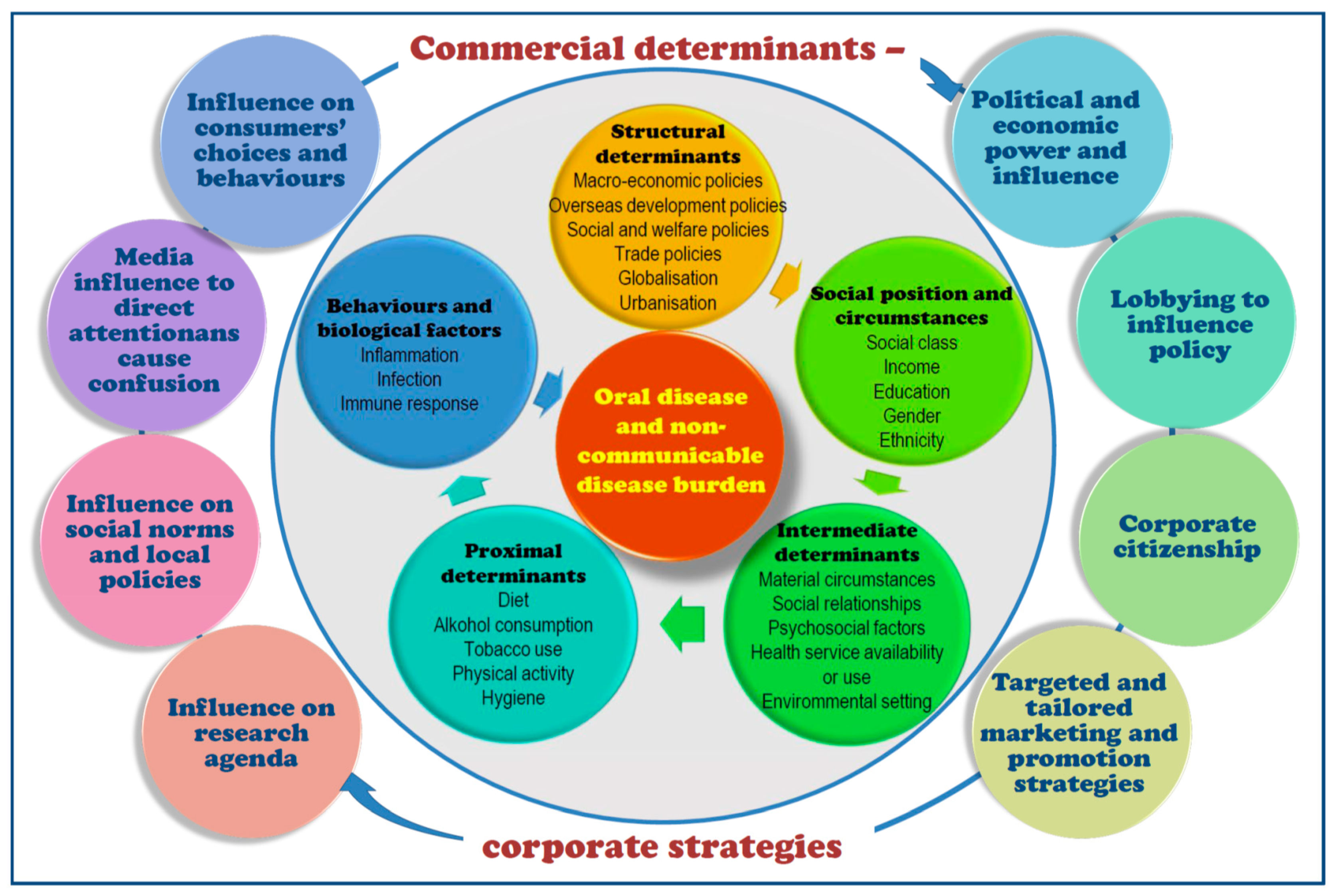
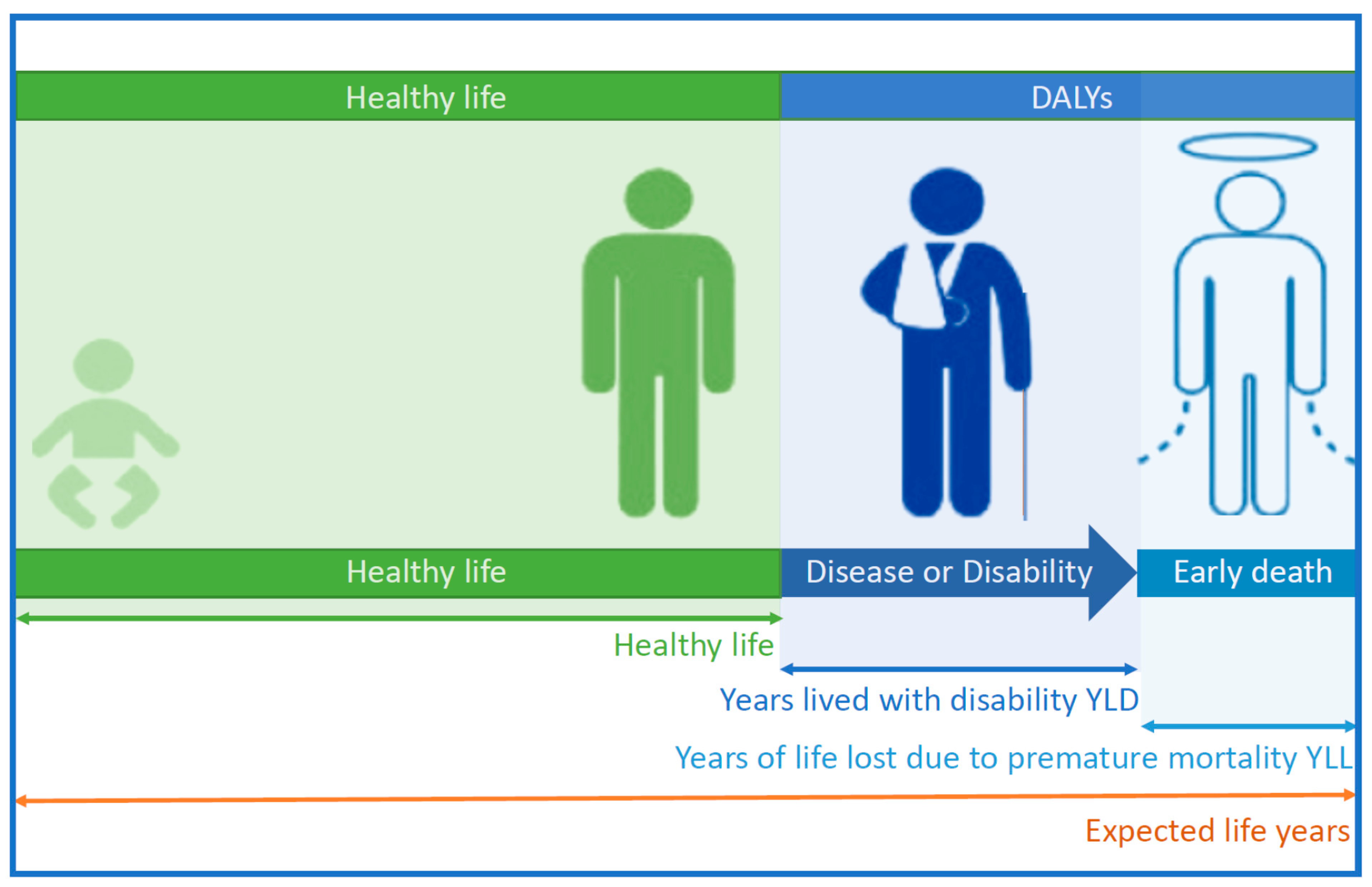
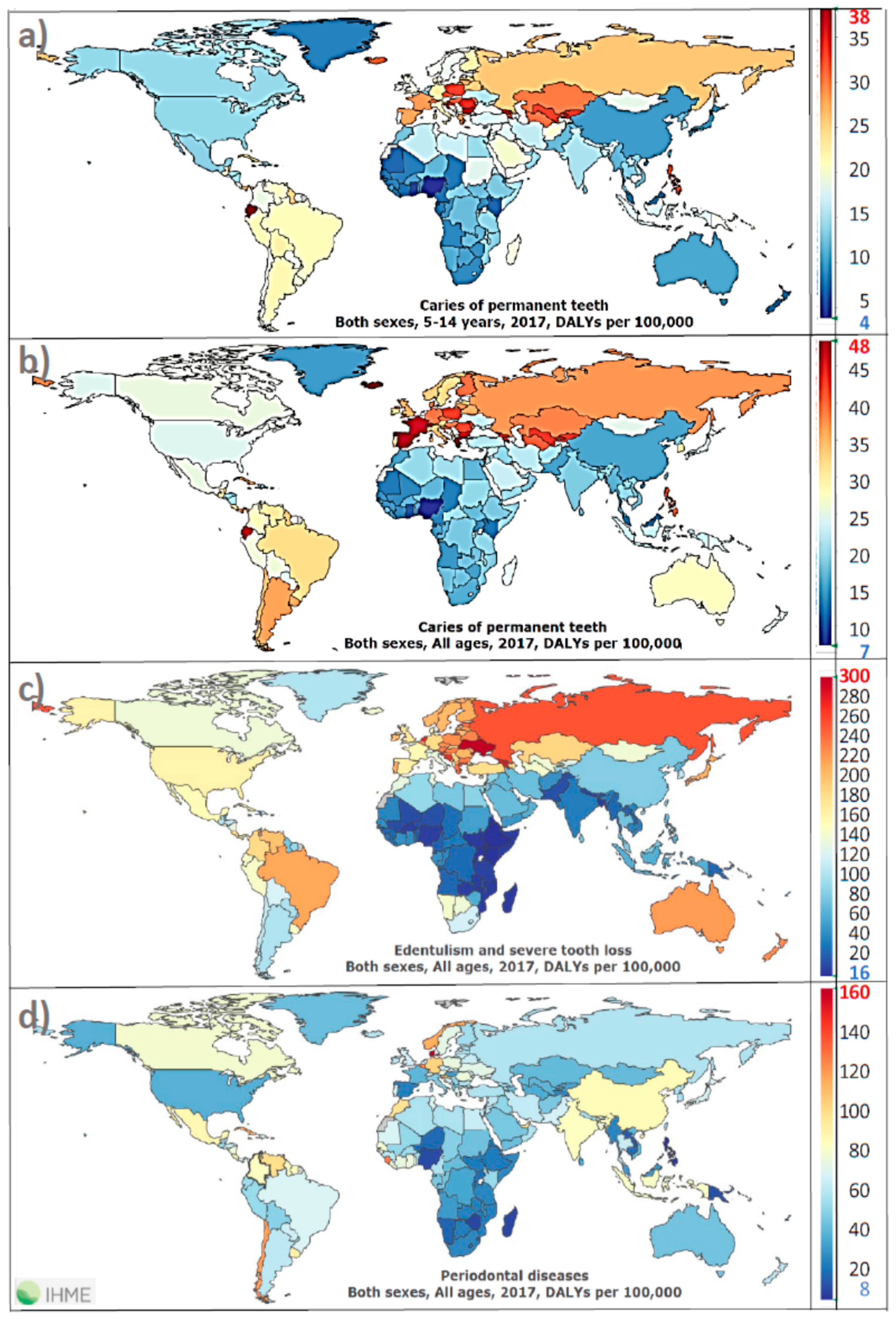
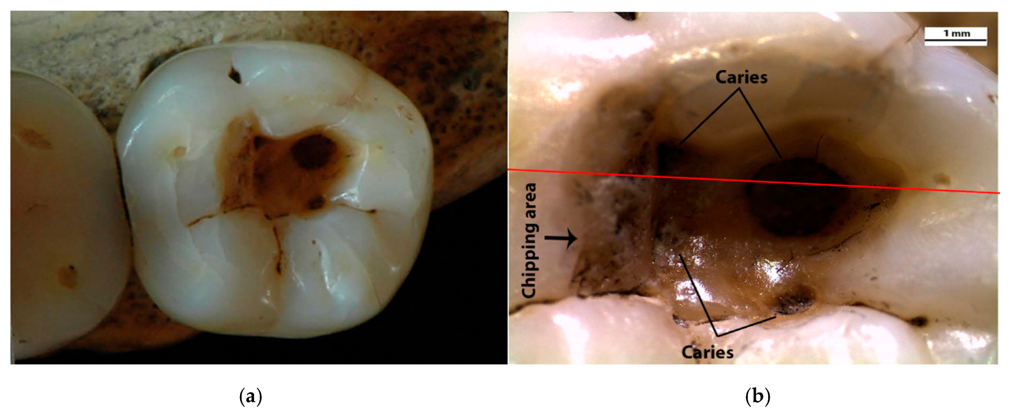
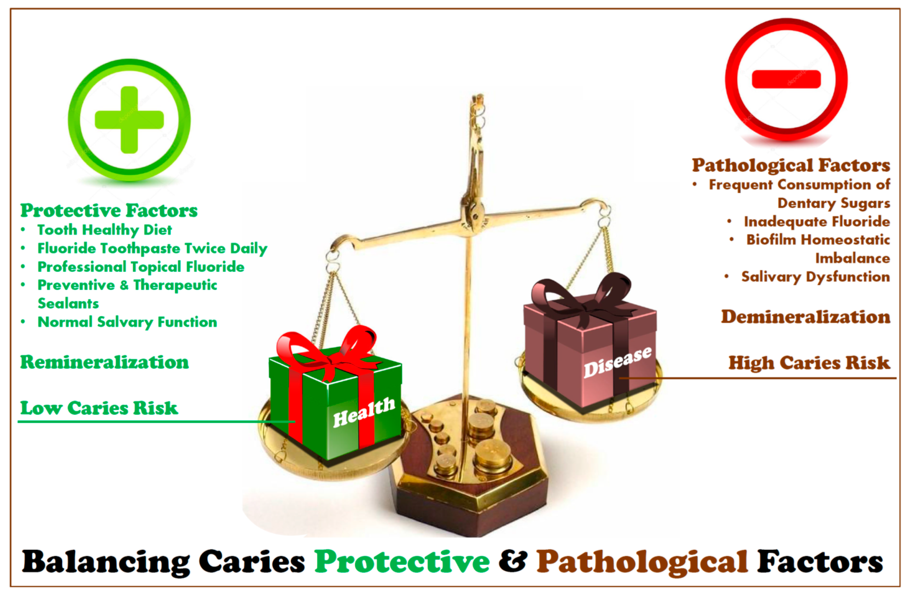
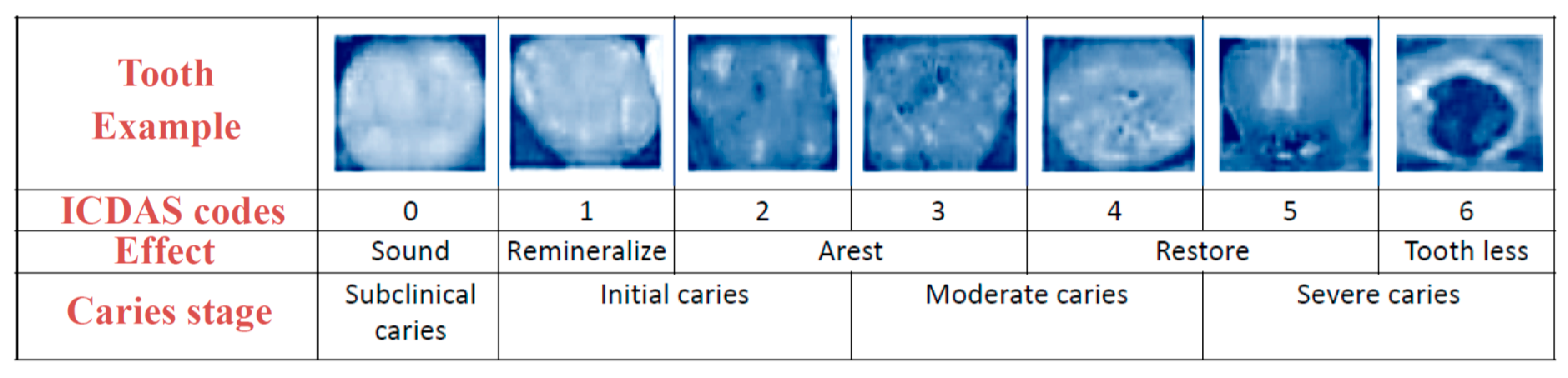
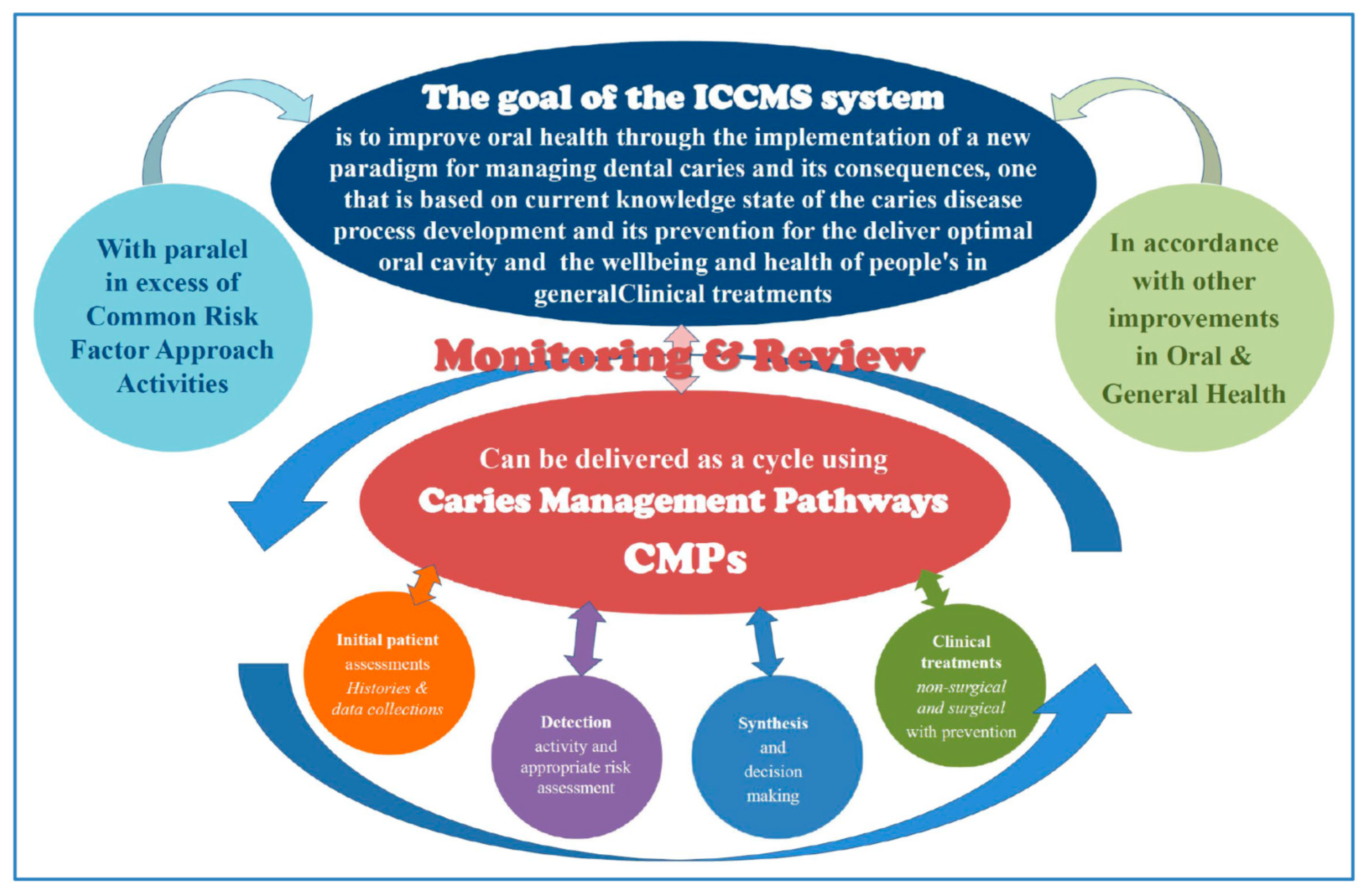
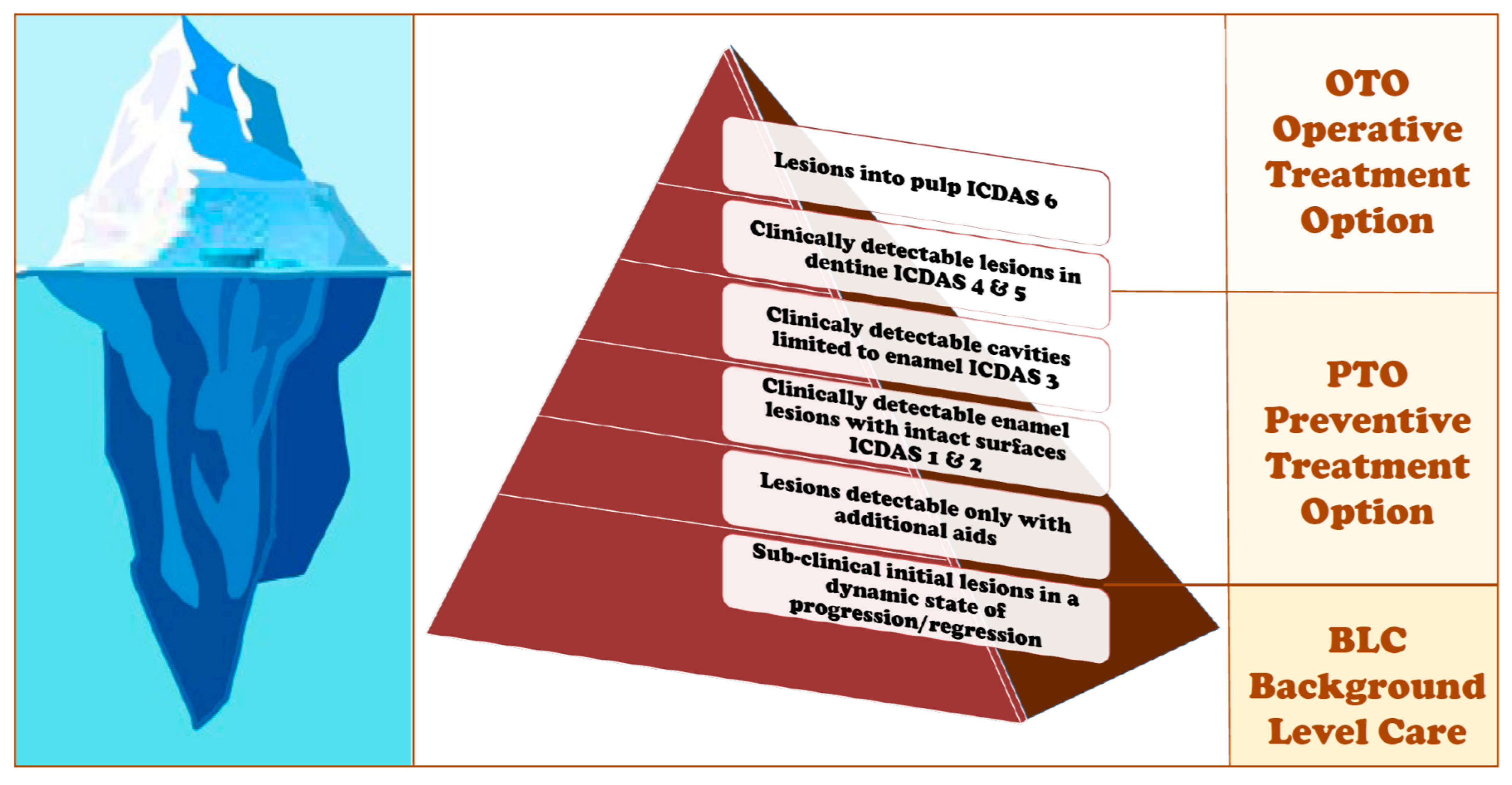
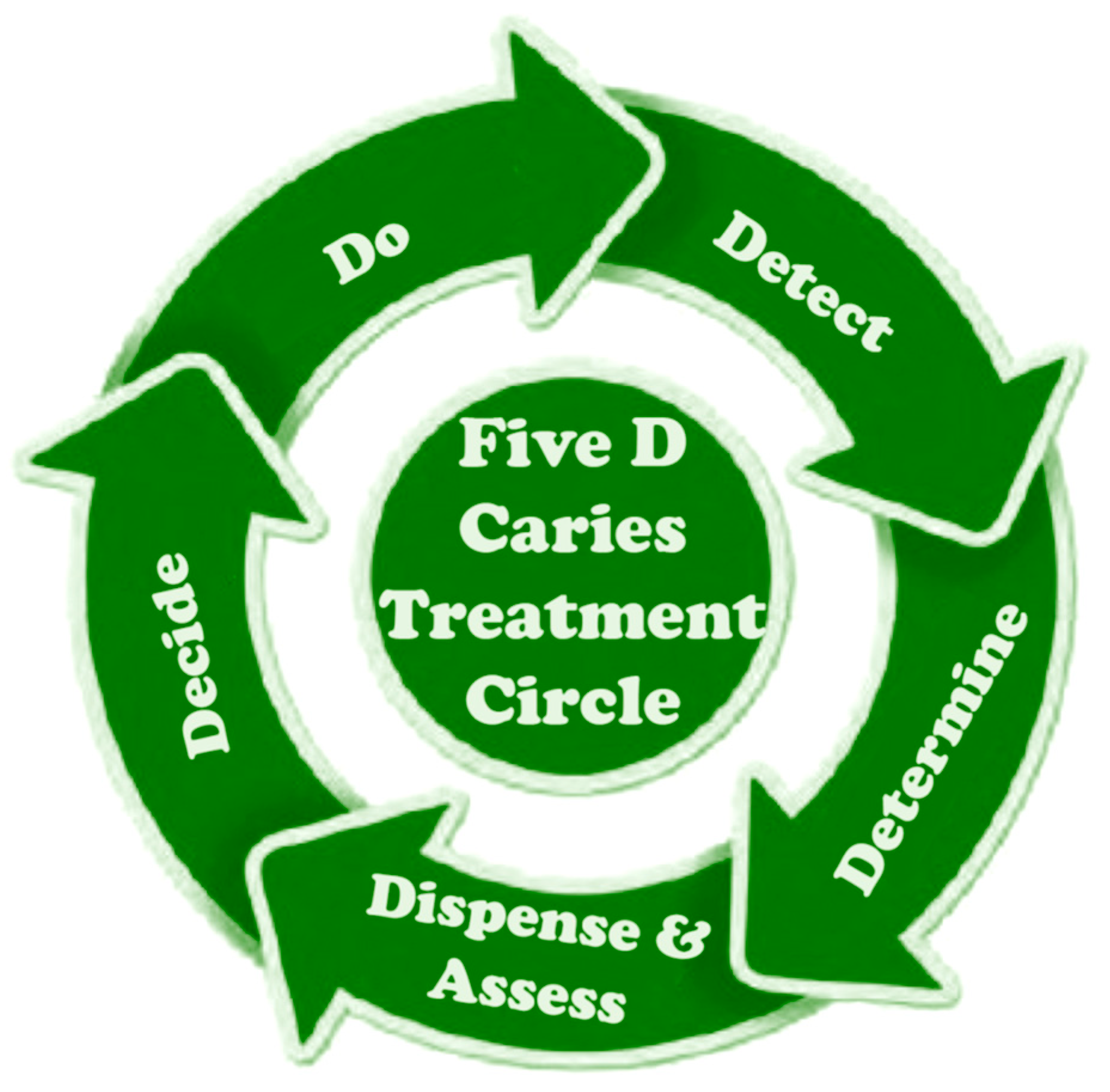
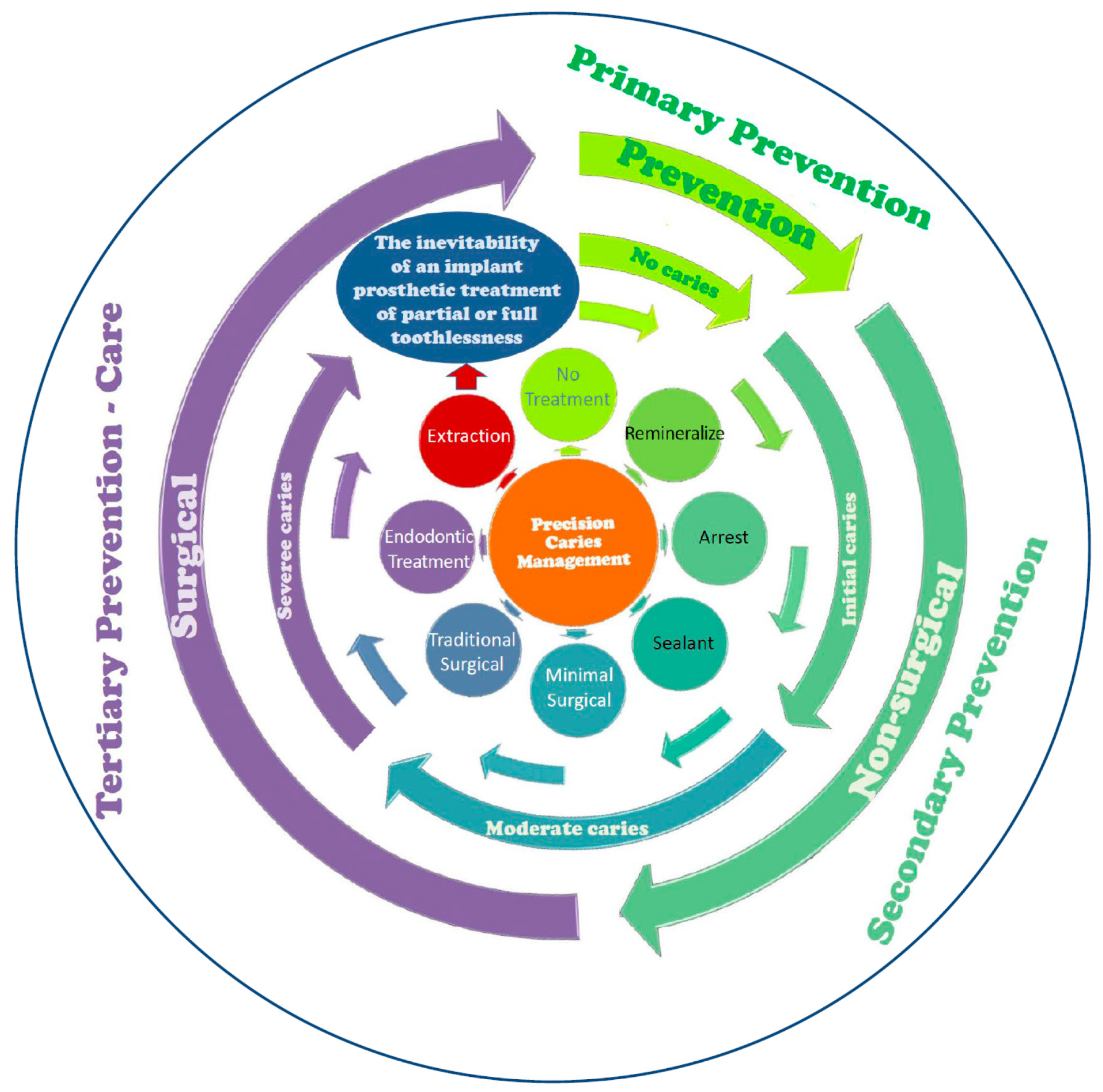
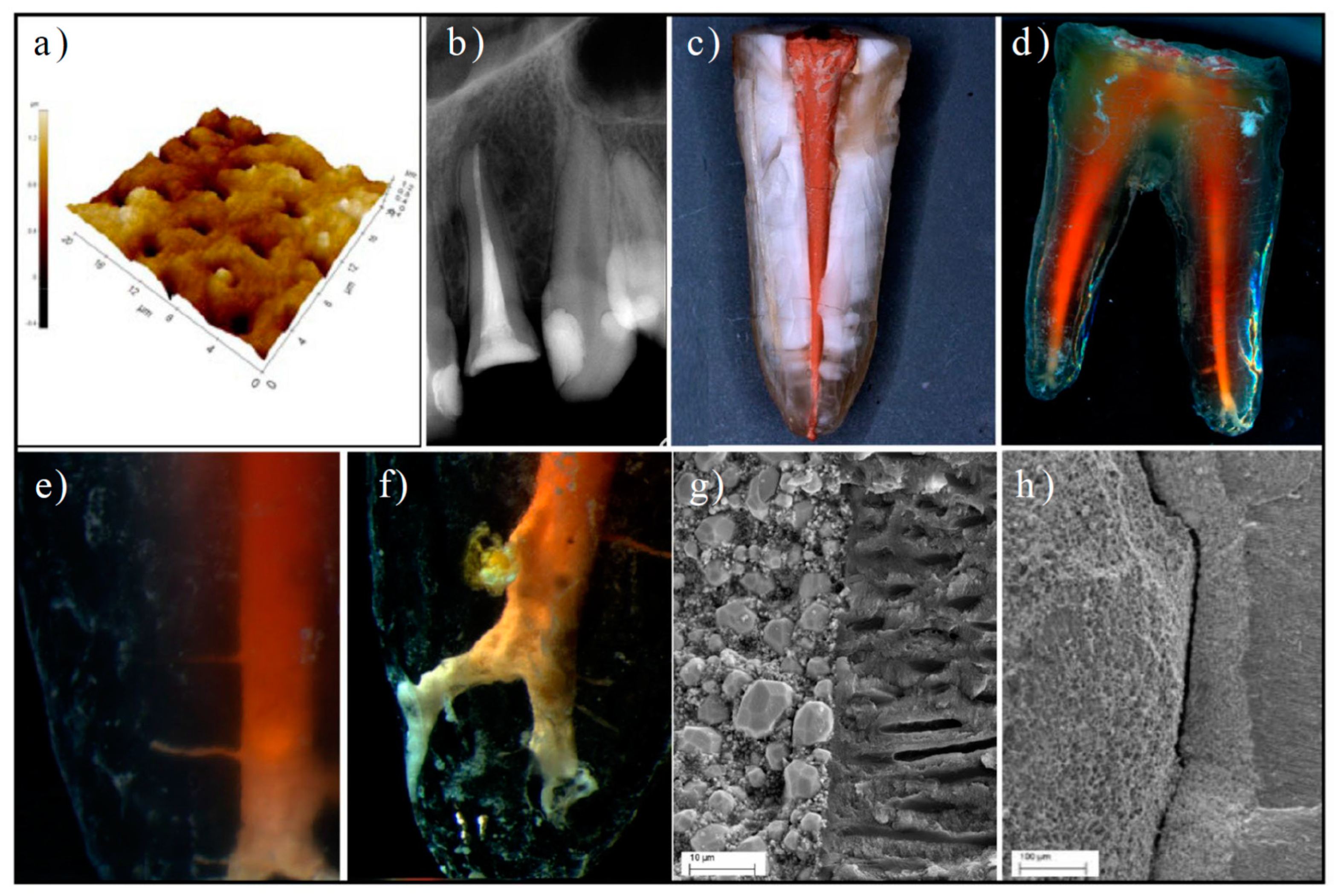
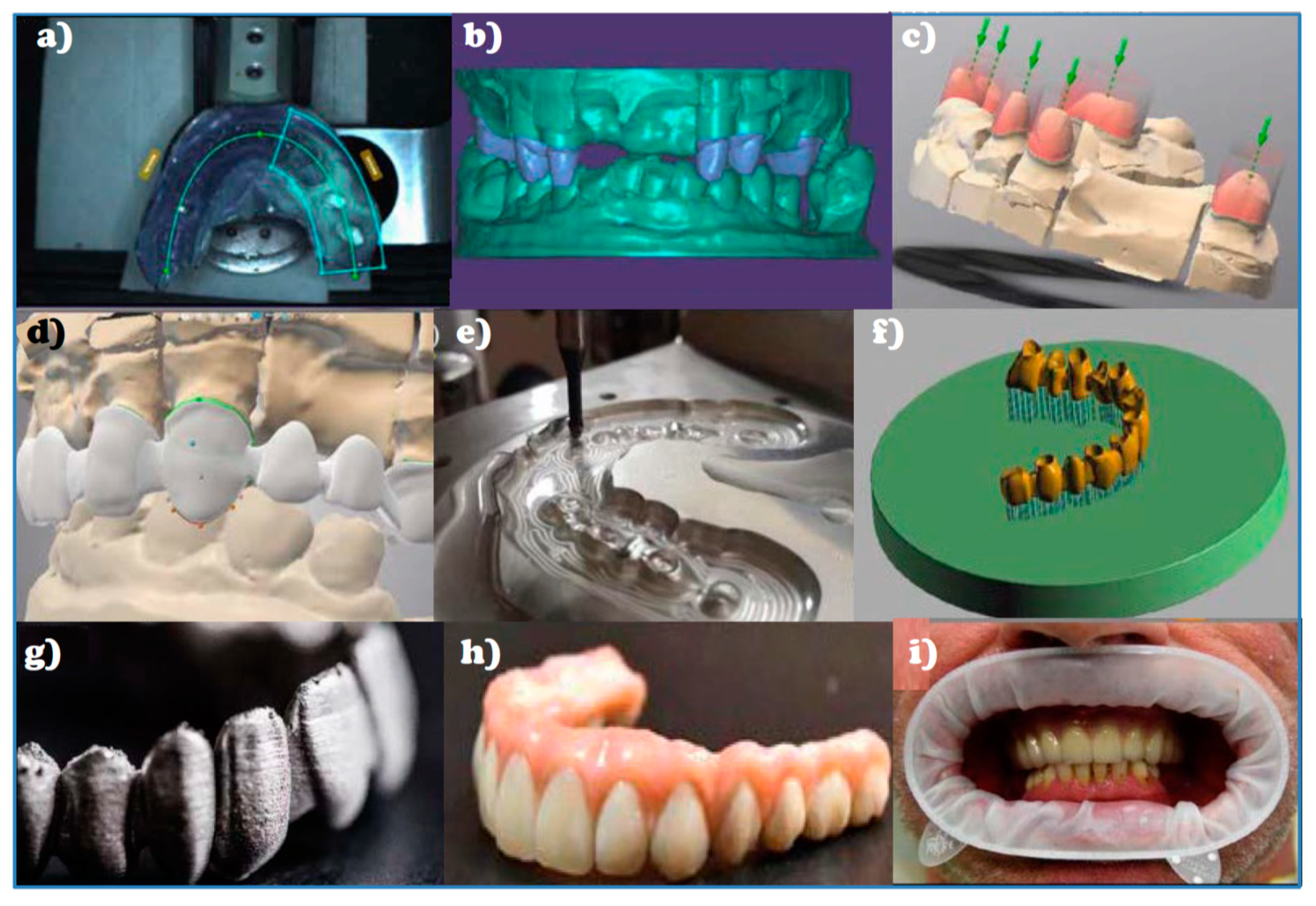
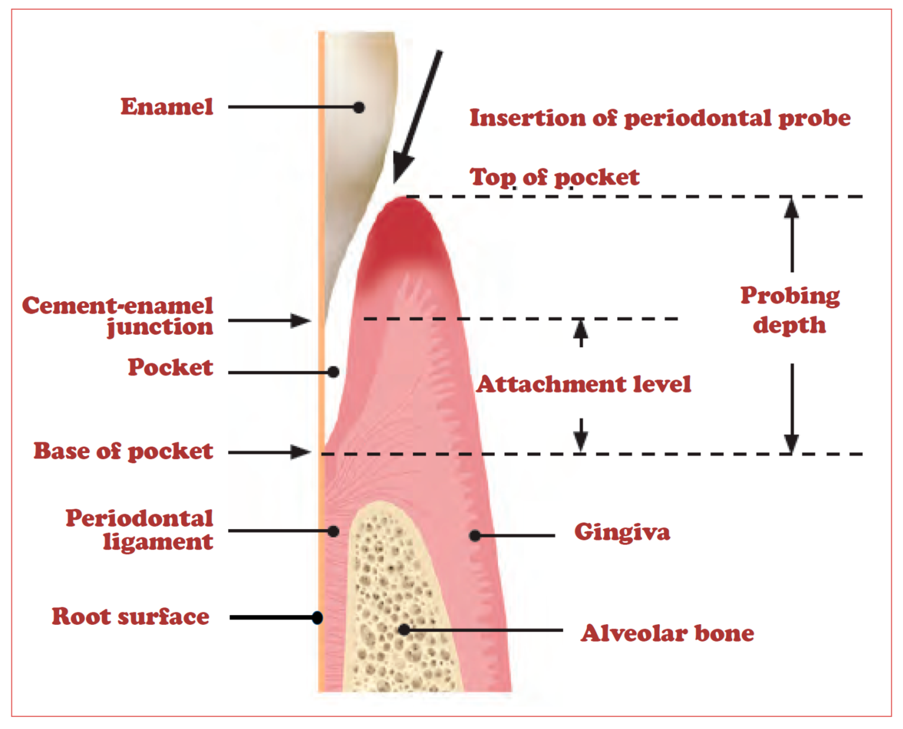
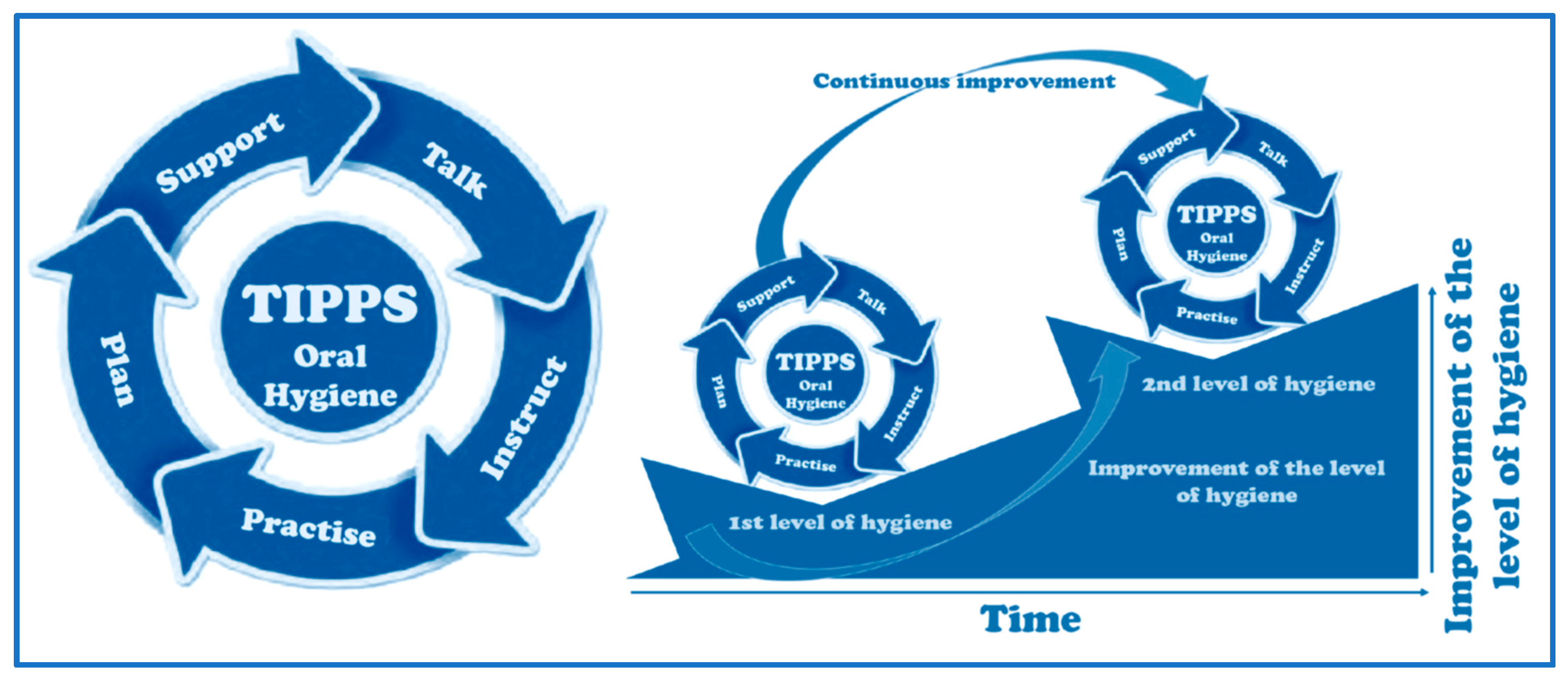
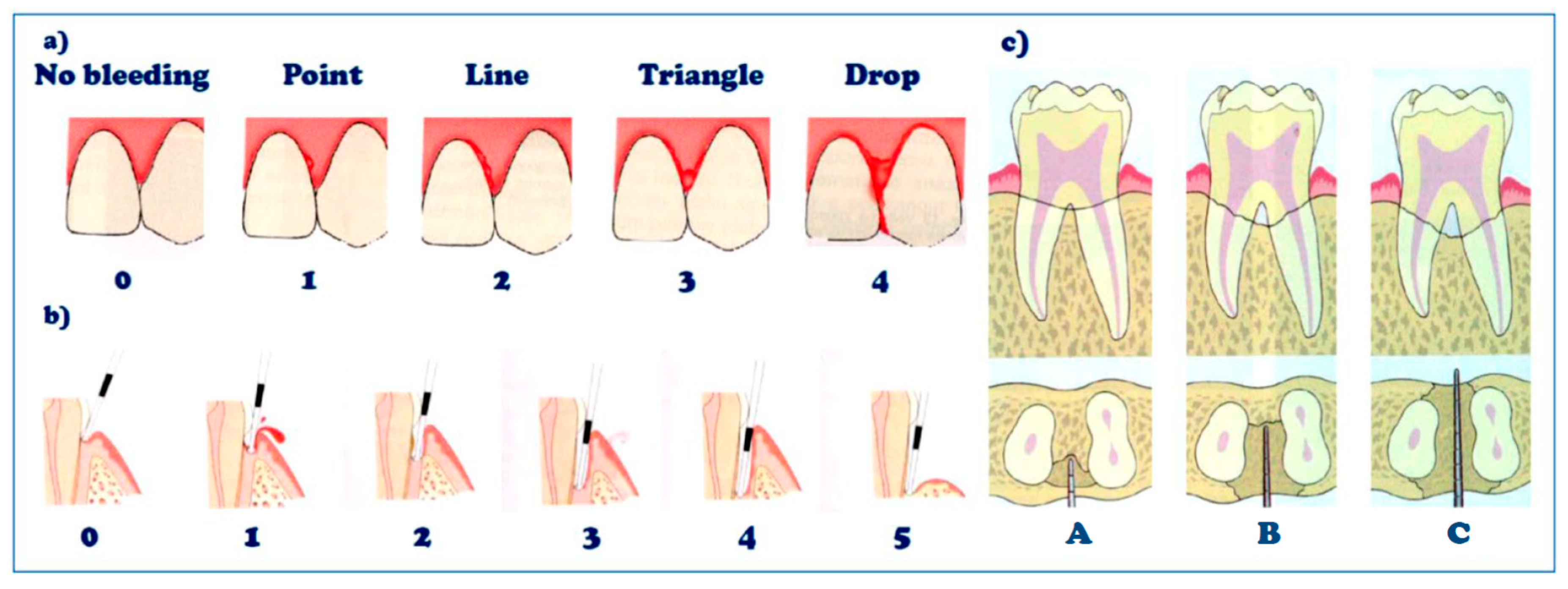
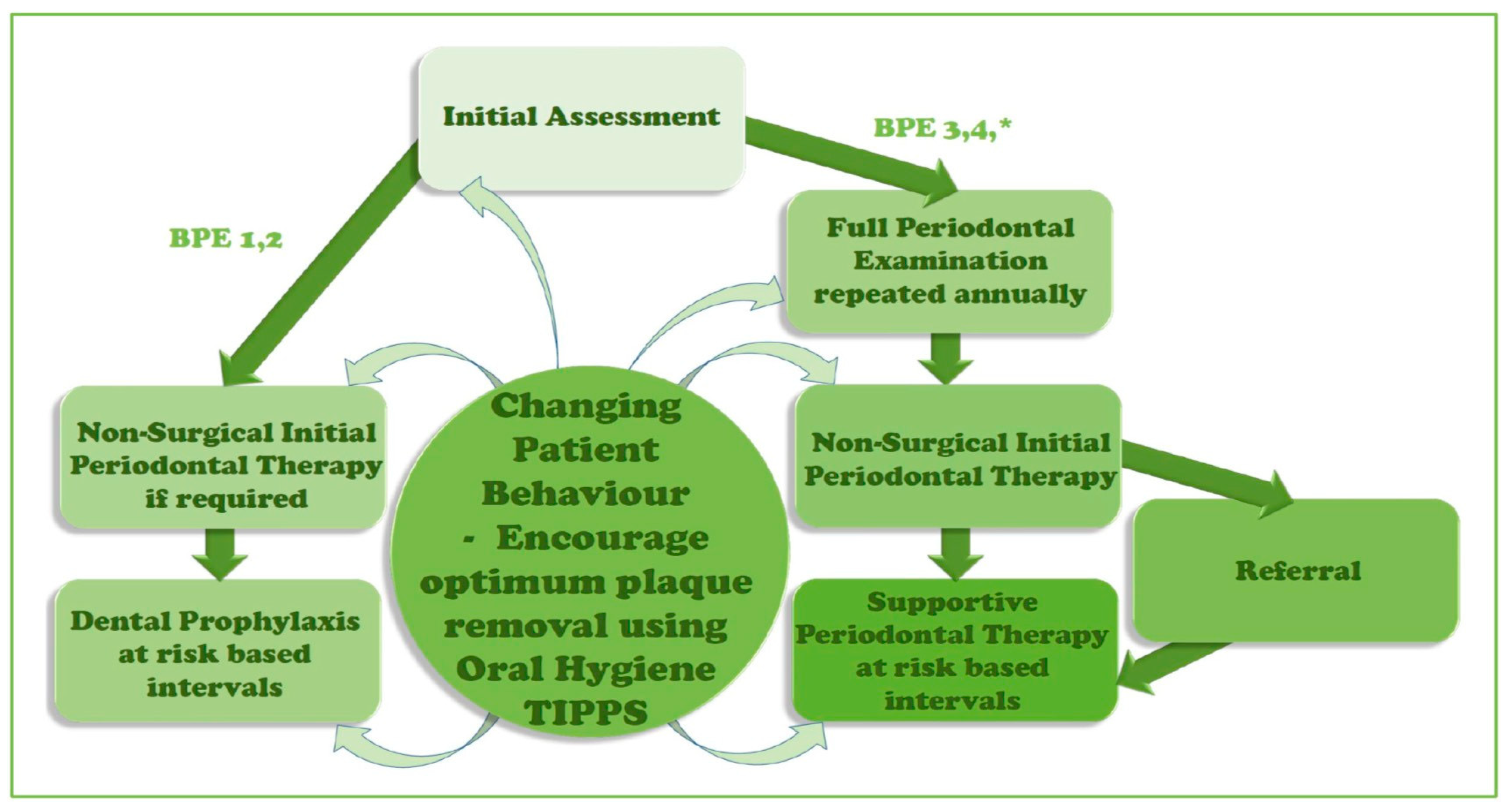
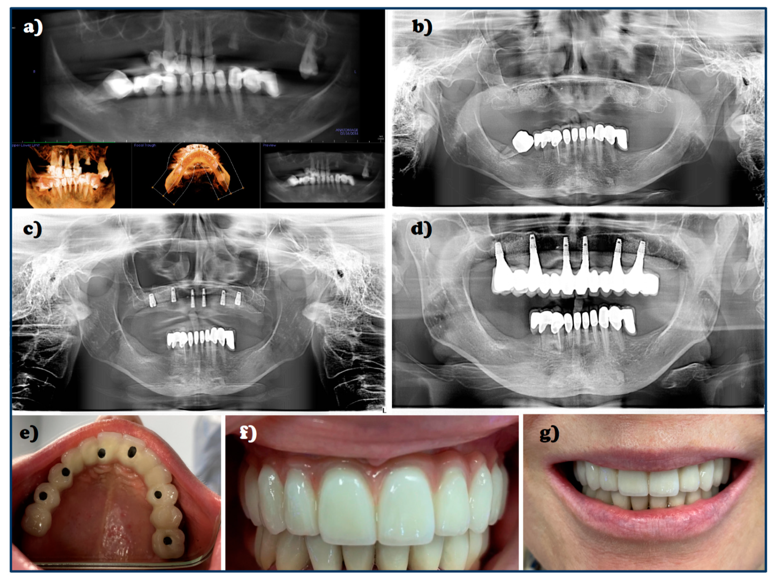
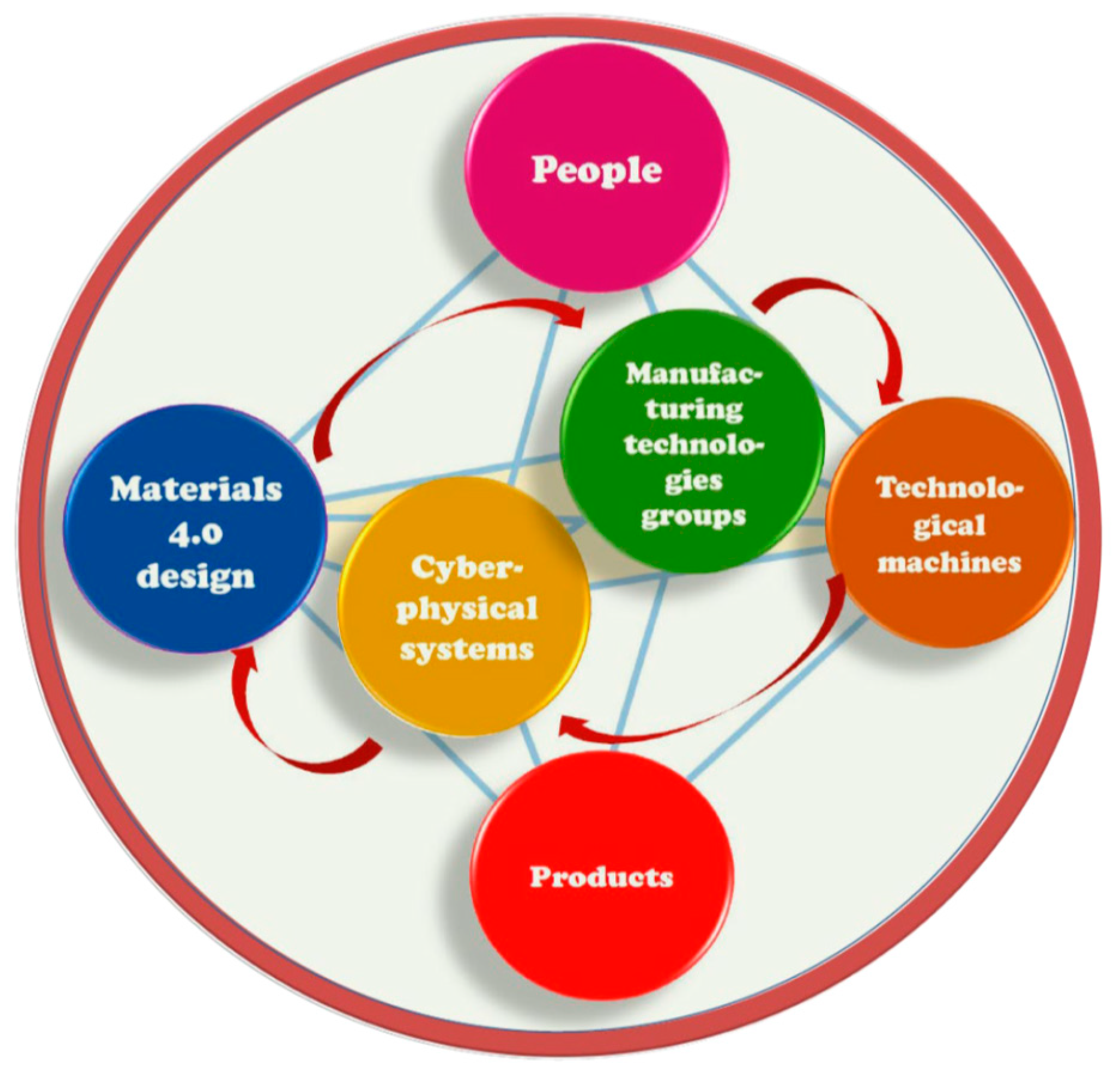
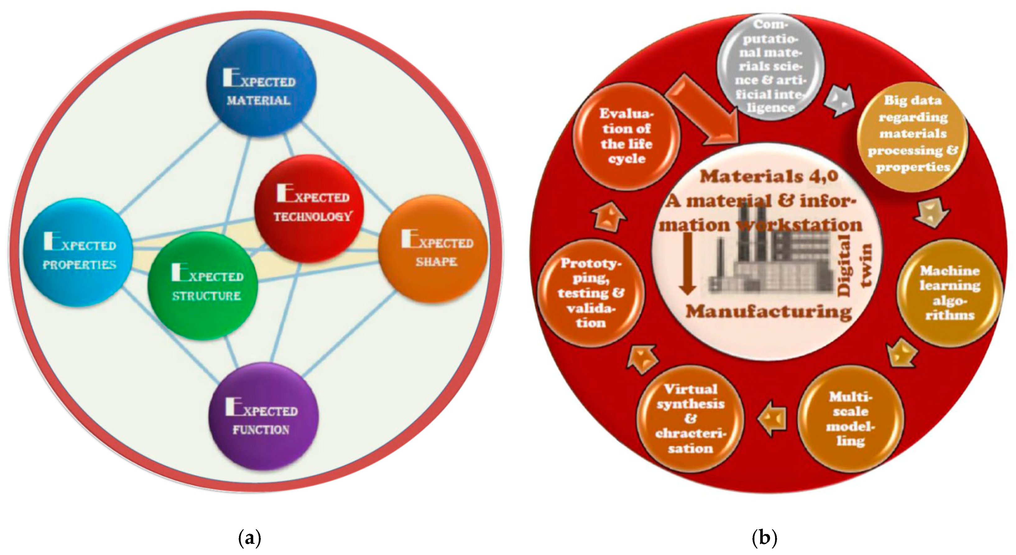
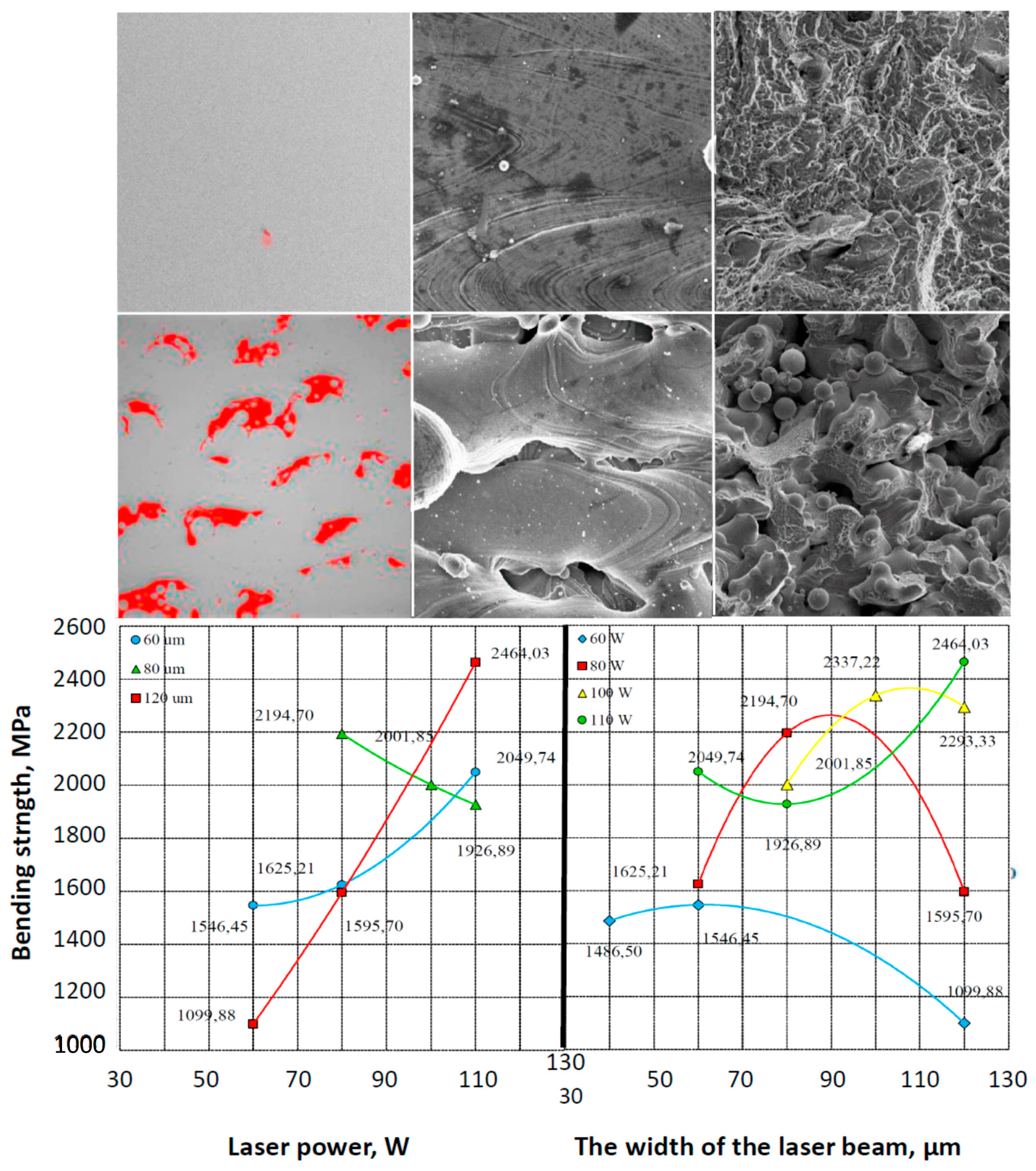
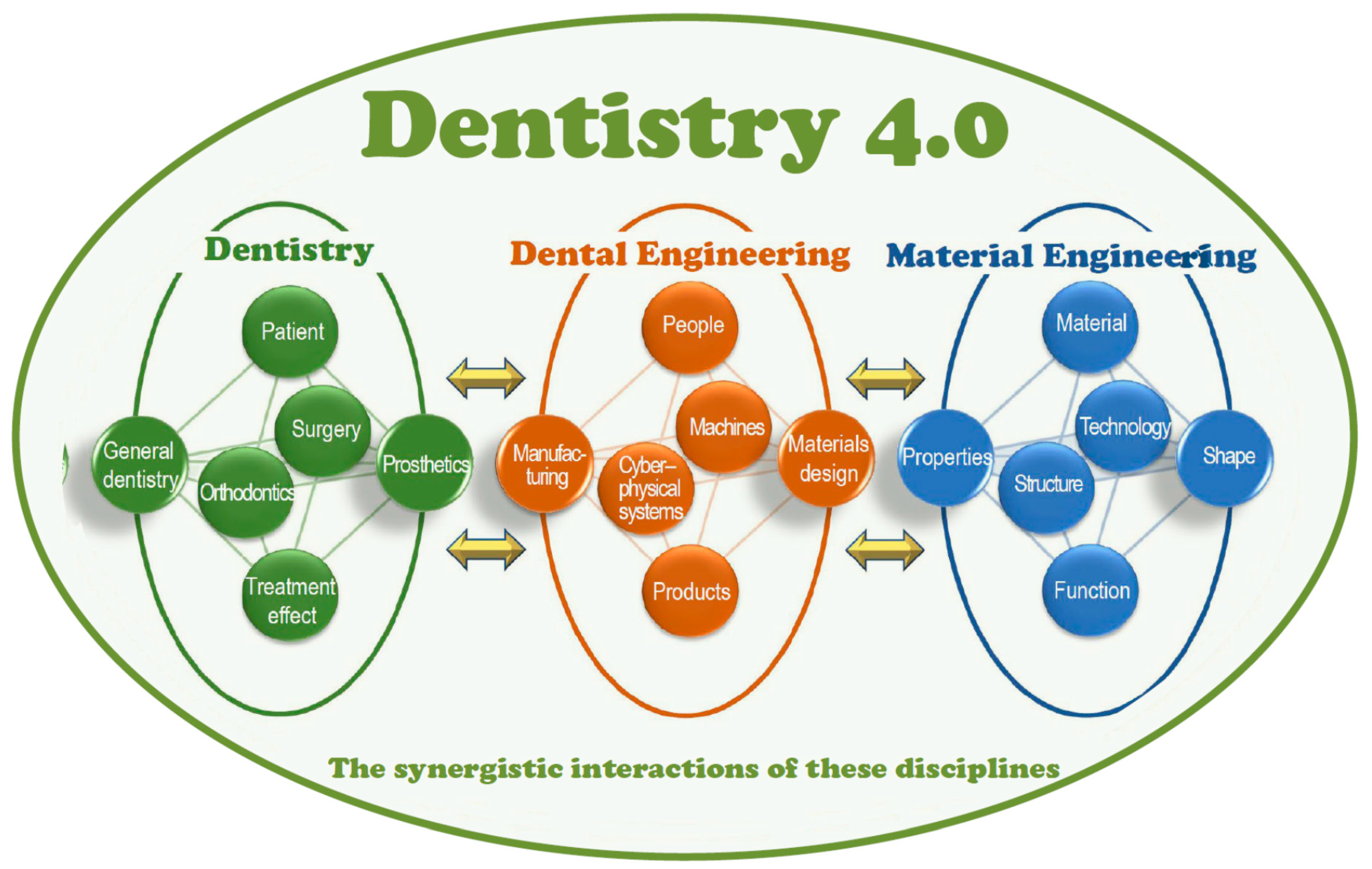
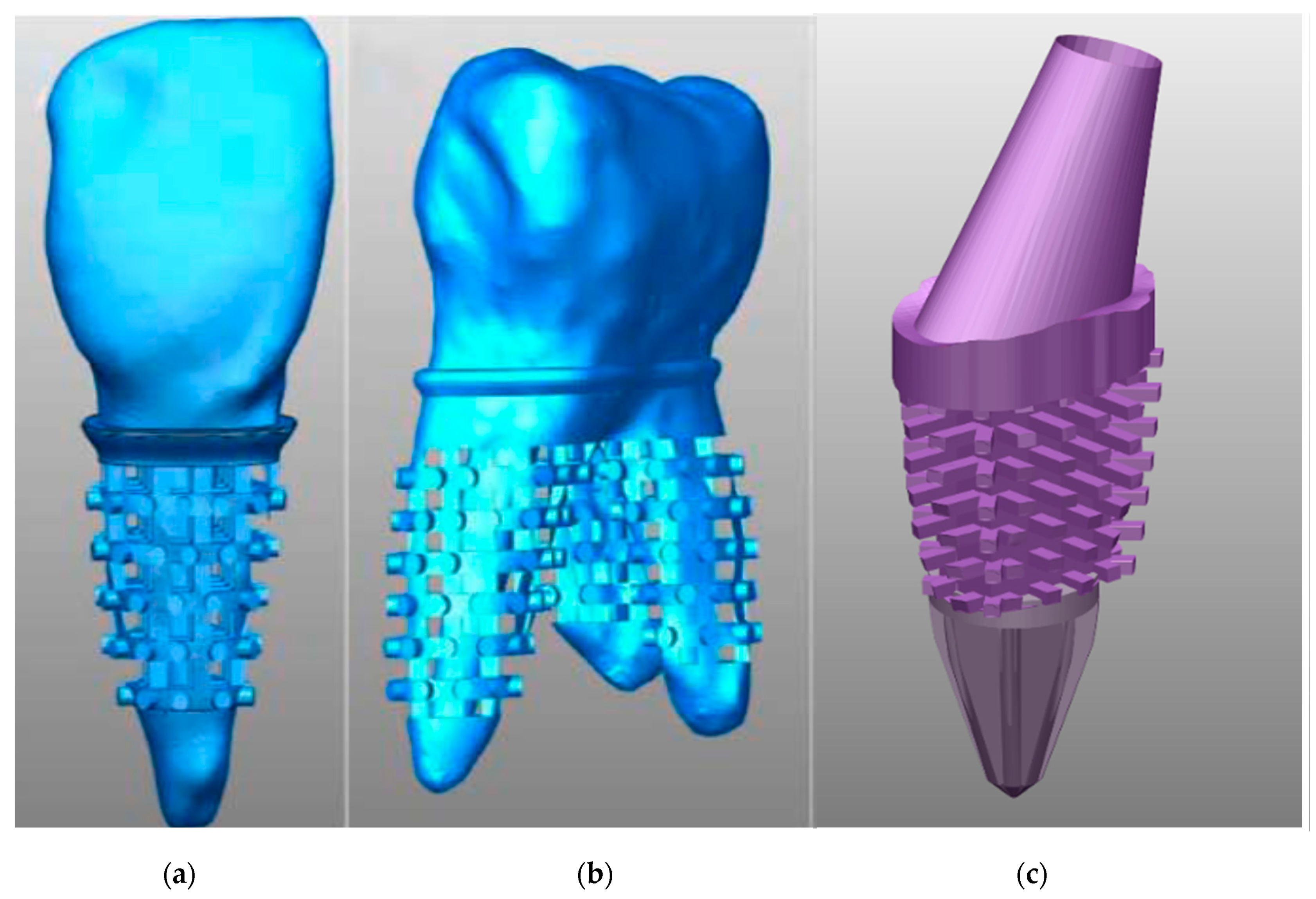
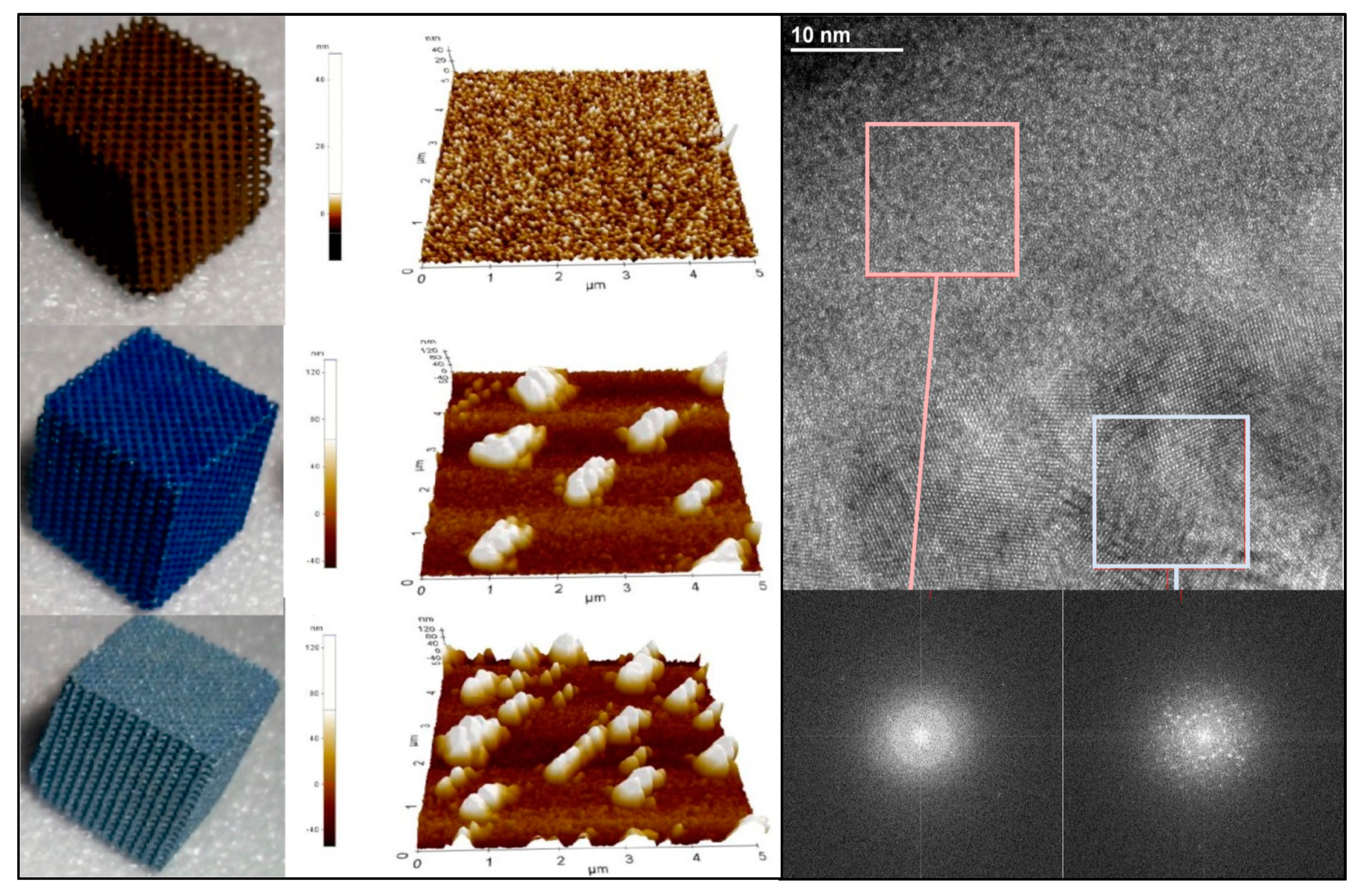
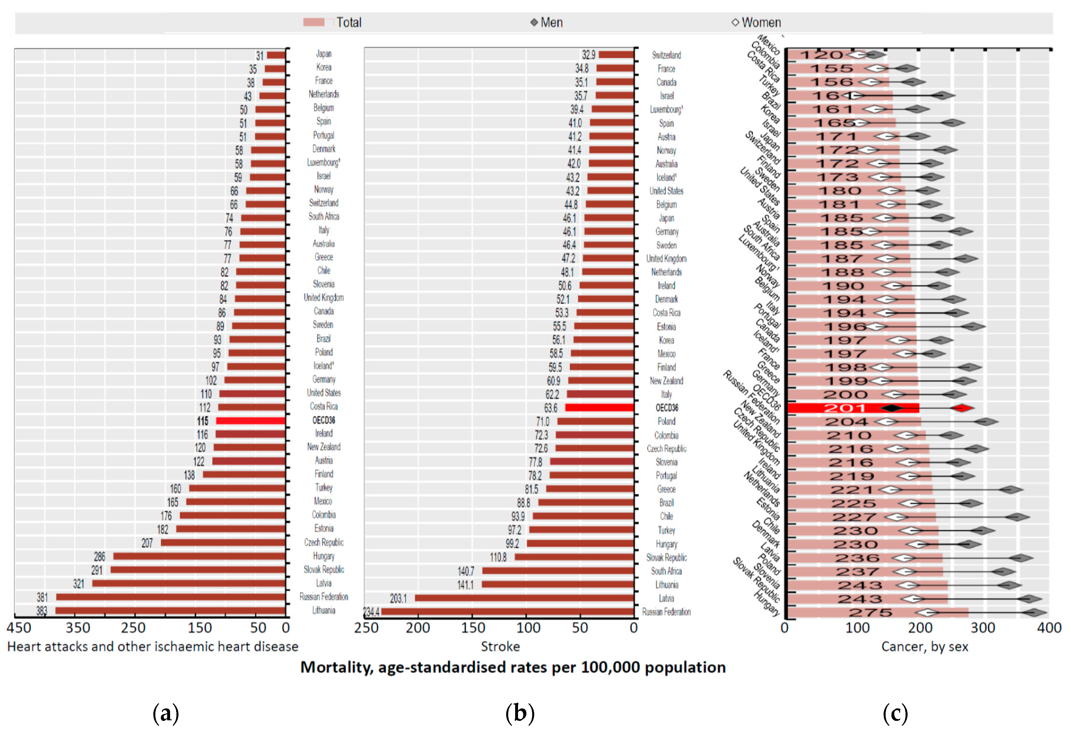
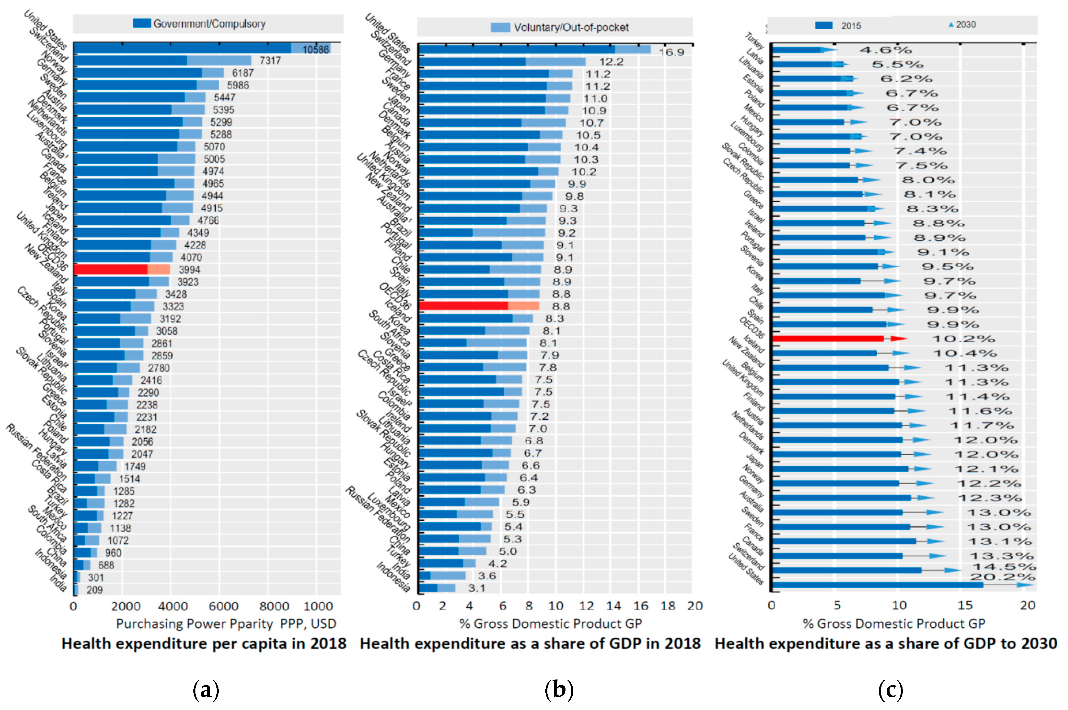
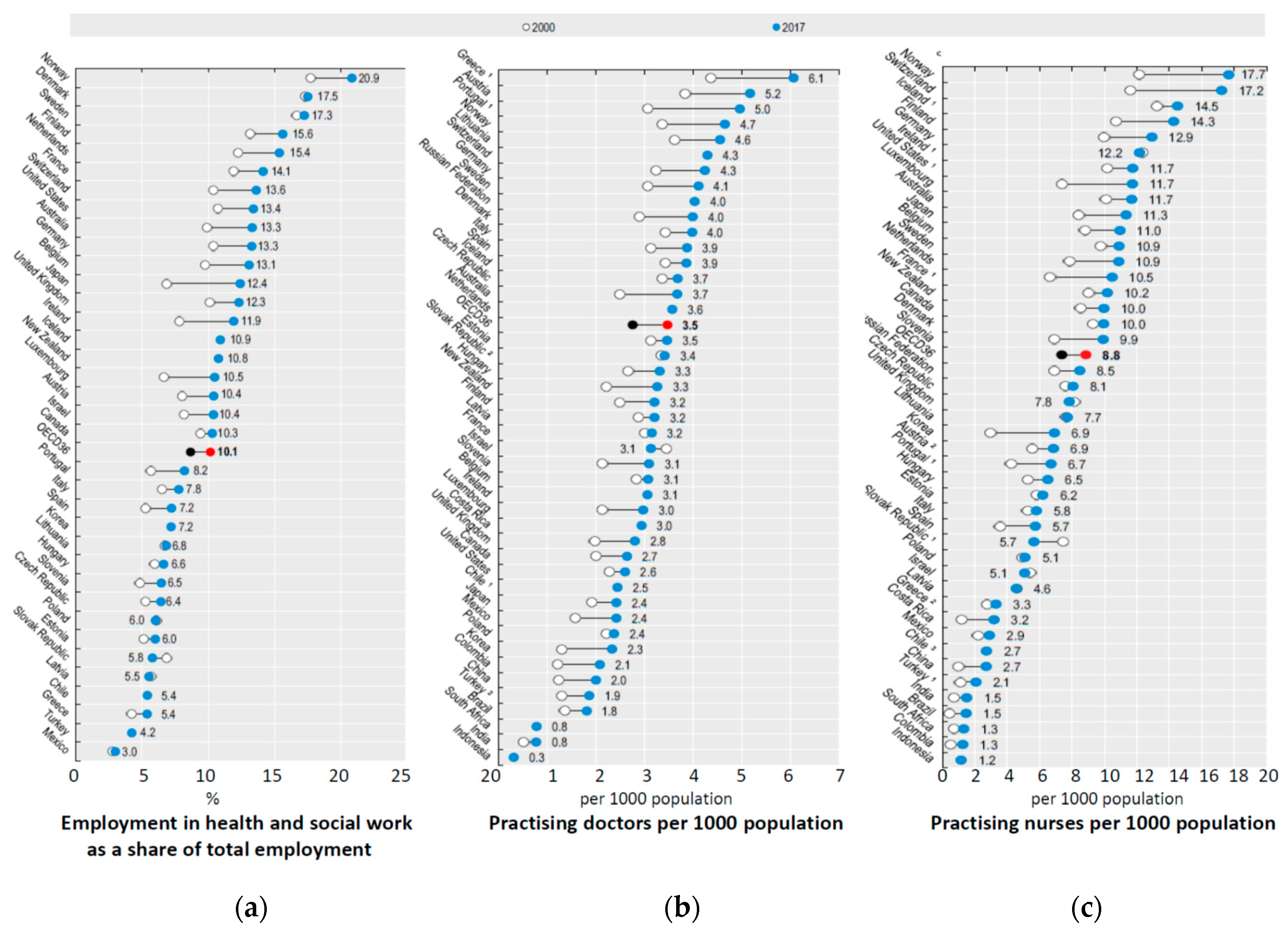
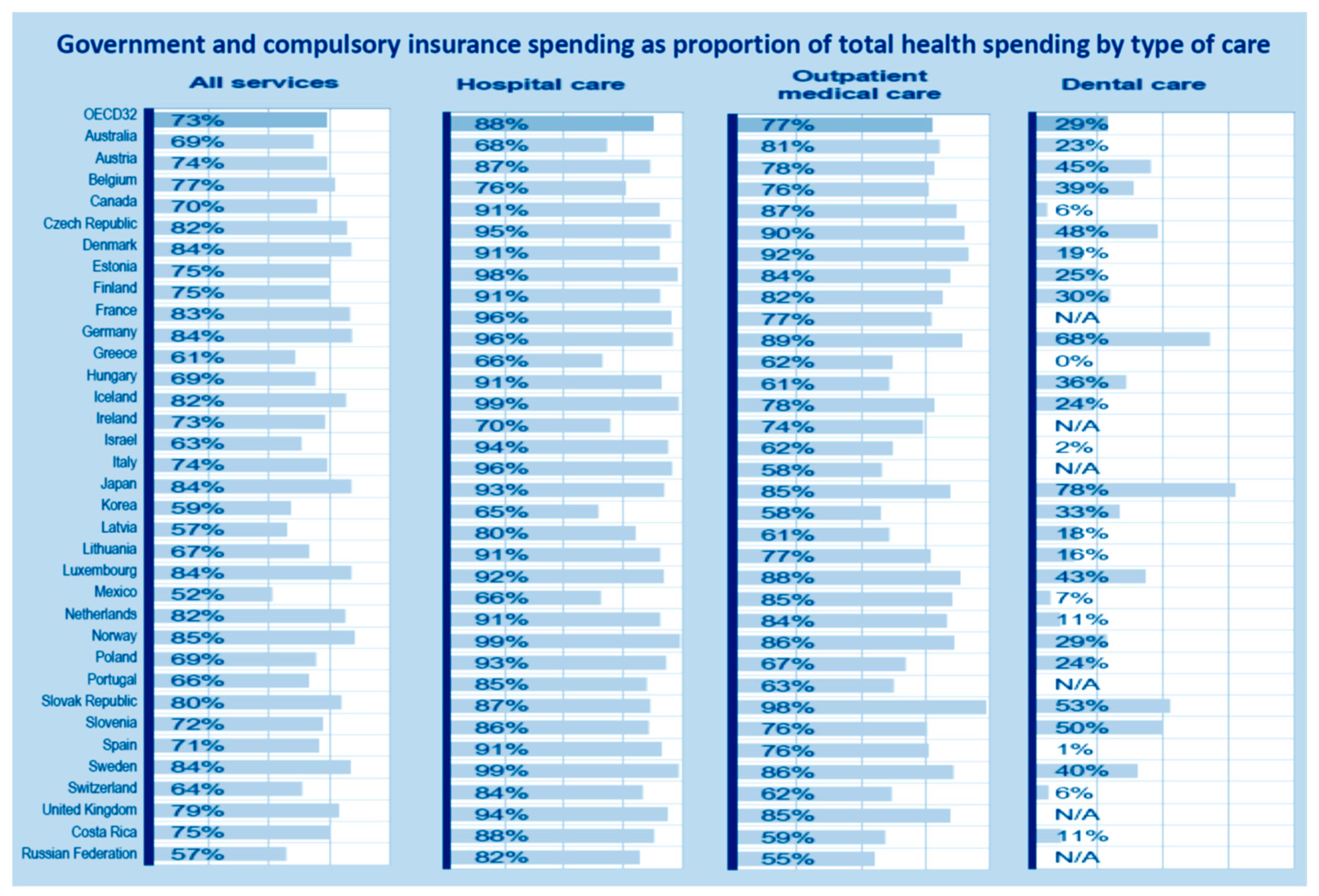
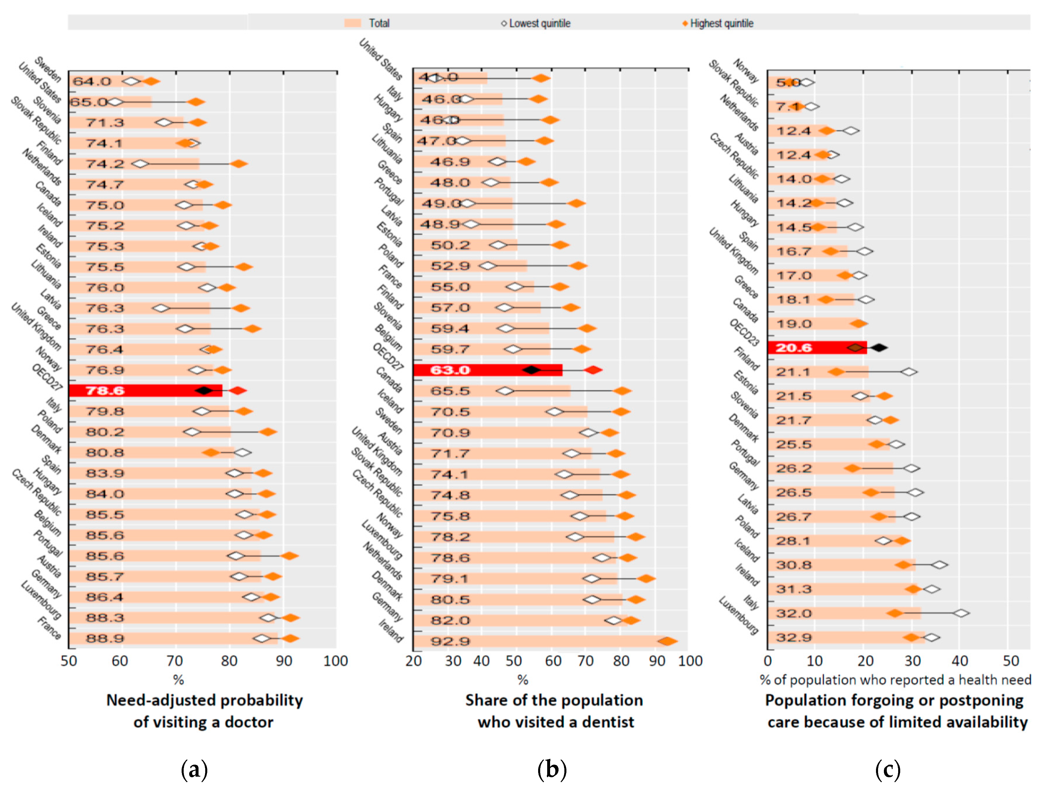
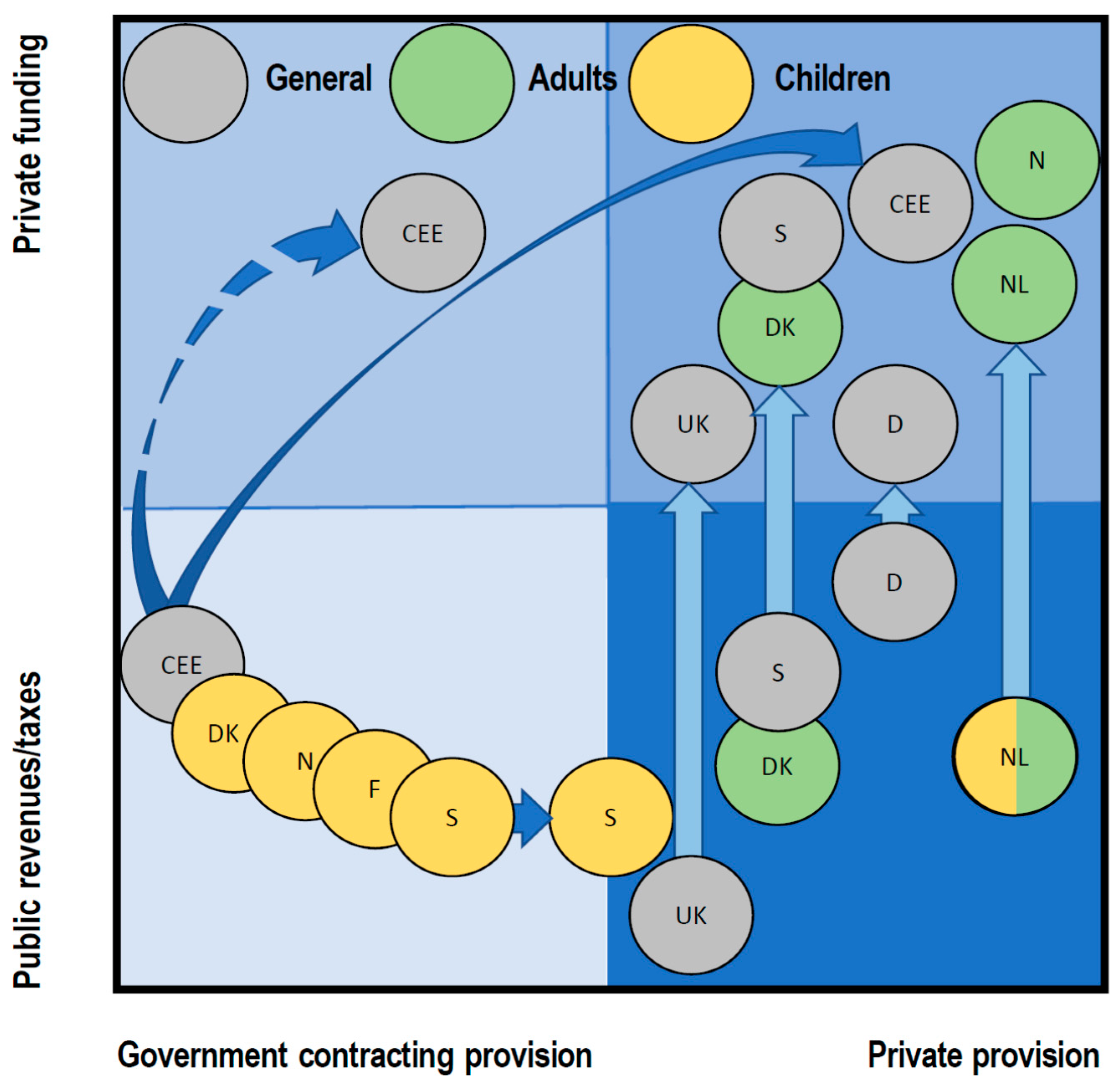
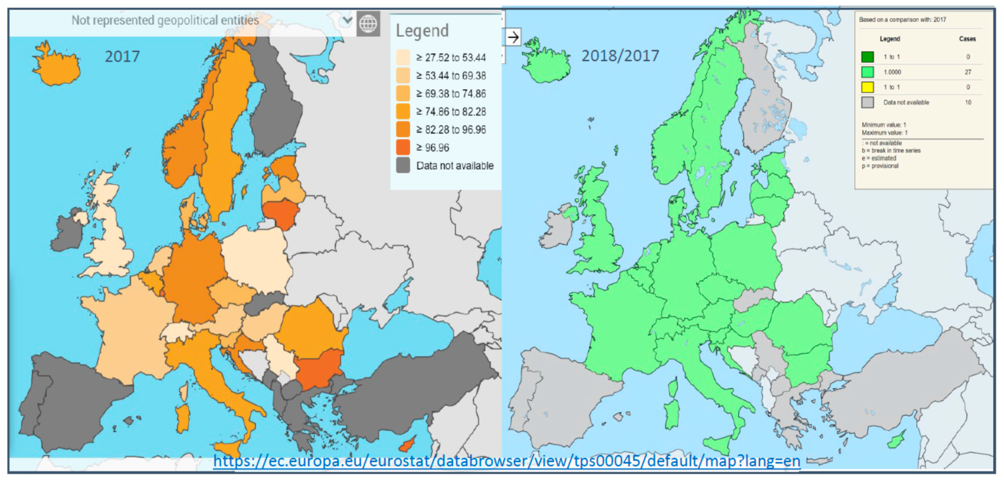
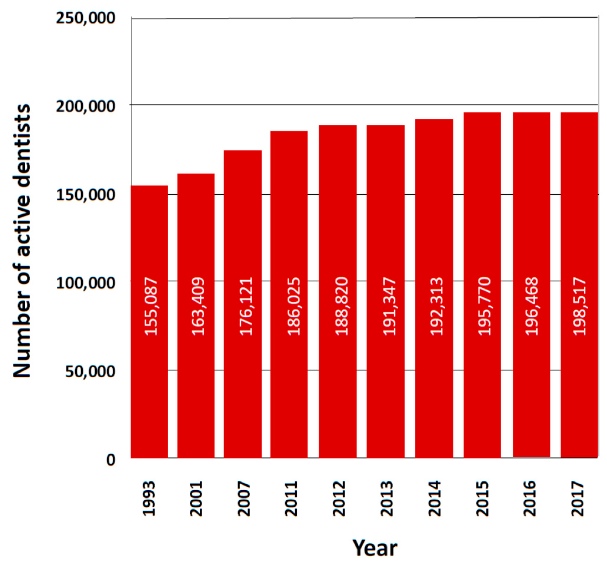
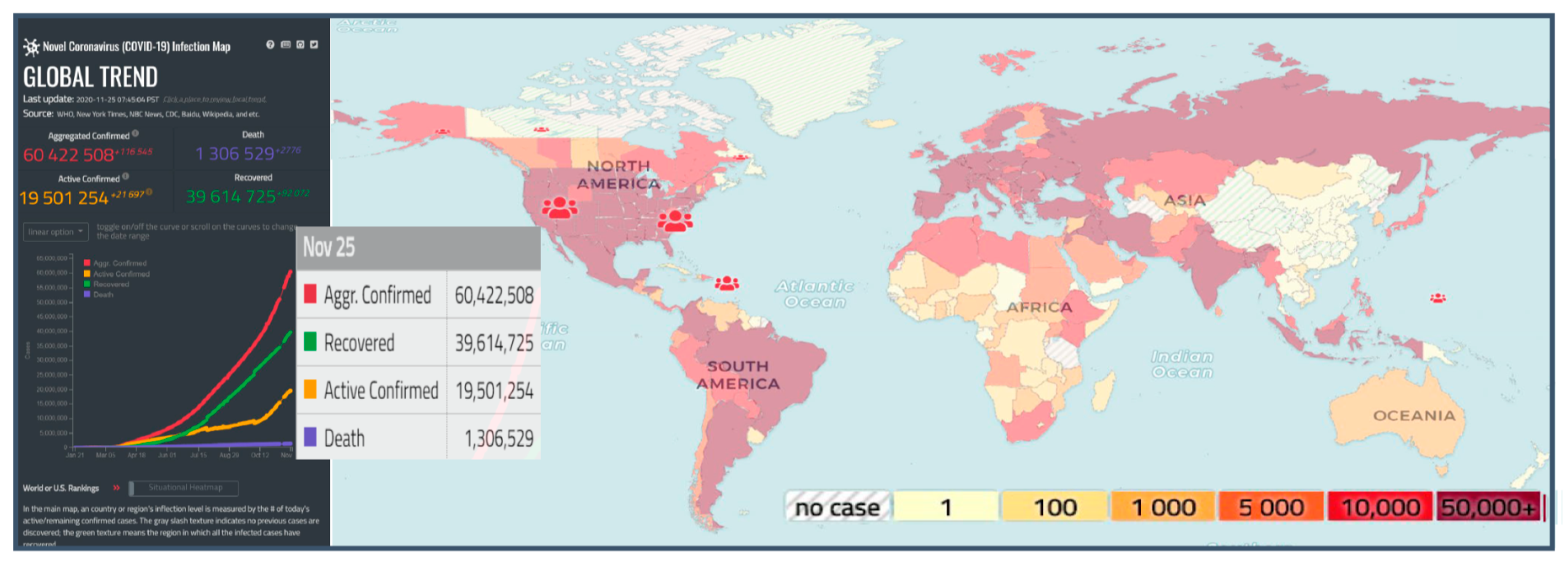
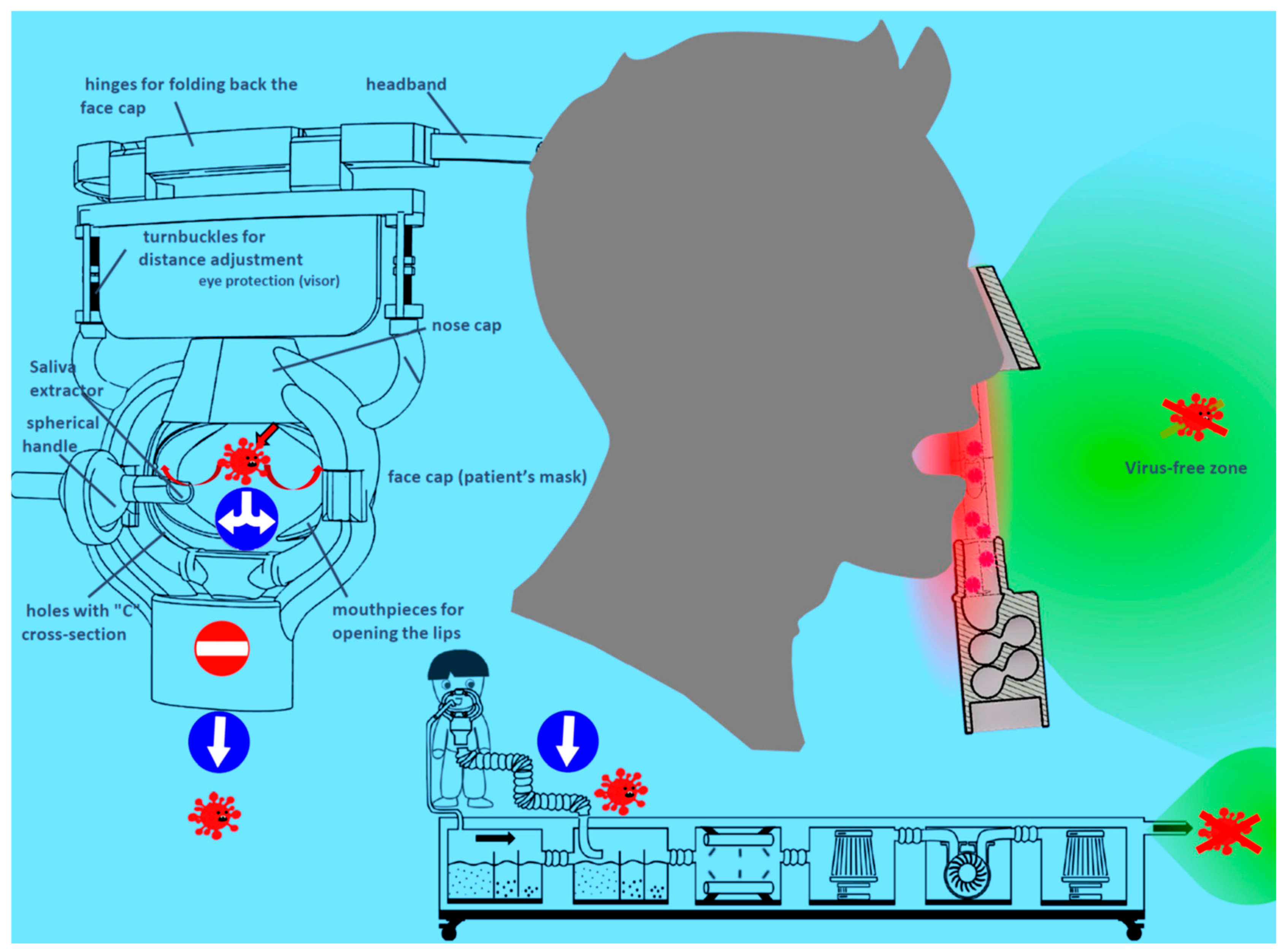
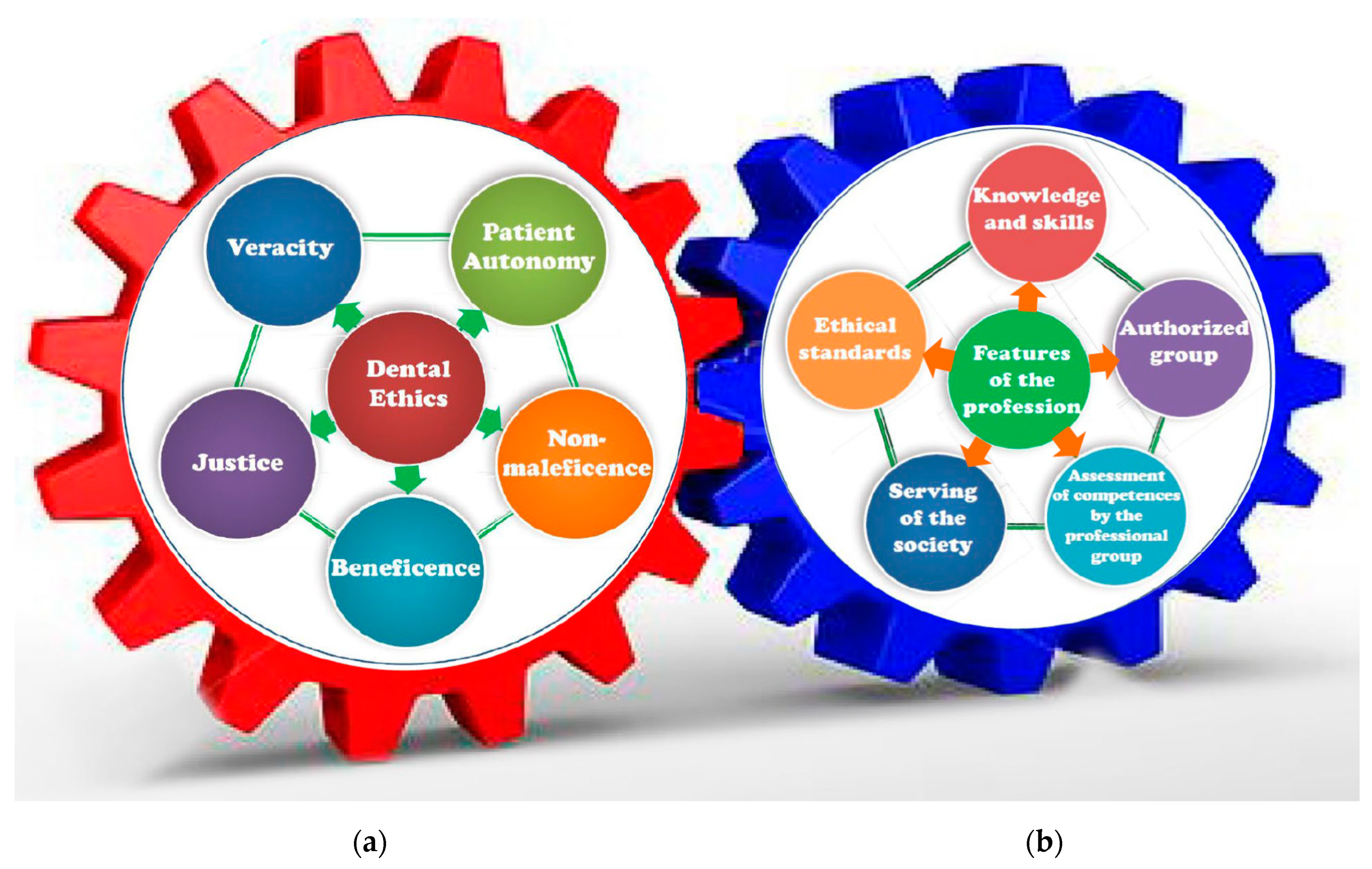
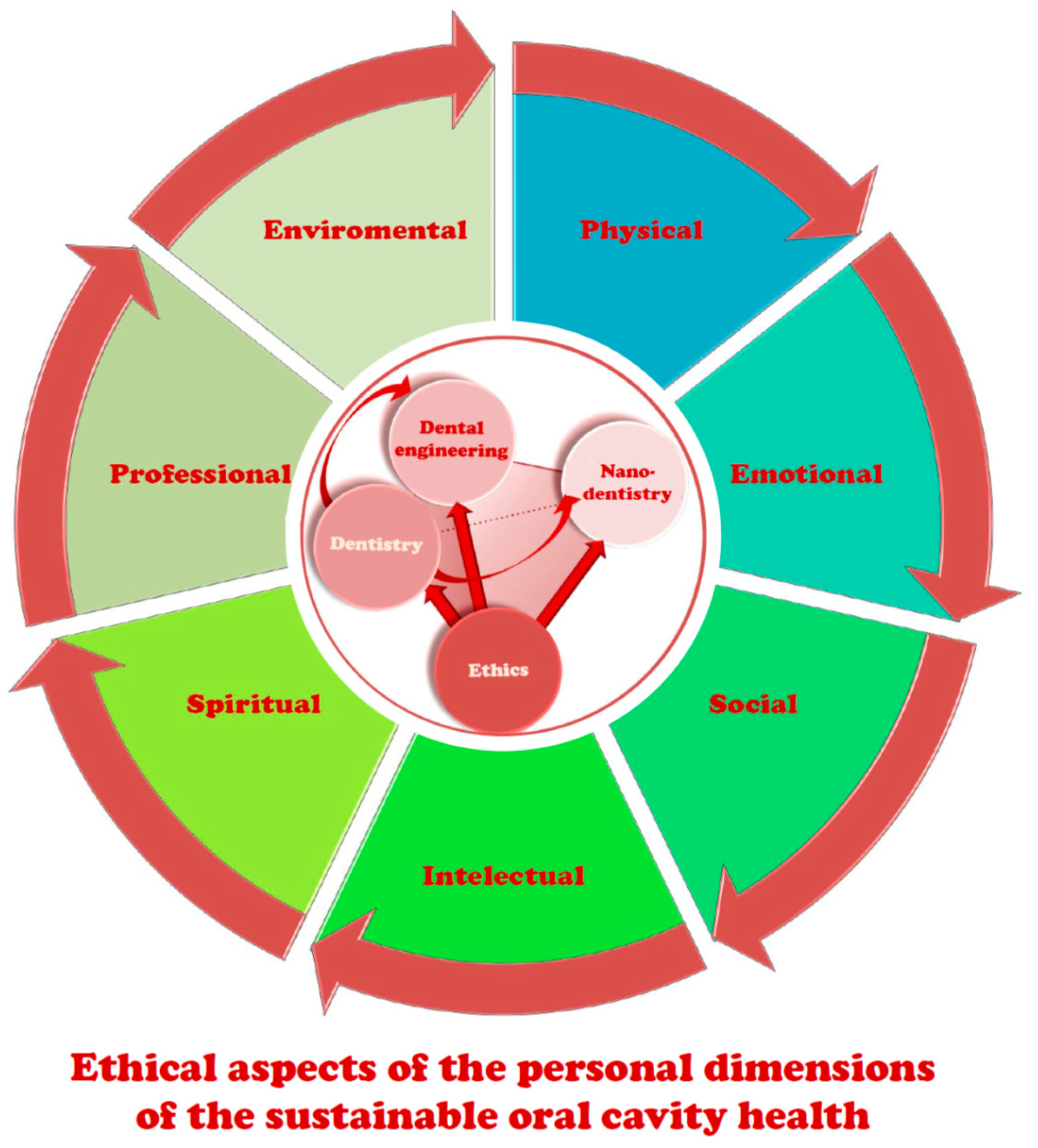
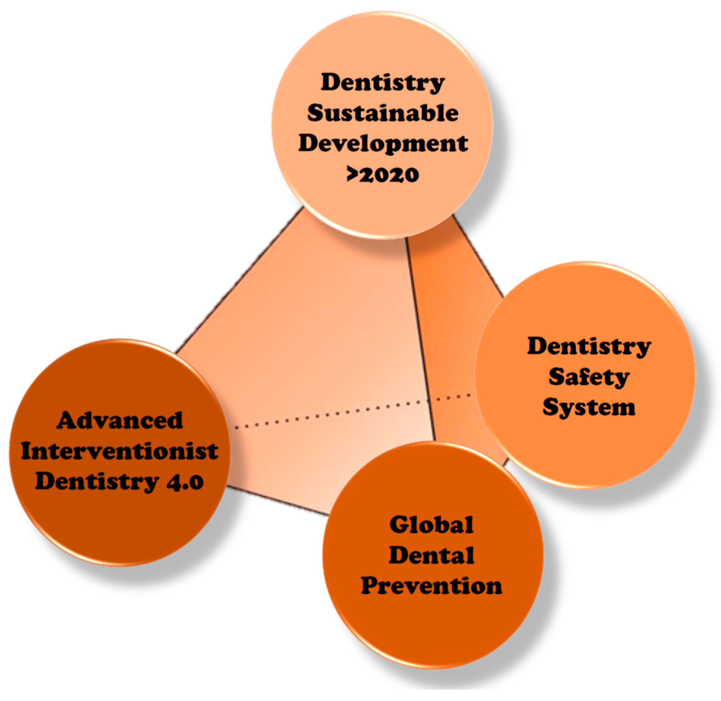
Figure 1. Chinese censer from the turn of the 19th and 20th centuries with three legs to ensure stability.
Processes 08 01605 g001
Figure 2. Social and commercial determinants of oral diseases.
Processes 08 01605 g002
Figure 3. Diagram explaining the meaning of the disability-adjusted life-years (DALY) indicator.
Processes 08 01605 g003
Figure 4. The spread of caries in 2017 in various countries of the world (no newer analyses); (a) in the 5–14 age group and (b) regardless of age; (c) The spread of edentualism and severe tooth loss; (d) The spread of periodontal diseases regardless of age.
Processes 08 01605 g004
Figure 5. Top view the lower right third molar (RM3) of the man from Villabruna from the Late Upper Palaeolithic about 14,000 years ago; (a) Occlusal view; (b) detailed view of the occlusal cavity, with the four carious lesions and the chipping area on the mesial wall; red line is directed mesio-distally, passing through the larger carious lesion [109].
Processes 08 01605 g005
Figure 6. Diagram of the dynamics of interactions between protective and pathological factors that can shift the balance in the oral cavity towards health or disease, respectively.
Processes 08 01605 g006
Figure 7. Scheme of the so-called caries continuum with codes from the ICDAS system.
Processes 08 01605 g007
Figure 8. Scheme of the International Caries Classification and Management System (ICCMS).
Processes 08 01605 g008
Figure 9. Scheme of the caries development pyramid (CDP) with indications ranging from background-level care (BLC) to preventive treatment options (PTOs) and the operative treatment option (OTO) in comparison with the general view of an iceberg submerged largely in water.
Processes 08 01605 g009
Figure 10. Diagram of Deming’s circle adopted for the fives’ rule on the 5D Caries Management Cycle Rules 5D (CMCRs) for the continuous improvement of the level of the patient’s oral health state.
Processes 08 01605 g010
Figure 11. Precision Caries Management (PCM) schematic diagram developed in the form of the complex developed concentric circles by analogy to the Deming circle with a wide range of possible therapeutic decisions depending on the stage of caries development.
Processes 08 01605 g011
Figure 12. Examples of endodontically treated teeth: (a) morphology of the transverse fracture of the canal dentin (atomic force microscope); (bg) root canal filled with gutta-percha material with AH Plus sealant by the thermoplastic method: (b) X-ray of the patient; (c) longitudinal fracture of the tooth; (df) decalcified teeth; (e) filling the side branches of the main canal; (f) root delta ((cf) Stereo Discovery stereo light microscope with AxioCam HRC digital camera by Zeiss at 8–50× magnification); (g) tight connection of the root canal with the material based on gutta-percha and sealant in all sections of the root canal; (h) after an incorrectly performed procedure, border of three layers, tightly connected dentine of the root canal with a thick intermediate layer of sealant and a leak between the sealant and the material on the gutta-percha matrix covered with a sealant layer (g,h)—scanning electron microscope).
Processes 08 01605 g012
Figure 13. An example of a circumferential bridge after the extraction of the patient’s teeth as a result of neglecting the treatment of caries, alternatively manufactured using the technology of milling solid-state metal blocks or selective laser sintering (SLS) from powders of the Co25Cr5W5MoSi alloy: (a) scan of the impression, (b) model with finger dentition occlusion, (c) verification of the parallelism of the pillars, (d) finished design of the skeleton, € milling of the bridge in a controlled numerically controlled center (CNC), (f) layout scheme of the bridge selectively sintered with a set of supports on the work table, (g) foundation of the bridge manufactured using one of the given technological methods, (h) finished bridge veneered with ceramics, (i) bridge installed in the patient’s mouth.
Processes 08 01605 g013
Figure 14. Diagram of a longitudinal section through the periodontal pocket illustrating the gingival recession and the exposure of the tooth root surface.
Processes 08 01605 g014
Figure 15. Diagram of Deming’s circle adopted for the prophylaxis of periodontal diseases and a diagram of the continuous improvement of the level of oral hygiene with a TIPPS circle.
Processes 08 01605 g015
Figure 16. Characteristic damage caused by the development of the periodontal disease: (a) papilla bleeding; (b) depth of periodontal pockets; (c) classes of furcations.
Processes 08 01605 g016
Figure 17. The scheme of the main elements of the prevention and treatment of periodontal diseases in primary care.
Processes 08 01605 g017
Figure 18. An example of an implant-prosthetics treatment of the female patient with 3-degree unstable (MOB = 3) prosthetic restoration of the 12–24 segment due to periodontal disease. (ad) The pantographic X-ray images: (a) revealing significant bone loss; (b) after tooth extraction from periodontal diseases reasons; (c) after the insertion of the implants using a surgical template; (d) after installing the finished prosthetic restoration; (eg) view of the prosthetic restoration: (e) in the oral cavity; (f) full-arch prosthetic bridge; (g) the patient’s full smile after treatment.
Processes 08 01605 g018
Figure 19. Schematic of the extended holistic Industry 4.0 model.
Processes 08 01605 g019
Figure 20. Material engineering paradigm (a) and a diagram of the Materials 4.0 methodology of material design as part of engineering design (b).
Processes 08 01605 g020
Figure 21. Influence of laser power and laser spot width on the bending strength and structure of a selectively laser-sintered Ti6Al4V alloy; the top-row of photos the structure of this material produced by the SLS method in appropriately selected conditions, guaranteeing a bending strength of approx. 2464 MPa; the middle-row of photos the structure of material ensuring the strength of only approx. 1099 MPa; the bottom-row with diagrams - the effect of the laser power (first) and the width of the laser spot (second) on the bending strength; first column of photos the illustration in red of the pores at a magnification of 100×, in the first-row with a pore area fraction of 0.03% and in the second-row a fraction of 10.53%; in both cases, the width of the laser dot is 120 µm, while the laser power is 110 and 60 W, respectively; the presence of these pores in the second case in the second column at the bottom is visualized in a scanning electron microscope; the third column—the fractures of the tested specimens after bending—in the first-row fracture with a homogeneous structure, without pores and visible boundaries of the sintered powder particles; in the second-row, the structure of the fracture heterogeneous, with visible fragments where there are open pores and spherical particles, completely or partially unsintered.
Processes 08 01605 g021
Figure 22. General diagram of the synergistic relations between dentistry, dental engineering, and materials engineering in the Dentistry 4.0 model.
Processes 08 01605 g022
Figure 23. Examples of implant-scaffolds (a,b) with a porous structure and (c) with protrusions on the surface.
Processes 08 01605 g023
Figure 24. Examples of pure titanium porous samples manufactured by the selective laser sintering SLS method and coated with the atomic layers deposition ALD method inside the pores with a TiO2 nano-layer; the first column—the models of fully porous implant-scaffolds with dimensions of 10 × 10 × 10 mm, made of pure titanium, coated in the ALD process with TiO2 nanolayers successively in 500, 1000 and 1500 cycles.; the middle column—the surface morphology of these samples observed in the Atomic Force Microscope AFM corresponding to these thicknesses; the last column—Fast Fourier Transform FFT of pure titanium sample film coated with a TiO2 layer for 1500 cycles tested in a high-resolution transmission electron microscope HRTEM confirming the crystalline Ti substrate structure (pink) and amorphous structure of TiO2 layer (violet).
Processes 08 01605 g024
Figure 25. Comparison of morality due to (a) heart attacks and other ischemic heart diseases, (b) stroke, and (c) cancer by sex.
Processes 08 01605 g025
Figure 26. Comparison of the health expenditure in 2018 in the OECD countries (a) per capita, (b) as a share of GDP, (c) as a share of the GDP projected in 2030.
Processes 08 01605 g026
Figure 27. Changes in the years 2000–2017: (a) employment in health and social work as a share of total employment, (b) practising doctors per 1000 population, (c) practising nurses per 1000 population.
Processes 08 01605 g027
Figure 28. The extent of coverage of financing medical services in 2017 by government and compulsory insurance including that dedicated to dental care.
Processes 08 01605 g028
Figure 29. The statistics in 2014 for visits to (a) doctors and (b) dentists, and (c) the population forgoing or postponing care because of limited availability.
Processes 08 01605 g029
Figure 30. A scheme of exemplary changes in the method of financing health care in some European countries generally for all habitants, whether adults or children.
Processes 08 01605 g030
Figure 31. The detailed distribution of dentists in individual European countries (* Supreme Medical Chamber in Poland on behalf of the Government of Poland on 10 February 2020, sent a letter to Eurostat, correcting incorrect information because, in Poland, 42,425 people currently have the right to practice the profession of dentists in Poland, most of whom are dentists registered as practicing this profession. No. of dentist in 100,000 inhabitants should be 112.1).
Processes 08 01605 g031
Figure 32. Number of practicing dentists in various European countries per 100,000 inhabitants in 2017 and the rate of changes in the number of dentists in 2018 compared to 2017.
Processes 08 01605 g032
Figure 33. Changes in the number of practicing dentists in the USA in the period 1993–2017.
Processes 08 01605 g033
Figure 34. Epidemiological situation in various countries of the world on 25 November 2020 caused by the COVID-19 disease.
Processes 08 01605 g034
Figure 35. Diagram of SARS-CoV-2 transmission via a dentist in the event of one infected patient appearing in a dental clinic.
Processes 08 01605 g035
Figure 36. Diagram of one of the face cap solutions and an example of its use with a set of devices shown in the lower part of the drawing.
Processes 08 01605 g036
Figure 37. Diagram of the basic principles of dental (a) and engineering (b) ethics in the form of gears symbolizing the necessary cooperation between these areas.
Processes 08 01605 g037
Figure 38. The scheme of the ethical aspects of the personal dimensions of sustainable oral cavity health.
Processes 08 01605 g038
Figure 39. Model of the sustainable development of modern dentistry from 2020.
Processes 08 01605 g039
Table 1. Principal carious lesion classification criteria.
No.Classification BasisClassification
1Treatment of CariesD = decayed or caries lesions, M = missing owing to extraction, F = filled or restored caries lesions
2Morphology (Location of the Lesion)Occlusal caries, smooth-surface caries and root caries
3Prior Condition of the ToothPrimary caries, secondary (recurrent) caries
4Severity and Rate of Caries ProgressionAcute caries, chronic caries, active caries and arrested caries
5Extent of the LesionIncipient caries, advanced caries
6Chronology or AgeEarly childhood caries, adolescent caries, adult caries
7Etiology (Causes or Origins of Caries)Baby bottle tooth decay
8Affected tissuesEnamel, dentin, cementum
Table 2. Presentation of the basic symptoms accompanying different stages of the development of plaque diseases.
Disease Development StagesBasic Symptoms in the Subsequent Stages of the Disease Development
GingivitisThis is caused by plaque; there are red, swollen tissues that bleed when brushing and probing.
Chronic periodontitisThere is a slow destruction of the connective epithelium and attachment of the connective tissue of the tooth, bone destruction with a loss of bone mass, and the formation of periodontal pockets.
Aggressive periodontitisThe degree of destruction of the attachment of connective tissue and bone is severe at the rapid progression of the disease; severe condition in the group of younger patients is related to a family history of aggressive periodontitis.
Necrotizing ulcerative gingivitis (NUG)Necrotizing ulcerative periodontitis (NUP) is present in the presence of the loss of connective tissue and bone destruction, manifested by the painful ulceration of the ends of the interdental papillae with visible gray necrotic tissue accompanied by halitosis.
Periodontal abscessThere is acute or chronic and asymptomatic freely draining infection of the periodontal pocket.
Perio-endo lesionsThe source of the bacteria is from the periodontium or the root canal system, while lesions may heal or may be independent.
Gingival enlargementIrritation from plaque or tartar, as well as repeated rubbing or trauma, as well as fluctuating hormone levels or the use of certain medications, cause the gingivae to thicken.
Table 3. A new classification scheme for periodontal and peri-implant diseases and conditions.
Periodontal Diseases and Conditions
Periodontal Health, Gingival Diseases and ConditionsForms of PeriodontitisPeriodontal Manifestations of Systemic Diseases And Developmental and Acquired Conditions
  • Periodontal health and gingival health
    • Clinical gingival health on an intact periodontium
    • Clinical gingival health on a reduced periodontium
      • Stable periodontitis patient
      • Non-periodontitis patient
  • Necrotizing
    Periodontal Diseases
    • Necrotizing Gingivitis
    • Necrotizing Periodontitis
    • Necrotizing Stomatitis
  • Systemie diseases or conditions affecting the periodontal supporting tissues
2.
Other Periodontal Conditions
  • Periodontal Abscesses
  • Endodontic-Periodontal Les ions
2.
Gingivitis—dental biofilm-induced
  • Associated with dental biofilm alone
  • Mediated by systemie or locaI risk factors
  • Drug-influenced gingival enlargement
2.
Periodontitis as Manifestation of Systemic Diseases
Classification of these conditions should be based on the primary systemic disease according to the International Statistical Classification of Diseases
and Related Health Problems codes
3.
Mucogingival deformities and conditions around teeth
  • Gingival phenotype
  • Gingivaljsoft tissue recession
  • Lack of gingiva
  • Decreased vestibular depth
  • Aberrant frenum/muscle posit ion
  • Gingiva l excess
  • Abno rmal color
  • Condition of the exposed root surface
4.
Traumatic occlusal forces
  • Primary occlusal trauma
  • Secondary occlusal trauma
  • Orthodontic forces
3.
Gingival diseases—non-dental biofilm induced
  • Genetic/developmental disorders
  • Specific infections
  • lnflammatory and immune conditions
  • Reactive processes
  • Neoplasms
  • Endocrine, nutritional & metabolic diseases
  • Traumatic lesions
  • Gingival pigmentation
3.
Periodontitis
  • Stages: Based on Severity 1 and Complexity of Management 2
    • Stage I: lnitial Periodontitis
    • Stage II: Moderate Periodontitis
    • Stage III: Severe Periodontitis with potential for additional tooth loss
    • Stage IV: Severe Periodontitis with potentiaI for loss of the dentition
  • Extent and distribution 3: localized; generalized; molar-incisor distribution
  • Grades: Evidence or risk of rapid progression 4 anticipated treatment response 5
    • Grade A: Slow rate of progression
    • Grade B: Moderate rate of progression
    • Grade C: Rapid rate of progression
5.
Prostheses and tooth-related factors that modify or predispose to plaque- induced gingival diseases/periodontitis
  • Localized tooth -relat ed factors
  • Localized dental prostheses-related factors
1 Severity: Interdental clinical attachment level (CAL) at site with greatest loss; radiographic bone loss and tooth loss. 2 Complexity of management: probing depths, pattern of bone loss, furcation lesions, number of remaining teeth, tooth mobility, ridge defects, masticatory dysfunction. 3 Add to stage as descriptor: localized <30% teeth, generalized 30% teeth. 4 Risk of progression: direct evidence by paradiographs or CAL loss, or indirect (bone loss/age ratio). 5 Anticipated treatment response: case phenotype, smoking, hyperglycemia.
Table 4. Method of determining Basic Periodontal Examination (BPE) indicators.
NoSteps by the dentist to assess BPE
1A World Health Organization WHO BPE probe is used. This has a “ball end” 0.5 mm in diameter, and a black band from 3.5 to 5.5 mm. Light probing force should be used (20–25 g).
2The dentition is divided into 6 sextants:
upper right (17 to 14), upper anterior (13 to 23), upper left (24 to 27).
lower right (47 to 44), lower anterior (43 to 33), lower left (34 to 37).
3All teeth in each sextant are examined (with the exception of 3rd molars).
4For a sextant to qualify for recording, it must contain at least 2 teeth (if only 1 tooth is present in a sextant, the score for that tooth is included in the recording for the adjoining sextant).
5The probe should be “walked around” the sulcus/pockets in each sextant, and the highest score recorded. As soon as a code 4 is identified in a sextant, the clinician may then move directly on to the next sextant, though it is better to continue to examine all sites in the sextant. This will help to gain a fuller understanding of the periodontal condition, and will make sure that furcation involvements are not missed. If a code 4 is not detected, then all sites should be examined to ensure that the highest score in the sextant is recorded before moving on to the next sextant.
Table 5. Method of determining the scoring codes value when evaluating Basic Periodontal Examination (BPE) indicators.
Code BPEDescription
0No pockets > 3.5 mm, no calculus/overhangs, no bleeding after probing (black band completely visible).
1No pockets > 3.5 mm, no calculus/overhangs, but bleeding after probing (black band completely visible).
2No pockets > 3.5 mm, but supra- or subgingival calculus/overhangs (black band completely visible).
3Probing depth 3.5–5.5 mm (black band partially visible, indicating pocket of 4–5 mm).
4Probing depth > 5.5 mm (black band entirely within the pocket, indicating pocket of 6 mm or more).
*Furcation involvement.
Comments: Both the number and the * should be recorded if a furcation is detected—e.g., the score for a sextant could be 3 * (e.g., indicating probing depth 3.5–5.5 mm plus furcation involvement in the sextant).
An example BPE score grid:433 *
-24 *
Table 6. Indicators determined in order to objectively assess the condition of the periodontium and its diseases.
Indicator Type Description or Specification
BOP
Bleeding on Probing
(Papilla Bleeding Index PBI)
Assesses the presence of bleeding gums; a patient with less than 10% bleeding is counted as having a healthy mouth cavity; this indicator is assessed using a WHO or North Carolina probe; the probe should “go around” the pocket on each side and if there is bleeding, the site is noted on the plot (at the mesial, distal, buccal, palatal and lingual surfaces); after using the appropriate computer software, the system calculates the proportion of bleeding areas.
PI
Plaque Index
(Approximal Plaque Index API)
Assesses the presence of plaque; this indicator is assessed using a WHO or North Carolina probe or by plate staining; the probe should “go around” the tooth on each side and if plaque is present, the site is noted on the plot (at the mesial, distal, buccal, palatal and lingual surfaces); after using the appropriate computer software, the system calculates the proportion of biofilm area.
PPDs
Periodontal Probing Depths
Assesses the depth of the pockets; this index is assessed using the North Carolina probe; the probe should “go around” the pocket on each side and record the measurement results in millimeters on a plot with the location (at the mesial, distal, buccal, palatal and lingual surfaces).
REC
Gum Recession Index
Assesses gingival recessions; this index is assessed using the North Carolina probe; the results of measurements of the distance in millimeters from the cement-enamel junction (CEJ) [232] to the gingival margin should be made and recorded.
MOB
Mobility Index
Assesses the degree of tooth mobility: the other end of the mirror and a metal probe can be used to determine this index; While holding the lock with two tools, move it in all directions.
CodeGrade on a scale of 0–3.
0No movable property.
1Gentle mobility.
2The tooth moves horizontally >1 mm.
3The tooth moves horizontally and vertically >2 mm.
FUR
Furcation Index
Assesses the degree of furcation using a furcation probe.
CodeMeasure with an appropriate furcation probe.
AMeasurement with a furcation probe 1–3 mm.
BMeasurement with a furcation probe 4–6 mm.
CMeasurement with furcation probe >7 mm.
Table 7. Preventive and/or therapeutic procedures depending on the results of activities carried out in the hygienization phase of periodontal disease treatment and the BPE score results.
DiagnosisPreventive and/or Therapeutic Measures Necessary to Be Taken by a DentistTreatment Phase
Code BPEDescription
0normalNo need for periodontal treatment.0No treatment required
1bleeding after probing OHIIHygienic training
2dental stone supra- or subgingival, iatrogenic injuries OHI, the removal of plaque retentive factors, including all supra- and subgingival calculus.III + scaling supra- and subgingival
3depth of periodontal pockets 3.5–5.5 mmOHI, RSD
4depth of periodontal pockets >6 mmOHI, RSD. Assess the need for more complex treatment; referral to a specialist may be indicated.IIII = II + surgical treatment
*furcationsOHI, RSD. Assess the need for more complex treatment; referral to a specialist may be indicated.
Comments: As a general rule, radiographs to assess alveolar bone levels should be obtained for teeth or sextants where BPE codes 3 or 4 are found.
Abbreviations: OHI—oral hygiene instruction; RSD—root surface debridement.
Table 8. The potential and attractiveness of each group of methods of protection against the effects of the SARS-CoV-2 coronavirus.
NoType of Method Known in the Literature, State-of-the-Art Including Own SolutionPotentialAttracti-Veness
1Installation of air filtering devices in the treatment room 6.74.0
2Equipment operating with HEPA or carbon filters or with UV radiation 5.954.0
3Dental saliva extractor attachments 3.35.8
4“Extract from the treatment area” devices 3.76.6
5Patient enclosures in the form of glass or PMMA boxes 1.453.9
6Original solution eliminating the threat at the source with the set of devices for virus elimination 9.08.5
Publisher’s Note: MDPI stays neutral with regard to jurisdictional claims in published maps and institutional affiliations.

