Article

Enhanced Production of Anti-PD1 Antibody in CHO Cells through Transient Co-Transfection with Anti-Apoptotic Gene Bcl-xL Combined with Rapamycin

1
Engineering Research Center of Cell & Therapeutic Antibody, Ministry of Education, School of Pharmacy, Shanghai Jiao Tong University, 800 Dongchuan Road, Shanghai 200240, China
2
Jecho Laboratories, Inc. 7320 Executive Way, Frederick, MD 21704, USA
*
Author to whom correspondence should be addressed.
These authors contributed equally to this work.
Processes 2019, 7(6), 329; https://doi.org/10.3390/pr7060329
Submission received: 30 April 2019 / Revised: 20 May 2019 / Accepted: 24 May 2019 / Published: 1 June 2019
(This article belongs to the Special Issue Transient Gene Expression for Rapid Protein and Virus-Vector Supply)

Abstract

:
CHO cells are often used to produce monoclonal antibodies in mammalian cell expression systems. In the process of large-scale cell culture, apoptosis is related to cell survival and product quality. Over-expressing an anti-apoptotic gene to delay apoptosis and improve cell growth is one of the strategies for improving productivity of monoclonal antibodies. Autophagy inducer rapamycin can extend the culture duration of CHO cells and affect the yield of antibodies. A method was developed for transient co-transfection of anti-apoptotic genes and genes of interest combined with rapamycin to increase the transient expression of the anti-PD1 antibody. Under the optimal transfection conditions, the combination of Bcl-xL and rapamycin can significantly delay cell apoptosis, inhibit cell proliferation, and prolong cell life-time. As a result, anti-PD1 monoclonal antibody expression levels are increased by more than 2 times.

1. Introduction

In recent years, biopharmaceuticals have developed rapidly. CHO cells (Chinese hamster ovary cells) are the main expression host of recombinant protein drugs and monoclonal antibody drugs, and have the advantages of a high-density suspension culture, high yield, transfection and safety of therapeutic applications. It is often widely used in the production and research of recombinant proteins [1]. In recent years, transient expression technology has become a popular area of research due to its simplicity, short experimental period, low cost and the product’s conformity to clinical standards [2,3]. In the transient expression system of CHO cells, the expression level can reach in the order of g/L. In 2014, the MedImmune team established a CHO high expression system with a yield of up to 2 g/L [4]. They engineered the CHO-K1 cell line with co-expression of EBNA-1 and GS (the gene for glutamine synthetase). In 2015, it was reported that a yield of up to 1 g/L was achieved by optimizing the transient method using PEI (Polyetherimide) as a transfection reagent [5,6]. A CHO Cell Transient Expression Kit (ExpiCHO) has been developed, which is reported to produce up to 3 g/L [7]. In 2019, it was shown that transient expression in a Chinese hamster ovary (CHO) host (ExpiCHO-STM) is capable of achieving a high recombinant antibody expression of 1 g/L [8].
Mammalian cells are very sensitive to the culture environment. Nutrients and growth factor deficiency, hypoxia, and viral infection can induce apoptosis. Cell apoptosis is a type I programmed death, accounting for 80% of cell death. It affects the survival state of cells and the quality of protein products. In apoptosis, the Bcl-2 family [9] plays a key role, including two kinds of genes, one with anti-apoptotic functions, mainly Bcl-xL, Mcl-1, Bcl-2, Bcl-w, etc. The other genes with pro-apoptotic functions, mainly including Bax, Bak, Bad and so on, have the function of promoting death. Overexpression of an anti-apoptotic gene to delay apoptosis and extend the culture duration of cells is one of the methods used to increase protein expression yield and reduce production cost [10]. It has been reported that simultaneous expression of anti-apoptotic genes and target genes can increase cell viability and increase protein yield [11,12]. Our previous work has shown that the yield can be increased by 80% by co-transfecting the anti-apoptotic genes Bcl-xL, Mcl-1 and the target gene [13]. We found that transfecting with 10% Bcl-xL (the optimal dosage) increased the yield by 83%, and 50% of Mcl-1 (the optimal dosage), increasing the protein yield by 34%. In this study, we choose Bxl-xL as an anti-apoptotic gene.
Autophagy is a type II programmed cell death, which was proposed after the discovery of the phenomenon of “self-feeding” in cells in 1962 [14]. It is closely related to cell death, cell survival, and cell maintenance. The mTOR target has been seen as a sensor for amino acid and other nutrients, and a switch for autophagy activation, thus playing a vital role [15]. mTOR kinase, linked to phosphatidylinositol kinase, is thought to be involved in cell growth, cell cycle regulation, and protein synthesis. Studies have shown that rapamycin is a mTOR target inhibitor and it has been widely used in vivo and in vitro studies on autophagy [16]. Rapamycin causes cells to arrest in the G1 phase, slowing cell proliferation [17]. The Balcarcel and Stephanopoulos team demonstrated that rapamycin caused hybridoma cell death and increased antibody expression by a factor of two in fed-batch culture [18]. Rapamycin can increase CHO cell viability by 21% and the yield by 30% [16]. Also, inoculation of cultures in the presence of rapamycin can increase viability and final titer in CHOK1 cells [19]. Therefore, we deduced that the anti-apoptotic gene shared with rapamycin may also have certain effects on CHO cell culture and protein expression after transient transfection.

2. Materials and Methods

2.1. Cell Lines and Plasmids

CHO cell line was obtained from Invitrogen (Thermo Fisher Scientific, Carsbad, CA, USA). Bcl-xL/PCDNA3.1(-) plasmid and PD-1 antibody light chain and heavy chain were constructed as in our previous work [13,20]. All media and supplemental components used in this study were from Invitrogen (Thermo Fisher Scientific, Carsbad, CA, USA). Rapamycin was from Gene Operation (Shanghai, China). CHO cells were propagated in a serum-free CD-CHO medium supplemented with 8 mM GlutaMAX. Routine cultures were passaged at a cell concentration of 2 × 10⁵ cells/mL, using a working volume of 20 mL in 125 mL shake flasks (Corning, New York, NY, USA), and were maintained in a 37 °C incubator (Thermo Fisher Scientific, Shanghai, China) with 5% CO2 and shaken at 125 rpm. Cell density and cell viability were measured with a trypan blue (1:1, v/v; AMRESCO, Solon, OH, USA) staining method, which is counted by a cell counter (Ruiyu Biotech, Shanghai, China).

2.2. Cell Culture and Transfection

According to [12], we transfected 1 × 10⁶ cells/mL with a DNA/PEI ratio of 1:2 (w/w) and a DNA concentration of 3.5 μg/mL. The culture volume was 20–25 mL. Rapamycin’s working concentration was 100 nM. Rapamycin was dissolved in DMSO to a stock concentration of 200 μM and stored at −20 °C.

2.3. Analysis of Apoptosis

Cells (1 × 10⁶) were washed twice with pre-cooled PBS (10 mM Na2HPO4, 1.8 mM KH2PO4, 2.7 mM KCl, 137 mM NaCl, pH7.4) (Sinopharm Chemical Reagent Co., Ltd, Shanghai, China) and resuspended in 100 μL of binding buffer. Then, 5 μL of Annexin V-FITC and 5 μL of PI was added for staining, and the solution was incubated for 10–15 min at room temperature. After the incubation, binding buffer was added and flow cytometry was performed.

2.4. Analysis of Cell Cycle

Cells were collected at 2–5 × 10⁶ cells and washed twice with an appropriate amount of PBS (the same as Section 2.3) with centrifugation at 500× g for 5 min at 4 °C each time. Supernatant was discarded and an appropriate amount of PBS was added, gently pipetting the cells. The cell suspension was fixed to 1 mL of 75% ethanol (Sinopharm Chemical Reagent Co., Ltd, Shanghai, China), and then placed in a refrigerator at 4 °C overnight. The cells were collected by centrifugation at 1000× g for 5 min at 4 °C, and washed with an appropriate amount of 4 °C pre-cooled PBS. Cells were resuspended in 400 μL cold PBS and 0.5 mL of 50 μg/mL PI solution containing RNase A Solution (Takara, Dalian, Liaoning, China) was added to stain cells for 30 min at room temperature (protected from light). After filtration, flow cytometry (BD Bioscience, San Jose, CA, USA) was used for detection.

2.5. Analysis of Protein Expression in Apoptosis and Autophagy

During cell culture, the same volume of cell suspension was taken, centrifuged and lysed with RIPA lysis buffer (137 mM NaCl, 20 mM Tris, 1 mM MgCl2, 1 mM CaCl2, 1% NP-40, 0.5% dexycholate, 0.1% SDS, pH7.4) (Beyotime, Shanghai, China) supplemented with protease inhibitors. The enhanced BCA protein assay kit (Beyotime, Shanghai, China) was used to determine concentration of total protein in clarified lysates. Western blotting was used to measure the protein expression levels of LC3-I, LC3-II, Pho-mTOR, and Bcl-xL in cells. Tubulin (55 kDa) was used as an internal reference protein. The total number of cells lysed during loading remained essentially the same. Samples were loaded on a 10-well SDS–PAGE (10% or 12%) gel. Then, proteins were transferred to PVDF membrane (Millipore, Darmstadt, Germany). The primary antibodies that we used were Anti-Bcl-xL antibody (Cell signaling Technology, Danvers, MA, USA), Anti-Phos-m TOR antibody (Cell signaling Technology, Danvers, MA, USA), Anti-LC3-I, LC3-II antibody (Cell signaling Technology, Danvers, MA, USA), and a HRP-anti-rabbit IgG antibody (Cell Signaling Technology, Danvers, MA, USA).

2.6. Production of Anti-PD1 Monoclonal Antibody

The transient expression of anti-PD1 antibody was detected by ELISA. After the cell viability was less than 50%, the cells were centrifuged at 1000 rpm for 5 min at low speed to remove most of the cells, followed by high-speed centrifugation at 12,000 rpm for 10 min to further remove the remaining cells or debris. The supernatant after final centrifugation was taken for ELISA. A 96-well plate was coated with monoclonal anti-human κ chain antibody (Merck Millipore, Shanghai, China) as a capture antibody. Then, samples harvested from cell culture supernatants were added. A monoclonal HRP-anti-human IgG antibody (FC specific; Jackson Immu-noResearch, West Grove, PA, USA) was used for detecting enzymatic oxidation reaction with TMB substrate (BD Bioscience, San Jose, CA, USA). The signal was assessed by a spectrophotometer at 450 and 630 nm. The data was calculated according to the found standard curve.

3. Results

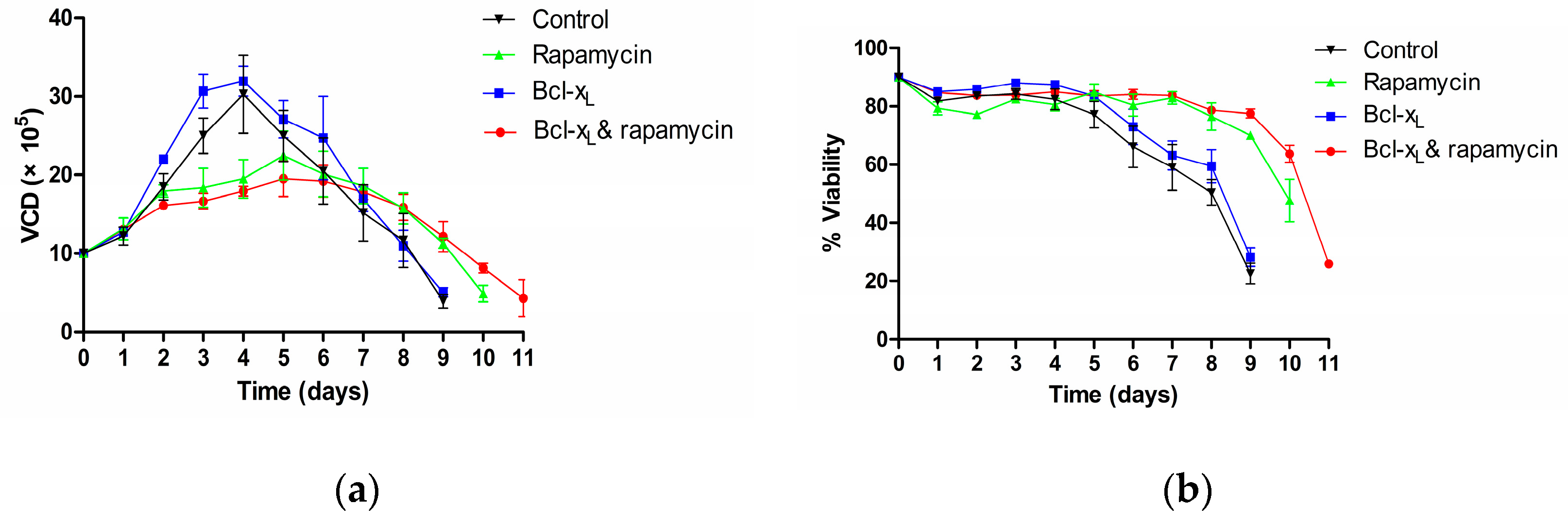
3.1. Analysis of Cell Viability and Density

After transfection, samples were taken daily. Cell density and cell viability were measured, and a curve was drawn as shown in Figure 1. In the Control group, only the anti-PD1 monoclonal antibody plasmid was transfected; in the rapamycin group, 100 nM rapamycin was added after transfecting the same amount of anti-PD1 monoclonal antibody plasmid as in the control group for 24 h; in the Bcl-xL group, 10% Bcl-xL plasmid was co-transfected with 90% anti-PD1 monoclonal antibody plasmid; in the Bcl-xL & rapamycin group, 10% Bcl-xL plasmid was co-transfected with 90% anti-PD1 monoclonal antibody plasmid with 100 nM rapamycin added after 24 h of the transfection. The total amount of transfected DNA in all groups was consistent.
As shown in Figure 1a, in the rapamycin group and Bcl-xL & rapamycin group, the maximum cell density was significantly lower than that of the Bcl-xL group and the control group. Meanwhile, compared with the control group (with 9 days of survival), the rapamycin group prolonged the cell survival by two days (11 days in total), and the Bcl-xL & rapamycin group prolonged survival by three days (12 days in total). There was no significant difference in cell density between the control group and the Bcl-xL group.
As shown in Figure 1b, after a plateau, cell viability gradually decreased until it was below 50%. Results showed that the cell viability of the Bcl-xL group was generally higher than that of the control group. Both of the viabilities in these two groups decreased more quickly (at about day 5) than the other two groups (at about day 9 or 10). Compared with the control group, the rapamycin group caused a short-term decrease in cell viability after the second day of dosing, gradually began to increase on the fourth day, and then maintained a high survival rate until the 11th day. The Bcl-xL & rapamycin group did not show a short-term decrease in cell viability at the initial stage, and maintained high cell viability throughout the culture until the 12th day. Moreover, it had the longest cell culture duration.

3.2. Analysis of Apoptosis

After transfection, samples were taken at the same time every day to monitor the apoptosis of the cells. As shown in Figure 2a, in the Bcl-xL group, the proportion of apoptotic cells decreased at 72 h after transfection, much lower than the control group. Compared with the control group, the apoptosis of the rapamycin group was basically the same at 24–48 h. Overall, the total average apoptosis of the rapamycin group was higher than that of the control group, which showed at 72 h. The apoptosis of the Bcl-xL & rapamycin group was consistent with the control group. Meanwhile, its mean value was better than that of the rapamycin group and lower than that of the Bcl-xL group. As shown in Figure 2b, the status of late apoptosis was consistent with total apoptosis (Figure 2a), and there was no a higher proportion of late apoptosis at 72 h in the rapamycin group.

3.3. Analysis of Cell Cycle

Samples were taken at the same time on the second, third and fourth day after transfection and the cell cycle was monitored. As shown in Table 1, the rapamycin group had a higher ratio of cells in the G1 phase than that of the control group. Similarly, the Bcl-xL & rapamycin group had a higher ratio of cells in the G1 phase than that of the Bcl-xL group.

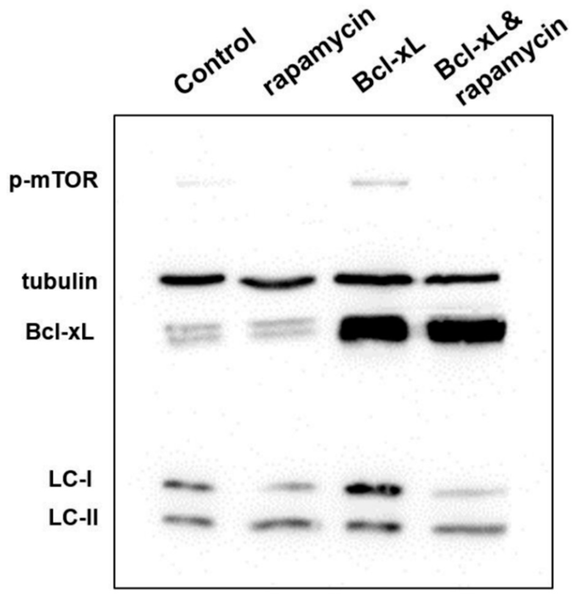
3.4. Analysis of Protein Expression in Apoptosis and Autophagy

When autophagy occurs, it is accompanied by a transition from LC3-I to LC3-II. Therefore, the decrease of LC3-I and increase of LC3-II indicates a regulation of autophagy [21]. Study shows that [22] rapamycin is an mTOR inhibitor, and it may reduce mTOR protein expression. The expression levels of LC3-I (16 kDa), LC3-II (14 kDa), phosphate-mTOR (289 kDa) and Bcl-xL (35 kDa) were detected by Western blotting on the fifth day. As shown in Figure 3, both rapamycin groups (rapamycin group and the Bcl-xL & rapamycin group) reduced LC3-I expression and increased LC3-II expression, while the expression of phosphate-mTOR was significantly lower than that of the control group and the Bcl-xL group. Bcl-xL gene was still overexpressed until the fifth day in both Bcl-xL groups (Bcl-xL group and the Bcl-xL & rapamycin group). According to the results, we deduced that rapamycin could promote autophagy.

3.5. Analysis of Anti-PD1 Monoclonal Antibody Expression Quantity

After transfection, when the cell viability declined to 50%, the cell supernatant was taken and the expression level was measured by ELISA, as shown in Figure 4. Compared with the control group, the expression was higher in the rapamycin group. Co-transfection of 10% Bcl-xL significantly increased the expression of anti-PD1 monoclonal antibody compared with the control group (13.3 μg/mL vs. 7.1 μg/mL), which was 87.3% higher. The combination of Bcl-xL and rapamycin further increased the expression level to 19.3 μg/mL, which was statistically different from the control group and the Bcl-xL group. (T test, p = 0.023).

4. Discussion

In this study, we studied the effect of rapamycin and the anti-apoptotic gene Bcl-xL on the expression of anti-PD1 monoclonal antibody in shake flask culture within four groups. It has been reported that stimulation of autophagy (or the presence of autophagosomes) often accompanies cell death [23]. This is consistent with the results of our study. The results for cell density and cell viability show that rapamycin may inhibit cell proliferation and slow down the growth rate, thus extending cell culture duration although it does damage cells to some extent. As shown in Figure 1a, the rapamycin group and the Bcl-xL & rapamycin group showed significantly inhibited cell proliferation and slower growth rates (the maximum cell density was significantly lower than that of the Bcl-xL group and the control group) throughout the 12-day cell culture. In Figure 1b, the cell viability of all groups decreased slightly after transfection, and then increased slightly. This was most likely caused by cell damage due to transfection [23]. The experimental results showed that the cell viability of the Bcl-xL group was generally higher than that of the control group. The cell viability of the rapamycin group and the Bcl-xL & rapamycin group was much higher than that of the control group. We speculated that rapamycin might indeed extend cell survival when added alone but might also damage the cells at the initial addition, while co-transfection of Bcl-xL gene could improve rapamycin’s negative effect on apoptosis. It has been reported that anti-apoptotic proteins like Bcl-xL and Bcl-2 inhibit both apoptosis and autophagy. They prevent the formation of autophagic vacuole through binding the BH3 site on Beclin-1 [24,25,26].
The transfection of the anti-apoptotic gene Bcl-xL reduced cell apoptosis; this might be due to the influence of cell transfection on cell status. In the Bcl-xL group, the proportion of apoptotic cells decreased at 72 h after transfection, and was much lower than the control group. It was speculated that the anti-apoptotic gene might improve the apoptotic state [25] and resist transfection damage [11]. It is hypothesized that in the early stage of the addition of rapamycin, it will cause apoptosis in certain CHO cells. Rapamycin significantly prolonged cell survival. In the cell cycle, rapamycin kept cells in the G1 phase, delayed cell division and reduced cell proliferation. In addition, rapamycin can induce autophagy, which was shown by reducing the expression of phosphorylated mTOR and promoting LC3-I transformation to LC3-II. The average expression of anti-PD1 monoclonal antibody for the rapamycin group was slightly higher than the control group. In a previous report, rapamycin was found to increase antibody yield by 30%, and it played a role in causing apoptosis [16], however, rapamycin did not show a significant apoptosis-inducing effect in this study. In the Bcl-xL & rapamycin group, the apoptosis was lower than that of the rapamycin group and higher than that of the Bcl-xL group. The reason was that the presence of anti-apoptotic genes might ameliorate the negative effects of external pressures such as transfection and the addition of other drugs inducing apoptosis [23].

5. Conclusions

This study aimed to develop a method of increasing the transient expression of anti-PD1 antibodies in a CHO cell expression system by co-transfection of anti-apoptotic gene Bcl-xL combined with rapamycin. The results showed that this method could significantly delay cell apoptosis, inhibit cell proliferation, and prolong cell survival, and anti-PD1 monoclonal antibody expression levels could be increased by more than 2 times. In addition, we attempted to simultaneously regulate apoptosis and autophagy in the CHO cell expression system, and to improve cell growth status through the combination of rapamycin and anti-apoptotic genes, thereby realizing higher expression levels. This work provides a cost-effective and time-saving method for improving cell survival and increasing the yields of biopharmaceuticals.

Author Contributions

Y.L. and X.Z. contributed to the experiments and draft manuscript. L.W. helped with transient transfection. H.Z., Y.Y. and L.H. assisted in experimental techniques and data analysis. X.L., C.X. and J.Z. (Jingyi Zhang) helped with cell culture and reagents preparation. J.Z. (Jianwei Zhu) initiated the project and co-designed the research. B.Z. initiated the project, co-designed the research, supervised experiments, and revised the manuscript.

Funding

This research was funded by the Natural Science Foundation of Shanghai, grant number 15ZR1423800.

Acknowledgments

This research was supported (in part) by Jecho Biopharmaceuticals Co., Ltd. Tianjin China for their technical assistance.

Conflicts of Interest

The authors declare no conflict of interest.

References

  1. Trill, J.J.; Shatzman, A.R.; Ganguly, S. Production of monoclonal antibodies in COS and CHO cells. Curr. Opin. Biotechnol. 1995, 6, 553–560. [Google Scholar] [CrossRef]
  2. Zhu, J. Mammalian cell protein expression for biopharmaceutical production. Biotechnol. Adv. 2012, 30, 1158–1170. [Google Scholar] [CrossRef]
  3. Hacker, D.L.; De Jesus, M.; Wurm, F.M. 25 years of recombinant proteins from reactor-grown cells—Where do we go from here? Biotechnol. Adv. 2009, 27, 1023–1027. [Google Scholar] [CrossRef]
  4. Daramola, O.; Stevenson, J.; Dean, G.; Hatton, D.; Pettman, G.; Holmes, W.; Field, R. A high-yielding CHO transient system: Coexpression of genes encoding EBNA-1 and GS enhances transient protein expression. Biotechnol. Prog. 2014, 30, 132–141. [Google Scholar] [CrossRef] [PubMed]
  5. Rajendra, Y.; Hougland, M.D.; Alam, R.; Morehead, T.A.; Barnard, G.C. A high cell density transient transfection system for therapeutic protein expression based on a CHO GS-knockout cell line: Process development and product quality assessment. Biotechnol. Bioeng. 2015, 112, 977–986. [Google Scholar] [CrossRef] [PubMed]
  6. Steger, K.; Brady, J.; Wang, W.; Duskin, M.; Donato, K.; Peshwa, M. CHO-S antibody titers >1 gram/liter using flow electroporation-mediated transient gene expression followed by rapid migration to high-yield stable cell lines. J. Biomol. Screen. 2015, 20, 545–551. [Google Scholar] [CrossRef] [PubMed]
  7. Jain, N.K.; Barkowski-Clark, S.; Altman, R.; Johnson, K.; Sun, F.; Zmuda, J.; Liu, C.Y.; Kita, A.; Schulz, R.; Neill, A.; et al. A high density CHO-S transient transfection system: Comparison of ExpiCHO and Expi293. Protein Expr. Purif. 2017, 134, 38–46. [Google Scholar] [CrossRef] [PubMed]
  8. Zhong, X.; Ma, W.; Meade, C.L.; Tam, A.S.; Llewellyn, E.; Cornell, R.; Cote, K.; Scarcelli, J.J.; Marshall, J.K.; Tzvetkova, B.; et al. Transient CHO expression platform for robust antibody production and its enhanced N-glycan sialylation on therapeutic glycoproteins. Biotechnol. Prog. 2019, 35, e2724. [Google Scholar] [CrossRef]
  9. Danial, N.N. BCL-2 family proteins: Critical checkpoints of apoptotic cell death. Clin. Cancer Res. 2007, 13, 7254–7263. [Google Scholar] [CrossRef]
  10. Baek, E.; Noh, S.M.; Lee, G.M. Anti-apoptosis engineering for improved protein production from CHO cells. Methods Mol. Biol. 2017, 1603, 71–85. [Google Scholar] [PubMed]
  11. Lee, S.; Lee, G.M. Bcl-2 overexpression in CHO cells improves polyethylenimine-mediated gene transfection. Process Biochem. 2013, 48, 1436–1440. [Google Scholar] [CrossRef]
  12. Zustiak, M.P.; Jose, L.; Xie, Y.; Zhu, J.; Betenbaugh, M.J. Enhanced transient recombinant protein production in CHO cells through the co-transfection of the product gene with Bcl-xL. Biotechnol. J. 2014, 9, 1164–1174. [Google Scholar] [CrossRef]
  13. Zhang, X.; Han, L.; Zong, H.; Ding, K.; Yuan, Y.; Bai, J.; Zhou, Y.; Zhang, B.; Zhu, J. Enhanced production of anti-PD1 antibody in CHO cells through transient co-transfection with anti-apoptotic genes Bcl-x L and Mcl-1. Bioprocess Biosyst. Eng. 2018, 41, 633–640. [Google Scholar] [CrossRef]
  14. Ashford, T.P. Cytoplasmic components in hepatic cell lysosomes. J. Cell Biol. 1962, 12, 198–202. [Google Scholar] [CrossRef] [PubMed]
  15. Edros, R.; Mcdonnell, S.; Al-Rubeai, M. The relationship between mTOR signalling pathway and recombinant antibody productivity in CHO cell lines. BMC Biotechnol. 2014, 14, 15. [Google Scholar] [CrossRef]
  16. Lee, J.S.; Lee, G.M. Rapamycin treatment inhibits CHO cell death in a serum-free suspension culture by autophagy induction. Biotechnol. Bioeng. 2012, 109, 3093–3102. [Google Scholar] [CrossRef] [PubMed]
  17. Pan, X.; Alsayyari, A.A.; Dalm, C.; Hageman, J.A.; Wijffels, R.H.; Martens, D.E. Transcriptome analysis of CHO cell size increase during a fed-batch process. Biotechnol. J. 2019, 14, 1800156. [Google Scholar] [CrossRef] [PubMed]
  18. Balcarcel, R.R.; Stephanopoulos, G. Rapamycin reduces hybridoma cell death and enhances monoclonal antibody production. Biotechnol. Bioeng. 2001, 76, 1–10. [Google Scholar] [CrossRef]
  19. Dadehbeigi, N.; Dickson, A.J. Chemical manipulation of the mTORC1 pathway in industrially relevant CHOK1 cells enhances production of therapeutic proteins. Biotechnol. J. 2015, 10, 1041–1050. [Google Scholar] [CrossRef]
  20. Ding, K.; Han, L.; Zong, H.; Chen, J.; Zhang, B.; Zhu, J. Production process reproducibility and product quality consistency of transient gene expression in HEK293 cells with anti-PD1 antibody as the model protein. Appl. Microbiol. Biotechnol. 2017, 101, 1889–1898. [Google Scholar] [CrossRef] [PubMed]
  21. Kabeya, Y.; Mizushima, N.; Yamamoto, A.; Oshitani-Okamoto, S.; Ohsumi, Y.; Yoshimori, T. LC3, GABARAP and GATE16 localize to autophagosomal membrane depending on form-II formation. J. Cell Sci. 2004, 117 Pt 13, 2805–2812. [Google Scholar] [CrossRef] [Green Version]
  22. Dai, C.; Ciccotosto, G.D.; Cappai, R.; Wang, Y.; Tang, S.; Hoyer, D.; Schneider, E.K.; Velkov, T.; Xiao, X. Rapamycin confers neuroprotection against colistin-induced oxidative stress, mitochondria dysfunction, and apoptosis through the activation of autophagy and mTOR/Akt/CREB signaling pathways. ACS Chem. Neurosci. 2018, 9, 824–837. [Google Scholar] [CrossRef] [PubMed]
  23. Lalaoui, N.; Lindqvist, L.M.; Sandow, J.J.; Ekert, P.G. The molecular relationships between apoptosis, autophagy and necroptosis. Semin. Cell Dev. Biol. 2015, 39, 63–69. [Google Scholar] [CrossRef] [PubMed]
  24. Sever, O.N.; Demir, O.G. Autophagy: Cell death or survive mechanism. J. Oncol. Sci. 2017, 3, 37–44. [Google Scholar] [CrossRef]
  25. Levine, B.; Sinha, S.C.; Kroemer, G. Bcl-2 family members: Dual regulators of apoptosis and autophagy. Autophagy 2008, 4, 600–606. [Google Scholar] [CrossRef] [Green Version]
  26. Noble, C.G.; Dong, J.M.; Manser, E.; Song, H. Bcl-xL and UVRAG cause a monomer-dimer switch in Beclin1. J. Biol. Chem. 2008, 283, 26274–26282. [Google Scholar] [CrossRef]
Figure 1. Assessment of cell density and viability with control group, rapamycin group, Bcl-xL group and Bcl-xL & rapamycin group throughout the 12-day cell culture. (a) Cell density; and (b) cell viability. The control group was transfected with anti-PD1 plasmids only. Error bars represent mean ± SEM for 3–4 independent experiments.
Processes 07 00329 g001
Figure 2. Comparison of apoptosis with the rapamycin, Bcl-xL and Bcl-xL & rapamycin group throughout the initial 3-days cell culture. (a) Total apoptosis, and (b) late stage apoptosis. Error bars represent mean ± SEM for 3–4 independent experiments. (*, **, *** indicate p < 0.05, p < 0.01, p < 0.001, respectively).
Processes 07 00329 g002
Figure 3. Western blotting of LC3-I, LC3-II, phosphate-mTOR and Bcl-xL on day 5 in the rapamycin, Bcl-xL and the Bcl-xL & rapamycin group.
Processes 07 00329 g003
Figure 4. Comparison of production with the control group, rapamycin group, Bcl-xL group and the Bcl-xL & rapamycin group. Error bars represent mean ± SEM for 3–4 independent experiments. (*, **, *** indicate p < 0.05, p < 0.01, p < 0.001, respectively).
Processes 07 00329 g004
Table 1. Comparison of cell cycle with rapamycin, Bcl-xL or combination of Bcl-xL and rapamycin from the second to forth days after transfection (mean ± SD).
DayCell CycleControlRapamycinBcl-xLBcl-xL & Rapamycin
D2G153.2 ± 1.061.5 ± 1.449.7 ± 0.859.6 ± 1.9
S25.9 ± 3.117.8 ± 1.926.1 ± 2.625.3 ± 1.5
G2/M15.8 ± 0.816.8 ± 1.219.6 ± 1.514.5 ± 1.9
D3G146.6 ± 2.656.7 ± 1.046.9 ± 1.956.9 ± 2.7
S31.3 ± 3.426.0 ± 2.628.6 ± 2.321.0 ± 3.0
G2/M18.2 ± 1.014.5 ± 3.022.2 ± 1.117.7 ± 1.7
D4G142.9 ± 1.556.4 ± 2.546.5 ± 1.253.6 ± 2.6
S32.3 ± 2.223.0 ± 4.235.0 ± 2.927.1 ± 1.3
G2/M24.2 ± 1.016.2 ± 2.017.9 ± 2.017.3 ± 1.8

