Article

Genomic and Proteomic Insights into Arsenic Detoxification and Alternative Transformation Pathways in Microbacterium oxydans AE038-20


by
Florencia Cecilia Spuches
1,†,
Andrés Hernán Morales
1,†,
Johan Sebastian Hero
1,2,
José Horacio Pisa
1,2,3,
Adriana Emilce Galván
4,
Marcela Alejandra Ferrero
5,6 and
Cintia Mariana Romero
1,2,*
1
Facultad de Bioquímica, Química y Farmacia, Universidad Nacional de Tucumán (UNT), Chacabuco 461, San Miguel de Tucumán T4000, Tucumán, Argentina
2
Planta Piloto de Procesos Industriales Microbiológicos PROIMI-CONICET, Av. Belgrano y Pasaje Caseros, San Miguel de Tucumán T4000, Tucumán, Argentina
3
Instituto de Desarrollo e Innovación Tecnológica para la Competitividad Territorial (IDITEC), Universidad de San Pablo (USP-T), San Martín 435, San Miguel de Tucumán T4000, Tucumán, Argentina
4
Department of Cellular Biology and Pharmacology, Herbert Wertheim College of Medicine, Florida International University, Miami, FL 33199, USA
5
Instituto de Ingeniería y Agronomía, Universiad Nacional Arturo Jauretche (UNAJ), Av. Calchaquí 6200, Florencio Varela 1888, Buenos Aires, Argentina
6
Consejo Nacional de Investigaciones Científicas y Técnicas (CONICET), Godoy Cruz 2290, Ciudad Autónoma de Buenos Aires, Argentina
*
Author to whom correspondence should be addressed.
These authors contributed equally to this work.
Processes 2026, 14(9), 1395; https://doi.org/10.3390/pr14091395
Submission received: 27 March 2026 / Revised: 18 April 2026 / Accepted: 22 April 2026 / Published: 27 April 2026

Abstract

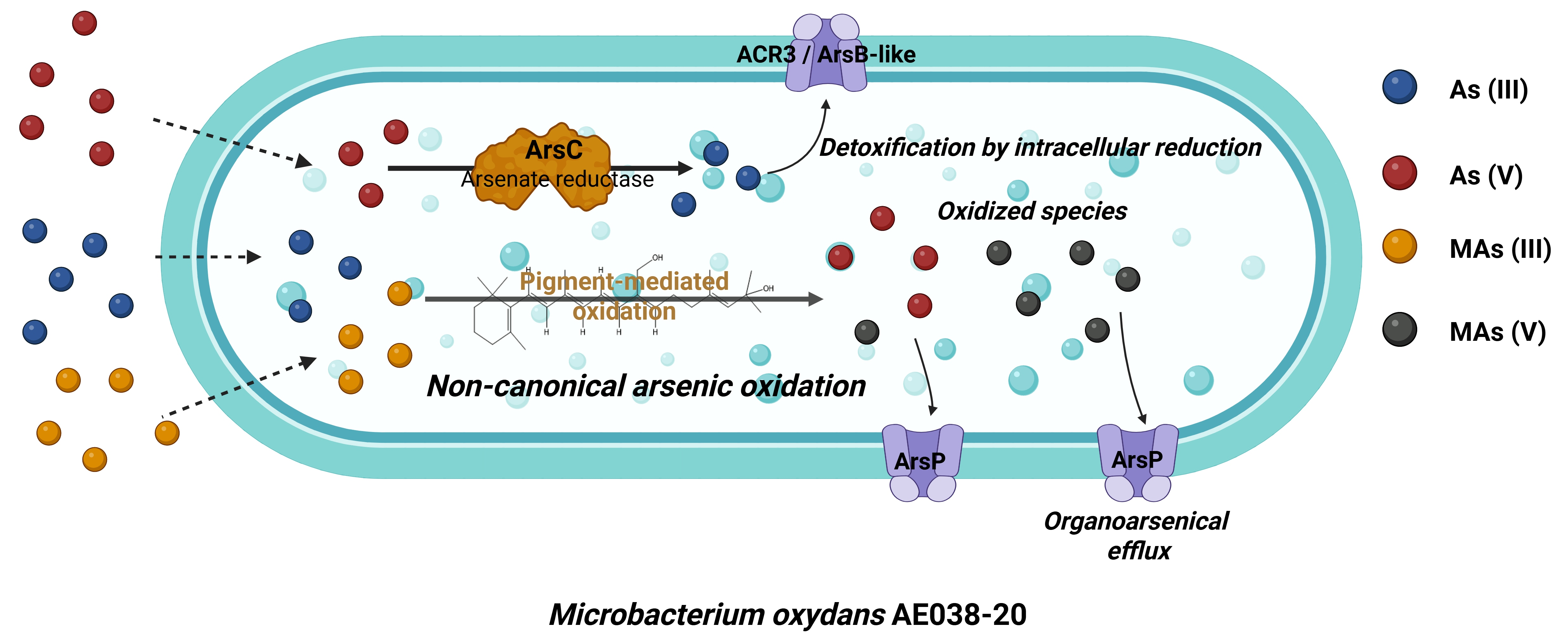
Arsenic-contaminated groundwater is a major environmental concern, particularly in northern Argentina. Here, Microbacterium oxydans AE038-20, isolated from arsenic-rich groundwater, was investigated to elucidate its tolerance and transformation capacity. Growth assays showed that the strain tolerates inorganic arsenic [As(III), As(V)] and methylarsenite [MAs(III)] without significant inhibition. Speciation analyses revealed progressive oxidation of As(III) to As(V), reaching near-complete conversion after 10 days. Similarly, MAs(III) was fully oxidized to MAs(V). Genome sequencing identified ars-related determinants, including arsR, arsC, putative arsenite efflux systems, and arsP, supporting detoxification via arsenate reduction and arsenite efflux. Proteomic analyses confirmed the expression of proteins related to arsenic resistance, oxidative stress response, and metal transport. However, no canonical arsenite oxidases were detected at either the genomic or proteomic level. Despite this, M. oxydans AE038-20 exhibited clear arsenic oxidation activity. The detection of pigment-associated proteins and in vitro oxidation assays suggest an alternative mechanism potentially mediated by redox-active pigments. These findings highlight an alternative pathway for arsenic transformation in environmental bacteria.

Graphical Abstract

1. Introduction

Arsenic (As) is a metalloid widely distributed in the Earth’s crust, and its presence in groundwater constitutes one of the most serious environmental and health problems worldwide. It is generally found in trace amounts, primarily in igneous and sedimentary rocks and soils, with an average crustal concentration of 1.5 mg/kg [1]. Its origin can be either natural, associated with the weathering of arsenical minerals and geochemical processes, or anthropogenic, linked to mining, agricultural, and industrial activities [2]. Currently, more than one hundred countries have regions with high concentrations of arsenic in drinking water, affecting millions of people [3]. In Latin America, Argentina is among the most affected countries, especially in the Chaco-Pampean region and northwestern Argentina, where levels frequently exceed the limit recommended by the World Health Organization (10 µg/L) [4,5,6,7,8]. HACRE (endemic regional chronic hydroarsenicism), a pathology caused by chronic ingestion of arsenic in water and food, leads to skin conditions (leukoderma and/or keratosis), skin, lung, bladder, and kidney cancer, among others, developmental disorders, cardiovascular conditions, neurotoxicity, and diabetes [9]. The molecular mechanisms of action of arsenite and arsenate are binding to sulfhydryl groups and phosphate substitution, respectively, while at the subcellular and cellular levels, the generation of reactive oxygen species (ROS) and the generation of reactive nitrogen species (RNS) are the most studied mechanisms of toxic action [10]. This highlights the urgent need to develop efficient and sustainable strategies for their removal.
In the environment, arsenic is found mainly in its inorganic forms arsenite [As(III)] and arsenate [As(V)], whose transformations are strongly influenced by physicochemical conditions and microbial activity. Microorganisms have developed diverse strategies to cope with environmental arsenic, including transport and biotransformation such as redox and methylation cycles [11]. These processes not only allow bacterial survival in contaminated environments but also form the basis of biotechnological bioremediation strategies [12].
In this context, the genus Microbacterium, belonging to the phylum Actinobacteria, has demonstrated remarkable metabolic versatility and adaptability to adverse environmental conditions [13]. Several strains have been reported as tolerant to heavy metals, with the potential to participate in redox transformation processes and contaminant removal. In particular, Microbacterium oxydans AE038-20, isolated from water wells in the town of Los Pereyra (Tucumán, Argentina), exhibits a remarkable capacity for the biotransformation of inorganic and organic arsenical species, as well as efficient biofilm formation. This strain demonstrated resistance to high concentrations of As(III) and As(V) in liquid medium, as well as tolerance to Cu(II), Cr(VI), and Cd(II) [14].
Within this framework, the objective of this work was to analyze the genomic basis and proteomic profile of Microbacterium oxydans AE038-20 in order to understand the molecular mechanisms involved in its tolerance and transformation capacity in the presence of various arsenic species.

2. Materials and Methods

2.1. Microorganism and Culture Conditions

Microbacterium oxydans AE038-20 (GenBank accession number KX369591) was obtained from the strain collection of the Planta Piloto de Procesos Industriales Microbiológicos (PROIMI-CONICET, Argentina). This strain was originally isolated from groundwater wells containing elevated arsenic concentrations (2 mg/L), located in Los Pereyra, Cruz Alta, Tucumán Province, Argentina (26°56′47″ S, 64°52′55″ W; 380 m a.s.l.), as previously reported by Maizel et al., 2018 [14]. M. oxydans AE038-20 was routinely cultured in Luria–Bertani (LB) medium composed of NaCl (10 g/L), yeast extract (5 g/L), and meat peptone (10 g/L), under constant agitation (180 rpm) at 30 °C. For long-term preservation, bacterial cells were stored at −20 °C in cryovials containing glycerol as a cryoprotective agent.

2.2. Growth and Arsenic Tolerance Assays

To evaluate growth performance and the biotransformation capacity of M. oxydans AE038-20 toward inorganic and organic arsenic species, batch cultures were carried out in Luria–Bertani (LB) medium supplemented or not with arsenic compounds at a final concentration of 3 µM. Erlenmeyer flasks containing the culture medium were inoculated with M. oxydans AE038-20 and incubated at 30 °C under constant agitation (180 rpm).
For inorganic arsenic assays, cultures were supplemented with arsenite [As(III)] and incubated for up to 10 days. Bacterial growth was monitored by measuring the optical density at 600 nm (OD600) using a UV–visible spectrophotometer (Beckman DU® 7400, Brea, CA, USA), and growth curves were constructed from the obtained data.
For organic arsenic assays, cultures were supplemented with methylarsenite [MAs(III)] at 3 µM. MAs(III) was chemically synthesized according to Stice et al. [15]. Growth experiments were conducted for 24 h, whereas oxidation assays were extended up to 10 days. During biotransformation experiments, samples were collected at 24 h intervals to determine OD600 values and generate growth curves. Non-inoculated LB medium supplemented with MAs(III) was used as a control.

2.3. Arsenic Speciation and Intracellular Arsenic Determination

For arsenic speciation analyses, 0.5 mL aliquots of culture samples were collected and centrifuged at 16,200× g for 5 min at 4 °C. The resulting supernatants were filtered through 3 kDa ultrafiltration membranes (Amicon®, MilliporeSigma, Burlington, MA, USA) and analyzed for soluble arsenic species by high-performance liquid chromatography coupled to inductively coupled plasma mass spectrometry (HPLC–ICP-MS).
Arsenic species were separated using a reverse-phase C18 column (BioBasic-18, 250 mm × 4.6 mm, 5 µm; Thermo Fisher Scientific, Waltham, MA, USA) under isocratic elution with a mobile phase consisting of 3 mM malonic acid and 5% (v/v) methanol (pH 5.6 adjusted with tetrabutylammonium hydroxide) at a flow rate of 1.0 mL/min. Arsenic species were identified based on the retention times of As(III) and As(V) standards.
For intracellular arsenic determination in MAs(III)-exposed cultures, bacterial cells were harvested by centrifugation and disrupted using liquid nitrogen. Subsequently, 1 mL of 20 mM ammonium phosphate buffer (pH 5.6) was added, and arsenic species were extracted by ultrasonication for 15 min. Samples were then centrifuged at 6200× g for 5 min, and the supernatants were analyzed by HPLC–ICP-MS as described above.

2.4. Genomic Sequencing and Bioinformatic Analysis

Genomic DNA from M. oxydans AE038-20 was extracted using a cetyltrimethylammonium bromide (CTAB)-based method, as previously described by Ellis et al. [16]. Whole-genome sequencing was performed by MR DNA (Molecular Research LP, Shallowater, TX, USA).
The quality of raw sequencing reads was assessed using FASTQC v0.11.3, and low-quality bases (Q < 28) were removed. Genome assembly was carried out using SPAdes v3.0 [17]. Gene prediction and functional annotation were performed using the RAST v2.0 platform (Rapid Annotation using Subsystem Technology). The annotated genome was analyzed to identify genes potentially involved in arsenic resistance and metabolism.
Sequencing data have been deposited in the NCBI Sequence Read Archive (SRA) under BioProject PRJNA1445915 (BioSample SAMN56804610, SRA accession SRR37871623). Genome quality was further evaluated using CheckM v1.2.5 [18] to estimate completeness and contamination based on lineage-specific marker genes.

2.5. Proteomic Analysis

For proteomic studies, M. oxydans AE038-20 was cultivated in LB medium without supplementation or supplemented with NaAsO2, Na3AsO4, or methylarsenite [MAs(III)] at a final concentration of 3 µM. Cultures were incubated at 30 °C with agitation (180 rpm) for 24 h. Cells were harvested by centrifugation and washed twice with physiological saline solution. Bacterial cells were then disrupted using liquid nitrogen, containing cytoplasmic and membrane proteins. Membrane proteins were solubilized in 20 mM potassium phosphate buffer (pH 6.4) containing 1% (w/v) n-dodecyl-β-d-maltoside (DDM), following an adapted protocol described by Everberg et al. [19].
Total protein concentration was determined using the Lowry method [20], with bovine serum albumin as the standard. Protein profiles were analyzed by sodium dodecyl sulfate–polyacrylamide gel electrophoresis (SDS–PAGE) followed by colloidal Coomassie Blue staining.
Proteomic analyses were performed at the Centro de Estudios Químicos y Biológicos por Espectrometría de Masas (CEQUIBIEM, Facultad de Ciencias Exactas y Naturales, Universidad de Buenos Aires). Protein samples were reduced with dithiothreitol, alkylated with iodoacetamide, and digested with trypsin. Peptides were desalted using C18 ZipTips and analyzed by nanoLC–MS/MS using a Q Exactive Orbitrap mass spectrometer (Thermo Fisher Scientific, Waltham, MA, USA) equipped with an EASY-SPRAY electrospray ionization source (Thermo Fisher Scientific, Waltham, MA, USA). Protein identification and data analysis were conducted through Proteome Discoverer v. 2.2 software (Thermo Fisher Scientific, Waltham, MA, USA) using translated protein sequences from the M. oxydans genome as the database. The proteomic approach was intended to provide a qualitative and semi-quantitative overview of the proteins detected at a single sampling time (24 h), rather than an exhaustive quantitative comparison among treatments. Accordingly, the dataset was used primarily for descriptive and exploratory purposes.
Proteomic data supporting the findings of this study are provided as Supplementary Material. Supplementary Tables S1–S4 contain the complete lists of proteins identified under basal conditions and after exposure to As(III), As(V), and MAs(III), including emPAI-based semi-quantitative values. For heatmap construction, emPAI values obtained for each detected protein under each condition were log10transformed prior to visualization. Proteins not detected under a given condition were treated as missing values and displayed as blank cells. Such non-detection was not interpreted as protein absence, since detectability may be affected by factors such as sample complexity, protein abundance, and the analytical depth of the method.

2.6. Pigment Extraction and Purification

Pigments were extracted following Meddeb-Mouelhi et al. [21] with minor modifications. M. oxydans AE038-20 cells were grown in 200 mL Luria-Bertani (LB) medium (30 °C, 180–200 rpm) for 24 h, harvested by centrifugation (8000 rpm, 10 min, 4 °C), washed twice with physiological saline solution, and stored at 4 °C until use. Wet biomass was weighed and mixed with methanol (10 mL per g wet cells). Cell disruption was performed by vortex for 20 min, followed by orbital shaking (200 rpm, 20 °C) for 30 min. The mixture was centrifuged to remove the decolorized cell pellet, and the organic phase was recovered by centrifugation (14,000 rpm, 15 min, 4 °C). The crude pigment extract was concentrated under vacuum (Savant Universal Vacuum Systems, UVS400A, Thermo Fisher Scientific, Waltham, MA, USA) until solvent removal.

2.7. Pigment Redox Assay Toward Inorganic Arsenic

To evaluate pigment redox activity toward inorganic arsenic, pigments were resuspended in 2 mL bidistilled water and incubated with 0.004 mg NaAsO2 (20 °C, 24 h, 180 rpm). After incubation, As(III) and As(V) species were determined as described above by high-performance liquid chromatography coupled to inductively coupled plasma mass spectrometry (HPLC–ICP–MS) at the Mass Spectrometry Laboratory, Faculty of Chemistry, Biochemistry and Pharmacy, National University of San Luis, San Luis, Argentina.

2.8. Statistical Analysis

Results are presented as mean ± standard deviation, with at least three replicates of each experiment. Statistical analyses were performed using the relevant parametric tests based on experimental design. Student’s t-tests or one-way analysis of variance (ANOVA) was used to compare control and arsenic-treated cultures, if appropriate. Differences were considered statistically significant at p < 0.05.
Genomic and proteomic analyses were primarily qualitative and descriptive; therefore, no statistical inference was applied to these datasets.

3. Results and Discussion

3.1. Growth Response and Arsenic Biotransformation by M. oxydans AE038-20

3.1.1. Growth in the Presence of Inorganic Arsenic Species

The growth of M. oxydans AE038-20 was evaluated in LB medium supplemented with 3 µM As(III), As(V), or MAs(III), and compared with non-supplemented controls. During the early exponential phase, no major differences in growth kinetics were observed between arsenic-exposed and control cultures (Figure 1). However, from approximately 10 h onwards, control cultures consistently reached slightly higher OD600 values, suggesting a moderate inhibitory effect of arsenic on biomass accumulation. This trend resulted in a modest reduction in final OD600 in arsenic-supplemented cultures compared to the control. Overall, these results indicate that M. oxydans AE038-20 is able to grow in the presence of both inorganic and organic arsenic species, although a slight impact on late-stage growth cannot be excluded under the conditions tested.

3.1.2. Oxidation of As(III)

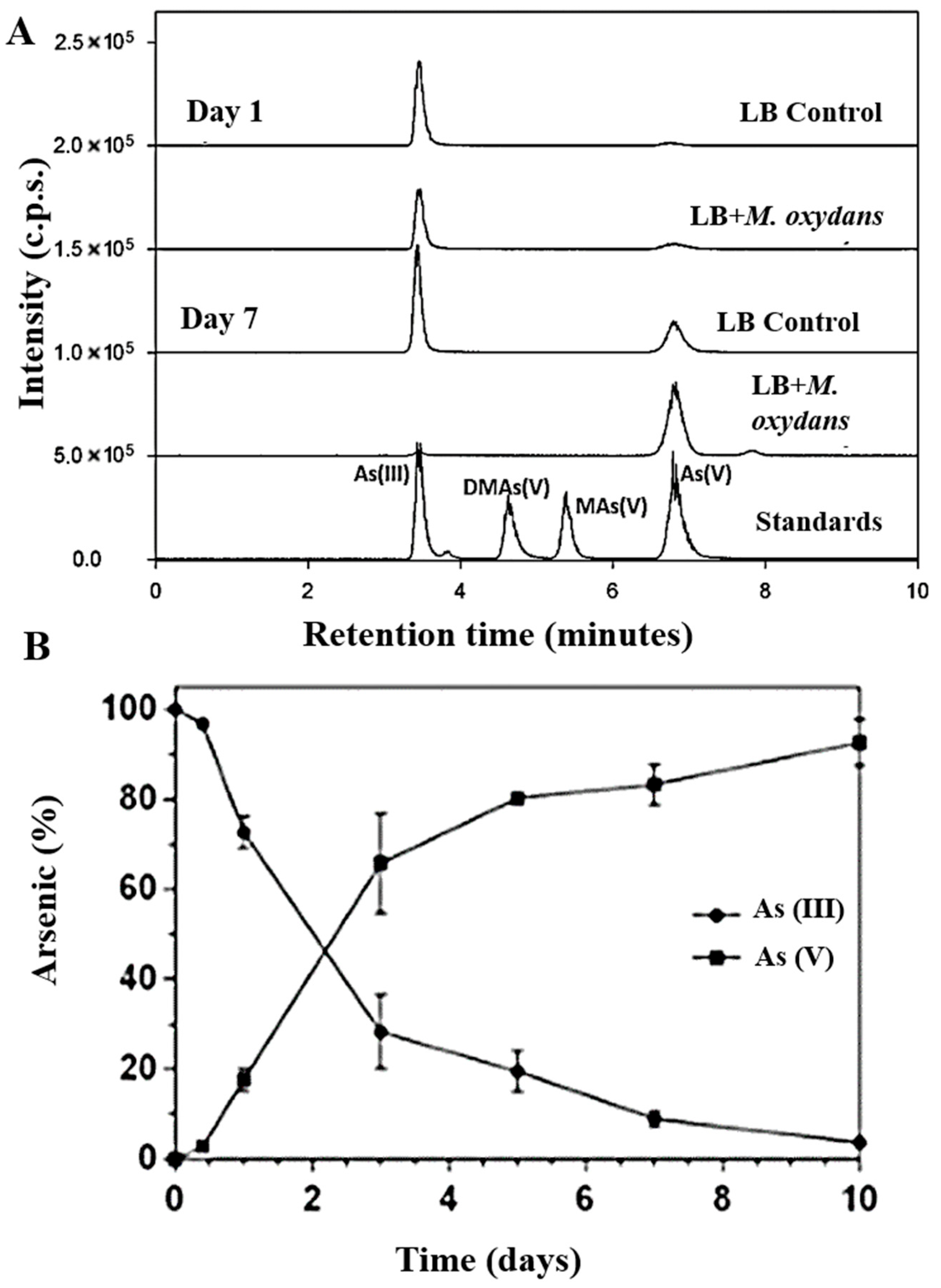
Arsenic speciation analysis by HPLC–ICP-MS revealed that M. oxydans AE038-20 promoted the oxidation of As(III) to As(V) in LB medium (Figure 2A). At day 1, chromatograms showed a single peak corresponding to As(III). By day 7, a marked decrease in the As(III) signal was observed together with the appearance of a new peak consistent with As(V). No significant changes were detected in abiotic controls, confirming that the transformation was biologically mediated.
Time-course analysis (Figure 2B) demonstrated a progressive decline in the relative proportion of As(III), reaching near-complete oxidation by day 10, while As(V) increased proportionally to account for nearly 100% of total arsenic. Notably, oxidation occurred gradually over extended incubation, particularly during late exponential and stationary phases, suggesting that the process may be associated with metabolic shifts occurring during prolonged growth [22,23].

3.1.3. Growth and Oxidation of MAs(III)

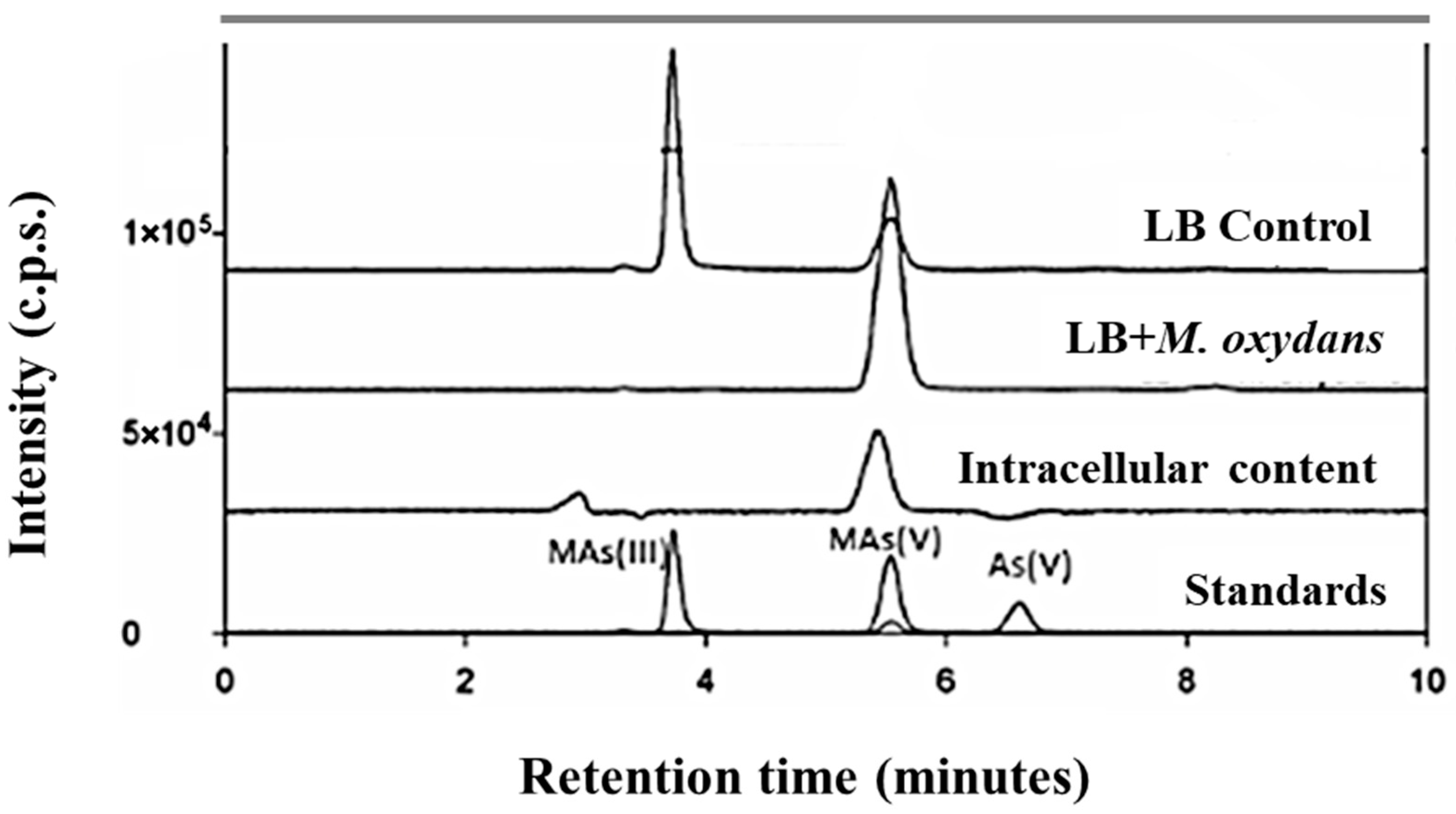
When cultured in LB supplemented with 3 µM MAs(III) HPLC–ICP-MS analysis of culture supernatants showed progressive oxidation of MAs(III) to MAs(V), reaching complete conversion after 7 days of incubation (Figure 3). Abiotic controls did not show detectable transformation, confirming the biological origin of the oxidation process. Intracellular arsenic speciation analysis following cell disruption revealed the presence of MAs(V) within the cellular fraction (Figure 3), indicating that oxidation is not exclusively extracellular and suggesting either intracellular transformation or subsequent uptake of oxidized species.

3.2. Genomic Insights into Arsenic Resistance in M. oxydans AE038-20

To investigate the molecular basis underlying the observed arsenic tolerance and oxidative transformation capacity of M. oxydans AE038-20, whole-genome sequencing and functional annotation were performed. Genome analysis aimed to identify genes potentially involved in arsenic resistance, transport, and redox transformation processes that could account for the phenotypic traits described above [24].
Comparative analysis of the 30 publicly available M. oxydans genomes showed that AE038-20 falls within the overall genomic range described for the species (Supplementary Table S5). The draft genome of AE038-20 (3.62 Mb; 3472 protein-coding genes; 3525 total genes; 69.6% GC) was smaller and encoded fewer genes than the average of the comparison set, but remained within the observed ranges for genome size (3.47–4.55 Mb), coding capacity (2889–3966 protein-coding genes), and total gene content (3383–4745 genes). In contrast, its GC content was located toward the upper end of the distribution, although still within the interval observed among the available M. oxydans genomes (68.0–70.0% GC). In addition, a genome quality assessment using CheckM indicated a completeness of 98.99% with 0% contamination, supporting that the assembled genome represents a near-complete and high-quality draft. Overall, these results indicate that AE038-20 is genomically consistent with the diversity currently represented within publicly available M. oxydans genomes.
Genome annotation revealed the presence of ars-related determinants, including arsR-like regulators, arsC-like genes, and genes encoding putative arsenite efflux systems (Table 1). Notably, an acr3-like transporter and an additional arsenic efflux pump-related protein were identified. These systems are commonly associated with the extrusion of As(III), supporting a detoxification strategy primarily based on cytoplasmic arsenite efflux. Additionally, the genome annotation indicated the presence of arsP, encoding a permease associated with resistance to organoarsenicals such as methylarsenite [MAs(III)]. Overall, this genetic repertoire is consistent with the high tolerance of M. oxydans AE038-20 to both inorganic and methylated arsenic species observed experimentally.
The gene arsC, encoding an arsenate reductase, was also detected, indicating the capacity to reduce As(V) to As(III), thereby coupling reduction with subsequent extrusion via efflux pumps. Regulation of the operon is likely mediated by ArsR, which responds to intracellular As(III). On the other hand, no genes encoding known arsenite oxidases (e.g., aioAB) or characterized methylarsenite oxidizing enzymes were identified in the genome. This absence is particularly noteworthy given that experimental assays demonstrated the transformation of reduced arsenic species. The lack of canonical redox genes suggests that arsenic biotransformation in M. oxydans AE038-20 may rely on alternative, yet-undescribed mechanisms.
Notably, comparative analysis of the publicly available M. oxydans genomes showed that arsenite oxidase genes (aioA/aioB) were not detected in any of the strains analyzed, in agreement with the result obtained for AE038-20 (Supplementary Table S5). This was further supported by the HMM-based screening of MicroTrait bioelement cycling families, which also failed to recover aioAB-related hits. In contrast, the number of arsC-related genes varied among the available genomes, suggesting greater diversity in the reductive detoxification repertoire than in oxidative pathways within the species. Together, these observations indicate that the absence of an aioAB-based arsenite oxidation system is not unique to M. oxydans AE038-20 and reinforce the view that the oxidative transformation phenotype observed in this strain may depend on alternative mechanisms.

3.3. Proteomic Response of M. oxydans AE038-20 to Arsenic Species

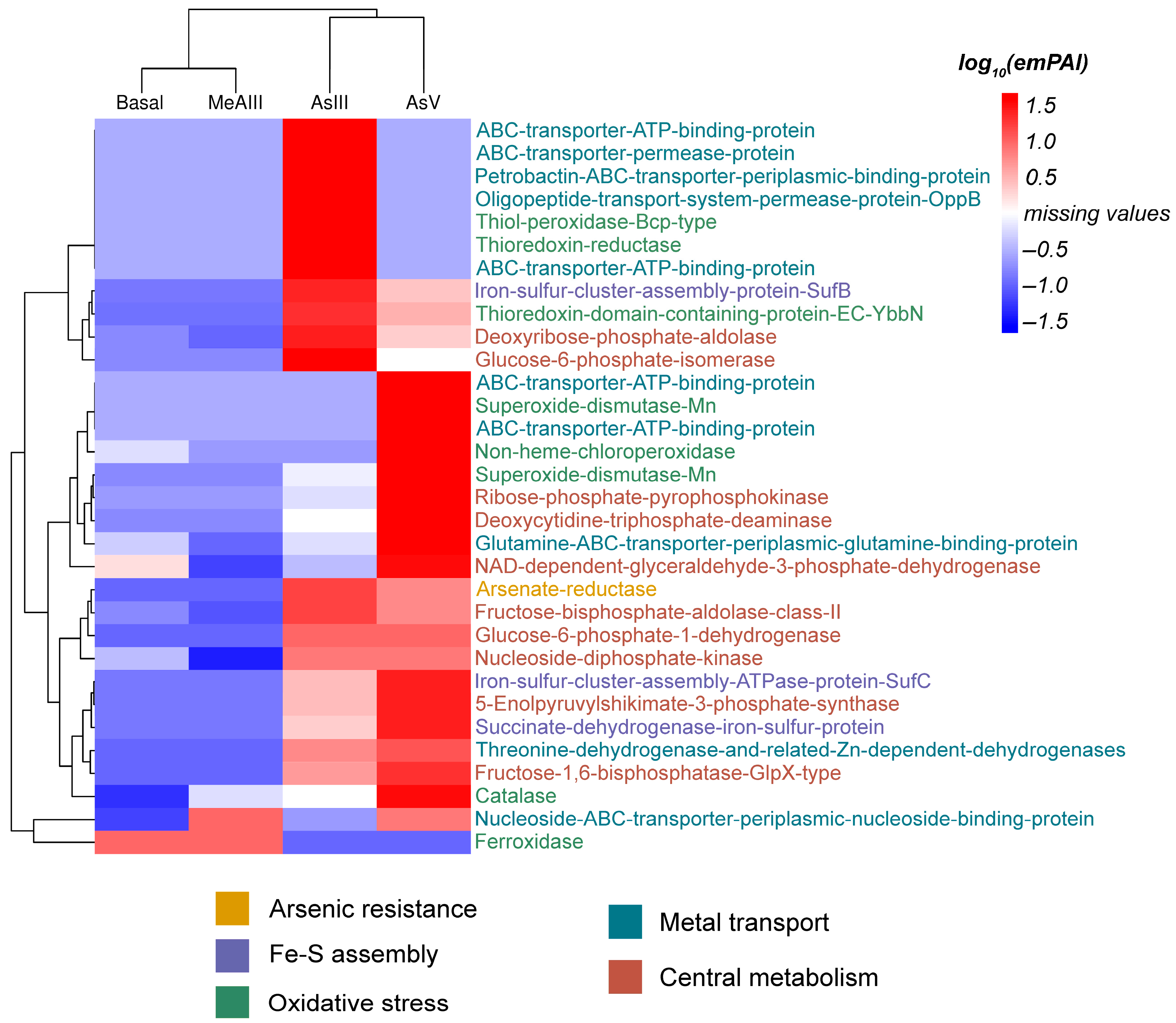
To characterize the molecular response of M. oxydans AE038-20 to different arsenic species, a comparative proteomic analysis was performed under four conditions: control (LB), As(III), As(V), and MAs(III) (Figure 4).
Proteomic heatmap provides an overview of the functional protein repertoire detected under basal conditions and after exposure to different arsenic species. Within the arsenic resistance module, proteins associated with the classical ars detoxification system were consistently detected, including the transcriptional regulator ArsR, the arsenate reductase ArsC, and membrane transport systems related to ACR3/ArsB-like arsenite efflux. The detection of ArsC supports the intracellular reduction of arsenate [As(V)] to arsenite [As(III)], a key step in the canonical bacterial detoxification pathway. In turn, the presence of ACR3/ArsB-type transporters is consistent with the active extrusion of arsenite from the cytoplasm, representing the primary route of arsenic detoxification in this strain. Proteins related to organoarsenical resistance, such as ArsP, were also detected, suggesting the capability to cope with methylated arsenic species such as MAs(III). Beyond the core ars system, the heatmap also reveals the presence of multiple proteins involved in oxidative stress response, including catalases, superoxide dismutases, and thiol-dependent peroxidases. These enzymes are commonly associated with arsenic exposure due to the oxidative imbalance generated during arsenic detoxification and redox cycling. Finally, several proteins associated with central metabolism and energy generation were consistently identified across conditions, reflecting the maintenance of basal metabolic activity during arsenic exposure. Together, the qualitative proteomic patterns suggest that arsenic handling in M. oxydans AE038-20 involves a coordinated response combining the canonical ars detoxification pathway with broader stress-response mechanisms and metabolic adaptation.

3.3.1. Basal Proteome

Under control conditions, the proteome reflected an actively growing and metabolically robust cell. Central carbon metabolism was well represented, including glycolytic enzymes (glyceraldehyde-3-phosphate dehydrogenase, enolase, fructose-bisphosphate aldolase), pentose phosphate pathway enzymes (transaldolase), and tricarboxylic acid cycle components (citrate synthase, fumarase, succinate dehydrogenase). Multiple ATP synthase subunits were detected, supporting efficient oxidative phosphorylation.
Proteins involved in redox homeostasis were also present under basal conditions, including superoxide dismutase (SOD), catalase, NADPH:quinone oxidoreductase, and Dps protein (involved in iron storage and ferroxidase activity). The constitutive expression of these systems suggests a preparedness to counteract oxidative stress derived from respiratory metabolism.
Chaperones and ATP-dependent proteases (DnaK, GroEL, ClpC) were consistently detected, indicating active proteostasis. Membrane-associated transporters, including ABC systems and OppA, revealed a nutrient-scavenging strategy typical of heterotrophic growth in LB medium.
Finally, enzymes with distinct bacterial metabolism functions were identified, such as catechol 2,3-dioxygenase (involved in the degradation of aromatic compounds) and a non-heme chloroperoxidase, indicating metabolic versatility that could be linked to adaptation to complex environmental niches and the production of extracellular biofilm components.
When combined, the baseline proteomic state of M. oxydans AE038-20 indicates a physiologically active cell with a strong energy metabolism, a competent antioxidant defense system, effective protein regulatory mechanisms, and a diversified capability for food absorption and utilization. This basal profile serves as a basis for differential responses to arsenical species, allowing for the differentiation between generic response elements and the unique adaptations generated by each chemical form of arsenic.

3.3.2. Response to As(III)

Exposure to As(III) triggered a response strongly oriented toward redox management and detoxification. ArsC (arsenate reductase) was detected under these conditions, indicating activation of the canonical ars system. Although As(III) was supplied externally, the presence of ArsC suggests intracellular interconversion and dynamic handling of arsenic species. Moreover, a marked reinforcement of antioxidant defenses was observed, including increased catalase, SOD, and peroxiredoxin/thiol-peroxidase systems, together with ferredoxin–NADP+ reductase. This profile is consistent with the known ability of As(III) to promote ROS formation and interact with thiol groups [25].
Proteins involved in Fe–S cluster biogenesis and repair (SufC, SufD) were also induced, supporting the notion that arsenite disrupts Fe–S-containing enzymes. Additionally, polyphosphate kinase (PPK) was detected exclusively under arsenic exposure, suggesting a potential role for polyphosphate metabolism in arsenic sequestration or phosphate balance.
Notably, no dedicated arsenite oxidase was detected, despite the clear oxidation of As(III) to As(V) observed at the physiological level in experimental tests. This discrepancy suggests that arsenite oxidation may not rely on a canonical enzymatic system.

3.3.3. Response to As(V)

The proteomic response to As(V) shared core features with As(III), including ArsC detection and activation of Fe–S repair systems (SufB, SufD, SufS). However, As(V) induced additional adjustments linked to phosphate homeostasis.
PhoH was specifically detected under As(V), indicating activation of phosphate starvation-related pathways, likely due to structural competition between arsenate and phosphate. This distinguishes the As(V) response from the primarily redox-centered As(III) profile. Metabolic reprogramming was also more pronounced, with differential expression of enzymes involved in nucleotide and amino acid biosynthesis (adenylosuccinate synthase, IMP dehydrogenase, ornithine carbamoyltransferase), suggesting broader metabolic adaptation. As in the As(III) condition, no canonical arsenite oxidase was identified.

3.3.4. Response to MAs(III)

MAs(III) exposure resulted in a broader and less specific detoxification profile compared to inorganic species. In addition to antioxidant enzymes, several P-type ATPases involved in heavy metal efflux were induced. The activation of these pumps, which are often linked with heavy metal expulsion, shows that MAs(III) causes a broad efflux response to toxins, rather than a specialized mechanism for arsenic removal. This pattern distinguishes the MAs (III) condition from the inorganic ones, which were mostly concerned with ArsC and systems involved in phosphate or Fe-S metabolism.
Proteins associated with intracellular survival and xenobiotic tolerance, including an Eis-like protein (Enhanced intracellular survival), were detected exclusively in this condition. Enhanced expression of aminopeptidases and peptide transporters indicated increased protein turnover, consistent with elevated cellular damage. This pattern demonstrates that, whereas As (III) and As (V) elicit responses more focused on specific detoxification and phosphate or Fe-S homeostasis, MAs (III) promotes a broader and non-specific response based on the activation of general metal tolerance mechanisms, xenobiotic detoxification, and intracellular resilience. Finally, no proteins previously described as canonical MAs(III) oxidases were detected under the tested conditions.

3.4. Integrated Genomic–Proteomic Analysis

The combined analysis of genomic and proteomic data provides a more comprehensive understanding of the adaptive response of M. oxydans AE038-20 to arsenic exposure. Genomic identification of the ars operon, including arsR, arsC, arsB, and acr3, is consistent with the proteomic detection of proteins involved in detoxification, oxidative stress response, and membrane transport systems, supporting an integrated resistance strategy based on arsenate reduction and arsenite efflux. Moreover, the presence of proteins associated with redox balance and stress mitigation reinforces the physiological observations of sustained growth under arsenic exposure.
In the present study, this combined approach was mainly aimed at elucidating the arsenic biotransformation phenotype observed in M. oxydans AE038-20, rather than at addressing in detail the differential gene expression and regulatory mechanisms governing such responses. Although transcriptomic analysis was beyond the scope of this work, future gene expression studies under arsenic exposure will be valuable to clarify the regulatory basis of the physiological and proteomic patterns identified here.
However, this integrative approach also highlights a key discrepancy: despite clear experimental evidence of arsenite and methylarsenite oxidation, neither genomic nor proteomic analyses revealed enzymes typically associated with these processes. This suggests that arsenic transformation in this strain may involve alternative biochemical pathways not directly encoded by canonical genes or not detectable under the experimental conditions used. Together, these findings underscore the importance of combining multi-omics approaches to uncover both expected and unconventional mechanisms of microbial adaptation to toxic environments.

3.5. Preliminary Pigment Assays

Based on the characteristic yellow coloration observed in colonies grown in the presence of arsenic and light, pigments produced by M. oxydans AE038-20 were extracted and characterized to evaluate their potential contribution to arsenic redox transformation.
Incubation of the pigment preparation with As(III) resulted in the formation of detectable As(V) after 24 h as determined by HPLC–ICP-MS (702.62 mg/L of As3+ and 56.24 mg/L of As5+ were recorded), demonstrating that the pigment preparation has the capacity to oxidize As(III) to As(V) under the tested conditions. In the context of the genome analysis, where no canonical arsenite oxidase genes (e.g., aioAB) were identified, these results support a pigment-mediated contribution to the oxidative transformation observed in whole-cell assays.

3.6. Integrated Model of Arsenic Detoxification and Transformation

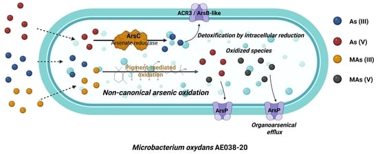
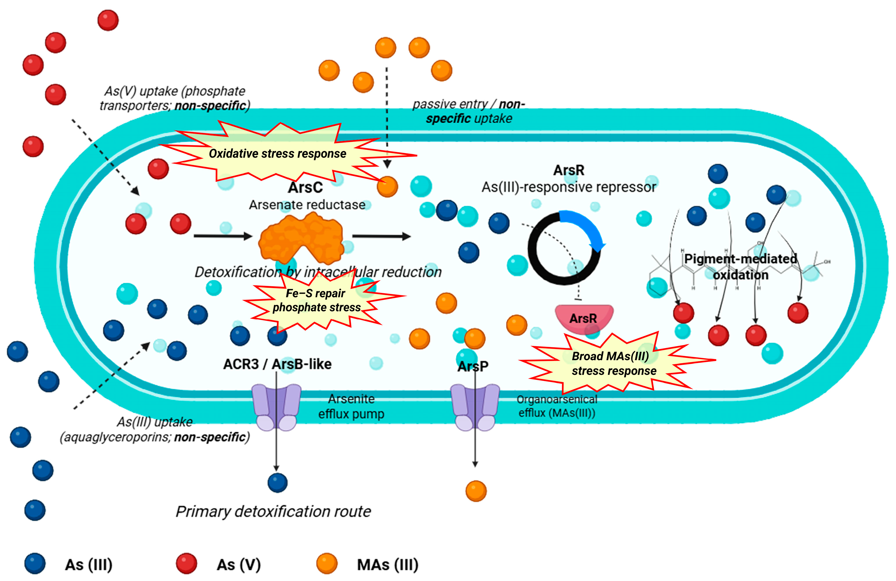
Taken together, the physiological, genomic, proteomic, and pigment redox analyses support an integrated model of arsenic detoxification and transformation in M. oxydans AE038-20 (Figure 5). Genome annotation revealed the presence of a classical ars resistance module including arsR, arsC, and membrane transporters related to ACR3/ArsB-like arsenite efflux, which are widely recognized as the core components of bacterial arsenic detoxification systems. Consistent with this genetic potential, proteomic analyses confirmed the detection of proteins associated with these functions, supporting the physiological relevance of this pathway under the tested conditions.
Within this framework, arsenate [As(V)] likely enters the cell through non-specific phosphate transport systems, a well-known route resulting from the structural similarity between arsenate and phosphate. Once inside the cytoplasm, ArsC-mediated reduction converts As(V) into arsenite [As(III)], which represents the central detoxification step of the ars system. Because arsenite is more mobile and highly toxic, its intracellular accumulation is prevented by active efflux through ACR3/ArsB-type transporters, effectively exporting the reduced species back into the extracellular environment. In addition to arsenate-derived arsenite, As(III) present in the environment may also enter the cell through aquaglyceroporins or other non-specific channels, further reinforcing the importance of the arsenite efflux systems as the primary detoxification route. The detection of ArsP suggests that the strain also possesses the capacity to cope with organoarsenical compounds, providing a specialized efflux mechanism for methylated species such as MAs(III).
In addition to the core detoxification machinery, the proteomic profile also revealed associated stress-response processes that likely contribute to arsenic adaptation in this strain. These included proteins related to oxidative stress mitigation, Fe–S cluster repair, phosphate-stress response, and a broader stress response under MAs(III) exposure (Figure 4). Although these functions do not constitute the central detoxification route itself, their detection suggests that arsenic exposure triggers a wider physiological adjustment that may help sustain cellular homeostasis under metalloid stress.
Beyond this canonical intracellular detoxification cycle, experimental observations from pigment extraction and redox assays indicate the presence of pigment-associated redox activity capable of promoting arsenite oxidation. Importantly, genomic analysis did not reveal arsenite oxidase genes (e.g., aioA/aioB), suggesting that the observed As(III) oxidation may occur through non-enzymatic or indirectly mediated mechanisms, likely associated with extracellular or surface-associated pigments. Such redox-active metabolites could facilitate the oxidation of arsenite to arsenate in the surrounding medium, thereby influencing arsenic speciation outside the cell. This complementary process provides a plausible explanation for the experimentally observed oxidative transformation of arsenic and highlights the potential role of secondary metabolites in arsenic cycling.
Although gene knock-out approaches would provide direct functional validation, such analyses were beyond the scope of the present study. Because the oxidative phenotype was not associated with well-characterized arsenite oxidation genes, no clear target for mutational analysis could be defined at this stage. Therefore, the mechanism proposed here should be considered a working hypothesis that will require future genetic or biochemical validation.
Together, these observations support the integrated model of arsenic detoxification and transformation summarized in Figure 5.
Overall, the integrated evidence indicates that M. oxydans AE038-20 combines intracellular detoxification via the ars system with extracellular redox processes that may contribute to arsenic speciation. This dual strategy may favor adaptation in arsenic-impacted environments and may also contribute to arsenic biogeochemical cycling [26]. In this context, the observed oxidation of inorganic and methylated arsenic species in the absence of identifiable oxidases supports the possibility that alternative, possibly metabolite-mediated, processes participate in arsenic transformation, although their molecular basis remains unresolved. Thus, further studies will be required to identify the compounds involved and to determine their specific role under environmentally relevant conditions. Nevertheless, these findings broaden current understanding of microbial arsenic cycling and suggest future opportunities to explore such processes in biotechnological arsenic remediation.

4. Conclusions

This study demonstrates that Microbacterium oxydans AE038-20 exhibited a remarkable tolerance to both inorganic and methylated arsenic species and was capable of oxidizing As(III) and MAs(III) to their corresponding pentavalent forms under the tested conditions. The combined physiological, genomic, and proteomic analyses provide new insights into the adaptive strategies employed by this strain to cope with arsenic stress.
The integration of genomic and proteomic approaches enabled the identification of genes, metabolic pathways, and proteins associated with arsenic resistance, oxidative stress response, and membrane transport systems, offering a comprehensive view of the molecular mechanisms potentially involved in arsenic detoxification. In particular, the presence and expression of components of the ars resistance system support a detoxification strategy primarily based on arsenate reduction and arsenite efflux.
However, despite the clear experimental evidence of arsenite and methylarsenite oxidation, neither genomic nor proteomic analyses revealed canonical enzymes typically associated with these processes. This discrepancy suggests that arsenic oxidation in M. oxydans AE038-20 may involve alternative mechanisms. Preliminary observations indicating that pigment extracts can mediate As(III) oxidation point to a possible role of redox-active pigments in this process. Nevertheless, the precise biochemical mechanisms underlying this phenomenon remain unresolved and require further investigation.
The results presented here expand current knowledge on microbial arsenic transformation and highlight the importance of integrating multi-omics approaches to unravel complex detoxification strategies in environmental microorganisms. These findings also provide a basis for future studies aimed at clarifying the molecular basis of alternative arsenic transformation processes and exploring their potential biotechnological applications in arsenic remediation.

Supplementary Materials

The following supporting information can be downloaded at: https://www.mdpi.com/article/10.3390/pr14091395/s1. Table S1. Proteins identified in Microbacterium oxydans AE038-20 under basal conditions (LB medium), including emPAI-based semi-quantitative values. Table S2. Proteins identified in Microbacterium oxydans AE038-20 after exposure to As(III), including emPAI-based semi-quantitative values. Table S3. Proteins identified in Microbacterium oxydans AE038-20 after exposure to As(V), including emPAI-based semi-quantitative values. Table S4. Proteins identified in Microbacterium oxydans AE038-20 after exposure to MAs(III), including emPAI-based semi-quantitative values. Table S5. Comparative genomic features of publicly available Microbacterium oxydans genomes, including assembly quality metrics, accession data, FastANI analysis, and MicroTrait HMM-based screening.

Author Contributions

Conceptualization, A.H.M., M.A.F. and C.M.R.; Methodology, F.C.S. and A.E.G.; Software, J.S.H. and J.H.P.; Validation, A.H.M.; Formal analysis, F.C.S., J.S.H. and J.H.P.; Investigation, F.C.S. and A.E.G.; Resources, C.M.R.; Data curation, A.H.M., J.S.H., J.H.P. and A.E.G.; Writing—original draft, F.C.S., A.H.M. and J.S.H.; Writing—review and editing, J.H.P., M.A.F. and C.M.R.; Visualization, F.C.S., A.H.M. and J.S.H.; Supervision, M.A.F. and C.M.R.; Project administration, C.M.R.; Funding acquisition, C.M.R. All authors have read and agreed to the published version of the manuscript.

Funding

This work was supported by National University of Tucumán, PIUNTD730.

Data Availability Statement

The original contributions presented in this study are included in the article/Supplementary Materials. Further inquiries can be directed to the corresponding author.

Conflicts of Interest

The authors declare no conflicts of interest.

References

  1. Drewniak, L.; Sklodowska, A. Arsenic-transforming microbes and their role in biomining processes. Environ. Sci. Pollut. Res. 2013, 20, 7728–7739. [Google Scholar] [CrossRef]
  2. Herath, I.; Vithanage, M.; Bundschuh, J.; Maity, J.P.; Bhattacharya, P. Natural arsenic in global groundwaters: Distribution and geochemical triggers for mobilization. Curr. Pollut. Rep. 2016, 2, 68–89. [Google Scholar] [CrossRef]
  3. Podgorski, J.; Berg, M. Global threat of arsenic in groundwater. Science 2020, 368, 845–850. [Google Scholar] [CrossRef]
  4. Smedley, P.L.; Kinniburgh, D.G. A review of the source, behaviour and distribution of arsenic in natural waters. Appl. Geochem. 2002, 17, 517–568. [Google Scholar] [CrossRef]
  5. Farías, S.S.; Casa, V.A.; Vázquez, C.; Ferpozzi, L.; Pucci, G.N.; Cohen, I.M. Natural contamination with arsenic and other trace elements in ground waters of Argentine Pampean Plain. Sci. Total Environ. 2003, 309, 187–199. [Google Scholar] [CrossRef] [PubMed]
  6. Bundschuh, J.; Farias, B.; Martin, R.; Storniolo, A.; Bhattacharya, P.; Cortes, J.; Albouy, R. Groundwater arsenic in the Chaco-Pampean plain, Argentina: Case study from Robles County, Santiago del Estero province. Appl. Geochem. 2004, 19, 231–243. [Google Scholar] [CrossRef]
  7. Nicolli, H.B.; Bundschuh, J.; Blanco, M.D.C.; Tujchneider, O.C.; Panarello, H.O.; Dapeña, C.; Rusansky, J.E. Arsenic and associated trace-elements in groundwater from the Chaco-Pampean plain, Argentina: Results from 100 years of research. Sci. Total Environ. 2012, 429, 36–56. [Google Scholar] [CrossRef]
  8. Bardach, A.E.; Ciapponi, A.; Soto, N.; Chaparro, M.R.; Calderon, M.; Briatore, A.; Cadoppi, N.; Tassara, R.; Litter, M.I. Epidemiology of chronic disease related to arsenic in Argentina: A systematic review. Sci. Total Environ. 2015, 538, 802–816. [Google Scholar] [CrossRef]
  9. Lepori, E.C.V. Hidroarsenicismo crónico regional endémico en Argentina. Acta Bioquím. Clín. Latinoam. 2015, 49, 83–104. [Google Scholar] [CrossRef]
  10. Olmos, V.; Ridolfi, A.S. Hidroarsenicismo: Mecanismos de acción asociados a la toxicidad del arsénico. Acta Toxicol. Argent 2018, 26, 32–44. [Google Scholar]
  11. Mathivanan, K.; Chandirika, J.U.; Vinothkanna, A.; Yin, H.; Liu, X.; Meng, D. Bacterial adaptive strategies to cope with metal toxicity in the contaminated environment—A review. Ecotoxicol. Environ. Saf. 2021, 226, 112863. [Google Scholar] [CrossRef]
  12. Mohsin, H.; Shafique, M.; Zaid, M.; Rehman, Y. Microbial biochemical pathways of arsenic biotransformation and their application for bioremediation. Folia Microbiol. 2023, 68, 507–535. [Google Scholar] [CrossRef]
  13. Rodríguez-Castrejón, U.E.; Serafín-Muñoz, A.H.; Álvarez-Vargas, A.; Cruz-Jiménez, G.; Gutiérrez-Ortega, N.L.; Miranda-Avilés, R.; Cano-Canchola, M.C. Assessment of arsenite removal efficiency, resistance, and biotransformation by Microbacterium hydroxycarbonoxydans isolated from contaminated sites. Sci. Rep. 2025, 15, 18494. [Google Scholar] [CrossRef]
  14. Maizel, D.; Balverdi, P.; Rosen, B.; Sales, A.M.; Ferrero, M.A. Arsenic-hypertolerant and arsenic-reducing bacteria isolated from wells in Tucumán, Argentina. Can. J. Microbiol. 2018, 64, 876–886. [Google Scholar] [CrossRef] [PubMed]
  15. Stice, S.; Liu, G.; Matulis, S.; Boise, L.H.; Cai, Y. Determination of multiple human arsenic metabolites employing high performance liquid chromatography inductively coupled plasma mass spectrometry. J. Chromatogr. B 2016, 1009, 55–65. [Google Scholar] [CrossRef]
  16. Ellis, R.J.; Thompson, I.P.; Bailey, M.J. Temporal fluctuations in the pseudomonad population associated with sugar beet leaves. FEMS Microbiol. Ecol. 1999, 28, 345–356. [Google Scholar] [CrossRef]
  17. Bankevich, A.; Nurk, S.; Antipov, D.; Gurevich, A.A.; Dvorkin, M.; Kulikov, A.S.; Lesin, V.M.; Nikolenko, S.I.; Pham, S.; Prjibelski, A.D.; et al. SPAdes: A new genome assembly algorithm and its applications to single-cell sequencing. J. Comput. Biol. 2012, 19, 455–477. [Google Scholar] [CrossRef]
  18. Parks, D.H.; Imelfort, M.; Skennerton, C.T.; Hugenholtz, P.; Tyson, G.W. CheckM: Assessing the quality of microbial genomes recovered from isolates, single cells, and metagenomes. Genome Res. 2015, 25, 1043–1055. [Google Scholar] [CrossRef]
  19. Everberg, H.; Sivars, U.; Emanuelsson, C.; Persson, C.; Englund, A.K.; Haneskog, L.; Lipniunas, P.; Jörntén-Karlsson, M.; Tjerneld, F. Protein pre-fractionation in detergent–polymer aqueous two-phase systems for facilitated proteomic studies of membrane proteins. J. Chromatogr. A 2004, 1029, 113–124. [Google Scholar] [CrossRef] [PubMed]
  20. Lowry, O.H.; Rosebrough, N.J.; Farr, A.L.; Randall, R.J. Protein measurement with the Folin phenol reagent. J. Biol. Chem. 1951, 193, 265–275. [Google Scholar] [CrossRef]
  21. Meddeb-Mouelhi, F.; Moisan, J.K.; Bergeron, J.; Daoust, B.; Beauregard, M. Structural characterization of a novel antioxidant pigment produced by a photochromogenic Microbacterium oxydans strain. Appl. Biochem. Biotechnol. 2016, 180, 1286–1300. [Google Scholar] [CrossRef]
  22. Lescure, T.; Moreau, J.; Charles, C.; Ben Ali Saanda, T.; Thouin, H.; Pillas, N.; Bauda, P.; Lamy, I.; Battaglia-Brunet, F. Influence of organic matters on AsIII oxidation by the microflora of polluted soils. Environ. Geochem. Health 2016, 38, 911–925. [Google Scholar] [CrossRef] [PubMed]
  23. Bermanec, V.; Paradžik, T.; Kazazić, S.P.; Venter, C.; Hrenović, J.; Vujaklija, D.; Boev, B. Novel arsenic hyper-resistant bacteria from an extreme environment, Crven Dol mine, Allchar, North Macedonia. J. Hazard. Mater. 2021, 402, 123437. [Google Scholar] [CrossRef] [PubMed]
  24. Jain, C.; Rodriguez-R, L.M.; Phillippy, A.M.; Konstantinidis, K.T.; Aluru, S. High throughput ANI analysis of 90K prokaryotic genomes reveals clear species boundaries. Nat. Commun. 2018, 9, 5114. [Google Scholar] [CrossRef] [PubMed]
  25. Larson, J.D. Investigating the Metalloproteome of Bacteria and Archaea. Ph.D. Thesis, Montana State University, Bozeman, MT, USA, 2024. [Google Scholar]
  26. Zhu, Y.G.; Xue, X.M.; Kappler, A.; Rosen, B.P.; Meharg, A.A. Linking genes to microbial biogeochemical cycling: Lessons from arsenic. Environ. Sci. Technol. 2017, 51, 7326–7339. [Google Scholar] [CrossRef]
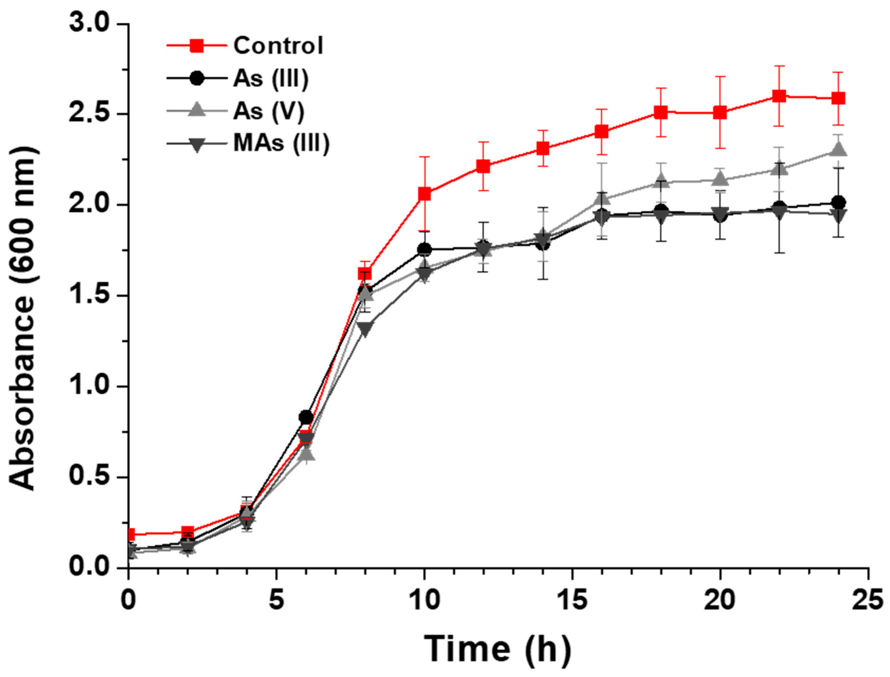
Figure 1. Growth of M. oxydans AE038-20 in LB medium without supplementation and supplemented with As(III), As(V) and MAs(III). All determinations were performed in triplicate. Error bars represent standard deviation (SD).
Processes 14 01395 g001
Figure 2. (A) Representative chromatogram illustrating the oxidation of As(III) to As(V) in LB medium by M. oxydans AE038-20 at 1 and 7 days of incubation. Retention times of arsenic standards are indicated. (B) Time-course profile of As(III) oxidation to As(V) over 10 days. Values represent the relative percentage of each arsenic species over time.
Processes 14 01395 g002
Figure 3. Chromatogram corresponding to the oxidation of MAs (III) to MAs (V) by M. oxydans AE038-20 in LB medium. Retention times of arsenic species standards are included.
Processes 14 01395 g003
Figure 4. Functional proteomic signature of Microbacterium oxydans AE038-20 under different arsenic conditions. Heatmap showing the relative abundance of selected proteins detected by LC–MS/MS in basal conditions and after exposure to As(III), As(V), and MAs(III). Protein abundance is represented as log10-transformed emPAI values, with color intensity indicating increasing relative protein abundance. Proteins were grouped according to their functional classification based on RAST subsystem annotations, including arsenic detoxification (ars system), oxidative stress and redox defense, Fe–S cluster assembly and repair, metal transport systems, and central carbon metabolism.
Processes 14 01395 g004
Figure 5. Integrated model of arsenic detoxification and transformation by Microbacterium oxydans AE038-20. Arsenate [As(V)] enters the cell through non-specific phosphate transport systems and is reduced intracellularly to arsenite [As(III)] by ArsC. Arsenite, which also enters passively through aquaglyceroporins, is actively expelled by ACR3/ArsB-like efflux pumps, representing the primary detoxification route. Organoarsenicals such as MAs(III) are exported by ArsP. In addition, the model incorporates proteome-associated stress-response processes, including oxidative stress response, Fe–S repair/phosphate stress, and a broader response to MAs(III) exposure. Although canonical arsenite oxidase genes (aioA/aioB) were not detected in the genome, experimental data support extracellular oxidation of As(III) to As(V), consistent with a pigment-mediated, non-enzymatic redox process operating at the cell–environment interface.
Processes 14 01395 g005
Table 1. Arsenic-related genes identified in the draft genome of M. oxydans AE038-20 and their putative roles in arsenic response.
Gene/System 1Predicted FunctionNo. of Copies 2Putative Role
arsR-likeArsenical resistance operon repressor2Putatively involved in the regulation of arsenic resistance genes under arsenic stress.
acr3-likeArsenical-resistance protein ACR31Putatively associated with arsenite efflux and intracellular detoxification.
arsC-likeArsenate reductase
(EC 1.20.4.1)
5Putatively involved in arsenate reduction as part of the canonical detoxification pathway.
arsenic efflux pump-likeArsenic efflux pump protein1Putatively associated with arsenic export and resistance to arsenic exposure.
aioABCanonical arsenite oxidaseNot identifiedNo canonical arsenite oxidation system was identified in the current genome.
arrABRespiratory arsenate reductaseNot identifiedNo evidence of respiratory arsenate reduction was found in the current genome annotation.
1 The suffix “-like” indicates similarity to annotated arsenic-related proteins but does not imply functional validation. 2 Gene assignments were based on RAST annotation of the draft genome.
Disclaimer/Publisher’s Note: The statements, opinions and data contained in all publications are solely those of the individual author(s) and contributor(s) and not of MDPI and/or the editor(s). MDPI and/or the editor(s) disclaim responsibility for any injury to people or property resulting from any ideas, methods, instructions or products referred to in the content.