Article

Hydrophobic Deep Eutectic Solvents (HDES) as an Alternative for the Extraction of Omega-3 Fatty Acids from Octopus maya By-Products


by
Daniela Aguilar-González
1,
Ian López-Álvarez
2,
Juan V. Cauich-Rodríguez
3,
Teresa Cerón-Carrillo
4,
Ingrid Mayanin Rodríguez-Buenfil
1 and
Manuel Octavio Ramírez-Sucre
1,*
1
Sede Sureste, Centro de Investigación y Asistencia en Tecnología y Diseño del Estado de Jalisco A.C., Tablaje Catastral 31264, Km 5.5 Carretera Sierra Papacal-Chuburná Puerto, Parque Científico y Tecnológico de Yucatán, Mérida C.P. 97302, Yucatán, Mexico
2
Departamento de Ingeniería Química y Bioquímica, Tecnológico Nacional de México, Campus Tuxtla Gutiérrez, Carretera Panamericana Km. 1080, Colonia Juan Crispín, Tuxtla Gutiérrez C.P. 29050, Chiapas, Mexico
3
Unidad de Materiales, Centro de Investigación Científica de Yucatán, Calle 43 No. 130 x 32 y 34, Colonia Chuburná de Hidalgo, Mérida C.P. 97205, Yucatán, Mexico
4
Facultad de administración, Benemérita Universidad Autónoma de Puebla, Universidad en Puebla Zaragoza, Av. San Claudio s/n, Colonia San Manuel, Puebla C.P. 72592, Puebla, Mexico
*
Author to whom correspondence should be addressed.
Processes 2026, 14(9), 1384; https://doi.org/10.3390/pr14091384
Submission received: 18 March 2026 / Revised: 22 April 2026 / Accepted: 23 April 2026 / Published: 26 April 2026
(This article belongs to the Special Issue Applications of Ultrasound and Other Technologies in Food Processing)

Abstract

This study evaluated the use of HDES for omega-3 recovery from by-products of Octopus maya, an endemic species of the Yucatán Peninsula, Mexico. A 2 × 3 × 2 factorial design was applied to assess the effect of: (1) the hydrogen bond acceptor (HBA, menthol or eucalyptol) of hydrophobic deep eutectic solvents (HDES) with oleic acid as the hydrogen bond donor; (2) the molar ratio (MR) (1:1, 1:2, or 2:1); and (3) ultrasound-assisted extraction time (ET) (30 or 60 min) in omega-3 equivalents (EO3, mg/mL), determined by UV–Vis spectrophotometry and viscosity characterization to the Octopus maya extracted samples (n = 2), reported as x ¯ ± SD. The effects of the factors studied were analyzed by a DOE methodology with Minitab® (version 18). Samples with the highest omega-3 were selected and their composition was confirmed by FTIR, Raman spectroscopy and gas chromatography. Eucalyptol at a molar ratio of 1:2 and an extraction time of 30 min yielded the highest OE3 (0.70 mg/mL). The statistical analysis revealed that the extraction of omega-3 determined by UV–Vis spectrophotometry was significantly influenced by the triple interaction of HBA × MR × ET (p < 0.05), indicating that extraction performance depends on the combined effect of solvent composition and processing conditions. All extracts showed Newtonian behavior with viscosities between 0.011 and 0.036 Pa·s, with eucalyptol formulations presenting the lowest values (0.011–0.023 Pa·s). Fatty acid profile allowed to quantify C16:0; C18:0; C18:1 n-9; C18:2 n-6; and C18:3 n-3, palmitic, stearic, oleic, linoleic and linolenic fatty acids, respectively, showing greater affinity for polyunsaturated fatty acids, mainly omega-6 (23.45–27.91%), and lower affinity for saturated fatty acids such as palmitic and stearic acids, indicating HDES as a sustainable alternative for selective extractions.

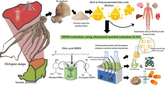
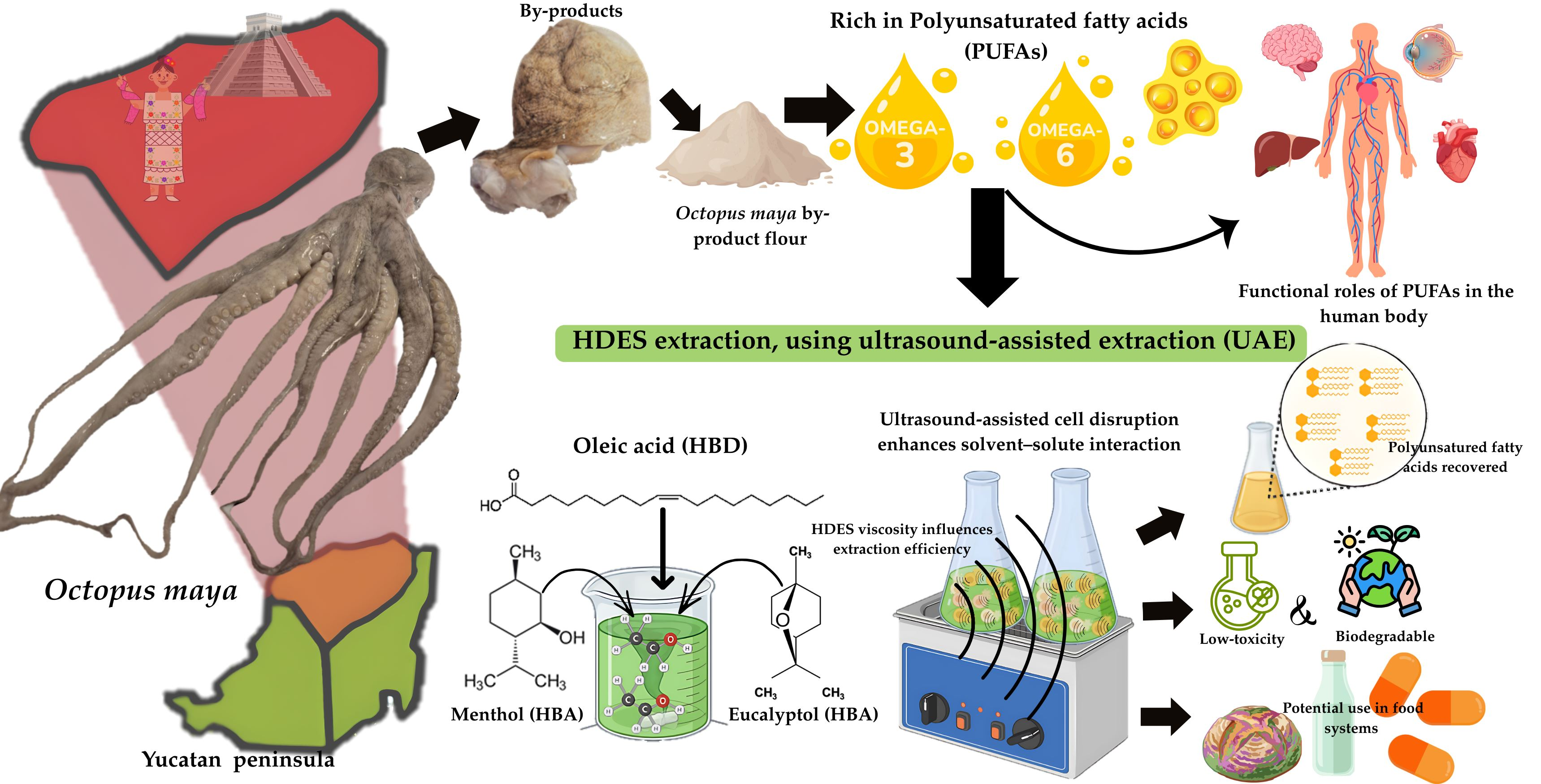
Graphical Abstract

1. Introduction

The octopus production in the Yucatan Peninsula, Mexico, is predominantly composed of Octopus maya (75%), an endemic species that generates approximately 16,000 jobs and an annual income of up to USD 36 million [1,2,3] and reaches a production volume of 25,462 tons in 2025 [4].
The processing of Octopus maya in packing facilities and gourmet restaurants generates by-products such as the tips of the tentacle and mantles (the correct term for the heads), which are not utilized and often discarded, leading to environmental contamination of water, soil, and air [5]. However, these by-products represent a potential source of high-value biomolecules, including proteins (collagen, 77.5 g/100 g DW) and polyunsaturated fatty acids (PUFA), particularly omega-6 (110 mg/100 g WW) and omega-3 (144 mg/100 g WW), such as eicosapentaenoic acid (EPA, 20:5 n-3) and docosahexaenoic acid (DHA, 22:6 n-3), which are present in Octopus maya [6]. These compounds are essential lipids in the human diet and health due to their (1) anti-inflammatory properties, (2) capacity of reducing the risk of cardiovascular diseases (CVD), (3) contribution to visual health, and (4) involvement in the proper functioning of key organs such as the brain, liver, and heart. Calder et al. [7] reported that an adequate omega-3 intake (250 mg/day) for the general population may help prevent the development of neurodegenerative diseases such as Parkinson’s and Alzheimer’s. However, to observe these biological effects, the dietary intake ratio of omega-6 (ω-6) to omega-3 (ω-3) should be maintained between 1:1 and 5:1 [8,9].
PUFA in octopus are mainly associated with muscle tissues and cell membranes in the form of triacylglycerols and phospholipids, which can limit their interaction with solvents and reduce extraction yields [10]. Therefore, the selection of an appropriate extraction method and solvent system is critical to improve lipid recovery and selectivity.
To obtain these fatty acids from marine by-products, efficient extraction methods are required. Commonly organic solvents are volatile and nonpolar compounds that enable efficient extraction of lipophilic compounds; however, their toxicity, flammability, and emission of volatile organic compounds represent risks to human health and the environment [11,12]. Ethanol has been explored as an option to optimize the extraction of total lipids (TL) and PUFA (ω-3) from Octopus vulgaris by-products as an alternative to conventional solvents, achieving a lipid recovery of 54.6% [13]. Although this represents an improvement over conventional solvents, challenges related to solvent consumption and selectivity toward specific PUFA remain. To improve solute–solvent interactions ultra-sound-assisted extraction (UAE) has been used to enhance lipid recovery by disrupting the cell structures through cavitation, leading to higher extraction efficiency, reduced extraction time, and lower solvent consumption [14]. However, this method still depends on solvents’ use. Consequently, there has been increasing interest in green and biodegradable extraction alternatives that align with the 12 principles of green chemistry being the use of natural deep eutectic solvents (NADES), a promising option [12,15].
NADES have emerged as an alternative group of low-toxicity, biodegradable, and stable solvents with modifiable physicochemical properties. These systems consist of a mixture of a hydrogen bond donor (HBD) and a hydrogen bond acceptor (HBA) at a specific molar ratio. When subjected to temperatures (>40 °C) and agitation, they form a stable liquid through hydrogen bonding and Van der Waals interactions, resulting in a stable liquid at room temperature [16,17,18].
Hydrophilic NADES are commonly formulated with an organic salt such as choline chloride (HBA) combined with sugars (hydrophilic HBD, e.g., fructose) or organic acids (e.g., lactic acid) with varying proportions of water to extract compounds such as polyphenols, flavonoids, terpenes, and catechins [19]. However, the hydrophilic nature of NADES limits their ability to extract lipophilic (nonpolar) compounds such as PUFA, making hydrophobic deep eutectic solvents (HDES) a promising solution for lipid extraction due to their affinity for nonpolar compounds and tailorable physicochemical properties.
HDES are characterized by their affinity for nonpolar compounds, low water solubility, density, and viscosity. In HDES, HBAs may include phenols, carboxylic acids, alcohols, and glycols or non-ionic, including monoterpenes such as menthol or thymol [20]. For HDES formulated with menthol and thymol combined with decanoic acid, densities of 0.896 and 0.918 g cm−3 and viscosities of 0.012 and 0.011 Pa·s have been reported [21,22]. These properties allow improved penetration of the solvent into the cell wall, which may be associated with enhanced solute extraction capacity.
Terpenes are natural compounds found in plant essential oils, composed of two isoprene units (C10) that may form aromatic rings or saturated cyclic structures, conferring hydrophobic character and affinity for nonpolar compounds [21,23]. An HDES formulated with menthol and lidocaine at a 1:1 molar ratio has been reported as a potential alternative for the extraction of EPA (172.04 μg g−1 DW of biomass) and DHA (602.79 μg g−1 DW of biomass) from mussels [24]. Menthol and thymol combined with carvacrol (a phenolic monoterpene) have also been applied for total lipid extraction from fish (TL = 87%) and mollusks (TL = 43–47%) [25], and HDES-based methodologies have additionally been explored for lipid extraction from seeds.
In this context, terpenes have emerged as key compounds for HDES formulation due to their nonpolar nature and suitability for lipid extraction. Menthol and eucalyptol have shown affinity for lipophilic matrices and solubilization capacity in long-chain organic acids such as oleic acid. In addition, previous studies have reported that terpene-based HDES are biodegradable, with low toxicity and low viscosity, properties that favor solvent penetration into the cell wall for a better PUFA recovery. Therefore, it is hypothesized that terpene-based HDES, particularly those formulated with menthol or eucalyptol and oleic acid, can enhance the extraction efficiency of PUFA from Octopus maya by-products due to their affinity for lipophilic compounds and favorable physicochemical properties. Based on this perspective, the present study applied eucalyptol and menthol combined with oleic acid at different molar ratios, using ultrasound-assisted extraction (UAE) to enhance the extraction of PUFA from Octopus maya by-products. The viscosity of the HDES, fatty acid profile determined by gas chromatography, and omega-3 were evaluated. The presence of lipid compounds was confirmed by FTIR and Raman spectroscopy, and the extraction performance of the HDES was assessed as an alternative to organic solvents for the recovery of lipid compounds from Octopus maya by-products.

2. Materials and Methods

2.1. Solvents and Reagents

Menthol (crystals, 100%), Dermiquim chemical solutions, (Jalisco, Mexico); oleic acid (≥99%), Favela Pro, S.A. de C.V. (Sinaloa, Mexico); eucalyptol (99%), SAFC Supply Solutions (MO, USA); ethanol (99.9%), Chemical products Monterrey S.A. de C.V. (N.L., Mexico); methanol, Sigma-Aldrich (MO, USA); and chloroform, Mallinckrodt Baker S.A. de C.V. (Edo. Mex, Mexico), of analytical grade, were purchased from commercial suppliers (B.MEDINA, Mérida, Yucatan, Mexico). The omega-3 (100% Norwegian salmon oil, Natural Health II®, 52.68% ω-3 declared on the label) standard used to construct the calibration curve was obtained from a local pharmacy (Costco, Mérida, Yucatán, Mexico).

2.2. Octopus maya Sample Collection

The Octopus maya specimens were captured along the coast of the state of Yucatán, Mexico, during the 2023–2024 fishing season with the minimal requirements (NOM-008-PESC-1993), as part of the project “Plataforma tecnológica pulpo maya para el desarrollo de productos de alto valor agregado 6559”. They were frozen for further analysis.

2.3. Processing of Octopus maya By-Product Samples

From this section onward the term by-products refers exclusively to the mantles (heads) of Octopus maya.
Specimens were randomly selected, and the by-product (mantle) was removed by cutting at the base of the arms. The by-products were washed, blended, and stored in 250 g (Ziploc® SC Johnson, Racine, WI, USA) bags at −20 °C for 72 h. Then the samples were freeze-dried for 96 h at −84 °C (Freezone, LABCONCO®, Kansas City, MO, USA), 2.5 L Benchtop. After lyophilization, the yield of dry material obtained from Octopus maya by-products was approximately 11.23%. The freeze-dried material was then ground using a commercial blender (Oster®, model BLST4127, USA) and sieved through a #35 stainless steel mesh (Fisher Scientific, Boston, MA, USA) with a pore size of 500 μm to obtain a uniform particle size fraction. The resulting by-product flours were stored in airtight containers and kept in a desiccator at room temperature to prevent moisture uptake and ensure sample stability.

2.4. Conventional Oil Extraction (COE)

Oil extraction was performed following the methodology described by Kuo et al. [26], with modifications. Briefly, 10 g of freeze-dried Octopus maya by-product flour was mixed with 100 mL of ethanol and homogenized using a high-speed disperser (ULTRA-TURRAX® T 18 digital, IKA Works Inc., Staufen, Germany) at 15,000 rpm for 1 min. The mixture was subjected to ultrasound-assisted extraction (UAE) for 30 min using an ultrasonic bath (BRANSON®, model 351, 42 kHz, Danbury, CT, USA). The sample was then centrifuged at 4500 rpm for 30 min at 4 °C to sediment residual solid material. The organic phase (ethanol) was collected into tubes and centrifuged again under the same conditions to ensure the removal of remaining impurities. To separate the lipid phase, the extracts were concentrated using a rotary evaporator (Laborota V-700, Heidolph Instruments GmbH & Co. KG, Schwabach, Germany) at 300 mbar and 60 °C using a water bath.
The extracts were stored at −20 °C until further analysis.
The conventional ethanol extraction was included as a reference method to confirm the presence of lipid compounds in Octopus maya by-products and to provide a basis for semi-quantitative comparison with HDES extraction performance. Due to differences in extraction processes and analytical approaches, this comparison was intended to identify relative trends rather than establish direct quantitative equivalence.

2.5. Preparation of HDES

HDES were prepared following the methodology described by Ramírez-Sucre et al. [27], with modifications. Oleic acid (282.46 g/mol) was used as the hydrogen bond donor (HBD), while menthol (156.27 g/mol) and eucalyptol (154.25 g/mol) were used as hydrogen bond acceptors (HBA) at molar ratios of 1:1, 1:2, and 2:1 (mol/mol). HDES composed of oleic acid (O) and menthol (M) was heated to 40 °C (manufacturer’s recommendation) under continuous stirring until a homogeneous mixture was obtained, whereas HDES formulated with eucalyptol (E) was stirred at room temperature until complete homogenization of both components was achieved.

2.6. Fatty Acid Extraction Using HDES Under UAE

To evaluate the effect of the hydrogen bond acceptor (HBA), molar ratio (MR), and extraction time (ET) of ultrasound-assisted extraction (UAE) on the omega-3 recovered (reported as equivalents of ω-3) from Octopus maya by-products using HDES, a 2 × 3 × 2 factorial design (duplicates) was established. The factor levels were HBA (M or E), MR (1:1, 1:2, or 2:1), and ET (30 or 60 min), as shown in Table 1.
PUFA extraction using HDES was performed following the methodology described by Ramírez-Sucre et al. [27], with modifications. Ten grams of HDES were mixed with 1 g of Octopus maya by-product flour. The mixture was subjected to ultrasound-assisted extraction (UAE) using an ultrasonic bath (BRANSON®, model 351, 42 kHz, Danbury, CT, USA) for 30 or 60 min, according to the experimental design. The samples were centrifuged at 4500 rpm for 15 min at 4 °C, and the supernatant was recovered. The extracts were stored at 4 °C for further analysis.

2.7. Viscosity Analysis of HDES Extracts

The viscosity of HDES extracts obtained from Octopus maya by-products was determined using a controlled-stress rheometer (Discovery Hybrid Rheometer DHR-2, TA Instruments, New Castle, DE, USA) equipped with a parallel plate geometry (40 mm diameter) and a gap of 1050 μm, following the methodology described by Ramírez-Sucre and Baigts-Allende [28]. Flow behavior was evaluated through viscosity curves (Pa·s) as a function of shear rate (0.001 to 100 s−1) at a constant temperature of 25 °C.

2.8. Determination of Omega-3 Equivalents Obtained by UV–Vis Spectrophotometry

The determination of omega-3 equivalents (EO3) in HDES extracts was performed using UV-Vis spectrophotometry according to the methodology described by Oliveira et al. [29], with modifications. A five-point calibration curve (50, 200, 300, 600, and 1200 μg/mL) was prepared using omega-3 (100% salmon, 52.68% ω-3) diluted in a chloroform:methanol (2:1, v/v) solution. Each calibration point was measured using a spectrophotometer UV–Vis (THERMO SCIENTIFIC®, Genesys 140, Mexico City, Mexico) at 202 nm.
The calibration curve showed a correlation coefficient (R2) of 0.961, with the corresponding equation:
y =   1191.3 x     257.4 1000
where y represents the EO3 (mg/mL) and x corresponds to the absorbance measured at 202 nm. This equation was used to estimate the relative concentration of omega-3 equivalents in the HDES extracts based on their measured absorbance values.
It is important to note that the UV–Vis absorbance at 202 nm is associated with the presence of unsaturated compounds and is not specific to omega-3 fatty acids. Therefore, the values reported as EO3 (mg/mL) represent a relative and indirect estimation based on the response of the calibration standard (52.68% omega-3 in the sample salmon oil used as standard), rather than an absolute quantification of omega-3 fatty acids.
Thus, UV–Vis analysis was employed as a semi-quantitative approach to compare relative differences among treatments. The results, expressed as mg/mL of EO3, were interpreted as reflecting relative variations in unsaturation rather than absolute omega-3 content.
Based on these results, the four samples with the highest EO3 (mg/mL) were selected for fatty acid profile characterization and were additionally analyzed by Fourier transform infrared spectroscopy (FTIR) and Raman spectroscopy.

2.9. Fatty Acid Profiling by Gas Chromatography (GC)

Fatty acid characterization was performed according to the method described in the NMX-F-490-1999-NORMEX standard. Sample methylation was carried out by adding sodium hydroxide in methanol to a flask containing glass beads, which was connected to a Friedrich condenser and placed in a water bath at 70 °C. The mixture was maintained under reflux for 12 min from the appearance of the first drop. Subsequently, boron trifluoride (BF3) in methanol was added, and reflux was continued for 2 additional min. Heptane was then added, and the mixture was refluxed for 1 min.
After completion of the reaction, the flask was removed, temporarily capped, and cooled with water. A saturated sodium chloride (NaCl) solution was added to promote phase separation. The upper phase was transferred to a 2 mL vial and sealed. The methylated sample was then injected into a PerkinElmer gas chromatograph equipped with a flame ionization detector (FID). A 1 μL injection was performed in split mode (40:1). A DB-23 capillary column (60 m × 0.25 mm × 0.25 μm) was used. The injector temperature was set at 220 °C, the oven temperature was programmed from 60 °C to 250 °C at 25 °C/min, and the FID temperature was maintained at 275 °C.
Quantification of the fatty acids [palmitic acid (C16:0), stearic acid (C18:0), oleic acid (C18:1 n-9), linoleic acid (C18:2 n-6), linolenic acid (C18:3 n-3), omega-3 (ω-3), Omega-6 (ω-6) and Omega-9 (ω-9)] was performed using the normalization method described in NMX-F-490-1999-NORMEX, and results were reported as fatty acid percentage (%).

2.10. Fourier Transform Infrared Spectroscopy (FTIR)

Functional group characterization of the HDES extracts was performed following the methodology described by Áviles-Betanzos et al. [30], with modifications. The analysis was conducted using a spectrometer (Nicolet iS5, Thermo Fisher Scientific Inc., Madison, WI, USA) equipped with an attenuated total reflection (ATR) accessory with a germanium crystal. Spectral scans were recorded over a range of 4000 to 500 cm−1 using 64 background scans and a resolution of 8 cm−1 in transmittance mode (%). FTIR spectra were visualized and processed using OriginLab® software, version 2025b, SR1 (No_H_234).

2.11. RAMAN Spectroscopy

Raman spectroscopy was employed to evaluate the structural composition of the HDES extracts exhibiting the highest ω-3 concentrations. Raman spectra were acquired using a Renishaw InVia Confocal spectrometer (Wotton-under-Edge, Gloucestershire, UK) over a spectral range of 200–3200 cm−1. A red laser (633 nm) was used as the excitation source, operating at 100% of its total power (17 mW), with an exposure time of 20 s [31]. Raman spectra were visualized and processed using OriginLab® software, version 2025b, SR1 (No_H_234).

2.12. Statistical Analysis

All results were reported as mean values ± standard deviation. A full factorial design of experiments (DOE) was implemented to evaluate the effects of hydrogen bond acceptor (HBA: menthol or eucalyptol), molar ratio (MR: 1:1, 1:2, and 2:1), and extraction time (ET: 30 and 60 min) on ω-3 and viscosity. Each response variable was analyzed independently. Experimental data were analyzed using Minitab® software (version 18) using analysis of variance (ANOVA) at a 95% confidence level (p < 0.05), in order to determine the statistical significance of factor effects and their interactions on the response variables. Additionally, Fisher’s least significant difference (LSD) test was applied as a post hoc analysis for the comparison of means among treatments.
All experiments were conducted in duplicate (due to the limited amount of extract obtained from the freeze dry process, 11.23% yield); the statistical analysis was interpreted considering this limited number of replicates. Therefore, the identification of significant effects and interactions is indicative of trends under the evaluated conditions. This approach allows the identification of consistent patterns among treatments while acknowledging the limitations in statistical treatment.
For fatty acid profile analysis, a reduced factorial design (2 × 2) was applied, considering HBA (menthol or eucalyptol) and MR (1:1 and 1:2) as factors. These data were analyzed following the same statistical approach described above.

3. Results and Discussion

3.1. Viscosity of HDES Extracts

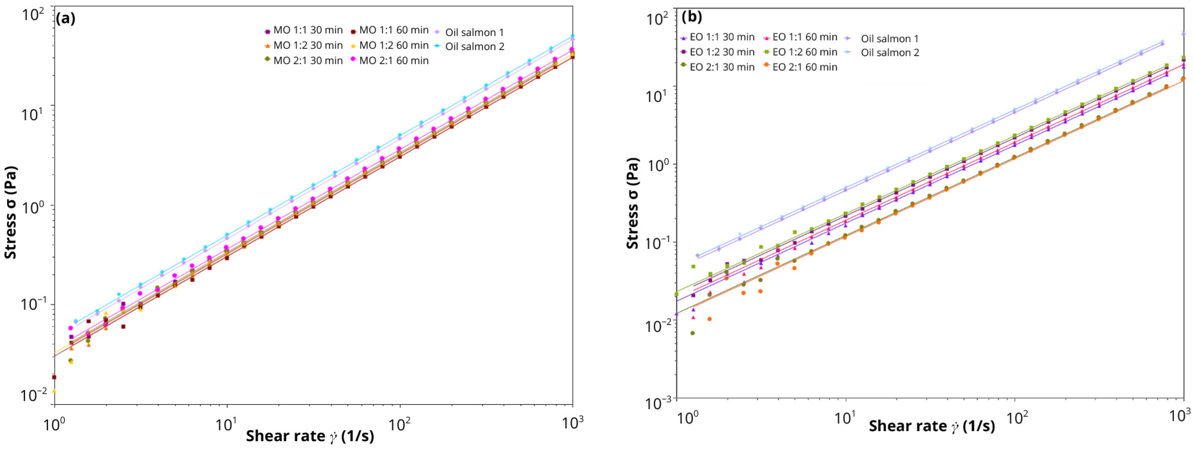
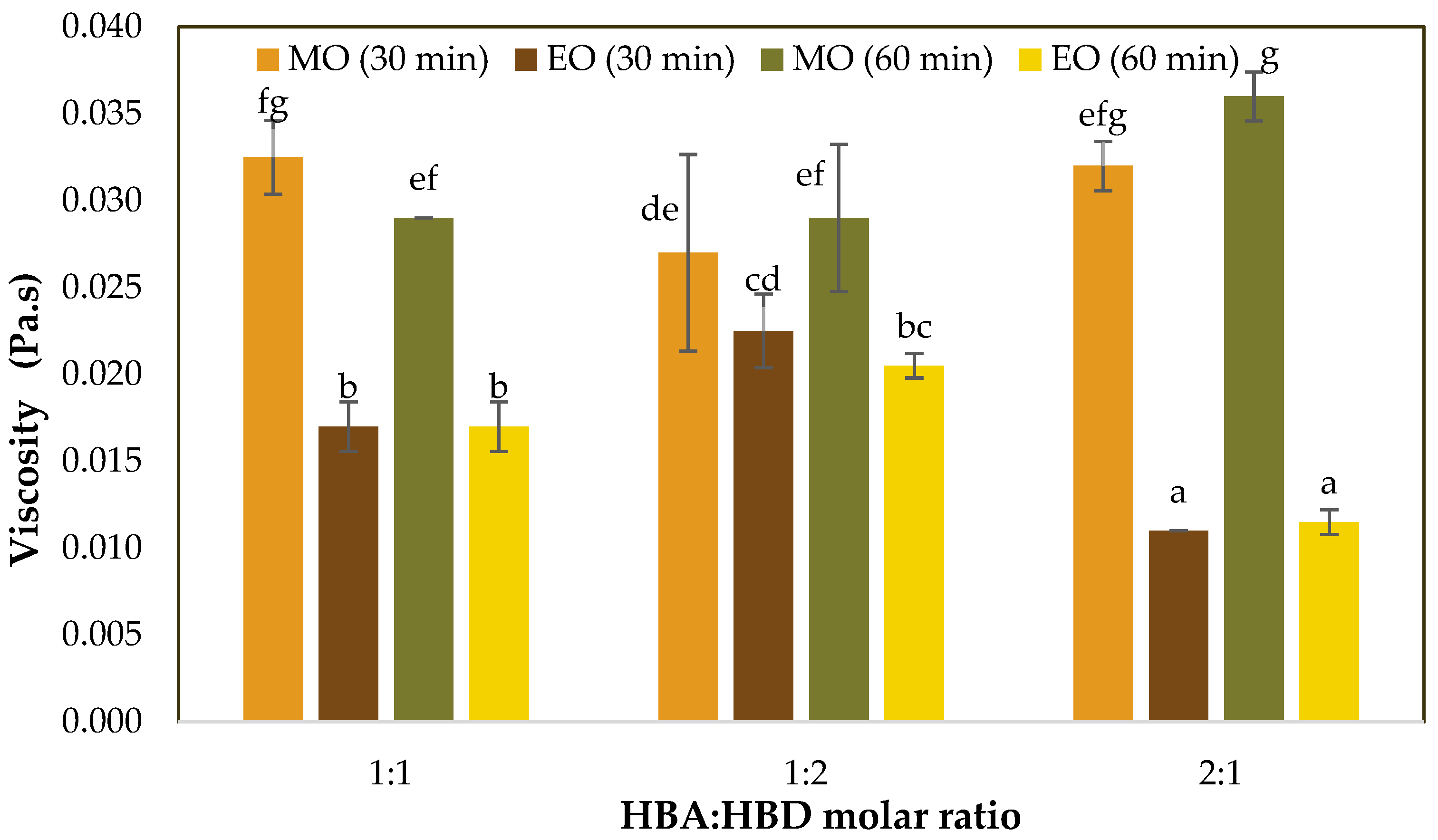
The flow behavior of the HDES is shown in Figure 1, while the average viscosity values and their standard deviations at a shear rate of 10 s−1 are summarized in Figure 2. The viscosities of the HDES extracts ranged from 0.011 to 0.036 Pa·s, showing Newtonian behavior in all cases within the analyzed range. Significant differences (p < 0.05) were also observed among treatments due to the studied factors: hydrogen bond acceptor (HBA), HBA:HBD molar ratio (MR), and extraction time (ET).
The results indicate that samples of HDES extracts formulated (HBA/O(MR)ET) with eucalyptol showed lower viscosities (p < 0.05) compared with menthol-based HDES. The eucalyptol formulations EO(1:2)30 and EO(1:2)60 presented values of 0.023 ± 0.002 Pa·s and 0.021 ± 0.001 Pa·s, respectively, which correspond to the highest viscosities among the eucalyptol-based HDES. Formulations EO(2:1)30 and EO(2:1)60 showed the lowest viscosity values, with 0.011 ± 0 Pa·s and 0.012 ± 0.001 Pa·s, respectively. In contrast, menthol-based systems were characterized by higher viscosity (p < 0.05). For example, the formulations MO(2:1)60, MO(2:1)30, and MO(1:1)30 showed viscosities ranging from 0.032 to 0.036 Pa·s.
Furthermore, the analysis of the experimental design (Table 2) showed that the interaction between HBA and MR had a statistically significant effect (p < 0.05), suggesting that the viscosity behavior of HDES extracts may be influenced by these factors.
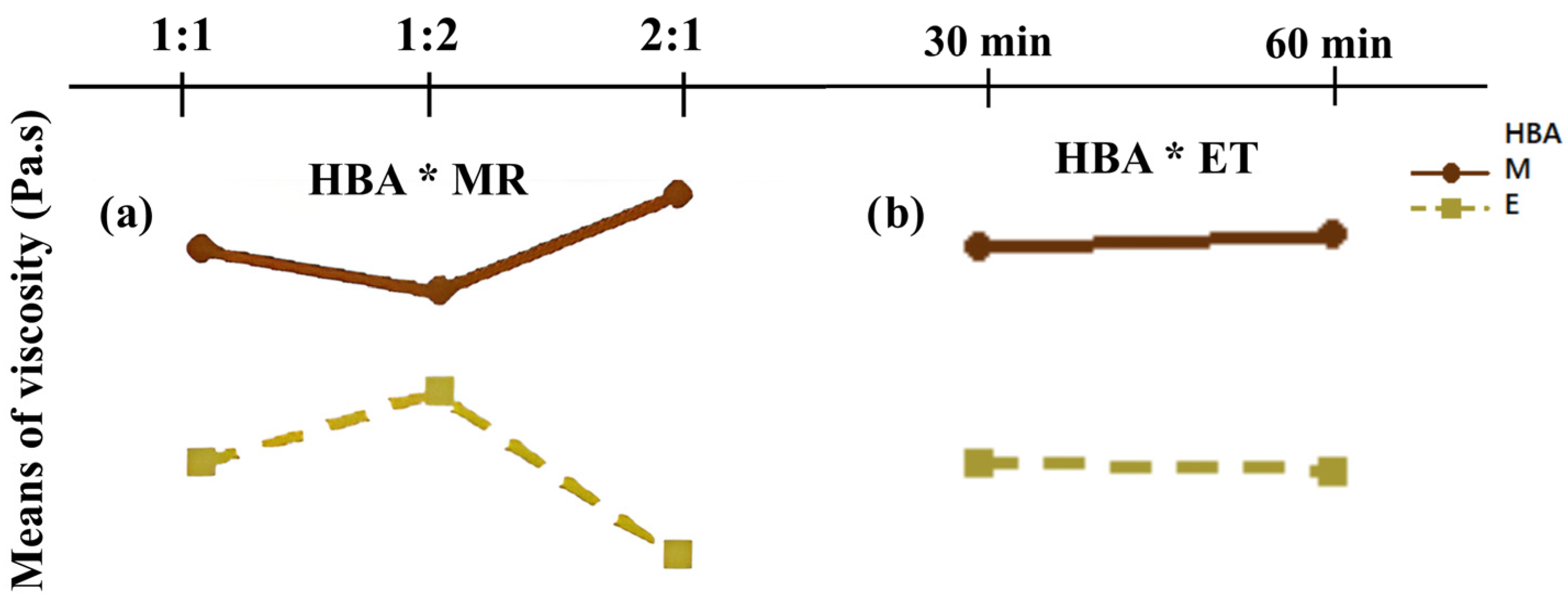
In line with these observations, the interaction plots (Figure 3a) indicate that menthol, particularly at a molar ratio of 2:1, tends to be associated with higher viscosity values in HDES systems. In contrast, eucalyptol at the same molar ratio tends to result in systems with lower viscosity. These trends are consistent with the viscosity ranges observed in this study which fall within reported values of Adeoye et al. [32], who evaluated the viscosity of pure HDES formulated with menthol and fatty acids at a fixed molar ratio (1:1). In that study, an increase in viscosity was associated with the alkyl chain length of the fatty acid. Reported viscosities were 0.012, 0.016, and 0.018 Pa·s for octanoic (C8:0), decanoic (C10:0), and dodecanoic (C12:0) acids, respectively. However, those authors characterized HDES in their pure form, without subjecting them to an extraction process. In this context, the viscosities of the eucalyptol-based systems were similar, whereas menthol-based systems showed higher viscosity values (≤0.037 Pa·s), an effect attributed to both the number of carbon atoms in oleic acid (C18:1) and intrinsic viscosities of the HBA systems (ηmenthol > ηeucalyptol).
On the other hand, Dabbagh et al. [33] reported that hydrogen-bonding interactions play an important role in the viscosity of HDES. In their study, based on molecular dynamics simulations, they examined the molecular properties of HDES formulations composed of fatty acids (valeric, enanthic, and pelargonic acids) as hydrogen bond donors (HBD) and menthol as the hydrogen bond acceptor (HBA), at three HBD:HBA molar ratios (1:2, 1:1, and 2:1). From these simulations, the authors observed that increasing the molar proportion of fatty acids resulted in lower viscosities. This behavior was attributed to changes in the network of intermolecular interactions between fatty acids and menthol. This finding is consistent with the behavior observed experimentally in the menthol-based HDES extracts of the present study, where a higher oleic acid content (1:2) led to lower viscosity values (0.027–0.029 Pa·s), whereas lower oleic acid content (2:1) resulted in higher viscosities (0.032–0.036 Pa·s). The similarities suggest that intermolecular hydrogen-bonding interactions, arising from the molecular structure of the HDES components, play a key factor in governing viscosity behavior. In this context, viscosity can be related to the transport capacity between the solvent and the solute as reported by Trenzado et al. [21]. High-viscosity systems tend to restrict molecular mobility thereby limiting diffusion rates within the medium. Lower viscosity, often associated with shorter chains and/or a higher molar proportion of fatty acids in an HDES system [32,33], may facilitate solvent penetration into the biological matrix and enhance mass transfer. However, extraction efficiency in HDES systems is determined by viscosity, the interplay between transport phenomena and molecular interactions. In addition to viscosity, factors such as solvent polarity, hydrogen-bonding capacity, and solute–solvent affinity is essential for governing extraction performance. The thermodynamic compatibility between the solvent and the target lipid compounds influences their solubilization, while mass transfer governs their release from the biological matrix [34].
At the molecular level, the hydrogen-bonding network affects viscosity and also contributes to the solvent’s ability to interact with solvate lipid compounds, as well as to disrupt structural components of the matrix. Therefore, formulations with a higher molar proportion of fatty acids (oleic acid), which exhibit lower viscosity, may promote improved solvent–matrix interactions and potentially enhance the recovery of polyunsaturated fatty acids (PUFAs). Additionally, the Newtonian behavior observed for all HDES extracts (Figure 1), with viscosities below 0.04 Pa·s, suggests favorable flow properties that may support solvent transport within the matrix and contribute to the extraction process.
However, considering that the experiments were conducted in duplicate, these results should be interpreted as indicative trends rather than definitive evidence.

3.2. Omega-3 Equivalents in HDES Extracts (UV–Vis)

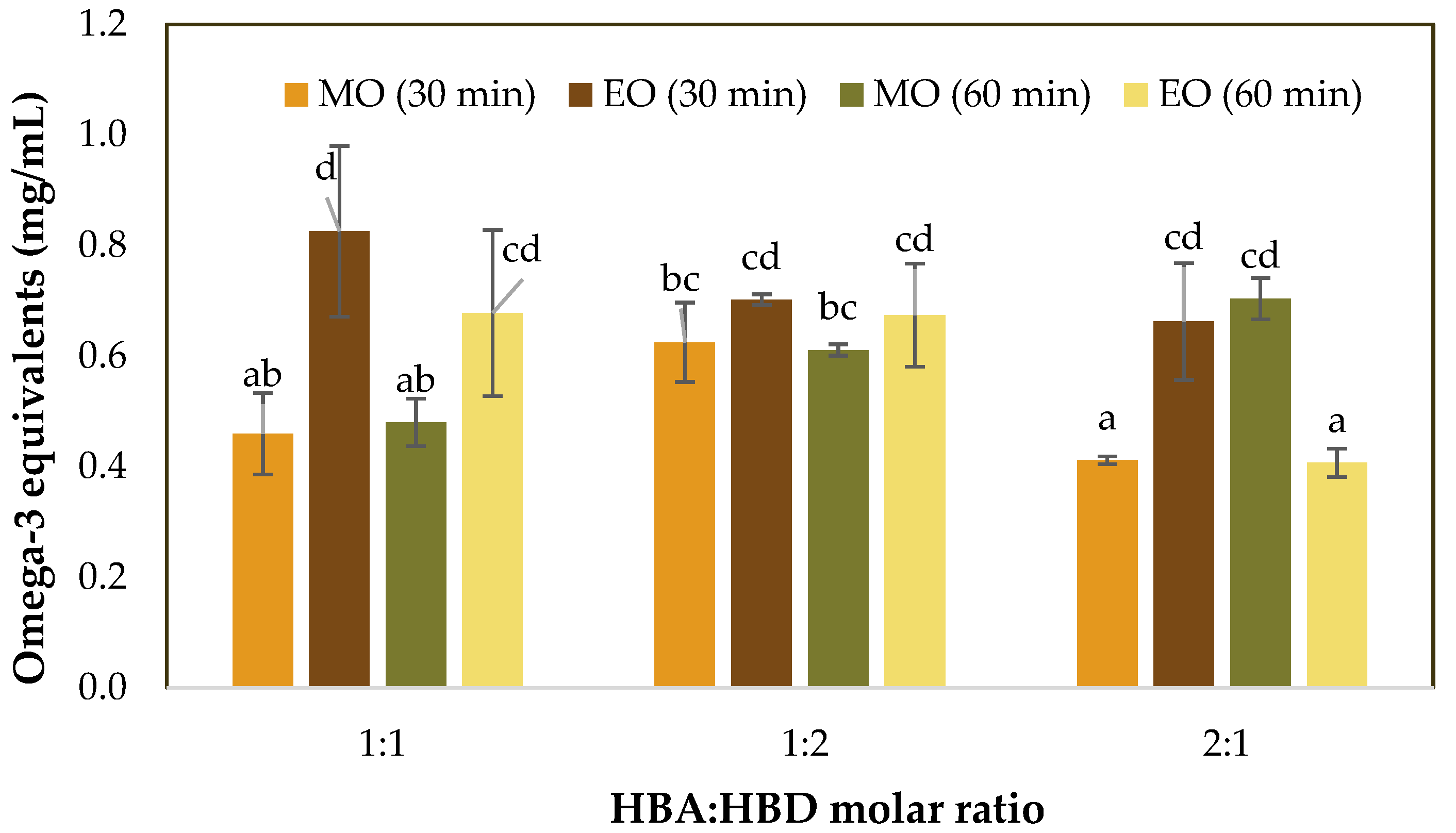
The omega-3 equivalents (EO3) in HDES extracts obtained from by-products of Octopus maya, analyzed by UV–Vis spectrophotometry (based on the absorption of light by functional groups, allowing an indirect estimation of unsaturated compounds) [35], showed significant differences (p < 0.05) among the treatments (Figure 4). The formulation EO(1:1)30 presented the highest value of EO3 (0.83 ± 0.15 mg/mL). However, no significant differences were found compared with other eucalyptol-based treatments, which showed values ranging from 0.66 to 0.70 mg/mL, indicating that these formulations have a similar extraction performance. In general, all menthol-based HDES showed lower EO3 (0.4 < EO3 mg/mL < 0.70).
In interpreting these results, it is important to consider the scope and limitations of the analytical method used. As reported by Oliveira et al. [29], UV–Vis spectrophotometry has been applied for the estimation of unsaturated bonds associated with fatty acids (ω-3, ω-6, and ω-9) in fresh tissues of Tambaqui (Colossoma macropomum). In the present study, this approach was specifically applied to estimate omega-3 as EO3 equivalents. Accordingly, the values obtained in the present study reflect relative differences among treatments in unsaturation-related compounds, rather than a direct quantification of ω-3 fatty acids. Within this context, the experimental design allowed evaluating how the factors, hydrogen bond acceptor (HBA), molar ratio (MR) and extraction time (ET), influenced these relative responses.
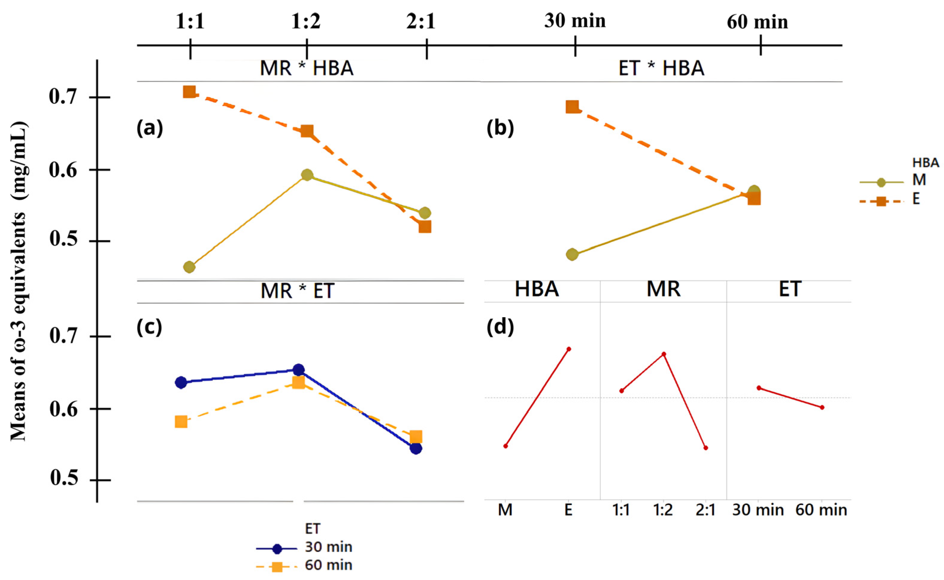
After analyzing the experimental design, one triple interaction (HBA × MR × ET) and two double interactions (HBA × MR) and (HBA × ET) showed significant effects (p < 0.05) on the EO3 of HDES extracts obtained from Octopus maya by-products (Table 2). The interaction effects HBA × MR and HBA × ET are presented in Figure 5, together with the MR × ET interaction and the main effects plot. However, these results have been interpreted considering the limited number of replicates (n = 2), which may influence the sensitivity of the statistical analysis.
The triple interaction indicates that the composition of the HDES (HBA/HBD), the molar ratio, and the extraction time significantly influence the recovery of EO3. Regarding the HBA × MR interaction (Figure 5a), eucalyptol at a molar ratio of 1:1 showed the highest values of EO3 (0.83 ± 0.15 mg/mL), suggesting a favorable condition under the evaluated parameters. For the HBA × ET interaction (Figure 5b), eucalyptol exhibited higher values at 30 min of UAE, whereas menthol showed lower values, indicating a possible time-dependent behavior between acceptors. In the case of the MR × ET interaction (Figure 5c), a molar ratio of 1:2 combined with 30 min of UAE resulted in EO3.
Finally, the main effects plot (Figure 5d) revealed that eucalyptol-based systems tended to show higher EO3 than menthol-based ones. In addition, a molar ratio of 1:2 appeared to be the most favorable condition across MR levels, while an extraction time of 30 min tended to result in higher EO3 compared to 60 min. Overall, these findings suggest that the composition of the HDES (HBA/HBD) appears to have a significant influence on the values of EO3 obtained.
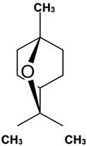
In this context, the observed differences between eucalyptol- and menthol-based systems may be associated with the molecular structure of HBA. Eucalyptol or 1,8-cineole (1,3,3-trimethyl-2-oxabicyclo[2.2.2]octane) is a monoterpene with a rigid and compact bicyclic structure composed mainly of carbon and oxygen atoms and lacking –OH groups. This characteristic prevents the formation of hydrogen bonds, limits intermolecular interactions, and contributes to a low melting point, allowing it to remain liquid at room temperature [36]. These structural features may influence the physicochemical properties of the resulting HDES.
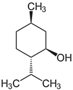
In contrast, menthol (2-isopropyl-5-methylcyclohexanol), a cyclic monoterpene that contains a hydroxyl group (–OH) capable of forming hydrogen bonds. These interactions favor the formation of intermolecular networks that, in the pure state, lead to a stable crystalline organization. However, when menthol is mixed with another component (such as fatty acids), its melting point decreases, forming a stable liquid with higher viscosity [37]. In this study, the highest EO3 obtained with eucalyptol-based HDES corresponded to the lowest viscosity values (0.012–0.023 Pa·s), compared with menthol-based formulations (0.027–0.036 Pa·s), which showed lower EO3 and greater variability (0.41–0.70 mg/mL).
Although lower viscosity may facilitate solvent mobility and promote better interaction with the lipid matrix, potentially improving extraction capacity [21], it should not be considered as the sole factor governing the process. In this sense, the lower viscosity observed (ηmenthol > ηeucalyptol) may be associated with higher EO3 (EO3menthol = 0.41–0.70 mg/mL < EO3eucalyptol = 0.41–0.83 mg/mL), suggesting a possible contribution of this property to the observed behavior. Instead, this behavior may be related to the combined influence of molecular structure, solvent–solute affinity, and intermolecular interactions, together with processing parameters such as extraction time, HBA/HBD type, and molar ratio, which collectively affect polarity, hydrogen bonding strength and viscosity [38].
These experimental results are like those reported by Strieder et al. [39], who designed and characterized HDES formulated with eucalyptol as HBA and oleic acid, tetradecanol, menthol, or camphor as HBD for the recovery of fatty acids from almond and peanut milk by-products. In their study, low viscosity values (0.004–0.005 Pa·s) were associated with improved extraction of fatty acids.
In addition, similarly, Bagović et al. [40] reported that menthol tends to form more viscous HDES. In their study, they formulated several HDES to evaluate their physicochemical properties and found that menthol–linoleic acid and menthol–thymol showed higher viscosities (0.027 and 0.029 Pa·s, respectively) compared with menthol–camphor and menthol–octanoic acid systems. These values of viscosity are like those obtained in the present study for menthol-based HDES, which also showed the lowest EO3.
Therefore, the present study provides complementary experimental evidence, suggesting that extraction behavior in HDES systems arises from the combined and interdependent effects of multiple physicochemical properties, rather than a single parameter. Although lower viscosity may facilitate solvent mobility, the values of EO3 reflect the overall interaction of factors such as viscosity, solvent–solute affinity, and intermolecular interactions within the system. In line with these observations, the statistical analysis indicated that the composition of the HDES, particularly in terms of the hydrogen bond acceptor (HBA) and its molar ratio, together with extraction time, influenced the EO3 obtained. These results highlight that the observed behavior is not governed by a single variable, but rather by the combined and interacting effects of formulation conditions and processing.
Based on the results showing the highest EO3 (mg/mL) obtained by UV–Vis spectrophotometry, the HDES extracts selected for characterization by Fourier transform infrared spectroscopy (FTIR), Raman spectroscopy, and fatty acid profiling were MO(1:1)30, MO(1:2)30, EO(1:1)30, and EO(1:2)30. Since the selected samples shared the same extraction time, a 2 × 2 factorial design was applied, in which the evaluated factors were the hydrogen bond acceptor (HBA: menthol or eucalyptol) and the molar ratio (MR: 1:1 or 1:2).

3.3. FTIR and Raman Characterization of HDES Extracts from Octopus maya By-Products

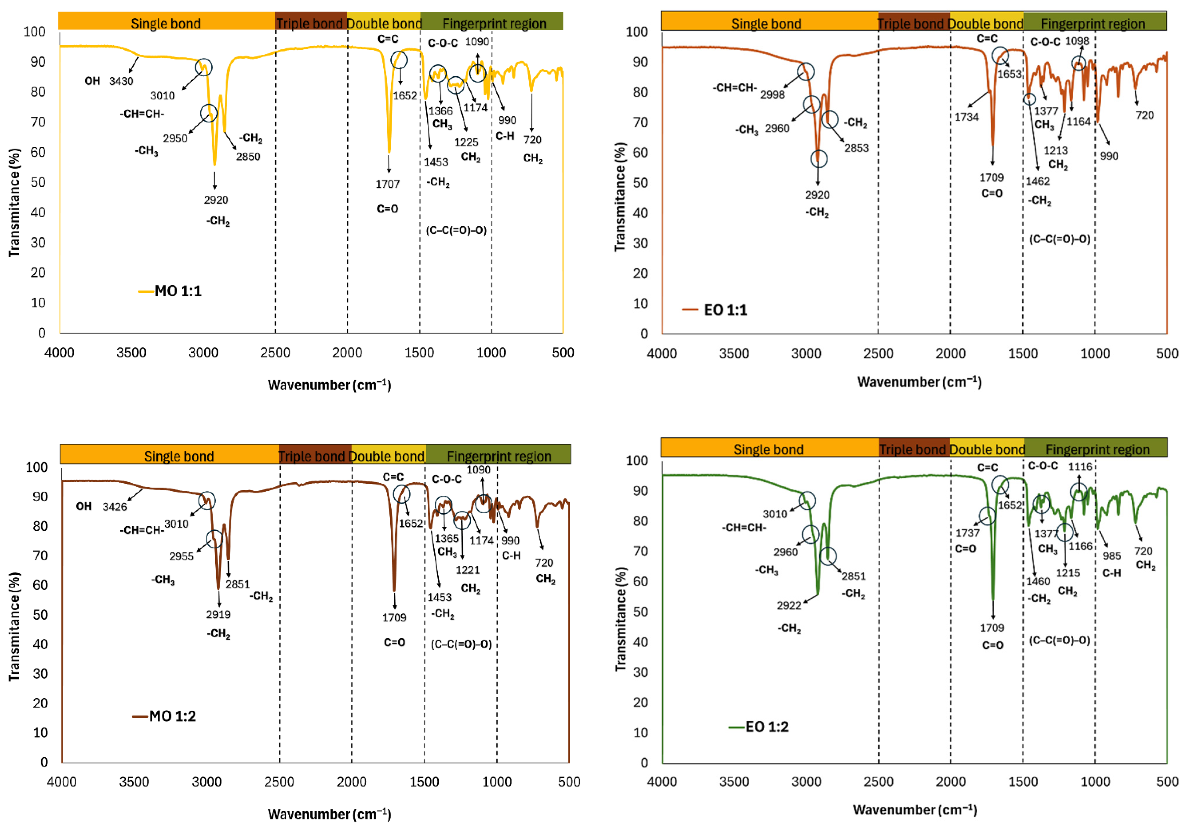
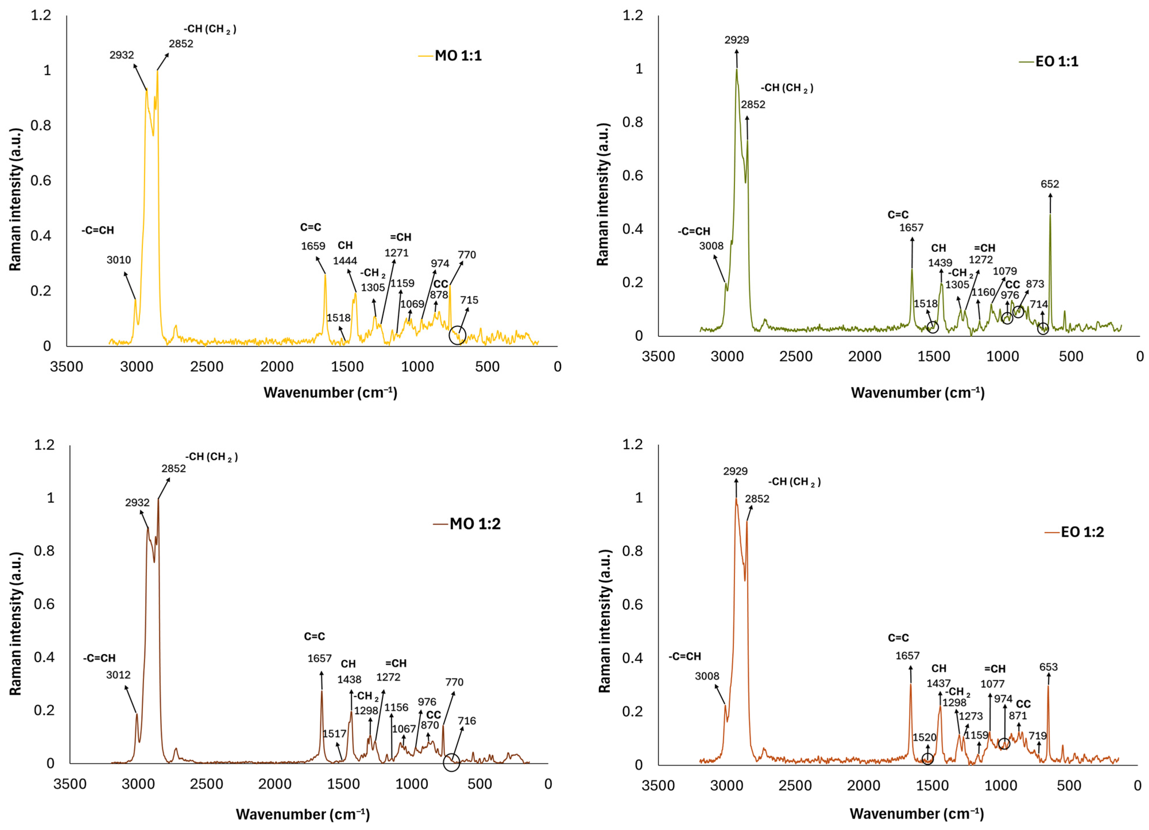
The FTIR spectra of the selected HDES extracts MO(1:1)30, MO(1:2)30, EO(1:1)30, and EO30(1:2) were measured over a wavelength range of 4000 to 500 cm−1. Each spectrum was divided into four regions to identify the functional groups and molecular composition of the extracts. In addition, Raman spectra were obtained to support the identification of bonds within the skeletal structures. The FTIR and Raman spectra of each HDES are shown in Figure 6 and Figure 7, respectively.
HDES formulated with menthol (MO(1:1)30 and MO(1:2)30) showed broad bands in the region of 3600–3100 cm−1, which correspond to the –OH stretching vibrations of menthol [32,41], although carboxylic acids can also exhibit it. In contrast, this broad peak was not detected in eucalyptol-based HDES, which can be attributed to the absence of a hydroxyl group in the eucalyptol molecule [36]. This behavior has also been reported by Strieder et al. [39], who observed a broad band at 3383 cm−1 in menthol-based HDES, whereas no stretching bands in this region were detected in eucalyptol–oleic acid formulations.
On the other hand, HDES extracts unsaturation were identified in the FTIR spectra through well-defined stretching peaks at 3010 cm−1 for MO(1:1)30, MO(1:2)30, and EO(1:2)30, and at 2998 cm−1 for EO(1:1)30. These peaks correspond to –CH=CH– double bonds (asymmetric stretching), which may be associated with the unsaturated fatty acids ω-3, ω-6, ω-9, or the oleic acid of the HBD present in the HDES system [42]. These signals were confirmed in the Raman spectra, which showed stretching bands at 3012–3008 cm−1 (–C=C–H), deformation bands at 1657–1658 cm−1 (C=C), and deformation bands at 1271–1273 cm−1 (=C–H), associated with the presence of ω-3 fatty acids [43,44]. These peaks may be attributed to the compounds recovered by the HDES from Octopus maya by-products, since studies such as Adeoye et al. [32], who formulated HDES based on menthol and short-chain fatty acids, and Strieder et al. [39], who prepared HDES with eucalyptol and oleic acid, did not report bands at these wavenumbers in their FTIR spectra. It is important to note that those studies analyzed HDES in its pure form with no subsequent extraction process.
Meanwhile, saturated fats correspond to well-defined peaks in the FTIR spectra at wavenumbers of 2920 and 2852 cm−1 (–CH3) [41], which were confirmed by the intensity of these peaks in the Raman spectra at 2932 and 2852 cm−1 [44]. These bands with high intensity may be associated with the presence of oleic acid, since its alkyl chain is mainly composed of saturated bonds, which is consistent with the fatty acid profile obtained by GC analysis (Section 3.4), where saturated fatty acids were also identified in the extracts. This behavior is consistent with that reported for HDES formulated with fatty acids. Adeoye et al. [32] identified stretching bands in the region of 2840 to 3000 cm−1 attributed to C–H bonds of alkanes in HDES formulated with menthol and short-chain fatty acids. Their study described that the shifts and intensity changes observed at wavenumbers at 2840 to 3000 cm−1 arise from hydrogen bond interactions between the hydrogen of the carboxyl group (–COOH) of the fatty acid and the oxygen of the hydroxyl group (–OH) of menthol, resulting in the formation of the HDES. This finding may explain that the formation of well-defined peaks in the region of 2850 to 3000 cm−1 could also be related to interactions between oleic acid and menthol or eucalyptol.
Another functional group identified in the FTIR spectra was the carbonyl group (–C=O), which showed well-defined bands in 1707–1709 cm−1. These bands are attributed to ester functional groups, commonly associated with lipids and fatty acids or free fatty acids [42].
In the Raman spectra of the eucalyptol-based HDES, shifts in the C=O wavenumber were observed, which may be associated with the presence of ω-3 ethyl esters [44]. However, Strieder et al. [39], reported that stretching bands in the region of 1700–1710 cm−1 in FTIR spectra of HDES formulated with eucalyptol and oleic acid may also result from the formation of hydrogen bonds between the HDES components.
Similarly, Adeoye et al. [32] observed that FTIR spectra of short-chain fatty acids showed a C=O stretching band at 1707 cm−1, that represent associated carbonyls related to fatty acids intermolecular interactions, which shifted to 1710 cm−1 when HDES based on menthol with short-chain fatty acids were analyzed. A similar behavior was observed in the present study, where HDES formulated with menthol and eucalyptol showed defined C=O bands in this spectral region. This suggests the preservation of the carbonyl group of oleic acid and its participation in interactions within the HDES system.

3.4. Fatty Acid Profile of Octopus maya By-Products

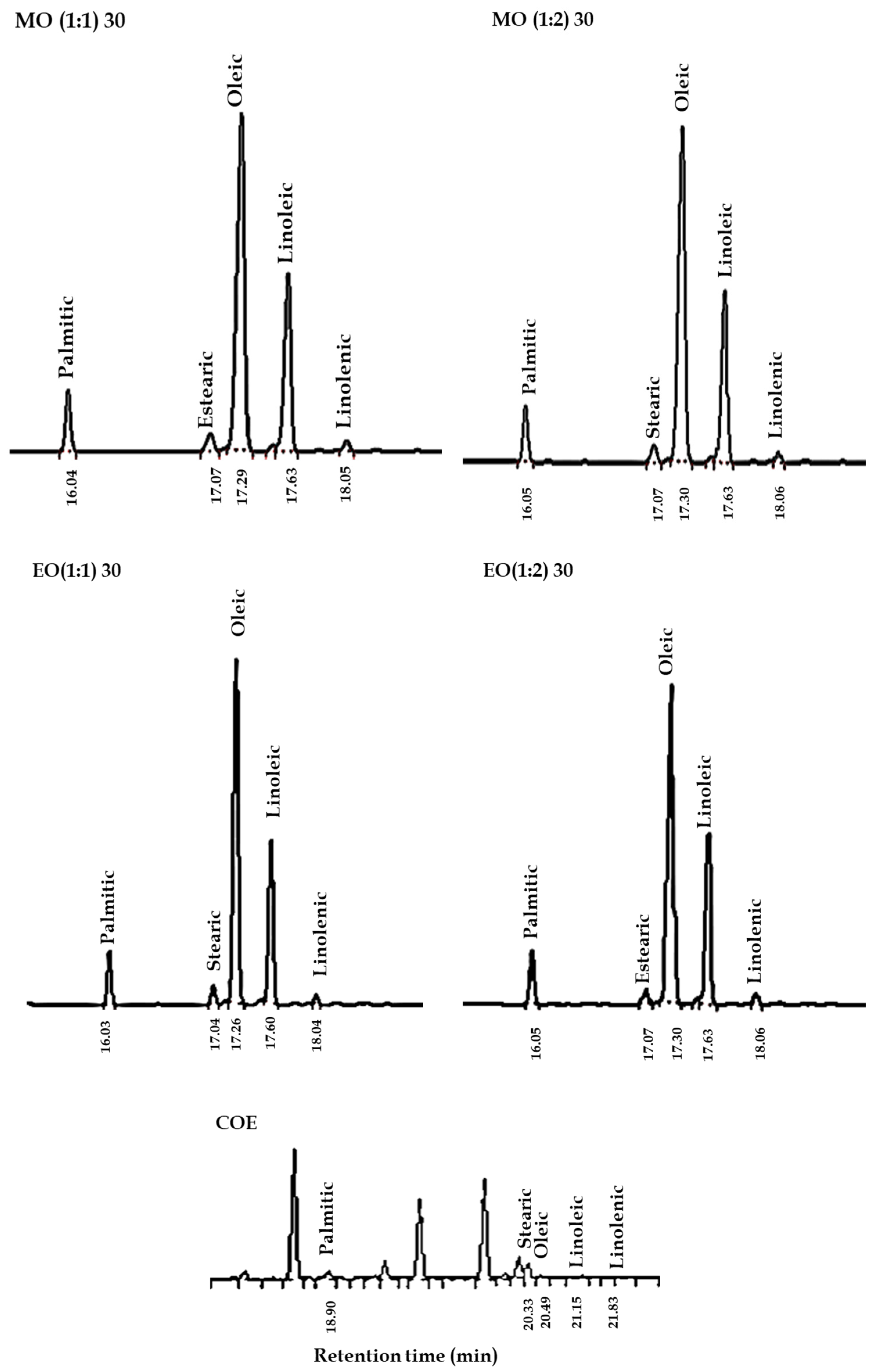
Gas chromatography (GC) is a reliable and widely used technique for fatty acid analysis, as it enables the separation and accurate quantification of individual compounds, being considered a reference method in lipid characterization. In contrast, UV–Vis provides an indirect estimation of unsaturated compounds without distinguishing specific fatty acids in complex mixtures [35,45]. Therefore, the interpretation of the omega-3 extraction capacity of HDES is supported by the fatty acid profile obtained by GC, which identifies: C16:0 (palmitic acid); C18:0 (stearic acid); C18:1 n-9 (oleic acid); C18:2 n-6 (linoleic acid); C18:3 n-3 (linolenic acid).
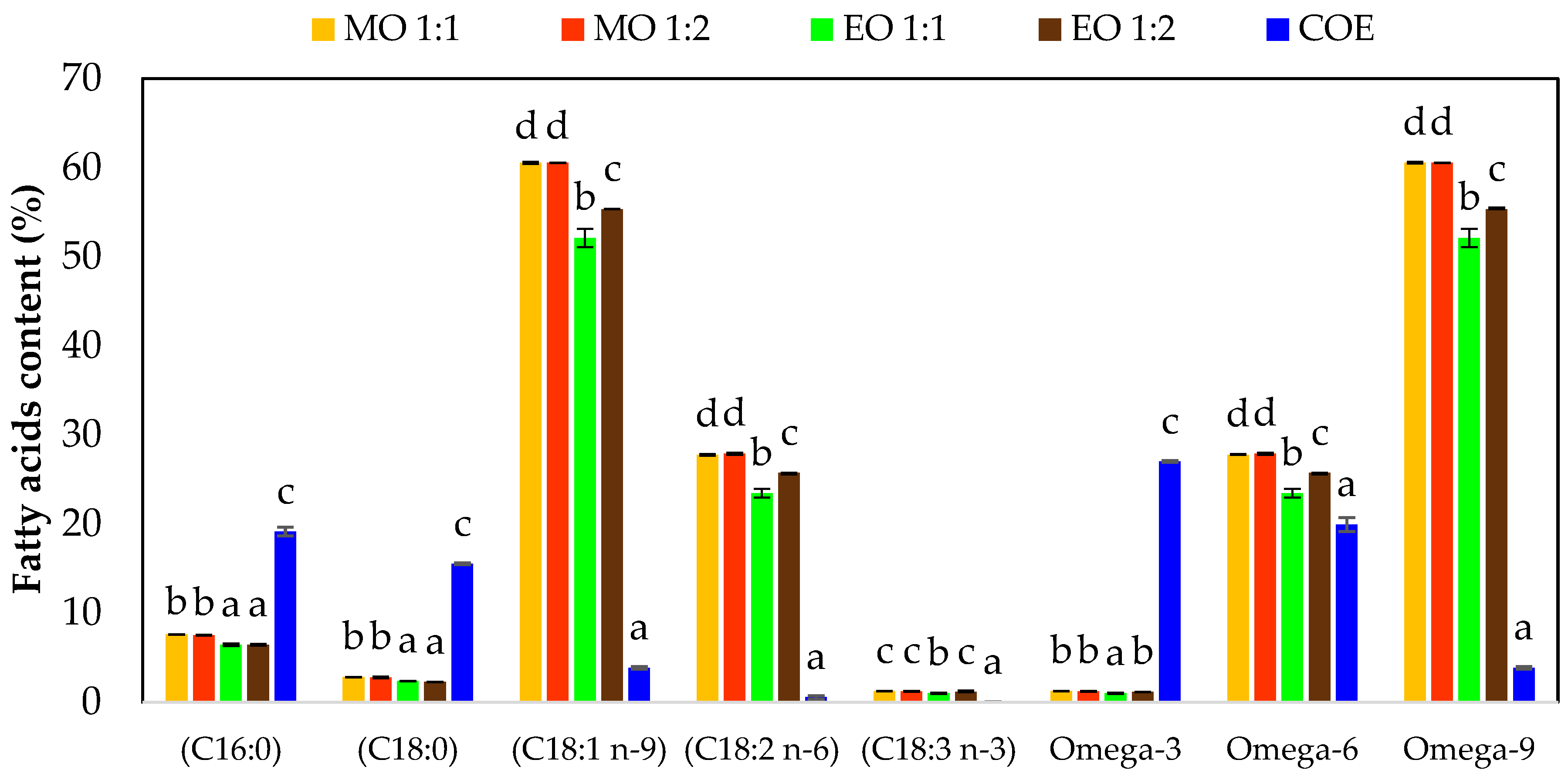
The means and standard deviations of the fatty acid profile groups (GC, expressed as percentage) from Octopus maya by-products obtained using HDES and the conventional method (COE) are summarized in Figure 8, where significant differences (p < 0.05) among treatments were observed for each fatty acid group. These results allow a direct comparison between conventional extraction (COE) and HDES systems in terms of fatty acid composition.
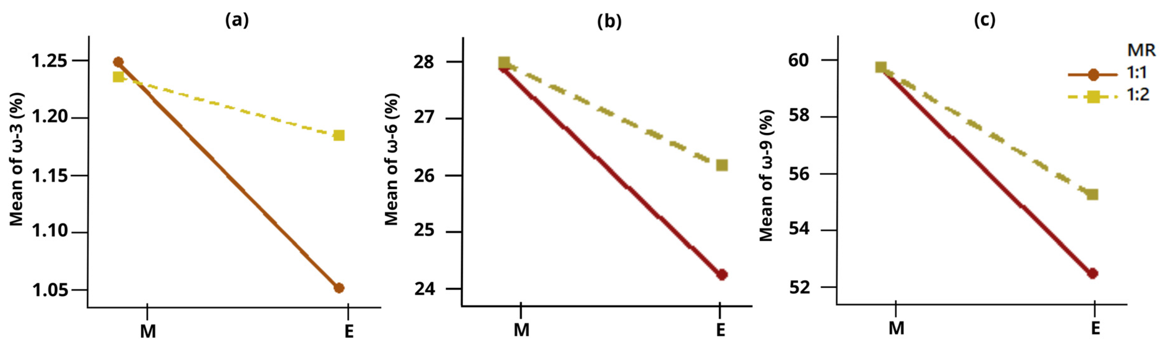
According to the experimental design analysis of HDES extracts based on GC results, the interaction between the hydrogen bond acceptor and molar ratio (HBA × MR) had a significant effect (p < 0.05) on ω-3 content (Table 3), highlighting the potential influence of intermolecular interactions among HDES components on extraction performance. The interaction plot (Figure 9a) shows that menthol at a molar ratio of 1:1 resulted in the highest ω-3 recovery (1.25%). Although these results show consistent trends among treatments, they should be interpreted considering the limited number of experimental replicates.
Figure 8. Fatty acid profile expressed as percentage (%) in extracts of HDES of by-products Octopus maya and conventional method (the corresponding GC chromatograms are presented in Figure 10). Values are reported as mean ± standard deviation (SD, n = 2). Different letters indicate statistically significant differences (LSD, p < 0.05) among each fatty acid group. Note: C16:0 (palmitic acid); C18:0 (stearic acid); C18:1 n-9 (oleic acid); C18:2 n-6 (linoleic acid); C18:3 n-3 (linolenic acid). MO: menthol–oleic acid; EO: eucalyptol-oleic acid; COE: conventional oil extraction.
Processes 14 01384 g008
Figure 9. Interaction plot (HBA × MR) showing the effect of hydrogen bond acceptor and molar ratio on (a) ω-3, (b) ω-6 and (c) ω-9 content (%) in HDES extracts from Octopus maya by-products.
Processes 14 01384 g009
Figure 10. Fatty acid profile of HDES extracts obtained from Octopus maya by-products (MO 1:1 30, MO 1:2 30, EO 1:1 30, and EO 1:2 30; nomenclature as defined in Table 1) and conventional oil extraction (COE): GC chromatograms.
Processes 14 01384 g010
Overall, the HDES extracts obtained from the by-products showed ω-3 GC values ranging from 1.02 to 1.25%, which are consistent with the levels observed for α-linolenic acid (ALA, C18:3 n-3), indicating that the ω-3 fraction recovered by HDES is primarily associated with this fatty acid. This behavior suggests a selective extraction toward shorter-chain polyunsaturated fatty acids and a limited affinity for long-chain ω-3 compounds such as EPA and DHA, likely due to steric and structural constraints that affect solvent–solute interactions [34].
From a complementary perspective, UV–Vis spectrophotometric analysis showed higher values of ω-3 equivalents in eucalyptol-based systems (0.83 mg/mL). This behavior may be explained by the non-specific detection of unsaturated compounds at this wavelength, which reflects the overall degree of unsaturation rather than the actual ω-3 content. Therefore, the higher UV–Vis response observed in eucalyptol-based systems likely corresponds to a greater presence of unsaturated compounds in general and does not necessarily reflect a higher recovery of ω-3 fatty acids [35]. In this context, GC results provide a more accurate representation of the extraction behavior, indicating that menthol-based systems achieved higher ω-3 recovery (1.23–1.25%).
Based on the results obtained by GC, this behavior suggests that the molecular structure of the hydrogen bond acceptor (HBA) influences the extraction performance of the system. Eucalyptol, which lacks hydroxyl groups, forms weaker intermolecular interactions, resulting in lower viscosity (0.011–0.023 Pa·s) and improved diffusion but reduced capacity to selectively solubilize specific fatty acids [36]. In contrast, menthol contains a hydroxyl group (–OH) capable of forming hydrogen bonds, promoting stronger intermolecular interactions and a more structured solvent network [37]. Although this leads to higher viscosity (0.027–0.036 Pa·s), it may enhance specific interactions with lipid molecules, favoring the extraction of ω-3 fatty acids detected by GC.
Anstiss et al. [24] reported that HDES formulated with menthol–lidocaine favors the recovery of ω-3 fatty acids (EPA and DHA) from mussels (Perna canaliculus). The authors attributed this behavior to the molecular affinity between the HDES and ω-3 fatty acids, which in addition to their hydrophobic character, contain carboxyl groups capable of participating in hydrogen-bonding. In this context, the presence of functional groups able to promote intermolecular interactions appears to play a key role in enhancing solute–solvent affinity and, consequently, extraction selectivity, beyond the effect of transport properties alone [34]. Furthermore, Topal et al. [25] noted that the affinity of HDES for fatty acids depends on the interactions between the components of the HDES system and the nature of the target lipids. In their study, menthol–based systems (menthol–carvacrol and menthol–thymol) showed good extraction capacity for total lipids in fish, which are rich in neutral fats (triacylglycerols and sterols), but lower efficiency in mussels, where polar lipids (phospholipids) predominate. This behavior was attributed to the non-polar properties of these HDES, which limit their affinity toward polar lipid compounds. These findings are consistent with the results of the present study, suggesting that extraction efficiency is driven by the combined influence between solvent polarity, hydrogen bonding capacity, and molecular interactions. This indicates that the appropriate selection of HDES components is important to improve extraction capacity to tailor selectivity toward specific lipid fractions. In this context, a distinct selective behavior was also observed for other polyunsaturated fatty acids [38].
On the other hand, conventional extraction (COE) showed a significantly higher ω-3 content (19.95 ± 0.78%) compared to the extracts obtained with the HDES. This fraction in COE is mainly composed of long-chain ω-3 fatty acids, such as eicosapentaenoic acid (EPA), docosahexaenoic acid (DHA), and alpha-linolenic acid (ALA, C18:3 n-3) [46]. The latter presented a value of 0.05%, suggesting that approximately 19.9% could correspond mainly to EPA and DHA, and to a much lesser extent to DPA (docosapentaenoic acid, C22:5 n-3), since, according to Torrinha et al. [6], this fatty acid is present in very small amounts in Octopus maya (4.3 mg/100 g of C22:5 n-3).
From a comparative perspective, conventional ethanol extraction (COE) and HDES systems exhibited markedly different behaviors in terms of GC ω-3 recovery. While COE showed higher total ω-3 percentages, primarily associated with long-chain fatty acids such as EPA and DHA, HDES extracts presented lower ω-3 values within a narrower range (1.02–1.25%), corresponding mainly to α-linolenic acid (ALA). This contrast indicates that, rather than maximizing total ω-3 recovery, HDES systems may exhibit more selective extraction depending on the molecular characteristics of the target compounds (short-chain fatty acids) which may limit the recovery of high-value compounds such as EPA and DHA under the evaluated conditions of composition (HBA:HBD). Therefore, although HDES do not surpass conventional extraction in terms of total ω-3 yield, they may offer advantages in terms of selectivity and the ability to tailor extraction conditions.
In addition to ω-3 fatty acids experimental design, analysis revealed that the interaction between the hydrogen bond acceptor and molar ratio (HBA × MR) had a significant effect (p < 0.05) on ω-6 values. These results indicate that HDES compositions favor improved recovery of these fatty acids. The interaction plot (Figure 9b) shows that menthol-based formulations at an MR of 1:2 resulted in the highest ω-6 extraction yield (27.91%), corresponding with linoleic acid (C18:2 n-6), suggesting this as the major fraction within the ω-6 group in HDES extracts.
By comparison, the COE extract showed a lower content of C18:2 n-6 (0.55 ± 0.2%) but a higher total percentage of ω-6 (19.95 ± 0.78%), reflecting the presence of ω-6 fatty acids such as linoleic acid, γ-linolenic acid (GLA) and arachidonic acid (AA) [46]. This difference in compound distribution indicates that, while the conventional method enables a broader extraction of the ω-6 fraction, HDES systems tend to selectively concentrate specific components, a trend also observed for ω-3 extraction.
This behavior may be attributed to the molecular compatibility between HDES components with linoleic acid and both linoleic and α-linolenic acid, whose structural features (chain length and degree of unsaturation) favor van der Waals forces and hydrogen bonding with the solvent system [34].
In particular, the presence of OH groups [37] may promote the preferential solubilization of these fatty acids. Overall, these findings highlight the capacity of HDES to enable targeted extraction of specific ω-6 and ω-3 fatty acids.
Consequently, this selective behavior has important implications for the targeted recovery of high-value lipids, suggesting that HDES can be designed to preferentially extract specific polyunsaturated fatty acids depending on the desired application.
On the other hand, the content of oleic acid (C18:1 n-9) predominated in the HDES extracts, attributed to its presence as a principal component of the solvent formulation. In contrast, the extract obtained using the conventional method (COE) showed a content of 3.87 ± 0.59%, a value close to that reported by Gullian et al. [47] in their analysis of the fatty acid profile of Octopus maya, where a conventional extraction method was used (5.74% of C18:1 n-9).
Nevertheless, the significant differences (p < 0.05) observed between menthol- and eucalyptol-based systems (HBA × MR) indicate that HDES composition influences the final proportion of oleic acid in extracts. In this regard, menthol-based systems (molar ratios 1:1 and 1:2, Figure 9c) showed higher oleic acid contents (60.55%), which may be associated with the ability of menthol to establish stronger intermolecular interactions through hydroxyl groups (–OH), compared to eucalyptol [36,37].
Finally, saturated fatty acids (palmitic acid, C16:0 and stearic acid, C18:0), obtained using the conventional method (COE), showed significantly higher values (19.16 ± 0.49 and 15.55 ± 0.12%, respectively) compared to HDES extracts, which ranged from 6% < C16:0 < 8% and 2% < C18:0 < 3%. According to the experimental design analysis, the type of HBA had a significant effect (p < 0.05), with menthol showing the highest proportion of these saturated fatty acids among the evaluated systems. This behavior may be attributed to the low polarity and absence of reactive sites, which restrict their interaction with the hydrogen-bonding network and structural organization of HDES [36]. Thus, the lower recovery of these compounds reflects both the physicochemical limitations of the system and the reduced affinity of the solvent for these lipids (palmitic and stearic acid).
From a comparative perspective, these results indicate that, unlike the conventional method, HDES limit the extraction of saturated fatty acids. This characteristic may be considered advantageous in terms of selectivity, particularly when the goal is to enrich lipid fractions with higher functional or nutritional value [48].

4. Conclusions

In this study, hydrophobic deep eutectic solvents (HDES) formulated from oleic acid in combination with menthol or eucalyptol were evaluated as alternative systems for the extraction and valorization of fatty acids from Octopus maya by-products, with particular emphasis on ω-3 recovery. Based on gas chromatography (GC) results, it was observed that solvent composition, particularly the type of hydrogen bond acceptor (HBA) and its molar ratio (MR), significantly influences the recovery of different fatty acid groups, evidencing formulation-dependent extraction behavior.
The results indicate that HDES systems exhibit a distinct extraction profile compared to ethanol, showing affinity toward specific compounds rather than enabling broad-spectrum extraction. Menthol-based formulations favored the recovery of linoleic acid (ω-6) and, to a lesser extent, α-linolenic acid (ω-3), whereas the conventional method (COE) achieved a higher overall recovery of fatty acids. This contrast highlights that, under the evaluated conditions, HDES do not maximize total ω-3 recovery, but instead allow adjustment of the lipid profile obtained, reflecting their selective behavior.
This selectivity may be associated with the molecular compatibility between the solvent system and the target compounds, through HBA and MR of HDES. Therefore, HDES can be considered a viable alternative when the objective is the selective recovery of specific lipid fractions rather than exhaustive extraction. However, this also implies that extraction efficiency depends on the chemical nature of fatty acids. Thus, the recovery of long-chain ω-3 fatty acids (EPA and DHA) requires further optimization of solvent design to enhance affinity and solubilization.
It is important to note that the interpretation of ω-3 recovery, considering the limited replicates, is primarily supported by GC data, while UV–Vis results were used only as complementary indicators of relative trends due to their non-specific nature.
Future work should focus on establishing particular relationships between HDES composition and fatty acid selectivity, as well as optimizing the parameters studied (HBA type, molar ratio, UAE time).
The design of HDES for lipid recovery from marine by-products may enable the selective extraction of omega fatty acids, which could be attractive to the ingredients industry for incorporation into food matrices to enhance nutritional value or to develop functional foods.

Author Contributions

Conceptualization, M.O.R.-S., I.M.R.-B. and D.A.-G.; methodology, M.O.R.-S., I.M.R.-B., J.V.C.-R., D.A.-G. and I.L.-Á.; software, M.O.R.-S., D.A.-G. and J.V.C.-R.; validation, M.O.R.-S., I.M.R.-B. and J.V.C.-R.; formal analysis, M.O.R.-S. and D.A.-G.; investigation, M.O.R.-S., D.A.-G. and I.L.-Á.; resources, M.O.R.-S.; data curation, M.O.R.-S.; writing—original draft preparation, D.A.-G.; writing—review and editing, M.O.R.-S., I.M.R.-B., J.V.C.-R. and T.C.-C.; visualization, M.O.R.-S.; supervision, M.O.R.-S.; project administration, M.O.R.-S.; funding acquisition, M.O.R.-S. All authors have read and agreed to the published version of the manuscript.

Funding

To the project 6559 “Plataforma tecnológica pulpo maya para el desarrollo de productos de alto valor agregado” and to SECIHTI for the scholarship No. 4019459 granted to Daniela Aguilar-González.

Data Availability Statement

The original contributions presented in this study are included in the article. Further inquiries can be directed to the corresponding author.

Acknowledgments

Rossana Faride Vargas Coronado for her technical assistance in FTIR and Raman spectra data acquisition and to Rubén Cejas Gutiérrez for the support in GC analysis.

Conflicts of Interest

The authors declare no conflicts of interest.

References

  1. Gobierno de México, Secretaría de Agricultura y Desarrollo Rural. El Pulpo: La Pesquería más Importante de Yucatán Inició este 1° de Agosto. 31 July 2024. Available online: https://www.gob.mx/agricultura/yucatan/articulos/el-pulpo-la-pesqueria-mas-importante-de-yucatan-inicia-este-1o-de-agosto?idiom=es (accessed on 4 February 2026).
  2. Pineda-Suazo, D.; Escobedo-Hinojosa, W.; Fabian-Canseco, L.E.; Gallardo, P.; Moguel-Ojeda, C.; Caamal-Monsreal, C.; Sánchez-Arteaga, A.; Rosas, C. Evaluation of Octopus maya enzyme activity of the digestive gland and gastric juice. Biol. Open 2024, 13, bio060429. [Google Scholar] [CrossRef] [PubMed]
  3. Rosas, C.; Markaida, U.; López Rocha, J. Octopus maya, the Mayan Octopus. In Octopus Biology and Ecology; Rosa, E.R., Gleadall, I., Villanueva, G.P.R., Eds.; Elsevier: Amsterdam, The Netherlands, 2024; pp. 97–119. [Google Scholar] [CrossRef]
  4. Gobierno del Estado de Yucatán. Costa Yucateca Cierra Exitosa Temporada de Pulpo [Comunicado de Prensa]. 18 December 2025. Available online: https://www.yucatan.gob.mx/saladeprensa/ver_nota.php?id=10213 (accessed on 9 February 2026).
  5. Rasmiya Begum, S.L.; Himaya, S.M.M.S.; Imthiyas, M.S.M.; Afreen, S.M.M.S. Fish Waste: Understanding the pollution potential and sustainable mitigation strategies. In Fish Waste to Valuable Products; Springer Nature: Singapore, 2024; pp. 427–440. [Google Scholar] [CrossRef]
  6. Torrinha, Á.; Cruz, R.; Gomes, F.; Mendes, E.; Casal, S.; Morais, S. Octopus lipid and vitamin E composition: Interspecies, interorigin, and nutritional variability. J. Agric. Food Chem. 2014, 62, 8508–8517. [Google Scholar] [CrossRef] [PubMed]
  7. Calder, P.C.; Cawood, A.L.; James, C.; Page, F.; Putnam, S.; Minihane, A.M. An overview of national and international long chain omega-3 polyunsaturated fatty acid intake recommendations for healthy populations. Nutr. Res. Rev. 2025, 39, e6. [Google Scholar] [CrossRef]
  8. Banaszak, M.; Dobrzyńska, M.; Kawka, A.; Górna, I.; Woźniak, D.; Przysławski, J.; Drzymała-Czyż, S. Role of Omega-3 fatty acids eicosapentaenoic (EPA) and docosahexaenoic (DHA) as modulatory and anti-inflammatory agents in noncommunicable diet-related diseases–Reports from the last 10 years. Clin. Nutr. ESPEN 2024, 63, 240–258. [Google Scholar] [CrossRef]
  9. Gutierres, D.; Pacheco, R.; Reis, C.P. The Role of Omega-3 and Omega-6 Polyunsaturated Fatty Acid Supplementation in Human Health. Foods 2025, 14, 3299. [Google Scholar] [CrossRef]
  10. Gaspar, L.; Ricardo, F.; Melo, T.; Domingues, P.; Domingues, M.R.; Calado, R.; Rey, F. Lipidomics of common octopus’(Octopus vulgaris) arm muscle using untargeted high-resolution liquid chromatography-mass spectrometry. J. Food Compos. Anal. 2023, 115, 104871. [Google Scholar] [CrossRef]
  11. Rahman, N.; Hashem, S.; Akther, S.; Jothi, J.S. Impact of various extraction methods on fatty acid profile, physicochemical properties, and nutritional quality index of Pangus fish oil. Food Sci. Nutr. 2023, 11, 4688–4699. [Google Scholar] [CrossRef] [PubMed]
  12. David, E.; Niculescu, V.C. Volatile organic compounds (VOCs) as environmental pollutants: Occurrence and mitigation using nanomaterials. Int. J. Environ. Res. Public Health 2021, 18, 13147. [Google Scholar] [CrossRef]
  13. Méndez, L.; Rodríguez, A.; Aubourg, S.P.; Medina, I. Low-toxicity solvents for the extraction of valuable lipid compounds from octopus (Octopus vulgaris) waste. Foods 2023, 12, 3631. [Google Scholar] [CrossRef]
  14. Martins, R.; Barbosa, A.; Advinha, B.; Sales, H.; Pontes, R.; Nunes, J. Green extraction techniques of bioactive compounds: A state-of-the-art review. Processes 2023, 11, 2255. [Google Scholar] [CrossRef]
  15. Ivanković, A.; Dronjić, A.; Martinović Bevanda, A.; Talić, S. Review of 12 Principles of Green Chemistry in Practice. Int. J. Sustain. Green Energy 2017, 6, 39–48. [Google Scholar] [CrossRef]
  16. Devi, M.; Moral, R.; Thakuria, S.; Mitra, A.; Paul, S. Hydrophobic deep eutectic solvents as greener substitutes for conventional extraction media: Examples and techniques. ACS Omega 2023, 8, 9702–9728. [Google Scholar] [CrossRef]
  17. Martel-Martín, S.; Di Pietro, M.E.; Gutiérrez, A.; Aguilar, N.; Bol-Arreba, A.; Aparicio, S.; Matroodi, F.; Rossi, B.; Mele, A. A paradigm for natural eutectic solvents based on fatty acids: Molecular interactions and toxicological considerations. J. Mol. Liq. 2024, 414, 126148. [Google Scholar] [CrossRef]
  18. Ristivojević, P.; Krstić Ristivojević, M.; Stanković, D.; Cvijetić, I. Advances in extracting bioactive compounds from food and agricultural waste and by-products using natural deep eutectic solvents: A circular economy perspective. Molecules 2024, 29, 4717. [Google Scholar] [CrossRef] [PubMed]
  19. Trusheva, B.; Petkov, H.; Chimshirova, R.; Popova, M.; Dimitrova, L.; Zaharieva, M.M.; Ilieva, Y.; Vasileva, B.; Tsvetkova, I.; Najdenski, H.; et al. Insight into the influence of natural deep eutectic solvents on the extraction of phenolic compounds from poplar type propolis: Composition and in vitro biological activity. Heliyon 2024, 10, e28621. [Google Scholar] [CrossRef] [PubMed]
  20. Makoś, P.; Słupek, E.; Gębicki, J. Hydrophobic deep eutectic solvents in microextraction techniques–A review. Microchem. J. 2020, 152, 104384. [Google Scholar] [CrossRef]
  21. Trenzado, J.L.; Benito, C.; Atilhan, M.; Aparicio, S. Hydrophobic Deep eutectic Solvents based on cineole and organic acids. J. Mol. Liq. 2023, 377, 121322. [Google Scholar] [CrossRef]
  22. Aguilar, N.; Benito, C.; Martel-Martín, S.; Gutiérrez, A.; Rozas, S.; Marcos, P.A.; Bol-Arreba, A.; Atilhan, M.; Aparicio, S. Insights into Carvone: Fatty Acid Hydrophobic NADES for Alkane Solubilization. Energy Fuels 2024, 38, 23633–23653. [Google Scholar] [CrossRef]
  23. Rozas, S.; Zamora, L.; Benito, C.; Atilhan, M.; Aparicio, S. A study on monoterpenoid-based natural deep eutectic solvents. Green Chem. Eng. 2023, 4, 99–114. [Google Scholar] [CrossRef]
  24. Anstiss, L.; Weber, C.C.; Baroutian, S.; Shahbaz, K. Menthol-based deep eutectic solvents as green extractants for the isolation of omega-3 polyunsaturated fatty acids from Perna canaliculus. J. Chem. Technol. Biotechnol. 2023, 98, 1791–1802. [Google Scholar] [CrossRef]
  25. Topal, T.; Card, A.; Mackenzie, A.D.; Lagutin, K.; Marshall, S.N.; Cumming, A.H.; Killeen, D.P. Hydrophobic natural deep eutectic solvents for marine lipid extraction. J. Am. Oil Chem. Soc. 2024, 101, 361–367. [Google Scholar] [CrossRef]
  26. Kuo, C.H.; Liao, H.Z.; Wang, Y.H.; Wang, H.M.D.; Shieh, C.J.; Tseng, C.Y. Highly efficient extraction of EPA/DHA-enriched oil from cobia liver using homogenization plus sonication. Eur. J. Lipid Sci. Technol. 2017, 119, 1600466. [Google Scholar] [CrossRef]
  27. Ramírez-Sucre, M.O.; Avilés-Betanzos, K.A.; López-Martínez, A.; Rodríguez-Buenfil, I.M. Evaluation of polyphenol profile from citrus peel obtained by natural deep eutectic solvent/ultrasound extraction. Processes 2024, 12, 2072. [Google Scholar] [CrossRef]
  28. Ramírez-Sucre, M.O.; Baigts-Allende, D.K. Efecto del tratamiento térmico en el comportamiento reológico de salsas de chile habanero (Capsicum chinense) adicionadas con gomas guar y xantana. Agrociencia 2016, 50, 837–847. [Google Scholar]
  29. Oliveira, C.; Santos, J.; Sousa, R.G. UV Spectrophotometry applied to the quantification of omega-3, -6 and -9 in fresh tissues of wild and farmed tambaqui. Int. J. Innov. Educ. Res. 2020, 8, 183–195. [Google Scholar] [CrossRef]
  30. Avilés-Betanzos, K.A.; Cauich-Rodríguez, J.V.; Ramírez-Sucre, M.O.; Rodríguez-Buenfil, I.M. Optimization of Spray Drying Conditions for a Capsicum chinense Leaf Extract Rich in Polyphenols Obtained by Ultrasonic Probe/NADES. ChemEngineering 2024, 8, 131. [Google Scholar] [CrossRef]
  31. Pech-Pisté, R.; Pérez-Aranda, C.; Balam, A.; Vargas-Coronado, R.; Cauich-Rodríguez, J.V.; Avilés, F. Piezoimpedance of carbon nanotube yarns coupled with Raman spectroscopy and its implementation for sensing polymerization kinetics. Carbon 2023, 213, 118246. [Google Scholar] [CrossRef]
  32. Adeoye, D.O.; Gano, Z.S.; Ahmed, O.U.; Shuwa, S.M.; Atta, A.Y.; Iwarere, S.A.; Jubril, B.Y.; Daramola, M.O. Synthesis and characterisation of menthol-based hydrophobic deep eutectic solvents. Chem. Proc. 2023, 14, 98. [Google Scholar] [CrossRef]
  33. Dabbagh Hosseini Pour, M.; Jahanbin Sardroodi, J.; Hadidi, N.; Pazuki, G. Molecular dynamics and COSMO-RS model of menthol–fatty acid deep eutectic solvents: Thermodynamic, structural, and dynamics insights. Sci. Rep. 2025, 15, 41849. [Google Scholar] [CrossRef]
  34. Ferreira, C.; Sarraguça, M. A comprehensive review on deep eutectic solvents and its use to extract bioactive compounds of pharmaceutical interest. Pharmaceuticals 2024, 17, 124. [Google Scholar] [CrossRef] [PubMed]
  35. Rohman, A.; Irnawati; Windarsih, A.; Riswanto, F.D.O.; Indrayanto, G.; Fadzillah, N.A.; Riyanto, S.; Abu Bakar, N.K.A. Application of chromatographic and spectroscopic-based methods for analysis of Omega-3 (Ω-3 Fas) and Omega-6 (Ω-6 Fas) fatty acids in marine natural products. Molecules 2023, 28, 5524. [Google Scholar] [CrossRef] [PubMed]
  36. Hoch, C.C.; Petry, J.; Griesbaum, L.; Weiser, T.; Werner, K.; Ploch, M.; Verschoor, A.; Multhoff, G.; Dezfouli, A.B.; Wollenberg, B. 1, 8-cineole (eucalyptol): A versatile phytochemical with therapeutic applications across multiple diseases. Biomed. Pharmacother. 2023, 167, 115467. [Google Scholar] [CrossRef] [PubMed]
  37. Cherniakova, M.; Varchenko, V.; Belikov, K. Menthol-based (deep) eutectic solvents: A review on properties and application in extraction. Chem. Rec. 2024, 24, e202300267. [Google Scholar] [CrossRef]
  38. Şahin, S.; Kurtulbaş, E.; Toprakçı, I.; Anwar, F.; Khan, R.; Ciğeroğlu, Z.; Yardımcı, A.İ.; Torun, M.; Torun, F.B.; Falsafi, S.R. Deep Eutectic Solvents for Sustainable Extraction of Bioactive Compounds from Biomass: Mechanistic Insights and Scale-Up Challenges. Molecules 2026, 31, 880. [Google Scholar] [CrossRef]
  39. Strieder, M.M.; Bragagnolo, F.S.; Mendiola, J.A.; Rostagno, M.A.; Ibáñez, E. Screening and characterization of 1, 8-cineole-based solvents as an alternative to hexane for obtaining nonpolar compounds from plant-based milk coproducts. ACS Sustain. Chem. Eng. 2024, 12, 16052–16063. [Google Scholar] [CrossRef]
  40. Bagović Kolić, M.; Železnjak, M.; Markov, K.; Gaurina Srček, V.; Cvjetko Bubalo, M.; Radošević, K.; Radojčić Redovniković, I. Physicochemical and Biological Properties of Menthol and Thymol-Based Natural Deep Eutectic Solvents. Molecules 2025, 30, 1713. [Google Scholar] [CrossRef]
  41. Nandiyanto, A.B.D.; Oktiani, R.; Ragadhita, R. How to read and interpret FTIR spectroscope of organic material. Indones. J. Sci. Technol. 2019, 4, 97–118. [Google Scholar] [CrossRef]
  42. Vongsvivut, J.; Miller, M.R.; McNaughton, D.; Heraud, P.; Barrow, C.J. Rapid discrimination and determination of polyunsaturated fatty acid composition in marine oils by FTIR spectroscopy and multivariate data analysis. Food Bioprocess Technol. 2014, 7, 2410–2422. [Google Scholar] [CrossRef]
  43. Ahmmed, F.; Killeen, D.P.; Gordon, K.C.; Fraser-Miller, S.J. Rapid quantitation of adulterants in premium marine oils by Raman and IR spectroscopy: A data fusion approach. Molecules 2022, 27, 4534. [Google Scholar] [CrossRef] [PubMed]
  44. Ahmmed, F.; Gordon, K.C.; Killeen, D.P.; Fraser-Miller, S.J. Detection and quantification of adulteration in krill oil with Raman and infrared spectroscopic methods. Molecules 2023, 28, 3695. [Google Scholar] [CrossRef] [PubMed]
  45. Słomczyńska, P.; Siudem, P.; Białek, A.; Kaźmierski, S.; Paradowska, K. The Application of 1H Nuclear Magnetic Resonance (NMR), Gas Chromatography (GC) and Ultraviolet–Visible (UV-Vis) Spectroscopy Techniques to the Analysis of the Fatty Acid Profile as Quality of Argan Oil. Int. J. Mol. Sci. 2025, 26, 5322. [Google Scholar] [CrossRef] [PubMed]
  46. Swetha, N.; Mathanghi, S.K. Towards sustainable omega-3 fatty acids production–A comprehensive review on extraction methods, oxidative stability and bio-availability enhancement. Food Chem. Adv. 2024, 4, 100603. [Google Scholar] [CrossRef]
  47. Gullian-Klanian, M.; Terrats-Preciat, M.; Pech-Jiménez, E.C.; Cutz De Ocampo, J. Effect of frozen storage on protein denaturation and fatty acids profile of the red octopus (Octopus maya). J. Food Process. Preserv. 2017, 41, e13072. [Google Scholar] [CrossRef]
  48. Lawrence, G.D. Saturated Fats: Time to Assess Their Beneficial Role in a Healthful Diet. Dietetics 2024, 3, 452–462. [Google Scholar] [CrossRef]
Figure 1. Viscosity as a function of shear rate for HDES extracts obtained from Octopus maya by-products, compared with salmon oil. (a) Viscosity behavior of extracts formulated with menthol; (b) viscosity behavior of extracts formulated with eucalyptol.
Processes 14 01384 g001
Figure 2. Viscosity at shear rate 10 s−1 expressed in Pa·s from HDES extracts of by-products Octopus maya. M: menthol; E: eucalyptol; O: oleic acid. Molar ratio HBA:HBD. Values are expressed as mean ± standard deviation (SD, n = 2). Different letters indicate statistically significant differences (LSD, p < 0.05).
Processes 14 01384 g002
Figure 3. Interaction and main effects plots from a 2 × 3 × 2 full factorial design evaluating the influence of hydrogen bond acceptor type (HBA: M = menthol, E = eucalyptol), molar ratio (MR: 1:1, 1:2, 2:1), and extraction time (ET: 30 min, 60 min) on the means of viscosity. (a) HBA × MR and (b) HBA × ET.
Processes 14 01384 g003
Figure 4. Omega-3 equivalents (mg/mL) obtained in extracts of HDES of by-products Octopus maya evaluated by spectrophotometer UV-Vis. M = menthol; E = eucalyptol; O = oleic acid; in different HBA:HBD molar ratios. Values are expressed as mean ± standard deviation (SD, n = 2). Different letters indicate statistically significant differences (LSD, p < 0.05).
Processes 14 01384 g004
Figure 5. Interaction and main effects plots from a 2 × 3 × 2 full factorial design evaluating the influence of hydrogen bond acceptor type (HBA: M = menthol, E = eucalyptol), molar ratio (MR: 1:1, 1:2, 2:1), and extraction time (ET: 30 min, 60 min) on the mean yield of omega-3 equivalents (mg/mL): (a) MR × HBA (b) ET × HBA; (c) MR × ET and (d) main effects for each individual factor on EO3 (mg/mL) in HDES extracts obtained from Octopus maya by-products determined by spectrophotometer UV-Vis.
Processes 14 01384 g005
Figure 6. FTIR spectra of 30 min HDES extracts with the highest omega-3 equivalents (mg/mL), according to the UV–Vis spectrophotometric analysis. MO: menthol–oleic acid; EO: eucalyptol–oleic acid. The numbers shown at each peak correspond to the wavenumber at which the signal was observed.
Processes 14 01384 g006
Figure 7. Raman spectra of 30 min HDES extracts with the highest omega-3 equivalents (mg/mL), as determined by UV–Vis spectrophotometric analysis. MO: menthol–oleic acid; EO: eucalyptol–oleic acid. The numbers shown on each peak indicate the corresponding Raman shift at which the vibrational bands were detected.
Processes 14 01384 g007
Table 1. Proposed experimental design. Factors—hydrogen bond acceptor (HBA), molar ratios, and UAE time—and response variables.
Hydrogen Bond
Acceptor (HBA)
Hydrogen Bond Donor (HBD)Molar Ratio (HBA:HBD)Extraction Time (UAE) (min)NomenclatureResponse
Variables
Menthol (M)
(C10H20O)
Processes 14 01384 i001
Oleic acid (O)
(C18H34O2)
Processes 14 01384 i003
1:130MO(1:1)30
  • Omega-3 equivalents
  • Viscosity
1:2MO(1:2)30
2:1MO(2:1)30
1:160MO(1:1)60
1:2MO(1:2)60
2:1MO(2:1)60
Eucalyptol (E)
(C10H18O)
Processes 14 01384 i002
1:130EO(1:1)30
1:2EO(1:2)30
2:1EO(2:1)30
1:160EO(1:1)60
1:2EO(1:2)60
2:1EO(2:1)60
Table 2. Effect of the hydrogen bond acceptor (HBA), molar ratio (MR), and UAE time (ET); the effect of their double and triple interactions on the response variables at a 95% confidence level (p < 0.05).
Response Variablesp Values
Individual FactorsDouble InteractionsTriple
Interactions
HBAMR (mol/mol)ET (min)HBA × MRHBA × ETMR × ETHBA × MR × ET
Omega-3 equivalents (mg/mL)0.0060.0700.5290.0080.0030.6350.017
Viscosity (Pa·s)<0.0010.2400.867<0.0010.5060.2800.252
Note: Statistically significant effects (p < 0.05) are shown in bold.
Table 3. Effect on the fatty acids group of the hydrogen bond acceptor (HBA), molar ratio (MR) and their double interactions (HBA × MR) at a 95% confidence level (p < 0.05).
Response Variablesp Value
Individual FactorsDouble Interactions
HBAMRHBA × MR
C16:0<0.0010.4440.444
C18:0<0.0010.0340.092
C18:1 n-9<0.0010.0120.012
C18:2 n-6<0.0010.0030.005
C18:3 n-30.0050.0500.028
ω–30.0050.0500.028
ω–6<0.0010.0030.005
ω–9<0.0010.0120.012
Note: Statistically significant effects (p < 0.05) are shown in bold.
Disclaimer/Publisher’s Note: The statements, opinions and data contained in all publications are solely those of the individual author(s) and contributor(s) and not of MDPI and/or the editor(s). MDPI and/or the editor(s) disclaim responsibility for any injury to people or property resulting from any ideas, methods, instructions or products referred to in the content.