Article

Genomic Insights into a Thermophilic Bacillus licheniformis Strain Capable of Degrading Polyethylene Terephthalate Intermediate


by
Pedro Eugenio Sineli
1,†,
Fernando Gabriel Martínez
1,†,
Federico Zannier
1,
Luciana Costas
2,
José Horacio Pisa
1,
Analía Álvarez
1,3,* and
Cintia Mariana Romero
1,2,*
1
Pilot Plant for Industrial and Microbiological Processes (PROIMI-CONICET), Avenida Belgrano y Pasaje Caseros, San Miguel de Tucumán 4000, Argentina
2
Faculty of Biochemistry, Chemistry and Pharmacy, National University of Tucumán, Ayacucho 491, San Miguel de Tucumán 4000, Argentina
3
Natural Sciences College and the Miguel Lillo Institute, National University of Tucumán, Miguel Lillo 205, San Miguel de Tucumán 4000, Argentina
*
Authors to whom correspondence should be addressed.
These authors contributed equally to this work.
Processes 2026, 14(2), 381; https://doi.org/10.3390/pr14020381
Submission received: 22 December 2025 / Revised: 15 January 2026 / Accepted: 19 January 2026 / Published: 22 January 2026

Abstract

Bacillus licheniformis Mb1, a thermophilic strain isolated from the Yungas rainforest in northwestern Argentina, was analyzed through genomic and experimental approaches to explore its biotechnological potential. Phylogenomic analysis confirmed its close relationship with B. licheniformis reference strains. The genome revealed multiple genes associated with hydrolytic, oxidative, carbohydrate-active, and polyester-degrading activities, indicating a wide enzymatic capacity. Experimental assays demonstrated strong extracellular hydrolytic activities and efficient degradation of bis(2-hydroxyethyl) terephthalate (BHET), a key polyethylene terephthalate (PET) intermediate. In liquid cultures with 3 mg/mL BHET, B. licheniformis Mb1 achieved 99.9% depletion within four days, with transient BHET dimer accumulation and progressive terephthalic acid (TPA) production, reaching 1.17 mg/mL after 15 days. Mono (2-hydroxyethyl) terephthalate (MHET) and vanillic acid were not detected. Complete BHET and dimer degradation suggests the presence of versatile hydrolases acting on short-chain polyester intermediates. Sequence and molecular docking analyses identified a BHETase-like carboxylesterase as the main enzyme candidate, featuring a truncated lidC region that generates a more open catalytic cleft. This structural trait, not previously reported in bacterial BHETases, enables the accommodation of bulkier substrates such as BHET dimer. These findings highlight B. licheniformis Mb1 as a promising biocatalyst for polyester depolymerization and a valuable microbial resource for future enzyme discovery and plastic bioremediation strategies.

1. Introduction

Plastic pollution has emerged as one of the most critical environmental issues of the present era. Global synthetic polymer production exceeds hundreds of millions of tons per year, and their persistence in the ecosystems contributes to long-term ecological imbalance and potential health risks [1]. Polyethylene terephthalate (PET) is among the most widely used plastics, and despite its recyclability, post-consumer PET often accumulates in ecosystems because of inefficiencies in recycling processes and the durability of the polymer [2]. During enzymatic depolymerization of PET, the intermediates bis(2-hydroxyethyl) terephthalate (BHET) and mono(2-hydroxyethyl) terephthalate (MHET) play central roles. PETases catalyze the cleavage of ester bonds in PET, releasing MHET, BHET, and ethylene glycol (EG), while MHETases further hydrolyze MHET into terephthalic acid (TPA) and EG [3]. One of the obstacles in PET biodegradation is the accumulation of BHET, that can inhibit the enzymatic degradation. Therefore, an efficient microbial metabolism of BHET is essential for complete PET mineralization [4]. To address this limitation, several strategies have been proposed, including the design of PETase variants with reduced sensitivity to inhibition, and the incorporation of accessory enzymes with specific activity on BHET [5]. Thus, the search for enzymes capable of transforming BHET becomes particularly relevant. Recent studies have identified conserved BHETases in Streptomyces species, which degrade BHET and erode amorphous PET films under mild conditions. These findings highlight both the ecological prevalence and the catalytic significance of BHET-targeting esterases in the enzymatic degradation of PET [6].
Microorganisms from diverse environments have been explored as sources of PET-active enzymes, including cutinases, esterases, and engineered PETases [2]. Thermophilic bacteria are of particular interest, as higher temperatures improve polymer chain mobility and enzymatic activity [7]. Bacillus licheniformis is a spore-forming bacterium widely used in industry for its secretion of robust extracellular enzymes such as proteases, lipases, and esterases, many of which remain stable under harsh conditions [8]. Studies showing that B. licheniformis can degrade polymers like polylactic acid and polyethylene emphasize its potential for plastic biotransformation [9].
The Yungas rainforest in northwestern Argentina harbors a rich but underexplored microbial diversity with biotechnological potential. In this study, we report the whole genome of Bacillus licheniformis Mb1, a thermophilic strain isolated from this environment, and analyze its enzymatic potential. Genomic analysis focused on identifying enzymes potentially involved in the degradation of PET intermediates such as BHET.

2. Materials and Methods

2.1. Strain, Growth Conditions and DNA Extraction

Bacillus sp. Mb1 was previously isolated from soil samples collected in the Yungas region of Tucumán, Argentina and characterized as a thermophile strain. Regarding the time of isolation, the soil samples were subjected to selective thermal treatment followed by cultivation on nutrient agar. The Mb1 isolate was selected based on its ability to grow at elevated temperatures and its strong extracellular enzymatic activity. The strain was cultivated in YP medium at 45 °C with agitation at 150 rpm for 72 h. After centrifugation, the biomass was washed twice with phosphate-buffered saline (PBS, pH 7.0). Genomic DNA was extracted using the QIAamp Genomic DNA Kit (QIAGEN, Hilden, Germany) following the manufacturer’s protocol. DNA quality was assessed by agarose gel electrophoresis (0.8%), and concentration was determined with a NanoDrop spectrophotometer (Thermo Scientific, Waltham, MA, USA).

2.2. DNA Sequencing, Preprocessing Data and Genome Assembly

Genomic DNA from Bacillus sp. Mb1 was sequenced at Macrogen Inc. (Seoul, Republic of Korea) on the Illumina HiSeq platform using a TruSeq Nano DNA paired-end library with a read length of 151 bp. Raw sequence quality was assessed with FastQC v0.11.5 [10], and adapter trimming together with low-quality base removal was performed using Trimmomatic v0.36 [11]. Filtered high-quality reads were then assembled de novo with SPAdes v3.13.0 under default parameters, which applies a de Bruijn graph approach optimized for Illumina short reads [12]. Assembly validation was subsequently carried out by mapping the processed reads back to the draft genome and assessing completeness with BUSCO v3.0.2 using the bacteria_odb10 dataset [13]. Plasmid content was evaluated using PlasmidFinder [14] combined with BLAST v2.16.0 searches of small contigs to screen for plasmid replication genes. Species identification and phylogenetic placement were performed through Mash v2.0 [15] and Average Nucleotide Identity (ANI) calculations, comparing the draft genome against reference Bacillus genomes, also were performed a phylogenetics tree using Species Tree Builder v0.1.4.

2.3. Functional Annotations

The gene predictions of the assembled contigs were carried out with Prokka v1.14.6 [16], which integrates gene prediction with Prodigal, Barrnap, Aragorn, and other databases to identify protein-coding sequences, rRNAs, tRNAs, and tmRNA. Circular maps of the annotated genome were generated with Proksee [17]. Functional annotation was performed with eggNOG-mapper v2 [18] using the eggNOG v5.0 database, allowing assignment of proteins to orthologous groups and functional categories. Carbohydrate-active enzymes (CAZymes) were identified using the dbCAN2 meta server [19].

2.4. Evaluation of Hydrolytic and Redox Activities

As an initial exploratory step, qualitative plate assays were performed to assess the hydrolytic and reductive capabilities of Bacillus sp. Mb1. All plates were prepared on a YP 50% base (half-strength yeast extract–peptone; 1.3% agar) and supplemented with the test substrates as indicated below. For carbohydrate-active enzymes screening, YP medium was amended (w/v) with starch, carboxymethyl cellulose (CMC), mannan, or xylan at 0.5%, and with chitin at 0.1%. Proteolytic activity was evaluated on YP 50% agar containing 2% (w/v) reconstituted skim milk. Selenite reduction was assessed on YP 50% agar supplemented with sodium selenite (20 mg/L); selenite reduction was evaluated as a complementary functional assay to probe the reductive and nanoparticle-forming capacity of the strain [20]. Laccase activity was preliminarily screened on YP agar supplemented with guaiacol as phenolic indicator. Briefly, cultures were inoculated on YP 50% plates containing 0.05% (v/v) sterile guaiacol, added after medium cooling (50–55 °C) to avoid degradation. Plates were incubated at 30 °C, and extracellular laccase activity was considered positive by the appearance of an orange-reddish halo surrounding the colonies due to guaiacol oxidation. Polyester-degrading potential was evaluated on YP 50% agar containing either bis(2-hydroxyethyl) terephthalate (BHET, 0.1% w/v) or polycaprolactone (PCL, 0.1% w/v), each tested on separate plates. Plates were spot-inoculated with 10 µL drops of an actively growing Mb1 culture and incubated at 45 °C for 48 h. After incubation, polysaccharide plates (starch, CMC, mannan, xylan, chitin) were developed with 10% Lugol’s iodine.

2.5. Evaluation of BHETase Activity in Culture Supernatants

To assess the production of BHETase by Bacillus licheniformis Mb1, the strain was cultivated in YP 50% medium under three conditions: (i) unsupplemented YP 50% (control), (ii) YP 50% supplemented with BHET (3 mg/mL), and (iii) YP 50% supplemented with polycaprolactone (PCL; Sigma-Aldrich, St. Louis, MO, USA) at 3 mg/mL. Prior to addition, PCL was solubilized in acetone. Cultures were incubated at 45 °C with shaking (120 rpm) in an orbital incubator. Samples were withdrawn at 0, 1, 2, 3, 4, 5, 6, 7, and 15 days of incubation and centrifuged at 12,000 rpm for 5 min to obtain cell-free supernatants for enzymatic assays.
BHETase activity was quantified using a microplate-based colorimetric method adapted from standard procedures [21]. Reactions were performed in flat-bottom 96-well microplates in a final volume of 300 µL per well. Each reaction mixture consisted of 240 µL of buffer A (100 mM sodium phosphate, pH 7.0, containing 0.1% gum arabic, and 0.4% Triton X-100), 30 µL of culture supernatant, and 30 µL of 2 mM p-nitrophenyl butyrate (pNPB; Sigma-Aldrich, St. Louis, MO, USA) to initiate the reaction. Plates were incubated at 37 °C, and the release of p-nitrophenol (pNP) was monitored at 405 nm using a microplate reader. Quantification was performed using a calibration curve prepared with authentic pNP. The use of pNPB provides a sensitive and reproducible proxy to quantify esterase/cutinase-like activity in crude enzyme preparations. Short-chain p-nitrophenyl esters such as pNPB are efficiently hydrolyzed by cutinases and carboxylesterases without the need for interfacial activation, enabling reliable spectrophotometric detection of extracellular hydrolytic activity in culture supernatants [21]. Due to the lack of a chromogenic group in BHET, pNPB-based assays were employed to monitor enzyme production and secretion dynamics. Enzymatic activity was expressed as volumetric activity (U/L), defined as µmol of p-nitrophenol released per minute per liter of culture supernatant, allowing comparison of extracellular hydrolytic capacity across conditions.

2.6. BHET Degradation Assay

2.6.1. BHET Turnover in Liquid Cultures Monitored by Thin-Layer Chromatography (TLC)

Liquid cultures were prepared in YP 50% (half-strength yeast extract–peptone) supplemented with BHET (3 mg/mL) and sterilized by autoclaving [21]. For inoculation, 1% (v/v) fresh growth of Bacillus sp. Mb1 was used to inoculate 20 mL of liquid medium in 125-mL flasks. Cultures were incubated for 7 days at 45 °C and 120 rpm in an orbital shaker-incubator. Two controls accompanied each run: (i) abiotic, consisting of YP 50% + BHET without bacterial inoculum; and (ii) biotic, YP 50% inoculated with Bacillus sp. Mb1 but without BHET. At 0, 1, 4, and 7 days, 600 µL samples were withdrawn and extracted with 300 µL of ethyl acetate (Biopack, Zárate, Argentina) by gentle agitation for 5 min to ensure phase partitioning, followed by a brief 15 s spin down. Twenty-five microliters of the organic phase were applied onto TLC Silica gel 60 RP-18 F254 plates (Merck, Darmstadt, Germany). Plates were developed with chloroform:methanol (8:2, v/v) and visualized at 254 nm [21]. Authentic BHET, MHET, TPA, and vanillic acid (Sigma-Aldrich, St. Louis, MO, USA) were run alongside samples for spot comparison. These compounds were selected as reference analytes to track the key intermediates and end products typically generated during PET depolymerization.

2.6.2. Quantification by High-Performance Liquid Chromatography (HPLC)

For quantitative analysis, Mb1 was cultivated in YP 50% + BHET as above and incubated under the same conditions for 15 days, with matched biotic and abiotic controls. At 0, 1, 4, 7, and 15 days, 600 µL aliquots were mixed 1:1 with chilled acetone (600 µL) to precipitate proteins and stored overnight at −20 °C. Samples were centrifuged (12,000 rpm, 5 min, 4 °C), supernatants recovered, acetone evaporated in a fume hood, and volumes restored to 600 µL with deionized water. Each sample was then diluted 1:1 with the HPLC mobile phase (methanol/water 50:50, v/v, 0.1% acetic acid), filtered through 0.22 µm hydrophobic PTFE syringe filters (Thermo Scientific, Shanghai, China), and further diluted with mobile phase when required. HPLC runs employed a Phenomenex Gemini C18 column (5 µm, 4.6 mm i.d. × 250 mm) held at 30 °C. Elution was isocratic with the mobile phase above at 1.0 mL/min. 20 µL injections were monitored at 250 nm with spectral scanning from 200–400 nm. Calibration curves were generated from BHET, MHET, TPA, and vanillic acid standards at 0.100, 0.050, 0.025, 0.005, and 0.001 mg/mL, and used to quantify analytes in culture supernatants.

2.7. Identification and Sequence Analysis of Putative BHET-Degrading Enzymes

The genome of Bacillus licheniformis Mb1 was screened to identify enzymes with potential activity toward BHET. Hidden Markov model (HMM) searches were conducted using HMMER v3.3.2 against curated HMM profiles from the α/β-hydrolase (PF12697), carboxylesterase (PF00135), cutinase (PF01083), lipase (PF00151), and PET hydrolase-like (PF12740) families. Proteins matching these profiles with statistically significant E-values were retained as putative BHET-active candidates.
Candidate sequences were aligned with functionally validated PETases and BHETases using Clustal Omega v1.2.4 and visualized in ESPrint v3.2 [22]. Phylogenetic relationships were inferred with the Neighbor-Joining method and rendered in iTOL [23]. Pairwise sequence identities were calculated using Biopython v1.81 [24], and structural similarity was evaluated through TM-align [25] using AlphaFold 3–predicted protein models [26] and the crystallographic structure of TfCa [27].

2.8. Molecular Docking and Visualization

Docking simulations with BHET and BHET-dimer were performed using AutoDock 4 [28]. Ligand and receptor were prepared using prepare_ligand4.py and prepare_receptor4.py, respectively. Grid parameter files were created with prepare_gpf4.py using a 60 × 60 × 60 grid. Energy maps were computed with AutoGrid4, and docking runs were executed with AutoDock4 using default genetic algorithm settings. For downstream analyses, the lowest-energy pose was selected using pdb_poses.py, and its interactions examined and visualized in ChimeraX v1.10 [29].

3. Results and Discussion

3.1. General Features of Bacillus sp. Mb1 Genome

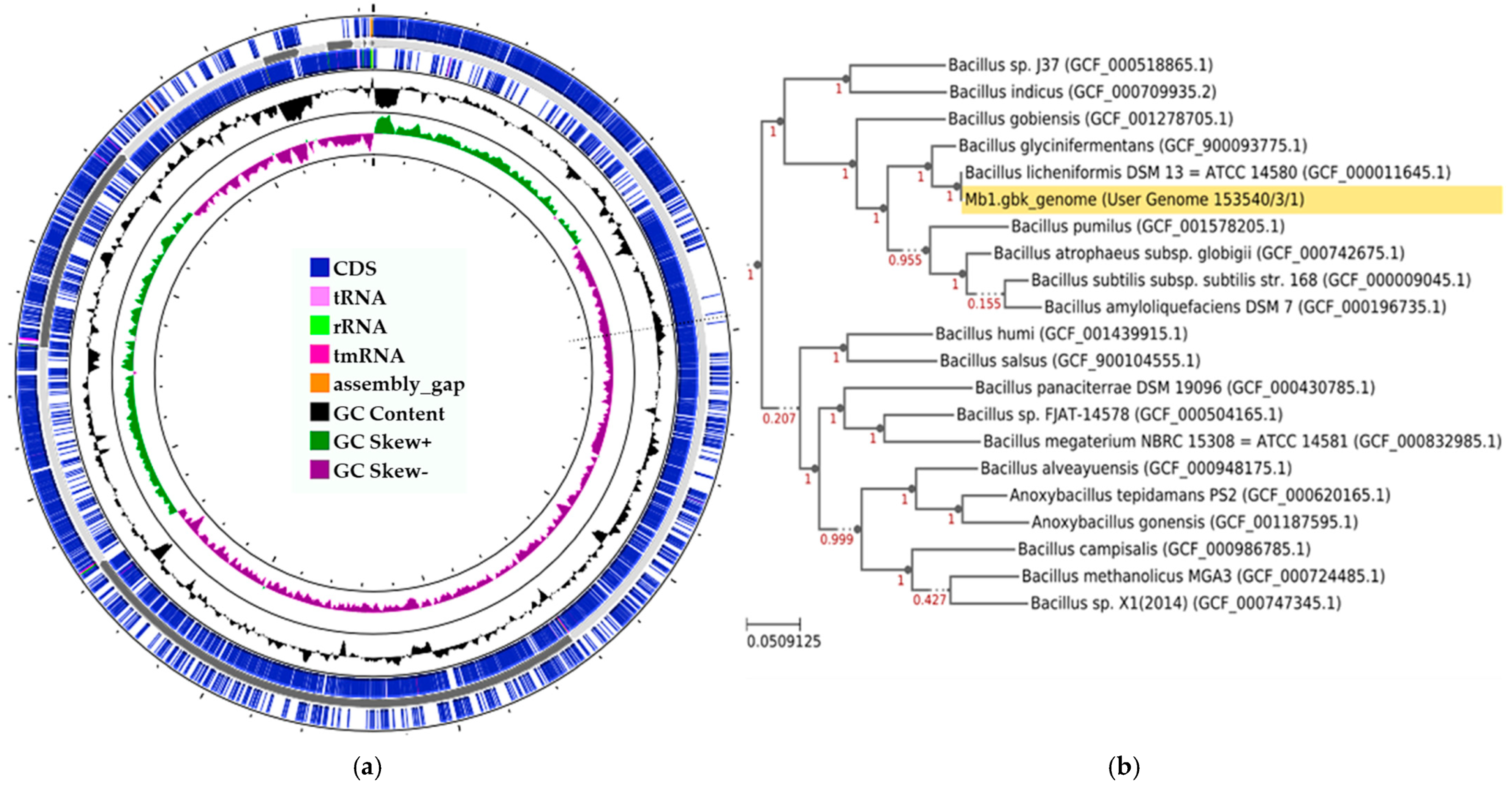
The paired-end reads were assembled de novo with SPAdes, resulting in a high-quality draft genome comprising 16 contigs (14 ≥ 1 kb) and a GC content of 45.6%. The assembly showed an N50 of 1.1 Mb, with the largest contig reaching 1.72 Mb. Read mapping demonstrated >99% of properly paired reads, an average coverage depth of 162×, and complete base coverage (≥10×), confirming the robustness of the assembly. Analysis of potential plasmid content using PlasmidFinder and BLAST did not detect plasmid replication genes. Genome annotation performed with Prokka predicted a total of 4542 genes, including 4453 protein-coding sequences (CDS) and 89 RNA genes, containing 11 rRNAs, 77 tRNAs, and 1 tmRNA, distributed across 14 contigs and a total length of 4,327,483 pb, consistent with assembly metrics (Figure 1a). Species-level identification using Mash distance analysis showed that Mb1 clustered within the Bacillus licheniformis group, with a Mash distance of 0.002969, and ANI analysis against reference genomes confirmed this classification, with the highest identity observed to Bacillus licheniformis strain DFI.1.225 (GCF_024463755.1), showing 99.93% ANI and 96.8% alignment coverage. In addition, a phylogenomic analysis grouped the Mb1 genome within the Bacillus licheniformis clade (Figure 1b).

3.2. Functional Inference and Identification of Biotechnologically Relevant Enzymes

Functional classification of the Bacillus licheniformis Mb1 genome with eggNOG-mapper assigned 3502 proteins (77% of the total) to COG categories. The largest groups corresponded to amino acid transport and metabolism (E), carbohydrate transport and metabolism (G), transcription (K), and energy production and conversion (C), which together accounted for a major fraction of the coding potential. Notably, 1024 proteins (23.6%) were classified in the S category (function unknown). This group includes not only proteins annotated as hypothetical but also conserved proteins without a defined biological role, indicating that a substantial part of the Mb1 genome remains functionally uncharacterized (Table S1). Furthermore, 1688 proteins were annotated as hypothetical of which 179 lacked any COG assignment, indicating sequences without detectable conservation or functional evidence.
In addition, 109 carbohydrate-active enzymes (CAZymes) were identified in Bacillus licheniformis Mb1 genome, consisting of 55 glycoside hydrolases (GHs), 28 glycosyl transferases (GTs), 10 carbohydrate esterases (Ces), and 8 polysaccharide lyases (PLs). This result place Mb1 as a highly CAZyme containing genomes within the Bacillus genera (Figure S1). These enzymes are associated with the degradation of major plant polysaccharides, including starch, cellulose, xylan, mannan, and chitin, underscoring the biotechnological potential of Mb1 in enzyme production.

3.3. Evaluation of Hydrolytic and Redox Activity of B. licheniformis Mb1

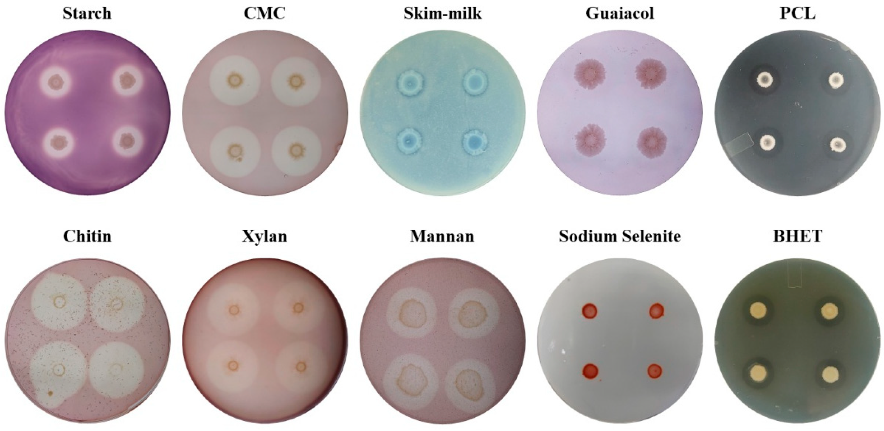
Qualitative plate assays revealed broad hydrolytic and redox capabilities in B. licheniformis Mb1 (Figure 2). Clear halos formed on YP 50% agar supplemented with starch, CMC, mannan, xylan, and chitin, indicating extracellular CAZyme activities spanning amylolytic, (hemi)cellulolytic, mannanolytic, xylanolytic, and chitinolytic functions (Figure 2). These phenotypes align with the recognized capacity of Bacillus spp.—including B. licheniformis—to secrete lignocellulose-active CAZymes and to serve as enzyme chassis for biomass deconstruction and bioprocessing [30,31]. Also, these finding are consistent with the high number of CAZyme identified in the Mb1 genome (Figure S1). In particular, robust plate clearing on xylan, mannan, and CMC is consistent with the CAZyme portfolios frequently reported for industrial Bacillus lineages and with their contribution to consolidated bioprocessing concepts [30,31].
A faint but discernible clearing zone on skim-milk plates indicated proteolytic activity (Figure 2), which is congruent with the long-standing use of B. licheniformis proteases in detergent, leather, feed, and other sectors [32,33]. Also, on plates with sodium selenite (20 mg/L), colonies developed a reddish coloration (Figure 2) that intensified over time, a typical indicator of microbial selenite reduction to biogenic selenium nanoparticles (SeNPs) [34,35]. This color change, reflects Se (IV) to Se (0) conversion and red SeNPs formation [36], supporting the potential relevance of the Mb1 strain’s redox phenotype. On the other side, laccase activity was not detected. Notably, Mb1 produced distinct clearing halos on BHET- and PCL-containing plates (Figure 2), consistent with extracellular BHETase/polyesterase activities and with the use of BHET and PCL as convenient model substrates to screen microbes for PET-degrading potential [37]. This type of plate assay is commonly employed as a preliminary screening method to identify microbial isolates with the potential to degrade PET or its intermediates, as it offers a simple and visual means of detecting extracellular hydrolytic activity [6,38].
Taken together, the plate-based screening highlights Bacillus licheniformis Mb1 as a genomically consistent, multifunctional biocatalyst with diverse capabilities: (i) CAZyme production, (ii) secretion of proteases, (iii) selenite reduction leading to SeNP formation, and (iv) extracellular polyester hydrolysis, a particularly relevant trait given the growing interest in microbial enzymes for plastic degradation and recycling.
Based on the multifunctional enzymatic profile revealed by the plate-based assays, the activities with potential relevance to PET depolymerization were selected for further investigation.

3.4. Differential BHETase-like Activity Induced by BHET and PCL

BHETase-like activity was monitored through the hydrolysis of the substrate Pnpb. Although this compound is not a structural analog of BHET, both substrates share ester bonds that are cleaved by enzymes of the α/β-hydrolase fold. Therefore, Pnpb hydrolysis reflects the presence and secretion of esterase/cutinase-like enzymes potentially involved in BHET transformation [21] (Figure S2).
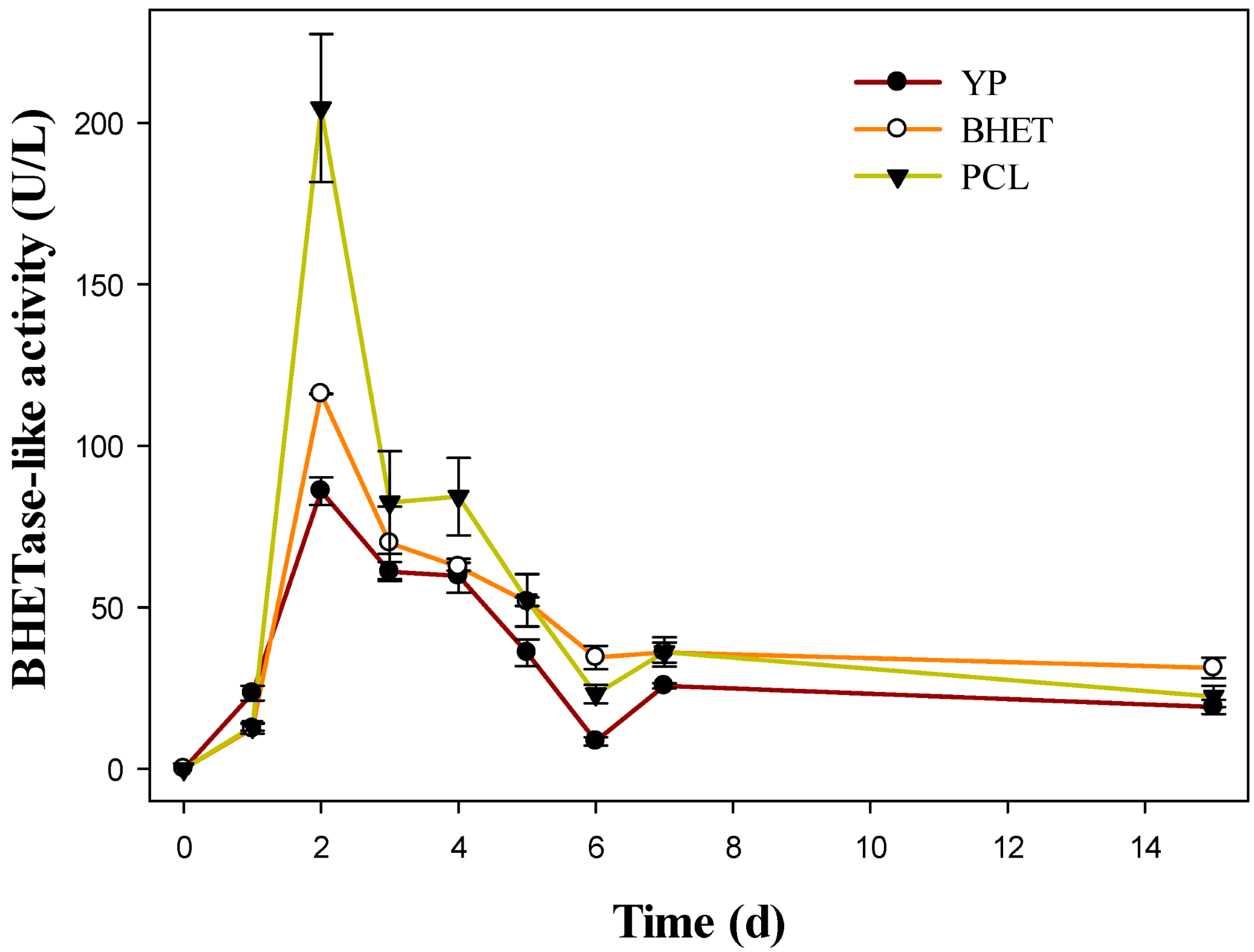
Enzymatic activity was initially absent in all conditions (0 days), but measurable levels appeared after 24 h. This indicates that B. licheniformis Mb1 produces BHETase early in growth, even without inducers (Figure 3).
A major induction peak occurred at day 2 in all conditions. YP 50% reached 86.0 ± 4.2 U/L, whereas BHET and PCL displayed substantially higher activities, measuring 116.1 ± 0.1 U/L and 204.6 ± 22.9 U/L, respectively. At this point, BHET medium showed ~1.4-fold activity increase than YP 50%, while PCL exhibited a pronounced ~2.4-fold increase compared to YP 50% (Figure 3). This demonstrates that both substrates enhance BHETase production, with PCL yielding the strongest induction.
Between days 3 and 4, all conditions showed a gradual decline from the day-2 maximum, but BHET and PCL maintained higher activities than YP 50%. At day 3, YP 50% reached 61.1 ± 3.0 U/L, while BHET and PCL remained elevated (69.9 ± 11.2 U/L and 82.5 ± 15.9 U/L). At day 4, BHET and PCL continued to support slightly higher activity than the control (Figure 3).
From day 5 onward, YP 50% showed a stronger decline, dropping to 35.9 ± 4.1 U/L at day 5 and 8.6 ± 1.3 U/L at day 6, while BHET and PCL retained substantially higher levels. At day 6, BHET and PCL activities (34.5 ± 3.7 U/L and 23.1 ± 2.8 U/L) were approximately 4.0-fold and 2.7-fold higher than YP 50%. By day 7, BHET and PCL remained elevated (36.1 ± 3.1 U/L and 36.2 ± 4.5 U/L) relative to YP 50% (25.8 ± 0.9 U/L). Finally, by day 15 all cultures showed reduced activity, with BHET and PCL maintaining slightly higher activity than YP 50%. Overall, the profiles indicate that B. licheniformis Mb1 produces significant extracellular BHETase-like activity in all media, with a maximum at 48 h. Both BHET and PCL function as inducers, particularly PCL, which supports the highest activities during the early and mid-incubation phases and maintains elevated levels relative to the control over extended culture periods.
The induction pattern aligns with the behavior reported for polyester-degrading Bacillus species [39], which activate hydrolases in response to synthetic substrates. The sustained activity in BHET- and PCL-supplemented cultures suggests that these polymers, or their intermediates, help maintain hydrolase expression beyond the initial peak.

3.5. Degradation of BHET by Bacillus licheniformis Mb1

TLC analyzed culture extracts at different incubation times. As illustrated in Figure S3, the BHET signal progressively decreased compared with the abiotic control, indicating its enzymatic breakdown. A partial loss of the substrate was already evident after 24 h (lane 1, Figure S3), and complete disappearance of the BHET spot occurred by day 4, remaining absent at day 7 (lanes 2 and 3, Figure S3). This pattern demonstrated that Mb1 efficiently metabolized BHET within the first days of incubation. No additional bands corresponding to intermediate or terminal hydrolysis products—such as MHET, TPA, or vanillic acid—were detected under these TLC conditions. The method was applied primarily as a qualitative tool to track the disappearance of the substrate rather than to resolve low-abundance by-products [21]. Hence, this assay provided a rapid and reliable indication of the strain’s ability to catalyze BHET hydrolysis in liquid culture.
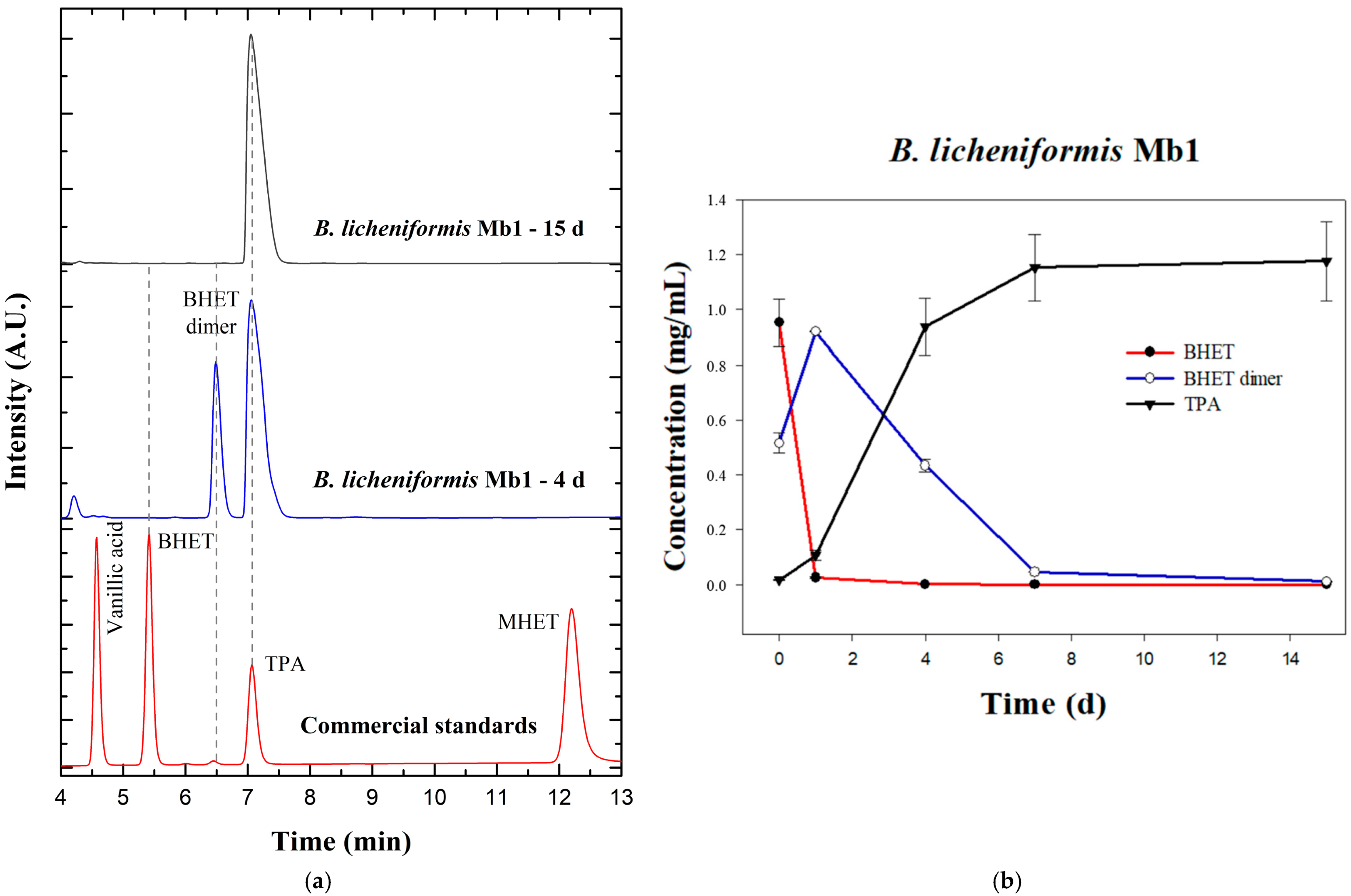
To gain a deeper understanding of the hydrolytic behavior of Bacillus licheniformis Mb1 toward BHET, culture supernatants obtained at different incubation times were analyzed by HPLC. Representative chromatograms are displayed in Figure 4a. BHET and MHET represent the sequential hydrolysis intermediates of PET, while TPA corresponds to the terminal monomer that signals complete depolymerization of the polyester backbone. Vanillic acid was incorporated as an aromatic derivative of interest, given that several microorganisms have been reported to oxidize PET-derived terephthalate into value-added aromatic molecules, including vanillic acid and related phenolics [40,41,42]. The inclusion of this extended set of standards allowed the identification not only of canonical degradation intermediates but also of potential secondary metabolites arising from bacterial aromatic metabolism.
In the abiotic control, two major chromatographic signals were detected: the characteristic BHET peak and an additional peak not corresponding to any of the standard compounds. Examination of its UV absorption spectrum (200–400 nm) revealed a spectral profile identical to that of BHET, suggesting that the extra signal corresponds to the BHET dimer as described in a previous study [21]. This interpretation is consistent with previous findings demonstrating that commercial BHET preparations often contain both monomeric and dimeric forms, which can be resolved chromatographically based on distinct retention times [43,44]. Furthermore, earlier reports have shown that exposure of BHET solutions to elevated temperatures such as those used for autoclaving may induce partial dimerization or formation of low-molecular-weight oligomers [21,45]. Thus, the secondary peak observed in the abiotic control likely results from heat-induced dimerization during media sterilization.
These observations reinforce the need to consider abiotic alterations of BHET when interpreting biodegradation assays. In PET enzymatic depolymerization systems, BHET dimers and oligomers are known to accumulate as reaction intermediates, potentially influencing substrate availability and enzymatic kinetics [38]. The chromatographic and detection parameters used here, C18 reversed-phase separation with UV monitoring at 250 nm, are consistent with standard analytical procedures for terephthalate ester quantification [43,46,47].
While TLC analysis was used as a qualitative screening tool and mainly confirmed BHET depletion, HPLC analysis provided higher sensitivity and revealed the presence of BHET dimer and TPA, allowing quantitative tracking of B. licheniformis Mb1-driven hydrolysis (Figure 4a,b). At the start of incubation, BHET was 0.9520 ± 0.0866 mg/mL (mean ± SE). After 1 day, BHET fell to 0.0250 ± 0.0020 mg/mL, i.e., 97.4% depletion relative to day 0 (remaining fraction 2.6%). By 4 days, BHET reached 0.0010 mg/mL (99.9% reduction), and it was below detection from day 7 onward (Figure 4b).
The BHET dimer exhibited a transient accumulation followed by clearance. It started at 0.5157 ± 0.0357 mg/mL at day 0 peaked at 0.9185 ± 0.0015 mg/mL after 1 day, then declined to 0.4330 ± 0.0240 mg/mL at 4 days, reaching 0.0453 ± 0.0013 mg/mL by 7 days and 0.0105 ± 0.0015 mg/mL by 15 days. This profile indicates that, once BHET is exhausted, B. licheniformis Mb1 progressively converts the accumulated dimer. Regarding to TPA, increased monotonically in parallel with substrate consumption, from 0.0165 ± 0.0025 mg/mL at day 0 to 0.1060 ± 0.0180 mg/mL at 1 day, 0.9380 ± 0.1020 mg/mL at 4 days, and 1.1520 ± 0.1220 mg/mL at 7 days, approaching a plateau at 1.1755 ± 0.1455 mg/mL by 15 days. Notably, MHET and vanillic acid were not detected under these chromatographic conditions in any biological sample in agreement with the TLC results (Figure S3).
These data delineate three phases: (i) first 24 h, rapid BHET depletion with transient BHET dimer accumulation and the onset of TPA release; (ii) 1–4 days, clearance of the dimer accompanied by a steep rise in TPA; and (iii) 4–15 days, near-asymptotic TPA levels with residual dimer conversion. The chromatographic overlays (Figure 4a) illustrate this trajectory, as BHET and the dimer dominate at early times (4 days), while the 15-day trace is characterized by a prominent TPA peak and minimal residual dimer, thus corroborating that B. licheniformis Mb1 drives BHET beyond oligomeric equilibria toward TPA accumulation. Taken together, this three-phase profile demonstrates that B. licheniformis Mb1 not only rapidly consumes BHET but also, over extended incubation, is capable of utilizing BHET-derived dimers as substrates, leading to sustained TPA production.
These results confirm that B. licheniformis Mb1 is capable of efficiently hydrolyzing BHET and producing TPA as the main soluble end product. The observed depletion of BHET and concomitant release of TPA by this strain is consistent with previous studies that highlight BHET as a central intermediate whose efficient bioconversion is essential for sustainable PET degradation strategies [40].
The complete degradation of BHET and its dimeric form observed for Bacillus licheniformis Mb1 contrasts with previous reports describing partial accumulation of BHET oligomers during enzymatic PET depolymerization. For instance, enzymatic systems such as PETase from Ideonella sakaiensis and cutinase-type enzymes from Thermobifida fusca typically exhibit limited activity toward short-chain substrates such as BHET dimers, which tend to accumulate due to suboptimal substrate binding within the active site groove of the enzyme [45,48]. This accumulation has been shown to interfere with enzymatic turnover and even modify the physicochemical structure of the polymer surface, reducing crystallinity and increasing brittleness as degradation proceeds [45]. Furthermore, the formation of insoluble BHET dimers has been associated with reduced accessibility of the catalytic site, potentially acting as competitive or physical inhibitors of hydrolytic activity [49].
In contrast, the efficient disappearance of both BHET and its dimer in cultures of B. licheniformis Mb1 suggests the presence of a more robust or versatile set of esterases or carboxylases capable of cleaving these oligomeric intermediates.

3.6. Genome Mining for Potential BHET-Hydrolyzing Enzymes

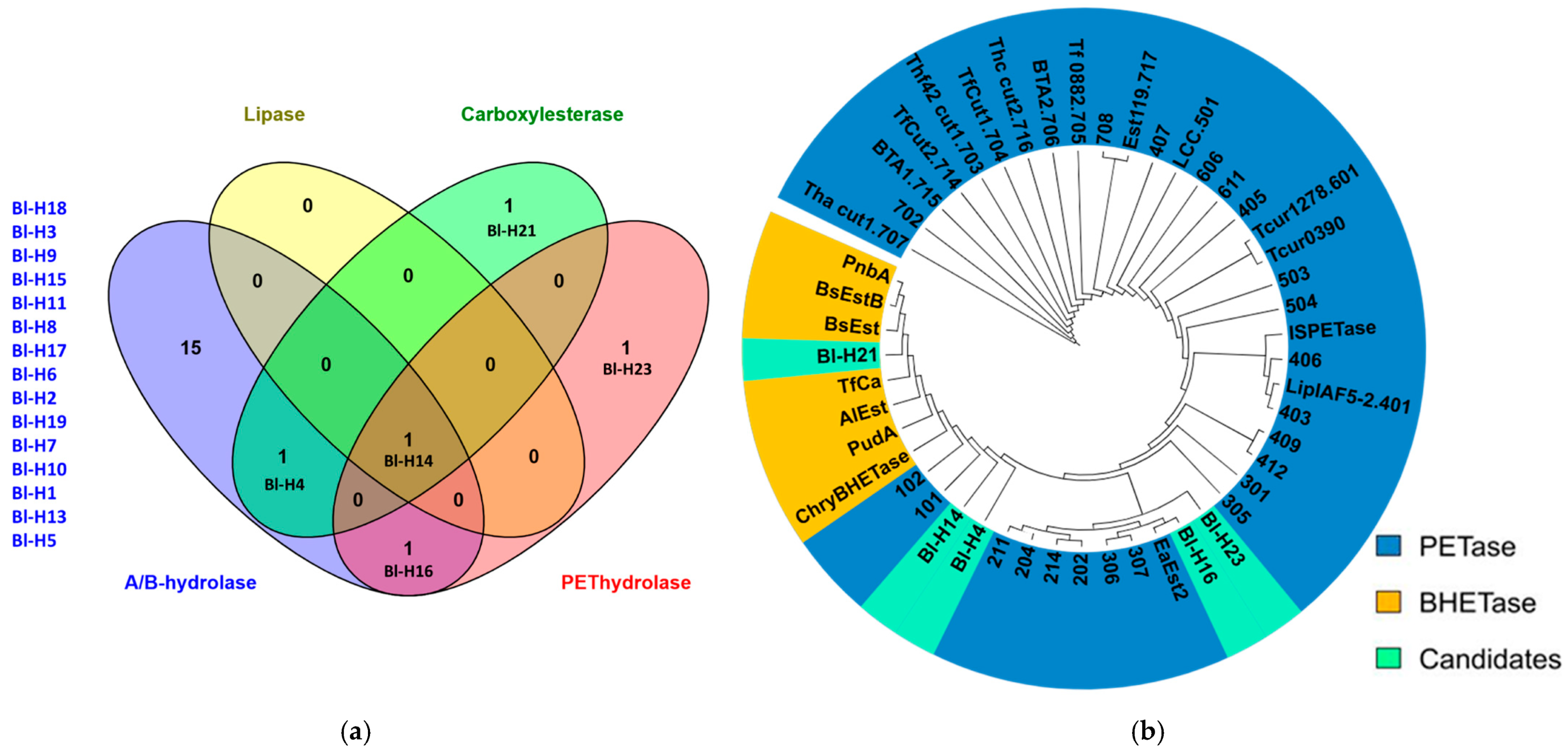
To explore the genetic basis of Bacillus licheniformis Mb1 for BHET-degrading activity, the genome was screened using HMM profiles from α/β-hydrolase families, including carboxylesterases, cutinases, lipases, and PET hydrolases. Twenty candidate sequences belonging to the α/β-hydrolase superfamily were retrieved. Most did not match known HMM profiles related to PET or BHET degradation. However, five sequences showed specific associations: Bl-H23 and Bl-H16 with PET hydrolases; Bl-H4 and Bl-H21 with carboxylesterases; and Bl-H14 with carboxylesterase, lipase, and PET hydrolase families. No matches were found for cutinase (Figure 5a).
Phylogenetic analysis with reference PETases and BHETases revealed that Mb1 candidates were distributed across both groups (Figure 5b), indicating different levels of functional relatedness. Notably, Bl-H21 clustered closely with reference BHETases, representing the most likely enzyme responsible for the observed BHET degradation. Conversely, Bl-H4, Bl-H14, Bl-H16, and Bl-H23 were grouped with structurally distant PETase variants described by Erickson et al. (2022) [50].

3.7. Structural Comparison and Active Site Interactions of Bl-H21

Sequence and structural analyses positioned Bl-H21 within the bacterial carboxylesterase group, displaying 58–59% sequence identity with BsEst, BsEstB, and PnbA [5,51,52], and a lower similarity with PudA (28%) [53] and AlEst [54], as well as for BHET-active enzymes TfCa (36.8%) [27] and ChryBHETase (25.4%) [5] (Table S2). Structural alignment confirmed a conserved α/β-hydrolase fold and catalytic triad Ser187–Glu308–His399 (Figure S4), consistent with the catalytic mechanism described for bacterial BHETases. TM-align scores supported this conservation, with values of 0.98 against BsEst, 0.89 against TfCa, and 0.78 against ChryBHETase (Table S3).
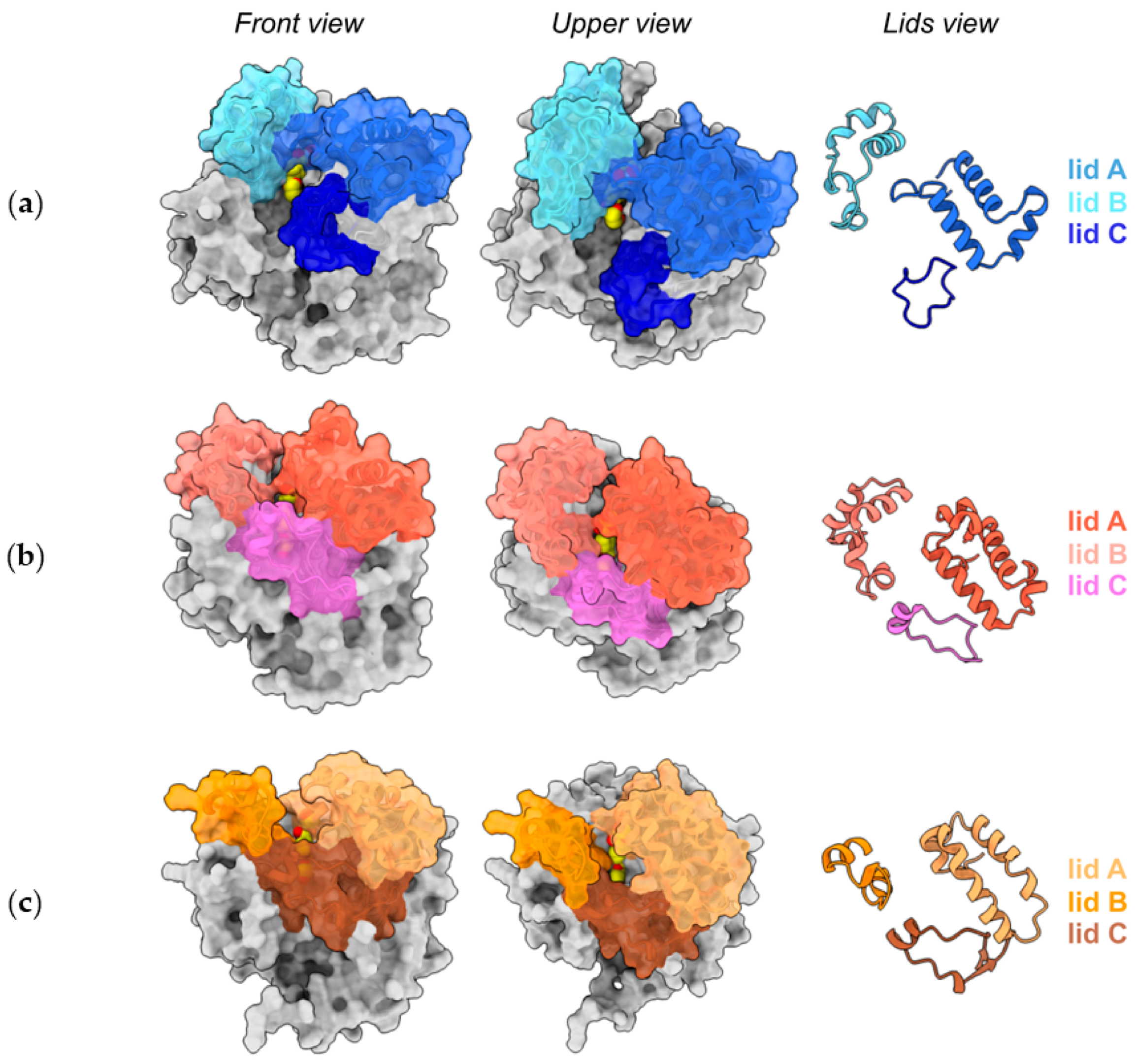
Lid domains appear to be pivotal for BHETase activity [5,27]. Comparative mapping of the lid regions revealed structural differences that may affect substrate accessibility (Figure 6). Bl-H21 lidA (residues 219–275) was similar in length and topology to TfCa, retaining a hydrophobic terminal segment likely involved in stabilizing aromatic ligands. LidB (308–352) was shorter than in TfCa but longer and more structured than the reduced lidB of ChryBHETase, which displays a truncated loop region (Figure 6).
LidC (59–77) appeared as a short, surface-exposed loop located near the MHET-binding subsite described for TfCa crystal structures (PDB 7W1I and 7W1L) [27]. This region, not previously described by von Haugwitz et al. (2022) [27], is here defined as the third lid (lidC). TfCa and ChryBHETase share a lidC of similar length and topology, both covering the cavity corresponding to the MHET-binding pocket and effectively closing the cleft. In contrast, Bl-H21 exhibits a shorter, truncated lidC that does not fully close the cavity, resulting in a more open conformation that may influence substrate entry and product release dynamics (Figure 6 and Figure 7).
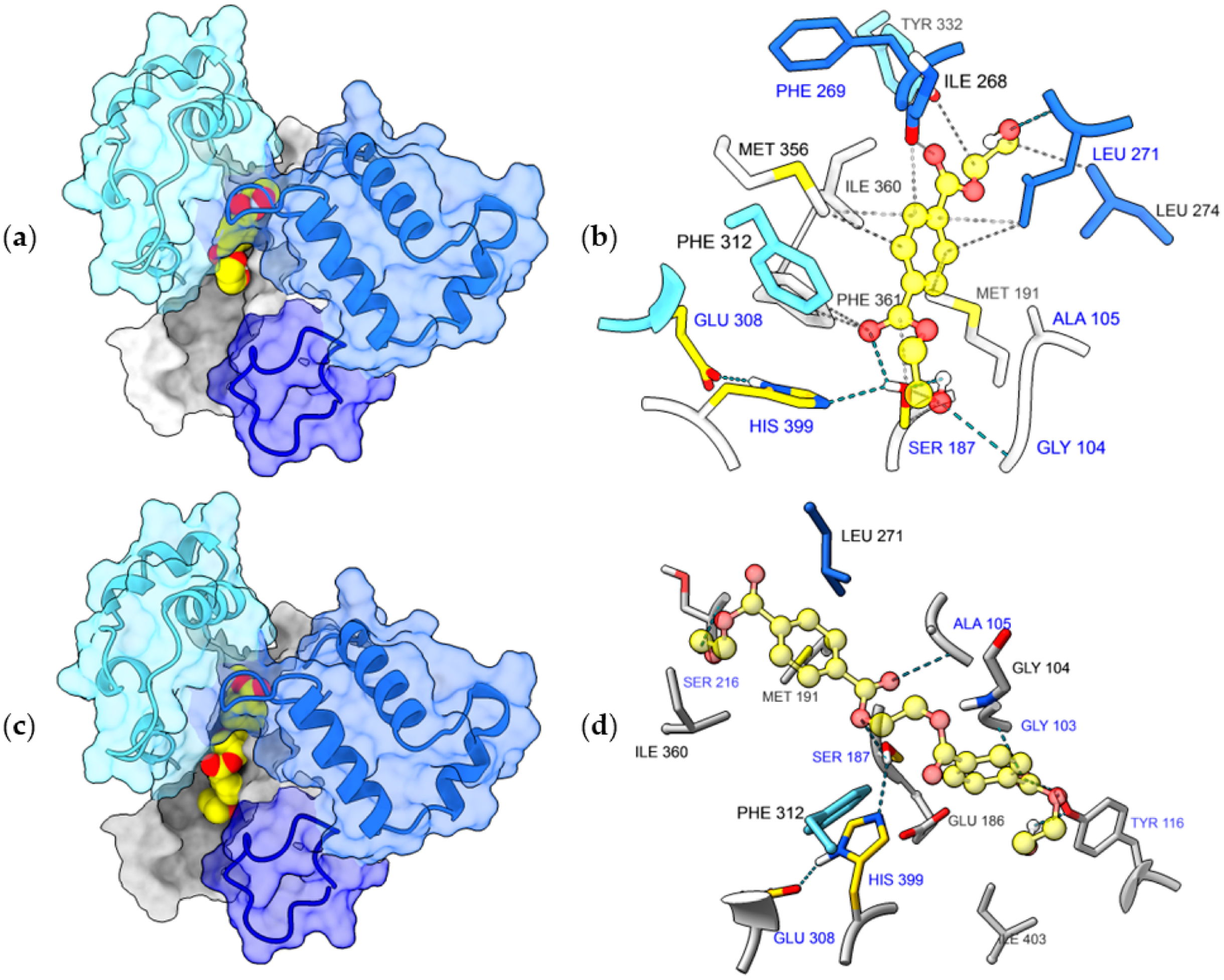
Docking analysis of BHET yielded multiple binding conformations, from which the one with the highest predicted affinity (−7.78 kcal/mol) was selected for interaction analysis (Figure 7a,b). In this configuration, BHET adopts a geometry compatible with hydrogen-bond formation -both in distance and angle- with Gly104, Ala105, Ser187, Phe269, and Leu271, involving the lid region, the catalytic serine, and the hydrophobic pocket wall. Additionally, residues Met191, Leu274, Phe312, Met356, Ile360, and Phe361 are positioned at van der Waals contact distance (<4 Å), delineating the groove that accommodates the BHET monomer.
To further evaluate substrate accommodation, docking was also performed using BHET dimer (Figure 7c,d). The best predicted conformation (ΔG = −7.25 kcal/mol) positioned the dimeric substrate deeper in the cleft, with Ser187, Gly103, Ala105, Tyr116, and Ser216 located at hydrogen-bonding distance, and Ile360, Phe312, Glu186, Gly104, Met191, and Leu271 at contact distance. The extended contact surface and stable geometry suggest that the wider, more open cavity of Bl-H21 can accommodate larger BHET-related substrates.
Together, these observations indicate that Bl-H21 retains the canonical catalytic core of bacterial carboxylesterases and BHETases while exhibiting structural features intermediate between TfCa and ChryBHETase. Importantly, the truncated lidC of Bl-H21 leaves the catalytic cleft more accessible, which may facilitate the entry and positioning of bulkier substrates such as BHET dimer, in agreement with the experimental BHET-degradation activity measured for B. licheniformis Mb1.

4. Conclusions

Bacillus licheniformis Mb1 is a promising strain for different areas of biotechnology, including enzyme production and plastic bioremediation. The combination of experimental results and global genome analysis shows that this strain carries multiple genes linked to hydrolytic, oxidative, carbohydrate-active, and polyester-degrading activities. The ability of B. licheniformis Mb1 to degrade BHET, supported by the presence of BHETase-like and other hydrolase coding genes, suggests its potential as a biocatalyst for polyester depolymerization. The candidate hydrolase Bl-H21 conserves the catalytic core of bacterial BHETases but presents a uniquely truncated lidC that creates a more open catalytic cleft, suggesting an enhanced capacity to accommodate larger BHET-related oligomeric substrates. This activity helps to overcome one of the main limitations in microbial PET degradation, highlighting the value of B. licheniformis Mb1 as a sustainable source of useful enzymes and the development of eco-friendly bioprocesses.

Supplementary Materials

The following supporting information can be downloaded at https://www.mdpi.com/article/10.3390/pr14020381/s1, Table S1: COG analysis showing different clusters of orthologous groups in Bacillus licheniformis Mb1 genome; Figure S1: Predicted CAZymes in Bacillus licheniformis Mb1 and selected Bacillus genomes from phylogenomic analysis; Figure S2: Schematic overview of PET intermediate degradation and the relationship between BHET hydrolysis and p-nitrophenyl butyrate (pNPB)-based enzymatic activity assays; Figure S3: Thin-layer chromatographic (TLC) analysis of BHET degradation by Bacillus licheniformis Mb1; Table S2: Identity matrix between Bl-H21 sequence and reference carboxylesterases with proved activity over plastics or PET derivatives (BHET); Figure S4: Multiple sequence alignment of Bl-H21 and reference carboxylesterases with confirmed BHET-hydrolyzing activity, generated using Clustal W; Table S3: Structural similarity matrix based on TM-score between Bl-H21 and reference carboxylesterases/BHETases.

Author Contributions

Conceptualization, P.E.S., F.G.M., C.M.R. and A.Á.; formal analysis, P.E.S., F.G.M. and F.Z.; investigation, P.E.S., F.G.M., F.Z., L.C. and J.H.P.; data curation, P.E.S., F.G.M., F.Z. and J.H.P.; writing—original draft preparation, P.E.S., F.G.M. and F.Z.; writing—review and editing, A.Á. and C.M.R.; visualization, P.E.S., F.G.M., F.Z. and L.C.; supervision, A.Á. and C.M.R.; project administration, A.Á. and C.M.R.; funding acquisition, A.Á. and C.M.R. All authors have read and agreed to the published version of the manuscript.

Funding

This research was funded by the National Scientific and Technical Research Council (CONICET, Argentina).

Data Availability Statement

The genome sequence has been submitted to the GenBank database with accession numbers: BioProject PRJNA1002392 and BioSample SAMN36843220. The protein sequence ID for Bl-H4, Bl-H14, Bl-H16, Bl-H21 and Bl-H23 are MFG5205724.1, MFG5207099.1, MFG5207509.1, MFG5207816.1, and MFG5207347.4, respectively.

Conflicts of Interest

The authors declare no conflicts of interest.

Abbreviations

The following abbreviations are used in this manuscript:
BHETbis (2-hydroxyethyl) terephthalate
MHETmono (2-hydroxyethyl) terephthalate
TPAterephthalic acid
TLCthin-layer chromatography
PCLPolycaprolactone
EGEthylene glycol

References

  1. Geyer, R.; Jambeck, J.R.; Law, K.L. Production, use, and fate of all plastics ever made. Sci. Adv. 2017, 3, e1700782. [Google Scholar] [CrossRef] [PubMed]
  2. Khairul Anuar, N.F.S.; Huyop, F.; Ur-Rehman, G.; Abdullah, F.; Normi, Y.M.; Sabullah, M.K.; Abdul Wahab, R. An overview into polyethylene terephthalate (PET) hydrolases and efforts in tailoring enzymes for improved plastic degradation. Int. J. Mol. Sci. 2022, 23, 12644. [Google Scholar] [CrossRef] [PubMed]
  3. Tegariyanto, M.; Titah, H.; Pratikno, H. Biodegradation of polyethylene terephthalate (PET) plastics using a combination of Aspergillus niger and Trichoderma harzianum. In IOP Conference Series: Earth and Environmental Science; IOP Publishing: Philadelphia, PA, USA, 2024. [Google Scholar] [CrossRef]
  4. Al-Sabagh, A.; Yehia, F.; Eshaq, G.; Rabie, A.; Elmetwally, A. Greener routes for recycling of polyethylene terephthalate. Egypt. J. Pet. 2016, 25, 53–64. [Google Scholar] [CrossRef]
  5. Li, A.; Sheng, Y.; Cui, H.; Wang, M.; Wu, L.; Song, Y.; Yang, R.; Li, X.; Huang, H. Discovery and mechanism-guided engineering of BHET hydrolases for improved PET recycling and upcycling. Nat. Commun. 2023, 14, 4169. [Google Scholar] [CrossRef]
  6. Verschoor, J.A.; Croese, M.R.; Lakemeier, S.E.; Mugge, A.; Burgers, C.M.; Innocenti, P.; Willemse, J.; Crooijmans, M.E.; Van Wezel, G.P.; Ram, A.F. Polyester degradation by soil bacteria: Identification of conserved BHETase enzymes in Streptomyces. Commun. Biol. 2024, 7, 725. [Google Scholar] [CrossRef]
  7. Mesbah, N.M. Industrial biotechnology based on enzymes from extreme environments. Front. Bioeng. Biotechnol. 2022, 10, 870083. [Google Scholar] [CrossRef]
  8. Schallmey, M.; Singh, A.; Ward, O.P. Developments in the use of Bacillus species for industrial production. Can. J. Microbiol. 2004, 50, 1–17. [Google Scholar] [CrossRef]
  9. Arena, M.; Abbate, C.; Fukushima, K.; Gennari, M. Degradation of poly (lactic acid) and nanocomposites by Bacillus licheniformis. Environ. Sci. Pollut. Res. 2011, 18, 865–870. [Google Scholar] [CrossRef]
  10. Simon Andrews, F.; Segonds-Pichon, A.; Biggins, L.; Krueger, C.; Wingett, S. FastQC: A Quality Control Tool for High Throughput Sequence Data. 2010. Available online: https://www.bioinformatics.babraham.ac.uk/projects/fastqc/ (accessed on 15 October 2025).
  11. Bolger, A.M.; Lohse, M.; Usadel, B. Trimmomatic: A flexible trimmer for Illumina sequence data. Bioinformatics 2014, 30, 2114–2120. [Google Scholar] [CrossRef]
  12. Bankevich, A.; Nurk, S.; Antipov, D.; Gurevich, A.A.; Dvorkin, M.; Kulikov, A.S.; Lesin, V.M.; Nikolenko, S.I.; Pham, S.; Prjibelski, A.D. SPAdes: A new genome assembly algorithm and its applications to single-cell sequencing. J. Comput. Biol. 2012, 19, 455–477. [Google Scholar] [CrossRef]
  13. Simão, F.A.; Waterhouse, R.M.; Ioannidis, P.; Kriventseva, E.V.; Zdobnov, E.M. BUSCO: Assessing genome assembly and annotation completeness with single-copy orthologs. Bioinformatics 2015, 31, 3210–3212. [Google Scholar] [CrossRef] [PubMed]
  14. Carattoli, A.; Zankari, E.; Garcìa-Fernandez, A.; Larsen, M.V.; Lund, O.; Villa, L.; Aarestrup, F.M.; Hasman, H. PlasmidFinder and pMLST: In silico detection and typing of plasmids. Antimicrob. Agents Chemother. 2014, 58, 3895–3903. [Google Scholar] [CrossRef] [PubMed]
  15. Ondov, B.D.; Treangen, T.J.; Melsted, P.; Mallonee, A.B.; Bergman, N.H.; Koren, S.; Phillippy, A.M. Mash: Fast genome and metagenome distance estimation using MinHash. Genome Biol. 2016, 17, 132. [Google Scholar] [CrossRef] [PubMed]
  16. Seemann, T. Prokka: Rapid prokaryotic genome annotation. Bioinformatics 2014, 30, 2068–2069. [Google Scholar] [CrossRef]
  17. Grant, J.R.; Enns, E.; Marinier, E.; Mandal, A.; Herman, E.K.; Chen, C.-Y.; Graham, M.; Van Domselaar, G.; Stothard, P. Proksee: In-depth characterization and visualization of bacterial genomes. Nucleic Acids Res. 2023, 51, W484–W492. [Google Scholar] [CrossRef]
  18. Cantalapiedra, C.P.; Hernández-Plaza, A.; Letunic, I.; Bork, P.; Huerta-Cepas, J. eggNOG-mapper v2: Functional annotation, orthology assignments, and domain prediction at the metagenomic scale. Mol. Biol. Evol. 2021, 38, 5825–5829. [Google Scholar] [CrossRef]
  19. Zhang, H.; Yohe, T.; Huang, L.; Entwistle, S.; Wu, P.; Yang, Z.; Busk, P.K.; Xu, Y.; Yin, Y. dbCAN2: A meta server for automated carbohydrate-active enzyme annotation. Nucleic Acids Res. 2018, 46, W95–W101. [Google Scholar] [CrossRef]
  20. Martínez, F.G.; Moreno-Martin, G.; Pescuma, M.; Madrid-Albarrán, Y.; Mozzi, F. Biotransformation of Selenium by Lactic Acid Bacteria: Formation of Seleno-Nanoparticles and Seleno-Amino Acids. Front. Bioeng. Biotechnol. 2020, 8, 506. [Google Scholar] [CrossRef]
  21. Martínez, F.G.; Canal Martínez, V.; Pereira, C.E.; Zannier, F.; Arnau, V.G.; Romero, C.M.; Álvarez, A. Enzymatic Potential of Schizophyllum commune BNT39 in BHET Hydrolysis and PET Biodegradation. Processes 2025, 13, 3663. [Google Scholar] [CrossRef]
  22. Gouet, P.; Robert, X.; Courcelle, E. ESPript/ENDscript: Extracting and rendering sequence and 3D information from atomic structures of proteins. Nucleic Acids Res. 2003, 31, 3320–3323. [Google Scholar] [CrossRef]
  23. Letunic, I.; Bork, P. Interactive Tree of Life (iTOL) v6: Recent updates to the phylogenetic tree display and annotation tool. Nucleic Acids Res. 2024, 52, W78–W82. [Google Scholar] [CrossRef] [PubMed]
  24. Cock, P.J.; Antao, T.; Chang, J.T.; Chapman, B.A.; Cox, C.J.; Dalke, A.; Friedberg, I.; Hamelryck, T.; Kauff, F.; Wilczynski, B. Biopython: Freely available Python tools for computational molecular biology and bioinformatics. Bioinformatics 2009, 25, 1422. [Google Scholar] [CrossRef] [PubMed]
  25. Zhang, Y.; Skolnick, J. TM-align: A protein structure alignment algorithm based on the TM-score. Nucleic Acids Res. 2005, 33, 2302–2309. [Google Scholar] [CrossRef] [PubMed]
  26. Abramson, J.; Adler, J.; Dunger, J.; Evans, R.; Green, T.; Pritzel, A.; Ronneberger, O.; Willmore, L.; Ballard, A.J.; Bambrick, J. Accurate structure prediction of biomolecular interactions with AlphaFold 3. Nature 2024, 630, 493–500. [Google Scholar] [CrossRef]
  27. Von Haugwitz, G.; Han, X.; Pfaff, L.; Li, Q.; Wei, H.; Gao, J.; Methling, K.; Ao, Y.; Brack, Y.; Mican, J. Structural insights into (tere) phthalate-ester hydrolysis by a carboxylesterase and its role in promoting PET depolymerization. ACS Catal. 2022, 12, 15259–15270. [Google Scholar] [CrossRef]
  28. Forli, S.; Huey, R.; Pique, M.E.; Sanner, M.F.; Goodsell, D.S.; Olson, A.J. Computational protein–ligand docking and virtual drug screening with the AutoDock suite. Nat. Protoc. 2016, 11, 905–919. [Google Scholar] [CrossRef]
  29. Meng, E.C.; Goddard, T.D.; Pettersen, E.F.; Couch, G.S.; Pearson, Z.J.; Morris, J.H.; Ferrin, T.E. UCSF ChimeraX: Tools for structure building and analysis. Protein Sci. 2023, 32, e4792. [Google Scholar] [CrossRef]
  30. Muras, A.; Romero, M.; Mayer, C.; Otero, A. Biotechnological applications of Bacillus licheniformis. Crit. Rev. Biotechnol. 2021, 41, 609–627. [Google Scholar] [CrossRef]
  31. Yadav, R.; Awathare, P.; Shitut, P.; Mahajan, V.; Nair, P.; Dharne, M. Genome mining of carbohydrate-active enzymes (CAZyme) and poly-γ-glutamic acid (γ-PGA) synthesis by Bacillus velezensis (WA11) directly from lignocellulosic biomass-based substrate. Biocatal. Agric. Biotechnol. 2025, 69, 103747. [Google Scholar] [CrossRef]
  32. Zhang, Y.; Hu, J.; Zhang, Q.; Cai, D.; Chen, S.; Wang, Y. Enhancement of alkaline protease production in recombinant Bacillus licheniformis by response surface methodology. Bioresour. Bioprocess. 2023, 10, 27. [Google Scholar] [CrossRef]
  33. Espoui, A.H.; Larimi, S.G.; Darzi, G.N. Optimization of protease production process using bran waste using Bacillus licheniformis. Korean J. Chem. Eng. 2022, 39, 674–683. [Google Scholar] [CrossRef]
  34. Martínez, F.G.; Moreno-Martín, G.; Mohamed, F.; Pescuma, M.; Madrid-Albarrán, Y.; Mozzi, F. Selenium-enriched fermented beverage with improved sensorial properties using lactic acid bacteria. J. Food Sci. Technol. 2024, 61, 2133–2144. [Google Scholar] [CrossRef] [PubMed]
  35. Martínez, F.G.; Moreno-Martin, G.; Mozzi, F.; Madrid, Y.; Pescuma, M. Selenium stress response of the fruit origin strain Fructobacillus tropaeoli CRL 2034. Appl. Microbiol. Biotechnol. 2023, 107, 1329–1339. [Google Scholar] [CrossRef] [PubMed]
  36. Wang, Z.; Li, N.; Zhou, X.; Wei, S.; Zhu, Y.; Li, M.; Gong, J.; He, Y.; Dong, X.; Gao, C. Optimization of fermentation parameters to improve the biosynthesis of selenium nanoparticles by Bacillus licheniformis F1 and its comprehensive application. BMC Microbiol. 2024, 24, 271. [Google Scholar] [CrossRef]
  37. Malafatti-Picca, L.; Bucioli, E.C.; De Barros Chaves, M.R.; De Castro, A.M.; Valoni, É.; De Oliveira, V.M.; Marsaioli, A.J.; Govone, J.S.; De Franceschi De Angelis, D.; Brienzo, M. Fungal screening for potential PET depolymerization. Polymers 2023, 15, 1581. [Google Scholar] [CrossRef]
  38. Liu, X.H.; Jin, J.L.; Sun, H.T.; Li, S.; Zhang, F.F.; Yu, X.H.; Cao, Q.Z.; Song, Y.X.; Li, N.; Lu, Z.H. Perspectives on the microorganisms with the potentials of PET-degradation. Front. Microbiol. 2025, 16, 1541913. [Google Scholar] [CrossRef]
  39. Sahu, S.; Kaur, A.; Khatri, M.; Singh, G.; Arya, S.K. A review on cutinases enzyme in degradation of microplastics. J. Environ. Manag. 2023, 347, 119193. [Google Scholar] [CrossRef]
  40. Ion, S.; Voicea, S.; Sora, C.; Gheorghita, G.; Tudorache, M.; Parvulescu, V.I. Sequential biocatalytic decomposition of BHET as valuable intermediator of PET recycling strategy. Catal. Today 2021, 366, 177–184. [Google Scholar] [CrossRef]
  41. Jiang, C.; Zhai, K.; Wright, R.C.; Chen, J. Engineered Yeasts Displaying PETase and MHETase as Whole-Cell Biocatalysts for the Degradation of Polyethylene Terephthalate (PET). ACS Synth. Biol. 2025, 14, 2810–2820. [Google Scholar] [CrossRef]
  42. Soong, Y.H.V.; Abid, U.; Chang, A.C.; Ayafor, C.; Patel, A.; Qin, J.; Xu, J.; Lawton, C.; Wong, H.W.; Sobkowicz, M.J. Enzyme selection, optimization, and production toward biodegradation of post-consumer poly (ethylene terephthalate) at scale. Biotechnol. J. 2023, 18, 2300119. [Google Scholar] [CrossRef]
  43. Kim, E.H.; Park, I.; Kim, S.; Kim, J.F.; Choi, Y.H.; Lee, H. Chemical recycling of polyethylene terephthalate using a micro-sized MgO-incorporated SiO2 catalyst to produce highly pure bis (2-hydroxyethyl) terephthalate in high yield. Chem. Eng. J. 2024, 499, 155865. [Google Scholar] [CrossRef]
  44. Zahova, S.; Tsacheva, I.; Troev, K.; Mitova, V. Conventional and MW assisted PET glycolysis promoted by titanium based catalyst. Polym. Degrad. Stab. 2023, 212, 110353. [Google Scholar] [CrossRef]
  45. Menzel, T.; Weigert, S.; Gagsteiger, A.; Eich, Y.; Sittl, S.; Papastavrou, G.; Ruckdäschel, H.; Altstädt, V.; Höcker, B. Impact of enzymatic degradation on the material properties of poly (ethylene terephthalate). Polymers 2021, 13, 3885. [Google Scholar] [CrossRef] [PubMed]
  46. Puspitasari, N.; Tsai, S.L.; Lee, C.K. Class I hydrophobins pretreatment stimulates PETase for monomers recycling of waste PETs. Int. J. Biol. Macromol. 2021, 176, 157–164. [Google Scholar] [CrossRef] [PubMed]
  47. Yao, J.; Liu, Y.; Gu, Z.; Zhang, L.; Guo, Z. Deconstructing PET: Advances in enzyme engineering for sustainable plastic degradation. Chem. Eng. J. 2024, 497, 154183. [Google Scholar] [CrossRef]
  48. Kawai, F.; Iizuka, R.; Kawabata, T. Engineered polyethylene terephthalate hydrolases: Perspectives and limits. Appl. Microbiol. Biotechnol. 2024, 108, 404. [Google Scholar] [CrossRef]
  49. Son, H.F.; Cho, I.J.; Joo, S.; Seo, H.; Sagong, H.Y.; Choi, S.Y.; Lee, S.Y.; Kim, K.J. Rational protein engineering of thermo-stable PETase from Ideonella sakaiensis for highly efficient PET degradation. ACS Catal. 2019, 9, 3519–3526. [Google Scholar] [CrossRef]
  50. Erickson, E.; Gado, J.E.; Avilán, L.; Bratti, F.; Brizendine, R.K.; Cox, P.A.; Gill, R.; Graham, R.; Kim, D.J.; König, G. Sourcing thermotolerant poly (ethylene terephthalate) hydrolase scaffolds from natural diversity. Nat. Commun. 2022, 13, 7850. [Google Scholar] [CrossRef]
  51. Ribitsch, D.; Heumann, S.; Trotscha, E.; Herrero Acero, E.; Greimel, K.; Leber, R.; Birner-Gruenberger, R.; Deller, S.; Eiteljoerg, I.; Remler, P. Hydrolysis of polyethyleneterephthalate by p-nitrobenzylesterase from Bacillus subtilis. Biotechnol. Prog. 2011, 27, 951–960. [Google Scholar] [CrossRef]
  52. Zock, J.; Cantwell, C.; Swartling, J.; Hodges, R.; Pohl, T.; Sutton, K.; Rosteck, P., Jr.; Mcgilvray, D.; Queener, S. The Bacillus subtilis pnbA gene encoding p-nitrobenzyl esterase: Cloning, sequence and high-level expression in Escherichia coli. Gene 1994, 151, 37–43. [Google Scholar] [CrossRef]
  53. Nomura, N.; Shigeno-Akutsu, Y.; Nakajima-Kambe, T.; Nakahara, T. Cloning and sequence analysis of a polyurethane esterase of Comamonas acidovorans TB-35. J. Ferment. Bioeng. 1998, 86, 339–345. [Google Scholar] [CrossRef]
  54. Tchigvintsev, A.; Tran, H.; Popovic, A.; Kovacic, F.; Brown, G.; Flick, R.; Hajighasemi, M.; Egorova, O.; Somody, J.C.; Tchigvintsev, D. The environment shapes microbial enzymes: Five cold-active and salt-resistant carboxylesterases from marine metagenomes. Appl. Microbiol. Biotechnol. 2015, 99, 2165–2178. [Google Scholar] [CrossRef]
Figure 1. Genome organization and phylogenomic placement of Bacillus licheniformis Mb1: (a) Genome map of Bacillus sp. Mb1 displaying distribution of CDS, RNAs and GC content; (b) Phylogenomic analysis of Bacillus licheniformis Mb1 (highlighted in yellow). The tree was constructed using a set of 20 Bacillus genomes.
Processes 14 00381 g001
Figure 2. Qualitative screening of enzymatic and redox activities in Bacillus licheniformis Mb1. Clear halos indicate positive hydrolytic activity on starch, chitin, CMC, xylan, and mannan, evidencing extracellular CAZyme activities. A faint clearing zone on skim-milk plates denotes proteolytic activity. Growth on sodium selenite plates resulted in red pigmentation characteristic of biogenic SeNPs. Laccase activity was not detected. Hydrolysis halos observed on plates containing BHET (bis(2-hydroxyethyl) terephthalate) and PCL (polycaprolactone) suggest the strain’s polyester-degrading potential.
Processes 14 00381 g002
Figure 3. Time-course BHETase-like activity produced by Bacillus licheniformis Mb1 cultivated in YP 50% (in red), YP 50% + BHET (in orange), and YP 50% + PCL (in green) media. Cultures were incubated at 45 °C, and enzymatic activity (U/L) was measured at the indicated time points using p-nitrophenyl butyrate (Pnpb) as substrate. Where one unit (U) corresponds to the release of 1 µmol of p-nitrophenol per minute under the assay conditions. Values represent volumetric extracellular activity measured in culture supernatants. Data represent mean values ± standard deviation from independent replicates.
Processes 14 00381 g003
Figure 4. HPLC analysis of BHET degradation by Bacillus licheniformis Mb1 grown on a liquid culture: (a) Stacked chromatograms of commercial standards (BHET, MHET, TPA, vanillic acid) in red; bacterial culture after 4 days incubation in blue, showing TPA release and an additional peak attributed to BHET dimer; and bacterial culture after 15 days in dark gray, showing complete BHET and BHET dimer depletion, and TPA release. (b) Time courses (mean ± SE, n = 3) of BHET, BHET dimer, and TPA concentration. A.U.: Arbitrary Units.
Processes 14 00381 g004
Figure 5. Identification and similarity analysis of Bacillus licheniformis Mb1 hydrolase candidates: (a) Venn diagram showing the distribution of Bacillus licheniformis Mb1 hydrolase candidates identified through HMM-based screening across α/β-hydrolase, lipase, carboxylesterase, and PET hydrolase families; (b) Similarity tree showing the relationship between Bacillus licheniformis Mb1 candidate hydrolases (green) and reference PETases (blue) and BHETases (orange).
Processes 14 00381 g005
Figure 6. Structural comparison of (a) Bl-H21, (b) TfCa (PDB: 7W1L), and (c) ChryBHETase, highlighting the organization of lid regions. Each structure is shown from a front view, upper view, and in a lid-only view panel where lidA, lidB, and lidC are color-coded, respectively. All structures are shown in complex with BHET, which is displayed in sphere representation and colored by heteroatoms (oxygen atoms in red and carbon atoms in yellow).
Processes 14 00381 g006
Figure 7. In silico docking and interaction maps of BHET (a,b) and BHET dimer (c,d) bound to Bl-H21. (a,c) Active site cavity isolated from the rest of the protein, highlighting the pocket floor (surface) and the three lids (lidA, lidB, and lidC) that frame the substrate-binding cleft. The docked ligands are positioned at the center of the cavity which are displayed in sphere representation and colored by heteroatoms (oxygen atoms in red and carbon atoms in yellow). (b,d) present close-up views of the predicted ligand-protein interactions. Residues labeled in blue lies at distances compatible with hydrogen-bond formation (dashed sky-blue lines), while residues labeled in black are positioned at van der Waals contact distances. The ligands are shown in ball-and-stick representation, with carbon atoms colored yellow and oxygen atoms colored red. The depicted conformations correspond to the highest predicted affinity.
Processes 14 00381 g007
Disclaimer/Publisher’s Note: The statements, opinions and data contained in all publications are solely those of the individual author(s) and contributor(s) and not of MDPI and/or the editor(s). MDPI and/or the editor(s) disclaim responsibility for any injury to people or property resulting from any ideas, methods, instructions or products referred to in the content.