Article

Effect of Storage Conditions on the Composition and Bioactivity of Freeze-Dried Lemongrass Oil Nanoemulsions Stabilized by Salt-Sensitive Cellulose Nanocrystals and Tween 80

by
Kaleb D. Fisher
1 and
Lingling Liu
2,*
1
Roy J. Carver Department of Biochemistry, Biophysics and Molecular Biology, Iowa State University, Ames, IA 50010, USA
2
School of Environmental, Civil, Agricultural and Mechanical Engineering, University of Georgia, Athens, GA 30602, USA
*
Author to whom correspondence should be addressed.
Processes 2025, 13(9), 2752; https://doi.org/10.3390/pr13092752
Submission received: 24 July 2025 / Revised: 20 August 2025 / Accepted: 26 August 2025 / Published: 28 August 2025
(This article belongs to the Special Issue Synthesis and Applications of Nanomaterials)

Abstract

Oil-in-water emulsions are widely used to enhance the solubility, stability, and bioactivity of essential oils in aqueous systems. Advancing the functionality and sustainability of these emulsions using renewable, eco-friendly ingredients remains an important research focus. This study developed and evaluated a lemongrass essential oil nanoemulsion stabilized by Tween 80, NaCl, and soybean stover-derived cellulose nanocrystals. After freeze-drying, the nanoemulsion was redispersed in water and analyzed for particle size, zeta potential, polydispersity index, and essential oil recovery. Freeze-drying led to significant bioactivity losses, with antifungal and antioxidant activities reduced by 77% and 31%, respectively. Antioxidant activity declined rapidly within the first two weeks of storage at room temperature but was not significantly impacted by light exposure. Storage conditions also altered the sample composition, with one new compound detected in samples stored without light exposure and eleven new peaks observed in light-exposed samples. This study provides insights into the effects of freeze-drying and storage on lemongrass essential oil-loaded nanoemulsion stabilized by Tween 80, NaCl, and cellulose nanocrystals. The findings highlight the challenges of preserving bioactivity and composition in lyophilized essential oil-loaded emulsions and suggest avenues for optimizing drying processes and formulations to improve storage stability and efficacy.

1. Introduction

Identifying renewable, eco-friendly products is a part of a growing body of research set on improving the quality and environmental impact of food storage and agricultural production and processes. Of these, many natural products are plant extracts known as essential oils, which are plant products comprised of many compounds, including terpenoid and non-terpenoid hydrocarbons, aromatic volatiles, fatty acids, and more [1]. Essential oils have been studied extensively and have been shown to exhibit antimicrobial and antifungal properties [2,3]. Further, research has shown that specific compounds within essential oils are responsible for antimicrobial activity [4]. These oils with components shown to exhibit antimicrobial and antifungal behavior can be derived from lemongrass and other plant sources [2,3,5,6,7,8].
Although shown to be effective, essential oils are restricted by certain factors such as low solubility in water, low bioactivity, and high volatility, reducing storage stability [9]. Further, storage conditions like temperature, light exposure, and time have a significant effect on the composition and properties of pure oils and emulsions [10,11,12,13]. As a potential solution to these limits, oil-in-water (O/W) nanoemulsions have been explored. Nanoemulsions are defined as mixtures of at least two liquids that result in droplets on the order of 100 nm and generally contain water, oil, and an emulsifier [14]. Nanoemulsions have been shown to be effective in improving the antioxidant activity, antifungal activity, and storage stability of essential oils [15,16,17].
In nanoemulsion research, green emulsion stabilizers such as cellulose nanocrystals (CNCs) have been investigated for their potential to stabilize emulsions [18]. CNCs are rod-shaped nanoparticles derived from renewable sources [19], making them an appealing option for researchers developing innovative nanoemulsion systems. While nanoemulsions are known to enhance various properties of oil-in-water mixtures, improving the stability and functionality of nanoemulsions remains an active area of study. One proposed method to enhance the stability of nanoemulsions is the removal of water through drying processes such as freeze-drying or lyophilization [20]. Additionally, researchers have explored various drying techniques to identify the most effective method for water removal that preserves or enhances the emulsions’ original properties [21,22,23,24].
In our previous publications, we showed that the addition of NaCl and CNC to an emulsion containing 10% Tween 80 and 5% lemongrass oil improved the thermodynamic stability of the emulsions [25,26]. In this paper, we build upon the previous work and explore a new method for further improving the storage stability of the nanoemulsion. We hypothesize that freeze-drying an essential oil-loaded nanoemulsion stabilized by salt-sensitive cellulose nanocrystals and Tween 80 will preserve the stability and bioactivity of the essential oil by delaying degradation. To test the hypothesis, we examined the effect that freeze-drying has on the antioxidant activity, antifungal activity, composition, and concentration of volatiles in nanoencapsulated lemongrass essential oil. Additionally, we explored the effect of room temperature storage and light exposure on the nanoemulsion after freeze-drying to test how these conditions change the composition and concentration of volatile components present in the nanoemulsions. As far as the authors are aware, this paper presents the first research available discussing the effects of freeze-drying on a nanoemulsion containing essential oil stabilized by cellulose nanocrystals.

2. Materials and Methods

2.1. Materials

Lemongrass essential oil (Cymbopogon Schoenanthus Oil, reagent grade, 100%) was acquired from Spectrum Chemical MFG Corp. (New Brunswick, NJ, USA). Sodium chloride (NaCl) was purchased from VWR chemicals (Radnor, PA, USA). Tween 80 (T80) was obtained from Research Products International (Mt Prospect, IL, USA). Organic solvents, methanol and acetonitrile were purchased from Fisher Chemical (Pittsburgh, PA, USA). Citral (cis and trans mixture) and 2,2-Diphenyl-2-picrylhydrazyl Free Radical were purchased from Tokyo Chemical Industry Co., Ltd. (Chuo-Ku, Tokyo, Japan). Geraniol 99% was obtained from Thermo Scientific (Waltham, MA, USA). Geranyl acetate was purchased from Sigma-Aldrich, Inc. (St. Louis, MO, USA). Aspergillus flavus Link 200026 NRRL 3357 strain was purchased from ATTC (Manassas, VA, USA). Potato dextrose agar (PDA) was bought from Becton, Dickinson and Company (Franklin Lakes, NJ, USA). Cellulose nanocrystals derived from soybean stover (SBS-CNC) were made according to our previous publication [25]. Namely, SBS was obtained on the farm, ground to below 1/8th of an inch, and washed. Samples then underwent an alkaline and bleaching process to remove hemicellulose and lignin, respectively. The final step included sulfuric acid hydrolysis and sonication to obtain soybean cellulose nanocrystals. Additionally, in our previous publication [26], SBS-CNC was characterized, and the zeta potential, particle size, and polydispersity index were measured to be –50 ± 1 mV, 197 ± 6 nm, and 0.65, respectively. The crystallinity index of dried SBS-CNC was found to be 82.29% as determined by X-ray diffraction methods. Further, using transmission electron microscopy, images of the nanocrystals were taken to determine the length and width of the nanocrystals, which were measured to be 117 ± 40 and 7.3 ± 2 nm, respectively.

2.2. Nanoemulsion Preparation and Freeze-Drying

To prepare a fresh nanoemulsion, calculations were completed to ensure a final lemongrass essential oil (LGEO) concentration of 5 wt%, soybean stover-derived cellulose nanocrystals (SBS-CNC) concentration of 1 wt%, Tween 80 (T80) concentration of 10 wt%, and NaCl concentration of 40 mM. Deionized water was added to achieve a final nanoemulsion mass of 10 g. Preparation began with mixing LGEO and T80 in a glass vial, followed by preparing the aqueous phase containing SBS-CNC, water, and NaCl, which was then gradually pipetted into the LGEO-T80 mixture with continuous stirring. After complete addition, the mixture was set on a stir plate at ~1500 rpm for 5 min. Samples were then sonicated, on ice, at 60% amplitude for 5 min (pulse: 5 s on/2 s off) using a 500 W ultrasonicator (Fisher Scientific, Hampton, NH, USA). After sonication, samples were immediately used for testing.
The freeze-drying process included first creating nanoemulsion samples containing deionized water, essential oil, NaCl, SBS-CNC, and Tween 80. Samples were then freeze-dried at a pressure close to 0 bar and a temperature around −84 °C for 48 h using the Labconco FreeZone freeze-drier (Kansas City, MO, USA).

2.3. Physicochemical Characterization of Redispersed Nanoemulsion

Following methods from our previous publications [25,26], we determined the zeta potential and particle size of freshly redispersed nanoemulsion samples. First, the freeze-dried nanoemulsion was dissolved in water and further diluted 100× using water. Approximately 1 mL of sample was then aliquoted into a folded capillary zeta cell. Particle size and zeta potential were measured immediately after dilution using the Zetasizer Nano ZS (Malvern Instruments Ltd., Worcestershire, UK). Further, the essential oil recovery percentage of redispersed nanoemulsion was determined. First, the peak of LGEO was found by measuring the oil at a range of 200–800 nm on a UV–Vis spectrophotometer (Azzota Scientific, Claymont, DE, USA). To measure the essential oil recovery, a standard curve of LGEO was created using a UV–vis spectrophotometer at 243 nm, the wavelength at which the LGEO peaked. The standard curve was made using pure LGEO and 95% ethanol as a diluent at different concentrations, including 2, 5, 7, and 10 µg/mL. To quantify the essential oil recovery after freeze-drying, the redispersed nanoemulsion was diluted to a detectable value on the spectrophotometer, and the absorbance at 243 nm was recorded. Lastly, the standard curve and measured absorbances of samples were used to calculate the concentration.

2.4. Effect of Light Exposure and Room Temperature Storage on Essential Oil Composition

To test the effect of light on freeze-dried nanoemulsions, samples were prepared in triplicate, freeze-dried, and the resulting powder was equally distributed between two glass petri dishes. One glass petri dish was designated for light exposure, and the other for no light exposure. For light exposure, a rectangular box was created using PVC plastic with a polycarbonate door on one side. At the top, two 27-watt, 6500 K, compact fluorescent light bulbs were attached to span most of the length of the box (Mandala Crafts, Inc., Cedar Park, TX, USA). For light exposure, samples were placed in a glass petri dish and gently flattened into a homogenous layer. Samples were placed 42 cm away from the surface of the light bulbs, side by side. Inside the light box, at a distance of 42 cm, there was a flux of 2950 ± 50 lux measured at the three sample locations determined by a 0–200,000 measurement range lux meter (Dr.meter, Newark, CA, USA). The door of the box was left open during experiments, and it was determined that at 42 cm, no change in the temperature of the samples would occur due to the light bulbs. Although the door remained open during experimentation, this side of the box was facing a wall, and ambient light was measured to be 0 lux in the box.
For samples receiving no light, they were similarly placed in a glass petri dish and gently flattened into a homogenous layer. These samples were placed in a drawer near the samples receiving light, and their location also received 0 lux. After 7 days, samples were rotated every 3.5 days to achieve uniform light exposure. Around 0.1 g of samples were collected from each petri dish for measurements on days 0, 7, 14, 21, and 30. A schematic detailing the design and key characteristics of the light box used for the experiments was created and included below (Figure 1).

2.5. Antioxidant Activity of Redispersed Nanoemulsion

2,2-diphenyl-1-picrylhydrazyl (DPPH) solution in methanol was prepared fresh and diluted using methanol to a concentration of 0.0631 mM, following Jiménez-Escrig et al. [27]. Following Baliyan et al. [28], a ratio of 3 mL of DPPH to 100 μL of sample was used for the assays. To test freeze-dried samples, a small amount of powder was added to a 20 mL scintillating vial. Then, DI water was added to achieve a sample with 5% EO concentration as determined by earlier UV–vis measurements. This sample was mixed with an appropriate amount of 0.0631 mM DPPH solution to achieve the previously stated ratio. The sample labeled as pure EO refers to the essential oil diluted in methanol to achieve a final concentration of 5% EO. For samples tested on days 0 and 30, the same vials containing freeze-dried product were used. Additionally, equivalent amounts of powder and water were used to redisperse the sample on days 0 and 30 to measure the degradation of EO in samples over time. For samples that were not redispersed, 100 μL of the liquid sample was pipetted directly into 3 mL of DPPH solution.
After the combination with DPPH, the samples were mixed well by swirling. After swirling, the test tubes were stored at room temperature, away from light, for 30 min. Following incubation, samples were removed from the cabinet and mixed by swirling again. Samples containing nanoemulsions (fresh and redispersed) were filtered using a 40 μm cell strainer. Then, 1 mL was removed from the solution and tested by UV–vis at 516 nm, the wavelength at which the spectrum of pure DPPH solution peaked. Lastly, to quantify the antioxidant properties of the sample, the following equation was used.
P e r c e n t I n h i b i t i o n % = C A 516 S A 516 C A 516 × 100 %
where CA516 is the absorbance of the control at 516 nm and SA516 is the absorbance of the samples being tested at 516 nm [29].

2.6. Fungal Cultures

Aspergillus flavus (A. flavus) cultures were grown on potato dextrose agar (PDA) containing 0.1% (w/v) Tween 80. Specifically, 14-day-old cultures were used to inoculate fresh plates and create a new generation for future use. This procedure was conducted every 14 days to maintain fungal cultures. Additionally, 14-day-old fungal cultures were used to create the spore suspension used in antifungal experiments. Specifically, sterile water was applied to 14-day-old plates, and the spores were scraped from the surface using a sterile glass microscope slide. The suspension was then diluted to 10^5 conidia/mL, counted using a hemocytometer.

2.7. Antifungal Activity of Redispersed Nanoemulsion

Potato dextrose agar containing 0.1% (w/v) T80 was created and sterilized by autoclaving. The media was then maintained at 55 °C in a water bath until use. For freeze-dried samples, nanoemulsion powder was dissolved in DI water and measured using a UV–vis spectrometer to determine concentration. The redispersed nanoemulsion was then added to the media until the desired concentration in the media was achieved. For samples with fresh nanoemulsion, the nanoemulsion was further diluted to a desired concentration. After the addition of the nanoemulsion, the media was dispersed between four petri dishes and then allowed to dry inside a biosafety cabinet until cool. Once the media cooled to room temperature, 10 µL of A. flavus at a concentration of 10^5 conidia/mL was pipetted onto the center of the plates and spread using a sterile plastic inoculation loop. A circular shape was used to determine the inoculation area and was constant among all samples. Plates were then incubated at 25 °C for 14 days. The growth diameter was measured vertically and horizontally every two days over a two-week period. These values, along with the control plates, were used to calculate the mycelial growth inhibition percentage (MGI%). Mycelial growth inhibition was found using the following equation;
M G I   ( % ) = C D a v g S D C D a v g × 100 %
In the equation above, CDavg is the average growth diameter of control samples, and SD is the respective sample growth diameter.

2.8. GC/Q-TOF Characterization and Quantification of Redispersed Nanoemulsion

Gas chromatography with quadrupole time-of-flight mass spectrometry (GC/Q-TOF) (Agilent 7250 GC/Q-TOF, Agilent Technologies Inc., Santa Clara, CA, USA) was used to determine chemical composition and concentration of essential oil in various samples. For compound determination, Agilent’s Masshunter Qualitative Analysis B.08.00 software and associated NIST molecular library were utilized.
For the determination of chemical compounds in LGEO, LGEO was diluted to 10,000 ppm and injected into the column (DB-5MS 30 m, 0.25 mm, 0.25 µm). GC/Q-TOF experiments utilized a hold time and temperature ramp similar to previously reported methods [30,31]. In our experiments, samples had a hold time of 3 min and a temperature ramp of 15 °C/min starting at 45 °C and ending at 320 °C, occurring over a 21.33 min run time. Because fresh and redispersed nanoemulsions containing LGEO were not suitable for GC analysis, the samples underwent an oil extraction process. The extraction process follows de Godoi et al. (2017) [32] with modifications. Specifically, 100 µL of nanoemulsion sample was heated at 50 °C for 15 min in a water bath. Then, the sample was combined with 1 mL of acetonitrile and centrifuged at 10,000 rpm for 10 min. Lastly, the supernatant was filtered using a syringe and a 0.7 µm membrane. Finally, to measure samples, 1 µL of the filtered supernatant was injected into the GC column.
For quantification, four standards were selected following analysis of pure EO injected into the GC column, including cis- and trans-citral, geraniol, and geranyl acetate. These standards were diluted on each day of GC measurements to varying concentrations and tested. The peak area was plotted against concentration and used to determine the unknown concentration of compounds in various samples.

2.9. Statistical Analysis

For statistical analyses, the program JMP Student Edition 18 was utilized. One-way ANOVA tests with Tukey post hoc tests were employed. Additionally, a significance threshold of p < 0.05 was used.

3. Results and Discussion

3.1. Physico-Chemical Characterization of Redispersed Nanoemulsion

As shown in Table 1, the zeta potential of the freeze-dried nanoemulsion after redispersion in water was found to be −30.6 ± 6.3 mV. In our previous publication [26], the zeta potential of the same sample before freeze-drying (fresh nanoemulsion) had a zeta potential of −39.2 ± 3.0 mV. The range for good physical stability is −30 to −60 mV, where any absolute value larger than 60 is considered to have excellent stability [29]. Within error, the redispersed sample fell within the range describing good physical stability on the day of redispersion.
The particle size of redispersed nanoemulsion samples was 158.4 ± 34.9 nm (Table 1). In our previous publication, Liu et al. [26], the particle size of fresh nanoemulsion was also determined and found to be smaller with a mean particle size of 137 ± 11 nm. In another paper, researchers noted a much larger increase in particle size of nanoemulsions after spray-drying [21]. With the increase in particle size, resolubilizing the freeze-dried nanoemulsion becomes more challenging. Further, aggregation and agglomeration that may occur during freeze-drying could lead to a reduction in the zeta potential of samples. The decreased zeta potential combined with the increased particle size suggests that the redispersed lyophilized nanoemulsion reduces its stability. Furthermore, the polydispersity index (PDI) of samples was found to be 0.6 ± 0.2, as shown in Table 1, which is in a similar range to those values of the fresh nanoemulsion, which were found to be 0.4 ± 0.1 in our previous publication [26]. Lastly, the essential oil recovery was determined to learn how freeze-drying affects the concentration of essential oil in the samples. To measure essential oil concentration in samples, a standard curve of LGEO was developed (Figure 2).
Using the standard curve, fresh and redispersed nanoemulsions were diluted and measured using a spectrophotometer. The recovery percentage was found by comparing the concentration found for redispersed samples to that found in fresh nanoemulsion samples. The essential oil recovery percentage was measured to be 68.7 ± 12.5%. This result indicates that essential oil concentration is decreased following freeze-drying. Similarly, in another study, the encapsulation efficiency of nanoemulsions containing krill oil after freeze-drying ranged from 51.9 to 58.2% [21]. The result is indicative of an initial loss of oil after freeze-drying in different nanoemulsion systems and agrees with the results presented here.

3.2. Effect of Freeze-Drying on the Antifungal Activity of Nanoemulsion Containing LGEO Against Aspergillus Flavus

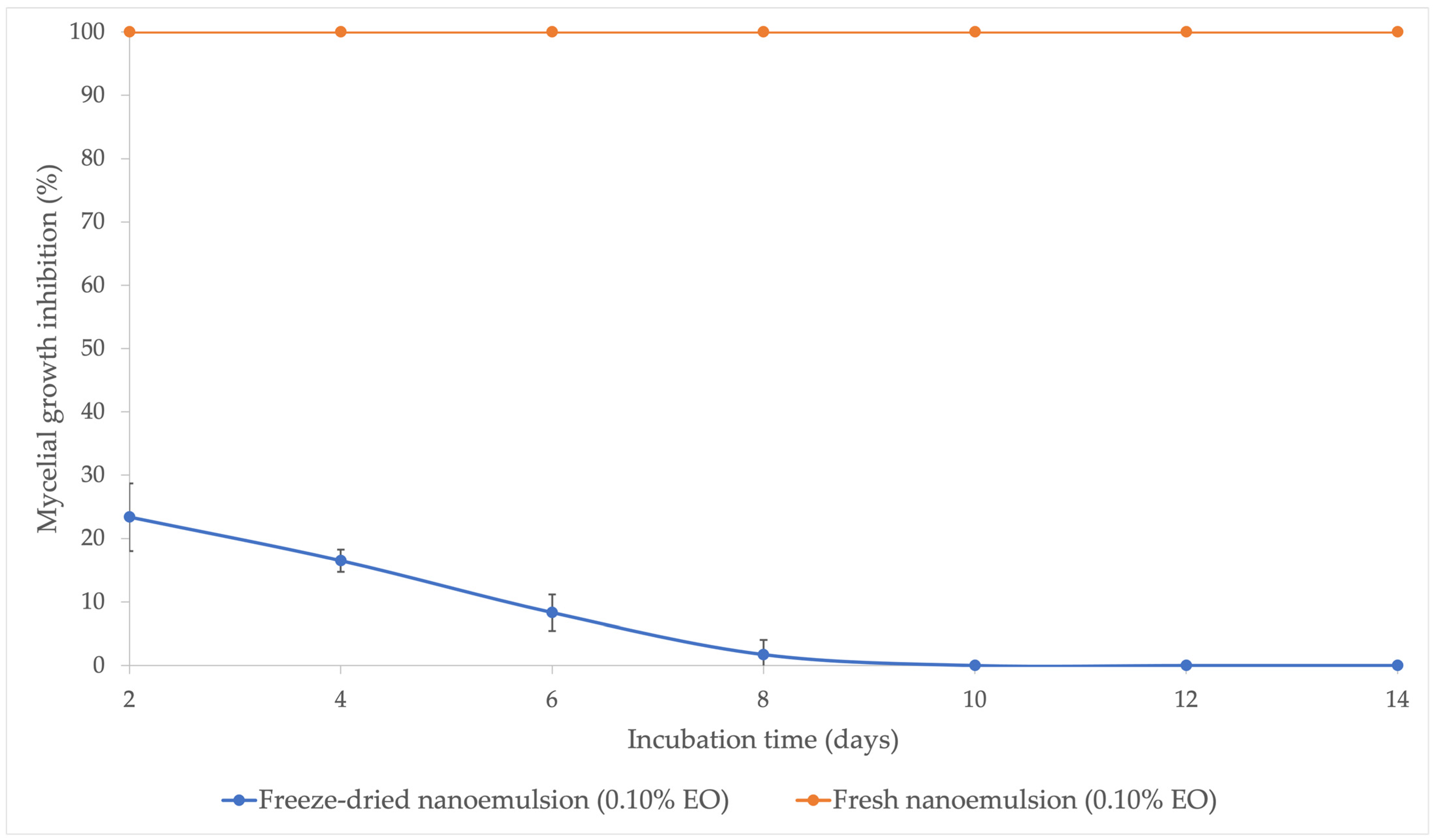
To determine the effect of freeze-drying on the antifungal activity of the nanoemulsion, mycelium growth inhibition experiments were conducted. Fresh nanoemulsion with 0.1% EO in media was able to completely inhibit mycelium growth over a 14-day period. In contrast, redispersed freeze-dried nanoemulsion at 0.10% EO in media showed an initial 77% loss in antifungal activity after only two days. At day 10, no more antifungal activity was observed (Figure 3). Additionally, images confirming the antifungal activity results are displayed in Supplementary Figure S1.
Many essential oils, including lemongrass, basil, Zatar, and others, have been tested against various organisms [6,14,15]. In these studies, essential oils were shown to inhibit the growth of specific fungal species. In particular, Martinazzo et al. [6] showed that Aspergillus flavus growth was inhibited by lemongrass oil. Additionally, lemongrass essential oil in nanoemulsion has shown complete growth inhibition and aflatoxin production in A. flavus [16]. Further, nanoemulsions containing Ocotea indecora essential oil were shown to inhibit the growth of multiple Aspergillus species, including A. flavus [33]. These results are in line with the fresh nanoemulsion result reported here (Figure 3).
In freeze-dried nanoemulsion, dramatic loss of antifungal activity is a likely consequence of vaporization of volatile components. Specifically, the loss of essential oil would be associated with a loss of volatile compounds such as citral, which makes up the majority of LGEO samples, as well as many other sources in the literature [5,34,35]. Additionally, individual components of LGEO, namely citral and geraniol, have been shown to exhibit antifungal activity when used alone [36,37]. With this, the significant loss of LGEO and its volatile components could explain the decrease in antifungal activity of nanoemulsions following freeze-drying.
Freeze-drying a nanoemulsion system containing cellulose nanocrystals can also result in changes to the morphology and physico-chemical characteristics of the CNC [22]. Some studies have shown that freeze-drying CNC results in dense agglomerates with particle sizes greater than 100 nm and flaky morphology [23]. Additionally, another source reported that after freeze-drying, CNCs were composed of large nonuniform flakes with highly decreased porosity as compared to other drying methods [24]. Changes in the shape and size of CNCs could affect the ability of the nanoemulsion to encapsulate the oil. Emulsion stabilizers and surfactants present in the nanoemulsion could reduce EO degradation or losses. For instance, Aw et al. [38] reported that CNCs can encapsulate essential oils by serving as both physical and chemical barriers, which the authors suggested was responsible for reducing oil degradation in emulsions. The results from the literature, along with the data presented in Figure 3, suggest that the barrier provided by CNCs, which initially stabilized the nanoemulsions, was lost or damaged during freeze-drying. This loss of the barrier appears to facilitate the vaporization of antifungal volatile components from the samples.

3.3. Antioxidant Activity of LGEO, Nanoemulsion, and Redispersed Nanoemulsion over 30-Day Period

3.3.1. Antioxidant Activity of Pure Essential Oil, Fresh Nanoemulsion, and Freshly Redispersed Freeze-Dried Nanoemulsion

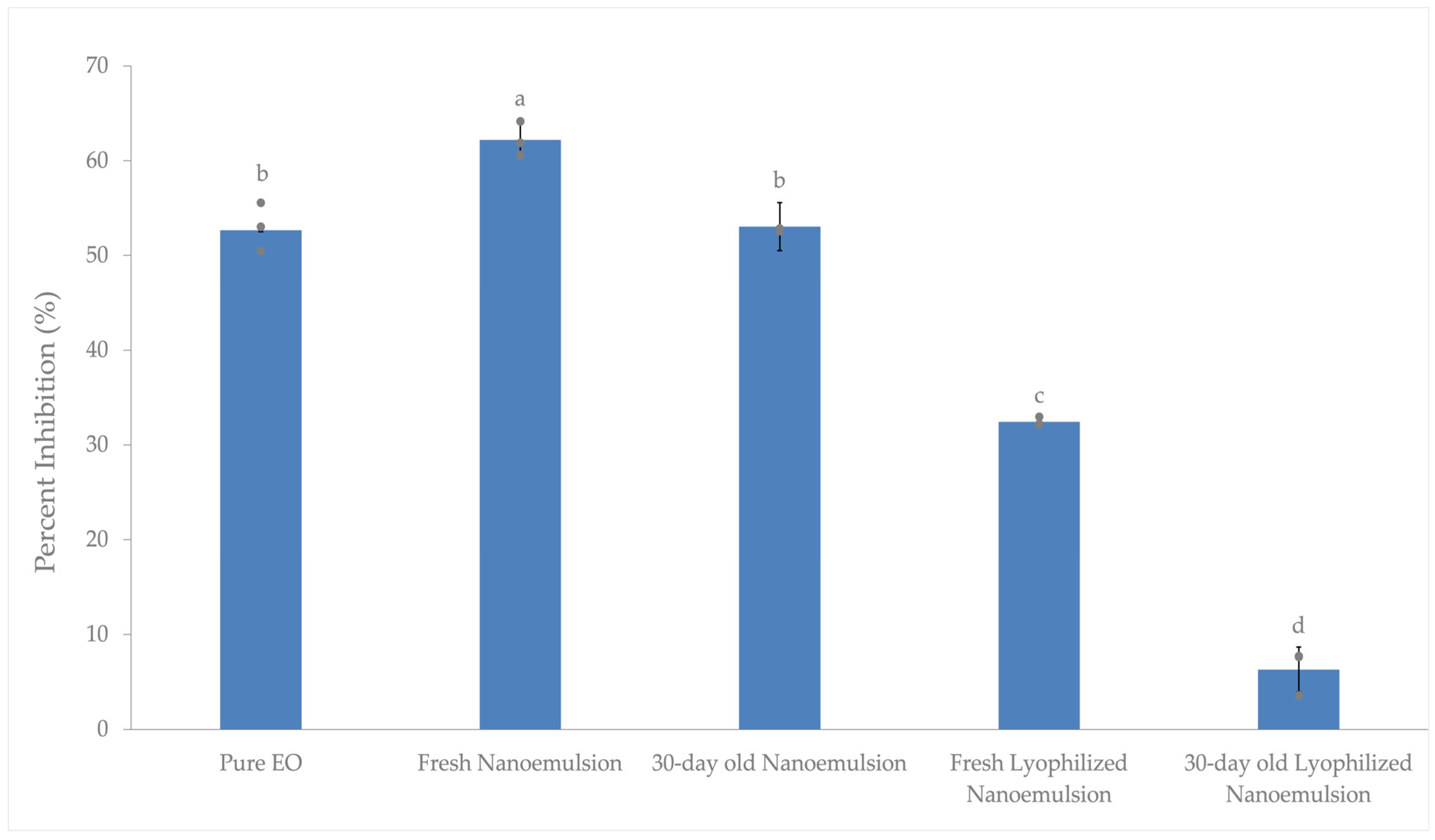
To test the antioxidant activity or free radical scavenging behavior of LGEO, nanoemulsion, and freeze-dried nanoemulsion, the DPPH assay was utilized. Essential oils, specifically LGEO, contain phenolic compounds known to have antioxidant activity [39,40]. On the day samples were created, 5% pure EO exhibited about 53% antioxidant activity, which was 9% lower than the 62% activity measured from fresh nanoemulsion samples made and tested on the same day (Figure 4). Both pure LGEO and fresh nanoemulsion had much higher activity than freshly redispersed nanoemulsion, which had about 33% antioxidant activity.
Results from Figure 4 suggest that the addition of nanoemulsion components increases the antioxidant activity of the sample. In other studies, nanoemulsion containing lemongrass oil, T80, ethanol, and water showed a similar result [17]. Specifically, nanoemulsion samples had around 9% higher antioxidant activity than pure EO [17]. An increase in radical scavenging activity may be attributable to a reduced loss of EO after the inclusion of Tween 80. In another study, researchers found that incorporating T80 into films at 2 and 4% caused a slight increase in the radical scavenging activity of samples [15]. These authors speculated that the increase in radical scavenging activity could be due to a reduced loss of EO upon T80 inclusion. Our result, in combination with data in the literature, may indicate that T80 aids in the increase in antioxidant activity by itself or by increasing the retention of EO. In contrast, another source reported that Tween 80 at 0.10% (v/v) was unable to scavenge free radicals in DPPH [41], the concentration of which is substantially lower than that used in our study (10 wt%).
Another ingredient, SBS-CNC, could also play a role in the increase in antioxidant activity seen between pure EO and fresh nanoemulsion. The inclusion of cellulose nanocrystals may be aiding in increasing the radical scavenging activity of the nanoemulsions. In Yu et al. [42], researchers concluded that the addition of cellulose nanocrystals (CNC) was responsible for the increased free radical scavenging activity in the 2,2′-azino-bis (3-ethylbenzothiazoline-6-sulfonic acid (ABTS) and DPPH assays tested on packaging films. Additionally, it was found that freeze-drying decreased the antioxidant activity of the nanoemulsion sample. As described earlier in Section 3.2, this might be caused by the changes in the stabilizing matrix, leading to oil evaporation and loss of antioxidant activity.

3.3.2. Antioxidant Activity of Pure Essential Oil, Fresh Nanoemulsion, and Freeze-Dried Nanoemulsion After 30 Days of Room Temperature Storage

Another variable tested in these experiments was the effect of room temperature storage for 30 days on the antioxidant activity of samples. After 30 days of room temperature storage, pure EO dissolved in methanol showed no significant difference in the antioxidant activity as compared to freshly diluted EO. As the pure EO sample was stored for 30 days diluted in methanol, the result is not comparable to other samples after storage that were not stored in methanol; thus, the result is not shown. Fresh nanoemulsion after 30 days of storage displayed a 9% decrease in antioxidant activity as compared to the fresh product (Figure 3). This result contrasts with the findings in another study, where it is reported that after 6 months, nanoemulsion containing EO, T80, ethanol, and water lost only 8% of antioxidant activity while the pure EO sample lost around 16% [17]. In this study, the largest loss in the antioxidant activity was measured in redispersed lyophilized nanoemulsion samples. Specifically, a 27% decrease in the antioxidant activity was observed after freeze-drying, and only 6% of antioxidant activity was left for a 30-day-old lyophilized nanoemulsion sample (Figure 4).
Overall, the antioxidant activity of the nanoemulsion samples declined after 30 days of room temperature storage, with the most significant decrease observed in the redispersed nanoemulsion sample. In contrast, pure LGEO diluted in methanol did not exhibit any significant changes (result not shown). These differences could be attributed to the varying compositions of the samples. The fresh nanoemulsion contained water, T80, soybean-CNC, NaCl, and LGEO, whereas the pure LGEO sample consisted solely of essential oil in methanol. One possible explanation for the stability of the pure LGEO sample is the difference in vapor pressure between methanol and the primary essential oil components. Methanol, with a vapor pressure of 96 mmHg at 20 °C and 127 mmHg at 25 °C (as reported by the Methanol Institute [43]), evaporates more readily than the essential oil components, such as geranyl acetate and citral, which have much lower vapor pressures (0.07 mmHg at 20 °C for geranyl acetate and approximately 0.09 mmHg at 25 °C for citral, according to Sigma Aldrich). As methanol evaporates, the sample may become enriched with LGEO, which could compensate for loss in antioxidant activity over time.

3.4. GC/QTOF Analysis of the Chemical Composition and Concentration of Volatile Components in Lemongrass Essential Oil and Its Nanoemulsion

3.4.1. GC/Q-TOF Analysis of Lemongrass Essential Oil

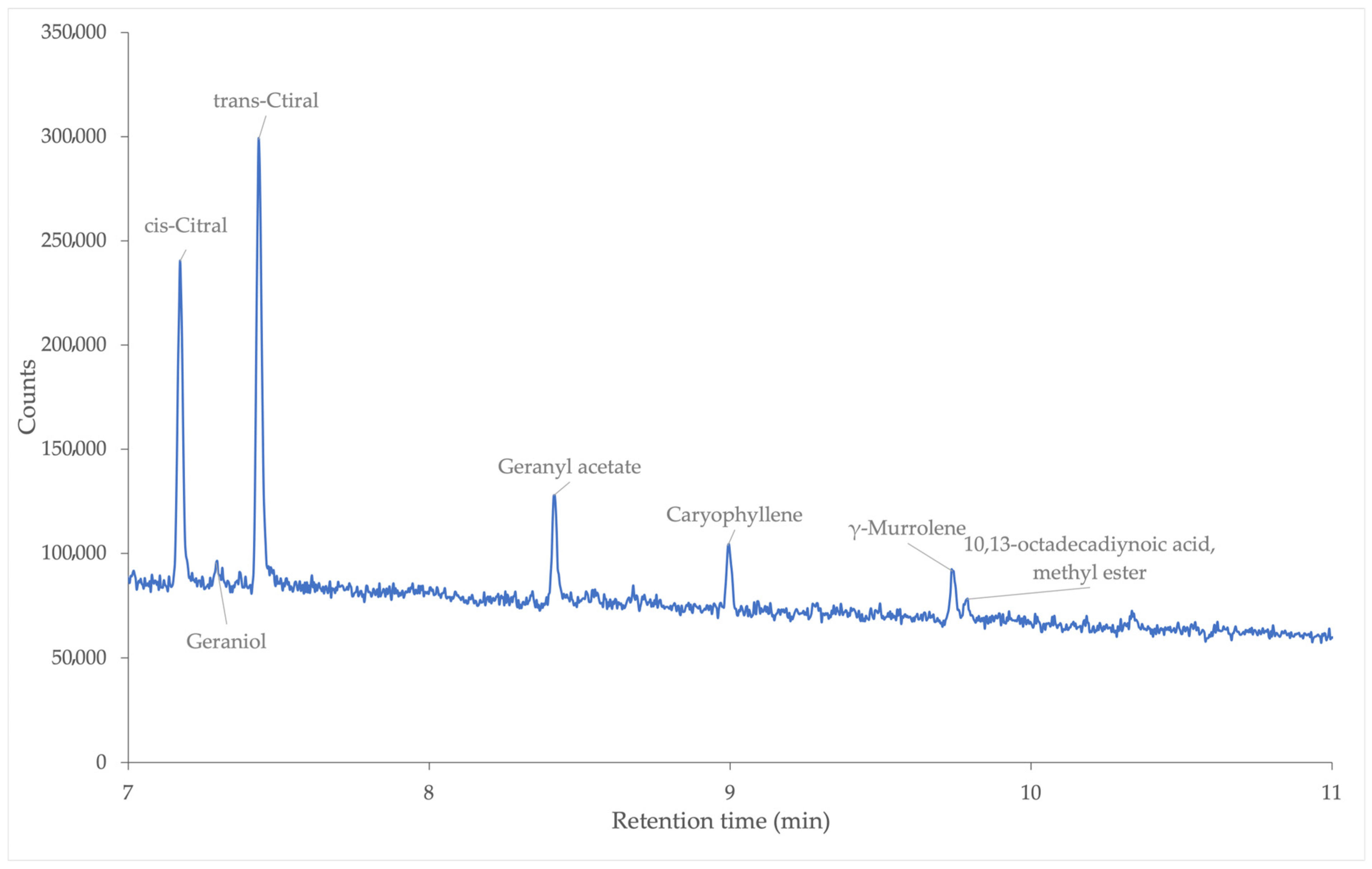
Analysis of pure lemongrass essential oil yielded multiple peaks of volatile compounds (Figure 5). Major components of LGEO were found to be cis and trans-citral, geranyl acetate, geraniol, caryophyllene, γ-Murrolene, and 10,13-octadecadiynoic acid, methyl ester.
The largest components within the lemongrass oil were cis and trans-citral with relative percentages of 30.00 and 43.20% respectively (Table 2). In Ali et al. [44], researchers also concluded that cis and trans-citral were the most concentrated components in their lemongrass (Cymbopogon Citratus) oil sample, with relative concentrations of 30.72 and 34.80% respectively. The third most concentrated compound in our results was caryophyllene at 11.65% while in Ali et al. [44], β-myrcene was the third highest component with a relative percentage of 11.28%.
The two primary compounds, cis- and trans-citral, appear as the most concentrated components in various studies on LGEO [5,34,35]. Other compounds such as geraniol, geranyl acetate, and caryophyllene also appear in these studies at different relative percentages. In other studies [34,35], myrcene is one of the more concentrated molecules in the lemongrass (Cymbopogon Citratus) oil, while in our results, it is not present. Another study, Mukarram et al. [5], also reported no myrcene peaks in their results. Furthermore, Mukarram et al. [5] conducted experiments using Cymbopogon flexuosus and noted that the lemongrass genus (Cymbopogon) comprises approximately 180 species. The two studies that reported the presence of myrcene utilized a species known as Cymbopogon Citratus, whereas our study focused on Cymbopogon Schoenanthus. Additionally, a study by Praveen et al. [45] identified γ-Muurolene in a lemongrass sample. The final compound detected in our analysis, 10,13-octadecadiynoic acid methyl ester, to the best of the authors’ knowledge, has not been previously reported in studies on lemongrass essential oil. However, this molecule has been identified in other plant extracts, such as those from Mentha spicata [45]. While this compound has not been documented in lemongrass essential oil studies, its detection in other plant extracts suggests its potential presence in botanical samples. The variation in chemical compounds among oil samples may be attributed to the use of different Cymbopogon species. Nevertheless, it is evident that cis- and trans-citral remain the most concentrated components in lemongrass essential oil, as confirmed by the data in Table 2.

3.4.2. GC/Q-TOF Analysis of Fresh Nanoemulsion Containing Lemongrass Essential Oil

In the fresh nanoemulsion sample, the characteristic peaks of cis and trans-citral, geraniol, geranyl acetate, caryophyllene, γ-Murrolene, and 10,13-octadecadiynoic acid methyl ester peaks were still observed (Figure 6).
Cis- and trans-citral were again identified as the compounds with the highest relative percentages, at 18.80% and 25.64%, respectively. However, unlike the pure EO sample, the third most abundant compound was identified at a retention time of 16.5 min as 9-Desoxo-9-x-acetoxy-3,8,12-tri-O-acetylingol. Additionally, new peaks emerged in the nanoemulsion as compared to pure EO, after the addition of T80, SBS-CNC, NaCl, and water. The most prominent new peaks appeared at retention times of approximately 13 min (two peaks) and 16.5 min (the strongest new peak) (Figure 6). At around 13 min, the two most probable compounds identified were 5,8,11-Heptadecatriynoic acid methyl ester and 1,8,15,22-Tricosatetrayne (Table 3). While these molecules have not been previously reported in lemongrass EO studies, their presence has been documented in other methanolic plant extracts [46,47]. Similarly, the compound detected at 16.5 min has not been identified in lemongrass oil studies but has been reported in the M. spicata species [45].
The peak at 16.5 min showed a relative percentage of 16.47%, followed by geranyl acetate at around 7.70% and multiple other molecules with relative percentages close to 3.00% (Table 3). Outside of citral, geraniol, and geranyl acetate, all peaks mentioned were not confirmed by use of a standard; rather, the peaks were identified using the Agilent Masshunter software and NIST library. Generally, the highest probability molecule was selected, and similar results in the literature were searched for. Outside of the molecules discussed already, there were others detected that are reported in Table 3. To our knowledge, every molecule in the table after caryophyllene oxide has not been reported in lemongrass essential oil studies in the literature. Many of them have been reported in other plant extract studies, while others have not been mentioned or noticed at all. Additionally, because standards were not purchased and tested for these molecules, we cannot confirm their existence in our tested samples. To aid in the confirmation of some of these molecules in our sample as either natural or degradation products, we researched the literature. To summarize the literature search for the source of these molecules, we created a table displaying this information in the Supplementary Materials Table S1.

3.5. Effect of Light Exposure and Room Temperature Storage on Freeze-Dried Nanoemulsion Containing LGEO

3.5.1. Effect of Light Exposure and Room Temperature Storage on the Antioxidant Activity of Freeze-Dried Nanoemulsion

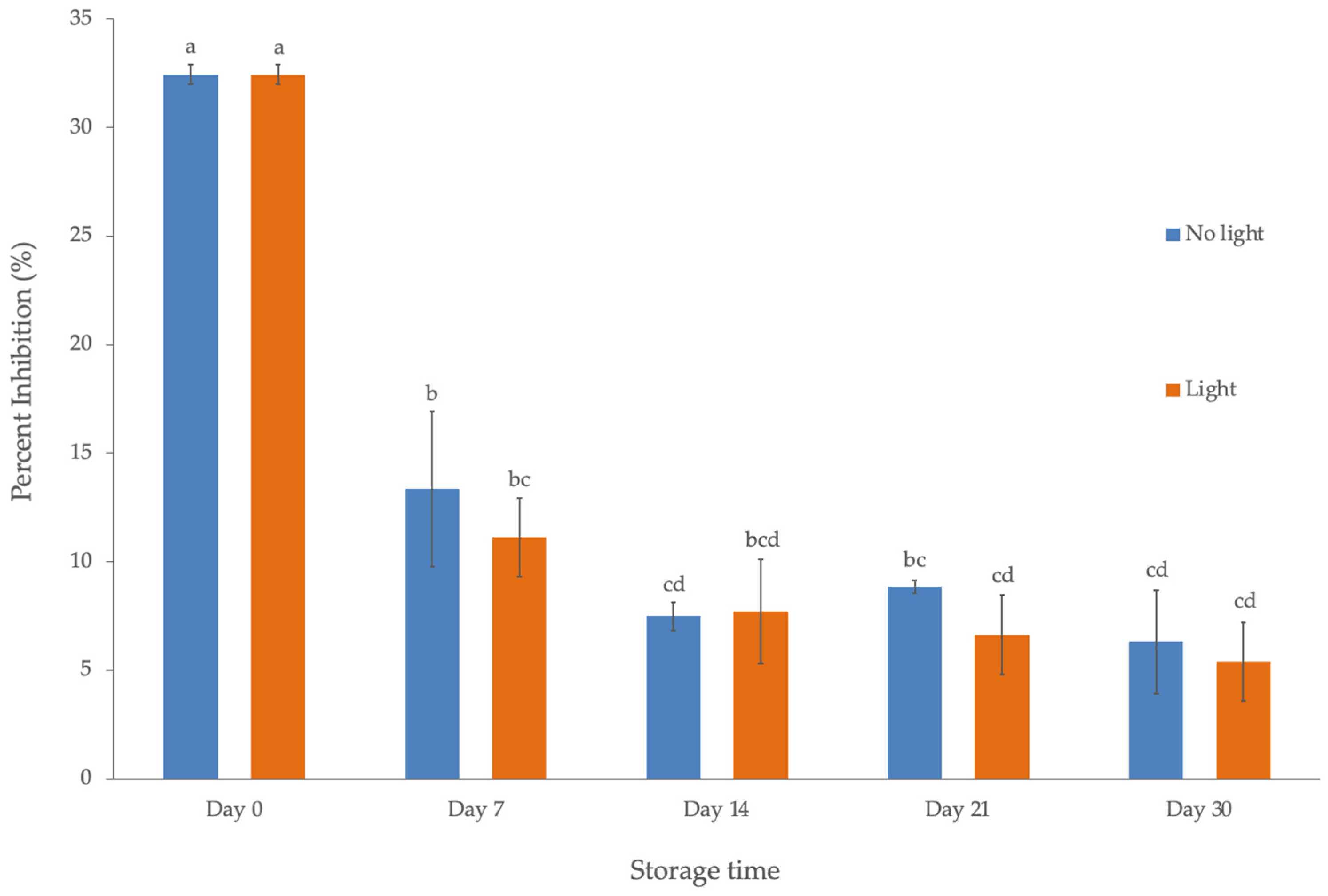
After examining the effect of room temperature storage on pure EO and fresh nanoemulsion, we investigated the impact of light exposure on freeze-dried nanoemulsion. For the control, freeze-dried nanoemulsion samples stored at −80 °C until the day of the experiment were used (labeled day 0), both with and without light exposure. These samples were then stored under their respective conditions for 30 days. The highest antioxidant activity in redispersed nanoemulsion samples after freeze-drying was observed in the sample that had neither been stored at room temperature nor exposed to light, with a value of 32.37% (Figure 7).
In comparison, fresh nanoemulsion retained nearly 63% antioxidant activity, while the redispersed lyophilized nanoemulsion showed only 32.37%, indicating that the antioxidant activity was nearly halved after freeze-drying. For antioxidant activity experiments, the redispersed nanoemulsion was dissolved in water and diluted to a measurable value on the UV–vis spectrometer. Calculations were performed to ensure that the initial concentration of EO in the redispersed samples was 5%, similar to that of the fresh nanoemulsion samples. Despite maintaining the same initial EO concentration, the significant decrease in antioxidant activity suggests that this loss is primarily due to the loss of the antioxidant compound in the lyophilized nanoemulsion.
Additionally, a significant decrease in the antioxidant activity was seen for all samples after 1 week of storage (Figure 7). This result aligns with a study in which citral essential oil (the main component of LGEO) was characterized in a nanoemulsion system [48]. In that study, the particle size was unstable and increased significantly every day during the first 7 days of storage and then began to reach thermodynamic equilibrium, resulting in a more stable particle size thereafter.
After 7 days of storage, the sample without light exposure had, on average, 2.5% more antioxidant activity than the sample exposed to light (Figure 7). On day 14, samples with and without light exposure had average activity that differed by around 0.5%. Finally, samples with and without light exposure had the lowest antioxidant activity after 30 days of storage (Figure 7). On day 30, samples without light exposure had an average activity of 6.31% while the sample with light exposure had an average activity of 5.39%. Overall, the experiments revealed a trend that the antioxidant activity of samples decreases during room temperature storage with or without light exposure. Statistical analysis of results revealed that light exposure did not significantly affect the antioxidant activity, but rather, time was the significant factor. After one week of storage, the most dramatic loss of activity was measured, and small variations were measured after that. Similar findings have been reported in the literature, showing a significant decrease in antioxidant activity over time [49]. In that study, exposure to light led to a significant reduction in the antioxidant capacity of nanoemulsions containing plant extracts over a 30-day period. In the same study, significant decreases in activity were observed for samples stored at room temperature without light exposure, indicating that some antioxidant capacity was lost during storage. This decline could be attributed to a decrease in antioxidant-contributing compounds, such as citral, over time. In a similar study, researchers found that citral content decreased consistently over a 30-day period in four different nanoemulsions containing octadecane, SDS, and Brij [50]. The findings here, along with those in the literature, suggest that components responsible for antioxidant activity are being lost during storage, a point that will be further explored in the next section.

3.5.2. Effect of Light Exposure and Room Temperature Storage Time on the Composition of Lemongrass Essential Oil-Loaded Nanoemulsion

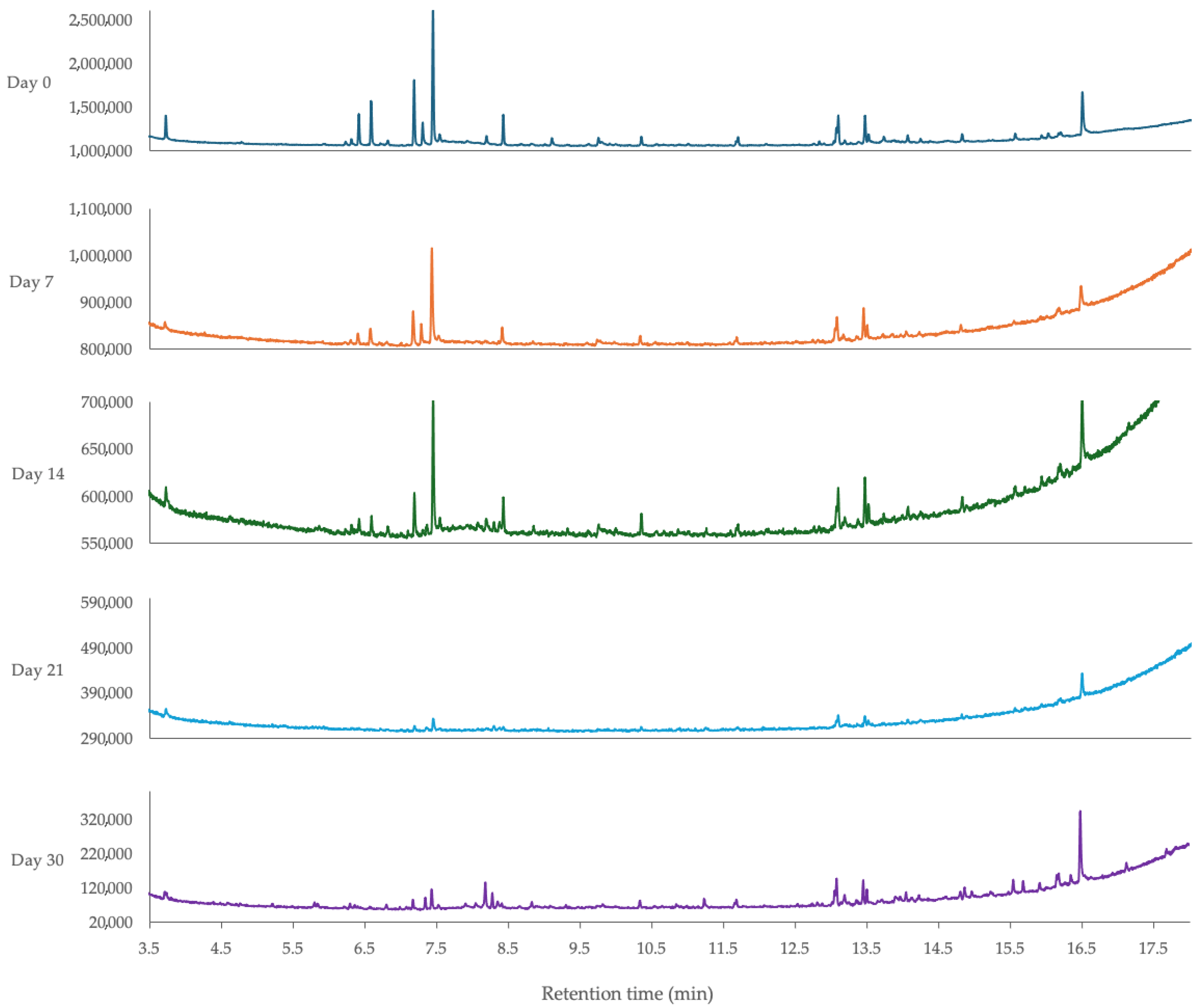
To examine the effect of encapsulated essential oil breakdown at room temperature, with and without light exposure, GC/Q-TOF compound detection and quantification methods were employed. Among the samples tested after freeze-drying and stored at room temperature without light exposure, several familiar peaks were observed (Figure 8).
Peaks corresponding to cis and trans-citral were present in all samples without light exposure, although at very low concentrations in some samples (Figure 8). While not detected in every sample, geraniol and geranyl acetate were present in samples up to day 14 (Figure 8). Recognizable peaks appeared at retention times around 13, 13.5, and 16.5 min and were detected in all samples. For these peaks, a significant decrease in peak area was observed after one week of storage, and generally, the peak areas continued to decline until day 21. On day 30, many of the peaks showed an increase in peak area; although this did not correlate with an average concentration increase (discussed in a later section), and was still significantly lower than the peaks observed on day 0.
A new peak, which had not been seen in previous samples, was discovered in freeze-dried samples. This peak appeared at around a 3.7 min retention time and was present in all replicates tested, although at significantly lower peak area values compared to day-0 samples. Among the replicates, two compounds were frequently suggested with high probabilities for this peak: styrene and benzeneethanamine, N-[(4-hydroxy)hydrocinnamoyl]-. Both compounds have been detected in plant products or essential oils; further verification with standards would be needed to confirm the compound being detected. Additionally, between 8.5 and 12.5 min, around eight peaks were detected on day 0. Each of these peaks, except for those at 10.3 and 11.7 min, was undetectable after one week.
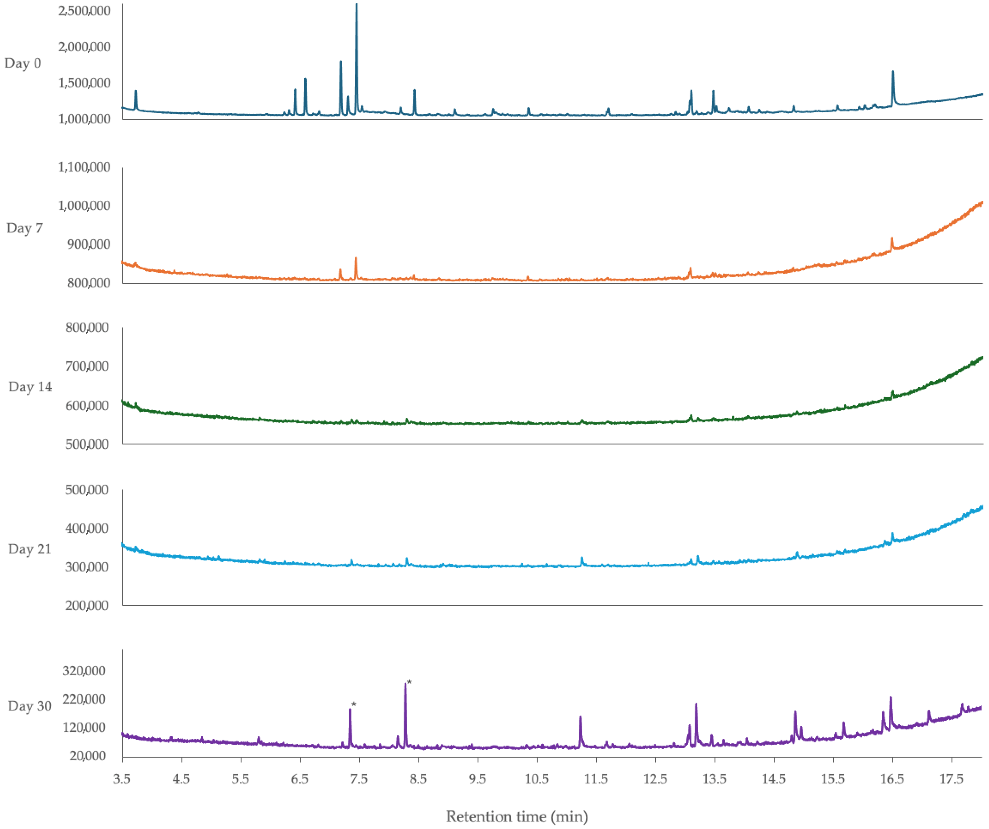
One peak that appeared only after storage was found at 11.2 min. This peak was only present after 30 days of storage in the samples without light exposure. It was also found in increasing concentrations starting on day 14 in samples exposed to light (Figure 9).
After light exposure, the loss of distinct peaks occurred more rapidly. The peaks that were not completely extinguished by day 7 are located at retention times of 7.17, 7.43, 13.05, 13.08, and 16.48 min. The peaks at 7.17 and 7.43 min correspond to cis and trans-citral, respectively. Although both citral compounds were present after 7 days of exposure, they were no longer detected in samples beyond that point. In contrast, the peaks at 13.05, 13.08, and 16.48 min were detected throughout the entire 30-day period, with an increase in concentration observed from day 21 to day 30.
In addition to the peak at 11.2 min, the samples exposed to light revealed the appearance of several new molecules. The first of these was located at around 5.8 min. This peak was faintly noticeable on days 14 and 21 but became most prominent after 30 days of light exposure. Other peaks following a similar trend, with the most prominent peak appearing on day 30, were located at 7.35, 8.15, 8.28, 11.24, 13.18, 14.85, 15.67, and 16.35 min (Figure 9).
A final note regarding samples in either exposure condition is the dramatic loss of volatiles after only 7 days of storage. As speculated earlier, the stabilizing effect of the essential oil, which was initially present in the nanoemulsions, was no longer observed, likely leading to significantly reduced peak sizes over time. Figure 8 and Figure 9, in combination with the earlier antifungal and antioxidant data, suggest that the morphology and physico-chemical characteristics that once stabilized the oil have become less effective. Variations in the most probable compound suggested by the software were noticed between replicates, making the determination of new peaks more difficult with our data set. For interested readers, a table describing the variation in names and their presence in the literature has been developed (Supplementary Materials Table S2).

3.5.3. Effect of Light Exposure and Room Temperature Storage on the Concentration of Four Volatile Components in Lemongrass Essential Oil-Loaded Nanoemulsion

Alongside compositional changes of the LGEO-loaded nanoemulsion, concentration changes for four volatile components were also quantified (Table 4).
For compounds except geranyl acetate, the highest concentrations of volatile components were observed on day 0, before any storage or light exposure. Geranyl acetate maintained its concentration through one week of storage but then declined, becoming undetectable by day 21. The other three peaks showed consistent decreases in concentration over the storage period. Specifically, both citral peaks decreased steadily, reaching their lowest concentrations on day 30. For geraniol, the peak disappeared by day 21 and remained undetectable on day 30 (Table 4).
The reduction in volatile compounds may explain the diminished stability of the nanoemulsions after freeze-drying, potentially facilitating greater vaporization of LGEO at room temperature. Similar trends were reported by Irmak et al. [51], who observed significant losses of phenolic content and antioxidant activity in rosemary oil stored at room temperature compared to 4 °C over four weeks. To determine if a similar rate of volatile loss occurs in fresh nanoemulsions, further experiments with fresh samples stored over a 30-day period are warranted.
Light-exposed samples produced markedly different results compared to those shielded from light. By day 21, both citral peaks were undetectable in light-exposed samples, while they remained measurable in the light-shielded samples (Table 4). Additionally, in light-exposed samples, geraniol was not detected at any point, and geranyl acetate was only observed after one week of exposure. These findings suggest that light exposure accelerates the degradation of LGEO volatiles. While volatile concentrations also decreased significantly in light-shielded samples, the process was notably slower under these conditions.
Although the data in Table 4 indicate that neither exposure condition preserved sample stability, the results align with trends observed in related studies. In all conditions, the concentrations of the four volatile components declined consistently, reaching either minimal values or zero after 30 days. Notably, even light-shielded samples stored solely at room temperature exhibited substantial volatile losses, suggesting an imperfect storage method. These findings are consistent with other studies involving freeze-dried nanoemulsions, which often report diminished activity and lower volatile concentrations compared to fresh counterparts.
This indicates that freeze-drying compromises EO stability within nanoemulsions. Previous research has compared various drying methods, such as spray drying, freeze-drying, and spray freeze-drying, in CNC-based formulations across different applications [22,23,24]. These studies suggest that alternative or combined drying methods could better preserve morphological and chemical features, thereby maintaining volatile component stability and bioactivity. Future investigations may need to explore these methods to optimize antifungal and antioxidant activity alongside the retention of volatile components.

4. Conclusions

In this study, we developed and characterized a nanoemulsion that was freeze-dried and subsequently redispersed in water. The freeze-drying process significantly impacted the physical and chemical properties of the nanoemulsion, reducing the zeta potential from 39.2 mV to 30.6 mV and increasing particle size from 137 nm to 158 nm. Additionally, approximately 30% of the essential oil concentration was lost during freeze-drying. Redispersed lyophilized nanoemulsions also exhibited a substantial decline in bioactivity, with antifungal activity decreasing by 77% and antioxidant activity by 31%. Storage conditions further influenced stability, with light exposure accelerating the degradation of key volatile compounds—cis- and trans-citral, geraniol, and geranyl acetate—detected via GC/Q-TOF. Antioxidant activity declined sharply after one week of storage under both light-exposed and light-shielded conditions, potentially due to thermodynamic stresses, before stabilizing at approximately 50% of the day 7 activity. Discrepancies between volatile degradation and antioxidant activity may stem from contributions by formulation components such as Tween 80, NaCl, and SBS-CNC. Light exposure also caused significant chemical changes, with 11 new compounds forming during storage compared to one in light-shielded samples. These findings suggest that light exposure accelerates chemical changes in the nanoemulsion, potentially further reducing its bioactivity and stability. Findings from this study highlight challenges in maintaining stability and bioactivity of essential oil-loaded nanoemulsions after freeze-drying, likely due to disruptions in stabilizing factors of the original O/W nanoemulsion. Despite these challenges, the fresh nanoemulsion demonstrated promising LGEO bioactivity, providing a foundation for further optimization. Refining freeze-drying protocols and formulation conditions could enhance stability and unlock new applications for renewable, bioactive nanoemulsions.

Supplementary Materials

The following supporting information can be downloaded at: https://www.mdpi.com/article/10.3390/pr13092752/s1, Figure S1. Images of antifungal activity experiments (corresponding to results in Figure 3). Images are representative of replicates.; Table S1. Literature search of components of fresh nanoemulsion extract detected by GC/Q-TOF. Columns highlight the name of the compound detected, whether the compound was detected in LGEO in other studies, whether the compound was detected in EO in general, the source of the molecule in the literature, and the citation of the publications citing the specific molecule.; Table S2. Compound compilation of molecules detected at similar retention times in freeze-dried nanoemulsion extract samples. Table contains a literature search of compounds detected in the freeze-dried nanoemulsion extract samples. Columns highlight the name of the compound detected, whether the compound was detected in LGEO in other studies, whether the compound was detected in EO in general, the source of the molecule in the literature, and the citation of the publications citing the specific molecule. References [5,34,35,44,45,46] are cited in the main text and Supplementary Materials. References [52,53,54,55,56,57,58,59,60,61,62,63,64,65,66,67,68,69,70,71,72,73,74,75,76,77,78,79,80,81,82,83,84,85,86,87,88,89,90,91,92,93,94] are cited in the Supplementary Materials.

Author Contributions

Conceptualization, L.L.; methodology, L.L. and K.D.F.; validation, L.L. and K.D.F.; formal analysis, L.L. and K.D.F.; investigation, L.L. and K.D.F.; resources, L.L.; data curation, L.L. and K.D.F.; writing—original draft preparation, K.D.F.; writing—review and editing, L.L. and K.D.F.; visualization, L.L. and K.D.F.; supervision, L.L.; project administration, L.L.; funding acquisition, L.L. All authors have read and agreed to the published version of the manuscript.

Funding

This research was supported by the Iowa Soybean Association.

Data Availability Statement

The original contributions presented in the study are included in the article; further inquiries can be directed to the corresponding author.

Acknowledgments

The authors would like to thank Lorien Radmer and Adina Howe from the Genomics and Environmental Research in Microbial Systems lab at Iowa State University for their generosity and assistance with freeze-drying samples.

Conflicts of Interest

The authors declare no conflicts of interest.

Abbreviations

The following abbreviations are used in this manuscript:
SBS-CNCSoybean stover-derived cellulose nanocrystals
O/WOil-in-water
LGEOLemongrass essential oil
EOEssential oil

References

  1. Hüsnü Can Baser, K.; Demirci, F. Chemistry of essential oils. In Flavours and Fragrances: Chemistry, Bioprocessing and Sustainability; Springer: Berlin/Heidelberg, Germany, 2007; pp. 43–86. [Google Scholar]
  2. Abers, M.; Schroeder, S.; Goelz, L.; Sulser, A.; St. Rose, T.; Puchalski, K.; Langland, J. Antimicrobial activity of the volatile substances from essential oils. BMC Complement. Med. Ther. 2021, 21, 124. [Google Scholar] [CrossRef]
  3. Hu, F.; Tu, X.; Thakur, K.; Hu, F.; Li, X.; Zhang, Y.; Zhang, J.; Wei, Z. Comparison of antifungal activity of essential oils from different plants against three fungi. Food Chem. Toxicol. 2019, 134, 110821. [Google Scholar] [CrossRef]
  4. Andoğan, B.C.; Baydar, H.; Kaya, S.; Demirci, M.; Özbaşar, D.; Mumcu, E. Antimicrobial activity and chemical composition of some essential oils. Arch. Pharmacal Res. 2002, 25, 860–864. [Google Scholar] [CrossRef]
  5. Mukarram, M.; Choudhary, S.; Khan, M.A.; Poltronieri, P.; Khan, M.M.A.; Ali, J.; Kurjak, D.; Shahid, M. Lemongrass Essential Oil Components with Antimicrobial and Anticancer Activities. Antioxidants 2022, 11, 20. [Google Scholar] [CrossRef]
  6. Martinazzo, A.P.; Oliveira, F.S.; Teodoro, C.E.S. Antifungal activity of Cymbopogon citratus essential oil against Aspergillus flavus. Cienca E Nat. 2022, 41, e20. [Google Scholar] [CrossRef]
  7. Oxenham, S.K.; Svoboda, K.P.; Walters, D.R. Antifungal activity of the essential oil of basil (Ocimum basilicum). J. Phytopathol. 2005, 153, 174–180. [Google Scholar] [CrossRef]
  8. Daouk, R.K.; Dagher, S.M.; Sattout, E.J. Antifungal activity of the essential oil of Origanum syriacum L. J. Food Prot. 1995, 58, 1147–1149. [Google Scholar] [CrossRef]
  9. Barradas, T.N.; Gyselle de Holanda e Silva, K. Nanoemulsions of essential oils to improve solubility, stability and permeability: A review. Environ. Chem. Lett. 2021, 19, 1153–1171. [Google Scholar] [CrossRef]
  10. Weisheimer, V.; Miron, D.; Silva, C.B.; Guterres, S.S.; Schapoval, E.E.S. Microparticles containing lemongrass volatile oil: Preparation, characterization and thermal stability. Die Pharm. Int. J. Pharm. Sci. 2010, 65, 885–890. [Google Scholar]
  11. Rowshan, V.; Bahmanzadegan, A.; Saharkhiz, M.J. Influence of storage conditions on the essential oil composition of Thymus daenensis Celak. Ind. Crops Prod. 2013, 49, 97–101. [Google Scholar] [CrossRef]
  12. Mehdizadeh, L.; Ghasemi Pirbalouti, A.; Moghaddam, M. Storage stability of essential oil of cumin (Cuminum cyminum L.) as a function of temperature. Int. J. Food Prop. 2017, 20 (Suppl. S2), 1742–1750. [Google Scholar] [CrossRef]
  13. Turek, C.; Stintzing, F.C. Impact of different storage conditions on the quality of selected essential oils. Food Res. Int. 2012, 46, 341–353. [Google Scholar] [CrossRef]
  14. Gupta, A.; Eral, H.B.; Hatton, T.A.; Doyle, P.S. Nanoemulsions: Formation, properties and applications. Soft Matter 2016, 12, 2826–2841. [Google Scholar] [CrossRef]
  15. Chu, Y.; Xu, T.; Gao, C.; Liu, X.; Zhang, N.I.; Feng, X.; Liu, X.; Shen, X.; Tang, X. Evaluations of physicochemical and biological properties of pullulan-based films incorporated with cinnamon essential oil and Tween 80. Int. J. Biol. Macromol. 2019, 122, 388–394. [Google Scholar] [CrossRef]
  16. Chaudhari, A.K.; Singh, V.K.; Das, S.; Dubey, N.K. Fabrication, characterization, and bioactivity assessment of chitosan nanoemulsion containing allspice essential oil to mitigate Aspergillus flavus contamination and aflatoxin B1 production in maize. Food Chem. 2022, 372, 131221. [Google Scholar] [CrossRef]
  17. Harleen, K.; Pranav, P.; Ramneek, K.; Shriya, A.; Manisha, S. Synthesis and characterization of Citrus limonum essential oil based nanoemulsion and its enhanced antioxidant activity with stability for transdermal application. J. Biomater. Nanobiotechnology 2020, 11, 215–236. [Google Scholar] [CrossRef]
  18. Gong, X.; Wang, Y.; Chen, L. Enhanced emulsifying properties of wood-based cellulose nanocrystals as Pickering emulsion stabilizer. Carbohydr. Polym. 2017, 169, 295–303. [Google Scholar] [CrossRef]
  19. Habibi, Y.; Lucia, L.A.; Rojas, O.J. Cellulose nanocrystals: Chemistry, self-assembly, and applications. Chem. Rev. 2010, 110, 3479–3500. [Google Scholar] [CrossRef]
  20. do Vale Morais, A.R.; do Nascimento Alencar, É.; Júnior, F.H.X.; De Oliveira, C.M.; Marcelino, H.R.; Barratt, G.; Fessi, H.; Do Egito, E.S.T.; Elaissari, A. Freeze-drying of emulsified systems: A review. Int. J. Pharm. 2016, 503, 102–114. [Google Scholar] [CrossRef] [PubMed]
  21. El-Messery, T.M.; Altuntas, U.; Altin, G.; Özçelik, B. The effect of spray-drying and freeze-drying on encapsulation efficiency, in vitro bioaccessibility and oxidative stability of krill oil nanoemulsion system. Food Hydrocoll. 2020, 106, 105890. [Google Scholar] [CrossRef]
  22. Alarcón-Moyano, J.; Acuña, D.; Matiacevich, S.; Caballero, L.; Melo, F.; Quero, F.; Díaz-Calderón, P. Physico-chemical and structural characterization of cellulose nanocrystals obtained by two drying methods: Freeze-drying and spray-drying. Food Hydrocoll. 2023, 140, 108571. [Google Scholar] [CrossRef]
  23. Khoshkava, V.; Kamal, M.R. Effect of drying conditions on cellulose nanocrystal (CNC) agglomerate porosity and dispersibility in polymer nanocomposites. Powder Technol. 2024, 261, 288–298. [Google Scholar] [CrossRef]
  24. Abdallah, W.; Kamal, M.R. Influence of process variables on physical characteristics of spray freeze dried cellulose nanocrystals. Cellulose 2018, 25, 5711–5730. [Google Scholar] [CrossRef]
  25. Liu, L.; Abiol, K.A.; Friest, M.A.; Fisher, K.D. Synergistic stabilization of nanoemulsion using nonionic surfactants and salt-sensitive cellulose nanocrystals. Polymers 2023, 15, 4682. [Google Scholar] [CrossRef]
  26. Liu, L.; Fisher, K.D.; Bussey, W.D. Comparison of emulsion stabilizers: Application for the enhancement of the bioactivity of lemongrass essential oil. Polymers 2024, 16, 415. [Google Scholar] [CrossRef]
  27. Jiménez-Escrig, A.; Jiménez-Jiménez, I.; Sánchez-Moreno, C.; Saura-Calixto, F. Evaluation of free radical scavenging of dietary carotenoids by the stable radical 2, 2-diphenyl-1-picrylhydrazyl. J. Sci. Food Agric. 2000, 80, 1686–1690. [Google Scholar] [CrossRef]
  28. Baliyan, S.; Mukherjee, R.; Priyadarshini, A.; Vibhuti, A.; Gupta, A.; Pandey, R.P.; Chang, C.M. Determination of antioxidants by DPPH radical scavenging activity and quantitative phytochemical analysis of Ficus religiosa. Molecules 2022, 27, 1326. [Google Scholar] [CrossRef] [PubMed]
  29. Kovacevic, A.; Savic, S.; Vuleta, G.; Mueller, R.H.; Keck, C.M. Polyhydroxy surfactants for the formulation of lipid nanoparticles (SLN and NLC): Effects on size, physical stability and particle matrix structure. Int. J. Pharm. 2011, 406, 163–172. [Google Scholar] [CrossRef]
  30. Farouil, L.; Dias, R.P.; Popotte-Julisson, G.; Bibian, G.; Adou, A.I.; de la Mata, A.P.; Muriel, S.; James, J.H.; Cebrián-Torrejón, G. The Metabolomic Profile of the Essential Oil from Zanthoxylum caribaeum (syn. chiloperone) Growing in Guadeloupe FWI using GC× GC-TOFMS. Metabolites 2022, 12, 1293. [Google Scholar] [CrossRef]
  31. Bashlouei, S.G.; Karimi, E.; Zareian, M.; Oskoueian, E.; Shakeri, M. Heracleum persicum essential oil nanoemulsion: A nanocarrier system for the delivery of promising anticancer and antioxidant bioactive agents. Antioxidants 2022, 11, 831. [Google Scholar] [CrossRef] [PubMed]
  32. de Godoi, S.N.; Quatrin, P.M.; Sagrillo, M.R.; Nascimento, K.; Wagner, R.; Klein, B.; Ourique, A.F. Evaluation of stability and in vitro security of nanoemulsions containing Eucalyptus globulus oil. BioMed Res. Int. 2017, 1, 2723418. [Google Scholar] [CrossRef]
  33. Pinto, L.D.A.; Machado, F.P.; Esteves, R.; Farias, V.M.; Köptcke, F.B.N.; Ricci-Junior, E.; Rocha, L.; Keller, L.A.M. Characterization and inhibitory effects of essential oil and nanoemulsion from Ocotea indecora (Shott) Mez in Aspergillus species. Molecules 2023, 28, 3437. [Google Scholar] [CrossRef]
  34. Soliman, W.S.; Salaheldin, S.; Amer, H.M. Chemical Composition Evaluation of Egyptian Lemongrass, Cymbopogon citratus, Essential Oil. Int. J. Sci. Eng. Res. 2017, 8, 630–634. [Google Scholar]
  35. Hanaa, A.M.; Sallam, Y.I.; El-Leithy, A.S.; Aly, S.E. Lemongrass (Cymbopogon citratus) essential oil as affected by drying methods. Ann. Agric. Sci. 2012, 57, 113–116. [Google Scholar] [CrossRef]
  36. Silva, C.D.B.D.; Guterres, S.S.; Weisheimer, V.; Schapoval, E.E. Antifungal activity of the lemongrass oil and citral against Candida spp. Braz. J. Infect. Dis. 2008, 12, 63–66. [Google Scholar]
  37. Tang, X.; Shao, Y.L.; Tang, Y.J.; Zhou, W.W. Antifungal activity of essential oil compounds (geraniol and citral) and inhibitory mechanisms on grain pathogens (Aspergillus flavus and Aspergillus ochraceus). Molecules 2018, 23, 2108. [Google Scholar] [CrossRef] [PubMed]
  38. Aw, Y.Z.; Lim, H.P.; Low, L.E.; Singh, C.K.S.; Chan, E.S.; Tey, B.T. Cellulose nanocrystal (CNC)-stabilized Pickering emulsion for improved curcumin storage stability. Lwt 2022, 159, 113249. [Google Scholar] [CrossRef]
  39. Anggraeni, N.I.; Hidayat, I.W.; Rachman, S.D.; Ersanda. Bioactivity of essential oil from lemongrass (Cymbopogon citratus Stapf) as antioxidant agent. AIP Conf. Proc. 2018, 1927, 030007. [Google Scholar] [CrossRef]
  40. Mirghani, M.E.S.; Liyana, Y.; Parveen, J. Bioactivity analysis of lemongrass (Cymbopogan citratus) essential oil. Int. Food Res. J. 2012, 19, 569–575. [Google Scholar]
  41. Pérez-Rosés, R.; Risco, E.; Vila, R.; Peñalver, P.; Cañigueral, S. Antioxidant activity of Tween-20 and Tween-80 evaluated through different in-vitro tests. J. Pharm. Pharmacol. 2015, 67, 666–672. [Google Scholar] [CrossRef]
  42. Yu, Z.; Sun, L.; Wang, W.; Zeng, W.; Mustapha, A.; Lin, M. Soy protein-based films incorporated with cellulose nanocrystals and pine needle extract for active packaging. Ind. Crops Prod. 2018, 112, 412–419. [Google Scholar] [CrossRef]
  43. Physical Properties of Pure Methanol. Available online: https://www.methanol.org/wp-content/uploads/2016/06/Physical-Properties-of-Pure-Methanol.pdf (accessed on 27 August 2025).
  44. Ali, M.M.; Yusuf, M.A.; Abdalaziz, M.N. GC-MS Analysis and Antimicrobial Screening of Essential Oil from Lemongrass (Cymbopogon citratus). Int. J. Pharm. Chem. 2017, 3, 72–76. [Google Scholar] [CrossRef]
  45. Praveen, T.; Krishnamoorthy, A.S.; Nakkeeran, S.; Sivakumar, U.; Amirtham, D. Antifungal volatiles from medicinal herbs suppress Fusarium oxysporum f. sp. lycopersici. J. Entomol. Zool. Stud. 2021, 9, 1083–1093. [Google Scholar]
  46. Altameme, H.J.M. GC-MS and FTIR analysis Phytocomponents on different parts of Capparis spinosa L. (Capparidaceae) in Iraq. J. Chem. Pharm. Sci. 2016, 4, 3269–3282. [Google Scholar]
  47. Pal, A.; Ray, R.; Acharya, K.; Paul, S. Assessment of the anti-leukemic and antioxidant potential of the methanol extract of a wild, edible, and novel mushroom, Astraeus hygrometricus, and unraveling its metabolomic profile. J. Adv. Biotechnol. Exp. Ther. 2021, 4, 388–404. [Google Scholar] [CrossRef]
  48. Lu, W.C.; Huang, D.W.; Wang, C.C.; Yeh, C.H.; Tsai, J.C.; Huang, Y.T.; Li, P.H. Preparation, characterization, and antimicrobial activity of nanoemulsions incorporating citral essential oil. J. Food Drug Anal. 2018, 26, 82–89. [Google Scholar] [CrossRef]
  49. Bazana, M.T.; da Silva, S.S.; Codevilla, C.F.; de Deus, C.; Lucas, B.N.; Ugalde, G.A.; Mazutti, M.A.; Flores, E.M.M.; Barin, J.S.; da Silva, C.D.B.; et al. Development of nanoemulsions containing Physalis peruviana calyx extract: A study on stability and antioxidant capacity. Food Res. Int. 2019, 125, 108645. [Google Scholar] [CrossRef] [PubMed]
  50. Mei, L.; Choi, S.J.; Alamed, J.; Henson, L.; Popplewell, M.; McClements, D.J.; Decker, E.A. Citral stability in oil-in-water emulsions with solid or liquid octadecane. J. Agric. Food Chem. 2010, 58, 533–536. [Google Scholar] [CrossRef]
  51. Irmak, S.; Solakyildirim, K.; Hesenov, A.; Erbatur, O. Study on the stability of supercritical fluid extracted rosemary (Rosmarinus offcinalis L.) essential oil. J. Anal. Chem. 2010, 65, 899–906. [Google Scholar] [CrossRef]
  52. Aguirre-Becerra, H.; Pineda-Nieto, S.A.; García-Trejo, J.F.; Guevara-González, R.G.; Feregrino-Pérez, A.A.; Álvarez-Mayorga, B.L.; Pastrana, D.M.R. Jacaranda flower (Jacaranda mimosifolia) as an alternative for antioxidant and antimicrobial use. Heliyon 2002, 6, e05802. [Google Scholar] [CrossRef]
  53. Ahmed, R.; Abdel-Rahman, A. Effect of Papaya Wastes on Quality Characteristics of Meat Burger. New Val. J. Agric. Sci. 2022, 2, 483–511. [Google Scholar] [CrossRef]
  54. Ahuchaogu, A.A.; Igara, C.E. Antibacterial, GC-MS and FTIR Chemical Profiling of Methanol Leaf Extract of Cymbopogon citratus Linn. ARC J. Pharm. Sci. 2011, 7, 1–9. [Google Scholar]
  55. Almalki, G.; Rabah, S.; Al-Faifi, Z.; Alharbi, A.; Sharma, M. Phytochemistry Screening, Antioxidant and Antimicrobial Activities of Euphorbia inarticulata Schweinf Plant Extract. Pharmacophore 2022, 13, 91–99. [Google Scholar] [CrossRef]
  56. Antwi-Agyakwa, A.K.; Yusuf, A.A.; Pirk, C.W.; Mohamed, S.A.; Ekesi, S.; Torto, B. Exploring non-host plant-based management strategy with lemongrass, garlic and guava volatiles for the African citrus triozid. J. Appl. Entomol. 2021, 145, 757–766. [Google Scholar] [CrossRef]
  57. Ayalew, A.A. Chromatographic and spectroscopic determination of solvent-extracted Lantana camara leaf oil. J. Int. Med. Res. 2020, 48, 300060520962344. [Google Scholar] [CrossRef]
  58. Begum, N.; Akhtar, K.; Ahanger, M.A.; Iqbal, M.; Wang, P.; Mustafa, N.S.; Zhang, L. Arbuscular mycorrhizal fungi improve growth, essential oil, secondary metabolism, and yield of tobacco (Nicotiana tabacum L.) under drought stress conditions. Environ. Sci. Pollut. Res. 2021, 28, 45276–45295. [Google Scholar] [CrossRef]
  59. Bennett, V.; Godwin, J. Proximate, Selected Metals and Metabolites Profile of Citrullus lanatus (Watermelon) Leaves. Int. J. Adv. Res. Chem. Sci. 2023, 10, 14–21. [Google Scholar] [CrossRef]
  60. Chairgulprasert, V.; Prasertsongskun, S.; Junpra-ob, S.; Sangjun, M. Chemical constituents of the essential oil, antioxidant and antibacterial activities from Elettariopsis curtisii Baker. Songklanakarin J. Sci. Technol. 2008, 30, 591–596. [Google Scholar]
  61. Chanthai, S.; Prachakoll, S.; Ruangviriyachai, C.; Luthria, D.L. Influence of extraction methodologies on the analysis of five major volatile aromatic compounds of citronella grass (Cymbopogon nardus) and lemongrass (Cymbopogon citratus) grown in Thailand. J. AOAC Int. 2012, 95, 763–772. [Google Scholar] [CrossRef]
  62. Chen, X.; Chen, H.; Xiao, J.; Liu, J.; Tang, N.; Zhou, A. Variations of volatile flavour compounds in finger citron (Citrus medica L. var. sarcodactylis) pickling process revealed by E-nose, HS-SPME-GC-MS and HS-GC-IMS. Food Res. Int. 2020, 138, 109717. [Google Scholar] [CrossRef] [PubMed]
  63. Chung, H.Y.; Fung, P.K.; Kim, J.S. Aroma impact components in commercial plain sufu. J. Agric. Food Chem. 2005, 53, 1684–1691. [Google Scholar] [CrossRef] [PubMed]
  64. Devi, M.A.; Singh, T.B. Composition of Essential Oil of Indigenous and Exotic Species of Lemongrass Growing in Manipur. Ann. Multidiscip. Res. Innov. Technol. 2023, 2, 58–64. [Google Scholar]
  65. Elsharkawy, E.R.; Abdallah, E.M.; Shiboob, M.H.; Alghanem, S. Phytochemical, antioxidant and antibacterial potential of Ducrosia anethifolia in northern border region of Saudi Arabia. J. Pharm. Res. Int. 2019, 31, 1–8. [Google Scholar] [CrossRef]
  66. Ghosh, S.; Derle, A.; Ahire, M.; More, P.; Jagtap, S.; Phadatare, S.D.; Patil, A.B.; Jabgunde, A.M.; Sharma, G.K.; Shinde, V.S.; et al. Phytochemical Analysis and Free Radical Scavenging Activity of Medicinal Plants Gnidia glauca and Dioscorea bulbifera. PLoS ONE 2012, 8, e82529. [Google Scholar] [CrossRef]
  67. Harlina, P.W.; Ma, M.; Shahzad, R.; Gouda, M.M.; Qiu, N. Effect of clove extract on lipid oxidation, antioxidant activity, volatile compounds and fatty acid composition of salted duck eggs. J. Food Sci. Technol. 2018, 55, 4719–4734. [Google Scholar] [CrossRef]
  68. Jaafar, F.M.; Osman, C.P.; Ismail, N.H.; Awang, K. Analysis of essential oils of leaves, stems, flowers and rhizomes of Etlingera elatior (Jack) RM Smith. Malays. J. Anal. Sci. 2007, 11, 269–273. [Google Scholar]
  69. Javaid, A.; Qudsia, H.; Khan, I.H.; Anwar, A.; Ferdosi, M.F. Antifungal activity of Senna occidentalis root extract against Macrophomina phaseolina and its GC-MS analysis. Pak. J. Weed Sci. Res. 2022, 28, 115–122. [Google Scholar] [CrossRef]
  70. Jisim, K.G.D.D.S.; Terbang, M. Analysis of essential oils of Etlingera sphaerocephala var. grandiflora by two-dimensional gas chromatography with time-of-flight mass spectrometry. Malays. J. Anal. Sci. 2010, 14, 32–40. [Google Scholar]
  71. Kamel, D.G.; Mansour, A.I.; El-Diin, M.A.N.; Hammam, A.R.; Mehta, D.; Abdel-Rahman, A.M. Using rosemary essential oil as a potential natural preservative during stirred-like yogurt making. Foods 2022, 11, 1993. [Google Scholar] [CrossRef] [PubMed]
  72. Kothiyal, G.; Singh, K.; Kishor, K.; Kamal, R.; Kunar, N. GC-MS Analysis For Determination of Bioactive Compounds of Phellinus Pectinatus: A Species of Wild Mushroom from Uttarakhand Himalaya, India. J. Chem. Health Risks 2024, 14, 2005–2013. [Google Scholar]
  73. Mahavirsing Dinore, J.; Shivaji Patil, H.; Farooqui, S.; Pradhan, V.; Farooqui, M. GC/MS and LC/MS phytochemical analysis of Vigna unguiculata L. Walp pod. Chem. Biodivers. 2023, 20, e202200048. [Google Scholar] [CrossRef]
  74. Mostafa, A.A.; Al-Rahmah, A.N.; Abdel-Megeed, A. Evaluation of some plant extracts for their antifungal and antiaflatoxigenic activities. J. Med. Plants Res. 2011, 5, 231–4238. [Google Scholar]
  75. Mathkoor, M.M.; Alkhfaji, E.N.; Oda, N.A. Using GC-MS Technology to Identify the Compounds Resulting from Mixing of Alcoholic Extracts of Some Medicinal Plants. J. Med. Chem. Sci. 2022, 6, 486–499. [Google Scholar]
  76. Mestour, S.; Meniai, A.H.; Melloul, S.; Louaer, M. Sustainability by extraction of bioactive compounds from onion seeds. Comptes Rendus Chimie 2024, 27 (Suppl. S3), 55–67. [Google Scholar] [CrossRef]
  77. Moni, J.N.R.; Adnan, M.; Tareq, A.M.; Kabir, M.I.; Reza, A.A.; Nasrin, M.S.; Capasso, R. Therapeutic potentials of syzygium fruticosum fruit (seed) reflected into an array of pharmacological assays and prospective receptors-mediated pathways. Life 2021, 11, 155. [Google Scholar] [CrossRef]
  78. Mwithiga, G.; Maina, S.; Muturi, P.; Gitari, J. Lemongrass (Cymbopogon flexuosus) agronomic traits, oil yield and oil quality under different agro-ecological zones. J. Agric. Food Res. 2022, 10, 100422. [Google Scholar] [CrossRef]
  79. Mwithiga, G.; Maina, S.; Muturi, P.; Gitari, J. Lemongrass (Cymbopogon flexuosus) growth rate, essential oil yield and composition as influenced by different soil conditioners under two watering regimes. Heliyon 2024, 10, e25540. [Google Scholar] [CrossRef] [PubMed]
  80. Ojewumi, M.E.; Adedokun, S.O.; Omodara, O.J.; Oyeniyi, E.A.; Taiwo, O.S.; Ojewumi, E.O. Phytochemical and antimicrobial activities of the leaf oil extract of Mentha spicata and its efficacy in repelling mosquito. Int. J. Pharm. Res. Allied Sci. 2017, 6, 17–27. [Google Scholar]
  81. Olayemi, R.F.; Jawonisi, I.O.; Samuel, J.A. Characterization and physico-chemical analysis of essential oil of Cymbopogon citratus leaves. Bayero J. Pure Appl. Sci. 2018, 11, 74–81. [Google Scholar] [CrossRef]
  82. Oso, B.J.; Boligon, A.A.; Oladiji, A.T. Metabolomic profiling of ethanolic extracts of the fruit of Xylopia aethiopica (Dunal) a. rich using gas chromatography and high-performance liquid chromatography techniques. J. Pharmacogn. Phytochem. 2018, 7, 2083–2090. [Google Scholar]
  83. Prakash, N.K.U.; Sripriya, N.S.; Raj, D.D.; Deepa, S.; Bhuvaneswari, S. Antioxidant potency and GC-MS composition of Origanum majorana Linn. Pak. J. Pharm. Sci. 2019, 32, 2117–2122. [Google Scholar] [PubMed]
  84. Selim, S.A.; Aziz, M.H.A.; Mashait, M.S.; Warrad, M.F. Antibacterial activities, chemical constitutes and acute toxicity of Egyptian Origanum majorana L., Peganum harmala L. and Salvia officinalis L. essential oils. Afr. J. Pharm. Pharmacol. 2013, 7, 725–735. [Google Scholar]
  85. Shruthi, C.N.; Kotresha, D. Chemical Profiling and in vitro Cytotoxic Properties of Themeda triandra Forssk. Against Human Bone Osteosarcoma Cell Line (MG-63). Asian J. Biol. Life Sci. 2024, 13, 83. [Google Scholar] [CrossRef]
  86. Singh, P.K.; Krishnan, N.; Raman, P. Quantification of hormones and profiling of metabolites from vegetative to reproductive growth phase shift in Lagenaria siceraria (Molina) Standl. Sci. Hortic. 2024, 323, 112464. [Google Scholar] [CrossRef]
  87. Steele, D.H.; Thornburg, M.J.; Stanley, J.S.; Miller, R.R.; Brooke, R.; Cushman, J.R.; Cruzan, G. Determination of styrene in selected foods. J. Agric. Food Chem. 1994, 42, 1661–1665. [Google Scholar] [CrossRef]
  88. Strelyaeva, A.V.; Kharitonova, A.G.; Vaskova, L.B.; Luferov, A.N.; Bokov, D.O.; Bondar, A.A.; Bobkova, N.V.; Jeremic, N.; Lazareva, Y.B.; Antsyshkina, A.M.; et al. Research on External Signs and Chemical Composition of Medicinal Plant Raw Material-Leaves of Ficus Elastica. Pharmacogn. J. 2022, 14, 958–972. [Google Scholar] [CrossRef]
  89. Tavanappanavar, A.N.; Mulla, S.I.; Seth, C.S.; Bagewadi, Z.K.; Rahamathulla, M.; Ahmed, M.M.; Farhana, S.A. Phytochemical analysis, GC–MS profile and determination of antibacterial, antifungal, anti-inflammatory, antioxidant activities of peel and seeds extracts (chloroform and ethyl acetate) of Tamarindus indica L. Saudi J. Biol. Sci. 2024, 31, 103878. [Google Scholar] [CrossRef]
  90. Ulqodry, T.Z.; Nugroho, R.Y.; Khotimah, N.N.; Putri, W.A.E.; Aryawati, R.; Mohamed, C.A.R. Insecticidal Activity and Phytochemical Profiles of Avicennia marina and Excoecaria agallocha Leaves Extracts. Indones. J. Mar. Sci. 2023, 28, 148–160. [Google Scholar]
  91. Villa-Ruano, N.; Pacheco-Hernández, Y.; Zárate-Reyes, J.A.; Cruz-Durán, R.; Lozoya-Gloria, E. Volatile composition and biological activities of the leaf essential oil from Zanthoxylum limoncello grown in Oaxaca, México. Chem. Biodivers. 2019, 16, e1800498. [Google Scholar] [CrossRef]
  92. Vintisen, R.; Krishnan, K.T.; Veloo, K.V.; Rajandas, H.; Parimannan, S.; Ravichandran, M. Gas chromatography-mass spectrometry (GC-MS) analysis of phytocompounds isolated from Hydnophytum formicarum Jack tuber extracts. J. Trop. Resour. Sustain. Sci. 2024, 12, 36–46. [Google Scholar] [CrossRef]
  93. Yan, L.; Xie, Y.; Wang, Y.; Zhu, J.; Li, M.; Liu, X.; Zhao, D. Variation in contents of active components and antibacterial activity in different parts of Thunb. Asian Biomed. 2020, 14, 19–26. [Google Scholar] [CrossRef]
  94. Zheljazkov, V.D.; Cantrell, C.L.; Astatkie, T.; Cannon, J.B. Lemongrass productivity, oil content, and composition as a function of nitrogen, sulfur, and harvest time. Agron. J. 2011, 103, 805–812. [Google Scholar] [CrossRef]
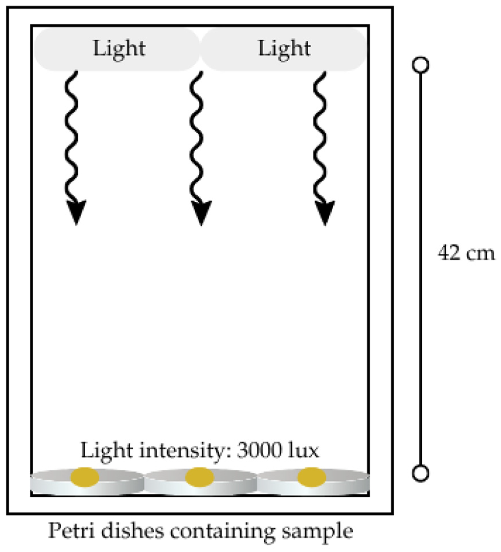
Figure 1. Diagram of the experimental setup used to expose samples to light intensity.
Processes 13 02752 g001
Figure 2. Standard curve of LGEO diluted in 95% ethanol.
Processes 13 02752 g002
Figure 3. Antifungal activity of fresh nanoemulsion and freeze-dried nanoemulsion at 0.10%. Data points represent averages (n = 4) and error bars represent standard deviations. Data for “Fresh nanoemulsion 0.10% EO” were reported in our earlier publication (Liu et al. [26]) and are presented here again for comparison.
Processes 13 02752 g003
Figure 4. Antioxidant assay of pure EO, fresh nanoemulsion, and freeze-dried (i.e., lyophilized) nanoemulsion on an initial date and 30 days later. Samples without shared letter(s) are significantly different. Note: Grey circles represent individual replicates, and error bars represent standard deviation.
Processes 13 02752 g004
Figure 5. GC/Q-TOF chromatogram of pure essential oil at 10,000 ppm. No peaks were observed before the retention time of 7 min or after 11 min.
Processes 13 02752 g005
Figure 6. GC/Q-TOF chromatogram of fresh nanoemulsion extraction.
Processes 13 02752 g006
Figure 7. Effect of room temperature, storage time and light exposure on the antioxidant activity of lyophilized nanoemulsions. Samples without shared letter(s) are significantly different.
Processes 13 02752 g007
Figure 8. GC/Q-TOF results of nanoemulsion stored at room temperature without light exposure over a 30-day period.
Processes 13 02752 g008
Figure 9. GC/Q-TOF results of nanoemulsion stored at room temperature with exposure to light over a 30-day time period. Note: * corresponds to peaks that appear to match peaks from the day 0 sample but have different retention times and represent different molecules.
Processes 13 02752 g009
Table 1. Physico-chemical characterization of freeze-dried nanoemulsion redispersed in water.
ParameterResult
Zeta Potential (mV)−30.6 ± 6.3
Particle Size (nm)158.4 ± 34.9
Polydispersity Index0.6 ± 0.2
Essential Oil Recovery Percentage (%)68.7 ± 12.5
Table 2. Compound names, retention time, and relative percentage of compounds in pure lemongrass essential oil.
CompoundRetention Time (min)Relative Percentage (%)
cis-Citral7.17230.00
Geraniol7.2952.14
trans-Citral7.43343.20
Geranyl acetate8.41711.65
Caryophyllene8.9966.13
γ-Muurolene9.7364.98
10,13-octadecadiynoic acid, methyl ester9.7891.90
Table 3. Names of compounds, retention time, and relative percentage in the fresh nanoemulsion extract. Compound names were determined based on the most probable compound suggested from a single chromatogram replicate of the fresh nanoemulsion extract. Compounds labeled as numbers correspond to compounds with long chemical names.
CompoundRetention Time (min)Relative Percentage (%)
Limonene oxide6.5780.783
cis-Citral7.17418.799
Geraniol7.2950.680
trans-Citral7.43925.644
Geranyl acetate8.4167.702
Caryophyllene8.9983.170
γ-Muurolene9.7422.454
Cadina-1(10),4-diene9.7851.562
Caryophyllene oxide10.3411.631
(1)11.6831.832
(2)12.8180.884
Methyl 5,8,11-heptadecatrienoate13.0523.181
1,8,15,22-Tricosatetrayne13.0796.815
(3)13.453.460
(4)13.7120.883
α-N-Normethadol14.0461.485
(5)14.8061.812
(6)15.5361.534
W-1816.47616.474
Note: Samples labeled with numbers 1–6 correspond to compounds with names that would not fit into the table. Compounds names are as follows: (1)—4-O-Methyl-12b,13,20-triacetoxy-2,9-dihydroxy-3a-carboxy-2,3-seco-tigla-1(10),6-diene-3,9-lactone; (2)—Benzene, 1,3,5-tris(3-methyl-3-butenyl)-; (3)—5,8,11-Heptadecatriynoic acid, methyl ester; (4)—3-Pyridinecarboxylic acid, 1,6-dihydro-4-hydroxy-2-methyl-6-oxo-1-phenyl-ethyl ester; (5)—not identified, (6)—(4-Isopropylidenebicyclo[3.2.0]hept-2-en-6-ylidene)acetic acid, methyl ester.
Table 4. Average concentrations of four primary peaks of lyophilized nanoemulsion samples as determined by GC/Q-TOF.
DayConditionCis-Citral Concentration (ppm)Trans-Citral Concentration (ppm)Geraniol Concentration (ppm)Geranyl Acetate Concentration (ppm)
0Day 01427.163804.20898.81107.41
7No light582.211674.69557.66111.76
14No light207.38679.18128.3160.45
21No light82.98169.5000
30No light35.5275.1000
7Light51.3562.43017.96
14Light9.364.6000
21Light0000
30Light0000
Disclaimer/Publisher’s Note: The statements, opinions and data contained in all publications are solely those of the individual author(s) and contributor(s) and not of MDPI and/or the editor(s). MDPI and/or the editor(s) disclaim responsibility for any injury to people or property resulting from any ideas, methods, instructions or products referred to in the content.