Article

Electron-Shuttling and Bioenergy-Stimulating Properties of Mulberry Anthocyanins: A Mechanistic Study Linking Redox Activity to MFC Performance and Receptor Affinity

by
Gilbert S. Sobremisana
1,†,
Po-Wei Tsai
2,†,
Christine Joyce F. Rejano
3,
Lemmuel L. Tayo
1,
Chung-Chuan Hsueh
4,
Cheng-Yang Hsieh
2,4 and
Bor-Yann Chen
4,*
1
Department of Biology, School of Health Sciences, Mapua University, Makati 1200, Philippines
2
Department of Food Science, National Taiwan Ocean University, Keelung 202, Taiwan
3
Department of Biological Engineering, School of Chemical, Biological, and Material Engineering and Sciences, Intramuros, Mapua University, Manila 1002, Philippines
4
Department of Chemical and Materials Engineering, National I-Lan University, I-Lan 260, Taiwan
*
Author to whom correspondence should be addressed.
These authors have equally contributed to this study.
Processes 2025, 13(7), 2290; https://doi.org/10.3390/pr13072290
Submission received: 15 May 2025 / Revised: 11 July 2025 / Accepted: 14 July 2025 / Published: 18 July 2025
(This article belongs to the Special Issue Advances in Renewable Energy Systems (2nd Edition))

Abstract

Oxidative stress overwhelms cellular antioxidant defenses, causing DNA damage and pro-tumorigenic signaling that accelerate cancer initiation and progression. Electron shuttles (ESs) from phytocompounds offer precise redox control but lack quantitative benchmarks. This study aims to give a clearer definition to electron shuttles by characterizing mulberry’s electrochemical capabilities via the three defined ES criteria and deciphering its mechanism against oxidative stress-related cancer. Using double-chambered microbial-fuel-cell power metrics, cyclic voltammetry, and compartmental fermentation modeling, we show that anthocyanin shows a significant difference (p < 0.05) in power density at ≥500 µg/mL (maximum of 2.06-fold power-density increase) and reversible redox cycling (ratio = 1.65), retaining >90% activity over four fermentation cycles. Molecular docking implicates meta-dihydroxyl motifs within the core scaffold in receptor binding, overturning the view that only ortho- and para-substituents participate in bioactivity. In vitro, anthocyanins both inhibit nitric oxide release and reduce DU-145 cell viability dose-dependently. Overall, our findings establish mulberry anthocyanins as robust electron shuttles with potential for integration into large-scale bio-electrochemical platforms and targeted redox-based cancer therapies.

1. Introduction

Morus alba, or mulberry, is a deciduous Chinese tree used globally for its nutritious fruit. The 20 m tree features heart-shaped leaves. Small, 2–3 cm fruits are white to black and sweet-tart. Morus alba fruits boost immunity, digestion, and well-being with vitamin C, iron, and fiber. They reduce inflammation and control blood sugar [1]. Due to its numerous nutrients and bioactive components, mulberry may prevent or treat chronic diseases including oxidative stress-associated cancer [1,2].
Compounds must give electrons under excess reactive oxygen species (ROS) and absorb electrons under normal conditions to control ROS levels. In bioprocesses, harnessing such electron-shuttle functionality opens the door to entirely new reactor configurations and treatment schemes [3]. For example, in microbial fuel cells (MFCs) [4], electron-shuttle-active phytochemicals can bridge microbial metabolism and the anode, improving power density while simultaneously mitigating oxidative stress in mixed-culture communities. In bio-electrosynthesis platforms, they can serve as redox mediators that shuttle electrons between electrodes and biocatalysts, driving target reactions more efficiently than direct electrochemical approaches. Even in wastewater treatment [5], integrating electron shuttles into aerobic or anaerobic bioreactors can enhance contaminant degradation by facilitating extracellular electron transfer, all while keeping ROS levels in check to protect sensitive microbial consortia. Thus, there is a pressing need to develop and characterize novel electron shuttle-based bioprocesses that leverage the dual antioxidant/electron-transfer roles of such compounds, ultimately enabling greener, more robust, and higher-performance bioenergy and bioremediation systems. ROS-interacting flavonoids and polyphenols like catechin modulate redox conditions [4]. Redox mediators or electron shuttles are organic molecules that may be oxidized and reduced, helping electron transport in redox processes without degrading [5]. The metabolism requires electron shuttles to efficiently transfer electrons, boosting microbial electroactivity. Ortho- and para-dihydroxyl phytochemicals increase electron shuttling, benefiting gut microbial consortia and abiotic electrochemical activities, according to Hsueh et al. [4]. Electron shuttles optimize microbial electroactivity in batch-fed fermentation reactors, and fermented food GRAS inoculants mimic gut probiotics [4,6,7,8].
To be effective as an electron shuttle and bioactive chemical, compounds must withstand degradation in models like batch fermentation employing GRAS inoculants. This preserves their structure, increases bioavailability, and prevents probiotics from using them as substrates or reactants. To study this phenomenon, compartmental modeling can be used, with ES compounds as intermediates. These compounds’ electrochemical characteristics have been connected to numerous illnesses, focusing on bioenergy production and target receptor affinity. Tsai et al. [9] found two-fold bioenergy amplification and dengue entry protein antiviral activity in Rheum palmatum water extract. Tsai et al. [10] found anti-COVID properties in Coffee arabica and 2.73 power density amplification. Electron shuttling, immunostimulant, and anti-pneumonia properties are also found in Jing Guan Fang’s plant extracts [11]. While these studies focus mainly on the bioactivity of ESs, the relationship between enhanced bioenergy output and the chemical affinity of plant extracts for their target receptors remains unclear. Moreover, assessing electron-shuttling solely by power-density amplification, without quantitative validation, poses two key challenges. First, it requires multiple dosing in bioreactors (e.g., double-chamber MFCs), which may be impractical when extract supplies are limited. Second, it lacks a standardized validation protocol to establish a practical threshold dose, applicable across different experimental setups.
Not only is oxidative stress from excess reactive oxygen species (ROS) a by-product of cancer but also acts as a signaling cue that drives epithelial–mesenchymal transition, invasion, angiogenesis, and metastasis according to Hayes et al. [12] and Sosa et al. [13]. DU-145 cells [14], a castration-resistant, androgen- and estrogen-receptor-negative prostate carcinoma line derived from a brain metastasis, maintain high ROS levels and reroute the metabolism toward glycolysis and the pentose phosphate pathway to buffer oxidative damage [12]. In addition, it produces high levels of pro-inflammatory cytokines (including elevated baseline nitric oxide [15]. Because cancer stem cells exploit enhanced antioxidant defenses to survive treatments like radiation, targeting ROS homeostasis offers a non-hormonal strategy for combating aggressive, treatment-resistant tumors. Aggressive, treatment-resistant tumors and androgen-independent signaling cell lines (e.g., DU-145) as a therapeutic target ensures that anti-proliferative effects are not simply due to interference with hormone pathways but reflect broader cytotoxic and electrochemical mechanisms.
ROS, produced by mitochondrial respiration and oxygen metabolism, are essential for cell signaling and immunology according to Aggarwal et al. [16]. ROS from transition metal ions in cells cause oxidative stress, antioxidant depletion, chronic inflammation, cancer, and aging [17]. Hydroxyl radicals damage DNA, increasing oncogenes and inactivating anti-tumor genes [18,19]. The RAS/MAPK pathway, boosted by mitochondrial ROS, promotes oncogenic signaling, cell proliferation, survival, and metastasis [17]. ROS oxidize RTKs, MAP3Ks, and growth factor receptors to activate this pathway [20]. MKP-1 degradation by highly reactive ROS maintains MAPK pathways and ERK activation [20].
Additionally, ROS influence other routes. The activation of NF-κB pathway by ROS causes nuclear translocation [21], and alters the NRF2 pathway, which regulates antioxidant gene expression and contributes to chemotherapeutic resistance [19]. The impairment of the antioxidant defense system leads to mitochondrial malfunction and oxidative damage, which increases pro-inflammatory cytokines and angiogenic factors and tumor growth [21,22]. Although ROS tend to cause oxidative damage, they also regulate respiration, enzyme function, and metabolism [23]. ATP synthesis produces ROS that affect energy production, mitochondrial biogenesis, dynamics, and mitophagy, and cell survival depends on redox balance to avoid oxidative stress [21,22].
ROS-activated pathways involve several receptors essential for disease therapy (Figure 1). TRPV1 affects cancer via increasing intracellular calcium, triggering the NF-κB pathway, boosting inflammation, cell survival, and tumor development [24]. The β3-adrenergic receptor (β3-AR) indirectly induces NR-F2 pathway activation via cAMP and PKA, resulting in antioxidant protein production and chemotherapy resistance in cancer cells [25,26]. Through adenylate cyclase, the A2A receptor activates the RAS/MAPK and ERK pathways, boosting cAMP and PKA, cell proliferation and survival, and carcinogenic signaling via a feedback loop with enhanced ROS [27]. A2A blockade reduces tumor development, immunological suppression, and angiogenesis [28]. TRPV1 is associated with the JNK pathway, cancer cell growth, and survival [29], β3-AR to cancer cachexia, migration, invasion, and apoptosis resistance [25], and A2A to anti-tumor immune response suppression [30,31]. Thus, cancer treatment requires targeting these receptors’ agonists and antagonists.
Although anthocyanins are widely recognized for their antioxidant and pharmacological activities, their role as engineered process intermediates, specifically, as electron shuttles (ESs), remains under-explored. Mulberry’s primary phytochemical, anthocyanin, to potentially target brain-metastatic cancer, has been proposed to possess ES functionality. Hsueh et al. [4] first identified structural motifs consistent with reversible electron transfer, and Xu et al. [32] further suggested that anthocyanins may mediate extracellular electron flux. However, these claims require systematic validation in engineered systems. To function effectively as an ES in bioenergy or bioremediation processes, an anthocyanin must both retain bioactivity after gastrointestinal-like stress (since it is absorbed via active transport [33,34]) and resist degradation pathways analogous to those observed for anthraquinones [31]. Moreover, it must meet three kinetic-electrochemical criteria: (1) possess the ES structural features detailed by Hsueh et al. [4]; (2) amplify MFC power density by at least twofold compared to abiotic controls [9]; and (3) remain stable over multiple probiotic-fermentation cycles, manifested by an α value <1 within 2–3 cycles [35,36] and a redox ratio approaching unity. Electrochemical bioprocess engineering has rarely been applied to drug discovery workflows. By integrating upstream production (e.g., optimized extraction of phytochemicals) with downstream screening (e.g., high-throughput bioassays and formulation), a bioprocess-driven approach offers several advantages: it enables scalable, cost-effective production of candidate compounds; ensures consistent quality and purity through controlled reactor conditions; and facilitates real-time monitoring of bioactivity during synthesis. This strategy can shorten timelines from molecule identification to preclinical testing while maintaining reproducibility and reducing resource consumption.
Therefore, this first-attempt study aims to integrate the three ES criteria in the design of a bioprocess model that (1) extracts anthocyanin-rich fresh and TCM mulberry, (2) evaluates and validates its stability, kinetics, and electrocatalytic performance using electrochemical assays and pilot-scale batch bioreactors [31,32], and, finally, (3) assesses such electrocatalysts for metastatic anti-cancer and NO-inhibition bioactivity through in vitro assays and in silico screening.

2. Materials and Methods

2.1. Sample Collection and Extraction

Sample preparation and extraction involved obtaining both fresh and dried commercial mulberries from a local TCM store in Tainan City, Taiwan. To reduce particle size, each part was mechanically crushed with a blender. Water and 95% ethanol were used in a 1:20 ratio (fruit:solvent) for extraction. The ethanol extract underwent reflux at 60 °C for 2 h, while the water extract was boiled in a traditional Chinese decoction pot until the volume was reduced to approximately 200 mL. Vacuum filtration was employed to filter the extracts, followed by concentration using a rotary evaporator. The concentrated samples were transferred to 50 mL centrifuge tubes and stored in the freezer for 24 h. Subsequently, the frozen crude samples underwent a 3-day lyophilization process using a freeze dryer. The sample set comprising the following mulberry extracts is presented in Table 1:

2.2. Determination of Total Polyphenol Content

The experimental procedures for all phytochemical tests described here were adopted from Tsai et al. [9]. All reagents were of analytical grade or higher. Folin–Ciocalteu phenol reagent (2N, ≥99.0%), sodium carbonate anhydrous (Na2CO3, ≥99.5%), and gallic acid (≥98.0%) were purchased from Sigma-Aldrich (St. Louis, MO, USA). The M. alba fruit extract solution was initially prepared at a concentration of ten milligrams per milliliter (10 mg/mL), from which a one milligram per milliliter (1 mg/mL) sample solution was prepared in ethanol. Next, 100 µL of the sample solution were combined with 500 µL of Folin’s reagent and 400 µL of Na2CO3. Stock solution A containing gallic acid at a concentration of 10 mg/mL in ethanol was prepared, and 50 µL of this stock solution were subjected to serial dilution in ethanol to obtain standard solutions at concentrations of 500, 250, 125, 62.5, 31.3, 15.6, and 7.81 µg/mL. Similarly to the sample solution, 100 µL of each standard solution and blank underwent treatment with 500 µL of Folin’s reagent and 400 µL of Na2CO3. All testing solutions were analyzed in triplicates using ELISA microplate reader (Thermo Scientific™ A51119700DPC, Taipei City, Taiwan) at a wavelength of 600 nm. A calibration curve was constructed using the standard solutions and used for subsequent calculations.

2.3. Determination of Total Flavonoid Content

Rutin (quercetin-3-rutinoside, ≥95% purity) and silver chloride (AgCl, ≥99.0% purity) were both purchased from Sigma-Aldrich (St. Louis, MO, USA). The sample solution (1 mg/mL) was prepared by diluting 10 mg/mL of the sample with 99% ethanol. In total, 0.4 mg/mL of rutin stock solution was prepared and diluted to prepare the standard solutions with the following concentrations: 400, 200-, 100-, 50.0-, 25.0-, 12.5-, and 6.25 µg/mL. Afterwards, five hundred microliters (500 µL) of the testing solutions (i.e., sample, standard, and blank) were incubated for 1 h, after treating with 2.0% AlCl3. Standard solutions and the samples were prepared in triplicates and analyzed on an ELISA reader at 430 nm wavelength [19].

2.4. Determination of Total Condensed Tannin Content

Fifty microliters of fruit extract solution (10 mg/mL) were diluted with 250 µL of 99% ethanol and treated with 600 µL of Vanillin (≥99% purity, from Sigma-Aldrich (St. Louis, MO, USA)) reagent. Through serial dilution, standard solutions (i.e., 160-, 80-, 40, 20-, 10-, 5.0-, and 2.5 µg/mL) were prepared from 160 µg/mL of catechin solution in ethanol. Then, 600 µL of Vanillin reagent were supplemented to 300 µL of standard solutions and blank (ethanol). The absorbance of the solutions (at 530 nm) was measured by using an ELISA microplate reader. All testing solutions were prepared in triplicates to ensure data reproducibility [9].

2.5. Determination of Total Anthocyanin Content

All reagents are of analytical grade or higher. Cyanidin-3-glucoside (C3G; ≥95% purity) is used as the reference standard. Total anthocyanin content (TAC) was quantified by the pH differential method as described by Mejias et al. [37]. Extracts were diluted in pH 1.0 potassium chloride buffer and pH 4.5 sodium acetate buffer, incubated 15 min at room temperature, then absorbances at 510 and 700 nm were measured using an ELISA microplate reader. TAC was calculated from the absorbance difference ΔA = (A510–A700)pH10 − (A510–A700)pH45. The molecular weight MW = 449 g/mol, ε = 26,900 L·mol−1·cm−1, and ℓ = 1 cm. Results are expressed as mg cyanidin-3-glucoside equivalents per g dry matter.

2.6. Determination of DPPH Free Radical Scavenging Activity

Ascorbic acid (≥99% purity), DPPH (2,2-diphenyl-1-picrylhydrazyl, ≥98% purity), and Trolox (6-hydroxy-2,5,7,8-tetramethylchroman-2-carboxylic acid, ≥97% purity) powders were all purchased from Sigma-Aldrich (St. Louis, MO, USA). The ascorbic acid solution (0.5 mg/mL in ethanol) was serially diluted to obtain concentrations of 500.0, 250.0-, 125.0-, 62.50-, 31.25-, 15.63-, and 7.813 µg/mL of standard solution. In a microliter plate, one hundred microliters (100 µL) of the standard solutions, control (ethanol) and extracts were taken and 150 µL of DPPH (200 µM) solution were added, and then incubated for 30 min. All testing solutions had to be simultaneously treated with DPPH solution. The testing solutions (treated with DPPH) and blank (250 µL) in a microliter plate were analyzed in an ELISA microplate reader at 517 nm. To calculate the percentage radical scavenging activity, the equation below was used, and the activity was plotted against its corresponding concentrations. Through linear regression, the 50% inhibitory concentration (IC50) was estimated which was determined at 50% radical scavenging activity [9].

2.7. Ferric Reduction Antioxidant Power (FRAP) Assay

By dissolving 10 mg of Trolox in 2.0 mL of ethanol and 3.0 mL of D.D. water, 2000 µg/mL of Trolox stock solution were prepared. Then, Trolox solution A was prepared by adding 500 µL of ethanol-D.D. water mixture (2:3) to 500 µg/mL of Trolox stock solution. Through serial dilution, a series of standard solutions (1000-, 500.0-, 250.0-, 125.0-, 62.50-, 31.25-, and 15.63 µg/mL) were prepared from Trolox solution A. The sample solution (10 mg/mL) was prepared by dissolving 10 mg of extract in 1.0 mL of ethanol-D.D. water (2:3). In a microplate, fifty microliters (50 µL) of extract and standard solution were treated with 1450 µL of FRAP reagent and analyzed in an ELISA reader at 593 nm. All testing solutions were prepared in triplicates [9].

2.8. Electron Shuttling Activity Analysis

2.8.1. Double-Chambered Microbial Fuel Cells

To evaluate the bioenergy-stimulating properties of different mulberry samples, an H-type double-chambered MFC was utilized. The setup was adopted from Tsai et al. [10]. Carbon rods (Grade: IGS743; Central Carbon Co., Ltd., Tapei City, Taiwan) were utilized as the electrode material. Each MFC chamber had a working volume of 200 mL and was divided by a proton exchange membrane (DuPontTM Nafion® NR-212, Chemours Company, Wilmington, DE, USA) with a contact area of 0.000452 m2 (ID = 1.2 cm). The cathodic chamber was filled with an electrolyte consisting of 6.38 g K3Fe(CN)6 (potassium ferricyanide, analytical grade; JTBaker®, Radnor, PA, USA) and 17.42 g K2HPO4 (dipotassium hydrogen phosphate, ≥99% purity; Sigma-Aldrich, St. Louis, MO, USA) dissolved in 200 mL deionized water. In contrast, the anodic chamber contained culture broth prepared as follows. Aeromonas hydrophila (NIU01) electroactive bacteria were precultured in 100 mL LB broth medium (DifcoTM LB Broth, Miller; Luria–Bertani, Franklin Lakes, NJ, USA) for 12 h at 30 °C and 125 rpm. In total 1 mL of the precultured broth was then inoculated into 18 flasks of 100 mL freshly sterilized LB broth for culture until the optical density (OD) at 600 nm reached approximately 2.1. The anodic chamber was then supplied with 200 mL of the culture fluid for power generation. The mulberry sample was prepared by measuring 1.2 g of the sample and dissolving it in 12 milliliters of deionized water.
Lastly, quantitative analysis was performed by adding various concentrations of the sample to the anode component (250 ppm, 500 ppm, 750 ppm, 1000 ppm, 1500 ppm, and 2000 ppm, respectively) while maintaining the working volume at 200 mL. To observe the effect of anthocyanin on respiratory ROS byproducts, the MFC setup was not purged of oxygen presence and was allowed to run in aerobic condition. After each concentration, the anode chamber and electrode were rinsed twice with sterile deionized water. To maintain consistent parameters throughout the experiment, the optical density (OD) was maintained at approximately 2.1, blank 1 was run prior to 250 ppm, and blank 2 was run after 2000 ppm. Dopamine was used as a standard by adding 0.1 mL of 0.6 M after Blank 2. Power and current densities were determined by the equation below:
P d e n s i t y = V M F C I M F C A A n o d e
I d e n s i t y = I M F C A A n o d e
A A n o d e = a p p a r e n t   w o r k i n g   a r e a   o f   t h e   g r a p h i t e   a n o d e
For V M F C and I M F C , these two parameters could be determined using linear sweep voltammetry on an electrochemical analysis workstation (Jiehan 5600, Jiehan Technology Corp., Taichung City, Taiwan). To provide a clear basis of electron shuttling and significant difference in the production of bioenergy, statistical analysis was deployed on power density.

2.8.2. Cyclic Voltammetry

Fifty cycles of cyclic voltammetry (CV) were conducted to assess the electrochemical performance of mulberry fruit extracts. The CV setup was adopted from Xu et al.’s [32] study. An electrochemical workstation (Jiehan 5640, Jiehan Technology Corp., Taichung City, Taiwan) was employed in a three-electrode system, comprising a glassy carbon working electrode (GCE, ID = 3 mm; model CHI104, CH Instruments Inc., Austin, TX, USA, A = 0.07 cm2), a reference electrode (Hg/Hg2Cl2 soaked in saturated KCl(aq)), and a platinum counter electrode (CE) (6.08 cm2). The CV was carried out within a voltage scan range of −1.5 V to +1.5 V at a scan rate of 0.01 V/s, following a previously described method [32]. Prior to testing, the electrodes were cleaned with D.D. water to prevent interference. The glassy carbon working electrode underwent polishing with 0.05 mm alumina polish and aluminum oxide, followed by rinsing with deionized water. The closed-loop areas of the curves were computed (e.g., V L V H i H i L d V ) using SigmaPlot 14.0 to serve as indicators of the electrochemical potentials of the examined herbs and formulations. While the decay ratio is computed as
D e c a y   r a t i o = 1 50 t h   c y c l e 10 t h   c y c l e × 100 %
While the redox ratio is calculated as follows:
Redox   Ratio = E p a E p c = P o t e n t i a l O x i d a t i o n P o t e n t i a l R e d u c t i o n
Wherein a redox ratio of 1 is ideal.

2.9. Fermentation Reactor Configuration and Compartmental Modeling

The fermentation of mulberry fruit extract was carried out in a bench-scale stirred-tank batch bioreactor with a working volume of 0.2 L. Two Generally Recognized as Safe (GRAS) bacterial inocula with 2.1 OD600 (isolated from fermented tofu and stinky tofu) were co-cultivated under aerobic conditions. The substrates consisted of 2 g L−1 total anthocyanins (equivalent to 2000 ppm MA-F-W) in 1 wt% NaCl (≥99.0% purity, Sigma-Aldrich, St. Louis, MO, USA) solution. Aerobic cultures were carried out at 30 °C, 125 rpm agitation without pH control. Cell density at 600 nm was measured every 12 h to determine the growth phenomena. Total Polyphenol Content, Total Flavonoid Content, and Total Anthocyanin Content were measured over time as well. The fermentation persisted until glucose level was almost zero. The glucose was measured using the OneTouch Select Plus blood glucose monitoring system machine.
To determine the kinetic constants of reaction rates (k1 and k2) of fermentation, compartmental modeling adopted from Sobremisana et al. [36] describes the dynamic transformation of phytochemicals in batch mode. In this model, TPC is assumed to be A (reactants) and TFC and TAC are assumed to be B (intermediates). Under perfect mixing and no inflow/outflow, the kinetic parameters were evaluated as follows:
(1)
The time at which the maximum concentration of B occurs in cultures:
t m a x = l n ( α 1 + a a α 2 ) k 1 ( α 1 ) .
(2)
Rate constant for the conversion from the reactants to the intermediates:
k 1 = l n ( α 1 + a a α 2 ) t m a x ( α 1 ) .
(3)
Prediction of the concentration of the intermediate over a time-course:
B A 0 B m a x A 0 = B B m a x = a e α k 1 t + 1 α 1 ( e k 1 t e a k 1 t ) 1 α 1 + a a α 2 α α 1 ( 1 + a a α )
where t = t 24 since a nearly constant biomass concentration can be achieved after 24 h (i.e., stationary phase).
Furthermore, the rate constants k were determined through minimization of the residual sum of the squared errors (RSS) using the relationship between the α and k1, k 2 = α k 1 ; α = k 2 k 1 . The corresponding α value that has the lowest RSS value was used as the rate constant for the model simulation.
R S S = i = 1 n C B ,   p r e d C B m a x C B ,   e x p e r i m e n t a l C B m a x 2
The algorithm of calculation for determination of the kinetic constant could be expressed as follows:
(i)
The given a = B 0 A 0 , equation for tmax could be used to determine α;
(ii)
Have some trial α value to satisfy RSS (i.e., M i n   k 1 R S S ) for the most appropriate k1 value;
(iii)
Use obtained k1 value to determine k2 and plot B A 0 ( t ) for time courses.
To globally optimize the model which was not accomplished in a prior study [32], the preceding algorithm was implemented using Python 3.12 via PyCharm 2024.1 (professional edition). The code implements an automated search for the best value through compartmental modeling defined by compartmental equation (Equation (7)). The code iterated through a range of values, calculated k1 and k2 and the optimal parameter minimized the residual sum of squares (RSS) for the most appropriate α value. The code also calculates other metrics such as root mean square deviation (RSMD) using the equation:
R M S D = i = 1 N x i x ^ i 2 N ,
where i is the variable i, N is the number of the non-missing data points, x i is experimental data, and x ^ i is the model predicted data at time ti of fermentation. While root mean square error (RSME) is calculated using:
R M S E = i = 1 N x i x ^ i 2 N P ,
where P is the number of parameters estimated including the constants and residual sum of squares (RSS) as given in Equation (8). Appending data on an existing set of data then recalibrates the model to search for the new best α value using the updated dataset. All of this can be accessed publicly in GitHub (v0.1.0) [36].

2.10. Multi-Receptor Virtual Screening

To capture the multifaceted bioactivity of mulberry anthocyanins in both therapeutic and bioprocess contexts, we selected three well-characterized protein targets: (1) the human adenosine A2A receptor (A2AR), which modulates inflammatory cascades and ROS production in the tumor microenvironment; (2) the β3-adrenergic receptor (β3-AR), implicated in cancer cell lipid metabolism and oxidative stress resilience; and (3) the TRPV1 ion channel, a key mediator of ROS-induced apoptosis in malignancies. All three have documented interactions with polyphenolic compounds and sit at critical nodes of redox signaling in cancer, making them ideal mechanistic probes for our docking study. Proteins considered in this study are obtained from RCSB Protein data bank [37,38,39] downloaded in PDB format from various studies of Doré et al. [40] for the A2A receptor (ID: 3PWH), Nagiri et al. [41] for the β3-adrenergic receptor (ID: 7DH5), and finally Neuberger et al. [42] for the TRPV1 receptor (ID: 8GFA). The .sdf files format of the ligands used in this study, namely, the anthocyanins compounds in mulberry fruit, were obtained from PubChem. (chem.ncbi.nlm.nih.gov). The structure of both the protein receptors and ligand were evaluated by Dockthor and binding sites were identified via the literature, and coordinates were identified using P2rank: Prankweb [42,43,44]. The binding sites for the different proteins are detailed in Table 2.
All crystallographic waters and co-crystallized ligands were removed from the receptors, polar hydrogens were added, and Kollman united-atom charges were assigned. Non-polar hydrogens were merged using GS-Lab software (version 1).
Ligand 3D coordinates for the anthocyanins were obtained from PubChem and energy-minimized with the MMFF94 force field. Each structure was loaded into AutoDockTools, Gasteiger charges were computed, non-polar hydrogens merged, and rotatable bonds were defined. Final ligand files were exported in .SDF format.
In this investigation, the Dockthor virtual screening tool was utilized to perform docking analyses of various ligands against three receptors. The anthocyanin structures were selected based on the quantitative HPLC–PDA survey of Morus alba fruit previously reported by Kim et al. [45]. PubChem structures of all selected compounds (Table 3) were integrated into our docking library. Subsequently, all selected ligands were docked against the human A2A adenosine receptor, TRPV1 receptor, and β3-adrenergic receptor.
All in silico analyses were performed on a Windows 11 computer with an i5 8th-generation processor, 516 GB ROM, and 8.00 GB RAM.

2.11. Cell Cultures

DU-145 cells were selected because they represent an advanced, brain-metastatic carcinoma—being both androgen- and estrogen-receptor negative, ROS-producing, and castration-resistant—making them an ideal model for probing non-hormonal, redox-mediated cytotoxic mechanisms of anthocyanin electron shuttles. DU-145 was purchased from American Type Culture Collection (Rockville, MD, USA). Cells were maintained and cultivated in Dulbecco’s modified Eagle’s medium (DMEM) supplemented with 10% fetal bovine serum (FBS), 100 IU/mL penicillin, and 100 µg/mL streptomycin (Gibco BRL, Grand Island, NY, USA) in a humidified incubator (5% CO2 at 37 °C) [10].
Collected cells (Rockville, MD, USA) were maintained and cultivated in Dulbecco’s modified Eagle’s medium (DMEM) supplemented with 10% fetal bovine serum (FBS), 100 IU/mL penicillin and 100 µg/mL streptomycin (Gibco BRL, Grand Island, NY, USA) in a humidified incubator (5% CO2 at 37 °C) [10].

2.12. Anti-Cancer Assay

A 96-well plate was used for culturing the cell lines overnight. Each well was seeded with a total volume of 1 mL of the cell suspension containing 4 × 10⁵ cells. Different extracts were added to each well along with LPS (500 ng/mL) treatment for 24 h. The positive control group received 1 mg/mL of 5-FU. Afterward, MTT solution (5 mg/mL) was added to each well and incubated for another 4 h. The spent cell media was eventually removed, and isopropanol was used to dissolve formazan overnight. The cell culture plates were analyzed at 570 nm using an ELISA plate reader [10].

2.13. Nitric Oxide (NO) Anti-Inflammatory Inhibition Assay

The 96-well plates were seeded with cell suspension containing 4 × 10⁵ cells per well. Different extracts were added to each well and treated with LPS (500 ng/mL) for a period of 24 h. Nitric oxide concentration was determined from the collected spent cell media after the addition of the Griess reagent with the use of the ELISA plate reader at 560 nm [9]. The NO percentage inhibition was calculated using the equation: % NO = [1 − (T/C)], where T and C represent the mean optical density of LPS-stimulated RAW 264.7 cells with and without sample extracts, respectively.

3. Results

3.1. Phytochemical Analysis

As shown in Table 4, the total polyphenol contents (mg gallic acid equivalent (GAE)/g of extracts) were in decreasing order as follows: MA-F-W (46.730 ± 1.703) > MA-Ft-E (17.217 ± 0.0651) > MA-F-E (16.335 ± 0.397) > MA-Ft-W (13.11 ± 0.0651). The total flavonoid contents (mg rutin/g of extracts) were in decreasing order as follows: MA-Ft-E (21.248 ± 0.257) > MA-F-W (6.895 ± 0.171) > MA-F-E (5.434 ± 0.265) > MA-Ft-W (2.500 ± 0.029). The total condensed tannin contents (mg catechin/g of extracts) were in decreasing order as follows: MA-Ft-W (19.557 ± 0.571) > MA-F-W (12.216 ± 0.741) > MA-F-E (8.152 ± 0.652) > MA-Ft-E (1.891 ± 0.036). The total anthocyanin (mg cyanidin-3-glucoside equivalent/g of extracts) were in decreasing order as follows: MA-F-W (8.906) > MA-F-E (2.325) > MA-Ft-E and MA-Ft-W (not detected).
As shown in Table 5, the DPPH assay IC50 values (mg/mL) in ascending order (indicating higher antioxidant activity with lower IC50 values) were as follows: MA-F-W (1.475 ± 0.020) < MA-Ft-E (4.206 ± 0.015) < MA-Ft-W (4.546 ± 0.235) < MA-F-E (5.171 ± 0.013). The FRAP assay results (mg Trolox/g extract) in descending order (higher values indicating stronger antioxidant capacity) were as follows: MA-F-W (45.031 ± 0.199) > MA-Ft-E (28.911 ± 0.118) > MA-Ft-W (23.476 ± 0.057) > MA-F-E (21.518 ± 0.229).
The One-way ANOVA revealed highly significant differences among the four extract types for all measured parameters in Table 6: total phenolic content (TPC; p = 7.99 × 10⁻¹¹, F = 1114), total flavonoid content (TFC; p = 6.52 × 10⁻¹⁵, F = 11,753), total condensed tannins (TCT; p = 1.84 × 10⁻⁹, F = 506.8), total anthocyanin content (TAC; p = 1.07 × 10⁻⁶, F = 100.9), DPPH IC50 (p = 1.45 × 10⁻¹², F = 3043), and FRAP reducing power (p = 7.36 × 10⁻¹⁴, F = 6410). Two-way ANOVA assessing the interaction between fruit maturity and solvent extraction likewise showed significant effects across all responses (TPC: p = 3.34 × 10⁻¹⁰, F = 1346.1; TFC: p = 1.40 × 10⁻¹⁴, F = 16,796; TCT: p = 3.15 × 10⁻⁸, F = 427.1; TAC: p = 6.69 × 10⁻⁵, F = 56.81; DPPH IC50: p = 2.10 × 10⁻¹², F = 4801; FRAP: p = 2.54 × 10⁻¹², F = 4577), indicating that both maturity and solvent choice jointly modulate extract composition and activity. Finally, multivariate ANOVA of the combined phytochemical profile confirmed a significant overall effect of extract type (p = 2.20 × 10⁻¹⁶, F = 6), which suggests distinct phytochemical profiles (e.g., flavonoids, tannins, anthocyanins) and antioxidant capacity among the samples.

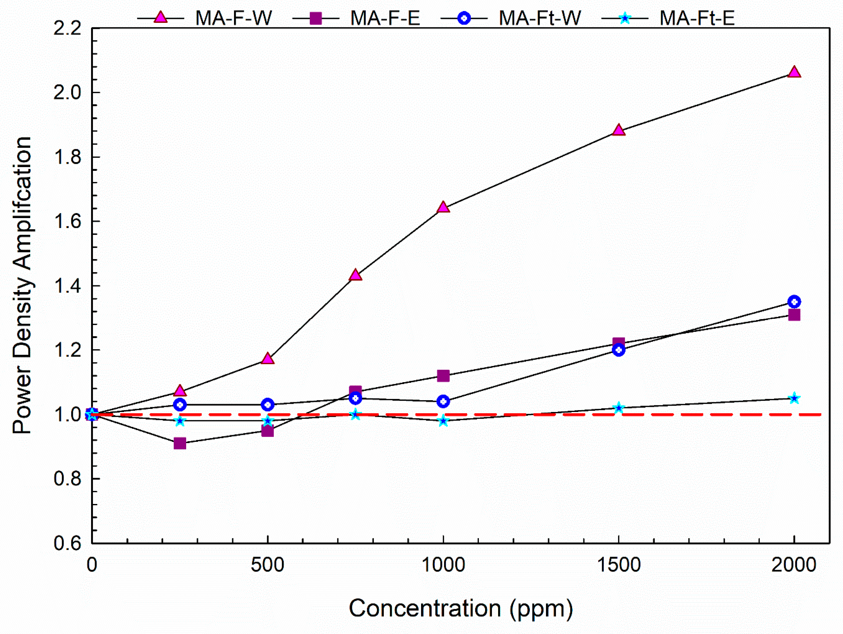
3.2. Electron-Shuttling Capability of Mulberry Fruit

The results demonstrated that mulberry fruit extracts effectively enhance electron transfer capabilities in microbial fuel cells (MFCs), suggesting substantial electron-mediated potential. Figure 2 illustrates the power density profiles obtained in DC-MFCs supplemented with mulberry fruit extracts across six concentrations. Among all tested samples, mulberry fresh commercial fruit water extract (MA-F-W) exhibited the highest power density amplification, approximately 2.06-fold (Figure 3). This significant enhancement of electron shuttling capability is closely linked to its high polyphenol content (46.730 ± 1.703 mg/g), as polyphenols serve as effective electron shuttles due to their aromatic rings and conjugated double bonds, contributing to antioxidant properties.
The antioxidant potential varied among extracts, as confirmed by DPPH and FRAP assays, correlating with their electrochemical properties and differing phytochemical compositions. The ripe mulberry fruit extract (MA-F-W) showed markedly higher bioenergy production compared to the unripe extract (MA-Ft-W), attributable to increased bioactive compound concentrations developed during fruit ripening. Specifically, anthocyanin accumulation, indicated by color transition from green to purple, enhanced electron shuttle properties in ripe fruits. The polyphenol content of MA-F-W aligns with maturity stages M5-M6 (46.730 ± 1.703 mg/g), whereas MA-Ft-W, with significantly lower polyphenol content (13.11 ± 0.0651 mg/g), corresponds to the M0 maturity stage. Consequently, mature fruit extracts significantly improved microbial activity and electron transfer efficiency within the MFC setup.
Paired t-test analysis confirms that the MA-F-W extract produced statistically significant effects on the measured parameter at concentrations of 500 µg/mL and above (p = 0.0215, 0.00199, 0.00184, 0.00209, and 0.00132 for 500, 750, 1000, 1500, and 2000 µg/mL, respectively) in Table 7. In contrast, MA-F-E did not reach significance at any concentration tested (all p > 0.05), nor did MA-Ft-W or MA-Ft-E (all p-values > 0.05), indicating that only the MA-F-W fraction elicited a consistent, dose-dependent bioenergetic response under aerobic DC-MFC.
The cyclic voltammetry (CV) results demonstrated distinct electrochemical behaviors among the mulberry fruit extracts. Figure 4 illustrates the CV profiles across multiple scan cycles (10th, 20th, and 50th cycles) for each extract. Notably, mulberry fresh commercial fruit water extract (MA-F-W) exhibited pronounced redox peak currents, indicating a robust electron transfer capability. Mulberry fresh commercial fruit ethanol extract (MA-F-E) also displayed significant electrochemical activity, albeit lower than MA-F-W. In comparison, both the TCM water extract (MA-Ft-W) and TCM ethanol extract (MA-Ft-E) showed relatively lower electrochemical activities, highlighting the influence of extraction solvents and maturity stage on electron mediation potentials. These CV profiles confirm the potential use of mulberry fruit extracts, particularly MA-F-W, in applications requiring enhanced electron mediation and antioxidant capacity.
Table 8 presents the CV area profiles, decay ratios, and redox ratios of the samples across the cycles. MA-F-W and MA-F-E exhibited stable reduction and oxidation peaks even after 20 CV cycles, with decay ratios of 62.48% and 79.85%, respectively, and favorable redox ratios of 1.65 and 1.27. This stability indicates the presence of electrochemically reversible electron shuttles suitable for bioenergy applications. Conversely, MA-Ft-E and MA-Ft-W, despite large CV areas, lacked clear reduction and oxidation peaks, indicating the presence of antioxidants that are consumed during reactions. These results suggest that MA-F-W contains electroactive electron shuttle species optimal for bioelectricity generation, whereas MA-Ft-E and MA-Ft-W possess antioxidants better suited for scavenging free radicals rather than electron shuttling activities.

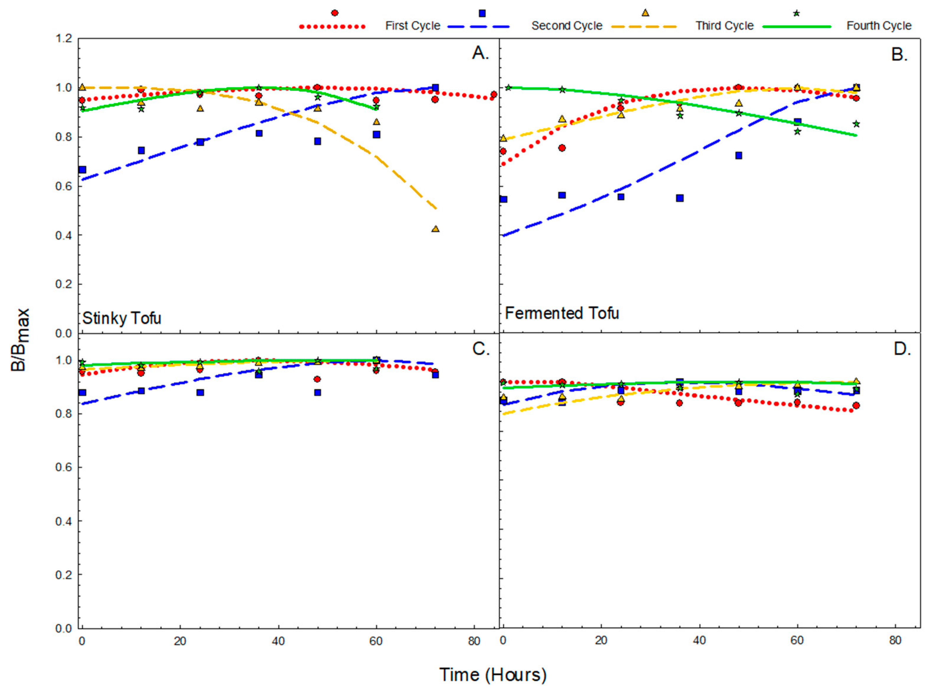
3.3. Anthocyanin Shows Persistence Against Degradation in Fermentation

As illustrated in Figure 5, the core structure of anthocyanin and different anthocyanidin intermediates, indicated by two alkyl groups, demonstrates that the presence of ortho/para substituents effectively distinguishes electron-shuttling compounds from pure antioxidants, confirming anthocyanin’s role as a stable electron shuttle facilitating efficient electron transfer and promoting the fermentation-driven conversion of total phenolic content (TPC) into total flavonoid content (TFC).
The batch-fed fermentation results provided insights into the transient dynamics of mulberry extracts exposed to microbial consortia derived from fermented tofu and stinky tofu. Over four fermentation cycles, distinct trends were observed in the total flavonoid content (TFC) and anthocyanin concentrations (Figure 6, Table 9 and Table 10).
For the stinky tofu-derived consortia, the TFC initially increased consistently up to cycle 2, peaking at an α value of 3.291, before declining slightly in subsequent cycles. In contrast, the fermented tofu-derived consortia exhibited a continuous increase in TFC up to cycle 3, with the highest α value observed at 3.961 during this cycle. The subsequent reduction in TFC after these peaks indicates potential degradation or metabolic shifts in microbial consortia activities.
Anthocyanin levels demonstrated remarkable stability across all fermentation cycles for both consortia. The α values for anthocyanin remained low, indicating that these compounds were not significantly consumed during fermentation but acted as stable electron shuttles or electrocatalysts, facilitating redox reactions. Specifically, the α values for anthocyanin ranged from 0.748 to 0.946 for stinky tofu-derived consortia and from 0.00479 to 0.71 for fermented tofu-derived consortia.
These observations underline the electrocatalytic role of anthocyanin in maintaining reaction efficiency and stability, further confirmed by consistently low α values, which indicate persistent catalytic rather than consumptive roles in the fermentation process.
Table 11 summarizes the calculated values of PDamplification, α, Rratio, and the presence of ortho or para constituents for various previously proposed electron shuttles from the literature. Chrysophanol (Rhubarb), Catechin (Green tea), Dopamine (pure), Ascorbic Acid (pure), and Anthocyanin (Mulberry) exhibited α values of 0.185, 0.0026, 0.4581, 1.3, and 0.0269, respectively. These metrics numerically confirm prior literature claims regarding the electron shuttle capabilities of these compounds. Specifically, the presence or absence of ortho/para constituents effectively discriminates between electron shuttle compounds and pure antioxidants. Anthocyanin, with a notably low α value of 0.0269 and presence of the essential structural characteristics (para or ortho dihydroxyl-bearing aromatic ring), was confirmed as an efficient electron shuttle. These results underline anthocyanin’s electrochemical stability and its capability to facilitate electron movement effectively within microbial fuel cells, highlighting its role as an electroactive catalyst promoting the conversion from TPC to TFC during fermentation.

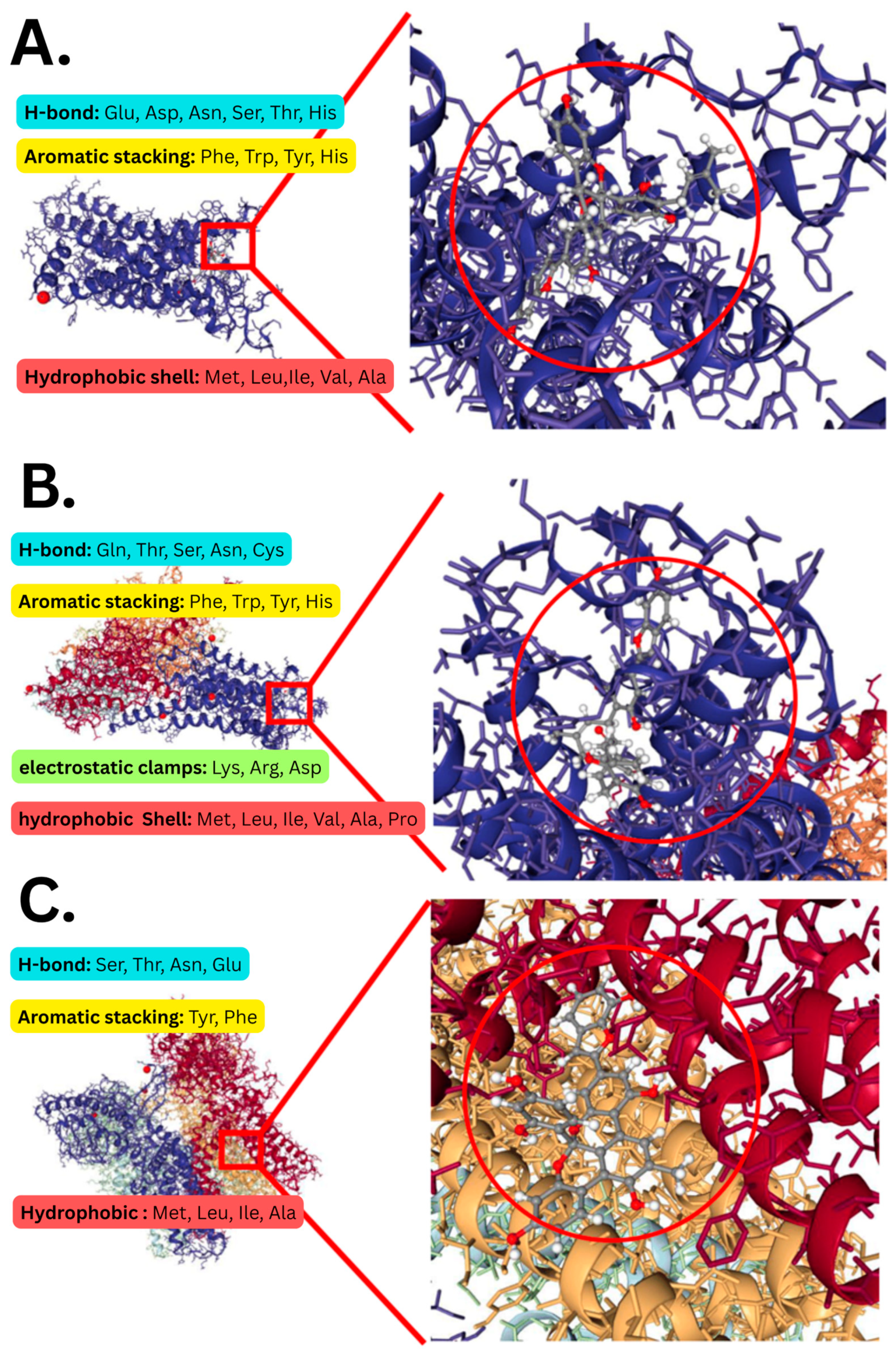
3.4. Delphinidin, Peonidin, and Cyanidin Have the Highest Receptor Affinity

The results of virtual screening identified key ligands exhibiting high affinities for critical targets associated with bioactivity, as presented in Table 12 and Figure 7. For the A2A receptor (Figure 7A), Delphinidin 3-glucoside chloride demonstrated the highest binding affinity with a score of −11.187, significantly surpassing the standard drug caffeine (−7.893). Cyanidin 3-O-rutinoside and Cyanidin-3-glucoside followed closely with scores of −10.961 and −10.64, respectively. In the case of the B3-Andregenic receptor (Figure 7B), Peonidin 3-glucoside exhibited the highest affinity, scoring −10.639, higher than the clinically used Miragebron (−9.915). Malvidin hexoside and Cyanidin 3-O-rutinoside also showed strong binding scores of −10.414 and −10.409, respectively.
For the TRPV1 receptor (Figure 7C), Cyanidin 3-O-rutinoside recorded the highest binding score of −10.286, again surpassing the standard drug Capsaicin (−9.332). Malvidin hexoside (−10.024) and Peonidin 3-glucoside (−9.916) were also identified as significant ligands. These binding interactions are influenced by the specific amino acid residues present at each receptor site. High-affinity interactions observed with the A2A receptor involve diverse interactions including hydrophobic contacts, hydrogen bonding, and ionic interactions with residues such as ILE, LEU, VAL, GLU, CYS, ARG, and HIS. Conversely, differences in amino acid compositions, specifically lacking certain charged or polar residues, explain the relatively lower affinity observed at TRPV1 and B3-AR sites. Furthermore, structural configurations like aromatic stacking facilitated by residues such as TYR, TRP, SER, THR, ASN, and ASP notably contribute to ligand stability at the B3-AR binding site. These observations underscore the intricate relationship between ligand molecular structures and receptor binding site characteristics, crucial for high-affinity ligand-protein interactions.
As shown in Table 13, one-way ANOVA indicates no significant variation in docking scores among the different anthocyanin ligands (p = 0.959), whereas there is a highly significant effect of receptor identity on binding affinity (p = 0.00154). One-sample, one-tailed t-tests comparing the average anthocyanin score to reference ligands reveal that anthocyanins bind significantly more strongly than caffeine (p = 1.03 × 10⁻⁵) and mirabegron (p = 4.49 × 10⁻³), but not significantly differently from capsaicin (p = 0.0603), highlighting the comparative strength of mulberry anthocyanins’ receptor interactions.

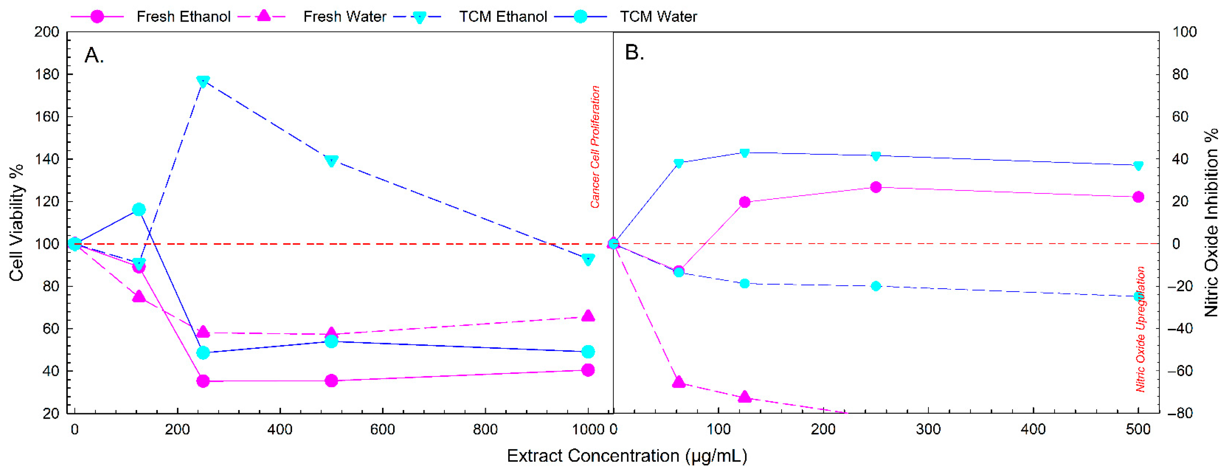
3.5. Fresh Mulberry Extract Shows Highest NO Inhibition and Anti-Cancer Activity
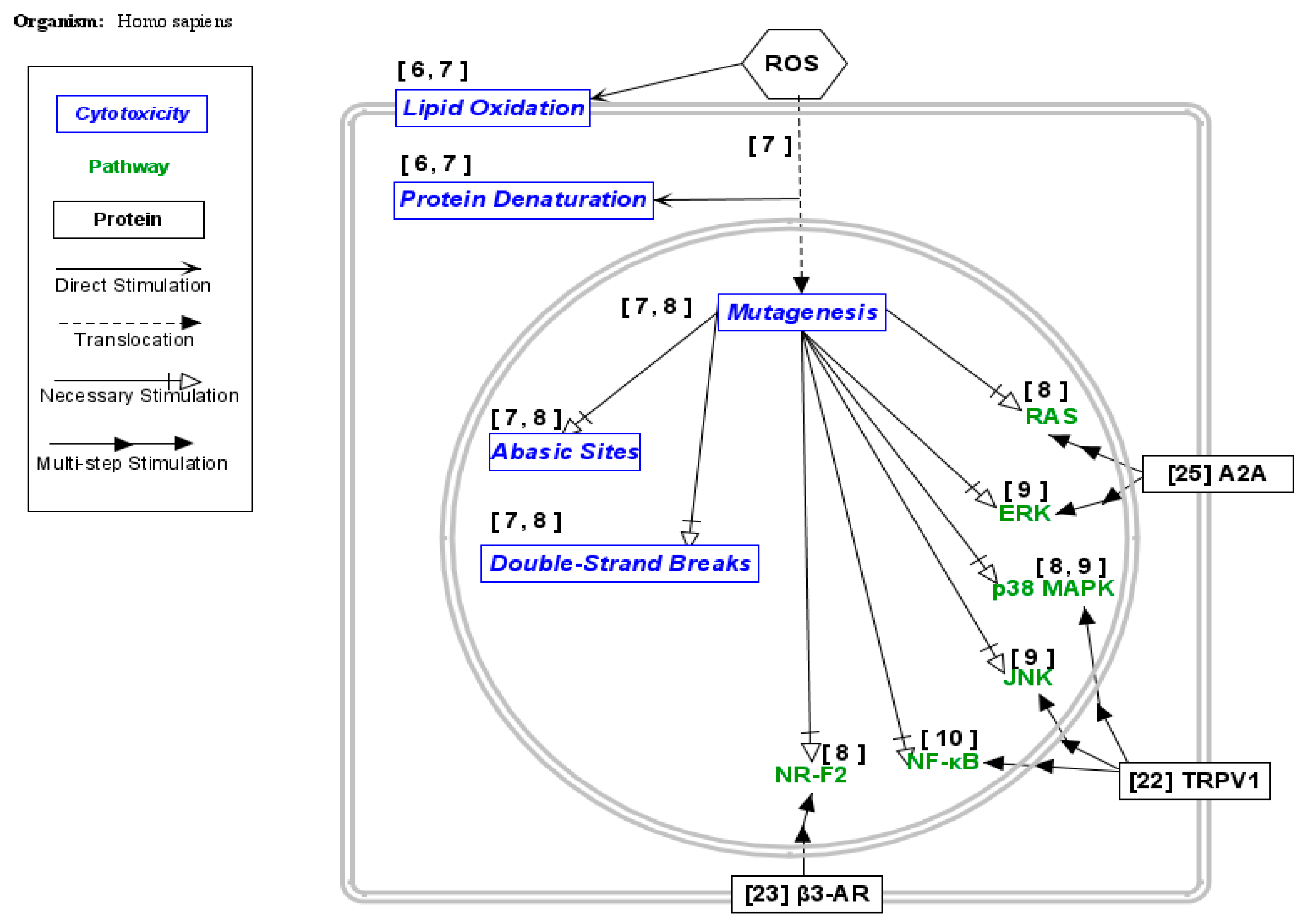
The anti-inflammatory and anti-cancer activities of mulberry fruit extracts, assessed by NO inhibition and cytotoxicity towards DU-145 cell lines, revealed distinctive bioactivity patterns among the samples (Figure 8). Mulberry fruit ethanol extract (MA-F-E) showed the strongest nitric oxide inhibition activity (Table 10), with an IC50 value of 0.486 µg/mL, indicating potent anti-inflammatory potential. Mulberry fruit TCM ethanol extract (MA-Ft-E) also exhibited NO inhibition, though less potent, with an IC50 value of 4.614 µg/mL.
The positive control, 5-FU (1 mg/mL), exhibited nearly 100% cytotoxicity (almost complete inhibition), whereas even at the highest tested concentration (500 µg/mL), the Fresh Ethanol (MA-F-E) and Fresh Water (MA-F-E) extracts in Figure 8 only achieved approximately 60% to 70% cytotoxicity, significantly lower than 5-FU. Moreover, TCM water and ethanol extracts (MA-Ft-W and MA-Ft-E) showed cell proliferation at lower concentrations, highlighting a significant difference in cytotoxic efficacy compared to 5-FU. Despite not matching the positive control drug’s potency, mulberry fresh commercial fruit water extract (MA-F-W) demonstrated the highest cytotoxic potential among tested extracts against DU-145 cells, with an IC50 value of 626.349 µg/mL, followed by TCM ethanol extract (MA-Ft-E) with an IC50 of 843.532 µg/mL. These findings suggest that ethanol-based mulberry fruit extracts, especially MA-F-E, hold promise in reducing inflammation potentially associated with tumor progression and metastasis, while water-based extracts, notably MA-F-W, display significant anti-cancer potential against prostate cancer cells. However, further studies optimizing concentration or exploring combination therapies may be necessary to enhance their anti-cancer efficacy to levels comparable with standard chemotherapeutics.
As shown in Table 14, only the MA-F-E extract achieved significant NO-inhibition activity (IC50 = 0.486 µg/mL; p = 0.022), whereas MA-F-W and MA-Ft-W showed no measurable inhibition under the conditions tested. In the anti-cancer assay, MA-F-W exhibited a potent cytotoxic effect (IC50 = 626.349 µg/mL; p = 0.001), while MA-Ft-E showed weaker activity (IC50 = 843.532 µg/mL) that did not reach the threshold for significance (p = 0.069). MA-Ft-W did not display significant activity in either assay. MA-F-E shows significant cytotoxicity compared to untreated but did not reach a 50% threshold.

4. Discussion

To reveal electrochemical activities as a possible measure for anti-oxidative stress by free radicals, the quantitative assessment of phytochemical constituents for total polyphenols, total flavonoids, and total condensed tannins in the M. alba fruit extracts using two solvents was implemented. The calibration curves clearly exhibited a promising linear relationship of a signal against the concentration of reference standards, with a determination coefficient (R2) greater than 0.99.
As indicated in Table 4, the total polyphenol content of commercial fruit mulberry water extract is higher than the ethanol extract, that is the majority of polyphenol from mulberry fruit are anthocyanins which are water soluble [54,55]. The statistically significant one-way ANOVA results (all p < 10−6; F-values from ~100 to >11,000) confirm that the choice of mulberry extract as substrate (e.g., fresh ripe vs. dried unripe) and extraction solvent (e.g., water vs. ethanol) produces different electrocatalyst profiles. It is also observed that MA-F-W water extract contains the highest total anthocyanin content [55].
The HPLC analysis (Figure S1, Supplementary Materials) of MA samples was conducted to evaluate and compare the phytochemical profiles of the mulberry extracts (MA-F-W, MA-F-E, MA-Ft-W, and MA-Ft-E). MA-F-W showed limited chromatographic peaks with lower intensity, predominantly within the initial 0–5 min retention time, indicating fewer water-soluble secondary metabolites. In contrast, MA-F-E demonstrated a moderate number of chromatographic peaks with higher intensity, mainly within 0–10 min, suggesting efficient extraction of ethanol-soluble phenolics and flavonoids. MA-Ft-W presented an increased number and intensity of peaks compared to the commercial water extract, with significant peaks distributed across 0–15 min, indicating higher concentrations of water-soluble polyphenols, anthocyanins, and glycosides. MA-Ft-E exhibited the most comprehensive chromatographic profile, characterized by numerous prominent peaks spanning 0–20 min, indicating superior extraction efficiency and a diverse range of bioactive compounds such as polyphenols, flavonoids, anthocyanins, and glycosides. Nevertheless, these HPLC chromatograms currently serve as fingerprints with unknown compounds. Future research will employ LC-MS analysis for identifying these unknown compounds, followed by detailed pharmacological investigations to elucidate their therapeutic potential.
According to Zhang et al. [55], five anthocyanin compounds are found in mulberry fruit, cyanidin 3-O-(6″-O-α-rhamnopyranosyl-β-d-glucopyranoside), cyanidin 3-O-(6″-O-α-rhamnopyranosyl-β-d-galactopyranoside), cyanidin 3-O-β-d-glucopyranoside, cyanidin 3-O-β-d-galactopyranoside, and cyanidin 7-O-β-d-glucopyranoside. Aside from anthocyanin, different polyphenol compounds (e.g., chlorogenic acid, resveratrol, and caffeic acid) can also be found in mulberry extract as observed by Jin et al. [56] from different mulberry fruit cultivars from China. Certain studies have shown that polyphenol can have an anti-cancer effect, Hazafa et al.’s [57] findings state that polyphenols combat cancer at the cellular level by inducing apoptosis (programmed cell death) in cancer cells. They achieve this by inhibiting the nuclear factor kappa B (NF-κB) pathway, which is known for its role in the regulation of immune response and cell survival. When these pathways are inhibited, it results in the reduction in cell proliferation and the induction of cell death in cancerous cells, providing a preventative and therapeutic approach against cancer [54]. Moreover, Thangapazham et al. [58] have showed up to 28% reduction in a dose dependent manner in terms of cancer cell invasion using polyphenols.
Certain studies have shown that anthocyanin possesses anti-cancer characteristics. As Lin et al. [59] found, anthocyanins could exert anti-carcinogenic effects by modulating gene expression through various signaling pathways, effectively preventing and treating cancer. For instance, cyanidin-3-glucoside (C-3-G) inhibits the activation of NF-κB, a transcription factor crucial for inflammatory responses, through the PI3K/PKB and MAPK pathways, reducing the expression of pro-inflammatory genes COX-2 and iNOS by over 50%. Furthermore, anthocyanins upregulate the expression of tumor suppressor genes like p53, which activates the transcription of cell cycle inhibitors such as p21 and p27, leading to cell cycle arrest and apoptosis in cancer cells. Delphinidin, for example, inhibits the Ras-ERK and PI3K/Akt pathways, blocking cell proliferation and inducing apoptosis, with studies showing a significant reduction in tumor size by 40–60% in treated mouse models. Moreover, anthocyanins like delphinidin can downregulate VEGF expression, inhibiting angiogenesis and metastasis, thereby effectively restricting tumor propagation and spread [59].
On the other hand, the sample with the highest flavonoid content is MA-Ft-E (see Table 3). In fact, ben Sghaier et al. [60] found that flavonoid compounds have anti-cancer properties through several mechanisms. They inhibit cancer cell viability in a dose-dependent manner, in particular. They are effective in HT29 cells, suggesting potential specificity for certain cancer types. Another study by Vafadar et al. [61] shows flavonoid’s ability to induce apoptosis in cancer cells by activating caspases (e.g., caspase-3 and caspase-9), and enhancing the release of cytochrome c. Quercetin also inhibits the PI3K/AKT/mTOR signaling pathway, leading to decreased cell survival and proliferation. Furthermore, it downregulates the expression of pro-survival proteins, including c-Myc and cyclin D1, while promoting the expression of tumor suppressor genes.
The highest tannin content for the fruit sample is 19.557 ± 0.571 for MA-Ft-W with the water extract for both TCM and commercial fruit generally being higher compared to ethanol extract. Tannins have been found to selectively induce apoptosis in various cancer cell lines (Caco-2 colon, MCF-7 and Hs578T breast, and DU 145 prostatic cancer cells) without affecting normal human fibroblast lung cells in a study conducted by Bawadi et al. [62]. The tannins inhibit cancer cell proliferation and migration, as evidenced by decreased ATP levels and suppressed cell movement towards chemoattractant. Moreover, they reduce the secretion and activity of matrix metalloproteinases (MMP-2 and MMP-9) and lower the levels of vascular endothelial growth factor (VEGF165), both of which are critical for angiogenesis and tumor progression. As a matter of fact, Almalki et al. [63] stated that tannin nanoparticles (NP99) enhance cancer treatment by either inducing apoptosis in MCF-7 breast cancer cells through a molecular mechanism involving the upregulation of the pro-apoptotic gene Bax and downregulation of the anti-apoptotic gene Bcl-2. This shift decreases the Bcl-2/Bax ratio, promoting apoptosis. In addition, NP99 generates oxidative stress and activates endogenous apoptotic pathways, which, in combination with Tamoxifen, a chemotherapeutic drug, results in synergistic cytotoxic effects that significantly reduce cell viability and proliferation.
The maturity and solvent interaction (all p < 10−4; F up to ~17,000) suggests that optimal electrocatalyst recovery requires appropriate solvent polarity along with fruit ripeness and processing. In a scale-up context, this finding supports a decision tree wherein early-harvest fruit might be processed with ethanol to maximize the yield of specific glycosides, whereas fully ripened or TCM fruit could be water-extracted to enhance overall redox capacity. Finally, the multivariate ANOVA (p = 2.20 × 10⁻¹⁶; F = 6) demonstrates that each extract has unique phytochemical profiles that can be utilized when designing MFC feed streams or biorefinery co-product workflows.
The free radical scavenging activity of the mulberry based on DPPH and FRAP is shown in Table 4. Such a scavenging property demonstrates that the duality of the compounds in mulberry could attenuate free radicals’ presence through redox reactions. As Du et al. [64] mentioned, when the concentration of mulberry anthocyanins (CMA) reached 0.40 mg/mL, it almost exhibited the same DPPH radical scavenging rate as vitamin C (standard) and the five anthocyanin monomers tested.
This clearly indicates that CMA is an excellent antioxidant agent. This antioxidant quality of mulberry is important in preventative medicine and slowing cancer progression because such a disease is known to have a relationship with the incidence of oxidative stress [12,13,14,15]. According to Katsube et al. [65], mulberry and its main components exhibited antioxidant activity by prolonging the oxidation process of lipids. Furthermore, the administration of mulberry resulted in improved liver function and increased levels of antioxidant enzymes. This improvement in antioxidant and liver enzymes helps to alter the body’s oxidant condition, leading to the production of anti-inflammatory agents.
Such results are important since oxidative stress-induced mutagenesis is mainly caused by free radicals or ROS which are highly reactive molecules capable of causing significant damage to cellular components, including DNA, proteins, and lipids. These initial results regarding the phytochemical and free radical scavenging activity of mulberry confirm its activity in terms of its oxidation potential.
Since the high content of polyphenol, as the previous literature pointed out, could signify an abundance of electron shuttles or an electrochemical catalyst [9,10], mulberry fruit can be evaluated for its ability to enhance the electron shuttling capability of living organisms using microbial fuel cells (MFCs). MFCs serve as a platform not only for assessing bioenergy production but also for evaluating electron shuttling enhancing capability. This study compared the bioenergy-generating characteristics of MFCs supplemented with mulberry fruit extracts at six different concentrations.
The power density profiles in DC-MFCs indicate the promising electron-mediated potentials of mulberry fruit extracts, enhancing the electron transfer capabilities of electroactive microbes in MFCs (see Figure 2). Despite the presence of oxygen in the anodic chamber which may hinder the capability of the anode to receive electrons, the high-power density suggests that anthocyanins might accept electrons from the bacteria more rapidly than O2 can, funneling them to the anode and restoring high coulombic efficiency (e.g., higher power density compared to blank). Among all the sample, mulberry fresh commercial fruit water extract (MA-F-W) has shown to have the highest PD amplification of ca. 2.06 (see Figure 3). Although a PD amplification of 2 was mentioned in previous papers [9,10,11] to be an indicator of electron shuttling capability, this claim provides no statistical basis. Utilizing statistical tests (e.g., T-test) provides a standardized and repeatable measurement of actual electron shuttle production without the need for dose-dependent PD amplification, especially on setups where substrate samples are limited. The confirmation of T-test p-value establishes a practical threshold dose—around 500 µg/mL of MA-F-W below which shuttle concentration is insufficient to impact MFC performance, and above which the redox-active compounds consistently boost electron transfer [62]. This statistically significant PD amplification for the MA-F-W can be attributed to its high anthocyanin content, which are water soluble compounds. Furthermore, the presence of aromatic rings and double bonds in the chemical groups of mulberry fruits (Table 1) contributes to their antioxidant properties. The antioxidant capabilities of these chemical moieties are influenced by the presence of electron donating and withdrawing substituents in the chemical structures [14]. The variations in fruit extract compositions and their electrochemical properties result in differences in antioxidant activities, as observed through DPPH and FRAP assays. These extracts may possess a synergistic nature to trigger redox-mediated potentials for medicinal purposes, such as in neurodegenerative diseases like Parkinson’s disease as mentioned by Chen et al. [53]. The low DPPH IC50 and high FRAP values observed in the MA-F-W fraction (Table 6) align with its cyclic-voltammetry performance and 2.06× power boost in MFCs, suggesting that the water extraction of fresh fruit yields a more effective intrinsic shuttle [66].
The observed difference in power density amplification between ripe mulberry fruit extract (Morus alba) and unripe Morus alba extract in double-chamber microbial fuel cells (MFCs) can be attributed to the varying composition of bioactive compounds and nutrient availability in the two extracts. Ripe mulberry fruits undergo significant changes in their biochemical composition as they mature, leading to an increase in the concentration of bioactive compounds. This development of the ripening of mulberry fruit extract serves as a “factory” to synthesize phytochemical compounds, like polyphenols and to a lesser extent, flavonoids. A study by Saensouk et al. [67] has observed that at different ripening stages of the mulberry, phenolics and flavonoids as well as the shift from green to purple color increases as the ripening stage progresses. The shifting from green to purple of the mulberry fruit is due to the formation of anthocyanin compounds in the fruit. The sample MA-F-W which contains 46.730 ± 1.70 mg/g of polyphenol, according to study [67] is between the M5 and M6 stage of maturity, while the MA-Ft-W which contains 13.11 ± 0.0651 mg/g of polyphenol could be around the M0 stage of maturity according to study [68].
On the other hand, unripe Morus alba extract may contain lower levels of these bioactive compounds, leading to reduced microbial activity and lower power generation in the MFC. Furthermore, the ripening process may also lead to the release of a specific compounds that bears the dihydroxyl aromatic group (e.g., anthocyanins) which act as electron shuttles or mediators, facilitating electron transfer between microbial cells and the anode. This was consistently observed in the total anthocyanin content comparison of ripe and unripe mulberry extract. No anthocyanins above detectable levels were quantified in unripe samples while the ripe mulberry contains up to 8.9 mg/g C3G equivalent of total anthocyanins. In fact, a study by Sangteerakij et al. [68] showed that the ripening process of mulberry did increase the anthocyanin content. These mediators can enhance the efficiency of electron transfer processes, further contributing to the observed higher power density in MFCs using ripe Morus alba extract.
Repeated cycles of cyclic voltammetry were conducted (Figure 4) to explore responses of serial oxidation and reduction processes and corresponding to the electroactive potentials of mulberry extracts. Cyclic voltammogram profiles provided insights into the herbs’ reversible electron transfer (ET) capabilities, distinguishing antioxidants from electron shuttles (ESs). The closed-loop areas, summarized in Table 8, reflected the electrochemical activities of the herbal extracts. As observed in Figure 4, the electrochemical activities of MA-F-W and MA-F-E, excluding MA-Ft-W and MA-Ft-E, gradually diminished after 50 cycles, indicating irreversible oxidation and/or reduction in electron donors and acceptors. This left only electrochemically stable, reversible ESs. Recent studies linked the antiviral properties of Traditional Chinese Medicine (TCM) to their electrochemical potential. The conversion of polyphenolic antioxidants in TCM herbs could enhance their electron-mediating properties, extending their anti-disease efficacy through electrochemical catalysis. Aware that the cyclic voltametric profile (CVP) of the material showed both reduction and oxidation potential peaks (Figure 4), this material was of course an ES. However, when CVP did not exhibit such characteristics, this simply indicates that the nature of ES cannot be revealed under such electrochemical conditions. This point does not simply exclude such material as ESs.
Additionally, MA-F-W and MA-F-E exhibited stable reduction and oxidation peaks even after 20 CV cycles, indicating the dominance of electrochemically stable and reversible ESs suitable for bioenergy applications. These results suggested that MA-F-W is abundant in electroactive ES species to escalate bioelectricity generation. In contrast, MA-Ft-E and MA-Ft-W showed significant large areas but lacked promising bioenergy-stimulating potential in DC-MFC, as well as reduction and oxidation peaks. This implies that MA-Ft-E and MA-Ft-W may contain more antioxidants, which are consumed during reactions, as confirmed by the DPPH assay. This difference may be attributed to the fact that MA-Ft-E and MA-Ft-W likely contain antioxidants effective for scavenging free radicals (like DPPH), but less efficient at reducing ferric ions, while MA-F-W may have antioxidants less efficient at scavenging free radicals but highly effective at reducing ferric ions.
Regarding electrochemical characteristics, due to the absence of standard redox peaks for Mulberry TCM samples, the Redox ratio was only experimentally determined from MA-F-W and MA-F-E. As indicated in Table 8, the Redox ratio of MA-F-W and MA-F-E which is ca. 1.65 and 1.8 falls into preferred values of the established criteria. That is, it has the ability to receive and withdraw electrons (i.e., typical characteristics of the electron shuttle) [4]. Similar phenomena were also observed by Petrovic [69], where cyclic voltammetry of catechin has a reduction potential of ca. −4 and oxidation potential of ca. 4 giving an almost ideal redox ratio of 1. Moreover, Qin et al. [70] mentioned that DC-MFC of a catechin-rich plant such as Camellia sinensis (green tea) shows more than 2-fold power density amplification compared to the blank control, which further supports the criteria for the electron shuttle.
Batch-fed fermentation is a biotic system that serves as a simplified model for studying the transient dynamics of plant extracts when exposed to microbial consortia. For this experimental design, cycles of fermentation were utilized to observe how microbial communities interact with different substrate loads by introducing microbial consortia derived from fermented tofu and stinky tofu into the setup.
After four cycles of serial acclimation, a notable trend emerged (see Figure 6). The total flavonoid content (TFC) showed a consistent increase up to cycle 2 for stinky tofu-derived consortia, while for fermented tofu-derived consortia, the increase continued until cycle 3. Chen et al. [35] have also observed an increase in maximal value-added production using similar consortia derived from fermented tofu for Rheum palmatum L. fermentation. The study has observed up to a 400% increase in anthraquinone compounds after the 3rd cycle. The observed increase in flavonoid concentration during batch-fed fermentation therefore can be attributed to the intricate enzymatic activities of bacteria, which may have converted polyphenols into much simpler flavonoids or flavonols. Redox reactions may also occur, where much more complex polyphenols undergo reduction or oxidation with the help of bacterial fermentation, resulting in the generation of different flavonoid compositions.
However, a noteworthy observation was the subsequent decline in TFC in further cycles. This decline could be attributed to the degradation or consumption of the microbial consortia present in the system. The diminishing trend in TFC beyond a certain fermentation cycle suggests that microbial communities may alter metabolic activity or derived composition, leading to a decrease in flavonoid production. If an electron shuttle remains undegraded throughout fermentation, its redox activity must be both fully reversible and catalytic rather than stoichiometric [71]. In practical terms, the anthocyanin continuously accepts and donates electrons without structural loss, implying a stable midpoint potential that does not drift under process conditions [71]. Such persistence indicates negligible side reactions or irreversible chemical modifications, so the shuttle can support countless redox cycles, maintain near-unity faradaic efficiency, and sustain extracellular electron transfer rates over the entire fermentation period [72]. Furthermore, Sobremisana et al. [36] also explored transient dynamics of fermentation using compartmental modeling and revealed that the 3rd cycle consistently has the lowest α value (k2/k1 ratio) for maximal treating capability. Meanwhile, electron shuttle compounds such as anthraquinones as per Chen et al. [35], have an alpha value of less than 1 in at least 2–3 cycles of fermentation.
Despite the decrease in TPC and the increase in TFC in the first 2 cycles then getting consumed in the later 2 cycles, anthocyanin content remains almost constant for all cycles. In fact, the α value of anthocyanin almost remains consistently low (stinky tofu: 0.00479–0.71 on Table 10 and for fermented tofu: 0.94–0.748 on Table 9) in all four cycles of the two-fermentation setup. This is an indication k1 is always greater than k2, in which the reaction does not proceed towards consumption or degradation of the compound by the consortia. Instead, anthocyanin acts as an electron shuttle or electrocatalyst for the chemical process (e.g., redox reactions) mentioned earlier, facilitating the reaction to move forward. According to Zhao et al. [73] if electrocatalysts persist, the transfer of electrons between electrodes and reactants will be stably proceeded in the highest reaction rate through routes of lowest activation energy. They function by facilitating intermediate chemical transformations described by overall half-reactions, effectively modifying reaction pathways, reducing activation barriers, and, importantly, enduring without being consumed in the ongoing reaction. This was further confirmed by Xu et al. [32], wherein supplementation of anthocyanin-rich plants in DC-MFC setup of NIU01 bacteria have more than 2-fold of power density amplification (ca 2.15 to 2.7 for L. ruthenicm Murr. and C. ternatea Linn., respectively) which was consistent with DC-MFC results as revealed herein (ca. 2.06) due to significant electron shuttling activity. As can be seen in Figure 5, anthocyanin contains the essential structure to be an electron shuttle (e.g., para or ortho dihydroxyl bearing aromatic ring). Therefore, anthocyanin may have been utilized as a catalyst in the conversion of TPC to TFC in the fermentation setup. Thus, such an observation supports the power density amplification and CV profiles of the electrocatalytic activity of anthocyanin.
According to Choudhary et al. [74], “If the said ratio is equal to 1, it means both the anodic and the cathodic peak currents are the same, and it talks about the reversible nature of the system”. This is crucial for ES compounds as this dictates their ability to conduct redox reactions reversibly. Though it may not initially appear to have significant impact, this is crucial in discriminating false positives (e.g., results that may show “apparent” high PD but does not possess the necessary structure). As observed, the Rratio focuses on redox reversibility, α highlights electrochemical stability and electron-mediating persistence, PD measures the performance index of practical applications; therefore, the presence of ortho/para constituents considers the structural suitability and structure-activity relationship.
Finally, various values of PDamplification, α, Rratio, as well as presence of ortho or para structure are shown in Table 8 of various previously proposed electron shuttles from literature. These various values were able to give numerical confirmation to previous claims of various compounds being an electron shuttle. It can also be observed that the presence or absence of ortho/para constituents was able to discriminate electron shuttles from pure antioxidant compounds. This confirms the prior claim of anthocyanin [32] as an electron shuttle. Such values also demonstrate that anthocyanin is effective in facilitating electron movement within the microbial fuel cell or other relevant systems. This also implies that the compound is electrochemically stable and not significantly transformed or consumed during the electron transfer process and ensures efficient electron transfer without favoring either oxidation or reduction excessively. As Figure 5 indicated, anthocyanin core structure contains the described characteristics to be an electron shuttle [4]. Therefore, anthocyanin seemed to have been utilized as an electroactive catalyst to convert TPC forward to TFC via fermentation.
Since the measurement of bioenergy production of the fruit extract shows amplification of power density which shows statistical significance with maximum PD amplification of more than 2-fold, virtual screening using plant compositions from the fruit extract, bearing structures essential for electrochemical activity were used. Critical targets like receptors for cancer are cost-effective, time-efficient, and expedite treatment candidate selection. Electron shuttles such as anthocyanin facilitate reversible single-electron transfers at the receptor interface, transiently altering the local electric field and microenvironment polarity. Such electrical modulation can (1) induce conformational changes in proteins, including movement of extracellular loops or allosteric domains, that stabilize intermediate states favorable for ligand binding. Reversible redox events have been shown to modulate receptor conformations, as evidenced by studies on β2-adrenergic receptors and IP3 receptors [75]. These changes can influence receptor dynamics, making the binding site more adaptable and conducive to ligand docking [75,76].
Electron shuttles can also (2) lower the activation energy for ligand docking by stabilizing intermediate states within the binding funnel [77]. The stabilization of conformational states through redox modulation is exemplified by integrin activation and its ligand-binding enhancement upon disulfide bond reduction [77]. Such redox effects, even at a distance, facilitate more favorable receptor-ligand interactions.
Reversible Redox Events also enhance binding affinity and signal transduction by inducing an electric field that pulls water molecules into position and modulates protonation states. For example, soluble guanylate cyclase (sGC) [78], a receptor regulated by redox conditions, shows restored ligand sensitivity through electron donation by H2S, converting the Fe3+ state back to Fe2+ [78]. This suggests how redox-driven receptor priming can enhance signaling activity.
Thus, the ~2 times power amplification we measure directly indicates that electron shuttles can generate local redox gradients and microcurrents strong enough to ‘prime’ receptor proteins for high-affinity interactions [79]. In essence, the bioenergy output reflects the electron shuttle redox “drive,” which electrically tunes receptor surfaces to enhance subsequent chemical binding, thereby influencing intracellular signal modulation [79].
Based on the result of the virtual screening, the top 5 ligands with the highest affinities for every receptor are shown in Table 12.
It can be observed that Delphinidin 3-glucoside chloride is listed as the top-ranking compound for A2A, with a binding score of −11.187 (see Table 9). The observed variations in the affinity of the provided ligand for different protein-binding sites can be attributed to the intricate interplay between the ligand’s molecular structure and the unique amino acid compositions of each binding site [73]. The high-affinity site of A2A (see Figure 5), exhibits a diverse array of amino acids that collectively create an optimal environment for multiple types of interactions with the ligand. These interactions include hydrophobic contacts facilitated by residues like ILE, LEU, and VAL [79,80] aiding in substrate recognition with neutral side chains and stabilization. Hydrogen bonding with amino acids like GLU and CYS [73], and potential ionic interactions involving positively charged residues like ARG and HIS [74,79]. The spatial arrangement and complementary nature of these interactions likely lead to a stable and favorable ligand-protein complex, resulting in higher binding affinity.
The one-way ANOVA of mulberry anthocyanins (p = 0.959) indicates that despite subtle structural variations (e.g., glycosylation patterns, hydroxylation positions such as ortho and para substituents), all tested anthocyanins engage receptor binding sites with comparable baseline affinity. This suggests that the core flavylium scaffold is the primary driver of binding. Moreover, as illustrated in Figure 5, the anthocyanin core contains a meta-substituent which was previously implicated to play no role in electron-shuttling bioenergetics [4]; our results suggest that this substituent may instead participate in different bioenergetics such as lowering the binding energy, thereby stabilizing ligand–receptor complexes.
Conversely, the lower affinity binding sites, exemplified by TRPV1 and B3-AR towards Delphinidin 3-glucoside chloride, lack the same degree of complementary amino acid residues required for robust interactions with the ligand. For instance, TRPV1 and B3-AR may lack specific polar or charged residues necessary for strong electrostatic or hydrogen bond formation. Furthermore, although TYR and TRP are configured for effective π-π interactions [81], the positioning of aromatic residues like TYR and TRP in those receptors is significant as well. In essence, the molecular landscapes of these lower affinity binding sites may not offer the same versatile and well-coordinated array of interactions as their high-affinity counterparts, leading to reduced binding affinity for the ligand.
Another noticeable result is the affinity of the TRPV1 receptor towards cyanidin 3-O-rutinoside (see Table 8). In the context of molecular interactions between proteins and the ligand, the binding site characteristics play a pivotal role in determining the affinity between them. The presence of amino acids with polar and charged side chains, such as LYS, ARG, GLU, ASN, HIS, and TYR [81,82], facilitates strong ionic and hydrogen bonds with the ligand’s hydroxyl groups, contributing to high affinity. Similarly, the TRPV1 binding site, (see Figure 7) featuring TYR and ASN [83] residues for hydrogen bonding and hydrophobic residues like LEU, PHE, and ALA [79,80] for interaction with hydrophobic ligand portions, has the molecular environment conducive to strong binding. Conversely, lower-affinity binding sites, such as those in A2A and B3-AR, may possess a suboptimal arrangement of polar and hydrophobic residues, impacting the ligand’s ability to form effective interactions. The spatial disposition of amino acids within the binding pocket may be crucial, influencing the overall affinity.
The B3-AR binding site exhibits distinctive features that could contribute to a high affinity for Peonidin 3-glucoside. The presence of hydrogen bonding-capable residues like SER, THR, TYR, and ASN allows for robust interactions with the numerous polyphenolic ligand’s hydroxyl groups [80]. Furthermore, the inclusion of aromatic amino acids, TRP, and TYR, provides potential sites for strong aromatic stacking interactions with the ligand’s benzene rings [84,85]. Electrostatic interactions are facilitated by amino acids like ASP [81], creating an environment to stabilize the positively charged oxygen in the ligand. In addition, hydrophobic residues such as VAL, PHE, and LEU offer the opportunity for hydrophobic interactions with the ligand’s aromatic rings, providing supplementary stabilization [79].
The one-way ANOVA across receptor targets is highly significant (p = 0.00154), which demonstrates that different anthocyanin compounds have different affinity depending on the protein context. This implies that different anthocyanin compounds could be receptor-selective, thereby binding to specific proteins which are important for bioactivity. Furthermore, one-sample, one-tailed t-tests reveal that the average anthocyanin binding score significantly exceeds that of caffeine (p = 1.034 × 10⁻⁵) and mirabegron (p = 4.49 × 10⁻³), indicating stronger predicted interactions than these small-molecule controls. By comparison with capsaicin, the difference is not statistically significant (p = 0.0603), although it trends toward greater affinity. This suggests that anthocyanins can be a potential competitive modulator at the TRPV1 site, with binding potency on par with a known agonist.
Regarding the most promising extracts of mulberry fruits, both of the ethanolic mulberry fruit samples show nitric oxide inhibition activity. The best was MA-F-E. According to Bruunsgaard, H. et al. [86] and Greten et al. [87] chronic inflammation such as that signified by nitric oxide production, creates a tumor-promoting environment, likely leading to tumor progression and metastasis in conditions (e.g., colorectal cancer). Inflammatory responses can lead to DNA damage and hinder proper DNA repair, potentially triggering DNA damage-induced inflammatory pathways. Furthermore, the activation of oncogenes is linked to the increased production of cytokines and chemokines, as well as the recruitment of myeloid cells, which can directly promote tumor growth or exhibit immunosuppressive effects.
The high-affinity docking of mulberry anthocyanins to key redox-sensitive receptors provides a mechanistic basis for their observed anti-inflammatory and cytotoxic effects. For example, delphinidin 3-glucoside chloride binds the A2A adenosine receptor [27] at −11.187 kcal/mol, well above caffeine’s −7.893 kcal/mol, suggesting a potent antagonism of A2A-driven cAMP/PKA signaling that normally upregulates NADPH oxidases and augments ROS production [28]. Similarly, cyanidin 3-O-rutinoside docks to TRPV1 [24] at −10.286 kcal/mol (versus capsaicin’s −9.332), implying a blockade of Ca2+-mediated mitochondrial ROS release and downstream NF-κB–driven inflammatory gene expression [29]. Finally, peonidin 3-glucoside exhibits a −10.639 kcal/mol affinity for β3-adrenergic receptors [25], likely inhibiting β3-AR–mediated Nrf2 activation and the resultant overproduction of phase II antioxidants that tumors exploit for chemoresistance [26].
By simultaneously suppressing A2A- and TRPV1-mediated ROS generation while preventing β3-AR–Nrf2–driven antioxidant overcompensation [30,31], these anthocyanins force a net intracellular redox imbalance. This manifests experimentally as potent NO inhibition (e.g., MA-F-E IC50 = 0.486 µg/mL) and significant DU-145 cell cytotoxicity (MA-F-W IC50 = 626 µg/mL), consistent with a decrease in ROS-dependent pro-survival signaling and an increase in apoptosis under oxidative stress. Thus, these results suggest that receptor engagement tightly correlates with the dual redox-modulatory and antiproliferative outcomes observed in vitro.
Regarding colorectal cancer [87], for example, the loss of tumor suppressors can inhibit DNA repair, leading to DNA damage-induced inflammatory pathways. Additionally, in some cases, the breakdown of the protective intestinal barrier at the site of tumor formation can lead to the translocation of commensal bacteria and their products. These bacterial products can trigger the production of interleukin-23 (IL-23) by tumor-associated myeloid cells, further contributing to an inflammatory environment that promotes cancer progression.
As revealed in Table 14, both the ethanol extract of fresh commercial fruit and dried TCM fruit have anti-inflammatory activity in nitric oxide assay. Specifically, MA-F-E shows the lowest IC50 value of 0.486. In comparison, a study by Amin et al. [88] showed the IC50 value of aspirin to be 3 mM (540.48 μg/mL when converted) while hydrocortisone shows an IC50 value of 3 µM (1087.41 µg/mL). This means that the ethanol extract of mulberry fruit has the potential to inhibit nitric oxide production and reduce any consequent inflammation caused by NO. The bioactivity profiles in Table 14 highlight that extract selection must be tailored to the intended application. Only the ethanol extract of fresh mulberry (MA-F-E) exhibited potent anti-inflammatory effects (p = 0.022), while the TCM ethanol extract (MA-Ft-E) showed moderate activity (p = 0.005) and the water-based extracts (MA-F-W, MA-Ft-W) were inactive in this assay.
In fact, Lim et al. [89] have showed that in vitro use of mulberry fruit as a supplement for mice contributed to a reduction in protein levels associated with oxidative stress markers, such as manganese superoxide dismutase, and inflammatory markers like monocyte chemoattractant protein-1, inducible nitric oxide synthase, C-reactive protein, tumor necrosis factor-α, and interleukin-1 in both the liver and adipose tissue.
As the target of the study is to explore the therapeutic characteristics of the mulberry fruit extract for oxidative stress, it is valuable to conduct in vitro tests to assess its potential efficacy, and if the in silico analysis results agree with the in vitro experiment.
Mulberry fruit extract has shown an IC50 value as low as 623.349 ug/mL for MA-F-W in DU-145 cell lines. Conversely, the fresh-fruit water extract (MA-F-W) was the only fraction to elicit a statistically significant cytotoxic effect against DU-145 cells (IC50 = 626.35 µg/mL; p = 0.001), whereas MA-Ft-E’s apparent IC50 of 843.53 µg/mL did not meet significance (p = 0.069) and MA-F-E lacked measurable anti-cancer activity. MA-F-W contains the highest total anthocyanin content and highest Power density amplification which firmly suggests that this bioactivity is strongly tied with bioenergetics (e.g., power density and lowering binding energies). This further confirms that the anthocyanins in the mulberry are both bioactive and electrochemically active.
In comparison to other cell lines, anthocyanin cyanidin-3-glucoside (C3G), one of the most abundant anthocyanins in mulberry, demonstrated dose-dependent cytotoxic effects on MCF-7 breast cancer cells with an IC50 value of 110 μg/mL after 24 h of treatment [90]. According to Mirmalek et al. [91], the compound induced 51.5% apoptosis in MCF-7 cells at this concentration, accompanied by the upregulation of pro-apoptotic genes including p53, Bax, and Caspase3, while downregulating the anti-apoptotic gene Bcl2 [90]. Lung adenocarcinoma A549 cells [92] also showed significant sensitivity to mulberry fruit extracts. Morus alba ripe fruit extracts demonstrated cytotoxicity with an IC50 of 18.4 ± 3.01 μg/mL compared to young fruit extracts (IC50 = 29.41 ± 3.6 μg/mL). Mulberry anthocyanins also showed promising results against thyroid cancer cell lines [93] SW1736 and HTh-7, suppressing cell proliferation in a time- and dose-dependent manner [93]. The treatment significantly increased apoptosis in both cell lines and enhanced autophagy, with cell death being partially abolished by autophagy inhibitors 3-methyladenine and chloroquine diphosphate salt [93]. The anti-cancer effects were mediated through the suppression of protein kinase B (Akt), mammalian target of rapamycin (mTOR), and ribosomal protein S6 (S6) activation. Finally, black mulberry fruit extract also demonstrated cytotoxic effects against PC3 prostate cancer cells [94], showing significant activity at 1/10 concentration compared to control groups, though the effects were not observed at lower concentrations (1/25, 1/50, 1/75, and 1/100). The bioactivity of mulberry across various cell lines demonstrates its potential as both an electron shuttle and therapeutic agent.
In addition to the electrochemical methods, this study has used other methods to elucidate the mulberry anthocyanin electrochemical mechanism in ROS, while previous studies have applied complementary techniques—such as fluorescence microscopy and flow cytometry, to directly visualize ROS dynamics following anthocyanin treatment. According to Xiang et al. [95], anthocyanin pretreatment significantly reduced ROS-induced fluorescence intensity in porcine granulosa cells [95], with quantitative analysis showing fluorescence intensity reductions of 2.94 times compared to oxidative stress controls. Elderberry anthocyanins, as published by Pahlke et al. [96] also demonstrated potent ROS-reducing effects in human colon carcinoma cells in flow cytometry. Cyanidin-3-glucoside reduced ROS levels to 11 ± 4% of control values at 200 μM concentration [96].
Although less than 300 ug/mL is the preferred IC50 value to be considered to have significant medicinal effects according to Asadi-Amani et al. [97], mulberry is a readily edible fruit that can be incorporated in one’s diet as a source of nutrition and supplement. This is a significantly higher amount of the fruit that can be consumed, apparently different from other plant extracts. This may overcome the issue of needing a higher amount or dosage of the fruit to be effective.
In addition, anthocyanins found in the fruit often need to be isolated before being used to achieve the maximum therapeutic effects. In the study of Chen et al. [98], Peonidin-3-glucoside, which is the anthocyanin showing to have the highest affinity with the B3 AR receptor has demonstrated the capability to inhibit the growth of cancer cells and resulted in the down-regulation of protein levels of cyclin-dependent kinase (CDK)-1, CDK-2, cyclin B1, and cyclin E. Katsube et al. [99] also showed that delphinidin 3-glucoside, the compound with the highest binding affinity to the A2A receptor, can induce apoptosis in human leukemia cell lines. Such an apoptotic mechanism was deciphered by Malik et al. [100] where anthocyanins induced significant G1/G0 phase arrest in HT-29 colon cancer cells [100], accompanied by an increased expression of p21WAF1 and p27KIP1 genes and decreased cyclin A and B expression. Anthocyanins also arrested MDA-MB-453 breast cancer cells in the G1 phase, with tortilla-derived anthocyanins showing enhanced efficacy compared to raw corn extracts. Anthocyanin from corn [101] also demonstrated preferential G2/M phase arrest mechanisms. Blueberry anthocyanins also induced G2/M arrest in HeLa cells at concentrations of 100–600 μg/mL, with anthocyanidins showing dual S and G2/M phase arrest at 400 μg/mL [102]. The capability of anthocyanin to induce apoptosis and arrest the cell cycle in cancer cells may also be due to their binding with apoptotic markers. Anthocyanin from Bilberry as reported by Jing et al. [103] increased caspase-3 activity by 169% and caspase-9 activity by 186% in MC38 colon cancer cells at 500 μM concentration.
Furthermore, as Cho et al. [104] revealed, although cyanidin-3-glucoside did not have the strongest affinity in the virtual screening result in this study, the isolated anthocyanin showed the ability to decrease cell viability in a dose-dependent manner, accompanied by changes in the levels of apoptotic proteins and DNA fragmentation. The above-mentioned findings indicated that isolating a single compound from a plant extract could offer several advantages. This includes higher purity and concentration of the active ingredient, potentially enhancing its anti-cancer properties. The isolated compound may also exhibit greater target specificity, allowing it selectively toward target cancer cells, and achieve a more potent and focused effect compared to the diverse array of compounds present in the whole fruit extract. Additionally, isolating a single compound eliminates potential interference from other compounds in the plant extract, ensuring that interactions do not diminish its efficacy. Evidently, precise dosing is crucial in in vitro assays, and using a purified compound allows for accurate dosing, attributing observed effects directly to the tested compound. Furthermore, the isolated compound may have improved bioavailability, enhancing its absorption by cells or tissues and thereby increasing its therapeutic potential.
Lastly, while plant-derived cancer medications have been instrumental in shaping modern oncological therapeutics, a vast majority of potentially bioactive phytocompounds remain unexplored. While paclitaxel [105] remains the archetypal plant-derived anti-cancer agent and has been shown to influence intracellular ROS levels through microtubule stabilization and mitochondrial stress pathways, mulberry anthocyanins appear to exert a more direct and tunable redox control as seen in the power density increase and redox ratio. Their polyhydroxylated flavylium core enables reversible single-electron transfers under physiological pH, rapidly scavenging superoxide and hydroxyl radicals without the collateral oxidative bursts sometimes seen with microtubule-targeting drugs. In contrast, paclitaxel’s ROS modulation is largely a downstream consequence of mitotic arrest rather than the intrinsic redox activity which anthocyanin demonstrates. This can offer an avenue for synergistic activity between anthocyanin and paclitaxel for ROS modulation bioactivity.
Thus, based on the prior literature and summarized results from this study, the deciphered mechanism is proposed as shown in Figure 9: 1. In the presence of a final electron receptor such as oxygen, anthocyanin could potentially be reversibly oxidized, becoming a stable radical according to Mattioli et al. [33], leading to the formation of a quinoidal base. These species (see Figure 5) can further oxidize to pseudoquinonic structures (e.g., a cyanidin quinoidal base), which can isomerize via keto-enol tautomerism. This oxidation involves the removal of electrons from the anthocyanin molecule, particularly from the oxygen-containing heterocyclic ring structure. As a result, the conjugated system within the anthocyanin molecule is altered, leading to a change in its color and an increase in reduction potential [106]; 2. Some anthocyanins could potentially gain an electron or a hydrogen proton, thus becoming deprotonated and reduced [107]. This form is often blue or pale in comparison to the oxidized form. The addition of an electron or a hydrogen proton reduces the overall positive charge on the molecule, affecting its conjugated system and ability to carry an electron [108,109]; 3. If some fully oxidized anthocyanin comes in contact with fully reduced anthocyanin, an exchange of electrons happens, resulting in both molecules returning to their original, partially reduced, and oxidized states, respectively. This reaction occurs reversibly without the anthocyanin being consumed in the process (as observed in the CV results and compartmental model values).
In the presence of the final electron receptor such as the anode in DC-MFC and little to no presence of oxygen in the anodic chamber, anthocyanins carry electrons from the cells to the anode rod causing power density amplification (as observed in DC-MFC results). However, in the presence of free radicals and endotoxins (e.g., DPPH and LPS), 4. as per Kostka et al. [109], anthocyanin acts as ‘protective scavenger’, reducing the threat by stabilizing it which was also observed by Garcia et al. [110] in which anthocyanins were not only found to act upon ROS themselves but also increase antioxidant enzyme activity and upregulate the expression of the nrf2 pathway, a critical regulator in antioxidant activity of cells; 5. then returning to the original state after getting deprotonated [108,109]; 6. Such stable radicals formed of anthocyanin can also then be recycled again by getting in contact with another reduced anthocyanin molecule, returning both molecules into their partial redox states. By donating an electron to O2 or OH, anthocyanin neutralizes those radicals before they damage cytochromes or pili, then the bacterial metabolism reduces the oxidized anthocyanin back again, closing a protective redox loop. The electron-shuttling capacity of mulberry anthocyanins, evidenced by a two-fold increase in power density in DC–MFCs and sharply defined redox peaks in CV—likely extends beyond simple extracellular electron transfer to the direct modulation of cellular redox networks. By facilitating rapid cycling between oxidized and reduced forms, these ES compounds can intercept mitochondrial and NADPH oxidase-derived electrons, attenuating excessive ROS generation at its source. Lowered ROS levels, in turn, reduce the activation of redox-sensitive kinases (e.g., MAPKs) and transcription factors such as NF-κB, thereby dampening pro-inflammatory cytokine production (e.g., TNF-α, IL-6). Simultaneously, sustained electron flow through ES ligands can maintain a more reduced intracellular milieu, favoring the nuclear translocation of Nrf2 and upregulation of phase II antioxidant enzymes (e.g., NQO1, GCLC), which further quells oxidative and inflammatory cascades. Together, this dual action—direct ROS buffering plus redox-sensor reprogramming—provides a coherent mechanistic bridge linking the electron-shuttle phenotype of anthocyanins to their anti-cancer and anti-inflammatory outcomes.

5. Conclusions

This study on mulberry fruit extracts provides evidence confirming anthocyanins as electron shuttles. This study also integrates the criteria in a bioprocess design for therapeutic screening. By using such established criteria for the characteristics of bioactive electron shuttles, it allows for a more targeted approach in identifying potential ES, saving time, materials, and financial resources. This is particularly important in fields where resource optimization is critical, such as bio-electrochemical systems and renewable energy technologies. Therefore, electron shuttles can then be defined as compounds that possess para or ortho dihydroxy analogs, that demonstrate statistically significant power density amplification (p < 0.05), resistance to degradation or consumption during reaction or biological processes (alpha value < 1), and possess unique redox properties of having distinct reduction and oxidation peaks (redox ratio = 1). Lastly, different fractions of the anthocyanin-rich extract have shown different in vitro activity. Thus, for an integrated bioprocess design, this means that (i) MA-F-E should be prioritized when targeting inflammatory pathways or ROS scavenging modules, and (ii) MA-F-W is the extract of choice to maximize electron-shuttling-driven therapeutic cytotoxicity. Combining these extracts, or optimizing extraction parameters to co-enrich key constituents, could yield a multifunctional feed stream that balances redox mediation with targeted bioactivity.

Supplementary Materials

The following supporting information can be downloaded at: https://www.mdpi.com/article/10.3390/pr13072290/s1, Figure S1: HPLC fingerprint analysis of Mulberry samples (280 nm): (A) MA-F-W; (B) MA-F-E; (C) MA-Ft-W; (D) MA-Ft-E.

Author Contributions

Conceptualization: G.S.S., B.-Y.C. and C.-C.H.; Methodology: P.-W.T., G.S.S., C.J.F.R. and C.-Y.H. and B.-Y.C. Writing—original draft preparation: P.-W.T., G.S.S. and B.-Y.C.; Writing—review and editing: P.-W.T., L.L.T., G.S.S. and B.-Y.C. All authors have read and agreed to the published version of the manuscript.

Funding

The authors sincerely appreciate the financial support (NSTC113-2221-E-197-001-MY2, NSTC 112-2221-E-197-004-MY3, MOST 109-2221-E-197-016-MY3, 110-2221-E-197-008, and 111-2221-E-197-001) from Taiwan’s National Science and Technology Council and 108-109 TEEP@AsiaPlus Program from Taiwan’s Ministry of Education.

Data Availability Statement

The data that support the findings of this study are available from the corresponding author upon reasonable request.

Acknowledgments

The authors sincerely appreciate the support of 108-109 TEEP@AsiaPlus Program from Taiwan’s Ministry of Education and Engineering Research and Development Technology, and the Department of Science and Technology, Philippines.

Conflicts of Interest

Author G.S.S is the creator and developer of GS-Lab software, which was partly used for receptor preparation. The author declares no other conflicts of interest.

References

  1. Yuan, Q.; Zhao, L. The Mulberry (Morus alba L.) Fruit—A Review of Characteristic Components and Health Benefits. J. Agric. Food Chem. 2017, 65, 10383–10394. [Google Scholar] [CrossRef] [PubMed]
  2. Chhikara, B.S.; Parang, K. Global Cancer Statistics 2022: The Trends Projection Analysis. Chem. Biol. Lett. 2023, 10, 451. [Google Scholar]
  3. Chen, H.; Simoska, O.; Lim, K.; Grattieri, M.; Yuan, M.; Dong, F.; Lee, Y.S.; Beaver, K.; Weliwatte, S.; Gaffney, E.M.; et al. Fundamentals, Applications, and Future Directions of Bioelectrocatalysis. Chem. Rev. 2020, 120, 12903–12993. [Google Scholar] [CrossRef] [PubMed]
  4. Hsueh, C.C.; Wu, C.C.; Chen, B.Y. Polyphenolic Compounds as Electron Shuttles for Sustainable Energy Utilization. Biotechnol. Biofuels 2019, 12, 271. [Google Scholar] [CrossRef] [PubMed]
  5. Deng, Y.; Zhang, K.; Zou, J.; Li, X.; Wang, Z.; Hu, C. Electron shuttles enhanced the removal of antibiotics and antibiotic resistance genes in anaerobic systems: A review. Front. Microbiol. 2022, 13, 1004589. [Google Scholar] [CrossRef] [PubMed]
  6. Venditti, P.; Di Meo, S. The Role of Reactive Oxygen Species in the Life Cycle of the Mitochondrion. Int. J. Mol. Sci. 2020, 21, 2173. [Google Scholar] [CrossRef] [PubMed]
  7. Watanabe, K.; Manefield, M.; Lee, M.; Kouzuma, A. Electron Shuttles in Biotechnology. Curr. Opin. Biotechnol. 2009, 20, 633–641. [Google Scholar] [CrossRef] [PubMed]
  8. Pérez-Burillo, S.; Molino, S.; Navajas-Porras, B.; Valverde-Moya, Á.J.; Hinojosa-Nogueira, D.; López-Maldonado, A.; Pastoriza, S.; Rufián-Henares, J.Á. An in Vitro Batch Fermentation Protocol for Studying the Contribution of Food to Gut Microbiota Composition and Functionality. Nat. Protoc. 2021, 16, 3186–3209. [Google Scholar] [CrossRef] [PubMed]
  9. Tsai, P.-W.; Hsieh, C.-Y.; Ting, J.U.; Rogio, K.G.G.; Lee, C.-J.; De Castro-Cruz, K.A.; Ciou, Y.-R.; Lien, T.-K.; Yang, L.-L.; Hsueh, C.-C.; et al. Unraveling the Bioenergy Production and Electron Transport Characteristics of Processed Rheum palmatum L. for Antiviral Drug Development. Ind. Crops Prod. 2023, 195, 116488. [Google Scholar] [CrossRef]
  10. Tsai, P.-W.; Tayo, L.L.; Ting, J.U.; Hsieh, C.-Y.; Lee, C.-J.; Chen, C.-L.; Yang, H.-C.; Tsai, H.-Y.; Hsueh, C.-C.; Chen, B.-Y. Interactive Deciphering Electron-Shuttling Characteristics of Coffea Arabica Leaves and Potential Bioenergy-Steered Anti-SARS-CoV-2 RdRp Inhibitor via Microbial Fuel Cells. Ind. Crops Prod. 2023, 191, 115944. [Google Scholar] [CrossRef] [PubMed]
  11. Tsai, P.-W.; Mailem, R.C.; Tayo, L.L.; Hsueh, C.-C.; Tseng, C.-C.; Chen, B.-Y. Interactive Network Pharmacology and Electrochemical Analysis Reveals Electron Transport-Mediating Characteristics of Chinese Medicine Formula Jing Guan Fang. J. Taiwan. Inst. Chem. Eng. 2023, 147, 104898. [Google Scholar] [CrossRef] [PubMed]
  12. Hayes, J.D.; Dinkova-Kostova, A.T.; Tew, K.D. Oxidative Stress in Cancer. Cancer Cell 2020, 38, 167–197. [Google Scholar] [CrossRef] [PubMed]
  13. Sosa, V.; Moliné, T.; Somoza, R.; Paciucci, R.; Kondoh, H.; LLeonart, M.E. Oxidative Stress and Cancer: An Overview. Ageing Res. Rev. 2013, 12, 376–390. [Google Scholar] [CrossRef] [PubMed]
  14. Alimirah, F.; Chen, J.; Basrawala, Z.; Xin, H.; Choubey, D. DU-145 and PC-3 human prostate cancer cell lines express androgen receptor: Implications for the androgen receptor functions and regulation. FEBS Lett. 2006, 580, 2294–2300. [Google Scholar] [CrossRef] [PubMed]
  15. Kacar, S.; Kar, F.; Hacioglu, C.; Kanbak, G.; Sahinturk, V. The effects of L-NAME on DU145 human prostate cancer cell line: A cytotoxicity-based study. Hum. Exp. Toxicol. 2020, 39, 182–193. [Google Scholar] [CrossRef] [PubMed]
  16. Aggarwal, V.; Tuli, H.S.; Varol, A.; Thakral, F.; Yerer, M.B.; Sak, K.; Varol, M.; Jain, A.; Khan, M.A.; Sethi, G. Role of Reactive Oxygen Species in Cancer Progression: Molecular Mechanisms and Recent Advancements. Biomolecules 2019, 9, 735. [Google Scholar] [CrossRef] [PubMed]
  17. Yu, T.-W.; Anderson, D. Reactive Oxygen Species-Induced DNA Damage and Its Modification: A Chemical Investigation. Mutat. Res./Fundam. Mol. Mech. Mutagen. 1997, 379, 201–210. [Google Scholar] [CrossRef] [PubMed]
  18. Sarmiento-Salinas, F.L.; Perez-Gonzalez, A.; Acosta-Casique, A.; Ix-Ballote, A.; Diaz, A.; Treviño, S.; Rosas-Murrieta, N.H.; Millán-Perez-Peña, L.; Maycotte, P. Reactive Oxygen Species: Role in Carcinogénesis, Cancer Cell Signaling and Tumor Progression. Life Sci. 2021, 284, 119942. [Google Scholar] [CrossRef] [PubMed]
  19. Son, Y.; Cheong, Y.-K.; Kim, N.-H.; Chung, H.-T.; Kang, D.G.; Pae, H.-O. Mitogen-activated Protein Kinases and Reactive Oxygen Species: How Can ROS Activate MAPK Pathways? J. Signal Transduct. 2011, 2011, 792639. [Google Scholar] [CrossRef] [PubMed]
  20. Nakajima, S.; Kitamura, M. Bidirectional Regulation of NF-ΚB by Reactive Oxygen Species: A Role of Unfolded Protein Response. Free Radic. Biol. Med. 2013, 65, 162–174. [Google Scholar] [CrossRef] [PubMed]
  21. Weinberg, F.; Ramnath, N.; Nagrath, D. Reactive Oxygen Species in the Tumor Microenvironment: An Overview. Cancers 2019, 11, 1191. [Google Scholar] [CrossRef] [PubMed]
  22. Policastro, L.L.; Ibañez, I.L.; Notcovich, C.; Duran, H.A.; Podhajcer, O.L. The Tumor Microenvironment: Characterization, Redox Considerations, and Novel Approaches for Reactive Oxygen Species-Targeted Gene Therapy. Antioxid. Redox Signal 2013, 19, 854–895. [Google Scholar] [CrossRef] [PubMed]
  23. Shields, H.J.; Traa, A.; Van Raamsdonk, J.M. Beneficial and Detrimental Effects of Reactive Oxygen Species on Lifespan: A Comprehensive Review of Comparative and Experimental Studies. Front. Cell Dev. Biol. 2021, 9, 628157. [Google Scholar] [CrossRef] [PubMed]
  24. Qu, Y.; Fu, Y.; Liu, Y.; Liu, C.; Xu, B.; Zhang, Q.; Jiang, P. The Role of TRPV1 in RA Pathogenesis: Worthy of Attention. Front. Immunol. 2023, 14, 1232013. [Google Scholar] [CrossRef] [PubMed]
  25. Chang, S.-H.; Jang, J.; Oh, S.; Yoon, J.-H.; Jo, D.-G.; Yun, U.J.; Park, K.W. Nrf2 Induces Ucp1 Expression in Adipocytes in Response to Β3-AR Stimulation and Enhances Oxygen Consumption in High-Fat Diet-Fed Obese Mice. BMB Rep. 2021, 54, 419–424. [Google Scholar] [CrossRef] [PubMed]
  26. Magnon, C.; Hall, S.J.; Lin, J.; Xue, X.; Gerber, L.; Freedland, S.J.; Frenette, P.S. Autonomic Nerve Development Contributes to Prostate Cancer Progression. Science (1979) 2013, 341, 1236361. [Google Scholar] [CrossRef] [PubMed]
  27. Hattori, N.; Nomoto, H.; Fukumitsu, H.; Mishima, S.; Furukawa, S. AMP N1-Oxide, a Unique Compound of Royal Jelly, Induces Neurite Outgrowth from PC12 Vells via Signaling by Protein Kinase A Independent of That by Mitogen-Activated Protein Kinase. Evid.-Based Complement. Altern. Med. 2010, 7, 63–68. [Google Scholar] [CrossRef] [PubMed]
  28. Yu, F.; Zhu, C.; Xie, Q.; Wang, Y. Adenosine A2A Receptor Antagonists for Cancer Immunotherapy: Miniperspective. J. Med. Chem. 2020, 63, 12196–12212. [Google Scholar] [CrossRef] [PubMed]
  29. Li, L.; Chen, C.; Chiang, C.; Xiao, T.; Chen, Y.; Zhao, Y.; Zheng, D. The Impact of TRPV1 on Cancer Pathogenesis and Therapy: A Systematic Review. Int. J. Biol. Sci. 2021, 17, 2034–2049. [Google Scholar] [CrossRef] [PubMed]
  30. Perrone, M.G.; Notarnicola, M.; Caruso, M.G.; Tutino, V.; Scilimati, A. Upregulation of Β3-Adrenergic Receptor MRNA in Human Colon Cancer: A Preliminary Study. Oncology 2008, 75, 224–229. [Google Scholar] [CrossRef] [PubMed]
  31. DeOliveira, C.C.; Gotardo, E.M.F.; Ribeiro, M.L.; Gambero, A. Role of A1 and A2A Adenosine Receptor Agonists in Adipose Tissue Inflammation Induced by Obesity in Mice. Eur. J. Pharmacol. 2017, 799, 154–159. [Google Scholar] [CrossRef] [PubMed]
  32. Xu, B.; Lan, J.C.; Sun, Q.; Hsueh, C.; Chen, B.-Y. Deciphering Optimal Biostimulation Strategy of Supplementing Anthocyanin-Abundant Plant Extracts for Bioelectricity Extraction in Microbial Fuel Cells. Biotechnol. Biofuels 2019, 12, 46. [Google Scholar] [CrossRef] [PubMed]
  33. Mattioli, R.; Francioso, A.; Mosca, L.; Silva, P. Anthocyanins: A Comprehensive Review of Their Chemical Properties and Health Effects on Cardiovascular and Neurodegenerative Diseases. Molecules 2020, 25, 3809. [Google Scholar] [CrossRef] [PubMed]
  34. Atnip, A.A.; Sigurdson, G.T.; Bomser, J.; Giusti, M.M. Time, Concentration, and PH-Dependent Transport and Uptake of Anthocyanins in a Human Gastric Epithelial (NCI-N87) Cell Line. Int. J. Mol. Sci. 2017, 18, 446. [Google Scholar] [CrossRef] [PubMed]
  35. Chen, B.-Y.; Hsueh, C.-C.; Tsai, P.-W.; Lin, Y.-H.; Tsai, P.-S.; Lien, T.-K.; Yang, C.-W.; Jiang, L.-D. Deciphering Biotransformation of Anthraquinone Electron Shuttles in Rheum palmatum L. for Value-Added Production. J. Taiwan. Inst. Chem. Eng. 2022, 139, 104508. [Google Scholar] [CrossRef]
  36. Sobremisana, G.; Ferrer, R.; Carpio, A.R.; Tayo, L.L.; Tsai, P.-W.; Hsueh, C.-C.; Chen, B.-Y. Exploring Characteristics of Value-Added Production of Anthraquinones in Rhubarb via Fermentation: Compartmental Modelling and Molecular Docking Analysis. J. Taiwan. Inst. Chem. Eng. 2023, 150, 105076. [Google Scholar] [CrossRef]
  37. Mejias, N.; Vega-Galvez, A.; Pasten, A.; Uribe, E.; Andrés, A.; Muñoz-Pina, S.; Khvostenko, K.; García-Segovia, P. Evaluating the Microstructure and Bioaccessibility of Bioactive Compounds and Antioxidant Activity After the Dehydration of Red Cabbage. Foods 2025, 14, 1932. [Google Scholar] [CrossRef] [PubMed]
  38. Berman, H.M.; Battistuz, T.; Bhat, T.N.; Bluhm, W.F.; Bourne, P.E.; Burkhardt, K.; Feng, Z.; Gilliland, G.L.; Iype, L.; Jain, S.; et al. The Protein Data Bank. Acta Crystallogr. D Biol. Crystallogr. 2002, 58, 899–907. [Google Scholar] [CrossRef] [PubMed]
  39. Burley, S.K.; Bhikadiya, C.; Bi, C.; Bittrich, S.; Chao, H.; Chen, L.; Craig, P.A.; Crichlow, G.V.; Dalenberg, K.; Duarte, J.M.; et al. RCSB Protein Data Bank (RCSB.org): Delivery of Experimentally-Determined PDB Structures alongside One Million Computed Structure Models of Proteins from Artificial Intelligence/Machine Learning. Nucleic Acids Res. 2023, 51, D488–D508. [Google Scholar] [CrossRef] [PubMed]
  40. Doré, A.S.; Robertson, N.; Errey, J.C.; Ng, I.; Hollenstein, K.; Tehan, B.; Hurrell, E.; Bennett, K.; Congreve, M.; Magnani, F.; et al. Structure of the Adenosine A2A Receptor in Complex with ZM241385 and the Xanthines XAC and Caffeine. Structure 2011, 19, 1283–1293. [Google Scholar] [CrossRef] [PubMed]
  41. Nagiri, C.; Kobayashi, K.; Tomita, A.; Kato, M.; Kobayashi, K.; Yamashita, K.; Nishizawa, T.; Inoue, A.; Shihoya, W.; Nureki, O. Cryo-EM Structure of the Β3-Adrenergic Receptor Reveals the Molecular Basis of Subtype Selectivity. Mol. Cell 2021, 81, 3205–3215.e5. [Google Scholar] [CrossRef] [PubMed]
  42. Neuberger, A.; Oda, M.; Nikolaev, Y.A.; Nadezhdin, K.D.; Gracheva, E.O.; Bagriantsev, S.N.; Sobolevsky, A.I. Human TRPV1 Structure and Inhibition by the Analgesic SB-366791. Nat. Commun. 2023, 14, 2451. [Google Scholar] [CrossRef] [PubMed]
  43. Gajos, M.; Jasnovidova, O.; van Bömmel, A.; Freier, S.; Vingron, M.; Mayer, A. Conserved DNA Sequence Features Underlie Pervasive RNA Polymerase Pausing. Nucleic Acids Res. 2021, 49, 4402–4420. [Google Scholar] [CrossRef] [PubMed]
  44. Krivák, R.; Hoksza, D. P2Rank: Machine Learning Based Tool for Rapid and Accurate Prediction of Ligand Binding Sites from Protein Structure. J. Cheminform. 2018, 10, 39. [Google Scholar] [CrossRef] [PubMed]
  45. Kim, I.; Lee, J. Variations in Anthocyanin Profiles and Antioxidant Activity of 12 Genotypes of Mulberry (Morus spp.) Fruits and Their Changes during Processing. Antioxidants 2020, 9, 242. [Google Scholar] [CrossRef] [PubMed]
  46. Shi, L.; Wang, Z.-F.; Wu, N.; Gou, G.-Z.; Chen, X.-L.; Yang, G.-M.; Liu, W. One-Pot Synthesis of N-Doped Reduced Graphene Oxide Decorated with Au-Pd@ Au Alloy Nanodendrites for Electrochemical Detection of Chrysophanol. Int. J. Electrochem. Sci. 2020, 15, 3660–3671. [Google Scholar] [CrossRef]
  47. Chen, B.-Y.; Liao, J.-H.; Hsueh, C.-C.; Qu, Z.; Hsu, A.-W.; Chang, C.-T.; Zhang, S. Deciphering Biostimulation Strategy of Using Medicinal Herbs and Tea Extracts for Bioelectricity Generation in Microbial Fuel Cells. Energy 2018, 161, 1042–1054. [Google Scholar] [CrossRef]
  48. Sobremisana, G.; Tayo, L.L.; Tsai, P.-W.; Hsueh, C.-C.; Chen, B.-Y. Machine Learning-Assisted Optimized Production of Quorum Quenching Anthraquinones in Rhubarb. J. Taiwan. Inst. Chem. Eng. 2024, 160, 105358. [Google Scholar] [CrossRef]
  49. Wang, R.; Sun, J.; Lassabliere, B.; Yu, B.; Liu, S.Q. Biotransformation of Green Tea (Camellia sinensis) by Wine Yeast Saccharomyces Cerevisiae. J. Food Sci. 2020, 85, 306–315. [Google Scholar] [CrossRef] [PubMed]
  50. Guo, L.-L.; Qin, L.-J.; Xu, B.; Wang, X.-Z.; Hsueh, C.-C.; Chen, B.-Y. Deciphering Electron-Shuttling Characteristics of Epinephrine and Dopamine for Bioenergy Extraction Using Microbial Fuel Cells. Biochem. Eng. J. 2019, 148, 57–64. [Google Scholar] [CrossRef]
  51. Huang, D.-Q.; Chen, C.; Wu, Y.-M.; Zhang, H.; Sheng, L.-Q.; Xu, H.-J.; Liu, Z.-D. The Determination of Dopamine Using Glassy Carbon Electrode Pretreated by a Simple Electrochemical Method. Int. J. Electrochem. Sci. 2012, 7, 5510–5520. [Google Scholar] [CrossRef]
  52. Ali, S.; Shultz, J.L. High Performance Microbiological Transformation of L-Tyrosine to L-Dopa by Yarrowia Lipolytica NRRL-143. BMC Biotechnol. 2007, 7, 50. [Google Scholar] [CrossRef] [PubMed]
  53. Chen, B.-Y.; Wu, Y.-C.; Lin, Y.-H.; Tayo, L.L.; Tacas, A.C.; Hsueh, C.-C.; Mattioli, R.; Francioso, A.; Mosca, L.; Silva, P. Deciphering Electron-Shuttling Characteristics of Parkinson’s Disease Medicines via Bioenergy Extraction in Microbial Fuel Cells. Ind. Eng. Chem. Res. 2020, 59, 17124–17136. [Google Scholar] [CrossRef]
  54. Fadhil, R.; Hayati, R.; Agustina, R. Quality Characteristics of Sauerkraut from Cabbage (Brassica oleracea) during Fermentation and Variation of Salt Concentration. Parameters 2019, 1, K3. [Google Scholar]
  55. Zhang, H.; Ma, Z.F.; Luo, X.; Li, X. Effects of Mulberry Fruit (Morus alba L.) Consumption on Health Outcomes: A Mini-Review. Antioxidants 2018, 7, 69. [Google Scholar] [CrossRef] [PubMed]
  56. Jin, Q.; Yang, J.; Ma, L.; Cai, J.; Li, J. Comparison of Polyphenol Profile and Inhibitory Activities against Oxidation and A-glucosidase in Mulberry (Genus Morus) Cultivars from China. J. Food Sci. 2015, 80, C2440–C2451. [Google Scholar] [CrossRef] [PubMed]
  57. Hazafa, A.; Rehman, K.-U.; Jahan, N.; Jabeen, Z. The Role of Polyphenol (Flavonoids) Compounds in the Treatment of Cancer Cells. Nutr. Cancer 2020, 72, 386–397. [Google Scholar] [CrossRef] [PubMed]
  58. Thangapazham, R.L.; Passi, N.; Maheshwari, R.K. Green Tea Polyphenol and Epigallocatechin Gallate Induce Apoptosis and Inhibit Invasion in Human Breast Cancer Cells. Cancer Biol. Ther. 2007, 6, 1938–1943. [Google Scholar] [CrossRef] [PubMed]
  59. Lin, B.; Gong, C.; Song, H.; Cui, Y. Effects of Anthocyanins on the Prevention and Treatment of Cancer. Br. J. Pharmacol. 2017, 174, 1226–1243. [Google Scholar] [CrossRef] [PubMed]
  60. ben Sghaier, M.; Pagano, A.; Mousslim, M.; Ammari, Y.; Kovacic, H.; Luis, J. Rutin Inhibits Proliferation, Attenuates Superoxide Production and Decreases Adhesion and Migration of Human Cancerous Cells. Biomed. Pharmacother. 2016, 84, 1972–1978. [Google Scholar] [CrossRef] [PubMed]
  61. Vafadar, A.; Shabaninejad, Z.; Movahedpour, A.; Fallahi, F.; Taghavipour, M.; Ghasemi, Y.; Akbari, M.; Shafiee, A.; Hajighadimi, S.; Moradizarmehri, S.; et al. Quercetin and Cancer: New Insights into Its Therapeutic Effects on Ovarian Cancer Cells. Cell Biosci. 2020, 10, 32. [Google Scholar] [CrossRef] [PubMed]
  62. Bawadi, H.A.; Bansode, R.R.; Trappey, A., II; Truax, R.E.; Losso, J.N. Inhibition of Caco-2 Colon, MCF-7 and Hs578T Breast, and DU 145 Prostatic Cancer Cell Proliferation by Water-Soluble Black Bean Condensed Tannins. Cancer Lett. 2005, 218, 153–162. [Google Scholar] [CrossRef] [PubMed]
  63. AlMalki, F.A.; Hassan, A.M.; Klaab, Z.M.; Abdulla, S.; Pizzi, A. Tannin Nanoparticles (NP99) Enhances the Anticancer Effect of Tamoxifen on ER+ Breast Cancer Cells. J. Renew. Mater. 2021, 9, 2077–2092. [Google Scholar] [CrossRef]
  64. Du, Q.-Z.; Zheng, J.; Xu, Y. Composition of Anthocyanins in Mulberry and Their Antioxidant Activity. J. Food Compos. Anal. 2008, 21, 390–395. [Google Scholar] [CrossRef]
  65. Katsube, T.; Yamasaki, M.; Shiwaku, K.; Ishijima, T.; Matsumoto, I.; Abe, K.; Yamasaki, Y. Effect of Flavonol Glycoside in Mulberry (Morus alba L.) Leaf on Glucose Metabolism and Oxidative Stress in Liver in Diet-induced Obese Mice. J. Sci. Food Agric. 2010, 90, 2386–2392. [Google Scholar] [CrossRef] [PubMed]
  66. Chen, B.-Y.; Ma, C.-M.; Han, K.; Yueh, P.-L.; Qin, L.-J.; Hsueh, C.-C. Influence of Textile Dye and Decolorized Metabolites on Microbial Fuel Cell-Assisted Bioremediation. Bioresour. Technol. 2016, 200, 1033–1038. [Google Scholar] [CrossRef] [PubMed]
  67. Saensouk, S.; Senavongse, R.; Papayrata, C.; Chumroenphat, T. Evaluation of Color, Phytochemical Compounds and Antioxidant Activities of Mulberry Fruit (Morus alba L.) during Ripening. Horticulturae 2022, 8, 1146. [Google Scholar] [CrossRef]
  68. Sangteerakij, D.; Rodyu, S.; Junden, S. Nutrition Composition and Anthocyanin Content of Mulberry Fruits (Morus alba L.) by Different Ripening Stage Grown in the South of Thailand, Applicants in Sorbet and Sherbet Ice-Cream and Analysis of Physical, Chemical and Sensory Evaluation. Emir. J. Food Agric. 2022, 35, 130–138. [Google Scholar] [CrossRef]
  69. Petrovic, S.C. Correlation of Perceived Wine Astringency to Cyclic Voltammetric Response. Am. J. Enol. Vitic. 2009, 60, 373–378. [Google Scholar] [CrossRef]
  70. Qin, L.; Guo, L.; Xu, B.; Hsueh, C.-C.; Jiang, M.; Chen, B.-Y. Exploring Community Evolutionary Characteristics of Microbial Populations with Supplementation of Camellia Green Tea Extracts in Microbial Fuel Cells. J. Taiwan. Inst. Chem. Eng. 2020, 113, 214–222. [Google Scholar] [CrossRef] [PubMed]
  71. Bond, D.R.; Lovley, D.R. Evidence for Involvement of an Electron Shuttle in Electricity Generation by Geothrix fermentans. Appl. Environ. Microbiol. 2005, 71, 2186–2189. [Google Scholar] [CrossRef] [PubMed]
  72. Sund, C.J.; McMasters, S.; Crittenden, S.R.; Harrell, L.E.; Sumner, J.J. Effect of electron mediators on current generation and fermentation in a microbial fuel cell. Appl. Microbiol. Biotechnol. 2007, 76, 561–568. [Google Scholar] [CrossRef] [PubMed]
  73. Zhao, Q.; Yu, H.; Wang, S.; Wang, W.; Liang, Z. Reference Module in Chemistry, Molecular Sciences and Chemical Engineering, 1st ed.; Elsevier: Amsterdam, The Netherlands, 2013. [Google Scholar]
  74. Choudhary, Y.S.; Jothi, L.; Nageswaran, G. Electrochemical Characterization. In Spectroscopic Methods for Nanomaterials Characterization; Elsevier: Amsterdam, The Netherlands, 2017; pp. 19–54. [Google Scholar]
  75. Rambacher, K.M.; Moniri, N.H. Cysteine redox state regulates human β2-adrenergic receptor binding and function. Sci. Rep. 2020, 10, 2934. [Google Scholar] [CrossRef] [PubMed]
  76. Kang, S.; Kang, J.; Kwon, H.; Frueh, D.; Seung, H.Y.; Wagner, G.; Park, S. Effects of redox potential and Ca2+ on the inositol 1,4,5-trisphosphate receptor L3-1 loop region: Implications for receptor regulation. J. Biol. Chem. 2008, 283, 25567–25575. [Google Scholar] [CrossRef] [PubMed]
  77. Yan, B.; Smith, J.W. Mechanism of integrin activation by disulfide bond reduction. Biochemistry 2001, 40, 8861–8867. [Google Scholar] [CrossRef] [PubMed]
  78. Zhou, Z.; Martin, E.; Sharina, I.; Esposito, I.; Szabo, C.; Bucci, M.; Cirino, G.; Papapetropoulos, A. Regulation of soluble guanylyl cyclase redox state by hydrogen sulfide. Pharmacol. Res. 2016, 111, 556–562. [Google Scholar] [CrossRef] [PubMed]
  79. Saura, P.; Frey, D.M.; Gamiz-Hernandez, A.P.; Kaila, V.R.I. Electric field modulated redox-driven protonation and hydration energetics in energy converting enzymes. Chem. Commun. 2019, 55, 6078–6081. [Google Scholar] [CrossRef] [PubMed]
  80. Olsbu, I.K.; Zoppellaro, G.; Andersson, K.K.; Boucher, J.; Hersleth, H. Importance of Val567 on Heme Environment and Substrate Recognition of Neuronal Nitric Oxide Synthase. FEBS Open Bio 2018, 8, 1553–1566. [Google Scholar] [CrossRef] [PubMed]
  81. Oue, S.; Okamoto, A.; Nakai, Y.; Nakahira, M.; Shibatan, T.; Hayashi, H.; Kagamiyama, H. Paracoccus Denitrificans Aromatic Amino Acid Aminotransferase: A Model Enzyme for the Study of Dual Substrate Recognition Mechanism. J. Biochem. 1997, 121, 161–171. [Google Scholar] [CrossRef] [PubMed]
  82. Alvarez, B.; Comini, M.; Salinas, G.; Trujillo, M. Redox Chemistry and Biology of Thiols; Academic Press: Cambridge, MA, USA, 2022; ISBN 0323915663. [Google Scholar]
  83. Chan, A.W.E.; Laskowski, R.A.; Selwood, D.L. Chemical Fragments That Hydrogen Bond to Asp, Glu, Arg, and His Side Chains in Protein Binding Sites. J. Med. Chem. 2010, 53, 3086–3094. [Google Scholar] [CrossRef] [PubMed]
  84. Chen, D.; Oezguen, N.; Urvil, P.; Ferguson, C.; Dann, S.M.; Savidge, T.C. Regulation of Protein-Ligand Binding Affinity by Hydrogen Bond Pairing. Sci. Adv. 2016, 2, e1501240. [Google Scholar] [CrossRef] [PubMed]
  85. Uyaver, S. Tyrosine, Phenylalanine, and Tryptophan Undergo Self-Aggregation in Similar and Different Manners. Atmosphere 2022, 13, 1448. [Google Scholar] [CrossRef]
  86. Patil, R.; Das, S.; Stanley, A.; Yadav, L.; Sudhakar, A.; Varma, A.K. Optimized Hydrophobic Interactions and Hydrogen Bonding at the Target-Ligand Interface Leads the Pathways of Drug-Designing. PLoS ONE 2010, 5, e12029. [Google Scholar] [CrossRef] [PubMed]
  87. Bruunsgaard, H. Physical Activity and Modulation of Systemic Low-Level Inflammation. J. Leukoc. Biol. 2005, 78, 819–835. [Google Scholar] [CrossRef] [PubMed]
  88. Greten, F.R.; Grivennikov, S.I. Inflammation and Cancer: Triggers, Mechanisms, and Consequences. Immunity 2019, 51, 27–41. [Google Scholar] [CrossRef] [PubMed]
  89. Amin, A.R.; Vyas, P.; Attur, M.; Leszczynska-Piziak, J.; Patel, I.R.; Weissmann, G.; Abramson, S.B. The Mode of Action of Aspirin-like Drugs: Effect on Inducible Nitric Oxide Synthase. Proc. Natl. Acad. Sci. USA 1995, 92, 7926–7930. [Google Scholar] [CrossRef] [PubMed]
  90. Lim, H.H.; Lee, S.O.; Kim, S.Y.; Yang, S.J.; Lim, Y. Anti-Inflammatory and Antiobesity Effects of Mulberry Leaf and Fruit Extract on High Fat Diet-Induced Obesity. Exp. Biol. Med. 2013, 238, 1160–1169. [Google Scholar] [CrossRef] [PubMed]
  91. Mirmalek, S.A.; Faraji, S.; Ranjbaran, S.; Aryan, H.; Arani, H.Z.; Jangholi, E.; Marzouni, H.Z.; Salimi-Tabatabaee, S.A. Cyanidin 3-glycoside induced apoptosis in MCF-7 breast cancer cell line. Arch. Med. Sci. AMS 2020, 19, 1092. [Google Scholar] [CrossRef] [PubMed]
  92. Sargara, P.; Nataraj, M.; Subramanian, R.B.; Thakkar, A.B. Hydromethanolic extracts of Morus alba L. (Mulberry) young and ripe fruit induces apoptosis in lung carcinoma cells (A549). Braz. J. Pharm. Sci. 2025, 61, e24112. [Google Scholar] [CrossRef]
  93. Long, H.L.; Zhang, F.F.; Wang, H.L.; Yang, W.S.; Hou, H.T.; Yu, J.K.; Liu, B. Mulberry anthocyanins improves thyroid cancer progression mainly by inducing apoptosis and autophagy cell death. Kaohsiung J. Med. Sci. 2018, 34, 255–262. [Google Scholar] [CrossRef] [PubMed]
  94. Dalkiliç, S.; Dalkiliç, L.K.; İnci, Ş.; Korkmaz, İ.; Kırbağ, S. Investigation of Cytotoxic Effect of Black Mulberry (Morus nigra L.) Fruit. Indian J. Tradit. Knowl. (IJTK) 2021, 20, 54–58. [Google Scholar] [CrossRef]
  95. Xiang, Y.; Lai, F.; He, G.; Li, Y.; Yang, L.; Shen, W.; Huo, H.; Zhu, J.; Dai, H.; Zhang, Y. Alleviation of Rosup-induced oxidative stress in porcine granulosa cells by anthocyanins from red-fleshed apples. PLoS ONE 2017, 12, e0184033. [Google Scholar] [CrossRef] [PubMed]
  96. Pahlke, G.; Ahlberg, K.; Oertel, A.; Janson-Schaffer, T.; Grabher, S.; Mock, H.P.; Matros, A.; Marko, D. Antioxidant Effects of Elderberry Anthocyanins in Human Colon Carcinoma Cells: A Study on Structure–Activity Relationships. Mol. Nutr. Food Res. 2021, 65, 2100229. [Google Scholar] [CrossRef] [PubMed]
  97. Asadi-Samani, M.; Rafieian-Kopaei, M.; Lorigooini, Z.; Shirzad, H. A Screening of Growth Inhibitory Activity of Iranian Medicinal Plants on Prostate Cancer Cell Lines. Biomedicine 2018, 8, 8. [Google Scholar] [CrossRef] [PubMed]
  98. Chen, P.-N.; Chu, S.-C.; Chiou, H.-L.; Chiang, C.-L.; Yang, S.-F.; Hsieh, Y.-S. Cyanidin 3-Glucoside and Peonidin 3-Glucoside Inhibit Tumor Cell Growth and Induce Apoptosis in Vitro and Suppress Tumor Growth in Vivo. Nutr. Cancer 2005, 53, 232–243. [Google Scholar] [CrossRef] [PubMed]
  99. Katsube, N.; Iwashita, K.; Tsushida, T.; Yamaki, K.; Kobori, M. Induction of Apoptosis in Cancer Cells by Bilberry (Vaccinium myrtillus) and the Anthocyanins. J. Agric. Food Chem. 2003, 51, 68–75. [Google Scholar] [CrossRef] [PubMed]
  100. Malik, M.; Zhao, C.; Schoene, N.; Guisti, M.M.; Moyer, M.P.; Magnuson, B.A. Anthocyanin-Rich Extract from Aronia meloncarpa E. Induces a Cell Cycle Block in Colon Cancer but Not Normal Colonic Cells. Nutr. Cancer 2003, 46, 186–196. [Google Scholar] [CrossRef] [PubMed]
  101. Herrera-Sotero, M.Y.; Cruz-Hernández, C.D.; Oliart-Ros, R.M.; Chávez-Servia, J.L.; Guzmán-Gerónimo, R.I.; González-Covarrubias, V.; Cruz-Burgos, M.; Rodríguez-Dorantes, M. Anthocyanins of Blue Corn and Tortilla Arrest Cell Cycle and Induce Apoptosis on Breast and Prostate Cancer Cells. Nutr. Cancer 2020, 72, 768–777. [Google Scholar] [CrossRef] [PubMed]
  102. Pan, F.; Liu, Y.; Liu, J.; Wang, E. Stability of blueberry anthocyanin, anthocyanidin and pyranoanthocyanidin pigments and their inhibitory effects and mechanisms in human cervical cancer HeLa cells. RSC Adv. 2019, 9, 10842–10853. [Google Scholar] [CrossRef] [PubMed]
  103. Jing, N.; Song, J.; Liu, Z.; Wang, L.; Jiang, G. Glycosylation of anthocyanins enhances the apoptosis of colon cancer cells by handicapping energy metabolism. BMC Complement. Med. Ther. 2020, 20, 312. [Google Scholar] [CrossRef] [PubMed]
  104. Cho, E.; Chung, E.Y.; Jang, H.-Y.; Hong, O.-Y.; Chae, H.S.; Jeong, Y.-J.; Kim, S.-Y.; Kim, B.-S.; Yoo, D.J.; Kim, J.-S.; et al. Anti-Cancer Effect of Cyanidin-3-Glucoside from Mulberry via Caspase-3 Cleavage and DNA Fragmentation in Vitro and in Vivo. Anti-Cancer Agents Med. Chem. 2017, 17, 1519–1525. [Google Scholar] [CrossRef] [PubMed]
  105. Lai, X.; Zhao, Y.; Shi, Z.; Xing, L.; Li, X.; Jia, L.; Lin, K. Plant-derived paclitaxel-loaded ultra-small Fe3O4 nanoparticles for MR imaging-mediated antitumor therapy. Ind. Crops Prod. 2025, 228, 120902. [Google Scholar] [CrossRef]
  106. Matsumura, E.; Matsuda, M.; Sato, F.; Minami, H.; Ramawat, K.G.; Mérillon, J.-M. Natural Products: Phytochemistry, Botany and Metabolism of Alkaloids, Phenolics and Terpenes; Springer: Berlin/Heidelberg, Germany, 2013. [Google Scholar]
  107. Ali, H.M.; Almagribi, W.; Al-Rashidi, M.N. Antiradical and Reductant Activities of Anthocyanidins and Anthocyanins, Structure–Activity Relationship and Synthesis. Food Chem. 2016, 194, 1275–1282. [Google Scholar] [CrossRef] [PubMed]
  108. Wahyuningsih, S.; Wulandari, L.; Wartono, M.W.; Munawaroh, H.; Ramelan, A.H. The Effect of PH and Color Stability of Anthocyanin on Food Colorant. In Proceedings of the IOP Conference Series: Materials Science and Engineering; IOP Publishing: Bristol, UK, 2017; Volume 193, p. 12047. [Google Scholar]
  109. Kostka, T.; Ostberg-Potthoff, J.J.; Briviba, K.; Matsugo, S.; Winterhalter, P.; Esatbeyoglu, T. Pomegranate (Punica granatum L.) Extract and Its Anthocyanin and Copigment Fractions—Free Radical Scavenging Activity and Influence on Cellular Oxidative Stress. Foods 2020, 9, 1617. [Google Scholar] [CrossRef] [PubMed]
  110. Garcia, C.; Blesso, C.N. Antioxidant Properties of Anthocyanins and Their Mechanism of Action in Atherosclerosis. Free Radic. Biol. Med. 2021, 172, 152–166. [Google Scholar] [CrossRef] [PubMed]
Figure 1. Proposed simple mechanism and pathway of oxidative stress-related cancer.
Processes 13 02290 g001
Figure 2. Power Density profiles of different mulberry fruit samples. MA-F-W is mulberry fruit water extract, MA-F-E is mulberry fruit ethanol extract. MA-Ft-W is mulberry fruit TCM water extract, and MA-Ft-E is mulberry fruit TCM ethanol extract.
Processes 13 02290 g002
Figure 3. Power Density Amplification of the samples in different concentrations. MA-F-W is mulberry fruit water extract, MA-F-E is mulberry fruit ethanol extract. MA-Ft-W is mulberry fruit TCM water extract, and MA-Ft-E is mulberry fruit TCM ethanol extract.
Processes 13 02290 g003
Figure 4. Cyclic Voltammetry profiles of the samples in the 10th, 20th, and 50th cycle. MA-F-W is mulberry fruit water extract, MA-F-E is mulberry fruit ethanol extract, MA-Ft-W is mulberry fruit TCM water extract, and MA-Ft-E is mulberry fruit TCM ethanol extract.
Processes 13 02290 g004
Figure 5. Core structure of anthocyanin and different anthocyanidin intermediates as indicated by two alkyl groups.
Processes 13 02290 g005
Figure 6. Compartmental modeling of the mulberry fermentation. (A) Stinky tofu TFC, (B) fermented tofu TFC, (C) fermented tofu anthocyanin, and (D) stinky tofu anthocyanin.
Processes 13 02290 g006
Figure 7. The pose for the top ligand and the corresponding amino acid interacting for (A) A2A receptor, (B) B3 AR, and (C) TRPV1.
Processes 13 02290 g007
Figure 8. (A) Cell viability percent and (B) nitric oxide percent inhibition. The red line indicates the area of proliferation and upregulation.
Processes 13 02290 g008
Figure 9. Proposed link between ES functionality and biological effects of anthocyanins’ electron shuttling mechanism which shows a reversible redox to decrease inflammation and demonstrate anti-cancer properties [33,106,107,108,109,110]. The sequence of events is indicated by the numbers.
Processes 13 02290 g009
Table 1. Extracts used in the study.
LegendPercent YieldDescription
MA-F-W33.30 ± 2.069Commercially obtained ripe Morus alba fruit water extract
MA-F-E15.99 ± 0.421Ripe Morus alba fruit ethanol extract
MA-Ft-W13.18 ± 0.251Dried unripe Morus alba traditional Chinese medicine water extract
MA-Ft-E5.087 ± 0.314Dried unripe Morus alba traditional Chinese medicine ethanol extract
Table 2. XYZ coordinates of the different protein receptors.
Protein ReceptorCoordinates
XYZ
TRPV1103.851181.034185.8088
β3-adrenergic (β3-AR)77.674473.631120.2751
A2A9.7198−34.0464−32.6645
Table 3. List of Anthocyanin compounds in the study.
CompoundIdentification Number
Cyanidin hexose deoxyhexose hexosidePubchem: 56671053
Cyanidin dihexosidePubchem: 44256718
Cyanidin 3-O-glucosidePubchem: 197081
Cyanidin 3-O-rutinosidePubchem: 441674
Pelargonidin 3-O-glucosidePubchem: 443648
Malvidin 3-O-beta-D-glucosidePubchem: 443652
pelargonidin 3-O-rutinosideCHEBI: 31968
Peonidin 3-glucosidePubchem: 443654
Cyanidin pentosidePubchem: 12137509
cyanidin 3-O-(6-O-malonyl-β-D-glucoside)CHEBI: 31442
Peonidin-3,5-O-di-β-glucopyranosideChemspider: 4589999
Cyanidin deoxyhexosidePubchem: 14138147
Delphinidin 3-glucoside chloridePubchem: 165558
Table 4. Phytochemical analysis of mulberry fruit, TCM and commercial.
SampleTotal Polyphenol Content Analysis
(Gallic Acid mg/g)
Total Flavonoids Content Analysis
(Rutin mg/g)
Total Condensed
Tannin Content Analysis
(Catechin mg/g)
Total Anthocyanin Content Analysis
(C3G mg/g)
MA-F-E16.335 ± 0.3975.434 ± 0.2658.152 ± 0.6522.325 ± 0.342
MA-F-W46.730 ± 1.7036.895 ± 0.17112.216 ± 0.7418.906 ± 1.206
MA-Ft-E17.217 ± 0.06521.248 ± 0.2571.891 ± 0.036N.D.
MA-Ft-W13.110 ± 0.0652.500 ± 0.02919.557 ± 0.571N.D.
Table 5. Antioxidant activity of mulberry fruit. MA-F-W is mulberry fruit water extract, MA-F-E is mulberry fruit ethanol extract, MA-Ft-W is mulberry fruit TCM water extract, and MA-Ft-E is mulberry fruit TCM ethanol extract.
SampleDPPH Assay IC50 (mg/mL)FRAP Assay (mg Trolox/g Extract)
MA-F-W1.475 ± 0.02045.031 ± 0.199
MA-F-E5.171 ± 0.01321.518 ± 0.229
MA-Ft-W4.546 ± 0.23523.476 ± 0.057
MA-Ft-E4.206 ± 0.01528.911 ± 0.118
Table 6. Summary of p-values and F-values from one-way ANOVA (extract type), two-way ANOVA (maturity × solvent interaction), and multivariate ANOVA (phytochemical profile) for total phenolic content (TPC), total flavonoid content (TFC), total condensed tannins (TCT), total anthocyanin content (TAC), DPPH radical-scavenging activity (IC50), and FRAP reducing power (p < 0.05).
TestOne-Way ANOVATwo-Way ANOVA
(Maturity:Solvent)
Multivariate ANOVA (Phytochemical Profile)
p-ValueF Valuep-ValueF Valuep-ValueF Value
TPC7.99 × 10−1111143.34 × 10−101346.12.20 × 10−166
TFC6.52 × 10−1511,7531.40 × 10−1416,796
TCT1.84 × 10−9506.83.15 × 10−8427.1
TAC1.07 × 10−6100.96.69 × 10−556.81
DPPH1.45 × 10−1230432.10 × 10−124801
FRAP7.36 × 10−1464102.54 × 10−124577
Table 7. Paired t-test p-values comparing pre- and post-treatment measurements for each mulberry extract across a concentration series (250–2000 µg/mL). p < 0.05 indicates a significant change in bioenergy production. Red box highlights the values with a statistically significant increase in power density.
Concentration (µg/mL)MA-F-WMA-F-EMA-Ft-WMA-Ft-E
2500.08140.1050.4760.419
5000.02150.2960.9960.348
7500.001990.3850.4950.681
10000.001840.2160.7480.52
15000.002090.1570.1740.946
20000.001320.07940.0660.587
Table 8. Area of CV profiles of the samples. Decay ratio is calculated using Equation (3) while Redox Ratio is determined using Equation (4).
SampleArea (uW)Decay RatioRedox Ratio
10th Cycle20th Cycle50th Cycle
MA-F-W15.2210.935.7162.48%1.65
MA-F-E10.773.342.1779.85%1.27
MA-Ft-W21.1720.2218.4212.99%No peaks
MA-Ft-E17.3715.226.3263.61%No peaks
Table 9. Comparison of k constants, alpha value, and evaluation metrics of fermented mulberry using fermented tofu. Red box highlights the values with a statistically significant increase in power density.
Fermented TofuTFCCycleαRSSK1K2RMSDRMSE
10.9780.0115280.0133540.013060.0444810.117687
23.8290.021213−0.01684−0.06450.0993170.262769
33.9610.003175−0.00771−0.030540.0272560.072112
42.470.0042840.0074070.0182950.0300150.079413
AnthocyaninCycleαRSSK1K2RMSDRMSE
10.004790.0058690.4507360.0021590.0308510.081623
20.04910.005954−0.0359−0.001760.0385320.10194
30.4710.0001630.0087430.0041180.0128940.034115
40.710.005954−0.00591−0.004190.0216220.057205
Table 10. Comparison of k constants, alpha value, and evaluation metrics of fermented mulberry using stinky tofu. Red box highlights the values with a statistically significant increase in power density.
Stinky TofuTFCCycleαRSSK1K2RMSDRMSE
13.2910.008915−0.00414−0.01360.025510.072153
21.8210.076641−0.01369−0.0240.088420.25009
32.6560.001033−0.00744−0.01970.074540.21083
43.0160.001576−0.00868−0.02610.0182270.051553
AnthocyaninCycleαRSSK1K2RMSDRMSE
10.940.0153610.0083930.007890.0303340.085796
20.9460.033728−0.0123−0.01160.051930.146879
30.7480.000224−0.00561−0.00410.0062840.017773
40.8110.004884−0.00477−0.00380.021310.060273
Table 11. ES values of previously proposed electron shuttles calculated from the literature. To be compared with this study.
Compound
(Sample)
PDamplificationOrtho/Para Constituents α ¯ RratioRemarksRef.
Chrysophanol
(Rhubarb)
2.39 ± 0.77Yes0.1851.07Electron shuttle; Demonstrated quorum quenching ability.[35,36,46]
Catechin
(Green tea)
2.67Yes0.00261.05Electron shuttle; Polyphenols were found to inhibit breast cancer cells growth.[47]
Dopamine
(pure)
3.72 ± 0.33Yes0.45810.788Electron Shuttle; Demonstrated potential activity for Parkinson’s disease[46,48,49,50,51,52,53]
Ascorbic Acid
(pure)
2.11± 0.27No1.31.7Not an Electron Shuttle; Widely used as a vitamin and antioxidant supplement.[46,51]
Anthocyanin
(Mulberry)
2.06 ± 0.10Yes0.02691.65Electron shuttle; Shows Dose-dependent activity against cancer.This study
Table 12. Bindings highlighted are compounds that have a higher binding score than the standard drug for the receptor. Standard drug *.
A2AScoreB3-AndregenicScoreTRPV1Score
Delphinidin 3-glucoside chloride−11.187Peonidin 3-glucoside−10.639Cyanidin 3-O-rutinoside−10.286
Cyanidin 3-O-rutinoside−10.961Malvidin hexoside−10.414Malvidin hexoside−10.024
Cyanidin-3-glucoside−10.64Cyanidin 3-O-rutinoside−10.409Peonidin 3-glucoside−9.916
Pelargonidin 3-glucoside−10.616Delphinidin 3-glucoside chloride−10.323Cyanidin pentoside−9.391
Cyanidin pentoside−10.52Pelargonidin 3-glucoside−10.049Delphinidin 3-glucoside chloride−9.131
Caffeine *−7.893Miragebron *−9.915Capsaicin *−9.332
Table 13. p-values from one-way ANOVA testing differences among anthocyanin docking scores and across receptor targets, and one-tailed t-tests comparing mean anthocyanin binding scores to benchmark ligands (caffeine, mirabegron, capsaicin); p < 0.05 indicates statistical significance.
ComparisonOne-Way ANOVAOne-Sample, One-Tailed t-Test
Between Anthocyanins0.959-
Between Receptors0.00154-
Anthocyanin vs. Caffeine-1.034 × 10⁻⁵
Anthocyanin vs. Miragebron-4.49 × 10⁻³
Anthocyanin vs. Capsaicin-0.06033
Table 14. IC50 values and one-tailed t-test p-values (versus control) for NO-inhibition and anti-cancer assays of mulberry fruit extracts; p < 0.05 indicates statistical significance.
SampleIC50 (µg/mL)One-Tailed t-Test
NO Inhibition AssayAnti-Cancer AssayNO Inhibition AssayAnti-Cancer Assay
MA-F-W-626.3490.9870.001
MA-F-E0.486-0.0220.015
MA-Ft-W--0.9250.845
MA-Ft-E4.614843.5320.0050.069
Disclaimer/Publisher’s Note: The statements, opinions and data contained in all publications are solely those of the individual author(s) and contributor(s) and not of MDPI and/or the editor(s). MDPI and/or the editor(s) disclaim responsibility for any injury to people or property resulting from any ideas, methods, instructions or products referred to in the content.

