

Evaluation of the Adsorption Capacity of the BiOX (X = Cl, I) and BiOX-GO Nanomaterials (NMs) for Water Treatment

 , , , , and
Abstract
1. Introduction
2. Materials and Methods
2.1. Chemicals and Materials
Synthesis and Characterization of BiOX and BiOX-GO NMs
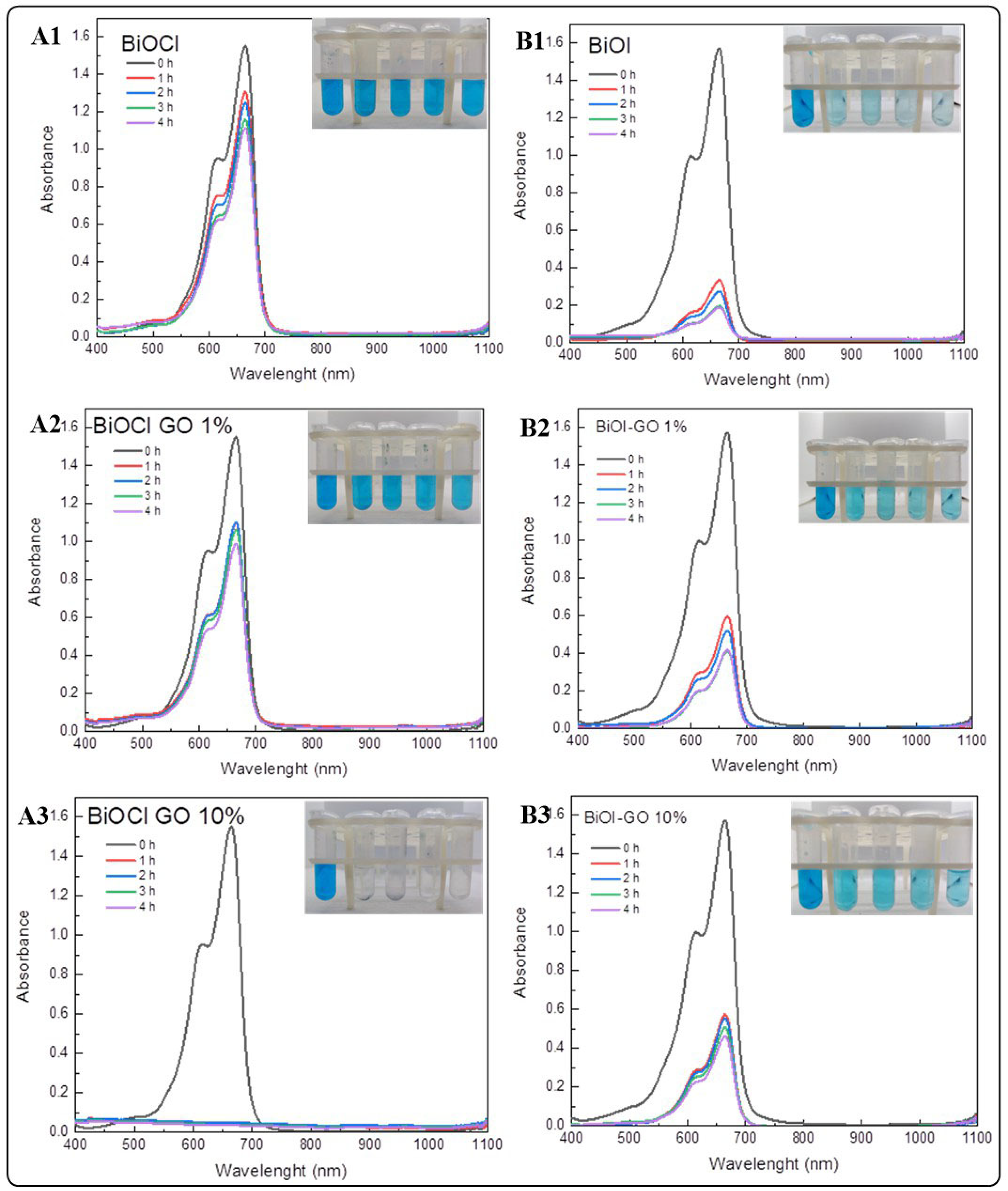
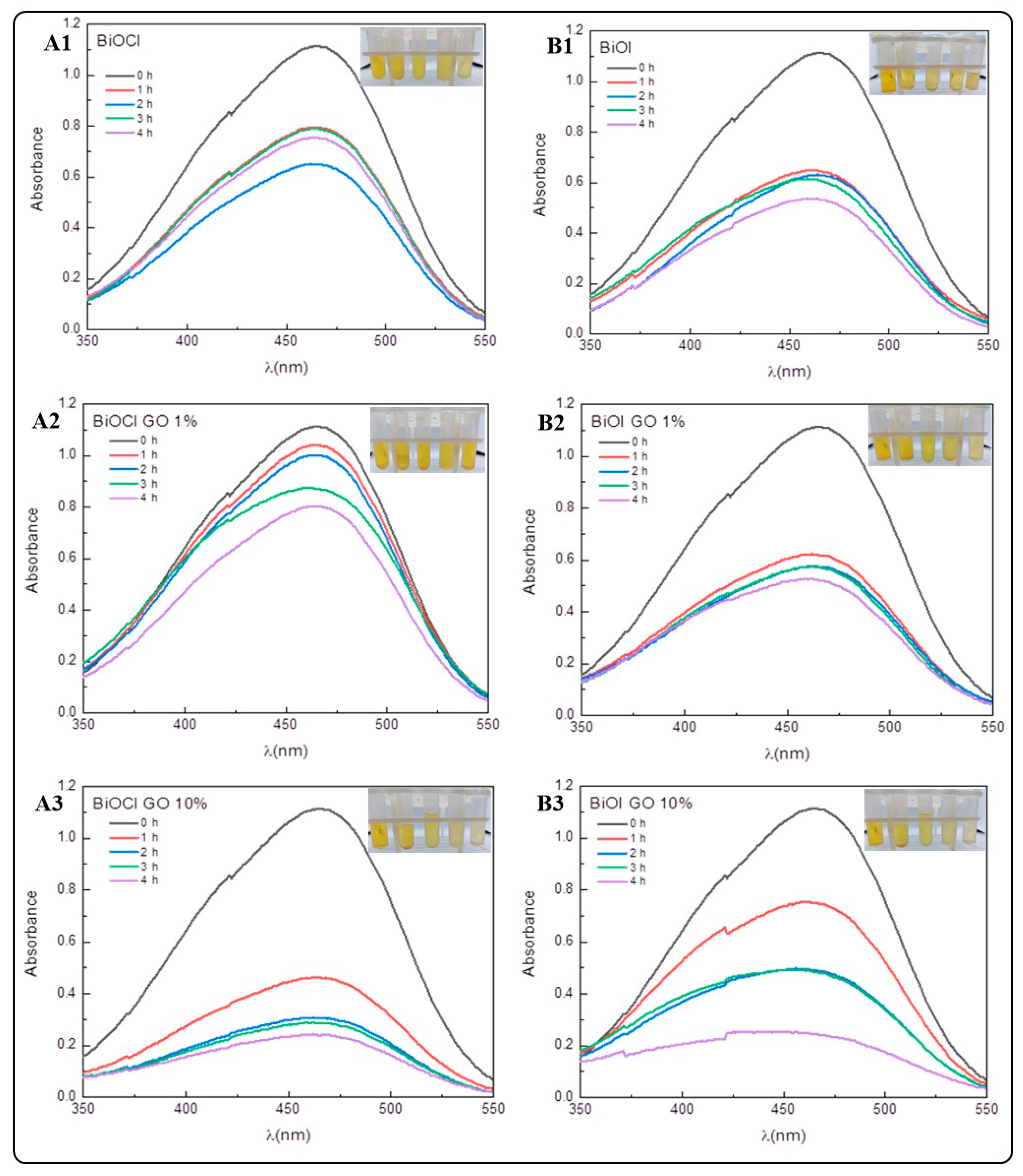
2.2. Methylene Blue (MB) and Methyl Orange (MO) Adsorption Assay
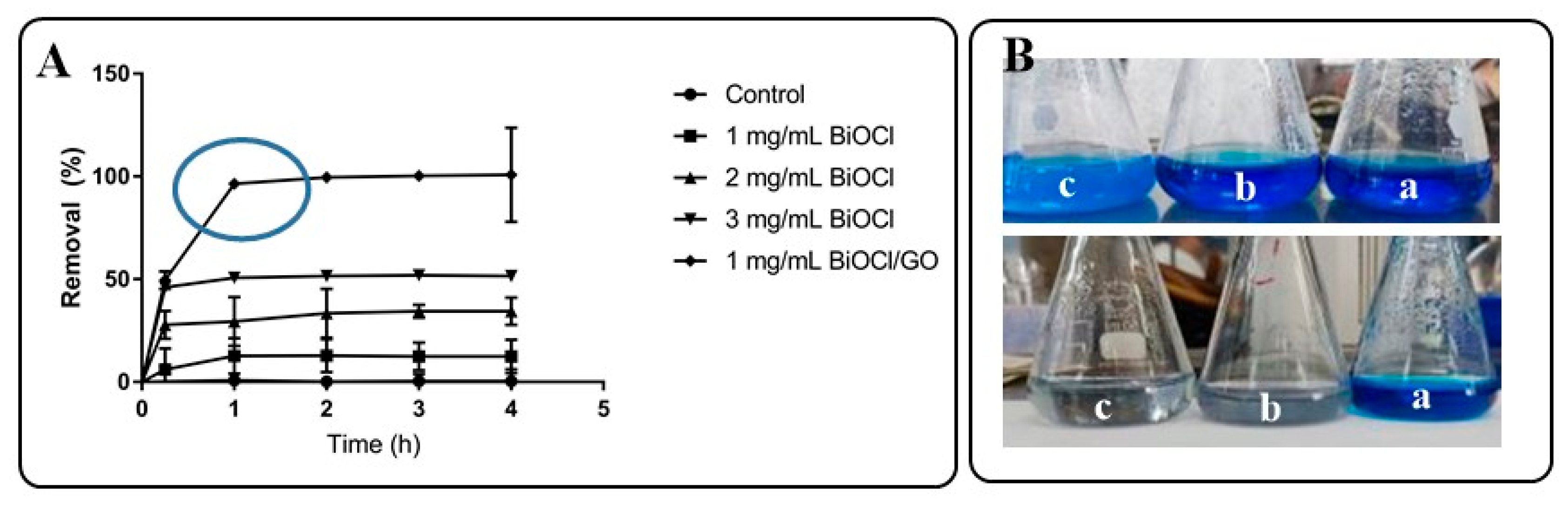
2.2.1. Screening Test for MB Adsorption
2.2.2. Evaluation of the MB and MO Removal Efficiency
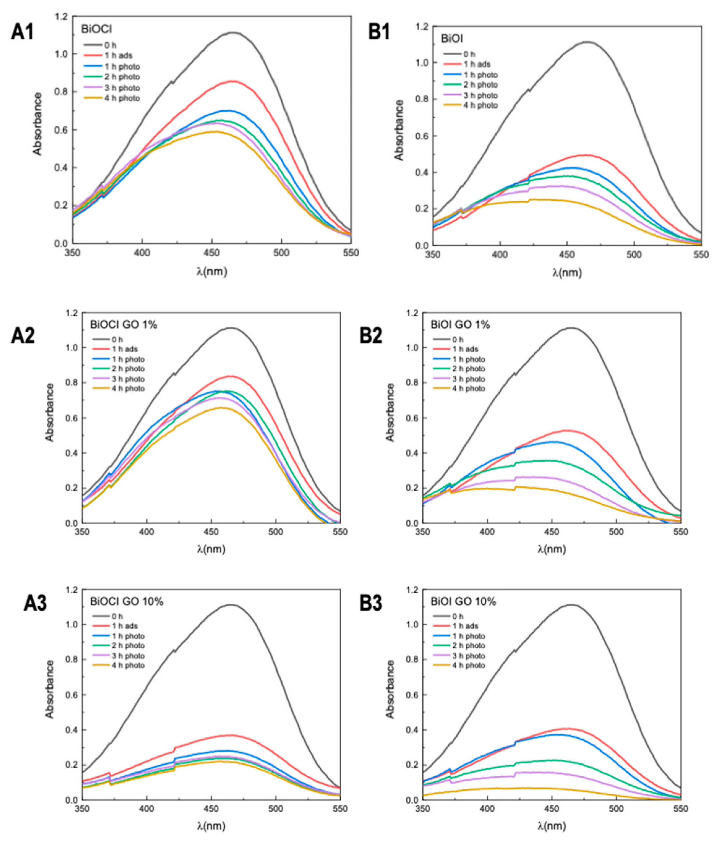
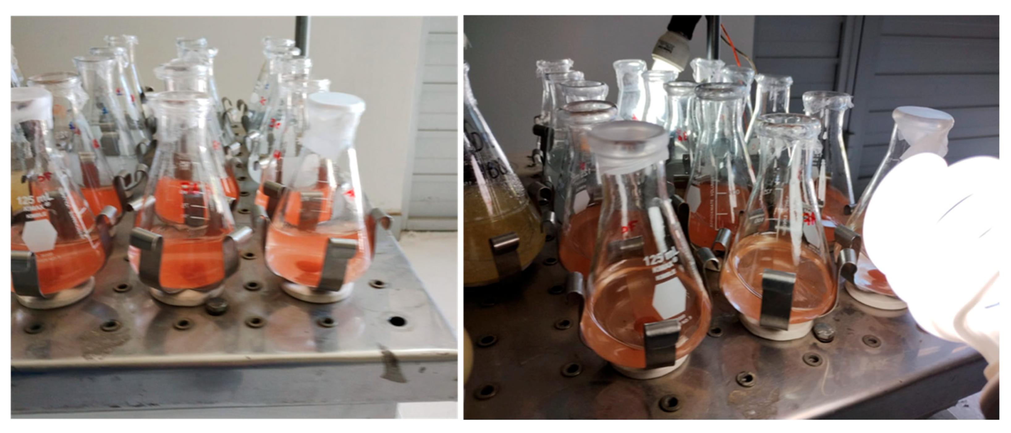
2.3. Photocatalytic Removal of MB or MO
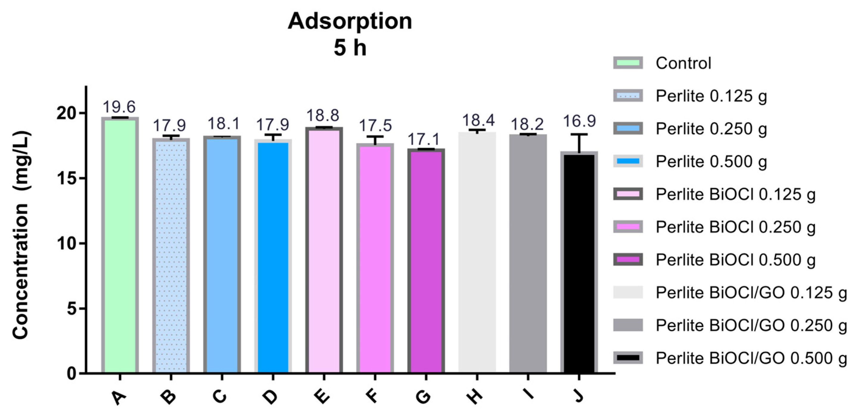
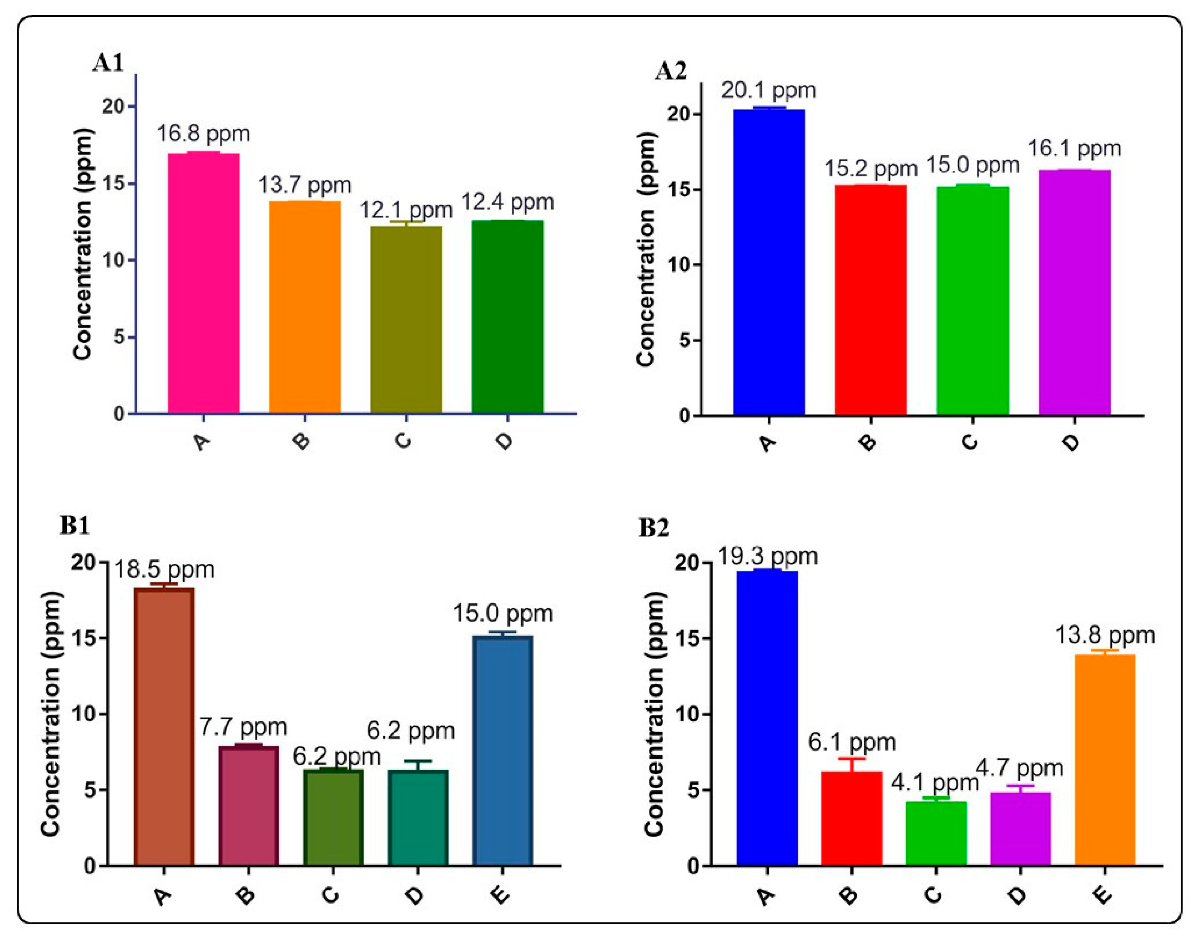
2.4. Arsenic Adsorption Assay
2.5. Photocatalytic Arsenic Removal Assay
3. Results and Discussion
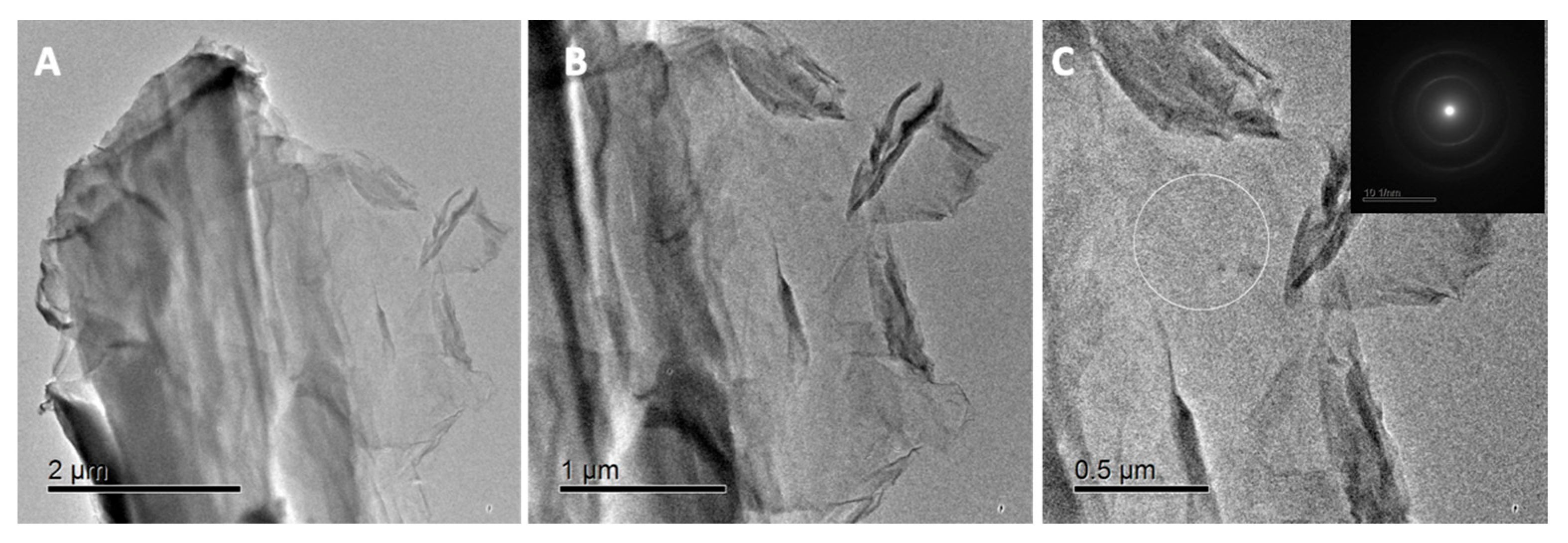
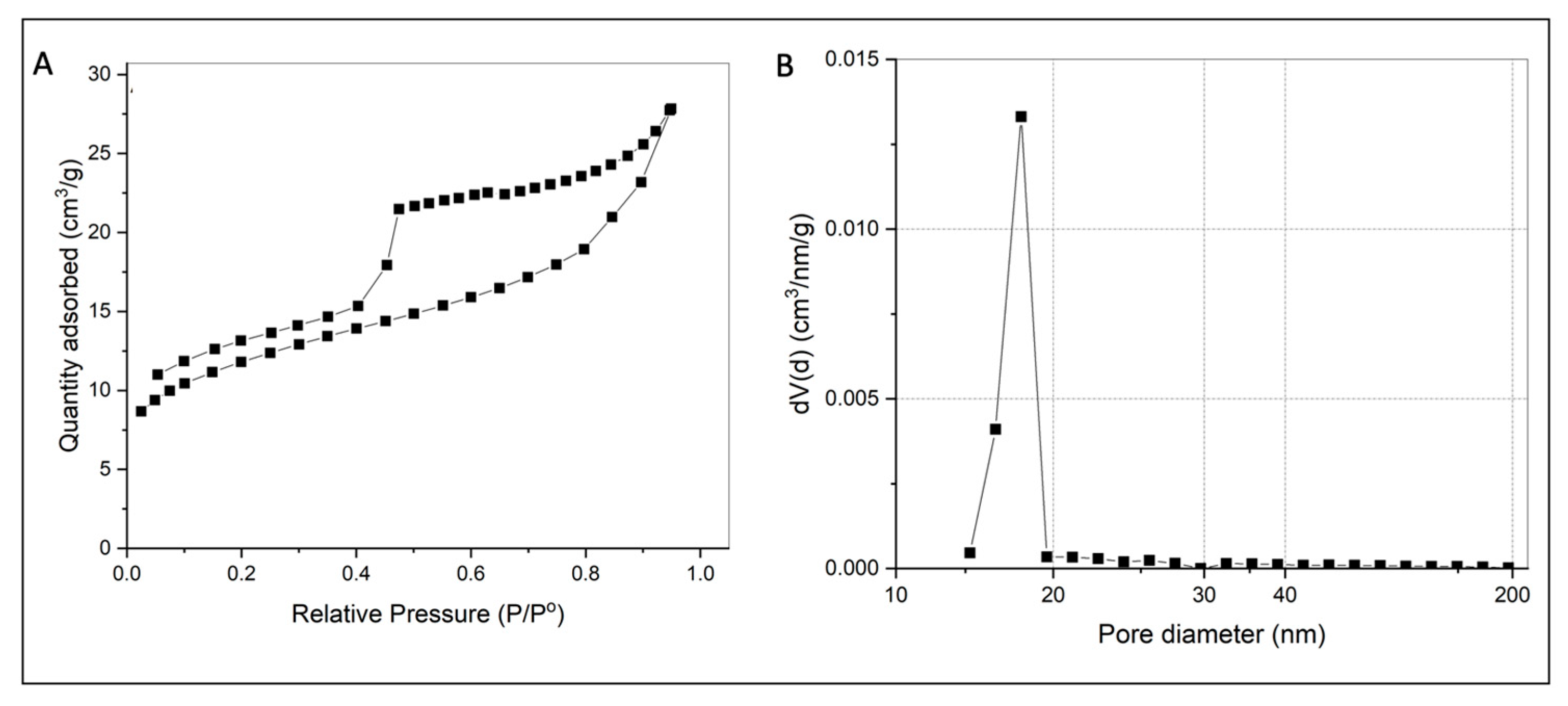
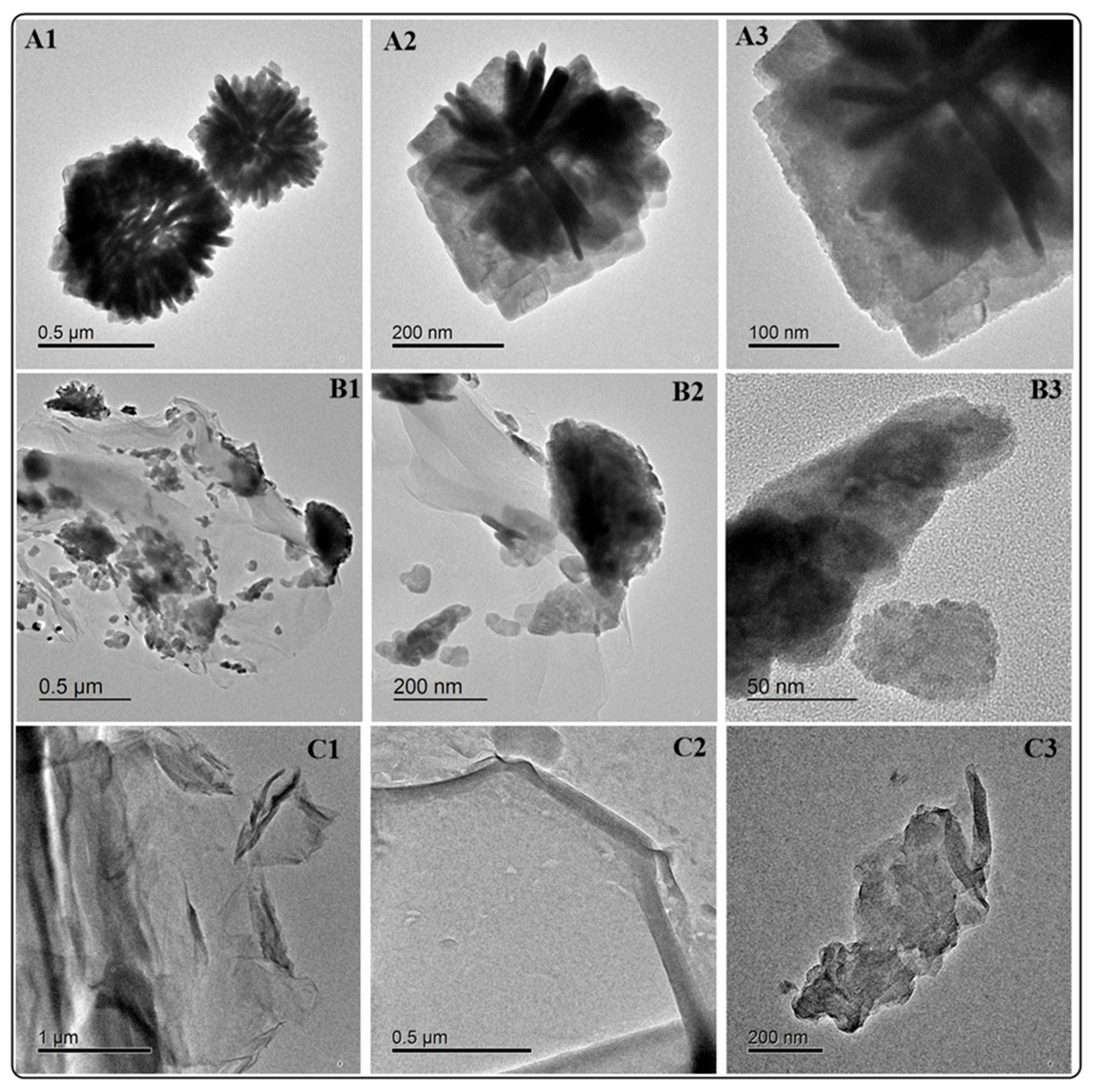
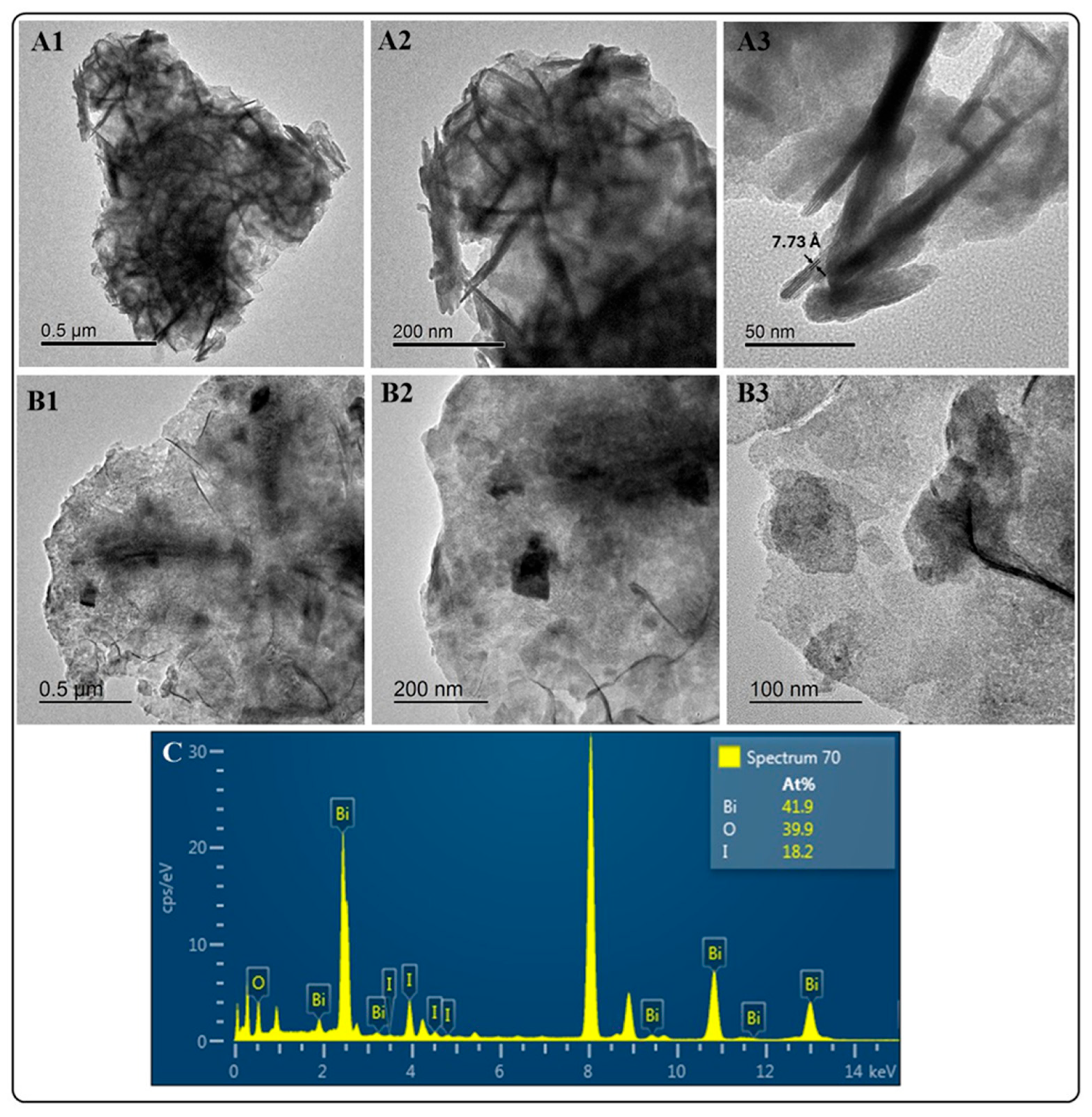
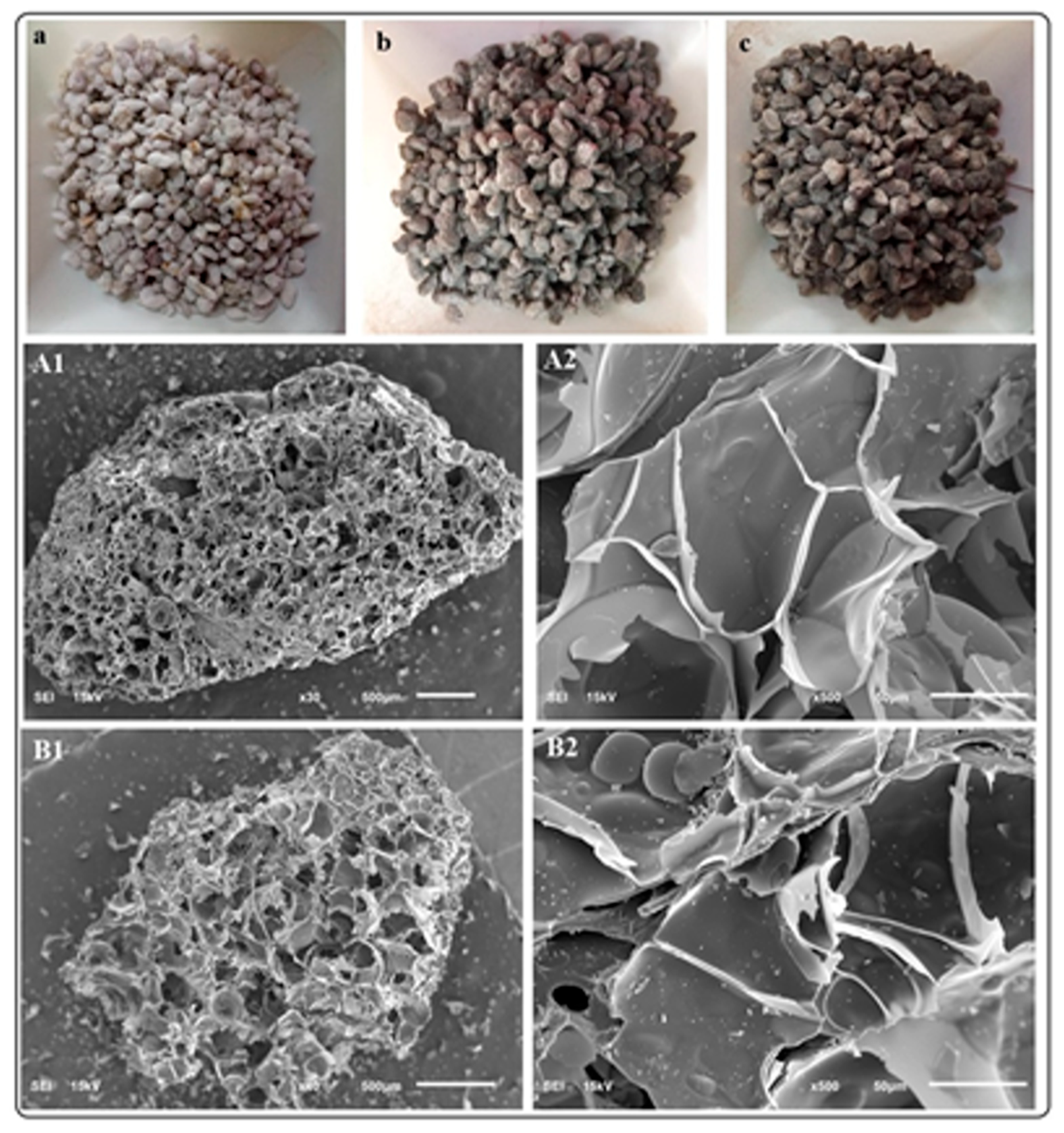
3.1. Synthesis and Characterization of BiOX (X = Cl, I) and BiOX-GO NMs
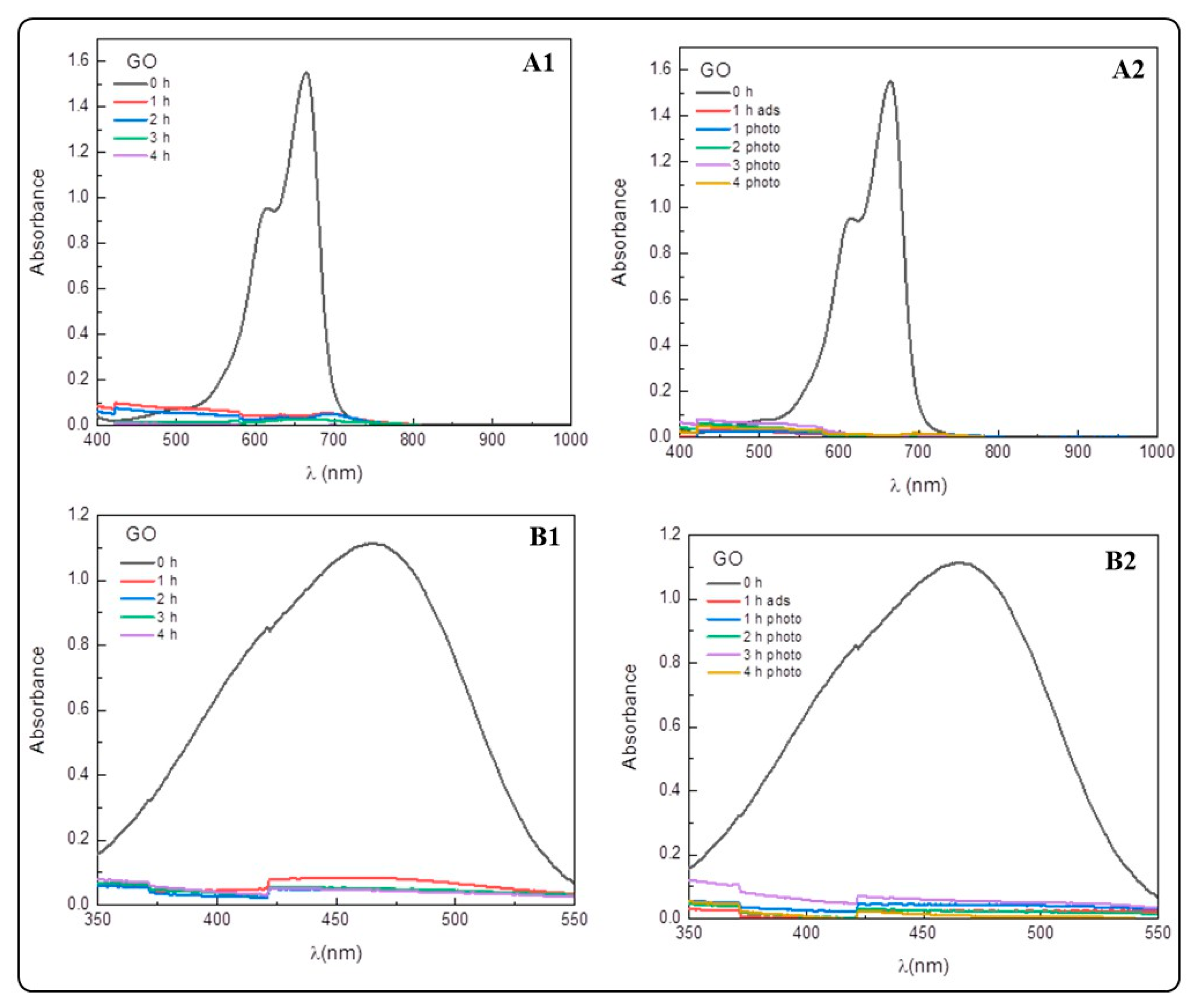
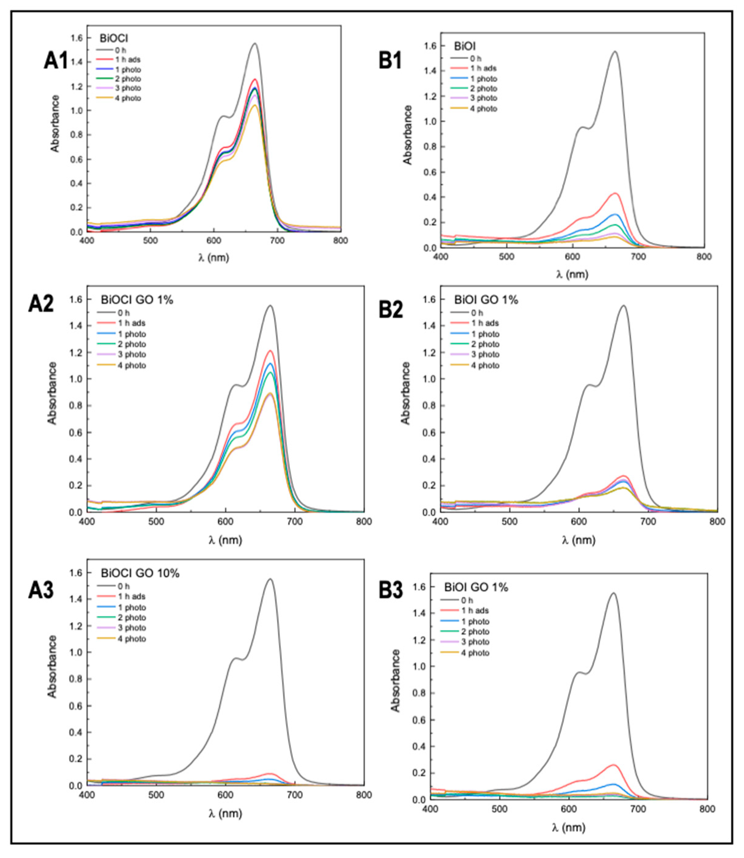
3.2. Methylene Blue and Methyl Orange Adsorption Assay
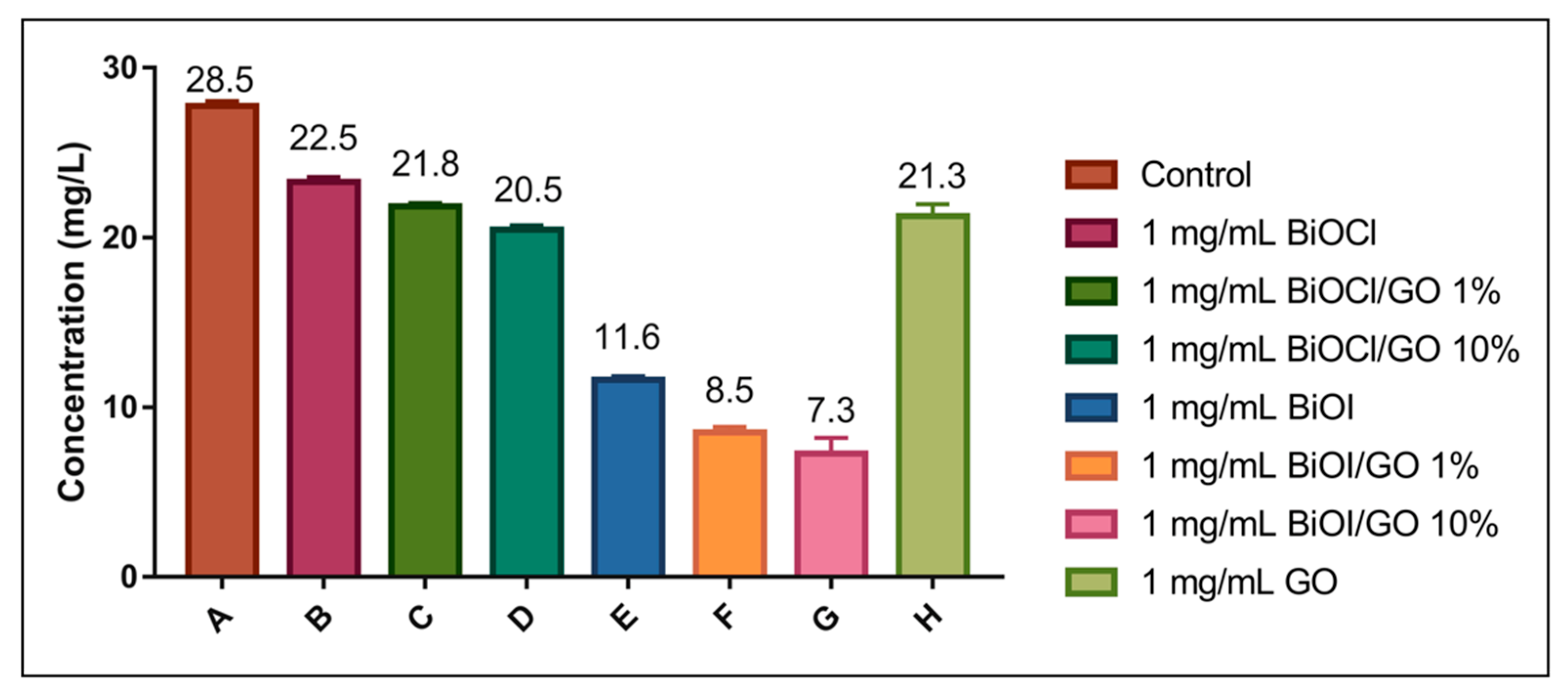
Arsenic Adsorption Assay
4. Conclusions
Author Contributions
Funding
Data Availability Statement
Conflicts of Interest
Appendix A

| Element (Wt. %) | BiOI | BiOI-GO 10% | BiOCl | BiOCl-GO 10% |
| Carbon | 12.57 | 15.28 | 10.84 | 18.63 |
| Oxygen | 19.21 | 20.62 | 27.34 | 23.16 |
| Sodium | 1.82 | 1.16 | 2.54 | 1.8 |
| Aluminum | 7.36 | 7.19 | 7.62 | 7.41 |
| Silicon | 39.72 | 36.59 | 38.35 | 36.63 |
| Potassium | 4.69 | 4.3 | 4.57 | 4.04 |
| Bismuth | 14.63 | 14.85 | 8.75 | 8.34 |
| Time (Hours) | BiOCl | BiOCl-GO 1% | BiOCl-GO 10% | BiOI | BiOI-GO 1% | BiOI-GO 10% |
| Degradation Percentage | ||||||
| 0 | 2 ± 0.5 | 6 ± 0.5 | 7 ± 0.5 | 5 ± 0.1 | 5 ± 0.1 | 7 ± 0.5 |
| 1 | 19 ± 0.1 | 22 ± 0.9 | 94 ± 0.3 | 72 ± 0.2 | 83 ± 0.5 | 83 ± 0.2 |
| 2 | 23 ± 0.5 | 28 ± 0.1 | 97 ± 0.1 | 83 ± 0.9 | 84 ± 0.3 | 93 ± 0.6 |
| 3 | 25 ± 0.6 | 32 ± 0.4 | 99 ± 0.9 | 88 ± 0.3 | 85 ± 0.2 | 97 ± 0.9 |
| 4 | 27 ± 0.4 | 43 ± 0.3 | 99 ± 0.2 | 93 ± 0.7 | 86 ± 0.2 | 97 ± 0.5 |
| 5 | 33 ± 0.6 | 45 ± 0.5 | 99 ± 0.3 | 95 ± 0.5 | 88 ± 0.4 | 98 ± 0.3 |
| Time (Hours) | BiOCl | BiOCl-GO 1% | BiOCl-GO 10% | BiOI | BiOI-GO 1% | BiOI-GO 10% |
| Degradation Percentage | ||||||
| 0 | 2 ± 0.3 | 6 ± 0.5 | 7 ± 0.5 | 3 ± 0.1 | 6 ± 0.1 | 7 ± 0.5 |
| 1 | 23 ± 0.2 | 25 ± 0.6 | 67 ± 0.9 | 55 ± 0.4 | 53 ± 0.5 | 64 ± 0.6 |
| 2 | 37 ± 0.1 | 34 ± 0.5 | 75 ± 0.7 | 63 ± 0.6 | 60 ± 0.9 | 67 ± 0.3 |
| 3 | 42 ± 0.3 | 33 ± 0.4 | 79 ± 0.7 | 67 ± 0.1 | 70 ± 0.6 | 81 ± 0.7 |
| 4 | 45 ± 0.6 | 37 ± 0.8 | 78 ± 0.8 | 73 ± 0.8 | 79 ± 0.8 | 87 ± 0.9 |
| 5 | 49 ± 0.6 | 42 ± 0.6 | 80 ± 0.4 | 80 ± 0.9 | 85 ± 0.7 | 94 ± 0.7 |
Appendix B
References
- Mahlknecht, J.; Aguilar-Barajas, I.; Farias, P.; Knappett, P.S.K.; Torres-Martínez, J.A.; Hoogesteger, J.; Lara, R.H.; Ramírez-Mendoza, R.A.; Mora, A. Hydrochemical Controls on Arsenic Contamination and Its Health Risks in the Comarca Lagunera Region (Mexico): Implications of the Scientific Evidence for Public Health Policy. Sci. Total Environ. 2023, 857, 159347. [Google Scholar] [CrossRef] [PubMed]
- Sánchez-Rodríguez, B.L.; Castillo-Maldonado, I.; Pedroza-Escobar, D.; Delgadillo-Guzmán, D.; Soto-Jiménez, M.F. Association of Obesity, Diabetes, and Hypertension with Arsenic in Drinking Water in the Comarca Lagunera Province (North-Central Mexico). Sci. Rep. 2023, 13, 9244. [Google Scholar] [CrossRef] [PubMed]
- García Salcedo, J.J.; Roh, T.; Nava Rivera, L.E.; Betancourt Martínez, N.D.; Carranza Rosales, P.; San Miguel Salazar, M.F.; Rivera Guillén, M.A.; Serrano Gallardo, L.B.; Niño Castañeda, M.S.; Guzmán Delgado, N.E.; et al. Comparative Biomonitoring of Arsenic Exposure in Mothers and Their Neonates in Comarca Lagunera, Mexico. Int. J. Environ. Res. Public Health 2022, 19, 16232. [Google Scholar] [CrossRef]
- Fisher, A.T.; López-Carrillo, L.; Gamboa-Loira, B.; Cebrián, M.E. Standards for Arsenic in Drinking Water: Implications for Policy in Mexico. J. Public Health Policy 2017, 38, 395–406. [Google Scholar] [CrossRef]
- Ahmad, A.; Bhattacharya, P. Arsenic in Drinking Water: Is 10 Μg/L a Safe Limit? Curr. Pollut. Rep. 2019, 5, 1–3. [Google Scholar] [CrossRef]
- Juárez-Aparicio, F.; Morales-Arredondo, J.I.; Armienta Hernández, M.A. Simultaneous Removal of Fluoride and Arsenic from Drinking Groundwater Using Limestones from Bajío Guanajuatense, Mexico. Arab. J. Geosci. 2024, 17, 109. [Google Scholar] [CrossRef]
- Alazaiza, M.Y.D.; Albahnasawi, A.; Ali, G.A.M.; Bashir, M.J.K.; Copty, N.K.; Amr, S.S.A.; Abushammala, M.F.M.; Al Maskari, T. Recent Advances of Nanoremediation Technologies for Soil and Groundwater Remediation: A Review. Water 2021, 13, 2186. [Google Scholar] [CrossRef]
- Siddiqui, S.I.; Naushad, M.; Chaudhry, S.A. Promising Prospects of Nanomaterials for Arsenic Water Remediation: A Comprehensive Review. Process Saf. Environ. Prot. 2019, 126, 60–97. [Google Scholar] [CrossRef]
- Maity, J.P.; Chen, C.Y.; Bhattacharya, P.; Sharma, R.K.; Ahmad, A.; Patnaik, S.; Bundschuh, J. Advanced Application of Nano-Technological and Biological Processes as Well as Mitigation Options for Arsenic Removal. J. Hazard. Mater. 2021, 405, 123885. [Google Scholar] [CrossRef]
- Wang, Y.; Liu, X.; Chen, Q.; Zhang, T.C.; Ouyang, L.; Yuan, S. Simultaneous Photocatalytic Oxidation and Adsorption for Efficient As(III) Removal by Magnetic BiOI/γ-Fe2O3 Core–Shell Nanoparticles. Mater. Today Chem. 2022, 24, 100823. [Google Scholar] [CrossRef]
- Rahidul Hassan, H. A Review on Different Arsenic Removal Techniques Used for Decontamination of Drinking Water. Environ. Pollut. Bioavailab. 2023, 35, 2165964. [Google Scholar] [CrossRef]
- Baig, N.; Kammakakam, I.; Falath, W.; Kammakakam, I. Nanomaterials: A Review of Synthesis Methods, Properties, Recent Progress, and Challenges. Mater. Adv. 2021, 2, 1821–1871. [Google Scholar] [CrossRef]
- Dudek, S.; Kołodyńska, D. Arsenic(V) Removal on the Lanthanum-Modified Ion Exchanger with Quaternary Ammonium Groups Based on Iron Oxide. J. Mol. Liq. 2022, 347, 117985. [Google Scholar] [CrossRef]
- Ma, Z.; Zhang, M.; Guo, J.; Liu, W.; Tong, M. Facile Synthesis of ZrO2 Coated BiOCl0.5I0.5 for Photocatalytic Oxidation-Adsorption of As(III) under Visible Light Irradiation. Chemosphere 2018, 211, 934–942. [Google Scholar] [CrossRef]
- Jimenez-Chavez, A.; Pedroza-Herrera, G.; Betancourt-Reyes, I.; De Vizcaya Ruiz, A.; Masuoka-Ito, D.; Zapien, J.A.; Medina-Ramirez, I.E. Aluminum Enhances the Oxidative Damage of ZnO NMs in the Human Neuroblastoma SH-SY5Y Cell Line. Discov. Nano 2024, 19, 36. [Google Scholar] [CrossRef]
- Khosrovyan, A.; Vodovnik, M.; Mortimer, M. Omics Approaches in Environmental Effect Assessment of Engineered Nanomaterials and Nanoplastics. Environ. Sci. Nano 2025, 12, 2551–2579. [Google Scholar] [CrossRef]
- Kong, L.; Wu, Y.; Li, C.; Liu, J.; Jia, J.; Zhou, H.; Yan, B. Nano-Cell and Nano-Pollutant Interactions Constitute Key Elements in Nanoparticle-Pollutant Combined Cytotoxicity. J. Hazard. Mater. 2021, 418, 126259. [Google Scholar] [CrossRef]
- Franceschini, F.; Jagdale, P.; Bartoli, M.; Tagliaferro, A. Perspectives on the Use of Bismuth-Based Materials for Sensing and Removal of Water Pollutants. Curr. Opin. Environ. Sci. Health 2022, 26, 100345. [Google Scholar] [CrossRef]
- Wang, J.; Zhang, T.; Jiang, S.; Ma, X.; Shao, X.; Liu, Y.; Wang, D.; Li, X.; Li, B. Controllable Self-Assembly of BiOI/Oxidized Mesocarbon Microbeads Core-Shell Composites: A Novel Hierarchical Structure Facilitated Photocatalytic Activities. Chem. Eng. Sci. 2020, 221, 115653. [Google Scholar] [CrossRef]
- Akash, S.; Rameshwar, S.S.; Rajamohan, N.; Rajasimman, M.; Vo, D.V.N. Metal Oxide Nanobiochar Materials to Remediate Heavy Metal and Dye Pollution: A Review. Environ. Chem. Lett. 2024, 22, 2091–2112. [Google Scholar] [CrossRef]
- Martínez-Montelongo, J.H.; Pineda-Arellano, C.A.; Hernandez-Rangel, R.; Jiménez-González, M.; Betancourt, I.; Peralta-Hernández, J.M.; Medina-Ramírez, I.E. Bismuth-Based Nanocomposites as Potential Materials for Indoor Air Treatment. Chemosphere 2024, 367, 43539. [Google Scholar] [CrossRef]
- Hu, J.; Weng, S.; Zheng, Z.; Pei, Z.; Huang, M.; Liu, P. Solvents Mediated-Synthesis of BiOI Photocatalysts with Tunable Morphologies and Their Visible-Light Driven Photocatalytic Performances in Removing of Arsenic from Water. J. Hazard. Mater. 2014, 264, 293–302. [Google Scholar] [CrossRef]
- Li, B.; Shao, L.; Zhang, B.; Wang, R.; Zhu, M.; Gu, X. Understanding Size-Dependent Properties of BiOCl Nanosheets and Exploring More Catalysis. J. Colloid Interface Sci. 2017, 505, 653–663. [Google Scholar] [CrossRef]
- Wang, X.; Li, Q.; Zhou, C.; Zhang, R. Iodine-Vacancy BiOI1-x Ultrathin Sheets for Improved Visible-Light Photooxidation Activities. Appl. Surf. Sci. 2019, 493, 657–664. [Google Scholar] [CrossRef]
- Dong, F.; Sun, Y.; Fu, M.; Wu, Z.; Lee, S.C. Room Temperature Synthesis and Highly Enhanced Visible Light Photocatalytic Activity of Porous BiOI/BiOCl Composites Nanoplates Microflowers. J. Hazard. Mater. 2012, 219–220, 26–34. [Google Scholar] [CrossRef] [PubMed]
- Han, Y.; Tao, X.; Yang, Z.; Li, K.; Yang, H.; Li, A.; Cheng, R. Effects of the Oxidation Degree of Graphene Oxide on the Adsorption of Methylene Blue. J. Hazard. Mater. 2014, 268, 191–198. [Google Scholar] [CrossRef]
- Guo, T.; Fan, X.; Jiang, X.; Qi, Y.; Du, J.; Zhang, A.; Wang, H. Engineering Shape of BiOCl Nanosheets with Improved Visible-Light Response for Superior Photocatalytic Degradation of Rhodamine B. J. Alloys Compd. 2023, 948, 169586. [Google Scholar] [CrossRef]
- Shah, A.H.; Gu, W.; Abideen, Z.U.; Teng, F. Removal of Chromium from Aqueous Solution by Porous Bi2MoO6@BiOCl Nanostructure. J. Solid State Chem. 2020, 292, 121719. [Google Scholar] [CrossRef]
- Zhong, S.; Zhou, H.; Shen, M.; Yao, Y.; Gao, Q. Rationally Designed a G-C3N4/BiOI/Bi2O2CO3 Composite with Promoted Photocatalytic Activity. J. Alloys Compd. 2021, 853, 157307. [Google Scholar] [CrossRef]
- Sharma, N.; Pap, Z.; Székely, I.; Focsan, M.; Karacs, G.; Nemeth, Z.; Garg, S.; Hernadi, K. Combination of Iodine-Deficient BiOI Phases in the Presence of CNT to Enhance Photocatalytic Activity towards Phenol Decomposition under Visible Light. Appl. Surf. Sci. 2021, 565, 150605. [Google Scholar] [CrossRef]
- Arumugam, M.; Seralathan, K.K.; Praserthdam, S.; Tahir, M.; Praserthdam, P. Synthesis of Novel Graphene Aerogel Encapsulated Bismuth Oxyiodide Composite towards Effective Removal of Methyl Orange Azo-Dye under Visible Light. Chemosphere 2022, 303, 135121. [Google Scholar] [CrossRef] [PubMed]
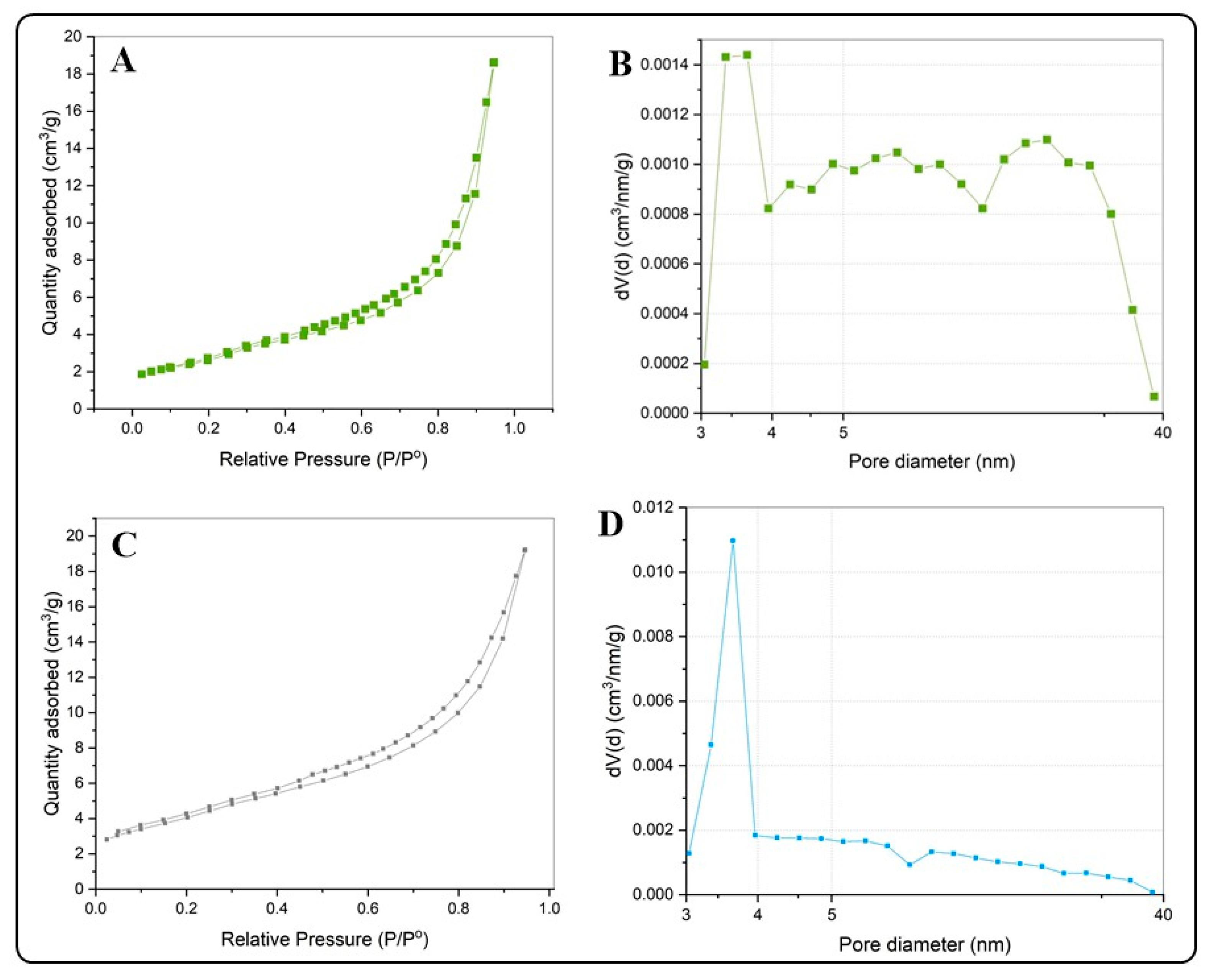
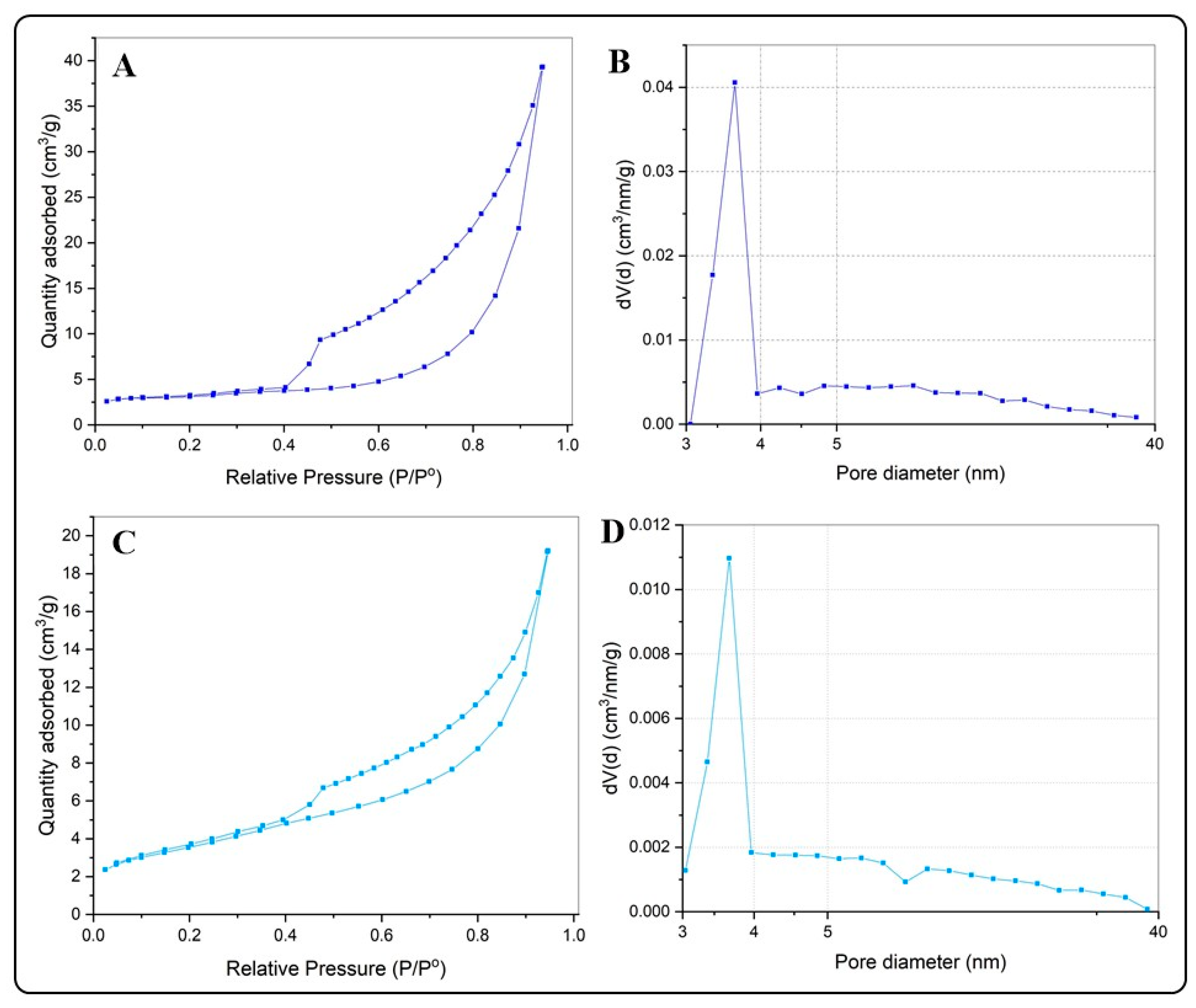
| Sample | Surface Area (m2g−1) | Pore Volume (cm3g−1) | Pore Diameter (nm) |
| BiOCl | 9.239 | 0.0518 | 15.330 |
| BiOCl-GO | 16.479 | 0.046 | 3.718 |
| BiOI | 29.388 | 0.1348 | 4.114 |
| BiOI-GO | 24.008 | 0.113 | 4.119 |
| GO | 47.703 | 0.053 | 4.02 |
| Time (Hours) | BiOCl | BiOCl-GO 1% | BiOCl-GO 10% | BiOI | BiOI-GO 1% | BiOI-GO 10% |
| Removal Percentage | ||||||
| 1 | 16 ± 0.15 | 30 ± 0.21 | 97 ± 0.23 | 91 ± 0.12 | 94 ± 0.06 | 97 ± 0.13 |
| 2 | 20 ± 0.11 | 34 ± 0.16 | 97 ± 0.18 | 93 ± 0.09 | 96 ± 0.14 | 98 ± 0.23 |
| 3 | 25 ± 0.16 | 32 ± 0.18 | 98 ± 0.14 | 95 ± 0.11 | 97 ± 0.10 | 98 ± 0.09 |
| 4 | 28 ± 0.06 | 37 ± 0.13 | 98 ± 0.19 | 98 ± 0.10 | 97 ± 0.16 | 99 ± 0.11 |
| Time (Hours) | BiOCl | BiOCl-GO 1% | BiOCl-GO 10% | BiOI | BiOI-GO 1% | BiOI-GO 10% |
| Removal Percentage | ||||||
| 1 | 28 ± 0.9 | 7 ± 0.8 | 59 ± 0.6 | 41 ± 0.7 | 44 ± 0.3 | 32 ± 0.5 |
| 2 | 29 ± 0.5 | 10 ± 0.2 | 72 ± 0.4 | 43 ± 0.3 | 48 ± 0.2 | 56 ± 0.7 |
| 3 | 32 ± 0.3 | 21 ± 0.1 | 74 ± 0.3 | 45 ± 0.1 | 48 ± 0.3 | 56 ± 0.2 |
| 4 | 41 ± 0.1 | 27 ± 0.1 | 78 ± 0.4 | 52 ± 0.6 | 53 ± 0.9 | 78 ± 0.9 |
| Time (Hours) | Adsorption | Photocatalysis | ||
| MB | MO | MB | MO | |
| 0 | 95.55 * | 94.88 * | ||
| 1 | 94.46 | 92.54 | 96.52 | 96.04 |
| 2 | 95.75 | 97.78 | 97.23 | 97.75 |
| 3 | 98.97 | 95.42 | 98.03 | 97.84 |
| 4 | 99.5 | 95.96 | 98.32 | 99.19 |
| Adsorbent | Arsenic/Time | ||
| * As(III)/5 h | ** As(III)/24 h | *** As(III)/5 h | |
| BiOCl | 18 ± 0.56 | 25 ± 0.35 | 21 ± 1.06 |
| BiOCl-GO 1% | 28 ± 2.55 | 25 ± 1.29 | 23 ± 0.63 |
| BiOCl-GO 10% | 26 ± 0.48 | 20 ± 0.51 | 28 ± 0.91 |
| BiOI | 32 ± 1.21 | 41 ± 5.43 | 59 ± 0.77 |
| BiOI-GO 1% | 38 ± 1.03 | 49 ± 2.17 | 70 ± 1.17 |
| BiOI-GO 10% | 36 ± 3.83 | 45 ± 3.30 | 45 ± 1.11 |
| GO | 17 ± 2.18 | 24 ± 2.50 | 20 ± 1.41 |
Disclaimer/Publisher’s Note: The statements, opinions and data contained in all publications are solely those of the individual author(s) and contributor(s) and not of MDPI and/or the editor(s). MDPI and/or the editor(s) disclaim responsibility for any injury to people or property resulting from any ideas, methods, instructions or products referred to in the content. |
© 2025 by the authors. Licensee MDPI, Basel, Switzerland. This article is an open access article distributed under the terms and conditions of the Creative Commons Attribution (CC BY) license (https://creativecommons.org/licenses/by/4.0/).
Share and Cite
Martinez-Montelongo, J.H.; Jiménez-González, M.L.; González-Pérez, A.; Mortimer, M.; Avelar-González, F.J.; Macias-Díaz, J.E.; Medina-Ramírez, I.E. Evaluation of the Adsorption Capacity of the BiOX (X = Cl, I) and BiOX-GO Nanomaterials (NMs) for Water Treatment. Processes 2025, 13, 2179. https://doi.org/10.3390/pr13072179
Martinez-Montelongo JH, Jiménez-González ML, González-Pérez A, Mortimer M, Avelar-González FJ, Macias-Díaz JE, Medina-Ramírez IE. Evaluation of the Adsorption Capacity of the BiOX (X = Cl, I) and BiOX-GO Nanomaterials (NMs) for Water Treatment. Processes. 2025; 13(7):2179. https://doi.org/10.3390/pr13072179
Chicago/Turabian StyleMartinez-Montelongo, Jorge H., Martha L. Jiménez-González, Abner González-Pérez, Monika Mortimer, F. J. Avelar-González, Jorge E. Macias-Díaz, and Iliana E. Medina-Ramírez. 2025. "Evaluation of the Adsorption Capacity of the BiOX (X = Cl, I) and BiOX-GO Nanomaterials (NMs) for Water Treatment" Processes 13, no. 7: 2179. https://doi.org/10.3390/pr13072179
APA StyleMartinez-Montelongo, J. H., Jiménez-González, M. L., González-Pérez, A., Mortimer, M., Avelar-González, F. J., Macias-Díaz, J. E., & Medina-Ramírez, I. E. (2025). Evaluation of the Adsorption Capacity of the BiOX (X = Cl, I) and BiOX-GO Nanomaterials (NMs) for Water Treatment. Processes, 13(7), 2179. https://doi.org/10.3390/pr13072179