Article

Effects of Denaturants on Early-Stage Prion Conversion: Insights from Molecular Dynamics Simulations

Department of Electrical and Computer Engineering, University of Alberta, Edmonton, AB T6G 2R3, Canada
*
Author to whom correspondence should be addressed.
Processes 2025, 13(7), 2151; https://doi.org/10.3390/pr13072151
Submission received: 15 May 2025 / Revised: 23 June 2025 / Accepted: 1 July 2025 / Published: 7 July 2025
(This article belongs to the Special Issue Advances in Computer Simulation of Condensed Matter Systems)

Abstract

Prion diseases such as chronic wasting disease involve the conformational conversion of the cellular prion protein (PrPC) into its misfolded, β-rich isoform (PrPSc). While chemical denaturants such as guanidine hydrochloride (GdnHCl) and urea are commonly used to study this process in vitro, their distinct molecular effects on native and misfolded PrP conformers remain incompletely understood. In this study, we employed 500 ns all-atom molecular dynamics simulations and essential collective dynamics analysis to investigate the differential effects of GdnHCl and urea on a composite PrPC/PrPSc system, where white-tailed deer PrPC interfaces with a corresponding PrPSc conformer. GdnHCl was found to preserve interfacial alignment and enhance β-sheet retention in PrPSc, while urea promoted partial β-strand dissolution and interfacial destabilization. Both denaturants formed transient contacts with PrP, but urea displaced water hydrogen bonds more extensively. Remarkably, we also observed long-range dynamical coupling across the PrPC/PrPSc interface and between transiently bound solutes and distal protein regions. These findings highlight distinct, denaturant-specific mechanisms of protein destabilization and suggest that localized interactions may propagate non-locally via mechanical or steric pathways. Our results provide molecular-scale insights relevant to prion conversion mechanisms and inform experimental strategies using GdnHCl and urea to modulate misfolding processes in vitro.

1. Introduction

The functions of protein molecules in biological systems are critically dependent on their complex three-dimensional structures. Unlike crystalline solids, which exhibit long-range periodic order, or simple fluids, which display uniform yet dynamic molecular arrangements, proteins adopt unique and often non-repeating conformations that determine their functional roles. These structures lack periodicity and can be highly sensitive to environmental changes. The specific conformation a protein adopts governs its stability and its ability to interact with other biomolecules. However, under conditions that remain incompletely understood, proteins can misfold into abnormal conformations. These misfolded species may initiate a cascade of self-perpetuating misfolding events, ultimately resulting in the formation of β-sheet-rich amyloid fibrils that accumulate in the brain and other tissues [1,2,3]. The discovery of this phenomenon revealed a previously unrecognized pathogenic mechanism underlying several neurodegenerative and systemic diseases [4,5]. To date, approximately 50 proteins have been identified that can form disease-associated misfolded aggregates [2]. Among them, the prion protein (PrP)—an extracellular protein found in many tissues but predominantly expressed in the nervous system—plays a central role in transmissible spongiform encephalopathies (TSEs), a group of fatal neurodegenerative disorders affecting both animals and humans [6]. Under pathological conditions, the native cellular form of PrP (PrPC) can be converted into a β-sheet-rich misfolded isoform known as “scrapie” (PrPSc), which is capable of templating its conformation onto other PrPC molecules. Animal prion diseases include scrapie in sheep and goats, bovine spongiform encephalopathy in cattle, and chronic wasting disease in cervids [7,8]. In humans, prion diseases can arise sporadically, be inherited, or be acquired and include conditions such as Creutzfeldt–Jakob disease, fatal familial insomnia, and Gerstmann–Sträussler–Scheinker syndrome [9].
Although the PrP amino acid sequence is highly conserved across vertebrate species, species-specific differences and naturally occurring polymorphisms can contribute to the emergence and propagation of distinct prion strains [1,2,3,6,8,10]. These strains, or variants, are believed to correspond to different self-propagating conformational states of PrPSc. While some strain diversity arises from species-specific sequence differences or polymorphisms, structurally distinct variants can also emerge from identical amino acid sequences, giving rise to different pathological phenotypes despite sequence identity. Prion conversion is further influenced by a range of cofactors that are known to modulate aggregation under certain conditions. Among the most frequently studied are glycosaminoglycans, nucleic acids, and specific lipid components, which have been implicated in altering aggregation in various experimental settings [1,11,12,13].
Despite extensive efforts, the precise sequence of molecular events that drive prion proteins from their native conformations to aberrant supramolecular aggregates remains incompletely understood [1,6,9]. Key uncertainties persist regarding the structure, stability, and morphology of intermediate misfolded PrP species, where multiple conformers, cofactors, and strain-specific features may be involved. These knowledge gaps hinder the development of effective preventive or disease-modifying treatments for PrP misfolding disorders. A detailed understanding of the pathways leading to misfolding and aggregation is therefore of paramount importance, as it offers critical insight into the mechanistic basis of pathogenesis and may identify transient intermediate conformers amenable to targeting by conformation-specific antibodies or small-molecule inhibitors. In this context, in vitro studies of PrP misfolding and aggregation have played a central role in disentangling the complexity of prion conversion. By enabling precise control over physicochemical conditions, in vitro experiments allow researchers to systematically isolate and assess the effects of individual factors—including prion strains, cofactors, and environmental modulators—on the conformational transition of PrP from its native form (PrPC) to the misfolded isoform (PrPSc) associated with pathogenic aggregation. The capacity to reproducibly generate and interrogate such conformations under defined conditions has been instrumental in elucidating key features of PrP structural conversion and in probing the influence of ligand interactions and solvent environments [10,13,14,15].
Among the most commonly used in vitro approaches are experimental systems employing chemical denaturants such as guanidine hydrochloride (GdnHCl) or urea, which enable controlled destabilization of PrPC and facilitate the formation and characterization of intermediate states and fibrillary PrPSc species [16,17,18,19,20,21]. Due to their distinct properties, these denaturants influence the misfolding process through different mechanisms. GdnHCl, an ionic denaturant, has been demonstrated to accelerate the formation of fibrils and oligomeric misfolded aggregates at moderate concentrations [17,21,22,23]. In contrast, PrP unfolding in urea, a polar but non-charged denaturant, seems to favor the accumulation of partially unfolded intermediates with residual secondary structure [19,20]. Despite their widespread use, important gaps remain in our understanding of how GdnHCl and urea interact with different classes of protein structures. While both denaturants are effective at destabilizing globular proteins, their effects on the formation and stability of β-sheet-rich amyloid fibrils are less well characterized. Notably, studies examining these effects have reported differing—and in some cases, seemingly divergent—trends for the two agents. Depending on the protein system, experimental conditions, and stage of aggregation, GdnHCl and urea can exhibit distinct influences on fibril formation, structural remodeling, or disassembly [24]. In particular, a deeper understanding of how these denaturants modulate the transition from native PrPC to its pathogenic PrPSc form is critical for elucidating the physicochemical mechanisms underlying prion conversion. This knowledge is important for the refinement of in vitro misfolding models, the identification of aggregation-prone intermediates, and the development of strategies to stabilize the native state or interfere with early misfolding events.
Molecular dynamics (MD) simulations offer a uniquely powerful approach for addressing these knowledge gaps by providing atomistic insights into the early unfolding events, intermediate conformers, and local rearrangements—insights that are difficult or impossible to obtain by other means. In particular, MD studies have revealed that GdnHCl and urea exhibit distinct preferences in destabilizing secondary structures of globular proteins and peptides. While several investigations indicate that β-sheets are more readily disrupted by urea than by GdnHCl [25,26,27], the effects of these denaturants on α-helices are more variable. Depending on the protein system, GdnHCl has been found either more or less efficient than urea in dissolving α-helical structures [25,27,28]. Comparative modeling studies further demonstrated that GdnHCl and urea unfold proteins through markedly different molecular mechanisms. GdnHCl, which dissociates into GdnH+ and Cl ions in aqueous solutions, has been proposed to destabilize proteins primarily by modifying electrostatic interactions [25,29,30,31]. This contrasts with urea, which has been found to accumulate within the protein’s solvation shell, forming multiple direct hydrogen bonds (HBs) with backbones and side chains [25,31,32,33], effectively displacing water molecules and weakening intramolecular HB networks. This differential behavior suggests that solvent-exposed structures might be particularly susceptible to urea-induced unfolding due to favorable HB interactions, while GdnHCl-induced destabilization is more influenced by electrostatic screening effects [27,29,30]. Nevertheless, the relative sensitivity of α-helices and β-structures, as well as the specific pathways of denaturation, appear highly protein-dependent, suggesting that no universal hierarchy of structural destabilization applies. Instead, the unfolding response reflects a combination of intrinsic protein topology, solvent exposure of secondary elements, and denaturant-specific interaction patterns.
Although MD simulations have provided valuable insights into the denaturation mechanisms of globular proteins and peptides, important questions remain regarding the effects of chemical denaturants on more complex architectures. In particular, the action of GdnHCl and urea on aggregation-prone, β-rich fibrillary seeds has not been systematically investigated. Available MD studies have largely focused on small, monomeric proteins with globular folds or short peptide fragments, limiting their relevance for understanding the behavior of more extended β-sheet assemblies. Moreover, the stability and unfolding dynamics of interfaces between native and misfolded protein forms, such as the alignment between PrPC and PrPSc during prion conversion, remain unexplored at the atomistic level under denaturing conditions. This represents an important gap, given that early-stage unfolding and mechanisms of misfolded state stabilization are critical for understanding the pathogenic conformational conversion process.
In this work, we apply all-atom MD simulations along with cutting-edge analysis tools to investigate the impact of GdnHCl and urea on the structural and dynamical stability of white-tailed deer (WTD) PrP, implicated in the ongoing spread of chronic wasting disease (CWD)—a form of transmissible spongiform encephalopathy affecting wild and farmed elk, deer, and other cervids [8,34]. The disease has been spreading rapidly over several decades and is currently found in four Canadian provinces including Alberta and Saskatchewan, as well as in twenty-six U.S. states. In addition to being fatal to infected animals, CWD results in long-term environmental contamination in affected regions and is transmissible to domestic cattle, sheep, and certain other mammals.
Our model system consists of the WTD PrPC (residues 93–233) structure in close contact with a matching PrPSc seed construct (Figure 1). For the latter, we employed a four-rung β-solenoid (4RβS) model [35], which offers a compact β-strand architecture compatible with a templated PrP conversion mechanism and consistent with X-ray diffraction data showing a characteristic 19.2 Å repeat in brain-derived prion assemblies [36]. This model has also been proposed to explain narrow fibrillar structures detected by negative-stain EM in ex vivo preparations [37] and has precedent in the β-solenoidal fold of the fungal heterokaryon incompatibility s (HET-s) prion [38]. An alternative architecture, based on parallel-in-register β-sheets (PIRIBS) and supported by high-resolution cryo-EM and solid-state nuclear magnetic resonance (NMR) studies of recombinant and ex vivo fibrils [39], typically exhibits a wide cross-sectional profile that generally requires pre-alignment of already unfolded PrP monomers. It should be noted that both in vitro and ex vivo studies have revealed a diversity of PrP fibril morphologies [17,23,40], and distinct conformers may coexist or predominate under specific physiological or experimental conditions. Here, we adopted the 4RβS framework as a plausible structure for exploring early-stage interfacial events in prion conversion.
Elucidating the effects of GdnHCl and urea on both PrPC and PrPSc within a unified simulation framework provides important insights into the molecular factors that govern prion protein destabilization and conformational conversion under denaturing conditions. By analyzing the structural and dynamical responses of the composite PrPC/PrPSc system to different solvent environments, this study contributes to a deeper understanding of early misfolding events and interfacial destabilization mechanisms, offering a foundation for future investigations into prion conversion and informing therapeutic strategies targeting early misfolding events in cervid prion diseases.
Figure 1. Starting composite structure of WTD PrPC (93–233) with the N-terminal region (residues 93–119) threaded onto the C-terminal segment of matching four-rung β-solenoid PrPSc seed structure 4RβS [35]. The color scheme represents the secondary structure: α-helices are shown in magenta, 310 helices in blue, β-strands in yellow, turns in cyan, and random coils in gray. Labels S1 and S2 mark the two β-strands, and H1–H3 denote the three α-helices in PrPC.
Processes 13 02151 g001

2. Materials and Methods

Two conformations of WTD prion protein, corresponding to the cellular form PrPC and the misfolded form PrPSc, were constructed. To construct the starting conformation of PrPSc, the β-solenoidal model described in [35] was used as a structural framework. The sequence corresponding to residues 89–230 (in mouse PrP numbering) was uploaded, and all mouse-to-WTD amino acid substitutions were introduced using Discovery Studio Visualizer [41] to produce a species-specific WTD PrPSc model. For PrPC, homology modeling was performed using the SWISS-MODEL server [42] with the WTD PrP sequence spanning residues 112–231. A total of 201 structural templates were identified. Among them, the template based on the crystal structure of deer PrPC (PDB ID: 4YXH [43], model 1, chain A) was selected for 3D modeling due to its high sequence identity and structural resolution. The protonation state of titratable residues corresponded to a neutral pH of around 7.4. To prepare the geometry for subsequent positioning of PrPC onto PrPSc as illustrated in Figure 1, the N-terminal segment of PrPC—residues 93–116—was modeled by adopting the backbone conformation of the corresponding β-rung from the PrPSc template using Discovery Studio Visualizer.
The PrP constructs were subjected to energy minimizations, equilibrations, and production MD simulations using the GROMACS software package [44] version 5.0.7, employing the Optimized Potentials for Liquid Simulations all-atom (OPLS-AA) force field. Initial in vacuo minimization was carried out separately for the PrPC and PrPSc models using 10,000 steps of the steepest descent algorithm. After minimization, the composite PrPC/PrPSc system was assembled by positioning the two monomeric units in a head-to-tail orientation, with the N-terminal segment of PrPC threaded onto the C-terminal rung of PrPSc. The spacing between rungs in the aligned region was set to 5.5 Å—slightly wider than the inter-rung distance in the PrPSc model—to allow room for structural adjustments. Structural alignment and system assembly were carried out using Visual Molecular Dynamics (VMD, [45]) and Discovery Studio Visualizer [41]. The composite system was minimized in vacuo again for 1000 steps with the same algorithm.
Two types of solvated systems were prepared, one containing 615 mM guanidine hydrochloride, represented as GdnH+ and Cl ions, and another with 6.5 M urea. These concentrations were selected to fall within the typical ranges used in in vitro PrP unfolding, aggregation, and amyloid seeding assays [18,19,20,21,22,23,24], ensuring relevance to commonly employed experimental conditions. A third system, devoid of denaturants, served as a control. All systems were solvated in rectangular boxes using the SPC/E water model, with 133 mM NaCl, modeled as Na+ and Cl ions, to maintain physiological ionic strength. Additional counterions were introduced as needed to neutralize the overall system charge. Following solvation, a multi-stage energy minimization was employed. This was followed by a series of entire systems’ minimizations with progressively decreasing positional restraints (Kposre = 1 × 10⁵, 1 × 10⁴, 1000, 100, 10, and 0 kJ mol−1 nm−2) applied to all non-hydrogen atoms of the protein to relieve steric constraints. The systems were gradually heated from 0 K to 310 K using the Berendsen thermostat [46], followed by NPT equilibration to adjust solvent density to 1 g/cm3. Both the equilibration and subsequent production simulations were carried out under constant pressure and temperature (NPT ensemble) at 310 K and 1 atm, using isotropic pressure coupling. Bond lengths were constrained using the LINCS algorithm with a fourth-order expansion. A cutoff of 1.4 nm was applied to both short-range electrostatic and van der Waals interactions. Long-range electrostatics were treated using the particle-mesh Ewald (PME) summation method, with a grid spacing of 0.135 nm for fast Fourier transform and cubic interpolation. Production MD trajectories of 500 ns were generated for systems containing either GdnH+/Cl or urea, as well as for a control system simulated without denaturants. This length was chosen to ensure that each trajectory explores a sufficiently broad range of conformations within the quasi-stable regimes typically reached after 100–200 ns of production simulation. Post-simulation structural analyses of the MD trajectories were conducted to evaluate the evolution of secondary structure and hydrogen bonding, utilizing built-in GROMACS and VMD tools and visualization through the VMD package.
To further probe the dynamics of denaturant-PrP interactions, we employed our group’s original essential collective dynamics (ECD) method. This approach is based on a statistical-mechanical framework [47,48] in which the macromolecule is described in terms of generalized Langevin dynamics involving a selected set of essential collective coordinates. These essential coordinates are identified as principal eigenvectors of the covariance matrix, obtained through principal component analysis (PCA) of the atomic displacement trajectories. Typically, 10–20 essential coordinates are sufficient to accurately sample the displacement for an MD trajectory. Each atom is represented by a point (“atom-image” r i ) in the 3K-dimensional collective coordinate space spanned by K essential eigenvectors [48,49]:
r i = r i 1 , r i 2 , . . . r i K ,   i = 1,2 , , N .
Here, N is the total number of atoms under analysis, and each r i k denotes a triplet of direction cosines E i , x k , E i , y k , E i , z k corresponding to the k-th essential eigenvector E k relative to the x-, y-, and z-degrees of freedom for atom i in the original 3N-dimensional configuration space. The full derivation and theoretical background of this representation are described elsewhere [47,48,49]. In particular, it has been demonstrated [48] that, under the assumptions of orthogonality and equipartition among the essential modes, the atom images r i are related to projected velocities v i of atoms according to ε r i 2 = v i 2 and ε r i r j 2 = v i v j 2 , where ε is a scaling coefficient. Therefore, the distance between two atom images in the 3K-dimensional space,
d i j = r i r j ,      i , j = 1,2 , , N ,
represents the magnitude of the difference between the projected velocities of atoms i and j [48]. The closeness of two atom images means that the corresponding atoms move synchronously, regardless of their spatial proximity in the protein structure. Importantly, it has been shown [48] that the distances d i j represent invariant (stable) dynamical couplings, enabling accurate prediction of persistent dynamical trends from relatively short segments of MD trajectories without exhaustive conformational sampling or equilibrium assumptions.
Several descriptors of dynamical coupling have been developed within the ECD framework. First, the pairwise distances between atom images d i j provide a direct measure of pair correlations across the analyzed system. Merely visualizing d i j in the form of a color-coded two-dimensional map offers an effective way to assess pair correlations between individual atoms or groups of atoms [49,50]. Although some patterns may resemble interatomic distances in protein structure, the ECD pair correlation analysis also captures indirect, mediated couplings that are not apparent from static structural information alone [49,51]. Additionally, the dynamics of selected atomic groups can be represented by calculating the centroids of the corresponding atom images. For example, the entire protein backbone can be represented by the centroid defined as c B = 1 N B r i B , where NB is the number of Cα atoms and the summation runs over the atom images r i B of these atoms. The following difference,
F i B = r i B c B ,      i = 1,2 , N B ,
measures the extent to which the motion of an individual Cα atom is coupled with the collective dynamics of the backbone as a whole, providing a means to assess local flexibility in the backbone [48,49,50,51,52,53]. In folded proteins or fibrils, the backbone flexibility descriptor F i B typically exhibits maxima at loops and disordered regions, whereas minima correspond to structured elements such as α-helices and β-stands. By definition, the ECD descriptors d i j and F i B are dimensionless quantities.
The ECD method has been successfully applied to characterize the dynamical stability of globular proteins, intrinsically disordered proteins, and fibrils [49,50,51,52,53,54], including PrP monomers and dimers [50,52,53,54]. Notably, ECD predictions show good agreement with experimental NMR-based experimental results obtained over much longer timescales [47,48,52,53,54], confirming that the predictions can be reliably extrapolated beyond the timescale of MD trajectories employed. As a result, exhaustive conformational sampling is not required to achieve accurate dynamical predictions.
In this work, we employ ECD to assess the dynamics of the action of GdnHCl and urea additions onto the composite PrPC/PrPSc system. The dynamics of the PrPC/PrPSc system were represented by all 282 Cα atoms of the backbone. Distinct from our previous studies, where the ECD method was applied to characterize protein molecules alone (albeit in solvent during MD simulations), here we explicitly included 380 GdnH+ cations, represented by their central carbon atoms, and 708 urea molecules, represented by carbonyl carbon atoms, in some of our dynamical analyses. The ECD descriptors, including backbone flexibility profiles and pair correlation maps, were calculated using K = 10 essential coordinates. For each analyzed system, we employed 100 segments, each of 0.2 ns, from the last 20 ns of the MD trajectories, to obtain averaged backbone flexibility and pair correlation descriptors. To facilitate comparison of the guanidinium- and urea-containing systems to each other and to the control system without denaturants, we have introduced proper scaling of ECD pair correlations, as described in Appendix A.

3. Results

3.1. Secondary Structure Evolution

To investigate the structural response of WTD PrPC and PrPSc to chemical denaturants, we carried out 500 ns all-atom MD simulations of the composite PrPC/PrPSc system, in three solvent environments: 615 mM guanidine hydrochloride, represented as GdnH+ and Cl ions; 6.5 M urea; and without added denaturants. Representative snapshots from the final simulation frames are shown in Figure 2a,c,e, while Figure 2b,d,f depict the corresponding secondary structure evolution over time during MD simulations. To illustrate the diversity of accessible conformations sampled in the simulations, snapshots at 200, 300, and 400 ns for each system are included in Figure S1 of the Supporting Information. The percentages of average α-helical and β-sheet contents, calculated from the last 250 ns of each simulation, are listed in Table 1. These data allow direct visual and quantitative comparison of how each denaturant affects the conformational stability of the PrPC/PrPSc complex.
In the presence of guanidine hydrochloride (Figure 2a,b and Figure S1a), PrPC retained its α-helical structure throughout the 500 ns simulation. The N-terminal region (residues 93–119), which was threaded onto the C-terminal rung of the fibrillar PrPSc seed, maintained this alignment. Notably, the β-solenoidal PrPSc structure remained largely intact and even exhibited elongation of its β-strands. In contrast, in the urea-containing system (Figure 2c,d and Figure S1b), the integrity of the PrPC/PrPSc interface threading was disrupted and PrPSc experienced a substantial loss of β-sheet content, indicating that urea, under these conditions, compromises the stability of the β-solenoidal fold and interfacial attachment. The α-helices of PrPC remained mostly preserved, with moderate transient unfolding in the C-terminal region of helix H2 and the N-terminal region of helix H3 (see Figure 2d). In the control simulation conducted without denaturants (Figure 2e,f and Figure S1c), PrPC remained structurally stable, with no significant loss of α-helical content by 500 ns, despite transient fluctuations at the termini of helices H2 and H3 (see Figure 2f). The PrPSc unit showed a moderate reduction in β-structure, but the extent of disruption was less than that observed in the urea-containing system.
Figure 2. Snapshots of the composite WTD PrPC/PrPSc system after 500 ns MD simulations in water solutions containing 615 mM GdnHCl (a), 6.5 M urea (c), and without denaturant additives (e), along with the corresponding secondary structure evolution plots over the course of the simulations (b,d,f). In panels (b,d,f), the X-axis represents simulation time in ns, while the Y-axis reflects aligned residue positions from PrPSc (residues 93–233) followed by PrPC (residues 93–233), both ordered upward from N- to C-terminus. In all panels (af), the secondary structure color-coding is the same as in Figure 1.
Processes 13 02151 g002
Consistent with these observations, the percentage of average α-helical content, listed in Table 1, remained largely stable across the three solvent conditions. The difference in α-helical content in the presence of urea (16.7%) and in the control simulation (16.6%) was within the threshold of statistical significance, although both values were somewhat less than the 20.4% observed in the presence of GdnHCl. In contrast, β-sheet content exhibited more pronounced differences. The GdnHCl presence resulted in a high β-sheet content of 19.5%, consistent with the visual observation of elongation and preservation of β-helices in Figure 2a and Figure S1a. This is higher than in the absence of denaturants, where the average β-sheet content decreased to 13.4%. In the presence of urea, only 10.0% of β-structure was found—almost half of the content observed under GdnHCl conditions, and about 30% lower than without denaturants. Since the S1-S2 β-sheet in PrPC remained intact (see Figure 2 and Figure S1), these variations in β-structure reflect the response of the PrPSc seed to interactions with the denaturants.
To compare the structural evolution of the three systems, Figure S2 shows the root-mean-square deviation (RMSD) between 200 and 500 ns of MD simulation. Over this interval, all systems exhibit quasi-stable RMSD behavior, although the trajectory for the urea-containing system fluctuates strongly compared to the other two systems. The origin of these fluctuations is examined in Figure S3, which shows RMSD profiles for specific regions of the PrPC/PrPSc construct in the urea-containing simulation: PrPC (93–119), PrPC (120–233), and PrPSc (93–233). As seen in the plot, the strongest fluctuations are associated with the PrPC (93–119) fragment, corresponding to the interface with PrPSc. Consistent with this observation, structural snapshots shown in Figure S1b and Figure 2c confirm substantial conformational variability in this region during the simulation in the presence of urea.
Based on these results, guanidine hydrochloride and urea exert markedly different effects on the structural stability of the misfolded PrPSc β-helical structure and interfacial region of PrPC, compared to each other and to the denaturant-free control. The presence of guanidine hydrochloride clearly stabilized the PrPSc seed, triggering an elongation of its β-strands, and the PrPC/PrPSc threading remained stable. In contrast, urea induced a pronounced loss of PrPSc β-sheet structure and disrupted N-terminal threading of PrPC over PrPSc, accompanied by strong fluctuations in the PrPC (93–119) segment at the interface. Across all three conditions, the secondary structure of PrPC remained largely stable, with only a slight loss of α-helical content observed near the C-terminal region of helix H2 and the N-terminal region of helix H3, across the intervening loop H2-H3. Notably, transient and reversible fluctuations in helical structure near these regions were also present in the denaturant-free control PrPC/PrPSc simulation (Figure 2f), suggesting that the presence of urea may preserve instabilities associated with the intrinsic dynamic behavior of PrPC rather than introduce entirely new structural disruptions.

3.2. Essential Collective Dynamics Analysis

While the secondary structure content provides valuable insights into the overall conformational state of the three PrPC/PrPSc systems, it primarily represents static structural features—either at specific time points or averaged over defined intervals. To evaluate structural stability in the context of atomic motions (dynamics) sampled over the course of the MD simulations, we applied our group’s original ECD method [47,48,49,50,51,52,53], as described in the Methods section.
Backbone flexibility profiles F B from ECD analysis of the PrPC/PrPSc complex, obtained over the final 20 ns of MD trajectories in solutions containing GdnHCl and urea, as well as in the absence of denaturants, are shown in Figure 3. The observed pattern of high flexibility in loop and terminal regions and low flexibility near major α-helices and β-strands is consistent with ECD results reported earlier for PrPC [50,52,53] and for fibrillar constructs [51]. In the PrPC region, all three flexibility profiles show pronounced minima at the locations of α-helices H2 and H3 and both β-strands, indicating structural rigidity under all solvent conditions. In the PrPSc region, the presence of GdnHCl during MD simulations resulted in consistently low flexibility values across all eight β-strand positions, reflecting preservation of the β-solenoidal architecture. By contrast, simulations in the presence of urea exhibited elevated flexibility at the edges of β-strands near residues 116, 187, and 225, suggesting partial destabilization of the fibrillar structure. The control simulations without denaturants showed intermediate behavior, with relatively rigid β-strands near residues 113–119 and 200, but a notable increase in the flexibility around β-strand locations at positions 190 and 225.
The most pronounced differences In flexibility minima are observed near the PrPC/PrPSc interface, where the N-terminal region of PrPC is threaded over the C-terminal rung of PrPSc. On the PrPSc side, within the β-strand region centered around residue 230, the guanidine-containing system displays a well-defined minimum in backbone flexibility, consistent with stabilized interfacial alignment. In contrast, the urea-containing system exhibits only a shallow minimum in this region, indicative of reduced dynamical stability. The denaturant-free system shows an intermediate reduction in flexibility, but this is accompanied by an additional, unexpectedly deep flexibility minimum near the C-terminus of PrPSc.
In the N-terminal rung of PrPC, both guanidine- and urea-containing systems exhibit a pronounced minimum in backbone flexibility between residues 93–103, whereas the denaturant-free system displays a sharp local peak. Instead, the flexibility profile for the denaturant-free system reveals a deep minimum shifted toward the 112–120 segment. At this location, the two denaturant-containing systems behave differently: guanidine produces a pronounced minimum, while urea results in increased flexibility. Although the entire interface region showed only transient formation of β-structure during the simulations (see Figure 2b,d,f), the flexibility profiles reveal three localized regions—the C-terminal segment of PrPSc, the N-terminal segment of PrPC, and residues 112–120 of PrPC—where minima in flexibility occur, although not simultaneously across all three systems. These rigidity patterns point to a trend toward interfacial association and early aggregation, even though stable cross-interface β-sheet formation was not observed within the simulation timescale. Notably, in this interfacial region, both the guanidine-containing system and the denaturant-free system develop two pronounced flexibility minima—each occurring at one of the three interfacial locations—whereas the urea-containing system exhibits only a single minimum at one of these locations. This selective emergence of rigidity suggests a site-specific tendency toward interfacial stabilization. However, in the presence of urea, the formation of a single rigidity minimum may reflect a limited ability to support simultaneous stabilization at multiple interfacial segments. In contrast, the appearance of two distinct rigid regions under guanidine and in the denaturant-free system suggests that these environments promote more extensive interfacial engagement between PrPC and PrPSc.
Despite solvent-dependent differences in backbone flexibility patterns, one consistent feature observed across all three systems is the overall number of pronounced flexibility minima. Specifically, the guanidine- and urea-containing systems each exhibit 14 distinct minima in their backbone flexibility profiles, while the denaturant-free system exhibits 13. Most of these minima coincide across conditions and correspond to regions of stable secondary structure. A few show condition-specific presence or absence of rigid segments, reflecting selective stabilization at certain sites—particularly at the three interfacial locations, as well as in the N-terminal rung and near residue 190 of PrPSc. Overall, however, the configuration of dynamically stable subunits appears largely conserved across the considered solvent environments.
Figure 3. ECD backbone flexibility profiles for the PrPC/PrPSc complex obtained from the last 20 ns of MD trajectories in solutions containing GdnHCl (gray curve), urea (red curve), and without denaturants (blue curve). The X-axis corresponds to aligned residue positions in PrPSc and PrPC. Lower values of the flexibility descriptor indicate more rigid regions, and higher values correspond to less constrained, more flexible segments. Magenta and yellow shading denote the positions of α-helices and β-strands, respectively.
Processes 13 02151 g003
To complement the backbone flexibility analysis, we next examined ECD pair correlations d. Unlike the previously discussed flexibility profiles, pair correlations reflect dynamical coupling across distinct regions of the analyzed system. In addition, the pair correlation analysis included guanidinium cations or urea molecules where applicable, in contrast to the flexibility profiles, which were computed for the PrPC/PrPSc complex alone. Figure 4a shows the resulting color-coded correlation map for the system containing GdnHCl. Figure 4b presents a fragment of the corresponding map for the urea-containing system, with the full version shown in Figure S4. Finally, Figure 4c displays the pair correlation map for the control system without denaturants. All the maps were averaged over 100 segments of 0.2 ns long segments from the final 20 ns of each simulation. To facilitate cross-system comparisons, the pair correlation values shown in Figure 4a,b and Figure S4 were uniformly rescaled as described in Appendix A.
As expected, the maps exhibit regions of strong pair correlations—shown in red, orange, and yellow—that involve structured or rigid segments of the PrPC/PrPSc complex. More flexible or unconstrained regions of the protein appear in green and blue. Guanidinium cations or urea molecules that are dynamically coupled with the PrP complex are visible as thin multicolored traces against a gray background, which represents freely moving, dynamically uncorrelated solute species.
Figure 4. ECD pair correlation maps obtained from the last 20 ns of MD simulations for systems containing GdnHCl (a), urea (b), and without denaturants (c). For the PrPC/PrPSc complex, correlations of Cα atoms are shown, with residues ordered along the X and Y axes. Panels (a,b) also display correlations involving guanidinium cations and urea molecules, respectively. Panel (b) presents a fragment of the corresponding correlation map, which is shown in full in Figure S4. The color legend indicates correlations: stronger dynamical coupling, corresponding to lower values of the descriptor (Equation (2)), are shown in red, orange, and yellow; weaker correlations, associated with higher values, are shown in blue. GdnH+ ions and urea molecules that are dynamically coupled with the PrP complex are visible as thin colored traces against the gray background.
Processes 13 02151 g004
Consistent with both the structural trends discussed in Section 3.1 and the backbone flexibility patterns illustrated in Figure 3, the presence of urea destabilizes the N-terminal rung of PrPSc and limits dynamical coupling between PrPC and PrPSc. This is evident when comparing the corresponding regions in Figure 4b to those in Figure 4a,c. In the guanidine-containing system shown in Figure 4a, the N-terminal segment of PrPC, which is In direct contact with the C-terminal rung of PrPSc, displays a pronounced pattern of strongly intercorrelated regions that are also dynamically coupled with the PrPSc construct. These intercorrelations span at least three of the four 4RβS rungs, indicating coupling not only at the immediate PrPC/PrPSc interface but also across more distant segments of PrPSc. Notably, similar patterns of distant coupling are also observed in the control system (Figure 4c); however, in the absence of denaturants, additional distantly intercoupled regions include PrPC’s α-helix H3 and β-strand S1. In the urea-containing system (Figure 4b), such distal coupling is more limited and largely confined to the N-terminal segment and β-strand S1 of PrPC.
As expected, guanidinium cations (Figure 4a) and urea molecules (Figure 4b and Figure S4) exhibit minimal dynamical correlation among themselves, as most behave as freely moving solutes. However, a subset of these molecules forms transient interactions with the PrPC/PrPSc complex, appearing as thin colored traces against the gray background in the correlation maps.
Interestingly, the strongest dynamical coupling between solute entities and the PrPC/PrPSc complex often exhibited a non-local character: some GdnH+ ions and urea molecules displayed strong dynamical correlations with extended regions of the PrP complex, not only with the local site of transient attachment. To illustrate these solute–protein interactions in greater detail, we examined individual examples of guanidinium cations and urea molecules that developed strong dynamical coupling with the PrPC/PrPSc complex. Figure 5a,b show the examples—for one GdnH+ ion and one urea molecule—both transiently attached to the PrPSc region and exhibiting dynamical coupling across the PrP complex. Additionally, Figure 5c presents an example of another urea molecule attached to the H2–H3 loop of PrPC, a region where modest disturbances in α-helical stability were also observed in the presence of urea. To provide structural context for these interactions, Figure 6 presents close-up views of the selected GdnH+ ion and urea molecules at their respective attachment sites on PrPSc and PrPC. The positions of these interaction regions within the overall PrPC/PrPSc assemblies are shown in Figure S5.
As seen in Figure 5a, the guanidinium cation that has attached to the PrPSc β-strand near acidic residues D147 and E149 exhibits significant dynamical coupling with most other PrPSc β-strand locations, as well as with PrPC N-terminal regions near residues 95 and 119, both PrPC β-strands S1 and S2, and the C-terminal residues. According to the corresponding close-ups in Figure 6a and Figure S5a, this attachment involves both salt bridges (SB) and hydrogen bonds between the GdnH+ cation and the side chains of PrPSc residues D147 and E149. The interaction with E149 occurs in the presence of a nearby R223 residue, which also interacts with E149 and contributes to the interface. Residue N111 is also located within the interfacial pocket. Other PrP residues are positioned too far to form direct contacts with the guanidinium cation; thus, the observed dynamical coupling with those regions is likely mediated. Additionally, a transient interaction is observed between GdnH+ and a Cl ion (depicted in Figure 6a as a green sphere) from the solvent-facing side.
The pair correlation profile for the urea molecule attached near PrPSc residue L128, shown in Figure 5b, exhibits strong dynamical coupling with N-terminal PrPSc residue W93, as well as with regions near Y153, P168, T194-T195, and T202, and the PrPSc/PrPC interface. This urea molecule also shows coupling with β-strands S1 and S2 and with the C-terminal region of helix H2 in PrPC. Inspection of the close-ups (Figure 6b and Figure S5b) reveals hydrogen bonds between this urea molecule and PrP residues G127, L128, and P168, with the first two involving backbone atoms. Additionally, four other urea molecules were observed near this interaction site—one occupying the same PrPSc cavity adjacent to L128, and three located near the loop spanning M157–P168. Collectively, these neighboring urea molecules formed multiple hydrogen bonds with residues including G129, M157, V164, Q163, Y165, R167, T195, N162, P168, and T194, potentially contributing to local interconnectivity within this binding region.
Figure 5. ECD pair correlations between a selected GdnH+ ion (a) and individual urea molecules at two distinct locations (b,c), and all residues in the PrPC/PrPSc complex at the end of the 500 ns MD simulations. Lower values of the correlation descriptor indicate stronger dynamical coupling, and higher values reflect a more independent motion. In each panel, the asterisk (★) marks the residue closest to the attachment site of the GdnH+ ion in (a) and the urea molecules in (b,c). Magenta and yellow shading denote the positions of α-helices and β-strands, respectively.
Processes 13 02151 g005
Figure 6. Close-up views of a GdnH+ ion (a) and urea molecules (b,c) transiently associated with PrPSc (a,b) and PrPC (c) within the PrPC/PrPSc complex. The illustrated solute entities correspond to those analyzed in Figure 5. In the GdnH+ ion and urea molecules, atoms are shown as spheres: carbon in beige, nitrogen in blue, oxygen in red, and hydrogen in white. In (a), a green sphere marks a Cl ion that transiently approached the guanidinium cation. PrPSc and PrPC residues near the attachment sites are shown with sticks and translucent surface areas. In the cartoon representation, α-helices are highlighted in magenta and β-strands in yellow. The positions of these regions within the overall PrPC/PrPSc assemblies are shown in Figure S5.
Processes 13 02151 g006
In Figure 5c, the urea molecule bound near residue E199 in the H2–H3 loop of PrPC loop H2-H3, exhibits dynamic correlations with PrPC β-strands S1 and S2, the N-terminal residue W99, and several regions of PrPSc. From the close-ups in Figure 6c and Figure S5c, it appears that this urea molecule formed two hydrogen bonds with the PrP backbone, both involving residue E199, as well as three hydrogen bonds with two neighboring urea molecules. These neighboring solutes, in turn, engaged in additional hydrogen bonds with PrP, including backbone interactions involving residues E199 and N200. While the interaction surface spans the K197–F201 segment, dynamic correlations with other regions are likely mediated indirectly.

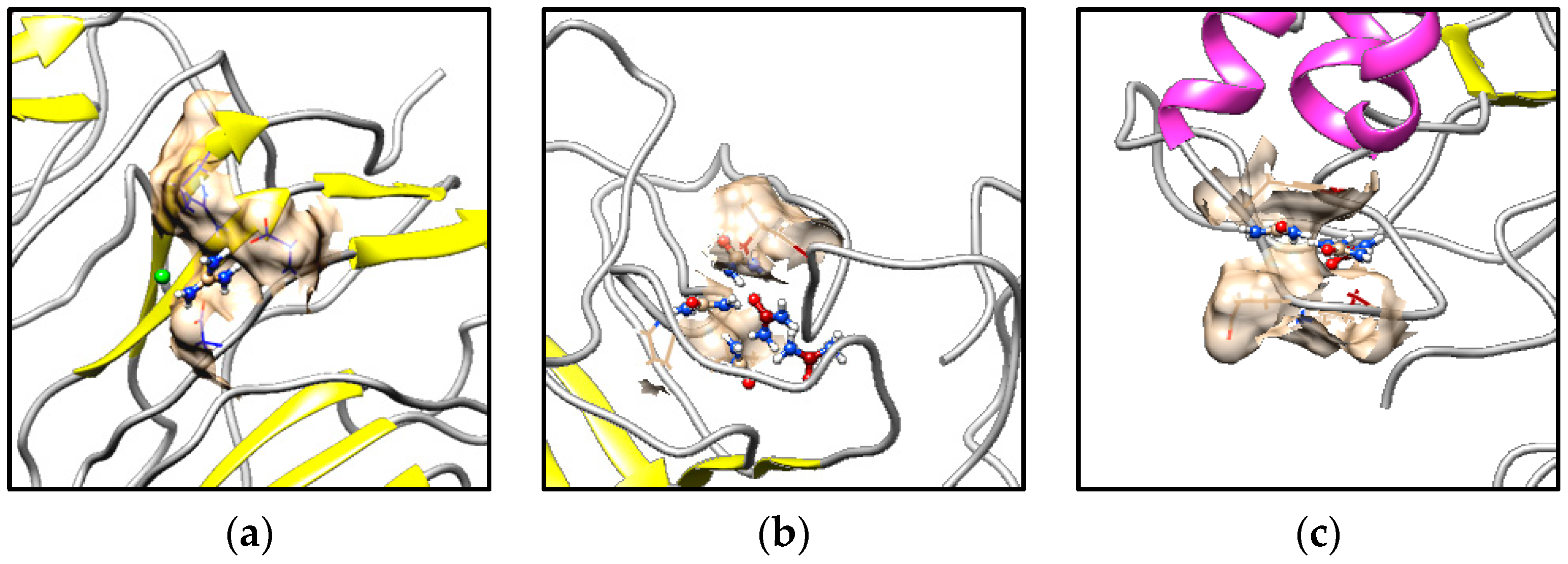
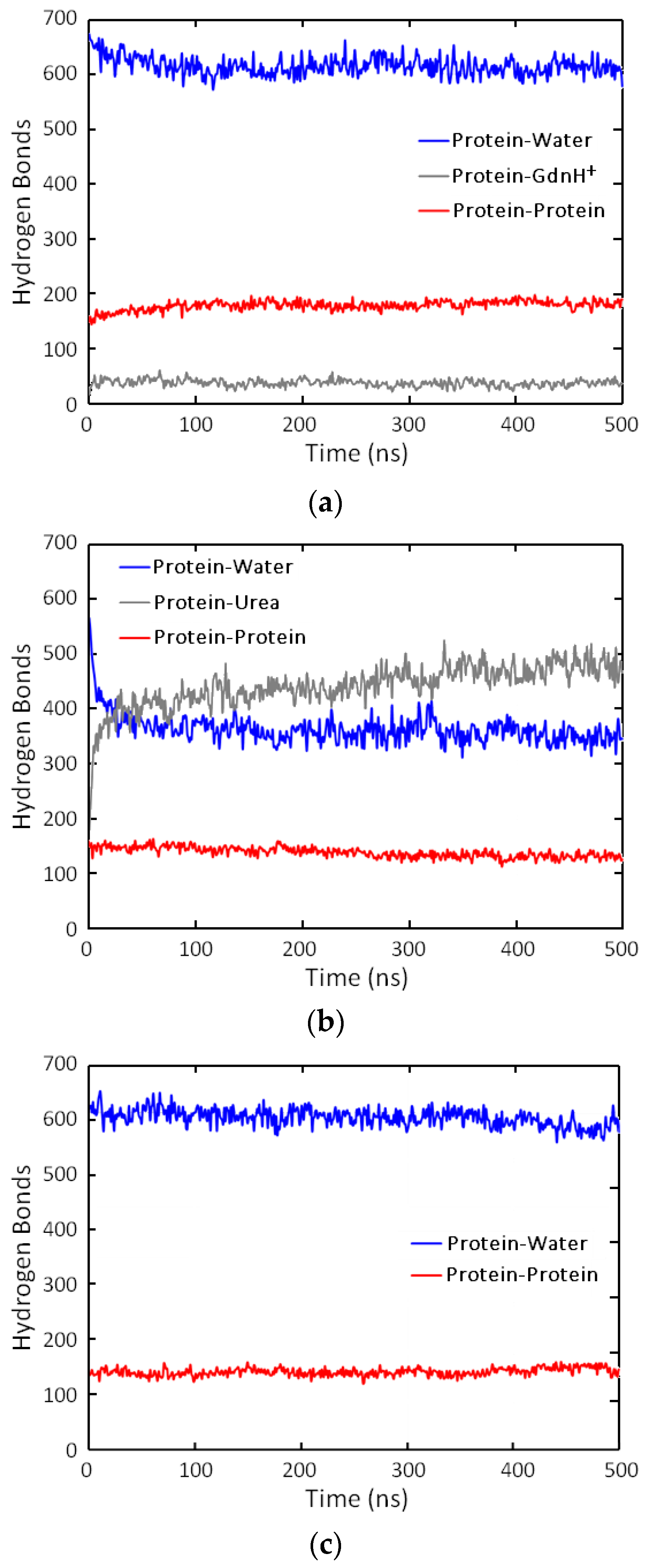
3.3. Hydrogen Bonding Under Denaturant Conditions

Given that hydrogen-bonding trends emerged as a key factor influencing the dynamics of protein–solvent interactions, we further examined the overall hydrogen-bonding and hydration behavior across the three simulated systems. Figure 7a–c present the time evolution of hydrogen-bonding interactions observed over the course of the 500 ns MD production simulations for the PrPC/PrPSc complex under GdnHCl, urea, and denaturant-free conditions, respectively. Each panel displays the total number of protein-protein, protein-water, and, where applicable, protein-denaturant hydrogen bonds as a function of simulation time.
In the GdnHCl-containing system (Figure 7a), protein-water interactions remain predominant throughout, while a modest but persistent number of HBs are formed between the protein and GdnH+ ions. These interactions are consistent with the localized binding observed at the interaction site illustrated in Figure 6a, where GdnH+ engages acidic residues via salt bridges but also forms hydrogen bonds, contributing to the stabilization of β-strands in PrPSc, which are themselves maintained by intra-protein hydrogen bonding.
In contrast, the urea-containing system (Figure 7b) shows a markedly different behavior. Protein-urea hydrogen bonding develops rapidly during the equilibration phase, gradually replacing protein-water interactions and eventually dominating. This shift reflects the accumulation of urea molecules in the solvation shell and the replacement of water by a hydrogen-bonding network formed with urea, as the structural observations illustrated by Figure 6b,c suggest.
In the control system without denaturants (Figure 7c), the number of protein-water and protein-protein hydrogen bonds remains steady, with consistently high protein-water interactions reflecting a relatively stable hydration environment. This behavior supports the moderate structural stability and flexibility presented earlier in Figure 2e,f and Figure 3, serving as a baseline for comparison with the two denaturant-containing systems. Together, these data further highlight the distinct effects that urea and guanidine hydrochloride exert on both protein hydration and internal hydrogen bonding.
Figure 7. Time evolution of hydrogen bonding during the 500 ns MD simulations for the PrPC/PrPSc complex in solutions containing (a) GdnHCl, (b) urea, and (c) no denaturants. The plots show the number of protein-water (blue), protein-protein (red), and protein-denaturant (gray) hydrogen bonds as a function of simulation time.
Processes 13 02151 g007

4. Discussion

This study investigated the differential effects of two widely used denaturants, guanidine hydrochloride and urea, on the conformational and dynamical stability of a PrPC/PrPSc complex representing a white-tailed deer cellular prion protein, PrPC, interfacing with a matching fibrillar 4RβS seed, PrPSc. Using 500 ns all-atom MD simulations, we conducted a comparative analysis of the denaturants’ impact on secondary structure retention, PrPC/PrPSc interfacial stability, collective dynamics behavior, and hydrogen-bonding evolution.
Over the timescale addressed in these simulations, the native module, PrPC, largely preserved its α-helical and β-sheet content (Figure 2 and Figure S1) in all the systems considered. Interestingly, according to Figure 2 and Table 1, the presence of GdnHCl or urea did not disrupt α-helices relative to the denaturant-free system. The overall stability of the folded PrPC domain, spanning from β-strand S1 to α-helix H3, is further supported by the consistency of the corresponding ECD backbone flexibility profiles and pair correlation maps across the three systems, as seen in Figure 3 and Figure 4.
The greatest differential response to the presence of denaturants was observed for the β-structure of the fibrillar PrPSc seed, as evident from Figure 2 and Figure S1. PrPSc β-sheet content was pronouncedly expanded in the GdnHCl-containing system, aligning well with published reports. It has been well-established experimentally that PrPC converts to PrPSc in the presence of GdnHCl [16,18,22], and this effect has been extensively used to trigger the PrP conversion in vitro [17,21]. In contrast to GdnHCl, the presence of urea induced partial dissolution of β-structure in PrPSc. Experimentally, this is supported indirectly by reports indicating the presence of misfolded PrP intermediates, rather than mature amyloid fibrils, in experiments on exposure of PrPC to urea in vitro [19,20], unless extensive exposures at acidic conditions were involved [55]. Published MD simulation studies, although primarily focused on other proteins, are broadly consistent with our findings in highlighting the distinct denaturing preferences of GdnHCl and urea, with the latter more strongly associated with β-structure disruption [25,26,27,28]. To our knowledge, however, stabilizing effects of GdnHCl on β-structure have not been reported in previous MD simulations.
The PrPC–PrPSc interface, modeled here by threading the N-terminal segment of PrPC onto the C-terminal rung of the 4RβS PrPSc seed, serves as a minimal yet informative representation of early conformational conversion. It contributes to addressing a gap in the characterization of early misfolding events under denaturing conditions. The divergent behavior observed at the PrPC–PrPSc interface is particularly revealing. The preservation of interfacial alignment in the GdnHCl system and its partial disruption in the urea simulation (Figure 2 and Figure S1), further supported by the ECD backbone flexibility and pair correlation analyses (Figure 3 and Figure 4), suggest that stabilization or destabilization at the boundary between native and fibrillar forms may influence the trajectory of conformational templating during prion conversion.
Another outcome of this study is the demonstration of long-range dynamical coupling across the PrPC–PrPSc interface. This effect is especially pronounced under GdnHCl but is also present in the denaturant-free system and, to a lesser extent, in the urea-containing system (see Figure 4). Such behavior appears to reflect the modular architecture of the PrPC/PrPSc complex, where relatively rigid structural segments are connected by flexible loops. The transmission of backbone tensions, steric interactions, and other mechanical constraints is expected to underlie communication between remote regions.
Notably, long-range dynamical coupling also extends to transiently associated guanidinium ions and urea molecules, some of which exhibited such coupling with remote structural elements of both PrPC and PrPSc. Examination of several strongly coupled solute entities, as identified by the ECD pair correlation analysis (see Figure 5 and Figure 6), revealed distinctions in the local interactions of the two solutes. A guanidinium cation transiently bound near acidic residues D147 and E149 in the PrPSc β-strand region formed both salt bridges and hydrogen bonds with these side chains, while also displaying dynamical coupling with adjacent β-strands and distal regions of the PrPC/PrPSc complex. In contrast, two urea molecules—one bound near L128 in PrPSc, the other near E199–N200 in PrPC—engaged in extensive hydrogen bonding within their respective binding pockets. These interactions involved PrP backbone and side-chain atoms as well as neighboring urea molecules, giving rise to an interconnected hydrogen-bonded network. Both urea molecules exhibited dynamical coupling with both nearby and remote regions of the PrPC/PrPSc complex.
The observed patterns of local binding for GdnH+ and urea are broadly consistent with previous molecular dynamics studies. Guanidinium ions have been shown to preferentially associate with negatively charged side chains, largely through electrostatic interactions [25,30,31]. Similarly, urea is known to accumulate within protein solvation shells and form multiple hydrogen bonds with both backbone and side-chain atoms, effectively displacing water and weakening native hydrogen-bonding networks [26,28,29,30]. These trends match the binding behaviors observed in our simulations. In contrast, the emergence of long-range dynamical coupling between these solute molecules and distal regions of the protein appears to be novel, especially in the context of transient solute–protein interactions in large, β-rich protein assemblies. This finding highlights the utility of the ECD approach in capturing non-local dynamical effects that may otherwise remain undetected in conventional structural analyses.
Finally, the hydrogen-bonding trends observed in our simulations (Figure 7) align well with existing computational findings. In the GdnHCl-containing system, the predominance of protein-water hydrogen bonding alongside a limited number of persistent HBs involving GdnH+ is consistent with earlier MD studies showing that guanidinium ions tend to engage selectively with charged side chains without broadly perturbing hydration shells [28,31]. Conversely, the extensive formation of protein-urea hydrogen bonds, accompanied by a reduction in water interactions and destabilization of β-structure, is in agreement with established models of urea action based on direct competition for backbone hydrogen-bonding sites [29,31,32]. This consistency confirms the validity of the structural and dynamical interpretations described above.
These findings carry implications for the molecular understanding of prion propagation in CWD and related disorders. In particular, the selective effects of urea and GdnHCl may serve as a model for understanding how different physicochemical microenvironments—ionic versus polar non-ionic—shape the stability and evolution of infectious aggregates. The ability of GdnHCl to preserve interfacial alignment and stabilize amyloid-like β-structure suggests a possible structural basis for its experimentally observed capacity to accelerate the prion conversion and may contribute to seeding specificity by maintaining compatible conformational interfaces. Meanwhile, the selective destabilizing action of urea may explain its tendency to promote intermediate states and nonproductive aggregation. These distinctions could inform experimental protocols aimed at probing strain-specific misfolding pathways or screening small molecules designed to stabilize native conformations. Although our simulations focused on a specific PrP sequence and PrPSc seed model, the general consistency of urea and GdnHCl effects across diverse protein systems suggests that the structural responses identified here are likely relevant to a broad range of PrP sequence variants, strains, and alternative PrPSc conformational models. In particular, our results provide molecular-level insights into in vitro strategies that employ varying proportions of guanidine hydrochloride-urea mixtures to modulate fibril growth, isolate conversion intermediates, or investigate the effect of denaturants on aggregation efficiency [16,17]. Notably, the observed long-range dynamical coupling across the PrPC/PrPSc complex suggests that structural perturbations initiated at localized sites, such as transient solute interactions, may propagate through an allosteric-like mechanism, providing a basis for coordinated responses at distant regions of amyloid-forming supramolecular assemblies.
Due to the inherent timescale limitations of all-atom MD simulations, an aspect not fully captured in this study is the unfolding behavior of the globular domain of PrPC, which contains three α-helices and two short β-strands. This region remained largely stable throughout the simulations, likely reflecting both its intrinsic structural resilience and the limited duration accessible to atomistic modeling. Future studies may explore longer simulation timescales to capture more advanced stages of structural transitions. In addition, the effects of alternative denaturants, cosolvents, PrP polymorphisms, or PrPSc models on the balance between folded and misfolded states could be examined. More complex conditions, such as membrane anchoring of PrPC or the presence of selected cofactors, may uncover new interaction modes that affect misfolding and conformational templating.

5. Conclusions

This study presents a comparative molecular-scale investigation into the effects of two common denaturants, guanidine hydrochloride and urea, on the conformational and dynamical stability of a composite PrPC/PrPSc system representing white-tailed deer prion protein interacting with matching fibrillar seed. Using all-atom molecular dynamics simulations in combination with essential collective dynamics analysis, we show that the presence of GdnHCl may enhance β-structure retention and preserve interfacial alignment between native and misfolded domains, while urea promotes β-structure disruption and interfacial destabilization. These findings complement prior experimental findings and published MD simulation results and underscore the distinct physicochemical mechanisms through which ionic and non-ionic polar denaturants modulate protein misfolding.
Beyond local structural impacts, the simulations reveal long-range dynamical coupling across the PrPC/PrPSc interface and between transiently bound solutes and distal protein regions—a behavior not previously reported in this context. These observations support a model in which mechanical constraints, steric effects, and modular connectivity within the protein enable propagation of dynamical perturbations across structurally distant regions. This suggests that transient solute interactions may influence remote structural responses through mechanically or sterically mediated pathways.
Overall, the results contribute to a better understanding of early misfolding events and prion conversion under denaturant conditions. They also provide molecular-scale insights relevant to in vitro strategies employing mixed GdnHCl-urea solutions to probe interfacial templating, conformational stability, and conversion mechanisms in prion misfolding. The present framework may serve as a reference point for future efforts to compare solvent-induced effects across distinct PrPSc structural templates and to examine how additional factors—including cofactors, disease-associated polymorphisms, and other modulators—influence early-stage misfolding and strain-specific conversion pathways.

Supplementary Materials

The following supporting information can be downloaded at https://www.mdpi.com/article/10.3390/pr13072151/s1. Figure S1: Snapshots of the PrPC/PrPSc system after 200 ns, 300 ns, and 400 ns MD simulations in water solutions with and without denaturant additives; Figure S2: RMSD as a function of time for the PrPC/PrPSc system; Figure S3: RMSD as a function of time for segments PrPC 93–119, PrPC 120–233, and PrPSc 93–233 of the PrPC/PrPSc; Figure S4: ECD pair correlation map for PrP Cα atoms in the PrPC/PrPSc complex and urea molecules, obtained from the last 20 ns of the MD simulation in urea-containing solution; Figure S5: Overview of the PrPC/PrPSc complexes showing the guanidinium cation (a) and urea molecules (b,c) attachment sites presented in Figure 6a and Figure 6b,c, respectively.

Author Contributions

Conceptualization, M.S.; methodology, L.D., M.W. and M.S.; software, M.S.; formal analysis, L.D. and M.W.; investigation, L.D., M.W. and M.S.; resources, M.S.; data curation, L.D. and M.W.; writing—original draft preparation, L.D. and M.S.; writing—review, and editing, M.S. and M.W.; visualization, L.D. and M.W.; supervision, M.S.; project administration, M.S.; funding acquisition, M.S. All authors have read and agreed to the published version of the manuscript.

Funding

This research was funded by the ALBERTA INNOVATES grant numbers 201700016 and 222300803.

Institutional Review Board Statement

Not applicable.

Data Availability Statement

The original contributions presented in this study are included in the article and Supplementary Material. Further inquiries can be directed to the corresponding author.

Acknowledgments

The authors thank Holger Wille for helpful discussions.

Conflicts of Interest

The authors declare no conflicts of interest.

Abbreviations

The following abbreviations are used in this manuscript:
PrPPrion protein
TSETransmissible spongiform encephalopathy
GdnHClGuanidine hydrochloride
GdnH+Guanidinium cation
MDMolecular dynamics
WTDWhite-tailed deer
CWDChronic wasting disease
4RβSFour-rung β-solenoid
EMElectron microscopy
cryo-EMElectron cryomicroscopy
HET-sHeterokaryon incompatibility protein s
PIRIBSParallel-in-register β-sheets
NMRNuclear magnetic resonance
ECDEssential collective dynamics
PCAPrincipal component analysis
dECD pair correlation descriptor (Equation (2))
F B ECD backbone flexibility descriptor (Equation (3))
RMSDRoot-mean-square deviation
SBSalt bridge
HBHydrogen bond

Appendix A

In the ECD framework [47,48,49,50,51,52,53], each atom in the system under analysis is represented by an “atom-image” vector r i , constructed from the components of the essential collective coordinates (see Equation (1)). These essential coordinates correspond to the K principal eigenvectors of the covariance matrix, obtained via PCA from the atomic displacements in the MD simulation. Each eigenvector spans the full configuration space of atomic degrees of freedom, comprising three Cartesian coordinates (x, y, z) per atom. In ECD, the directional cosines of the K principal eigenvectors along the Cartesian x-, y-, and z-axes for each atom i are concatenated into the corresponding atom-image vector r i in the 3K-dimensional essential coordinate space. Since all PCA eigenvectors are normalized by definition, ECD descriptors such as pairwise atom-image distances (see Equation (2)) are directly comparable within a given set of N atom images derived from a single MD trajectory.
Comparison of ECD descriptors across different MD trajectories is generally straightforward when the systems contain similar numbers of atoms and types of molecules, such that the overall structure of the essential coordinate space remains comparable. For example, in Figure 3, backbone flexibility profiles were compared across different MD simulations in which only the PrPC/PrPSc construct was analyzed. In this case, the construct was represented by 282 Cα atoms, and essential motion was equipartitioned among their corresponding degrees of freedom [48].
When additional molecular entities are included in the ECD analysis, the total essential motion becomes statistically partitioned across all degrees of freedom in the system. For example, in Figure 4a, the pair correlation map for the GdnHCl-containing system includes 380 guanidinium cations, whereas in Figure 4b and Figure S1, the corresponding map for the urea-containing system includes 708 urea molecules, in addition to the PrPC/PrPSc construct. Although the composition and physical behavior of the protein remain unchanged by the inclusion of the solute species into the analysis, the relative contribution of the protein to the essential modes is reduced due to the expansion of the overall dynamical system. A further consideration arises when the added entities consist of a large number of freely diffusing, weakly interacting solutes, such as guanidinium cations or urea molecules, each treated as a single dynamical unit. Each such solute contributes three translational degrees of freedom, and their motions are largely uncorrelated with each other and with the protein. In this context, when deriving equipartition-based scaling factors, treating the protein at the full resolution of 282 Cα atoms disproportionately inflates its contribution to the essential dynamics, since many of the atomic motions are intercorrelated. To maintain consistency, the protein must be represented at a decreased resolution that reflects its number of loosely coupled dynamical units. While this redundancy could be formally quantified by evaluating the effective dimensionality of protein contribution to the covariance matrix, such statistical-mechanical analysis is beyond the scope of this work. Here, we adopt a semi-empirical approach based on the ECD-derived backbone flexibility profiles shown in Figure 3. Specifically, we estimate the number of quasi-independent domains in the PrPC/PrPSc construct by counting the number of minima in the flexibility profile, which typically correspond to the most rigid backbone regions separated by more flexible loops. As these minima are broadly consistent with the expected pattern of secondary structure, the number of such segments offers a reasonable approximation of the number of dynamical units required. Based on this criterion, we have adopted NP =14 as the number of protein subunits for the purpose of scaling.
Each subunit is treated as contributing three degrees of freedom, yielding 3NP total for the protein, balanced against 3NS degrees of freedom from the solutes. Although the dimensionality of the essential coordinate space remains fixed at 3K, the variance is now statistically equipartitioned [48] across the combined 3 N P + 3 N S degrees of freedom. Under this assumption, the fraction of essential variance attributable to the protein is reduced by a factor of approximately N P / ( N P + N S ) . The squared distances between protein atom images are then scaled proportionally:
r i r j 2 = N P N P + N S r i r j r e f 2 ,      i , j = 1,2 , , N ,
where | r i r j | r e f 2 are the corresponding distances in the reference system without added solutes. While this contraction does not influence the relative distribution of intra-image distances within each simulation, inter-system comparisons are facilitated by uniformly rescaling the pair correlation descriptor, d i j = r i r j , by the inverse of the square root contraction factor in denaturant-containing analyses:
d i j , n o r m a l i z e d = d i j 1 + N S / N P ,      i , j = 1,2 , , N .
Here, NS = 380 for guanidinium-including analyses and NS =708 for urea-including analyses, NP = 14, and N = 282 is the number of Cα atoms representing the PrPC/PrPSc construct.
It should be noted that the effectiveness of the present normalization approach relies on the assumption that most of the solute ions or molecules are weakly interacting with each other and with the protein. While full dynamical decoupling is not required, statistical partitioning of essential motion depends on the assumption that the vast majority of the solute entities contribute largely uncorrelated translational motion. In the present simulations, a limited number of denaturant ions or molecules transiently bind to the protein, and in some cases these localized interactions exert non-local effects on protein dynamics. However, such strongly coupled solutes represent only a small fraction of the total NS, and their impact does not invalidate the global statistical partitioning assumption.
In more complex cases involving multiple structured macromolecules, stronger dynamical coupling may lead to deviations from ideal scaling. In such cases, the contraction of protein atom-image distances may be weaker than predicted by simple equipartition-based models. This effect could be accommodated by introducing a correction to the nominal number of contributing units, replacing NS with an effective count 1 δ N S where δ < 1 reflects partial dynamical correlation between added entities. The corrected normalization factor then becomes 1 + 1 δ N S / N P . While the precise form and magnitude of such corrections remain to be established, the statistical-mechanical rationale underlying the present normalization framework remains general and potentially adaptable to diverse multicomponent systems.

References

  1. Louros, N.; Schymkowitz, J.; Rousseau, F. Mechanisms and pathology of protein misfolding and aggregation. Nat. Rev. Mol. Cell Biol. 2023, 24, 912–933. [Google Scholar] [CrossRef] [PubMed]
  2. Chiti, F.; Dobson, C.M. Protein Misfolding, Amyloid Formation, and Human Disease: A Summary of Progress Over the Last Decade. Annu. Rev. Biochem. 2017, 86, 27–68. [Google Scholar] [CrossRef]
  3. Soto, C.; Pritzkow, S. Protein misfolding, aggregation, and conformational strains in neurodegenerative diseases. Nat. Neurosci. 2018, 21, 1332–1340. [Google Scholar] [CrossRef] [PubMed]
  4. Prusiner, S.B. Prions. Proc. Natl. Acad. Sci. USA 1998, 95, 13363–13383. [Google Scholar] [CrossRef]
  5. Goedert, M. Alzheimer’s and Parkinson’s diseases: The prion concept in relation to assembled Aβ, tau, and α-synuclein. Science 2015, 349, 1255555. [Google Scholar] [CrossRef] [PubMed]
  6. Casey, C.; Sleator, R.D. Prions: Structure, function, evolution, and disease. Arch. Microbiol. 2025, 207, 1. [Google Scholar] [CrossRef]
  7. Orge, L.; Lima, C.; Machado, C.; Tavares, P.; Mendonça, P.; Carvalho, P.; Silva, J.; de Lurdes Pinto, M.; Bastos, E.; Pereira, J.C.; et al. Neuropathology of Animal Prion Diseases. Biomolecules 2021, 11, 466. [Google Scholar] [CrossRef]
  8. Silva, C.J. Chronic Wasting Disease (CWD) in Cervids and the Consequences of a Mutable Protein Conformation. ACS Omega 2022, 7, 12474–12492. [Google Scholar] [CrossRef]
  9. Zerr, I.; Ladogana, A.; Mead, S.; Hermann, P.; Forloni, G.; Appleby, B.S. Creutzfeldt-Jakob disease and other prion diseases. Nat. Rev. Dis. Primers 2024, 10, 14. [Google Scholar] [CrossRef]
  10. Shoup, D.; Priola, S.A. Cell biology of prion strains in vivo and in vitro. Cell Tissue Res. 2023, 392, 269–283. [Google Scholar] [CrossRef]
  11. Alves Conceição, C.; Assis de Lemos, G.; Barros, C.A.; Vieira, T.C. What is the role of lipids in prion conversion and disease? Front. Mol. Neurosci. 2023, 15, 1032541. [Google Scholar] [CrossRef] [PubMed]
  12. Aguilar-Calvo, P.; Callender, J.A.; Sigurdson, C.J. Short and sweet: How glycans impact prion conversion, cofactor interactions, and cross-species transmission. PLoS Pathog. 2021, 17, e1009123. [Google Scholar] [CrossRef] [PubMed]
  13. Shafiq, M.; Da Vela, S.; Amin, L.; Younas, N.; Harris, D.A.; Zerr, I.; Altmeppen, H.C.; Svergun, D.; Glatzel, M. The prion protein and its ligands: Insights into structure-function relationships. Biochim. Biophys. Acta 2022, 1869, 119240. [Google Scholar] [CrossRef]
  14. Surewicz, W.K.; Jones, E.M.; Apetri, A.C. The emerging principles of mammalian prion propagation and transmissibility barriers: Insight from studies in vitro. Acc. Chem. Res. 2006, 39, 654–662. [Google Scholar] [CrossRef]
  15. Spagnolli, G.; Requena, J.R.; Biasini, E. Understanding prion structure and conversion. Prog. Mol. Biol. Transl. Sci. 2020, 175, 19–30. [Google Scholar] [PubMed]
  16. Bocharova, O.V.; Breydo, L.; Parfenov, A.S.; Salnikov, V.V.; Baskakov, I.V. In vitro conversion of full-length mammalian prion protein produces amyloid form with physical properties of PrPSc. J. Mol. Biol. 2005, 346, 645–659. [Google Scholar] [CrossRef]
  17. Fridmanis, J.; Toleikis, Z.; Sneideris, T.; Ziaunys, M.; Bobrovs, R.; Smirnovas, V.; Jaudzems, K. Aggregation Condition–Structure Relationship of Mouse Prion Protein Fibrils. Int. J. Mol. Sci. 2021, 22, 9635. [Google Scholar] [CrossRef]
  18. Hosszu, L.L.P.; Wells, M.A.; Jackson, G.S.; Jones, S.; Batchelor, M.; Clarke, A.R.; Craven, C.J.; Waltho, J.P.; Collinge, J. Definable equilibrium states in the folding of human prion protein. Biochemistry 2005, 44, 16649–16657. [Google Scholar] [CrossRef] [PubMed]
  19. Julien, O.; Chatterjee, S.; Thiessen, A.; Graether, S.P.; Sykes, B.D. Differential stability of the bovine prion protein upon urea unfolding. Prot. Sci. 2009, 18, 2172–2182. [Google Scholar] [CrossRef]
  20. Julien, O.; Chatterjee, S.; Bjorndahl, T.C.; Sweeting, B.; Acharya, S.; Semenchenko, V.; Chakrabartty, A.; Pai, E.F.; Wishart, D.S.; Sykes, B.D.; et al. Relative and regional stabilities of the hamster, mouse, rabbit, and bovine prion proteins toward urea unfolding assessed by nuclear magnetic resonance and circular dichroism spectroscopies. Biochemistry 2011, 50, 7536–7545. [Google Scholar] [CrossRef]
  21. Milto, K.; Michailova, K.; Smirnovas, V. Elongation of mouse prion protein amyloid-like fibrils: Effect of temperature and denaturant concentration. PLoS ONE 2014, 9, e94469. [Google Scholar] [CrossRef] [PubMed]
  22. Swietnicki, W.; Morillas, M.; Chen, S.G.; Gambetti, P.; Surewicz, W.K. Aggregation and fibrillization of the recombinant human prion protein huPrP90–231. Biochemistry 2000, 39, 424–431. [Google Scholar] [CrossRef] [PubMed]
  23. Polano, M.; Bek, A.; Benetti, F.; Lazzarino, M.; Legname, G. Structural insights into alternate aggregated prion protein forms. J. Mol. Biol. 2009, 393, 1033–1042. [Google Scholar] [CrossRef] [PubMed]
  24. Paladino, A.; Vitagliano, L.; Graziano, G. The action of chemical denaturants: From globular to intrinsically disordered proteins. Biology 2023, 12, 754. [Google Scholar] [CrossRef]
  25. Camilloni, C.; Tiana, G. Urea and guanidinium chloride denature protein L in different ways in molecular dynamics simulations. Biophys. J. 2008, 94, 4654–4661. [Google Scholar] [CrossRef]
  26. Meloni, R.; Camilloni CTiana, G. Sampling the Denatured State of Polypeptides in Water, Urea, and Guanidine Chloride to Strict Equilibrium Conditions with the Help of Massively Parallel Computers. J. Chem. Theory Comput. 2014, 10, 846–854. [Google Scholar] [CrossRef]
  27. Parui, S.; Jana, B. Relative Solvent Exposure of the Alpha-Helix and Beta-Sheet in Water Determines the Initial Stages of Urea and Guanidinium Chloride-Induced Denaturation of Alpha/Beta Proteins. J. Phys. Chem. B 2019, 123, 8889–8900. [Google Scholar] [CrossRef]
  28. Heyda, J.; Kožíšek, M.; Bednárová, L.; Thompson, G.; Konvalinka, J.; Vondrášek, J.; Jungwirth, P. Urea and Guanidinium Induced Denaturation of a Trp-Cage Miniprotein. J. Phys. Chem. B 2011, 115, 8910–8924. [Google Scholar] [CrossRef]
  29. Meloni, R.; Tiana, G. Thermodynamic and structural effect of urea and guanidine chloride on the helical and on a hairpin fragment of GB1 from molecular simulations. Proteins 2017, 85, 753–763. [Google Scholar] [CrossRef]
  30. Mandal, M.; Mukhopadhyay, C. Microsecond molecular dynamics simulation of guanidinium chloride induced unfolding of ubiquitin. Phys. Chem. Chem. Phys. 2014, 16, 21706–21716. [Google Scholar] [CrossRef]
  31. Ganguly, P.; Shea, J.-E. Distinct and Nonadditive Effects of Urea and Guanidinium Chloride on Peptide Solvation. J. Phys. Chem. Lett. 2019, 10, 7406–7413. [Google Scholar] [CrossRef] [PubMed]
  32. Das, A.; Mukhopadhyay, C. Atomistic mechanism of protein denaturation by urea. J. Phys. Chem. B 2008, 112, 7903–7908. [Google Scholar] [CrossRef]
  33. Eberini, I.; Emerson, A.; Sensi, C.; Ragona, L.; Ricchiuto, P.; Pedretti, A.; Gianazza, E.; Tramontano, A. Simulation of urea-induced protein unfolding: A lesson from bovine β-lactoglobulin. J. Mol. Graph. Model. 2011, 30, 24–30. [Google Scholar] [CrossRef]
  34. Rg, C.; Tallon, A.; Latch, E.K. Chronic Wasting Disease Research in North America: A systematic review highlighting species-wise and interdisciplinary research trends. Prion 2025, 19, 1–16. [Google Scholar] [CrossRef]
  35. Spagnolli, G.; Rigolli, M.; Orioli, S.; Sevillano, A.M.; Faccioli, P.; Wille, H.; Biasini, E.; Requena, J.R. Full atomistic model of prion structure and conversion. PLoS Pathog. 2019, 15, e1007864. [Google Scholar] [CrossRef]
  36. Wille, H.; Bian, W.; McDonald, M.; Kendall, A.; Colby, D.W.; Bloch, L.; Ollesch, J.; Borovinskiy, A.L.; Cohen, F.E.; Prusiner, S.B.; et al. Natural and synthetic prion structure from X-ray fiber diffraction. Proc. Natl. Acad. Sci. USA 2009, 106, 16990–16995. [Google Scholar] [CrossRef] [PubMed]
  37. Kamali-Jamil, R.; Vázquez-Fernández, E.; Tancowny, B.; Rathod, V.; Amidian, S.; Wang, X.; Tang, X.; Fang, A.; Senatore, A.; Hornemann, S.; et al. The ultrastructure of infectious L-type bovine spongiform encephalopathy prions constrains molecular models. PLoS Pathog. 2021, 17, e1009628. [Google Scholar] [CrossRef] [PubMed]
  38. Wasmer, C.; Lange, A.; Van Melckebeke, H.; Siemer, A.B.; Riek, R.; Meier, B.H. Amyloid fibrils of the HET-s (218–289) prion form a beta solenoid with a triangular hydrophobic core. Science 2008, 319, 1523–1526. [Google Scholar] [CrossRef]
  39. Manka, S.W.; Wenborn, A.; Collinge, J.; Wadsworth, J.D.F. Prion strains viewed through the lens of cryo-EM. Cell Tissue Res. 2023, 392, 167–178. [Google Scholar] [CrossRef]
  40. Ribes, J.M.; Patel, M.P.; Halim, H.A.; Berretta, A.; Tooze, S.A.; Klöhn, P.-C. Prion protein conversion at two distinct cellular sites precedes fibrillisation. Nat. Commun. 2023, 14, 8354. [Google Scholar] [CrossRef]
  41. BIOVIA Discovery Studio Visualizer, v. 19.1.0.1828; Dassault Systèmes: San Diego, CA, USA. Available online: https://www.3ds.com/products/biovia/discovery-studio (accessed on 17 July 2019).
  42. Waterhouse, A.; Bertoni, M.; Bienert, S.; Studer, G.; Tauriello, G.; Gumienny, R.; Heer, F.T.; de Beer, T.A.P.; Rempfer, C.; Bordoli, L.; et al. SWISS-MODEL: Homology modelling of protein structures and complexes. Nucleic Acids Res. 2018, 46, W296–W303. [Google Scholar] [CrossRef]
  43. Baral, P.K.; Swayampakula, M.; Aguzzi, A.; James, M.N. X-ray structural and molecular dynamical studies of the globular domains of cow, deer, elk and Syrian hamster prion proteins. J. Struct. Biol. 2015, 192, 37–47. [Google Scholar] [CrossRef]
  44. Berendsen, H.J.C.; van der Spoel, D.; van Drunen, R. GROMACS: A message-passing parallel molecular dynamics implementation. Comput. Phys. Commun. 1995, 91, 43–56. [Google Scholar] [CrossRef]
  45. Humphrey, W.; Dalke, A.; Schulten, K. VMD—Visual Molecular Dynamics. J. Mol. Graph. 1996, 14, 33–38. [Google Scholar] [CrossRef]
  46. Berendsen, H.J.C.; Postma, J.P.M.; van Gunsteren, W.F.; DiNola, A.; Haak, J.R. Molecular-Dynamics with Coupling to an External Bath. J. Chem. Phys. 1984, 81, 3684–3690. [Google Scholar] [CrossRef]
  47. Stepanova, M. Dynamics of essential collective motions in proteins: Theory. Phys. Rev. E. 2007, 76, 051918. [Google Scholar] [CrossRef] [PubMed]
  48. Potapov, A.; Stepanova, M. Conformational modes in biomolecules: Dynamics and approximate invariance. Phys. Rev. E. 2012, 85 Pt 1, R02090. [Google Scholar] [CrossRef] [PubMed]
  49. Dorosh, L.; Stepanova, M. Probing oligomerization of amyloid beta peptide in silico. Mol. Biosyst. 2017, 13, 165–182. [Google Scholar] [CrossRef]
  50. Issack, B.B.; Berjanskii, M.; Wishart, D.S.; Stepanova, M. Exploring the essential collective dynamics of interacting proteins: Application to prion protein dimers. Proteins Struct. Funct. Bioinforma. 2012, 80, 1847–1865. [Google Scholar] [CrossRef]
  51. Wu, M.; Wille, H.; Stepanova, M. Essential collective dynamics analysis reveals nonlocal interactions of alpha-synuclein38-95 monomers with fibrillar seeds. J. Chem. Phys. 2022, 157, 235101. [Google Scholar] [CrossRef]
  52. Blinov, N.; Berjanskii, M.; Wishart, D.S.; Stepanova, M. Structural domains and main-chain flexibility in prion proteins. Biochemistry 2009, 48, 1488–1497. [Google Scholar] [CrossRef] [PubMed]
  53. Santo, K.P.; Berjanskii, M.; Wishart, D.S.; Stepanova, M. Comparative analysis of essential collective dynamics and NMR-derived flexibility profiles in evolutionarily diverse prion proteins. Prion 2011, 5, 188–200. [Google Scholar] [CrossRef] [PubMed]
  54. Mercer, R.C.C.; Daude, N.; Dorosh, L.; Fu, Z.-L.; Mays, C.E.; Gapeshina, H.; Wohlgemuth, S.L.; Acevedo-Morantes, C.Y.; Yang, J.; Cashman, N.R.; et al. A novel Gerstmann-Sträussler-Scheinker disease mutation defines a precursor for amyloidogenic 8 kDa PrP fragments and reveals N-terminal structural changes shared by other GSS alleles. PLoS Pathog. 2018, 14, e1006826. [Google Scholar] [CrossRef] [PubMed]
  55. Morillas, M.; Vanik, D.L.; Surewicz, W.K. On the mechanism of α-helix to β-sheet transition in the recombinant prion protein. Biochemistry 2001, 40, 6982–6987. [Google Scholar] [CrossRef]
Table 1. Percentage of α-helical and β-sheet content in the PrPC/PrPSc composite system in average over the last 250 ns of the 500 ns MD simulations.
Denaturantα-Helical Content%β-Sheet Content%
GdnHCl20.4 ± 0.119.5 ± 0.1
Urea16.7 ± 0.110.0 ± 0.1
None16.6 ± 0.113.4 ± 0.1
Disclaimer/Publisher’s Note: The statements, opinions and data contained in all publications are solely those of the individual author(s) and contributor(s) and not of MDPI and/or the editor(s). MDPI and/or the editor(s) disclaim responsibility for any injury to people or property resulting from any ideas, methods, instructions or products referred to in the content.

