Article

Optimization of Immobilization, Characterization, and Environmental Applications of Laccases from Pycnoporus sanguineus UEM-20

by
Vinícius Mateus Salvatori Cheute
1,
Emanueli Backes
2,
Vanesa de Oliveira Pateis
1,
Verci Alves de Oliveira Junior
3,
Thaís Marques Uber
1,
José Rivaldo dos Santos Filho
1,
Luís Felipe Oliva dos Santos
3,
Rafael Castoldi
1,
Cristina Giatti Marques de Souza
1,
Julio Cesar Polonio
3,
Alex Graça Contato
4,*,
Adelar Bracht
1 and
Rosane Marina Peralta
1,2,3,*
1
Post-Graduate Program in Biochemistry, State University of Maringa, Maringa 87020-900, PR, Brazil
2
Post-Graduate Program in Food Science, State University of Maringa, Maringa 87020-900, PR, Brazil
3
Post-Graduate Program in Environmental Biotechnology, State University of Maringa, Maringa 87020-900, PR, Brazil
4
Chemistry Institute, Federal University of Rio de Janeiro, Rio de Janeiro 21941-598, RJ, Brazil
*
Authors to whom correspondence should be addressed.
Processes 2025, 13(6), 1800; https://doi.org/10.3390/pr13061800
Submission received: 22 April 2025 / Revised: 21 May 2025 / Accepted: 3 June 2025 / Published: 6 June 2025
(This article belongs to the Special Issue Bioprocess Design and Biomass Production Processes)

Abstract

:
The immobilization of a laccase from Pycnoporus sanguineus UEM-20 via the formation of cross-linked enzyme aggregates (CLEAs) was optimized through a central composite design (CCD) of response surface methodology (RSM). Both free and immobilized enzymes were investigated for their physico-chemical characteristics, and their adequacy in removing bisphenol A (BPA) and decolorizing malachite green dye in solution was evaluated. The immobilization caused only minor differences in thermostability. Upon immobilization, the enzyme experienced some changes in its kinetic properties. The Vmax decreased by a factor of 1.1, and the KM increased by a factor of 1.89. These kinetic changes did not modify in any remarkable way the capacity of the immobilized enzyme in degrading BPA and decolorizing malachite green dye. Its sensitivity to NaCl was also minimally affected by immobilization. However, its sensitivity to sodium sulfate was substantially decreased. After 1 month’s conservation, the activity of the free form had suffered a drastic drop. The immobilized form, by contrast, remained 100% active after 6 months. All these findings predict that the immobilized laccase from P. sanguineus UEM-20 may be useful in the enzymatic bioremediation of pollutants such as endocrine disruptors and synthetic dyes.

Graphical Abstract

1. Introduction

Laccases (EC 1.10.3.2; benzenediol/oxygen reductase) are enzymes containing several copper centers, making them able to catalyze the oxidation of aromatic compounds of various kinds as well as several other electron-rich compounds. Their catalytic action characteristically reduces molecular oxygen (O2) to water (H2O) [1]. These enzymes are widely distributed in nature, found in fungi, bacteria, plants, and insects, and they serve distinct biological functions such as lignification, delignification, pathogenicity, detoxification, morphogenesis, sporulation, and the polymerization of melanin precursors, among others [2]. Bacterial and fungal laccases have been extensively investigated for their potential in various technological applications owing to their broad substrate specificity, utilization of molecular oxygen as the electron acceptor, and independence from cofactors or hydrogen peroxide for their catalytic activity [3]. The combination of these advantageous properties in a single enzyme significantly enhances effectiveness in degrading toxic compounds, which are increasingly prevalent in the environment [4].
Recalcitrant xenobiotics are difficult to degrade, and they often accumulate, becoming, thus, environmentally harmful. Numerous studies have shown that laccases from white-rot fungi are proficient in the in vitro transformation or degradation of several xenobiotics, including polycyclic aromatic hydrocarbons (PAHs), plastics, phenolic compounds, dyes, and active pharmaceutical ingredients, among others [1]. Pycnoporus sanguineus [5] is a white-rot fungus that synthesizes laccase as its principal ligninolytic enzyme. P. sanguineus laccase has been described as a thermoresistant enzyme with a high capacity to degrade different xenobiotics in its free form or immobilized form using different techniques [6,7,8,9,10]. The immobilization of laccases is worthwhile as the maneuver generally enhances their application in large-scale technological processes, particularly in those that result in significant decreases in the stability of the free enzymes. This is the primary reason for the numerous attempts in recent years to obtain immobilized laccases with operational and high storage stability [11,12,13,14].
An effective enzyme immobilization strategy is the formation of cross-linked enzyme aggregates (CLEAs) [15]. This technique has been successfully applied to various enzymes, including laccases, and involves the controlled precipitation of the enzyme followed by cross-linking with bifunctional agents such as glutaraldehyde [15,16]. The CLEA method offers several notable advantages: it enables high enzyme concentration, enhanced catalytic and storage stability, and low production costs due to the absence of a supporting carrier, such as encapsulating agents [16]. While the first three benefits are typical of many immobilization techniques, the elimination of a support matrix provides an additional economic and operational advantage unique to CLEAs.
Based on these advantages, the present study pursued three main objectives. First, to produce laccase from a novel Pycnoporus sanguineus isolate cultivated under solid-state fermentation. Second, to immobilize the enzyme using the CLEA technique and evaluate its performance in degrading bisphenol A, a well-known endocrine-disrupting compound widely used in industrial polymer production. Finally, to assess the efficacy of the immobilized laccase in decolorizing the synthetic dye malachite green. Throughout the study, particular emphasis was placed on comparing the physico-chemical and kinetic properties of both the free and immobilized forms of the enzyme.

2. Materials and Methods

2.1. Chemicals

ABTS [diammonium salt of 2,2′-azino-bis (3-ethylbenzothiazoline-6-sulfonic acid)], and bisphenol A (2,2-bis(4-hydroxyphenyl)propane) were acquired from Sigma-Aldrich Co., Ltd. (Saint Louis, MO, USA). The other chemicals were all obtained from reliable sources.

2.2. Isolation, Taxonomic Identification, and Molecular Identification of a New Isolate Strain

The fungus used in this work was isolated in the urban area of Maringá as part of a project entitled “Bioprospecting of White-Rot Basidiomycetes”. Records were taken about the exact location, type of substrate, and habitat where the fungus was found, as well as data regarding appearance (color, size, shape, and consistency). After collecting, the basidiocarp was taken to the LBM-UEM. Photos of the isolate were incorporated into the image bank, and cultures are identified as UEM-20 in the Collection of Filamentous Fungi of LBM-UEM. The methodology used in the molecular identification of the fungus is presented in the Supplementary Materials. After molecular identification, it was incorporated into the Collection of Filamentous Fungi of the Laboratory of Biochemistry of Microorganisms at the State University of Maringá as Pycnoporus sanguineus UEM-20 (Figures S1 and S2, Supplementary Materials).

2.3. Culture Conditions for Laccase Production and Enzyme Extraction

Solid-state fermentation (SSF) was conducted using three mycelial plugs inoculated into autoclaved (121 °C for 15 min) Erlenmeyer flasks (0.25 L) fitted with cotton plugs, each containing 5 g of one of the following substrates: wheat bran (WB), sugar cane bagasse (SCB), corn cob (CC), eucalyptus sawdust (ES), coffee husk (CH), and rice husk (RH). The initial moisture content was adjusted using a mineral salt solution to achieve levels ranging from 50% to 90%. The cultures were incubated in ambient air at 28 °C in the absence of light for a duration of 5 to 13 days. The enzyme was extracted with cold distilled water (pH 5.0–5.1). To each flask, 30 mL was added, and the resulting suspension was agitated for 30 min at 10 °C. The resulting samples were centrifuged at 6000 rpm for 5 min, and the supernatant was designated as the crude enzyme extract.

2.4. Standard Laccase Assay

The laccase activity was determined using 2,2′-azino-bis(3-ethylbenzothiazoline-6-sulfonic acid) (ABTS) as the substrate in 0.1 M citrate buffer (pH 3.0) at 40 °C. Enzymatic oxidation of ABTS was monitored by measuring the increase in absorbance at 420 nm. An extinction coefficient of 36 mM−1·cm−1 was used to calculate enzyme activity, which was expressed in international units (U) per gram of dry substrate [17]. Relative residual activity after each reuse was expressed as a percentage of the initial activity measured during the first use.

2.5. Partial Purification and Concentration of Laccase

To the crude enzyme extract, ammonium sulfate was gradually added under gentle agitation to achieve a saturation of 55% (w/v). The material was kept in the refrigerator for 24 h. After this period, the material was centrifuged again for 10 min at 6026× g, and the supernatant was discarded. The precipitated material was resolubilized in distilled water and underwent dialysis using a dialysis bag (cut-off 10 kDa). The sample was frozen, lyophilized, and stored at −20 °C until use [18].

2.6. Electrophoretic Analysis

Native SDS-PAGE was performed based on a previously described method [19], with minor modifications. To preserve non-reducing conditions, β-mercaptoethanol was omitted, and samples were not subjected to heating prior to electrophoresis. Total protein from the P. sanguineus extract was quantified using the Bradford assay [20], and 25 µg of protein was loaded onto the gel. The laccase activity was subsequently visualized using the ABTS staining method, as previously described [21]. The apparent molecular mass of the laccase was estimated by comparing its electrophoretic mobility to that of molecular weight standards from Bio-Rad (Hercules, CA, USA), ranging from 10 to 250 kDa.

2.7. Immobilization of Laccase by Cross-Linked Enzyme Aggregates Using Response Surface Methodology

The entire immobilization procedure was conducted at temperatures below 4 °C. Ammonium sulfate was gradually added to the enzyme solution under continuous stirring to induce controlled precipitation. After 10 min of stirring, glutaraldehyde was introduced as a cross-linking agent. The suspension was maintained at 4 °C for 24 h to complete the cross-linking reaction. Subsequently, the mixture was centrifuged in Falcon tubes at 3075× g for 10 min. The resulting cross-linked enzyme aggregates (CLEAs) were washed four times with distilled water to remove residual ammonium sulfate and glutaraldehyde. The final CLEA pellets were stored in distilled water (pH 5.0) at 4 °C until further use. Two key parameters were assessed to evaluate the immobilization process, immobilization yield and residual activity, calculated using the following equations:
Immobilization   yield   ( % ) = ( UA UE ) UA × 100
Residual   activity = UH UA UE × 100
The meaning of the symbols are as follows: UA, added units; UE, units still in the solution after immobilization; UH, the immobilized units.
The optimization of immobilization was performed through a central composite design (CCD) using response surface methodology (RSM). The relationship between ammonium sulfate (X1, 21.8–78.2%, wt/v) and glutaraldehyde (X2, 29.5–170.5 mM) in the content was analyzed (Table 1).
Each independent variable had five levels, as presented in the experimental spreadsheet (Table 2). The RSM data were analyzed and fitted using multiple regression techniques with Statistica Software v10.0 according to Equation (3):
Y = β o + i = 1 n β i X i + i = 1 j > i n 1 j = 2 n β i j X i X j i + i = 1 n β i i X i 2
In the model, Y represents the dependent variable (response), corresponding to activity recovery, while Xi and Xj denote the independent variables—specifically, the levels of ammonium sulfate and glutaraldehyde. β0 is the constant (intercept) term, βij represents the linear coefficients, βii the interaction coefficients, and βii the quadratic coefficient. n refers to the number of independent variables included in the model. Analysis of variance (ANOVA) was conducted using Fisher’s F-test at a significance level of α = 0.05 to assess the adequacy of the fitted polynomial model. Following model fitting, process optimization was performed using the desirability function approach. For model validation, predicted values were compared with experimentally obtained results. Subsequently, the effect of cross-linking time on laccase immobilization was investigated under optimized conditions for X1 and X2 (ammonium sulfate and glutaraldehyde concentrations, respectively). Cross-linking was evaluated at several times—3, 6, 12, 24, and 36 h—since the initial experimental design was based on a 24 h cross-linking period.
Table 2. Experimental RSM (response surface methodology) matrix for the optimization of ammonium sulfate (X1) and glutaraldehyde (X2) contents in the immobilization of P. sanguineus laccase by cross-linking. The results are presented as relative activity (%) of the free enzyme.
RunX1X2Relative Activity (%)
1−1−118.20 ± 2.13
21−142.78 ± 1.61
3−1153.58 ± 1.02
41192.06 ± 2.31
5−1.41012.11 ± 0.15
61.41064.42 ± 3.72
70−1.4131.94 ± 1.89
801.4190.66 ± 2.46
90086.52 ± 1.56
100086.63 ± 0.92
110087.92 ± 2.20

2.8. Physico-Chemical Characterization of Free and Immobilized Laccases

To determine the optimal temperature for both free and immobilized laccases, ABTS oxidation was measured in sodium acetate buffer (50 mM; pH 5.0) over a temperature range of 30–80 °C. A temperature of 40 °C was selected to evaluate the optimal pH, which was varied from 2.0 to 8.0 using McIlvaine buffer. Enzymatic activity results were normalized to the highest observed activity, defined as 100%. Negative controls were run with inactivated enzymes.
Thermal stability was assessed by incubating enzyme samples at temperatures ranging from 40 to 65 °C for up to one hour in 50 mM sodium acetate buffer (pH 5.0), in the absence of substrate. pH stability was evaluated by maintaining the enzymes in McIlvaine buffer at different pH levels (2.0–8.0) for 3 h. Long-term stability (storage stability) was assessed by measuring residual activity after storage at 4–8 °C for up to six months in 50 mM sodium citrate buffer (pH 5.0).
To investigate the effects of salts on laccase activity, increasing concentrations (up to 0.5 M) of Na2SO4 and NaCl were added to the reaction medium. In all stability assays, enzyme activity was quantified using ABTS as substrate under previously established standard conditions as described in Section 2.3.

2.9. Determination of Kinetic Parameters

Kinetic modeling of the initial reaction rates for both free and immobilized laccases was performed using Scientist® software (version 2.0; MicroMath Scientific Software, Salt Lake City, UT, USA). The software uses iterative nonlinear least-squares fitting for estimating the parameters in the rate equations. Model selection was based on multiple criteria: the model selection criterion (MSC), the standard deviations of the fitted parameters, and the sum of squared deviations between experimental and calculated reaction rates. The MSC is defined as [22].
MSC = ln i = 1 n w i ( Y obs i Y ¯ obs ) 2 i = 1 n w i ( Y obs i Y cal ) 2 2 p n
The variables are Yobs, measured reaction rates; Y ¯ obs , mean of all reaction rates; Ycal, theoretical reaction rates; w, statistical weights. The calculations were done with n experimental observations and p parameters to be determined.

2.10. Application of Free and Immobilized Laccases in Bisphenol A (BPA) Degradation

BPA degradation was monitored by quantifying the residual BPA concentration using high-performance liquid chromatography (HPLC). The initial BPA concentration was set at 200 µM, and incubation was conducted under the same conditions used for enzyme activity assays, with the temperature maintained at 40 °C.
HPLC analysis was carried out using a C18 column (4.6 mm × 250 mm, 5 µm) at 40 °C, with a flow rate of 0.8 mL/min. Elution was performed using a methanol–water mixture (70:30, v/v), and detection was conducted spectrophotometrically at 290 nm. BPA quantification was based on a calibration curve, following the method described by Brugnari et al. [23].

2.11. Application of Free and Immobilized Laccases in the Decolorization of Malachite Green (MG) and Toxicity Analysis

MG discoloration tests were performed in glass Erlenmeyer flasks (125 mL) under stirring at 110 rpm. The final assay volume was 10 mL in water containing 100 ppm of MG. The final assay volume was 10 mL, and 1.0 U/mL of free or immobilized laccase was added. The reaction mixtures were kept at 40 °C in the absence of light, and a scan in the visible spectrum was performed after 6, 12, 24, and 48 h. To evaluate the effect of salts on the dye discoloration, NaCl or Na2SO4 were added to the reaction mixture for the final concentrations of 0.5 M. Color intensity was measured at 620 nm and expressed as % decolorization when necessary.
Toxicity tests were conducted using lettuce seeds (Lactuca sativa) as the biological model. The bioassay was performed at 30 °C using untreated malachite green (MG) and MG samples treated for 48 h with either free or immobilized laccase, in the presence or absence of 0.5 M NaCl or 0.5 M Na2SO4. Petri dishes (90 mm diameter) lined with filter paper were saturated with 3 mL of each test solution or tap water (used as the control), and 20 seeds were placed in each dish. After a 5-day incubation period, root lengths were measured and recorded as root elongation, expressed in centimeters [24].

2.12. Statistics

All analyses were repeated three times. The error parameters of the mean values were standard deviations. Errors of the optimized kinetic parameters are standard deviations. Statistical significances were derived from t-tests or ANOVA. The criterion of significance was p ≤ 0.05.

3. Results and Discussion

3.1. Production of Laccase by P. sanguineus UEM-20 in Solid-State Conditions

Three factors were considered in the production of laccase: type of substrate, initial moisture, and time of cultivation [10,25,26,27]. Solid-state cultures using wheat bran as substrate were significantly superior in terms of growth and laccase production when compared to the other substrates used. At 28 °C mycelial growth on wheat bran started after 3 days following inoculation. The medium was fully colonized after 9 days. Additionally, maximal laccase production (30 U/g substrate in a protein extract containing 434.88 µg/mL of protein) was obtained after 9 days of cultivation (Figure 1A–C) using an initial moisture of 83.3%. Under these conditions, a single laccase with an apparent molecular mass of 79 kDa was produced (Figure 1). Previous work has also found a single laccase with molecular mass of 61.4 kDa produced by P. sanguineus cultivated in submerged conditions. The latter was an isolate obtained in China [28]. Two isoforms of laccase were purified from liquid cultures of P. sanguineus CS43 and presented very similar molecular masses of 68 and 66 kDa [10].
P. sanguineus is a fungus recognized as an excellent producer of laccase, and solid-state cultures offer many advantages for enzyme production, including diminished energy requirements, the perspective of utilizing by-products and waste from food and agricultural industries as substrates, and increased levels of productivity [29,30,31,32,33].

3.2. Optimization of Laccase Immobilization by Cross-Linked Enzyme Aggregates Using Response Surface Methodology

Two factors with important roles in the synthesis of CLEAs are the presence of a precipitating agent and the presence of a cross-linking agent. In the literature, most of the studies that used this immobilization method reported a higher efficiency of laccase precipitation using ammonium sulfate as a precipitating agent and glutaraldehyde as a cross-linking agent for the synthesis of CLEAs. Thus, based on literature data and preliminary tests, available in the Supplementary Materials, the experimental ranges of ammonium sulfate and glutaraldehyde were defined, and are shown in Table 2.
The enzyme activity recovery values ranged from 12.11 ± 0.15 to 92.06 ± 2.31%. Run number 4 led to the highest laccase activity recovery (92.06 ± 2.31%) with the ammonium sulfate and glutaraldehyde contents set at +1 levels (70%, wt/v and 150 mM, respectively). Furthermore, the very close values observed in runs 9, 10, and 11 (repetitions of the central point of the experimental design) demonstrate and ensure the sufficient repeatability of the data.
The experimental data were analyzed using nonlinear least-squares estimation as given by Equation (1). Table 3 presents the effect values—linear, quadratic, and interaction terms—of the two independent variables along with their statistical significance. Although these effect values do not directly correspond to specific physical or chemical phenomena, they provide insight into the relative influence of each variable. Specifically, variables associated with larger absolute effect values exert a greater impact on the response.
A positive effect indicates that increasing the corresponding variable leads to an increase in the response, whereas the opposite holds for a a negative effect. Except for the linear effect of X2, all other factors exhibited high effect values, exerting, thus, a strong influence on the laccase activity. Moreover, a significant interaction between ammonium sulfate (X1) and glutaraldehyde (X2) was observed, which contributed to higher laccase activity recovery. The predicted model is described by Equation (5):
Y = 87.01 + 17.15 X 1 + 20.99 X 2 23.99 X 1 2 12.40 X 2 2 + 3.48 X 1 X 2
where Y is the response (activity recovery, %) and X1 and X2 are the coded values of the independent variables.
The statistical validity of the proposed quadratic model (Equation (2)) was evaluated by analysis of variance (ANOVA) using Fisher’s F-test at a significance level of α = 0.05, with results summarized in Table 4. The regression p-value of 0.000002 (<0.05) indicates that the model is statistically significant at the 95% confidence level, confirming that the selected factors have a meaningful impact on the response. Furthermore, the high values of R2 (0.99726) and adjusted R2 (0.99452) demonstrate the model’s excellent fit and predictive capability, with less than 1% of the response variability unexplained. This strong correlation is further supported visually by the “predicted vs. actual” plot (Figure S3 and Table S1), which shows a linear trend, confirming consistency between the independent variables and the response surface methodology (RSM) model predictions.
The lack of fit reveals the model’s ability to accurately describe the relationship between the experimental factors and the response variable. In this study, the non-significant lack of fit value (0.070145) indicates that the quadratic model fits the data well and is suitable for further analysis. Figure 2 illustrates the response surface and contour plots of the laccase activity recovery (%) as a function of X1 (ammonium sulfate) and X2 (glutaraldehyde) within the tested range. Initially, laccase activity recovery increased with higher concentrations of both ammonium sulfate and glutaraldehyde. However, beyond approximately the +1 coded level, the activity plateaued, suggesting an optimal interaction between the two additives. Further increases beyond this point did not significantly enhance the response, potentially reducing the cost-effectiveness of the process and increasing medium toxicity.
After statistical validation of the model, numerical optimization was performed to identify the factor levels that maximize the response. Table 5 summarizes the optimized conditions for X1 and X2 that yield the highest laccase activity recovery.
The maximum laccase activity recovery value of 100.14% was predicted to occur at 58.46% and 145.24 mM ammonium sulfate and glutaraldehyde contents, respectively. Under these optimized conditions, an experimental value of 98.79 ± 0.33 of the predicted yield was found. The low relative error level of both values (less than 5%) further corroborates the high accuracy of the proposed model. Figure S4 shows the scanning electron microscopy images of laccase CLEAs: 27× (A), 1000× (B), and 5000× (C).
Several research groups have previously achieved immobilization efficiencies exceeding 70% using laccase from Oudemansiella canarii [18], as well as laccases from Trametes versicolor [34], Coriolopsis polyzona [35], and Pleurotus ostreatus [36]. The success of laccase immobilization largely depends on the choice and concentration of the cross-linking agent. Glutaraldehyde, in particular, plays a critical role in this process. However, excessively high concentrations of glutaraldehyde may lead to enzyme denaturation and conformational alterations, potentially damaging the enzyme’s active site and impairing its catalytic function [37,38,39,40].

3.3. Physico-Chemical Characterization of Free and Immobilized P. sanguineus Laccase

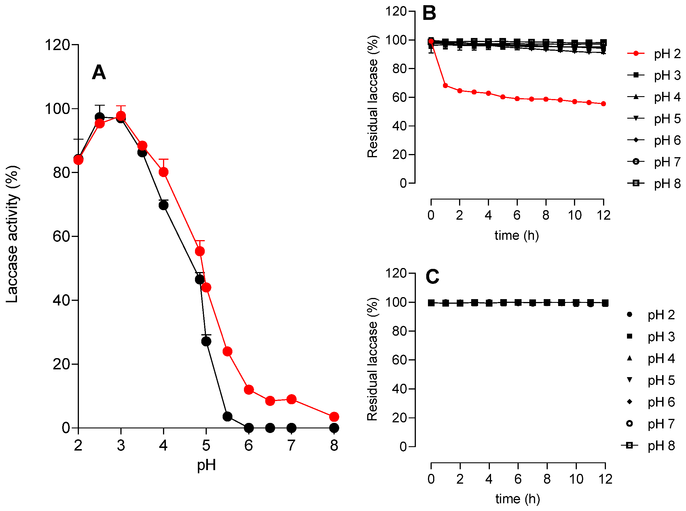
3.3.1. Effects of pH

The effects of pH on the activity and stability of both types of laccases were investigated in the pH range of 2.0 to 8.0, and the outcome is presented in Figure 3. Figure 3A shows that the optimal pH value was 3.0 for both enzymes. Experiments were also carried out to evaluate the enzymic stability with respect to of pH variations (Figure 3B,C). Although the P. sanguineus laccase has been shown to be active only in the acidic pH range, it was shown to be stable over a wide pH range: the free laccase form showed good stability at pHs between 3.0 and 8.0, showing a 20% reduction in activity when incubated at pH 2.0 (Figure 3B), while the immobilized enzyme was also stable at pH 2.0 (Figure 4C). The persistent stability of the immobilized form at the low pH of 2.0 when compared to the free form suggests the formation of a protective microenvironment around the enzyme, shielding it from denaturation under extreme conditions. This behavior supports the idea that immobilization can enhance structural robustness by multipoint attachment or spatial confinement. The optimal pH of 3.0, shared by both forms, aligns with previous studies with the P. sanguineus laccase [10] and also with the laccases of other white-rot fungi such as T. versicolor [41], P. ostreatus [42], and Coriolopsis gallica [43]. The phenomenon possibly reflects the acidic conditions under which these enzymes are most active. Even after immobilization, the activity dropped significantly at an alkaline pH, indicating that immobilization did not buffer the enzyme microenvironment against pH changes, an effect sometimes observed depending on the support material used. For some industrial applications of the laccases, such as the elimination of lignin by alkali treatment of lignocellulosic feedstocks or degradation of recalcitrant dye compounds, a high activity over alkaline pH ranges would be extremely desirable [44]. In these cases, the use of bacterial laccases, active in the alkaline pH range, such as the laccases from Pseudomonas putida [45], the laccase from Caldalkalibacillus thermarum, and that expressed in Escherichia coli [46], are more suitable options.

3.3.2. Effects of Temperature
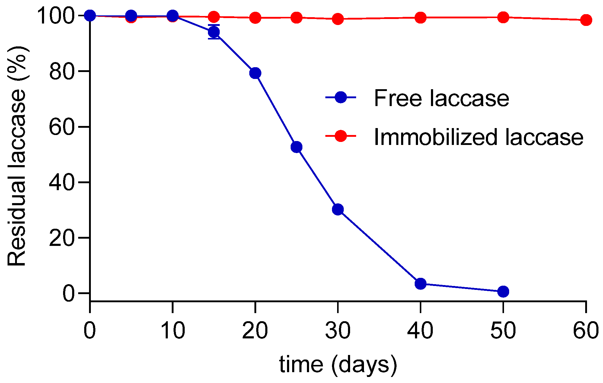
The catalytic performance of Pycnoporus sanguineus laccases, both free and immobilized, was evaluated under varying temperature conditions, with special attention to three critical aspects of enzymatic application: operational stability, thermostability, and storage stability (Figure 4 and Figure 5). A small alteration was observed in the operational stability after immobilization. The free enzyme showed maximum activity at a temperature of 70 °C, while the immobilized enzyme showed maximum activity between 50 and 70 °C (Figure 4A). It should be emphasized that in these experiments, the laccase was operating in the presence of substrate (ABTS) and under standard reaction conditions, that correspond to operational stability. Maintenance of operational stability at elevated temperatures, a crucial requirement, suggests that the immobilized enzyme is more adaptable to temperature fluctuations during catalysis, probably due to the physical stabilization imparted by the immobilization matrix. Interestingly, immobilization did not shift the optimal temperature, which remained high, a desirable feature for industrial processes that benefit from increased reaction rates at elevated temperatures. This pattern has also been observed for other fungal laccases, such as those from T. versicolor, where the optimal operational temperature remains stable or broadens after immobilization [14].
Results of the thermostability tests, which inform about the ability of the enzyme in resisting thermal denaturation over prolonged periods of time, are shown in Figure 4B,C. Both enzyme forms were stable from 30 to 60 °C, but at 70 °C the immobilized laccase (Figure 4C) exhibited slightly higher residual activity than the free form (Figure 4B). This modest gain is consistent with the literature, where it has been reported that enzyme immobilization can reduce conformational flexibility, thereby protecting the protein structure from thermal unfolding [47]. However, the difference was small, indicating that the native enzyme is already highly thermostable, and the immobilization conditions, though effective, did not significantly enhance this property. In fact, our data agree with previous works where Pycnoporus sanguineus laccases were immobilized by other methods without evident gains in terms of thermal stability [48]. This finding contrasts with more thermolabile laccases (e.g., P. ostreatus), where immobilization often results in marked improvement in thermostability [49,50,51].

3.3.3. Storage Time

The storage stabilities of both forms of laccase were evaluated at 4 °C for a period of up to 6 months (Figure 5). While the free enzyme had completely lost its activity after 50 days, the immobilized enzyme remained fully active after 60 days of storage. In fact, the immobilized enzyme remained completely active even up to 120 days of storage. In enzymatic processes, enzyme stability directly impacts cost-efficiency. This improvement is one of the most widely reported benefits of immobilization and is attributed to reduced conformational mobility and protection from degradation, autolysis, or microbial contamination during storage. Enzymes with low storage stability require more frequent replacement, increasing material and labor costs. Increased storage stability has been reported for laccases immobilized by different methods [52,53,54].

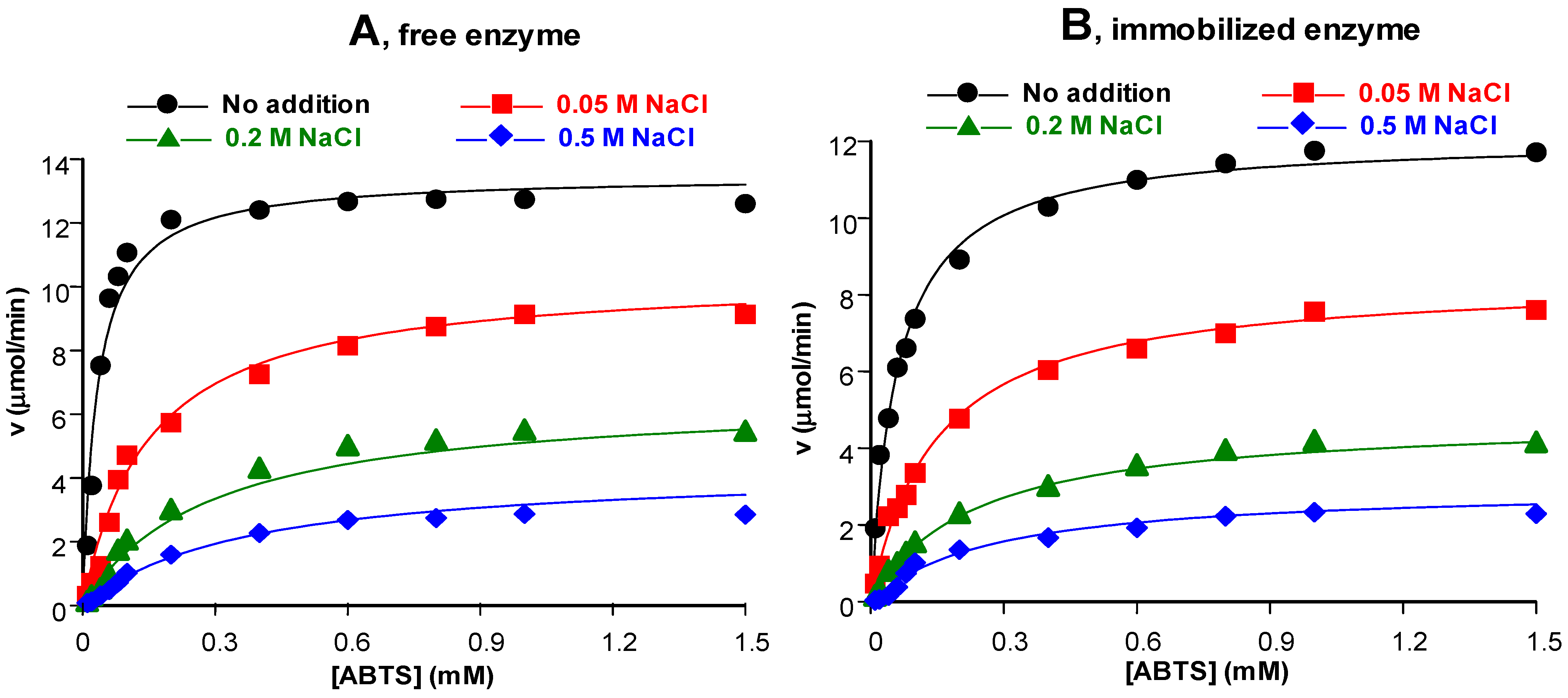
3.3.4. Influence of Salts in the Environment

Figure 6 shows how different concentrations of Na2SO4 and NaCl affected the activity of the P. sanguineus laccase using ABTS as substrate. The inhibition caused by both salts was concentration-dependent; however, NaCl caused greater inhibition than Na2SO4 within the same concentration range. In addition, the immobilized enzyme was only modestly inhibited by Na2SO4. The inhibition of Ganoderma lucidum laccase by NaCl was also reported by Zilly et al. [55], and unlike our study, the enzyme was slightly stimulated by Na2SO4. In another recent study, the immobilized laccase of O. canarii was much more strongly stimulated by Na2SO4 than the free form [41]. Furthermore, with respect to the inhibition by NaCl, immobilization of the O. canarii laccase considerably reduced this inhibition. Laccases from different origins, thus, differ largely not only in their sensitivity to salts but also in the way by which the response to salts is modified upon immobilization. Experiments on the effects of salts are important for the characterization of the enzyme and more rational biotechnological applications in environments containing various types of this kind of compound.

3.4. Kinetic Properties

A kinetic analysis with both enzyme forms was conducted to find out if immobilization caused substantial modifications in the enzyme–substrate interactions and also for obtaining more information about the inhibition caused by salts. The experimental approach to these questions consisted in the traditional protocol of varying simultaneously both the substrate and inhibitor (salts) concentrations while measuring the resulting reaction rates. Figure 7 shows the outcome of the experiments in which the substrate and NaCl concentrations were varied concomitantly in the reaction medium containing the free (A) and immobilized (B) enzymes, respectively.
Both free and immobilized enzymes displayed the traditional saturation curve in the absence of NaCl. Even on gross inspection, however, it is possible to deduce that immobilization caused changes in the enzymic kinetic behavior. For example, the free enzyme (Figure 7A) tended to a higher maximal rate when saturating substrate concentrations were approached, in comparison with the immobilized enzyme (Figure 7B). With respect to NaCl, each increase in its concentration produced a corresponding v vs. [S] curve running below the preceding one without any sign of convergence at saturating substrate concentrations. This behavior suggests non-competitive or mixed-type inhibition. For this reason, in the first attempt at obtaining kinetic parameters from the results, shown in Figure 8, the classical equation describing non-competitive (or mixed) inhibition, shown below, was fitted to the data:
v = V max [ S ] K M 1 + [ I ] K i 1 + [ S ] 1 + [ I ] K i 2
[S] represents the substrate concentration and v the reaction rate, whereas [I] is the inhibitor concentration (in this case NaCl). Vmax and KM are the maximal reaction rate and the Michaelis–Menten constant, respectively; Ki1 and Ki2 are the dissociation constants of EI and ESI complexes, respectively. Fitting this equation to the whole data set was not very satisfactory as systematic deviations were seen between theory and experiment, as shown in Figures S5 and S6 of the Supplementary Materials. For this reason, we tried to fit the equation resulting from the modifier mechanism. The latter consists in the following catalytic steps [56,57]:
E + S k 2 k 1 E S k 3 E + P
I + E K i 1 E I
I + E S K i 2 E S I
E I + S k 5 k 4 E S I k 6 E I + P
In the above mechanism, E, S, and I represent enzyme, substrate, and inhibitor, respectively, whereas Ki1 and Ki2 are the dissociation constants of the complexes EI and ESI. The rate constants for each step are represented by k1, k2, …, k6. The main difference with respect to the classical non-competitive inhibition model is that the ESI complex is not inactive. If k6 is greater than k3, i.e., k6 > k3, I is actually a stimulator; but, when the opposite occurs, i.e., when k6 < k3, I will be an inhibitor. It should be noted that unless k6 = 0, the inhibition will be incomplete, i.e., even in the present of very high inhibitor concentrations there will still be transformation of S into P, although at lower rates. This mechanism leads to the following equation under some simplifying assumptions [56,57]:
v = V 1 [ S ] + V 2 [ S ] [ I ] K i 2 K M 1 + [ I ] K i 1 + [ S ] 1 + [ I ] K i 2
In this equation, V1 = k3 [Et], and it corresponds to Vmax in the absence of an inhibitor (i.e., [I] = 0). V2 is the maximal reaction rate still observable at saturating inhibitor concentrations, i.e., V2 = k6 [Et]. For more information about the modifier mechanism, especially about the simplifying assumptions that lead to Equation (7), the reader is advised to consult reference [57]. As can be deduced even by simple inspection of Figure 7, Equation (7) is a reasonably good description of the kinetic behavior of both the free and immobilized laccases alone and in the presence of NaCl at various concentrations. This is also corroborated by the statistical measures of fit goodness, which are listed in Table 6 for Equation (7) and in the legends to Figures S3 and S4 in the Supplementary Materials for Equation (6). They are much more favorable for Equation (7) when compared to Equation (6). Values of the optimized parameters, V1, V2, KM, Ki1, and Ki2, are listed in Table 6. Immobilization reduces the maximal rate (Vmax) by 10.3%. This is not a very pronounced change. The KM, however, was nearly doubled, what can be regarded as a pronounced change because it indicates that the strength by which the immobilized enzyme binds the substrate was reduced by 50%. As far as the effects of NaCl are concerned, the inhibition of both enzyme forms is not complete, as revealed by the V2 values. For the free enzyme, the V2/V1 ratio, which was equal to 0.134, reveals that the maximal inhibition by NaCl at saturating substrate and NaCl concentrations can be predicted to reach 87%; for the immobilized enzyme, the V2/V1 ratio of 0.1 reveals that the maximal inhibition, 90%, was practically the same. This is obviously consistent with the fact that the reaction rate vs. NaCl concentration curves for both enzyme forms are quite similar, as shown in Figure 6. However, if one compares the Ki1 and Ki2 values obtained with both enzyme forms, it is easy to see that the responses of the free and immobilized forms to NaCl were not the same. In fact, immobilization decreased the affinity of the non-complexed enzyme (E) for the salt (Ki1 increased) but increased the affinity of the substrate-complexed enzyme (ES) for the same salt (Ki2 decreased). There is no definitive explanation for this phenomenon that could be deduced from the data obtained so far, except for the widespread general notion that immobilization always induces structural changes in the enzyme, which, in turn, generally modifies the response of the macromolecule to components of the surrounding microenvironment.
The kinetics of the effects of Na2SO4 were investigated using the same experimental protocol and theoretical analysis used when analyzing the effects of NaCl. The results are shown in Figure 8. The experiments shown in Figure 6B already revealed that inhibition by Na2SO4 is less pronounced, a phenomenon that occurs over the whole substrate concentration range. Equation (7) was also fitted to the curves in Figure 8. In the case of the free enzyme, the agreement between theory and experiment was very good, as the calculated curves cover quite well the experimental ones, as can be deduced by visual inspection. The optimized parameters are given in Table 7 together with the corresponding standard errors, which indicate a reasonably precise determination. It should be noted that the maximal inhibition at saturating concentrations of both substrate and Na2SO4 concentrations, which can be calculated from the V1 and V2 values, is equal to 60%. It is, thus, a clear case of incomplete inhibition.
Analysis of the data on the inhibition by Na2SO4 obtained with the immobilized enzyme was not so straightforward as that of the data obtained with the free form. This was caused by the low inhibition degree of the immobilized enzyme that was observed with Na2SO4 in the concentration range of up to 0.5 M. It was, notwithstanding, possible to fit Equation (7) to the experimental data, as revealed by Figure 8. The standard errors of the optimized parameters, except V1 and KM, however, could not be determined with a reasonable degree of accuracy. Even so, the mean values of the parameters are consistent with the experimental observation that inhibition by sulfate was substantially reduced. In fact, from the observed V1 and V2 values, one can predict that the maximal inhibition at saturating substrate and Na2SO4 concentrations will not exceed 44%. Inhibition of the immobilized laccase by Na2SO4 is, thus, also incomplete. However, an inhibition of 44% can only be expected to occur at unusually high Na2SO4 concentrations if one considers the high, though imprecise, values of both inhibitor constants given in Table 7.

3.5. Degradation of Bisphenol A (BPA) and Malachite Green by Free and Immobilized P. sanguineus Laccases

The capability of free and immobilized laccases to degrade BPA is shown in Figure 9. No significant difference was found in the degradation of BPA by free and immobilized laccases. Around 50% of BPA was degraded after 40 and 75 min when 0.5 and 0.1 U/mL of laccase were added in the reaction media, respectively. The use of 1.0 U/mL of laccase caused the disappearance of more than 95% of the initial BPA.
Bisphenol A (BPA) is a well-known endocrine disruptor that is challenging to remove effectively using conventional wastewater treatment methods. Consequently, alternative approaches, such as biodegradation and advanced oxidation processes (AOPs), have been extensively investigated [58,59]. The potential of laccases from several sources to biodegrade BPA has garnered significant attention over the years, with the employment of both free and immobilized enzymes for the purpose [23,60,61]. In certain protocols, redox mediators have been added to enhance laccase efficiency in degrading BPA. For instance, Singh et al. [62] reported a 90% degradation of 250 µM BPA after 24 h of treatment using T. versicolor laccase in the presence of 1-hydroxybenzotriazole as a redox mediator. However, some studies have demonstrated that free laccases can effectively degrade BPA without the addition of mediators.
Free and immobilized laccases have been used in the decolorization and biodegradation of malachite green [41,63,64]. The time course of the decolorization of MG by free and immobilized laccases in this work is shown in Figure 10. Both laccases can completely decolorize the dye, even in the presence of salts. Kinetic analyses of free and immobilized laccases performed in the presence of salts revealed that the latter, especially NaCl, function as enzyme inhibitors (Figure 7 and Figure 8). This inhibition by NaCl has already been reported for several laccases [7,53,65]. However, these inhibitions are reversible, so that when conducting long-term decolorization experiments (up to 48 h), it is possible to observe that complete decolorization of malachite green occurred even in the presence of salts. This fact is of great interest, considering that textile industry effluents are rich in salts, which are used to enhance the fixation of dyes in the fibers of different fabrics.

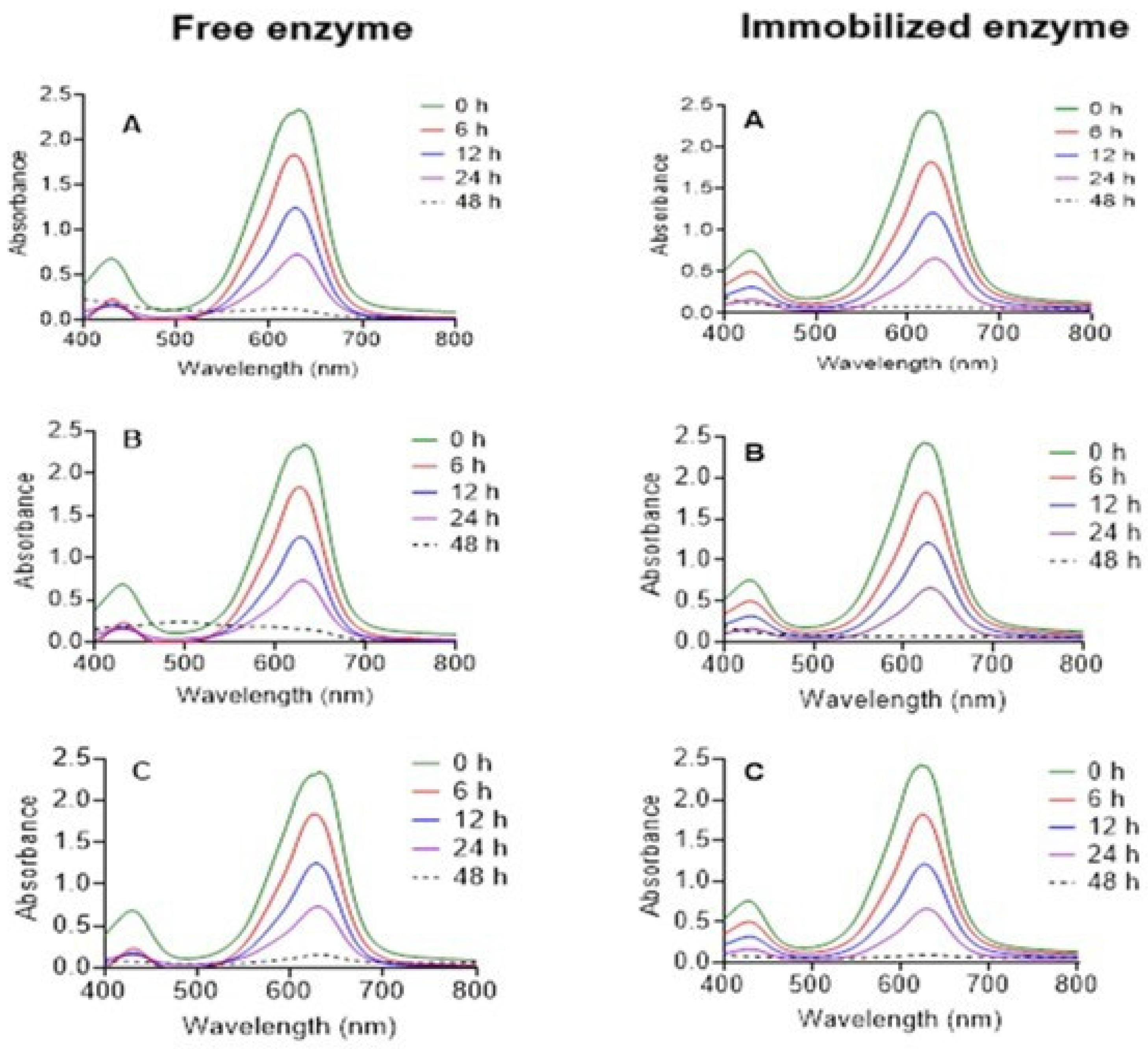
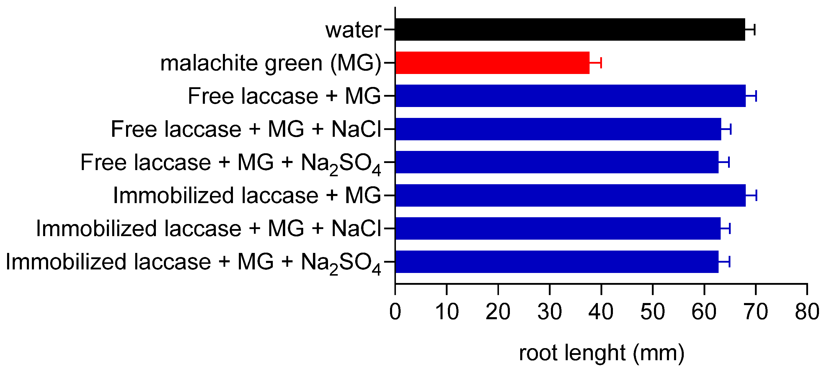
3.6. Actions of Laccase Treatment on Malachite Green Toxicity

Malachite green is a triphenylmethane dye widely used as a biocide in global aquaculture due to its effectiveness against major protozoal and fungal infections [66]. Primarily, it acts as an ectoparasiticide and is also employed to control skin and gill flukes. Beyond aquaculture, malachite green is also utilized as a food coloring agent, food additive, medical disinfectant, anthelmintic, and dye in industries such as silk, wool, jute, leather, cotton, paper, and acrylic production [67]. Despite its broad applications, malachite green is highly controversial because it is often associated with health risks, including adverse effects on the immune and reproductive systems, as well as having genotoxic and carcinogenic potential [68]. Although banned in several countries, its use persists in many regions due to its low cost, availability, and efficacy.
Several kinds of physico-chemical and biological procedures have been employed for decolorizing and degrading dyes in wastewater, including malachite green [41,68,69]. This is a field in which laccases may play a significant role. In many cases, decolorization results from minor chemical changes in the dye chromophore group, which do not correspond to the groups involved in toxicity. For this reason, it is of interest to investigate if the laccase produced herein also decreases toxicity in parallel with its malachite green decolorizing action. This issue was investigated by evaluating the root growth of Lactuca sativa, and the results are shown in Figure 11. Without previous treatment with laccase, MG clearly inhibited the root grow L. sativa. After incubation with laccase during 48 h, however, hte inhibition of root grow by MG was almost abolished. This occurred with both free and immobilized laccase and was only minimally influenced by the presence of the salts NaCl and Na2SO4 under the experimental conditions that were employed.

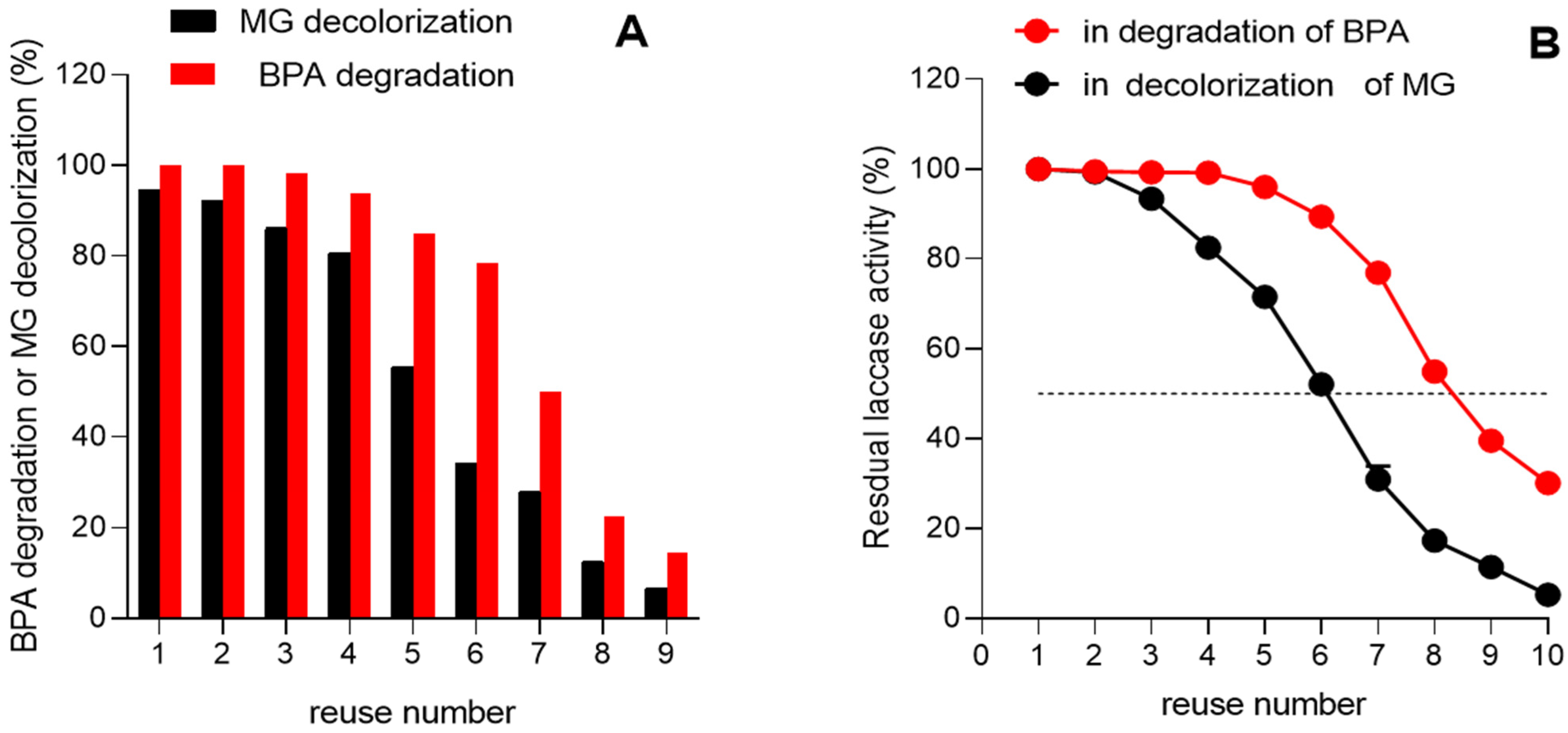
3.7. Reusability of Immobilized Laccase in the Degradation of BPA and Green Malachite

The use of free laccases for xenobiotic biodegradation faces several challenges, including high production costs, limited stability, and their inability to be reused. These drawbacks render large-scale applications of free enzymes economically unfeasible. Immobilization of laccases offers a promising strategy to overcome, at least partially, these significant limitations by enhancing enzyme stability and enabling repeated use.
Enzyme reusability is a key principle in enzymatic catalysis, influencing both industries that depend on enzyme technology and research aimed at understanding enzyme properties and their potential uses. The reuse of an enzyme must be tested, however, under the conditions of the application of the enzyme. As can be seen in Figure 12, the immobilized enzyme retained more than 50% of its initial capability to degrade BPA after seven cycles of reuse, and more than 50% of its initial capability to decolorize MG after five cycles of reuse. It is important to note that each reuse cycle of the immobilized laccase in BPA degradation lasts 2 h, while each reuse cycle in MG decolorization lasts 48, which may explain the greater number of reuses of the enzyme in BPA degradation.

4. Conclusions

The laccase of the new isolate of P. sanguineus (UEM-20) presents several favorable perspectives for its use in the detoxification of wastewater and in the decolorization of polluted waters, as evidenced by its efficiency in transforming bisphenol A (BPA) and in the decolorization of malachite green (MG). The latter activity was also accompanied by a significant reduction in toxicity. Furthermore, immobilization of the enzyme by means of the CLEA technique can be considered a successful undertaking. Upon immobilization, the enzyme experienced some changes in its kinetic properties. The Vmax decreased by a factor of 1.1, and the KM increased by a factor of 1.89. These kinetic changes did not modify in any remarkable way the capacity of the immobilized enzyme to degrade BPA. Its sensitivity to NaCl was minimally affected by immobilization. However, its sensitivity to sodium sulfate was substantially decreased, a point that may eventually become important under some specific circumstances. Significant advantages of immobilization can also be attributed to the enormous gain in storage ability, in addition to the possibility of reuse, a property that is inherent to all immobilized enzymes in general.

Supplementary Materials

The following supporting information can be downloaded at https://www.mdpi.com/article/10.3390/pr13061800/s1, Methodology: Molecular identification of new isolated fungi. Figure S1: Dendrogram showing phylogenetic relationships between UEM-20 isolate and other fungi with high percentage identity identified with BLAST v.2.15.0 in Genbank; Figure S2: Pycnoporus sanguineus UEM-20 (Trametes sanguinea); Table S1: Experimental and predicted model values of laccase immobilization and deviation for CCD; Figure S3: Predicted vs. observed laccase immobilization values; Figure S4: Scanning electron microscopy of laccase CLEAs: 27× (A), 1000× (B), and 5000×; Figure S5: Kinetics of the inhibition of the free laccase by chloride ions analyzed according to Equation (6); Figure S6: Kinetics of the inhibition of the immobilized laccase by chloride ions analyzed according to Equation (6). References cited in Supplementary Materials [70,71,72,73,74].

Author Contributions

Conceptualization, V.M.S.C. and R.M.P.; methodology, V.M.S.C., E.B., V.d.O.P., V.A.d.O.J., T.M.U., J.R.d.S.F. and L.F.O.d.S.; validation, R.C., C.G.M.d.S., J.C.P. and A.G.C.; formal analysis, A.G.C., A.B. and R.M.P.; writing—original draft preparation, V.M.S.C.; writing—review and editing, A.G.C., R.C., C.G.M.d.S., A.B. and R.M.P.; supervision, A.B. and R.M.P.; project administration, R.M.P.; funding acquisition, A.B. and R.M.P. All authors have read and agreed to the published version of the manuscript.

Funding

This research was funded by by grants from the Conselho Nacional de Desenvolvimento Científico e Tecnológico (CNPq 404589/2023-5), Fundação Araucária (FA 160/2022), and Fundação Carlos Chagas de Amparo à Pesquisa do Estado do Rio de Janeiro (FAPERJ), Brazil—grant number [E-26/210.537/2025].

Data Availability Statement

Data will be made available on request.

Conflicts of Interest

The authors declare no conflicts of interest.

References

  1. Janusz, G.; Pawlik, A.; Świderska-Burek, U.; Polak, J.; Sulej, J.; Jarosz-Wilkołazka, A.; Paszczyński, A. Laccase Properties, Physiological Functions, and Evolution. Int. J. Mol. Sci. 2020, 21, 966. [Google Scholar] [CrossRef] [PubMed]
  2. Dwivedi, U.N.; Singh, P.; Pandey, V.P.; Kumar, A. Structure–function relationship among bacterial, fungal and plant laccases. J. Mol. Catal. B Enzym. 2011, 68, 117–128. [Google Scholar] [CrossRef]
  3. Mahuri, M.; Paul, M.; Thatoi, H. A review of microbial laccase production and activity toward different biotechnological applications. Syst. Microbiol. Biomanuf. 2023, 3, 533–551. [Google Scholar] [CrossRef]
  4. Ahmad, I.; Pal, S.; Waseem, M.; Jamal, A.; Kamal, M.A.; Ahmad, F.; Haji, E.M.; Siddiqui, S.; Singh, A.K. Catalytic insights into laccase for sustainable remediation of multifaceted pharmaceutically active micropollutants from water matrices: A state-of-art review. J. Water Process. Eng. 2025, 70, 106901. [Google Scholar] [CrossRef]
  5. Murrill, W.A. The Polyporaceae of North America—VIII. Hapalopilus, Pycnoporus, and New Monotypic Genera. Bull. Torrey Bot. Club 1904, 31, 415–428. [Google Scholar] [CrossRef]
  6. Rodríguez-Delgado, M.; Orona-Navar, C.; García-Morales, R.; Hernandez-Luna, C.; Parra, R.; Mahlknecht, J.; Ornelas-Soto, N. Biotransformation Kinetics of Pharmaceutical and Industrial Micropollutants in Groundwaters by a Laccase Cocktail from Pycnoporus sanguineus CS43 Fungi. Int. Biodeterior. Biodegrad. 2016, 108, 34–41. [Google Scholar] [CrossRef]
  7. Yang, J.; Xu, X.; Yang, X.; Ye, X.; Lin, J. Cross-Linked Enzyme Aggregates of Cerrena Laccase: Preparation, Enhanced NaCl Tolerance and Decolorization of Remazol Brilliant Blue Reactive. J. Taiwan Inst. Chem. Eng. 2016, 65, 1–7. [Google Scholar] [CrossRef]
  8. Garcia-Morales, R.; Rodríguez-Delgado, M.; Gomez-Mariscal, K.; Orona-Navar, C.; Hernandez-Luna, C.; Torres, E.; Ornelas-Soto, N. Biotransformation of Endocrine-Disrupting Compounds in Groundwater: Bisphenol A, Nonylphenol, Ethynylestradiol and Triclosan by a Laccase Cocktail from Pycnoporus sanguineus CS43. Water Air Soil Pollut. 2015, 226, 1–14. [Google Scholar] [CrossRef]
  9. Cheute, V.M.S.; Uber, T.M.; dos Santos, L.F.O.; Backes, E.; Dantas, M.P.; Contato, A.G.; Castoldi, R.; de Souza, C.G.M.; Corrêa, R.C.G.; Bracht, A.; et al. Biotransformation of Pollutants by Pycnoporus spp. in Submerged and Solid-State Fermentation: Mechanisms, Achievements, and Perspectives. Biomass 2024, 4, 313–328. [Google Scholar] [CrossRef]
  10. Ramírez-Cavazos, L.I.; Junghanns, C.; Ornelas-Soto, N.; Cárdenas-Chávez, D.L.; Hernández-Luna, C.; Demarche, P.; Parra, R. Purification and Characterization of Two Thermostable Laccases from Pycnoporus sanguineus and Potential Role in Degradation of Endocrine Disrupting Chemicals. J. Mol. Catal. B Enzym. 2014, 108, 32–42. [Google Scholar] [CrossRef]
  11. Brugnari, T.; Pereira, M.G.; Bubna, G.A.; de Freitas, E.N.; Contato, A.G.; Corrêa, R.C.G.; Castoldi, R.; de Souza, C.G.M.; Polizeli, M.L.T.M.; Bracht, A.; et al. A highly reusable MANAE-agarose-immobilized Pleurotus ostreatus laccase for degradation of bisphenol A. Sci. Total. Environ. 2018, 634, 1346–1351. [Google Scholar] [CrossRef] [PubMed]
  12. Datta, S.; Veena, R.; Samuel, M.S.; Selvarajan, E. Immobilization of Laccases and Applications for the Detection and Remediation of Pollutants: A Review. Environ. Chem. Lett. 2021, 19, 521–538. [Google Scholar] [CrossRef]
  13. Ortolan, G.G.; Contato, A.G.; Aranha, G.M.; Salgado, J.C.S.; Alnoch, R.C.; Polizeli, M.L.T.M. Enhancing Laccase Production by Trametes hirsuta GMA-01 Using Response Surface Methodology and Orange Waste: A Novel Breakthrough in Sugarcane Bagasse Saccharification and Synthetic Dye Decolorization. Reactions 2024, 5, 635–650. [Google Scholar] [CrossRef]
  14. Uber, T.M.; Pateis, V.O.; Cheute, V.M.S.; dos Santos, L.F.O.; Trindade, A.R.F.; Contato, A.G.; dos Santos Filho, J.R.; Corrêa, R.C.G.; Castoldi, R.; de Souza, C.G.M.; et al. Immobilization of Trametes versicolor Laccase by Interlinked Enzyme Aggregates with Improved pH Stability and Its Application in the Degradation of Bisphenol A. Reactions 2025, 6, 9. [Google Scholar] [CrossRef]
  15. Velasco-Lozano, S.; López-Gallego, F.; Mateos-Díaz, J.C.; Favela-Torres, E. Cross-Linked Enzyme Aggregates (CLEA) in Enzyme Improvement—A Review. Biocatalysis 2016, 1, 166–177. [Google Scholar] [CrossRef]
  16. Bilal, M.; Zhao, Y.; Noreen, S.; Shah, S.Z.H.; Bharagava, R.N.; Iqbal, H.M.N. Modifying Bio-Catalytic Properties of Enzymes for Efficient Biocatalysis: A Review from Immobilization Strategies Viewpoint. Biocatal. Biotransform. 2019, 37, 159–182. [Google Scholar] [CrossRef]
  17. Mota, T.R.; Kato, C.G.; Peralta, R.A.; Bracht, A.; de Morais, G.R.; Baesso, M.L.; Peralta, R.M. Decolourization of Congo Red by Ganoderma lucidum Laccase: Evaluation of Degradation Products and Toxicity. Water Air Soil Pollut. 2015, 226, 1–11. [Google Scholar] [CrossRef]
  18. Uber, T.M.; Buzzo, A.J.D.R.; Scaratti, G.; Amorim, S.M.; Helm, C.V.; Maciel, G.M.; Peralta, R.M. Comparative Detoxification of Remazol Brilliant Blue R by Free and Immobilized Laccase of Oudemansiella canarii. Biocatal. Biotransform. 2020, 40, 17–28. [Google Scholar] [CrossRef]
  19. Laemmli, U.K. Cleavage of Structural Proteins During the Assembly of the Head of Bacteriophage T4. Nature 1970, 227, 680–685. [Google Scholar] [CrossRef]
  20. Bradford, M.M. A Rapid and Sensitive Method for the Quantitation of Microgram Quantities of Protein Utilizing the Principle of Protein-Dye Binding. Anal. Biochem. 1976, 72, 248–254. [Google Scholar] [CrossRef]
  21. Patel, H.; Gupte, S.; Gahlout, M.; Gupte, A. Purification and Characterization of an Extracellular Laccase from Solid-State Culture of Pleurotus ostreatus HP-1. 3 Biotech 2013, 16, 77–84. [Google Scholar] [CrossRef] [PubMed]
  22. Akaike, H. A new look at the statistical model identification. IEEE Trans. Autom. Control 1974, 19, 716–723. [Google Scholar] [CrossRef]
  23. Brugnari, T.; Contato, A.G.; Pereira, M.G.; Freitas, E.N.D.; Bubna, G.A.; Aranha, G.M.; Peralta, R.M. Characterisation of Free and Immobilised Laccases from Ganoderma lucidum: Application on Bisphenol A Degradation. Biocatal. Biotransform. 2021, 39, 71–80. [Google Scholar] [CrossRef]
  24. Coelho-Moreira, J.S.; Brugnari, T.; Sá-Nakanishi, A.B.; Castoldi, R.; Souza, C.G.M.; Bracht, A.; Peralta, R.M. Evaluation of Diuron Tolerance and Biotransformation by the White-Rot Fungus Ganoderma lucidum. Fungal Biol. 2018, 122, 471–488. [Google Scholar] [CrossRef]
  25. Durand, A. Bioreactor Designs for Solid State Fermentation. Biochem. Eng. J. 2003, 13, 113–125. [Google Scholar] [CrossRef]
  26. Abu Yazid, N.; Barrena, R.; Komilis, D.; Sánchez, A. Solid-State Fermentation as a Novel Paradigm for Organic Waste Valorization: A Review. Sustainability 2017, 9, 224. [Google Scholar] [CrossRef]
  27. Eugenio, M.E.; Carbajo, J.M.; Martín, J.A.; González, A.E.; Villar, J.C. Laccase production by Pycnoporus sanguineus under different culture conditions. J. Basic Microbiol. 2009, 49, 433–440. [Google Scholar] [CrossRef]
  28. Lu, L.; Zhao, M.; Zhang, B.B.; Yu, S.Y.; Bian, X.J.; Wang, W.; Wang, Y. Purification and Characterization of Laccase from Pycnoporus sanguineus and Decolorization of an Anthraquinone Dye by the Enzyme. Appl. Microbiol. Biotechnol. 2007, 74, 1232–1239. [Google Scholar] [CrossRef]
  29. Niladevi, K.N.; Sukumaran, R.K.; Prema, P. Utilization of Rice Straw for Laccase Production by Streptomyces psammoticus in Solid-State Fermentation. J. Ind. Microbiol. Biotechnol. 2007, 34, 665–674. [Google Scholar] [CrossRef]
  30. Soccol, C.R.; da Costa, E.S.F.; Letti, L.A.J.; Karp, S.G.; Woiciechowski, A.L.; de Souza Vandenberghe, L.P. Recent Developments and Innovations in Solid State Fermentation. Biotechnol. Res. Innov. 2017, 1, 52–71. [Google Scholar] [CrossRef]
  31. Zimbardi, A.L.; Camargo, P.F.; Carli, S.; Aquino Neto, S.; Meleiro, L.P.; Rosa, J.C.; Furriel, R.P. A High Redox Potential Laccase from Pycnoporus sanguineus RP15: Potential Application for Dye Decolorization. Int. J. Mol. Sci. 2016, 17, 672. [Google Scholar] [CrossRef] [PubMed]
  32. Iracheta-Cárdenas, M.M.; Rocha-Peña, M.A.; Galán-Wong, L.J.; Arévalo-Niño, K.; Tovar-Herrera, O.E. A Pycnoporus sanguineus Laccase for Denim Bleaching and Its Comparison with an Enzymatic Commercial Formulation. J. Environ. Manag. 2016, 177, 93–100. [Google Scholar] [CrossRef]
  33. Scarpa, J.D.C.P.; Marques, N.P.; Monteiro, D.A.; Martins, G.M.; de Paula, A.V.; Boscolo, M.; Bocchini, D.A. Saccharification of Pretreated Sugarcane Bagasse Using Enzymes Solution from Pycnoporus sanguineus MCA 16 and Cellulosic Ethanol Production. Ind. Crop. Prod. 2019, 141, 111795. [Google Scholar] [CrossRef]
  34. Fathali, Z.; Rezaei, S.; Faramarzi, M.A.; Habibi-Rezaei, M. Catalytic Phenol Removal Using Entrapped Cross-Linked Laccase Aggregates. Int. J. Biol. Macromol. 2019, 122, 359–366. [Google Scholar] [CrossRef]
  35. Cabana, H.; Jones, J.P.; Agathos, S.N. Preparation and Characterization of Cross-Linked Laccase Aggregates and Their Application to the Elimination of Endocrine Disrupting Chemicals. J. Biotechnol. 2007, 132, 23–31. [Google Scholar] [CrossRef] [PubMed]
  36. Kumar, V.V.; Kumar, M.P.; Thiruvenkadaravi, K.V.; Baskaralingam, P.; Kumar, P.S.; Sivanesan, S. Preparation and Characterization of Porous Cross Linked Laccase Aggregates for the Decolorization of Triphenyl Methane and Reactive Dyes. Bioresour. Technol. 2012, 119, 28–34. [Google Scholar] [CrossRef]
  37. Daâssi, D.; Rodríguez-Couto, S.; Nasri, M.; Mechichi, T. Biodegradation of Textile Dyes by Immobilized Laccase from Coriolopsis gallica into Ca-Alginate Beads. Int. Biocatal. Biotransform. 2014, 90, 71–78. [Google Scholar] [CrossRef]
  38. Zheng, F.; Cui, B.K.; Wu, X.J.; Meng, G.; Liu, H.X.; Si, J. Immobilization of Laccase onto Chitosan Beads to Enhance Its Capability to Degrade Synthetic Dyes. Int. Biocatal. Biotransform. 2016, 110, 69–78. [Google Scholar] [CrossRef]
  39. Guzik, U.; Hupert-Kocurek, K.; Wojcieszyńska, D. Immobilization as a Strategy for Improving Enzyme Properties—Application to Oxidoreductases. Molecules 2014, 19, 8995–9018. [Google Scholar] [CrossRef]
  40. Cao, L. Immobilised Enzymes: Science or Art? Curr. Opin. Chem. Biol. 2005, 9, 217–226. [Google Scholar] [CrossRef]
  41. Backes, E.; Kato, C.G.; da Silva, T.B.V.; Uber, T.M.; Pasquarelli, D.L.; Bracht, A.; Peralta, R.M. Production of fungal laccase on pineapple waste and application in detoxification of malachite green. J. Environ. Sci. Health 2022, 57, 90–101. [Google Scholar] [CrossRef] [PubMed]
  42. Isanapong, J.; Suwannoi, K.; Lertlattanapong, S.; Panchal, S. Purification, characterization of laccase from Pleurotus ostreatus HK35, and optimization for congo red biodecolorization using Box–Behnken design. 3 Biotech 2024, 14, 73–90. [Google Scholar] [CrossRef] [PubMed]
  43. Songulashvili, G.; Flahaut, S.; Demarez, M.; Tricot, C.; Bauvois, C.; Debaste, F.; Penninckx, M.J. High yield production in seven days of Coriolopsis gallica 1184 laccase at 50 L scale; enzyme purification and molecular characterization. Fungal Biol. 2016, 120, 481–488. [Google Scholar] [CrossRef] [PubMed]
  44. Sitarz, A.K.; Mikkelsen, J.D.; Meyer, A.S. Structure, functionality and tuning up of laccases for lignocellulose and other industrial applications. Crit. Rev. Biotechnol. 2016, 36, 70–86. [Google Scholar] [CrossRef]
  45. Kuddus, M.; Joseph, B.; Ramteke, P.W. Production of laccase from newly isolated Pseudomonas putida and its application in bioremediation of synthetic dyes and industrial effluents. Biocatal. Agric. Biotechnol. 2013, 2, 333–338. [Google Scholar] [CrossRef]
  46. Xu, K.; Huo, Y.; Tang, S.; Han, S.; Lin, Y.; Zheng, S. A novel laccase for alkaline medium temperature environments in the textile industry. Biotechnol. J. 2024, 19, e2400383. [Google Scholar] [CrossRef]
  47. Mate, D.M.; Alcaide, M. Laccase: A multi-purpose biocatalyst at the forefront of biotechnology. Microb. Biotechnol. 2017, 10, 1457–1467. [Google Scholar] [CrossRef]
  48. Gonzalez-Coronel, L.A.; Cobas, M.; Rostro-Alanis, M.J.; Parra-Saldívar, R.; Hernandez-Luna, C.; Pazos, M.; Ángeles Sanromán, M. Immobilization of laccase of Pycnoporus sanguineus CS43. New Biotechnol. 2017, 39, 141–149. [Google Scholar] [CrossRef]
  49. Asgher, M.; Kamal, S.; Iqbal, H.M. Improvement of Catalytic Efficiency, Thermo-stability and Dye Decolorization Capability of Pleurotus ostreatus IBL-02 laccase by Hydrophobic Sol Gel Entrapment. Chem. Cent. J. 2012, 6, 110. [Google Scholar] [CrossRef]
  50. Lettera, V.; Pezzella, C.; Cicatiello, P.; Piscitelli, A.; Giacobelli, V.G.; Galano, E.; Amoresano, A.; Sannia, G. Efficient immobilization of a fungal laccase and its exploitation in fruit juice clarification. Food Chem. 2016, 196, 1272–1278. [Google Scholar] [CrossRef]
  51. Wlizło, K.; Polak, J.; Jarosz-Wilkołazka, A.; Pogni, R.; Petricci, E. Novel textile dye obtained through transformation of 2-amino-3-methoxybenzoic acid by free and immobilised laccase from a Pleurotus ostreatus strain. Enzym. Microb. Technol. 2020, 132, 109398. [Google Scholar] [CrossRef] [PubMed]
  52. Almulaiky, Y.Q.; Alkabli, J.; El-Shishtawy, R.M. Improving enzyme immobilization: A new carrier-based magnetic polymer for enhanced covalent binding of laccase enzyme. Int. J. Biol. Macromol. 2024, 282, 137362. [Google Scholar] [CrossRef] [PubMed]
  53. Backes, E.; Alnoch, R.C.; Contato, A.G.; Castoldi, R.; Souza, C.G.M.; Kato, C.G.; Peralta, R.A.; Moreira, R.F.P.M.; Polizeli, M.L.T.M.; Bracht, A.; et al. Properties and Kinetic Behavior of Free and Immobilized Laccase from Oudemansiella canarii: Emphasis on the Effects of NaCl and Na2SO4 on Catalytic Activities. Int. J. Biol. Macromol. 2024, 281, 136565. [Google Scholar] [CrossRef] [PubMed]
  54. Vallejo, J.L.; Vallejos, S.; Trigo-López, M.; García, J.M.; Busto, M.D. Optimization and stability of a reusable laccase-polymer hybrid film for the removal of bisphenol A in water. Environ. Technol. Innov. 2025, 38, 104093. [Google Scholar] [CrossRef]
  55. Zilly, A.; da Silva Coelho-Moreira, J.; Bracht, A.; De Souza, C.G.M.; Carvajal, A.E.; Koehnlein, E.A.; Peralta, R.M. Influence of NaCl and Na2SO4 on the Kinetics and Dye Decolorization Ability of Crude Laccase from Ganoderma lucidum. Int. Biodeterior. Biodegrad. 2011, 65, 340–344. [Google Scholar] [CrossRef]
  56. Botts, J.; Morales, M. Analytical Description of the Effects of Modifiers and of Enzyme Multivalency upon the Steady State Catalyzed Reaction Rate. Trans. Faraday Soc. 1953, 49, 696–707. [Google Scholar] [CrossRef]
  57. Baici, A. Kinetics of Enzyme-Modifier Interactions. In The General Modifier Mechanism; Springer: Berlin, Germany, 2015; pp. 65–102. [Google Scholar] [CrossRef]
  58. Pan, Z.; Song, C.; Li, L.; Wang, H.; Pan, Y.; Wang, C.; Feng, X. Membrane Technology Coupled with Electrochemical Advanced Oxidation Processes for Organic Wastewater Treatment: Recent Advances and Future Prospects. Chem. Eng. J. 2019, 376, 120909. [Google Scholar] [CrossRef]
  59. Wang, J.; Zhuan, R. Degradation of Antibiotics by Advanced Oxidation Processes: An Overview. Sci. Total Environ. 2020, 701, 135023. [Google Scholar] [CrossRef]
  60. Ghobadi Nejad, Z.; Borghei, S.M.; Yaghmaei, S. Kinetic Studies of Bisphenol A in Aqueous Solutions by Enzymatic Treatment. Int. J. Environ. Sci. Technol. 2019, 16, 821–832. [Google Scholar] [CrossRef]
  61. Lassouane, F.; Aït-Amar, H.; Rodriguez-Couto, S. High BPA removal by immobilized crude laccase in a batch fluidized bed bioreactor. Biochem. Eng. J. 2022, 184, 108489. [Google Scholar] [CrossRef]
  62. Singh, J.; Kumar, P.; Saharan, V.; Kapoor, R.K. Simultaneous Laccase Production and Transformation of Bisphenol-A and Triclosan Using Trametes versicolor. 3 Biotech 2019, 9, 1–16. [Google Scholar] [CrossRef] [PubMed]
  63. Himanshu; Behera, B.; Kumari, N.; Maruthi, M.; Singh, R.K.; Saini, J.K. Appraisal of malachite green biodegradation and detoxification potential of laccase from Trametes cubensis. Bioresour. Technol. 2025, 417, 131869. [Google Scholar] [CrossRef] [PubMed]
  64. Thoa, L.T.K.; Thao, T.T.P.; Hung, N.B.; Khoo, K.S.; Quang, H.T.; Lan, T.T.; Hoang, V.D.; Park, S.-M.; Ooi, C.W.; Show, P.L.; et al. Biodegradation and Detoxification of Malachite Green Dye by Extracellular Laccase Expressed from Fusarium oxysporum. Waste Biomass-Valorization 2022, 13, 2511–2518. [Google Scholar] [CrossRef]
  65. Enaud, E.; Trovaslet, M.; Naveau, F.; Decristoforo, A.; Bizet, S.; Vanhulle, S.; Jolivalt, C. Laccase chloride inhibition reduction by an anthraquinonic substrate. Enzym. Microb. Technol. 2011, 49, 517–525. [Google Scholar] [CrossRef]
  66. Srivastava, S.; Sinha, R.; Roy, D. Toxicological effects of malachite green. Aquat. Toxicol. 2004, 66, 319–329. [Google Scholar] [CrossRef]
  67. Culp, S.J.; Beland, F.A. Malachite green: A toxicological review. J. Am. Coll. Toxicol. 1996, 15, 219–238. [Google Scholar] [CrossRef]
  68. Raval, N.P.; Shah, P.U.; Shah, N.K. Malachite green “a cationic dye” and its removal from aqueous solution by adsorption. Appl. Water Sci. 2017, 7, 3407–3445. [Google Scholar] [CrossRef]
  69. He, J.; Mo, P.; Luo, Y.-S.; Yang, P.-H. Strategies for solving the issue of malachite green residues in aquatic products: A review. Aquac. Res. 2023, 2023, 578570. [Google Scholar] [CrossRef]
  70. Raeder, U.; Broda, P. Rapid preparation of DNA from filamentous fungi. Lett. Appl. Microbiol. 1985, 1, 17–20. [Google Scholar] [CrossRef]
  71. Pamphile, J.A.; Azevedo, J.L. Molecular characterization of endophytic strains of Fusarium verticillioides. World J. Microbiol. Biotechnol. 2002, 18, 391–396. [Google Scholar] [CrossRef]
  72. Van Den Ende, A.H.G.; De Hoog, G.S. Variability and molecular diagnostics of the neurotropic species Cladophialophora bantiana. Stud. Mycol. 1999, 43, 151–162. [Google Scholar]
  73. Justo, A.; Miettinen, O.; Floudas, D.; Ortiz-Santana, B.; Sjökvist, E.; Lindner, D.; Nakasone, K.; Niemelã, T.; Larsson, K.-H.; Ryvarden, L.; et al. A revised family-level classification of the Polyporales (Basidiomycota). Fungal Biol. 2017, 121, 798–824. [Google Scholar] [CrossRef] [PubMed]
  74. Saitou, N.; Nei, M. The neighbor-joining method: A new method for reconstructing phylogenetic trees. Mol. Biol. Evol. 1987, 4, 406–425. [Google Scholar] [CrossRef] [PubMed]
Figure 1. Laccase production by P. sanguineus under solid-state fermentation using various agricultural lignocellulosic residues. Cultures were incubated at 28 °C with an initial moisture content of 83.3% for 9 days. Data are mean ± standard deviation (n = 3). (A): Laccase activity with different lignocellulosic substrates. (B,C): Macroscopic appearance of 9-day-old P. sanguineus cultures grown on wheat bran. (D): Non-denaturing SDS-PAGE analysis of P. sanguineus laccase. Lane 1: Molecular weight markers (Coomassie blue staining). Lane 2: Total protein extract from P. sanguineus (Coomassie blue staining). Lane 3: Laccase activity detected by zymogram.
Processes 13 01800 g001
Figure 2. Theoretical three-dimensional response surface (A) and contour chart (B) for the relative efficiency of laccase immobilization by CLEA as a function of ammonium sulfate and glutaraldehyde content. Data were derived from the second-order polynomial of Equation (3).
Processes 13 01800 g002
Figure 3. Influence of pH on the activity (A) and stability of free (B) and immobilized (C) P. sanguineus laccase. Free (B) and immobilized (C) laccase were incubated without substrate for up to 12 h at various pH values (2.0–8.0) using McIlvaine buffer. Residual enzyme activity was measured at 40 °C in 50 mM citrate buffer, pH 3.0. In (A): () immobilized enzyme; (●) free enzyme. Data are mean ± standard deviation (n = 3).
Processes 13 01800 g003
Figure 4. Influence of temperature on the activity and stability of laccase. In (A), free and immobilized enzymes were incubated in the presence of the substrate ABTS at different temperatures for 5 min. Free (B) and immobilized (C) laccase were incubated at various temperatures for up to 120 min without substrate. Residual activity was determined at 40 °C in 50 mM citrate buffer, pH 3.0. Data are expressed as mean ± standard deviation.
Processes 13 01800 g004
Figure 5. Storage stability of free and immobilized P. sanguineus laccase at 4–8 °C. Residual activity was measured at 40 °C in 50 mM citrate buffer, pH 3.0. Data are means ± standard deviation.
Processes 13 01800 g005
Figure 6. Effect of Na2SO4 (A) and NaCl (B) on the catalytic activity of free and immobilized P. sanguineus laccase. Experimental details are given in the Materials and Methods Section. Values are mean ± standard deviation.
Processes 13 01800 g006
Figure 7. Chloride ion inhibition of the free (A) and immobilized (B) P. sanguineus laccase at various substrate concentrations. The technique for measuring the reaction rates is described in the Materials and Methods Section. Equation (7) was fitted simultaneously to all curves as described in Section 2.9 with [S] and [I] as the independent variables and v as the single dependent variable. Theoretical curves (solid lines) were generated using the optimized kinetic constants and Equation (7) as described in Section 2.9. The optimized constants are listed in Table 6.
Processes 13 01800 g007
Figure 8. Sulfate ion inhibition of the free (A) and immobilized (B) P. sanguineus laccase at various substrate concentrations. The technique for measuring the reaction rates is described in the Materials and Methods Section. Equation (7) was fitted simultaneously to all curves of each experimental series, as described in Section 2.9, with [S] and [I] as the independent variables and v as the single dependent variable. Theoretical curves (solid lines) were generated using the optimized kinetic constants and Equation (7). The optimized constants are listed in Table 7.
Processes 13 01800 g008
Figure 9. Time course of BPA degradation by free (A) and immobilized (B) laccases. Reactional conditions: initial BPA concentration: 200 µM; temperature: 40 °C; pH 3.0 (in 50 mM citrate buffer); agitation: 110 rpm. Denatured enzymes were used as negative controls.
Processes 13 01800 g009
Figure 10. Decolorization of malachite green by free (left) and immobilized (right) laccases. The final assay volume was 10 mL in water containing 100 ppm of MG, and 1.0 U/mL of free or immobilized laccase. The reaction mixtures were kept at 40 °C in the absence of light, and a scan in the visible spectrum was performed after 6, 12, 24, and 48 h. To evaluate the effect of salts on the decolorization, experiments were performed in the absence of salts (A), presence of 0.5 M of NaCl (B), and presence of 0.5 M of Na2SO4 (C).
Processes 13 01800 g010
Figure 11. Evaluation of Lactuca sativa root growth in the presence of MG before and after laccase treatment. Toxicity was evaluated at room temperature in tap water using untreated MG and after 48 h after treatment with free and immobilized laccase under different conditions. The red bar is the positive control; the black bar is the negative control; blue bars are the test experiments.
Processes 13 01800 g011
Figure 12. Reusability potential of the immobilized laccase in cycles of BPA degradation and MG decolorization. (A): BPA degradation or MG decolorization; (B): residual laccase activity.
Processes 13 01800 g012
Table 1. Encoded and real values in the factorial CCD (central composite design) experiment with 5 levels of range.
Coded ValuesActual Values
X1 (%, wt/v)X2 (mM)
1.4178.2170.5
170.0150.0
050.0100.0
−130.050.0
−1.4121.829.5
Table 3. Effect estimates for laccase immobilization according to the central composite rotational design.
FactorEffectStandard ErrorT (5)p-Value
Mean87.01191.30656066.5962<0.000001
Linear
X134.30501.60260721.40570.000004
X2−47.98391.912327−25.09190.000002
Quadratic
X141.98721.60260726.19930.000002
X2−24.80781.912327−12.97250.000049
Interactive
X1 X26.95272.2630553.07220.027713
Table 4. Analysis of variance (ANOVA) for the quadratic polynomial model adjusted to laccase immobilization.
SourceSSdfMSF-Valuep-Value
Regression
Residual
9314.1982
25.6071
5
5
1862.840
5.121
363.7350.000002
Lack of fit
Pure error
24.395
1.212
3
2
8.132
0.606
13.4190.070145
Total9339.80510
F5,5,0.05: 0.198; R2: 0.99726; R2 adj: 0.99452. SS: sums of squares; df: degrees of freedom; MS: means of squares.
Table 5. Optimal variable condition (in coded and real values) for the relative activity (%) of the immobilized laccase and observational validation of the numerical mode.
FactorOptimal ConditionPredicted (%)Experimental (%)Relative Error (%)
X10.423 (58.46%, wt/v)100.1498.79 ± 0.331.22
X20.905 (145.27 mM)
The predicted and experimental laccase activities did not differ by Tukey’s test at a 95% confidence level.
Table 6. Optimized parameters ± standard deviations obtained by fitting Equation (7) to the data shown in Figure 7 (inhibition by NaCl). MSC is the model selection criterion as defined by Equation (4). Asterisks indicate p < 0.05 in Student’s t-test.
ParameterFree EnzymeImmobilized Enzyme
V1 (=Vmax) (µmol/min)13.49 ± 0.25 *12.09 ± 0.12 *
V2 (µmol/min)1.81 ± 0.53 *1.21 ± 0.19 *
KM (mM)0.0328 ± 0.0029 *0.0602 ± 0.0026 *
Ki1 (M)0.00968 ± 0.00133 *0.0187 ± 0.00728 *
Ki2 (M)0.138 ± 0.024 *0.0987 ± 0.00728 *
Correlation0.9930.998
MSC4.1655.438
Table 7. Optimized parameters ± standard deviations obtained by fitting Equation (7) to the data shown in Figure 8 (inhibition by Na2SO4). MSC is the model selection criterion as defined by Equation (4). Asterisks indicate p < 0.05 in Student’s t-test.
ParameterFree EnzymeImmobilized Enzyme
V1 (=Vmax) (µmol/min)13.57 ± 0.25 *12.21 ± 0.21 *
V2 (µmol/min)5.45 ± 0.876.77
KM (mM)0.0321 ± 0.0029 *0.0602 ± 0.0043 *
Ki1 (M)0.167 ± 0.0490.746
Ki2 (M)0.246 ± 0.0651.63
Correlation0.9880.995
MSC3.4794.214
Disclaimer/Publisher’s Note: The statements, opinions and data contained in all publications are solely those of the individual author(s) and contributor(s) and not of MDPI and/or the editor(s). MDPI and/or the editor(s) disclaim responsibility for any injury to people or property resulting from any ideas, methods, instructions or products referred to in the content.

