Article

Proteins from Kappaphycus alvarezii: Identification by Mass Spectrometry and Antifungal Potential

by
Pedro Filho Noronha Souza
1,2,3,4,*,
Patrícia Gomes Lima
2,3,
Nicholas Silva dos Santos Filho
1,2,
João Lucas Timbó Mororó
1,2,
Francisco Italo Rodrigues Gomes
1,2,
Amanda Vitória Carmo de Oliveira
2,
Guilherme Barbosa Malagueta
2,
Bruno Moreira Soares
5,
Emerson Lucena da Silva
1,
Débora de Souza Collares Maia Castelo-Branco
6,7,
José Hélio de Araújo Filho
8,
João Victor Serra Nunes
9,
Raquel Carvalho Montenegro
1,2 and
Felipe Pantoja Mesquita
1,2,*
1
Laboratory of Pharmacogenetics, Center for Drug Research and Development (NPDM), Federal University of Ceará, Fortaleza 60355-636, Brazil
2
Laboratory of Bioinformatics Applied to Human Health, Center for Drug Research and Development (NPDM), Federal University of Ceará, Fortaleza 60355-636, Brazil
3
National Institute of Science and Technology in Human Pathogenic Fungi (FunVir), Ribeirão Preto 14040-903, Brazil
4
Visiting Researcher at the Cearense Foundation to Support Scientific and Technological Development, Fortaleza 60325-452, Brazil
5
ProAlgae Institute, Parnaíba 50355-632, Brazil
6
Laboratory of Emerging and Reemerging Pathogens, Department of Pathology and Legal Medicine, Federal University of Ceará, Fortaleza 60355-636, Brazil
7
Postgraduate Program in Medical Microbiology, Group of Applied Medical Microbiology, Federal University of Ceará, Fortaleza 60355-636, Brazil
8
Biological Science Department, State University of Rio Grande do Norte, Mossoró 59607-360, Brazil
9
Central Analytical Facilities from the Federal University of Ceará, Fortaleza 60355-636, Brazil
*
Authors to whom correspondence should be addressed.
Processes 2025, 13(5), 1569; https://doi.org/10.3390/pr13051569
Submission received: 20 April 2025 / Revised: 8 May 2025 / Accepted: 15 May 2025 / Published: 19 May 2025

Abstract

:
K. alvarezii is a red macroalgae cultivated on a large scale in Asian countries. In Brazil, it is cultivated in states such as Piaui, Rio de Janeiro, and São Paulo due to the high economic value for the food industry given the high concentrations of carrageenan, a hydrocolloid formed mainly by carbohydrates, used as a gelling agent and emulsifier. Therefore, to aggregate value to its protein content, the goal was to identify the proteins from K. alvarezii and biotechnological potentials against human pathogens. The protein extract produced Na+-acetate buffer was the most efficient in inhibiting the growth of C. parapsilosis and C. krusei. The analysis of the mechanism of action revealed that proteins from K. alvarezii cause severe damage to cellular morphology, including the effect on the cell wall and membrane, as indicated by scanning electron microscopy (SEM). Fluorescence microscopy agreed with the SEM results, revealing an increase in membrane permeabilization and pore formation, in addition to high levels of ROS, followed by apoptosis triggered by caspase 3/7. Regarding the characterization of proteins, biochemical analysis revealed the presence of proteolytic enzymes and those involved in ROS metabolism. Proteomic analysis by LC-ESI-MS/MS identified 336 proteins involved in processes such as energetic and nucleotide metabolism, defense against (a)biotic stress, and protein folding. Our results revealed that K. alvarezii proteins presented potential against C. parapsilosis and C. krusei.

1. Introduction

The resistance to drugs has increased over the years, posing a severe threat to all public systems worldwide. The development of resistance is a natural process that occurs during cellular evolution [1,2]. In this context, fungi present a problem given the rapid development of resistance to drugs commercially available in the last few years [3,4,5,6]. Among fungi, the human-pathogenic yeasts, especially from the Candida genus, pose a critical position as resistant pathogens. Pathogens from the Candida genus are opportunistic and cause hospital-acquired infections, mainly in immuno-deficient patients (HIV+) with high mortality rates [7,8,9].
One of the most important species in the Candida genus is C. albicans. However, diseases caused by non-C. albicans such as C. krusei, C. glabrata, and C. parapsilosis have increased dramatically in the last few years [10,11,12]. A recent study posed C. parapsilosis as the second common agent causative of candidiasis [13]. Infections caused by C. parapsilosis are gaining attention due to its resistance to high-resistance to azole acquired in the last few years and the higher number of outbreaks in hospital environments caused by fluconazole-resistant C. parapsilosis [14,15]. Based on these data, seeking new molecules to overcome resistance is imperative.
Recently, the algae have gained attention as a repository of natural molecules (e.g., proteins) that could be used in the fight against resistant pathogens [16,17,18,19]. Therefore, the goal was to assess the biotechnological potential of proteins from the red macroalgae Kappaphycus alvarezii. K. alvarezii belongs to the phylum Rhodophyta, which is being cultivated on a large scale in many Asian and American countries [2,4]. This extensive cultivation is given a high economic value for the food industry based on producing carrageenan, a hydrocolloid formed mainly by carbohydrates, used as a gelling agent and emulsifier [4,5,6]. Here is the first time proteomic analysis is employed to identify and characterize the proteins from K. alvarezii and evaluate the biotechnological potential of those proteins.

2. Materials and Methods

2.1. Biological Materials and Chemicals

This work was possible because of a partnership between the Laboratory of Bioinformatics Applied to Human Health, LABIS, and the Instituto Pró Alga (Parnaíba, Piauí, Brazil), which kindly offered the algae K. alvarezii. The chemicals were purchased by Sigma-Aldrich (São Paulo, SP, Brazil). The microorganisms used in the research (C. krusei, C. albicans, C. parapsilosis, and C. albicans) were from LABIS at the Center for Drug Research and Development (NPDM) at the Federal University of Ceará, Fortaleza, Brazil.

2.2. Protein Extraction

For the extraction of proteins and production of protein extract from K. alvarezii, different buffers were used, such as distilled water, 0.15 M NaCl solution, and the buffers glycine-HCl (pH 2.3), sodium acetate (pH 5.2), sodium phosphate (pH 6.0), sodium phosphate (pH 7.0), Tris-HCl (pH 7.5), Tris-HCl (pH 8.5), and glycine-NaOH (pH 10), all at concentration of 0.05 M as described by [20]. First, the K. alvarezii was processed with a mortar and pestle to a fine powder. The buffers were added separately to produce each extract. Then, the liquid formed was filtered and centrifuged (12,000× g 10 min, and 4 °C). The supernatant was collected for dialysis, carried out in a 12 kDa cut-off cellulose membrane, under agitation, with water, with four changes in 24 h. After dialysis, another centrifugation with the same standards was conducted to collect the supernatant. The soluble proteins were measured following the Bradford assay [21].

2.3. Antifungal Assays

Anticandidal tests were carried out according to Lopes et al. [22]. For the Candida planktonic growth inhibition assay, 100 μL of each species (2.3 × 10³ CFU) in twice-concentrated Sabouraud broth and 100 μL of protein extracts (50 μg mL−1) were used. The negative control was each buffer used in the protein extractions. The positive control was NYS (Nystatin, 1000 μg mL−1) and ITR (Itraconazole, 1000 μg mL−1). The absorbance was read at 600 nm in an automated multi-mode reader, Cytation 3 Cell (BioTek®, Los Angeles, CA, USA). Finally, the assay was performed in triplicate, representing the mean ± standard deviation.

2.4. Mechanism of Action

2.4.1. Membrane Integrity Assay

After the inhibition test, samples were incubated with 5 μL of 10−3 M PI (propidium iodide) for 30 min in the dark [23]. Then, cells were centrifugated (12,000× g, 10 min, 4 °C), and pellets were washed with 0.15 M NaCl three times. Finally, cells were placed on the coverslip and visualized in fluorescence microscopy coupled to Cytation 3 Cell (BioTek®) equipment with excitation and emission wavelengths of 535 and 617 nm, respectively.
Therefore, the pore size experiment was conducted with 10 μL of 6 kDa FITC-Dextran at 10−2 M, placed in treatment tubes in the dark for 30 min. After this time, the pellets were centrifuged and washed as above. The cells were placed on the coverslip and visualized in fluorescence microscopy coupled to Cytation 3 Cell (BioTek®, Los Angeles, CA, USA) equipment with an excitation wavelength of 494 nm and an emission wavelength of 518 nm.

2.4.2. Detection of Reactive Oxygen Species (ROS) Assay

ROS accumulation was inferred as described [24]. After incubation of the treatments with the extracts, 10 μL of 10−2 M DCFH-DA (2′,7′ of dichlorofluorescein diacetate) was placed for 30 min in the dark. The cells were placed on the coverslip and visualized in fluorescence microscopy coupled to Cytation 3 Cell (BioTek®, Los Angeles, CA, USA) with excitation and emission wavelengths of 488 and 525 nm, respectively.

2.4.3. Morphological Assessments by Scanning Electron Microscopy (SEM)

The methodology for SEM analysis was performed following Staniszewska et al. [25]. The images were obtained using the equipment model of the low-energy FEI Inspect TM50 microscope (Everhart-Thornley, Hanover, Germany).

2.5. Enzymatic Activities

2.5.1. Total Protease Activity

The total protease activity was measured using the colorimetric substrate azocasein as [26]. First, 200 μL of the extracts were added, together with 2 mM DTT (dithiothreitol), 300 μL of 25 mM Na+-phosphate buffer (pH 6.0), and 200 μL of azocasein 1% w/v, and incubated in the water bath (37 °C, 1 h). Then, 300 μL of TCA (trichloroacetic acid) at 20% (v/v) was used to stop the reaction. After the interruption, the samples were centrifuged (for 10 min, 5000× g at 25 °C). Then, mixing 500 μL of NaOH with 500 μL of sample supernatants was necessary to read the absorbance. Absorbances were measured at a wavelength of 420 nm using an automated multi-mode microplate reader, Cytation 3 Cell (BioTek®). Protease activity was calculated following the standard of 1 AU equal to an increase of 0.01 at 420 nm, thus expressing the activity in activity units per milligram per minute (AU mgP−1 min−1).

2.5.2. Guaiacol Peroxidase (GPOX) Activity

In this assay, guaiacol was used as the substrate to determine the action of GPOX [27]. Thus, three main components were mixed, 900 μL of each buffer, 500 μL of 60 mM hydrogen peroxide (H2O2), and 500 μL of 20 mM guaiacol, followed by incubation at 30 °C for 10 min. One hundred microlitres of the extracts were placed in the mixture to start the reaction. Finally, the absorbances were read by Epoch Biotek (BioTek®, Los Angeles, CA, USA) (microplate reader) for 3 min, being visualized every 10 s at a wavelength of 480 nm. Considering that POX uses 4 mol of H2O2 to synthesize 1 mol of tetraguaiacol, the calculation and expression were made in μMol of H2O2 per milligram of protein per minute (μMol H2O2).

2.5.3. β-1,3-glucanase (β-1,3-GLU) Activity

The activity of β-1,3-GLU laminarin was used as a substrate, thus releasing D-glucose [28]. Next, three central components were mixed, each extract (50 μL), 900 μL of laminarin (2 mg mL−1), and 50 μL of 50 mM sodium acetate buffer pH 5.2, with incubation for 30 min at 50 °C. After making the mixture, it was boiled for 30 min and cooled in an ice bath for 5 min. Absorbance reads were performed at 520 nm. Finally, D-glucose released was evaluated based on a curve (4.1 to 13 × 104 nM of glucose). The unit of activity was nkat mgP, where 1 nanokatal (nkat) is considered 1.0 nM of D-glucose released by the β-1,3-glucanase activity per second.

2.5.4. Chitinase Activity (CHI)

Colloidal chitin (10 g/L) was used as a substrate to detect the presence of chitinase in the samples. Initially, 250 μL of colloidal chitin was mixed with 250 μL of the extracts and then incubated with gentle shaking at 37 °C for 1 h [28]. After incubation, to stop the reaction, the samples went through a cycle of boiling for 5 min and cooling for 5 min. The control group received 250 μL of colloidal chitin after the interrupted reaction. Subsequently, centrifugation (10 min, 10,000× g, at 10 °C), and the supernatants (300 μL) were collected. Then, 10 μL β-glucuronidase (198,455 units/mL) was added and incubated again at 37 °C for 1h. The samples were boiled and cooled to stop the reaction, and 1 mL of 10% DMAB [4-(dimethylamine) benzaldehyde] was added and incubated for 20 min at 37 °C. The chitinolytic activity was expressed as nanokatal per milligram of protein (nkat mg/P), and absorbance was measured at 585 nm.

2.5.5. Serine Protease Inhibitory Activity

In this assay, 680 μL of Tris-HCl buffer (50 mM, pH 7.5) containing 20 mM CaCl2 was mixed with 100 μL of each extract and with 20 μL of trypsin and incubated for 10 min at 37 °C. Subsequently, 500 μL of 1.25 mM Nα-benzoyl-DL-arginine-p-nitroanilide (BApNA) was added, undergoing another 15 min incubation at 37 °C. Then, the reaction was interrupted using 250 μL of 30% acetic acid. Trypsin inhibitory activity (IA) was expressed as IA per milligram of protein (IA mg/P), and finally, absorbance was read at 410 nm [26].

2.5.6. Cysteine Protease Inhibitory Activity

The cysteine protease inhibitory activity was assayed using papain as a model enzyme. The protein extract (100 μL) was incubated with 20 μL of papain solution (0.1 mg/mL in 25 mM of sodium phosphate buffer pH 6.0, 40 μL of 103 M EDTA, 103 M DTT, and 340 μL of 25 mM of sodium phosphate buffer pH 6.0 for 10 min at 37 °C. Subsequently, 100 μL of 1 mM BANA (N-α-benzoyl-DL-arginine-p-naphthylamide) was added and incubated for 20 min at 37 °C. One milliliter of 2% HCl in ethanol was added to stop the reaction, and 250 μL of DMACA (0.06% m/v in ethanol) was used for colorimetric assay. After incubation for 30 min, the absorbance was read at a wavelength of 540 nm. The inhibitory activity of papain (IA) was revealed as the AI per milligram of protein (AI mg/P) [26].

2.6. Protein Identification by Mass Spectrometry (MS/MS)

The proteins from the extract with the best antimicrobial activity were reduced with 10 mM of DTT (dithiothreitol) for 1 h at 37 °C in the dark and alkylated with 15 mM of IAA (iodoacetamide) for 30 min at 37 °C in the dark. Then, the proteins were digested with trypsin (Promega, Madison, WI, USA) at 1:20 (w/w enzyme: protein) [29]. The resulting peptides were chromatographed, and mass spectrometry analysis was performed according to pre-established methods [29] using an ESI Quad TOF LC mass spectrometer. The tool MS/MS Ion Search from the Mascot Server (https://www.matrixscience.com/) was employed to identify the proteins.
The PKL files from tandem MS spectra were used to identify the proteins by searching against UP2311_C_reinhardtii (AA) and SwissProt databases. The criteria for the search variable modifications to oxidation (O), carbamidomethyl (C), and 1% FDR were set in the search. The MS/MS tolerance was 0.6 DA; the peptide charge was set to 2+, 3+, and 4+, and finally, the instrument was set to ESI-QUAD-TOF. The gene ontology (GO) was performed using the blast2go program (https://www.blast2go.com/, accessed on 30 September 2024) according to biological activity, molecular function, and subcellular location.

2.7. Statistical Analysis

All assays were performed in independent triplicates, with statistics expressed as mean ± standard error. The Tukey test with p < 0.05 and ANOVA analysis were required using the GraphPad Prism 5.01 tool.

3. Results

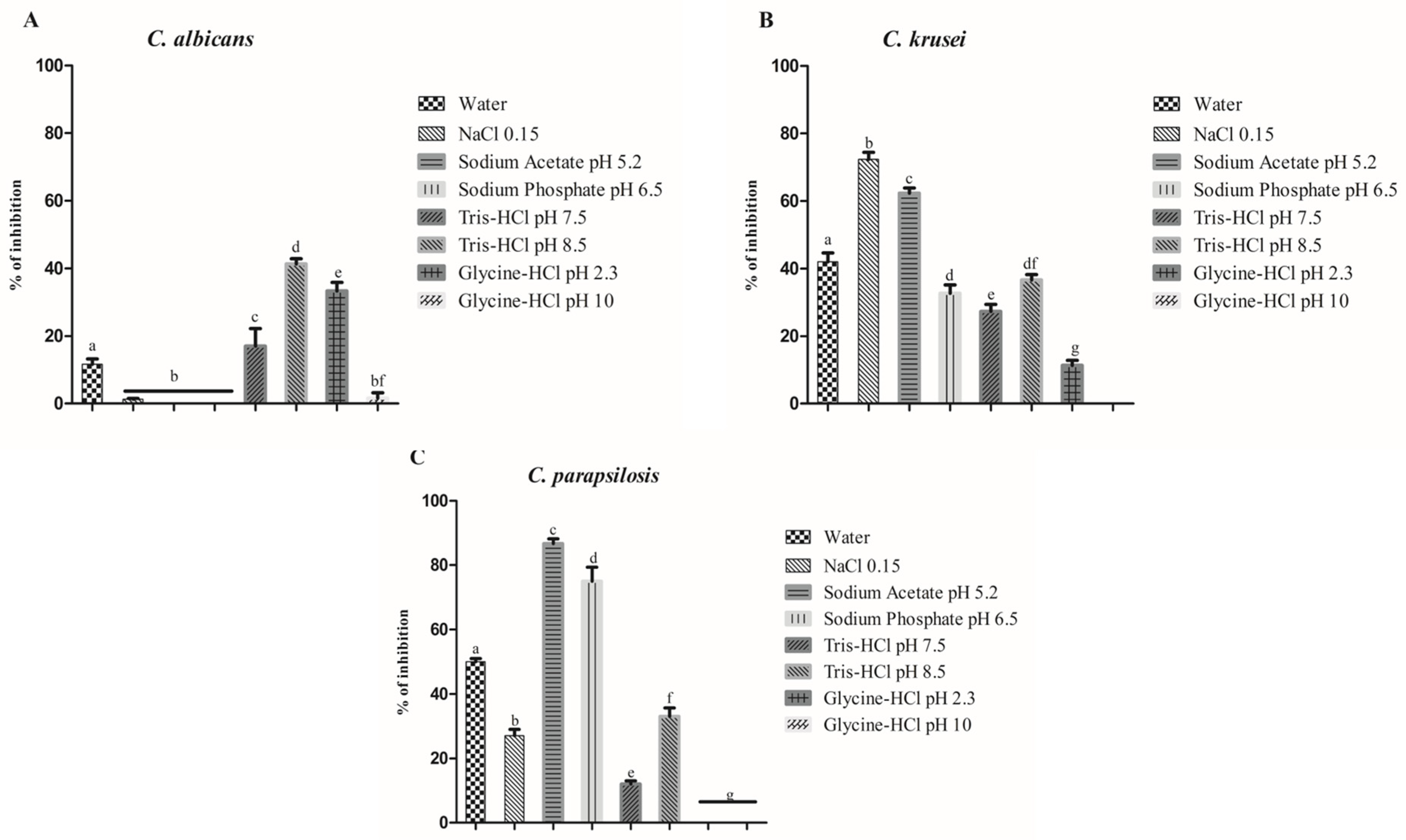
3.1. Protein Extraction and Antimicrobial Activity

Seeking the best way to extract proteins from K. alvarezii with biological properties, eight solutions were tested, covering a pH range of 2.3 to 10 (Table 1). Based on the results, the highest protein content, 2.55 mg P−1, was obtained with the alkaline buffer glycine-NaOH pH 10. The lowest content of protein, 0.53 mgP−1, was obtained with the acidic buffer glycine-HCl pH 2.3 (Table 1). The other buffers extracted the content of proteins ranging from 1.35 to 2.15 mg P−1 (Table 1).
Regarding the antimicrobial activity, although there were differences in protein content, all extracts were tested for antimicrobial activity. All the extracts were tested at a concentration of 50 µg mL−1. At the concentration tested, none of the extracts presented antibacterial activity. In contrast, they were active against Candida spp. (Figure 1). Against C. albicans, Tris-HCl pH 8.5 was the best by inhibiting 40% of its growth (Figure 1A). In the case of C. krusei, the best extracts were those made with 0.15 M NaCl and 0.05 M of sodium acetate pH 5.2 by inhibiting, respectively, 75% and 60% the growth (Figure 1B). Regarding C. parapsilosis, the best activity was found in the extract produced with 0.05 M of sodium acetate pH 5.2, achieving inhibition of 85% (Figure 1C). Based on these results, the proteins extract with 0.05 M of sodium acetate pH 5.2 was chosen to move forward in the studies of mechanisms of action against C. krusei and C. parapsilosis and protein characterization and identification.

3.2. Mechanisms of Action

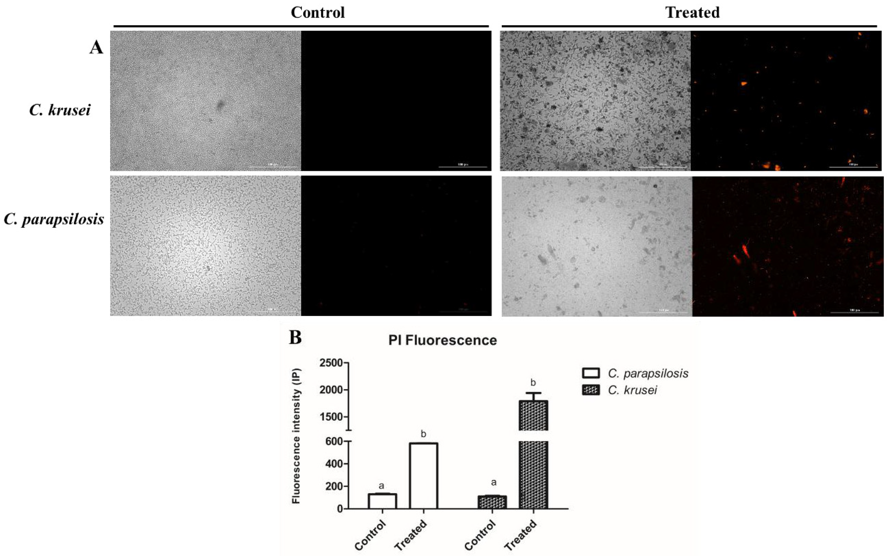
3.2.1. Membrane Permeability and Pore Formation

Aiming to understand the mechanism behind the anticandidal activity of proteins from K. alvarezii, the PI assay was performed (Figure 2). As expected, the control cells treated with only 50 mM of sodium acetate buffer pH 5.2 presented no fluorescence of PI, indicating the membrane is healthy because PI cannot pass through the membrane. In contrast, the red fluorescence in cells treated with sodium acetate proteins revealed increased membrane permeability, indicating damage in the cell membrane (Figure 2).
The data produced by PI are not enough to indicate pore formation on the membrane based on the size of PI. Based on that, cells were treated again with proteins extracted with acetate buffer and incubated with a 6 kDa dextran coupled with FITC. Only C. krusei cells presented fluorescence, showing the dextran-FITC within the cell, suggesting movement through the membrane, indicating a pore with a size of at least 6 kDa. In contrast, in C. parapsilosis cells, the absence of green fluorescence indicates the presence of a pore higher than PI but lower than dextran-FITC (Figure 2).

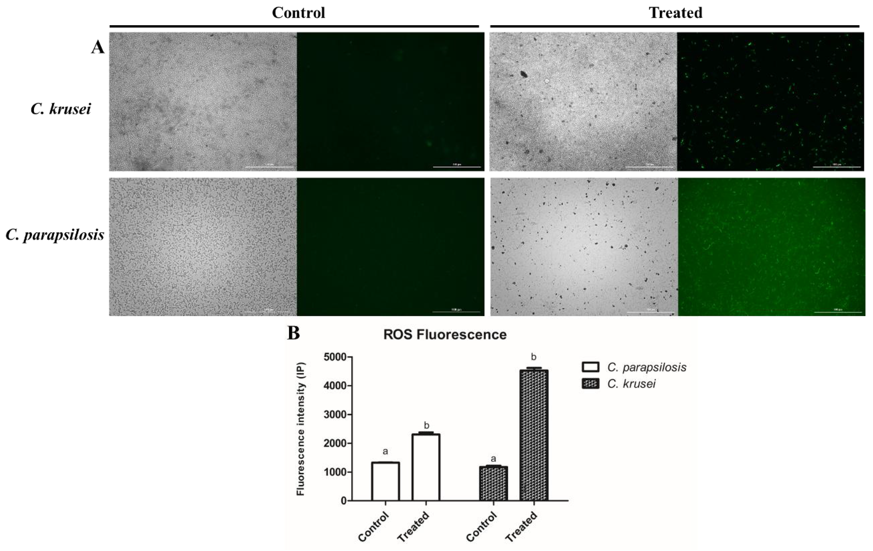
3.2.2. ROS Overaccumulation and Apoptosis

After analyzing the mechanisms of action, it was evaluated whether the proteins from K. alvarezii, the ROS overaccumulation, and apoptosis mediated by caspase 3/7 were assessed. First, the proteins from K. alvarezii induced ROS accumulation only in C. parapsilosis cells and not in C. krusei (Figure 3). In contrast, the proteins from K. alvarezii induced apoptosis mediated by 3/7 caspase in C. parapsilosis and C. krusei (Figure 4).

3.2.3. Evaluation of Change in Cell Morphology by Scanning Electron Microscopy (SEM)

The following steps evaluated whether the proteins from K. alvarezzi could induce damage in the morphology of Candida cells. As expected, the control cells from C. krusei (Figure 5A) and C. parapsilosis (Figure 6E) presented no damage or cracks on the cell structure. In the case of C. krusei treated with proteins from K. alvarezzi cells, scars (Figure 6B,C—white arrows), cracks, and damage on the cell wall were observed (Figure 6D). In some cases, it is evident that there is a loss of internal content (Figure 6B,C—white arrow), suggesting damage to the membrane. Regarding cells of C. parapsilosis treated with proteins from K. alvarezzi, it was observed that cells presented a scaly, rough surface and damaged morphology (Figure 6F). Additionally, cracks, damage on the cell wall, and loss of internal content were observed (Figure 6G,H).

3.3. Identification and Characterization of Proteins in the K. alvarezzi Extract

The antimicrobial assays revealed the biotechnological potential of proteins extracted from K. alvarezzi. Based on that, our further experiments focused on identifying the proteins in the extract. The LC-MS/MS analysis identified 336 proteins in the extract of K. alvarezzi extracted with sodium acetate buffer (Supplementary Table S1). The proteins identified cover all cellular compartments and develop several molecular functions in K. alvarezzi’s cells (Figure 7A,B). The biological processes displayed by those proteins are carbohydrate metabolism (3%), cell cycle, morphology, and growth (16%), cell signaling (5%), DNA metabolism (4%), energetic metabolism (7%), nucleotide metabolism (4%), photosynthesis (5%), protein metabolism (27%), stress response (6%), and unknown (27%) (Figure 7C).
In addition to the LC-MS/MS analysis, biochemical analysis revealed the presence of proteins such as proteinases, glucanases, chitinase, and serine and cysteine proteinase inhibitors, and proteins involved in redox metabolism, such as superoxide dismutase, catalase, ascorbate peroxidase, and phenol peroxidase (Table 2).

4. Discussion

Over the years, the red marine seaweed K. alvarezzi has mainly been employed to build sustainable and environmentally friendly agriculture and soil fertilization [30]. The content of nutrients in K. alvarezzi is the reason for this seaweed’s attention. Analysis of K. alvarezzi revealed the content of carbohydrates (26 g), protein (18 g), fats (2 g), and fibers (6 g) per 100 g of dry wet tissue [31]. Regarding micronutrients necessary for plant nutrition, K. alvarezzi has 15 mg of phosphorus, 10 mg of potassium, 5 mg of calcium, and 2 mg of magnesium per 100 mg of dry tissue [31]. Altogether, this content of nutrients made K. alvarezzi a good source of nutrients that are valuable in agriculture.
Despite its application in agriculture, K. alvarezzi has many applications in human health by presenting antibacterial, anticancer, antiviral, antifungal, and antiglycemic effects [31,32,33,34,35,36]. Most studies with K. alvarezzi focused on secondary compounds such as phenolic, terpenoids, flavonoids, and alkaloids, all extracted with organic solvents, which cannot extract proteins [31,32,33,34,35,36]. For example, Sit et al. [36] evaluated the antidermatophytic activity of K. alvarezzi with organic and aqueous extracts—the extract produced with water presented no activity against dermatophytes. In contrast, the extract prepared with hexane inhibited 50% of dermatophyte growth at a concentration 25-fold (1.25 mg mL−1) higher than that of proteins from K. alvarezzi.
However, a study by Xu et al. [32] showed the potential of proteins from K. alvarezzi differently. Based on in silico analysis, authors sought in the genome of K. alvarezzi a lectin with high potential to present anticancer activity. Based on the results, the authors produced the lectin by heterologous expression. They tested it in vitro against various types of cancer. Those results highlight the importance and pioneering nature of our study in identifying the proteins from K. alvarezzi by MS analysis and biochemical analysis associated with antifungal activity.
In the search for proteins with biological applications, screening employing different buffers is a common method [20,37,38]. Here, different aqueous buffers were tested to find the best extracting solution in terms of protein (Table 1). All buffers extracted proteins and presented some antifungal activity against C. parapsilosis and C. krusei. However, the best antifungal activity was found with proteins extracted with 50 mM Na+-acetate pH 5.2 (Figure 1). As in other works [27,35], the acidic buffer extracted the lowest protein. This is a common problem with acidic buffers due to their effect on protein structure and, thus, solubility. Silva et al. [20] showed that the same buffer extracted proteins similar to those in our study. The best antifungal activity was found here with proteins extracted with sodium acetate buffer, like those in Silva et al. [20]. In our case, the proteins extracted were more active against C. parapsilosis and C. krusei than those in Silva et al. [20], where proteins were more effective against C. krusei and C. albicans.
Based on the antifungal activity found, further analysis employed fluorescence and scanning electron microscopes to study the antifungal action of proteins from K. alvarezzi (Figure 2, Figure 3, Figure 4, Figure 5 and Figure 6). The proteins extracted with sodium-acetate buffer induced an increase in membrane permeabilization (Figure 2) but only induced a 6 kDa pore in C. krusei (Figure 3). An increase in membrane permeability is a usual mechanism of action caused by antifungal proteins. However, it is not enough to suggest the formation of pores in the cell membrane. Etxaniz et al. [39] revealed that the pores in the membrane that allow the movement of PI are too small (0.1 nm), and a cell can easily recover. But bigger pores, such as 6 kDa ones, cause considerable damage that cells cannot heal [39].
By attacking the cell membrane, the proteins from K. alvarezzi employed a mechanism of action different from commercial drugs with specific targets [40,41,42]. For example, the azoles inhibit proteins involved in ergosterol biosynthesis, and polyenes interact with the ergosterol in the membrane [3,9,19,43,44,45]. The common resistance mechanisms are changing the proteins involved in ergosterol and reducing the levels in the membranes, respectively [9]. By attacking the membranes, the proteins from K. alvarezzi make the acquisition of resistance by Candida cells hard.
On top of that, the hypothesis that proteins from K. alvarezzi attack the membrane, causing damage, was corroborated by the SEM analysis (Figure 6). As revealed (Figure 6), both C. parapsilosis and C. krusei cells are damaged, exhibiting abnormal morphology and a broken cell wall. Silva et al. [20] and Sousa et al. [37] found similar results using protein extracts. The mechanisms on the membrane and cell wall presented by proteins from K. alverezzi raise the question about the proteins in the extract. The answer comes from the LC-MS analysis (Figure 7 and Supplementary Table S1) and biochemical analysis (Table 2).
Mass spectrometry analysis of proteins from K. alvarezzi revealed the presence of proteins such as chitinases, Exo-beta-D-glucosaminidase, cysteine and serine proteinase inhibitors, and metallo (Supplementary Table S1) that could be involved in the antifungal activity of the extract from K. alvarezzi. Corroborating with the MS analysis, the biochemical analysis found the activity of chitinase, cysteine and serine proteinase inhibitors, and total proteinase activity (Table 2).
The presence of chitinase could be related to the damage associated with the cell wall in C. parapsilosis and C. krusei (Figure 6D,G—white arrows). Chitinases degrade chitin to N-acetyl-glucosamine [46,47]. It turns out that chitin is a significant component in yeast cell walls. The cell wall develops several functions in yeasts, including protection from physical damage, shape and rigidity, selective barrier, and adhesion [48]. Additionally, two identified enzymes could work coordinated with chitinase: (1) Exo-beta-D-glucosaminidase identified by LC-MS analysis (Supplementary Table S1) and (2) β-1,3 glucanase (Table 2) determined by biochemical assay. Together, these three proteins may be responsible for damaging the fungal cell wall, leading to cell death.
The cell wall and membrane are composed of proteins and carbohydrates. As such, proteases are proteins that could attack and damage the cell wall and membrane [20,38,49,50,51]. For example, Torres-Ossandón et al. [52] showed that a protease-rich fraction displayed antifungal activity against the fungus Botrytis cinerea. Freita et al. [49] reported that cysteine protease increases the membrane permeabilization in the membrane of Fusarium oxysporum and induces loss of internal content, as revealed by atomic force microscopy. Total protease activity was shown using azocasein as a substrate (Table 2). In addition, many types of proteases were found by LC-MS analysis, such as metallo, cysteine, and serine proteinases (Supplementary Table S1). Proteases are involved in protein metabolism by activating other proteins or degrading proteins to recycle the amino acids. They are also critical components of proteasome complexes involved in many cell functions. The gene ontology analysis revealed that 23% of the proteins identified were involved in the protein metabolism of K. alvarezzi cells (Figure 7C).
Together with proteases, MS analysis detected the presence of protease inhibitors of both classes, which regulate protease function [53,54]. Accordingly, biochemical analysis revealed the activity of both proteins (Table 2). Protease inhibitors have already been reported to display antimicrobial activity against C. albicans. A Kunitz trypsin inhibitor purified from Cassia leiandra induced membrane pore formation and the loss of cytoplasmic content in C. albicans cells. Those results are similar to the results presented by proteins from K. alvarezzi [55]. Melo et al. [56] reported that ClCPI, a cysteine protease inhibitor from C. leiandra, attacks C. tropicalis cells, leading to morphological alterations and cell death.

5. Conclusions

The protein profile and biotechnological potential of the proteins in K. alvarezzi are reported for the first time here. Our data provide insights about the mechanisms of action of proteins from K. alvarezzi, which are mediated by destabilizing the cell membrane and cell wall of C. parapsilosis and C. krusei by inducing pore formation, high levels of ROS, followed by apoptosis, and cell death. The results suggest that the protein obtained from K. alvarezzi could be a new alternative to control C. parapsilosis and C. krusei infections.

Supplementary Materials

The following supporting information can be downloaded at: https://www.mdpi.com/article/10.3390/pr13051569/s1, Table S1: Proteins from K. alvarezzi identified by LC-ESI-MS/MS analysis.

Author Contributions

Conceptualization, P.F.N.S., N.S.d.S.F., J.L.T.M., F.I.R.G., A.V.C.d.O., G.B.M., B.M.S., E.L.d.S., D.d.S.C.M.C.-B., J.H.d.A.F., J.V.S.N., R.C.M. and F.P.M.; Methodology, P.F.N.S., P.G.L., N.S.d.S.F., F.I.R.G., A.V.C.d.O., G.B.M., B.M.S., E.L.d.S., D.d.S.C.M.C.-B., J.H.d.A.F., J.V.S.N., R.C.M. and F.P.M.; Software, P.F.N.S., N.S.d.S.F., J.L.T.M., G.B.M. and R.C.M.; Validation, P.G.L. and F.P.M.; Formal analysis, P.F.N.S., P.G.L., J.L.T.M., A.V.C.d.O., E.L.d.S., D.d.S.C.M.C.-B., J.H.d.A.F., J.V.S.N. and F.P.M.; Investigation, P.F.N.S., P.G.L., J.L.T.M., F.I.R.G., A.V.C.d.O., B.M.S. and R.C.M.; Resources, P.G.L., N.S.d.S.F. and F.I.R.G.; Data curation, N.S.d.S.F., G.B.M., B.M.S., E.L.d.S. and J.H.d.A.F.; Writing—original draft, P.F.N.S.; Writing – review & editing, P.F.N.S.; Visualization, P.G.L. and J.V.S.N.; Supervision, P.F.N.S., F.I.R.G., A.V.C.d.O., E.L.d.S., J.H.d.A.F., J.V.S.N., R.C.M. and F.P.M.; Project administration, P.F.N.S., N.S.d.S.F., J.L.T.M., B.M.S., D.d.S.C.M.C.-B., J.H.d.A.F., R.C.M. and F.P.M.; Funding acquisition, P.F.N.S., J.L.T.M., G.B.M. and D.d.S.C.M.C.-B. All authors have read and agreed to the published version of the manuscript.

Funding

This work was supported by the National Council for Scientific and Technological Development (CNPq) for a research grant to Felipe P. Mesquita (Process number: 421392/2023-1). Raquel C. Montenegro thanks CNPq (Process number: 305459/2019-8) for a research productivity grant. Pedro F. N. Souza thanks the CNPq for the research productivity grant (Process number: 305003/2022-4) and the Cearense Foundation to Support Scientific and Technological Development (FUNCAP) for visiting research grant (process nº PVS-0215 00099.01.00/23). We also thank the Office of Coordination for the Improvement of Higher Education Personnel (CAPES). We also thank the Conselho Nacional de Desenvolvimento Científico e Tecnológico (CNPq) and Fundação Coordenação de Aperfeiçoamento do Pessoal do Ensino Superior (CAPES) grant number 405934/2022-0 (The National Institute of Science and Technology INCT Funvir), from Brazil to Pedro F. N. Souza.

Data Availability Statement

The original contributions presented in this study are included in the article/supplementary material. Further inquiries can be directed to the corresponding authors.

Acknowledgments

We are grateful to the staff of the central analytical facilities of UFC, Brazil, for the SEM analysis and the Multi-User Facility of the Drug Research and Development Center of the Federal University of Ceará for technical support with fluorescence analysis.

Conflicts of Interest

The authors declare no conflicts of interest.

References

  1. Pickering, T.D.; Skelton, P.; Sulu, R.J. Intentional introductions of commercially harvested alien seaweeds. Bot. Mar. 2007, 50, 338–350. [Google Scholar] [CrossRef]
  2. Hayashi, L.; Santos, A.A.; Faria, G.S.M.; Nunes, B.G.; Souza, M.S.; Fonseca, A.L.D.; Barreto, P.L.M.; Oliveira, E.C.; Bouzon, Z.L. Kappaphycus alvarezii (Rhodophyta, Areschougiaceae) cultivated in subtropical waters in Southern Brazil. J. Appl. Phycol. 2011, 23, 337–343. [Google Scholar] [CrossRef]
  3. Canuto, M.M.; Rodero, F.G. Antifungal drug resistance to azoles and polyenes. Lancet Infect. Dis. 2002, 2, 550–563. [Google Scholar] [CrossRef] [PubMed]
  4. Fayaz, M.; Namitha, K.K.; Murthy, K.N.C.; Swamy, M.M.; Sarada, R.; Khanam, S.; Subbarao, P.V.; Ravishankar, G.A. Chemical composition, iron bioavailability, and antioxidant activity of Kappaphycus alvarezzi (Doty). J. Agric. Food Chem. 2005, 53, 792–797. [Google Scholar] [CrossRef]
  5. Sanjivkumar, M.; Chandran, M.N.; Suganya, A.M.; Immanuel, G. Investigation on bio-properties and in-vivo antioxidant potential of carrageenans against alloxan induced oxidative stress in Wistar albino rats. Int. J. Biol. Macromol. 2020, 151, 650–662. [Google Scholar] [CrossRef]
  6. Raman, M.; Doble, M. κ-Carrageenan from marine red algae, Kappaphycus alvarezii—A functional food to prevent colon carcinogenesis. J. Funct. Foods 2015, 15, 354–364. [Google Scholar] [CrossRef]
  7. Morgan, J.; Meltzer, M.I.; Plikaytis, B.D.; Sofair, A.N.; Huie-White, S.; Wilcox, S.; Harrison, L.H.; Seaberg, E.C.; Hajjeh, R.A.; Teutsch, S.M. Excess Mortality, Hospital Stay, and Cost Due to Candidemia: A Case-Control Study Using Data From Population-Based Candidemia Surveillance. Infect. Control Hosp. Epidemiol. 2005, 26, 540–547. [Google Scholar] [CrossRef]
  8. Brown, G.D.; Denning, D.W.; Gow, N.A.R.; Levitz, S.M.; Netea, M.G.; White, T.C. Hidden Killers: Human Fungal Infections. Sci. Transl. Med. 2012, 4, 165rv13. [Google Scholar] [CrossRef]
  9. Sanguinetti, M.; Posteraro, B.; Lass-Flörl, C. Antifungal drug resistance among Candida species: Mechanisms and clinical impact. Mycoses 2015, 58 (Suppl. 2), 2–13. [Google Scholar] [CrossRef]
  10. Antinori, S.; Milazzo, L.; Sollima, S.; Galli, M.; Corbellino, M. Candidemia and invasive candidiasis in adults: A narrative review. Eur. J. Intern. Med. 2016, 34, 21–28. [Google Scholar] [CrossRef]
  11. Quindós, G. Epidemiology of candidaemia and invasive candidiasis. A changing face. Rev. Iberoam. Micol. 2014, 31, 42–48. [Google Scholar] [CrossRef] [PubMed]
  12. Schneider, J.; Mateo, E.; Marcos-Arias, C.; Eiró, N.; Vizoso, F.; Pérez-Fernández, R.; Eraso, E.; Quindós, G. Antifungal activity of the human uterine cervical stem cells conditioned Medium (hUCESC-CM) against Candida albicans and other medically relevant species of Candida. Front. Microbiol. 2018, 9, 2818. [Google Scholar] [CrossRef] [PubMed]
  13. Chaves, G.M.; Cavalcanti, M.A.Q.; Porto, A.L.F. Pathogenicity characteristics of stocked and fresh yeasts strains. Braz. J. Microbiol. 2003, 34, 197–202. [Google Scholar] [CrossRef]
  14. Pinto, T.N.; Kohn, A.; Da Costa, G.L.; Oliveira, L.M.A.; Pinto, T.C.A.; Oliveira, M.M.E. Candida guilliermondii as an agent of postpartum subacute mastitis in Rio de Janeiro, Brazil: Case report. Front. Microbiol. 2022, 13, 964685. [Google Scholar] [CrossRef]
  15. Naglik, J.R.; Challacombe, S.J.; Hube, B. Candida albicans secreted aspartyl proteinases in virulence and pathogenesis. Microbiol. Mol. Biol. Rev. 2003, 67, 400–428. [Google Scholar] [CrossRef]
  16. Puig-Asensio, M.; Pemán, J.; Zaragoza, R.; Garnacho-Montero, J.; Martín-Mazuelos, E.; Cuenca-Estrella, M.; Almirante, B. Impact of therapeutic strategies on the prognosis of Candidemia in the ICU. Crit. Care Med. 2014, 42, 1423–1432. [Google Scholar] [CrossRef] [PubMed]
  17. Monge, R.A.; Roman, E.; Nombela, C.; Pla, J. The MAP kinase signal transduction network in Candida albicans. Microbiology 2006, 152, 905–912. [Google Scholar] [CrossRef]
  18. Colombo, A.L.; Guimarães, T. Epidemiology of hematogenous infections due to Candida spp. Rev. Soc. Bras. Med. Trop. 2003, 36, 599–607. [Google Scholar] [CrossRef]
  19. Berkow, E.L.; Lockhart, S.R. Fluconazole resistance in Candida species: A current perspective. Infect. Drug Resist. 2017, 10, 237–245. [Google Scholar] [CrossRef]
  20. Silva, A.L.; Bezerra, L.P.; Freitas, C.D.T.; Silva, A.F.B.; Mesquita, F.P.; Neto, N.A.S.; Oliveira, J.P.B.; Aguiar, T.K.B.; Nagano, C.S.; Carneiro, R.F.; et al. Luffa operculata seed proteins: Identification by LC-ESI-MS/MS and biotechnological potential against Candida albicans and C. krusei. Anal. Biochem. 2022, 655, 114851. [Google Scholar] [CrossRef]
  21. Bradford, M.M. A rapid and sensitive method for the quantitation of microgram quantities of protein utilizing the principle of protein-dye binding. Anal. Biochem. 1976, 72, 248–254. [Google Scholar] [CrossRef]
  22. Lopes, F.E.S.; da Costa, H.P.S.; Souza, P.F.N.; Oliveira, J.P.B.; Ramos, M.V.; Freire, J.E.C.; Jucá, T.L.; Freitas, C.D.T. Peptide from thaumatin plant protein exhibits selective anticandidal activity by inducing apoptosis via membrane receptor. Phytochemistry 2019, 159, 46–55. [Google Scholar] [CrossRef] [PubMed]
  23. Crowley, L.C.; Scott, A.P.; Marfell, B.J.; Boughaba, J.A.; Chojnowski, G.; Waterhouse, N.J. Measuring cell death by propidium iodide uptake and flow cytometry. Cold Spring Harb. Protoc. 2016, 2016, 647–651. [Google Scholar] [CrossRef] [PubMed]
  24. Maurya, I.K.; Pathak, S.; Sharma, M.; Sanwal, H.; Chaudhary, P.; Tupe, S.; Deshpande, M.; Chauhan, V.S.; Prasad, R. Antifungal activity of novel synthetic peptides by accumulation of reactive oxygen species (ROS) and disruption of cell wall against Candida albicans. Peptides 2011, 32, 1732–1740. [Google Scholar] [CrossRef] [PubMed]
  25. Staniszewska, M.; Bondaryk, M.; Swoboda-Kopec, E.; Siennicka, K.; Sygitowicz, G.; Kurzatkowski, W. Candida albicans morphologies revealed by scanning electron microscopy analysis. Braz. J. Microbiol. 2013, 44, 813–821. [Google Scholar] [CrossRef]
  26. Xavier-Filho, J.; Campos, F.A.P.; Ary, M.B.; Silva, C.P.; Carvalho, M.M.M.; Macedo, M.L.R.; Lemos, F.J.A.; Grant, G. Poor Correlation between the Levels of Proteinase Inhibitors Found in Seeds of Different Cultivars of Cowpea (Vigna unguiculata) and the Resistance/Susceptibility to Predation by Callosobruchus maculatus. J. Agric. Food Chem. 1989, 37, 1139–1143. [Google Scholar] [CrossRef]
  27. Urbanek, H.; Kuzniak-Gebarowska, E.; Herka, K. Elicitation of defence responses in bean leaves by Botrytis cinerea polygalacturonase. Acta Physiol. Plant. 1991, 13, 43–50. [Google Scholar]
  28. Song, Y.Y.; Zeng, R.S.; Xu, J.F.; Li, J.; Shen, X. Interplant Communication of Tomato Plants through Underground Common Mycorrhizal. PLoS ONE 2010, 5, e13324. [Google Scholar] [CrossRef]
  29. Karpievitch, Y.V.; Polpitiya, A.D.; Anderson, G.A.; Smith, R.D.; Dabney, A.R. Liquid Chromatography Mass Spectrometry-Based Proteomics: Biological and Technological Aspects. Ann. Appl. Stat. 2010, 4, 1797. [Google Scholar] [CrossRef]
  30. Illera-Vives, M.; Seoane Labandeira, S.; Fernández-Labrada, M.; López-Mosquera, M.E. Agricultural Uses of Seaweed. In Sustainable Seaweed Technologies; Elsevier: Amsterdam, The Netherlands, 2020; pp. 591–612. [Google Scholar] [CrossRef]
  31. Khotijah, S.; Irfan, M.; Muchdar, F. Nutritional Composition of Seaweed Kappaphycus alvarezii. Agrikan J. Agribisnis Perikan. 2020, 13, 139–146. [Google Scholar] [CrossRef]
  32. Xu, T.; Wang, Y.C.; Ma, J.; Cui, Y.; Wang, L. In silico discovery and anti-tumor bioactivities validation of an algal lectin from Kappaphycus alvarezii genome. Int. J. Biol. Macromol. 2024, 275, 133311. [Google Scholar] [CrossRef]
  33. Hirayama, M.; Shibata, H.; Imamura, K.; Sakaguchi, T.; Hori, K. High-Mannose Specific Lectin and Its Recombinants from a Carrageenophyta Kappaphycus alvarezii Represent a Potent Anti-HIV Activity Through High-Affinity Binding to the Viral Envelope Glycoprotein gp120. Mar. Biotechnol. 2016, 18, 215–231. [Google Scholar] [CrossRef] [PubMed]
  34. Das, D.; Arulkumar, A.; Paramasivam, S.; Lopez-Santamarina, A.; del Carmen Mondragon, A.; Miranda Lopez, J.M. Phytochemical Constituents, Antimicrobial Properties and Bioactivity of Marine Red Seaweed (Kappaphycus alvarezii) and Seagrass (Cymodocea serrulata). Foods 2023, 12, 2811. [Google Scholar] [CrossRef]
  35. Yulianti, E.; Sunarti; Wahyuningsih, M.S.H. The effect of Kappaphycus alvarezii fraction on plasma glucose, Advanced Glycation End-products formation, and renal RAGE gene expression. Heliyon 2021, 7, e05978. [Google Scholar] [CrossRef] [PubMed]
  36. Sit, N.W.; Chan, Y.S.; Lai, S.C.; Lim, L.N.; Looi, G.T.; Tay, P.L.; Tee, Y.T.; Woon, Y.Y.; Khoo, K.S.; Ong, H.C. In vitro antidermatophytic activity and cytotoxicity of extracts derived from medicinal plants and marine algae. J. Mycol. Med. 2018, 28, 561–567. [Google Scholar] [CrossRef]
  37. Sousa, A.J.S.; Souza, P.F.N.; Gifoni, J.M.; Dias, L.P.; Freitas, C.D.T.; Oliveira, J.T.A.; Sousa, D.O.B.; Vasconcelos, I.M. Scanning electron microscopy reveals deleterious effects of Moringa oleifera seed exuded proteins on root-knot nematode Meloidogyne incognita eggs. Int. J. Biol. Macromol. 2020, 154, 1237–1244. [Google Scholar] [CrossRef] [PubMed]
  38. Mota, T.R.; Linhares, H.V.S.; Araújo-Filho, J.H.; Veras, D.M.; Costa, H.P.S.; Souza, C.M.P.; Souza, P.F.N.; Martins, T.F. Protein extract from Cereus jamacaru (DC.) inhibits Colletotrichum gloeosporioides growth by stimulating ROS generation and promoting severe cell membrane damage. Microb. Pathog. 2019, 130, 71–80. [Google Scholar] [CrossRef]
  39. Etxaniz, A.; González-Bullón, D.; Martín, C.; Ostolaza, H. Membrane Repair Mechanisms against Permeabilization by Pore-Forming Toxins. Toxins 2018, 10, 234. [Google Scholar] [CrossRef]
  40. Lima, A.M.; Azevedo, M.I.G.; Sousa, L.M.; Oliveira, N.S.; Andrade, C.R.; Freitas, C.D.T.; Souza, P.F.N. Plant antimicrobial peptides: An overview about classification, toxicity and clinical applications. Int. J. Biol. Macromol. 2022, 214, 10–21. [Google Scholar] [CrossRef]
  41. Lima, P.G.; Oliveira, J.T.A.; Amaral, J.L.; Freitas, C.D.T.; Souza, P.F.N. Synthetic antimicrobial peptides: Characteristics, design, and potential as alternative molecules to overcome microbial resistance. Life Sci. 2021, 278, 119647. [Google Scholar] [CrossRef]
  42. Souza, P.F.N.; Marques, L.S.M.; Oliveira, J.T.A.; Lima, P.G.; Dias, L.P.; Neto, N.A.S.; Lopes, F.E.S.; Sousa, J.S.; Silva, A.F.B.; Caneiro, R.F.; et al. Synthetic antimicrobial peptides: From choice of the best sequences to action mechanisms. Biochimie 2020, 175, 132–145. [Google Scholar] [CrossRef] [PubMed]
  43. Lockhart, S.R.; Chowdhary, A.; Gold, J.A.W.; Aspergillus, A. The rapid emergence of antifungal-resistant human-pathogenic fungi. Nat. Rev. Microbiol. 2023, 21, 818–832. [Google Scholar] [CrossRef]
  44. Taei, M.; Chadeganipour, M.; Mohammadi, R. An alarming rise of non-albicans Candida species and uncommon yeasts in the clinical samples; a combination of various molecular techniques for identification of etiologic agents. BMC Res. Notes 2019, 12, 779. [Google Scholar] [CrossRef]
  45. Berman, J.; Krysan, D.J. Drug resistance and tolerance in fungi. Nat. Rev. Microbiol. 2020, 18, 319–331. [Google Scholar] [CrossRef] [PubMed]
  46. Kumar, M.; Brar, A.; Yadav, M.; Chawade, A.; Vivekanand, V.; Pareek, N. Chitinases—Potential Candidates for Enhanced Plant Resistance towards Fungal Pathogens. Agriculture 2018, 8, 88. [Google Scholar] [CrossRef]
  47. Žiarovská, J.; Zamiešková, L.; Bilčíková, J.; Fialková, V.; Sabo, J.; Kunová, S.; Kačániová, M. Expression of Specific Class I Chitinase mRNA Levels in Different Grape Varieties and Their Antimicrobial Activity. Agronomy 2020, 10, 1176. [Google Scholar] [CrossRef]
  48. Stewart, G.G. The Structure and Function of the Yeast Cell Wall, Plasma Membrane and Periplasm. In Brewing and Distilling Yeasts; Springer: Berlin/Heidelberg, Germany, 2017; pp. 55–75. [Google Scholar] [CrossRef]
  49. Freitas, C.D.T.; Silva, R.O.; Ramos, M.V.; Porfírio, C.T.M.N.; Farias, D.F.; Sousa, J.S.; Oliveira, J.P.B.; Souza, P.F.N.; Dias, L.P.; Grangeiro, T.B. Identification, characterization, and antifungal activity of cysteine peptidases from Calotropis procera latex. Phytochemistry 2020, 169, 112163. [Google Scholar] [CrossRef]
  50. Díaz, M.E.; Rocha, G.F.; Kise, F.; Rosso, A.M.; Guevara, M.G.; Parisi, M.G. Antimicrobial activity of an aspartic protease from Salpichroa origanifolia fruits. Lett. Appl. Microbiol. 2018, 67, 168–174. [Google Scholar] [CrossRef]
  51. Guevara, M.G.; Oliva, C.R.; Huarte, M.; Daleo, G.R. An aspartic protease with antimicrobial activity is induced after infection and wounding in intercellular fluids of potato tubers. Eur. J. Plant Pathol. 2002, 108, 131–137. [Google Scholar] [CrossRef]
  52. Torres-Ossandón, M.J.; Vega-Gálvez, A.; Salas, C.E.; Rubio, J.; Silva-Moreno, E.; Castillo, L. Antifungal activity of proteolytic fraction (P1G10) from (Vasconcellea cundinamarcensis) latex inhibit cell growth and cell wall integrity in Botrytis cinerea. Int. J. Food Microbiol. 2019, 289, 7–16. [Google Scholar] [CrossRef]
  53. Rashed, N.A.; MacDonald, M.H.; Matthews, B.F. Protease Inhibitor Expression in Soybean Roots Exhibiting Susceptible and Resistant Interactions with Soybean Cyst Nematode. J. Nematol. 2008, 40, 138. [Google Scholar] [PubMed]
  54. Zhang, H.; Mao, J.; Liu, F.; Zeng, F. Expression of a nematode symbiotic bacterium-derived protease inhibitor protein in tobacco enhanced tolerance against Myzus persicae. Plant Cell Rep. 2012, 31, 1981–1989. [Google Scholar] [CrossRef] [PubMed]
  55. Araújo, N.M.S.; Dias, L.P.; Costa, H.P.S.; Sousa, D.O.B.; Vasconcelos, I.M.; de Morais, G.A.; Oliveira, J.T.A. ClTI, a Kunitz trypsin inhibitor purified from Cassia leiandra Benth. seeds, exerts a candidicidal effect on Candida albicans by inducing oxidative stress and necrosis. Biochim. Biophys. Acta Biomembr. 2019, 1861, 183032. [Google Scholar] [CrossRef] [PubMed]
  56. Melo, I.R.S.; Dias, L.P.; Araújo, N.M.S.; Vasconcelos, I.M.; Martins, T.F.; de Morais, G.A.; Gonçalves, J.F.C.; Nagano, C.S.; Carneiro, R.F.; Oliveira, J.T.A. ClCPI, a cysteine protease inhibitor purified from Cassia leiandra seeds has antifungal activity against Candida tropicalis by inducing disruption of the cell surface. Int. J. Biol. Macromol. 2019, 133, 1115–1124. [Google Scholar] [CrossRef]
Figure 1. The anticandidal activity of proteins extracted from K. alvarezzi against Candida ssp. C. albicans (A), C. krusei (B), and C. parapsilosis (C). The regression calculation is used to find the MIC50 of the best molecule against C. albicans (insert). The data are shown as the mean and standard deviation of three independent experiments. Different letters indicate statistical significance (p < 0.05) between the treatment and control groups.
Processes 13 01569 g001
Figure 2. The proteins from K. alvarezzi increased membrane permeability by PI uptake assay. (A) Images of the fluorescence intensity of C. parapsilosis and C. krusei with the control (DMSO) and proteins from K. alvarezzi, and (B) relative fluorescence intensity values of the comparison between the DMSO group and the group treated. The data are shown as the mean and standard deviation of three independent experiments. Different letters indicate statistical significance (p < 0.05) between the treatment and control groups. White bars: 100 µm.
Processes 13 01569 g002
Figure 3. The proteins from K. alvarezzi induced the pore formation on the membrane of Candida Cells. (A) Images of the fluorescence intensity of C. parapsilosis and C. krusei with the control (DMSO) and proteins from K. alvarezzi, and (B) relative fluorescence intensity values of the comparison between the DMSO group and the group treated. The data are shown as the mean and standard deviation of three independent experiments. Different letters indicate statistical significance (p < 0.05) between the treatment and control groups. White bars: 100 µm.
Processes 13 01569 g003
Figure 4. ROS overaccumulation in Candida cells induced by proteins from K. alvarezzi. (A) Images of the fluorescence intensity of C. parapsilosis and C. krusei with the control (DMSO) and proteins from. K. alvarezzi, and (B) relative fluorescence intensity values of the comparison between the DMSO group and the group treated. The data are shown as the mean and standard deviation of three independent experiments. Different letters indicate statistical significance (p < 0.05) between the treatment and control groups. White bars: 100 µm.
Processes 13 01569 g004
Figure 5. Protein from K. alvarezzi induced apoptosis in Candida cells mediated by caspase 3/7. (A) Images of the fluorescence intensity of C. parapsilosis and C. krusei with the control (DMSO) and proteins from K. alvarezzi, and (B) relative fluorescence intensity values of the comparison between the DMSO group and the group treated. The data are shown as the mean and standard deviation of three independent experiments. Different letters indicate statistical significance (p < 0.05) between the treatment and control groups. White bars: 100 µm.
Processes 13 01569 g005
Figure 6. Scanning electron microscopy (SEM) images revealed alterations in the Candida cells morphology induced by protein from K. alvarezzi. (A) the surface of the control cells of C. parapsilosis was covered by well-defined and organized structures. (BD) C. parapsilosis cells exposed to proteins from K. alvarezzi showed surface alterations, multiple buds, scars, cell wall damage, and internal content loss. (E) the surface of the control cells of C. krusei was covered by well-defined and organized structures. (FH) C. krusei cells exposed to proteins from K. alvarezzi showed surface alterations, multiple buds, scars, cell wall damage, and internal content loss. Arrows show the damage.
Processes 13 01569 g006
Figure 7. Proteins from K. alvarezzi cells were identified by LC-ESI-MS/MS analysis. Gene ontology analysis of proteins from K. alvarezzi extracted with 50 mM sodium acetate pH 5.2. (A) classification based on the cellular component, (B) molecular function, and (C) based on biological process.
Processes 13 01569 g007
Table 1. The amount of proteins extracted from algae K. alvarezii using different buffers.
Protein ExtractsmgP
Water1.35 ± 0.070
0.15 M NaCl1.87 ± 0.009
Sodium acetate pH 5.21.21 ± 0.002
Sodium phosphate pH 6.51.67 ± 0.018
Tris-HCl pH 7.51.76 ± 0.057
Tris-HCl pH 8.52.15 ± 0.005
Glycine-HCl pH 2.30.53 ± 0.007
Glycine-HCl pH 10.02.55 ± 0.025
Values are the means ± standard deviation (SD) of biological triplicates.
Table 2. Activities of proteins present in the extract from algae K. alvarezzi.
Protein ActivityUnits a
Proteolytic (UA mg−1 of protein min−1)581.70 ± 2.405
β-1,3-glucanase (ƞkatal mg−1 of protein)0.35 ± 0.007
Chitinase (ƞkatal mg−1 of protein)0.52 ± 0.012
Serine protease inhibitor activity (UI mg−1 of protein)250.30 ± 2.314
Cysteine protease inhibitor activity (UI mg−1 of protein)171.70 ± 0.723
Superoxide Dismutase (UI mg−1 of protein)50.12 ± 0.101
Catalase (UI mg−1 of protein)25.45 ± 0.001
Ascorbate Peroxidase (UI mg−1 of protein)38.15 ± 0.005
Guaiacol Peroxidase (UA mg−1 of protein)10.14 ± 0.002
a Values are the means ± standard deviation (SD) of biological triplicates.
Disclaimer/Publisher’s Note: The statements, opinions and data contained in all publications are solely those of the individual author(s) and contributor(s) and not of MDPI and/or the editor(s). MDPI and/or the editor(s) disclaim responsibility for any injury to people or property resulting from any ideas, methods, instructions or products referred to in the content.

