Review

Effects of Light Quality Adjustment in Microalgal Cultivation: Flashing Light and Wavelength Shifts in Photobioreactor Design

Engineering Department, University of Palermo, Viale delle Scienze ed. 6, 90128 Palermo, Italy
*
Author to whom correspondence should be addressed.
Processes 2025, 13(4), 1159; https://doi.org/10.3390/pr13041159
Submission received: 4 March 2025 / Revised: 28 March 2025 / Accepted: 7 April 2025 / Published: 11 April 2025
(This article belongs to the Special Issue Bioreactor Design and Optimization Process)

Abstract

:
The distribution of light within a microalgal culture and the choice of the best wavelengths are considered the most critical aspects in the scale-up of microalgal culture. Several studies have investigated these features, resulting in a substantial body of literature that analyzes the effects in terms of an increase in biomass production or shift in its composition. This work addresses two types of light quality adjustments: the application of flashing light and shifts in light wavelength. The effects on microalgal culture are examined. Later, the application of these light features to photobioreactor design is described. Specifically, three kinds of photobioreactors are examined: (1) reactors designed to minimize light gradients, (2) reactors where the geometry produces a flashing light effect on the cells and (3) reactors that use filters to obtain a shift in the sunlight wavelength. The results showed that both the effect of flashing lights and wavelength shift strongly depends on various parameters such as the alga taken into consideration, the light intensity, the agitation type, growth medium, light intensity and temperature and, regarding the flashing light also, the frequency and the duty cycle. Despite all these specific differences, this work aims to resume and provide specific instruments for choosing operational parameters in microalgal cultivation and in photobioreactor design to achieve targeted outcomes, such as an increase in biomass production or in high-value compound accumulation.

1. Introduction

Light is one of the most important parameters influencing the growth of microalgae, a group of photoautotrophic microorganisms with assorted genetic origin [1].
The homogeneity of light access, together with nutrient concentration, salinity, temperature and pH, has a key role in microalgae growth in photobioreactors [2,3]. The kinetics of cell growth are strongly influenced by the interaction of these factors, contributing to the overall complexity of photobioreactors. Among these factors, the distribution of light and its spectrum are considered the most critical [4]. The efficiency of photosynthesis is directly related to the amount and to the quality of light absorbed by microalgae [5]. In the context of a photobioreactor system, if a single cell absorbs a portion of the transmitted light, the remaining cells in the system will share the remaining flux. Consequently, light energy becomes not homogeneous within the volume of the photobioreactor. This phenomenon is called self-shading [6,7] and causes a decrease in light availability with the depth of the photobioreactor [8]. Typically, this light attenuation phenomenon is modeled using the Lambert–Beer Law [9]:
I(z) = I0 exp(−ka X z)
where I0 (µmol m−2 s−1) is the incident light intensity; kₐ (m2 g−1) is the Lambert–Beer light extinction coefficient; X (g m−3) is the biomass concentration inside the reactor; z (m) is the axial coordinate of the reactor depth. Although this equation helps with the description of the light attenuation under a quantitative point of view, the qualitative aspects, such as the timing of light distribution and the light wavelength, have also an important role in microalgal growth and in the quality of the obtained biomass. In fact, numerous studies have shown that using narrow spectra of specific wavelengths cannot only enhance the growth rate and modulate the biochemical composition of microalgal biomass but also reduce both the capital and operational costs of lighting systems [10,11,12,13].
Photobioreactors for microalgae cultivation are broadly categorized into open-air systems and closed systems, where the latter are typically designed for axenic single-species cultures. Light-emitting diodes (LEDs) are considered the most suitable light source for the closed system cultivation, offering a long working life and up to 90% efficiency in converting energy into light [14].
The use of artificial lighting systems offers easier control but significantly increases biomass production costs [15]. Solar energy is an available alternative containing photosynthetic active radiation (PAR), the spectrum used by algae for photosynthesis, with blue and red wavelengths being most efficient to produce not only biomass but also lipids and the optimization of pigment production [16]. Although the role of wavelengths in microalgal growth is very complex and depends over several factors, in general, it is possible to state that red and blue light wavelengths are absorbed by microalgae, while green light is reflected, due to the same colors in microalgae cell [17,18,19]. Nevertheless, some studies have shown that green light can penetrate more deeply into the internal structure of cells and more efficiently excite chlorophyll [12,20], while other studies showed that green light can penetrate in depth into dense cultures where other wavelengths are absorbed first [21]. The role of wavelength is, therefore, complex and needs to be clarified to optimize conditions for cultivating microalgae on an industrial scale.
In this work, two main aspects regarding the qualitative aspects of lighting of microalgal cultures are addressed: the way in which the light is distributed within the reactors (e.g., flashing or intermittent light) and the shift in light wavelength. After an introductive part that resumes the principles of photosynthesis, the effects of flashing lights and wavelength adjustments are addressed. In the final part, the application of these theoretical principles to photobioreactor design was analyzed based on the literature and described.
This work may serve as a reference guide for choosing operational parameters in microalgal cultivation and in photobioreactor design for obtaining a specific goal, such as an increase in biomass production or in high-value compound accumulation. Moreover, this work can contribute to the consolidation and growth of the microalgal industry.

2. Photosynthesis in Microalgae: Mechanisms and Energy Conversion Pathways

2.1. Properties of Light and Its Role in Photosynthesis

To comprehend the photosynthetic mechanisms, it is necessary to focus on the nature of light and how it interacts with the photosynthetic apparatus. Light is electromagnetic radiation, with visible light constituting only a small part of the entire electromagnetic spectrum, as shown in Figure 1. Radiation has wavelengths ranging from 103 to 10−12 m; on the base of the wavelengths, the spectrum can be divided into several parts, such as radio, infrared, X-ray, etc. Visible light ranges from approximately 380 nm to around 750 nm, corresponding to the violet and the far red, respectively.
The visible light wavelengths correspond also to PAR (photosynthetic active radiation), which is the portion of the light spectrum employed in photosynthesis as it can be absorbed by the photosynthetic pigments.
Within this range, blue and red wavelengths are the most efficient for the photosynthetic apparatus as they are absorbed by light-harvesting antennae in photosynthetic units [22]. As we are going to explore later, the ratio between the blue and the red light can influence the composition of microalgae or, in superior plants, the plant development [23].
According to quantum theory, light travels in packages called photons, and each of them has an energy equal to the product between the Plank constant and its frequency ν.
Figure 1. Electromagnetic spectrum [24].
Processes 13 01159 g001
E = h ν
where h is the Planck constant, equal to 6.626 × 10⁻³⁴ J s.
The relationship between the frequency ν and the wavelength λ implies that they are inversely proportional:
λ = c v
According to these equations, energy is inversely proportional to wavelength. Consequently, photons of smaller wavelengths, such as 400 nm, corresponding to the blue, are more energetic than one of 700 nm, which is on the red. Photosynthetic pigments capture the energy of the light and transfer it to the photochemical complexes, where it is transformed into chemical energy and used for the overall photosynthetic process. Light intensity is measured in several ways and by using several units. In this review, irradiance is adopted, which is the flux of photons that hits a surface in the unit of time, measured in μmol quanta m−2 s−1 or μE m−2 s−1. An alternative unit is W m−2.

2.2. Mechanism of Photosynthesis in Microalgae
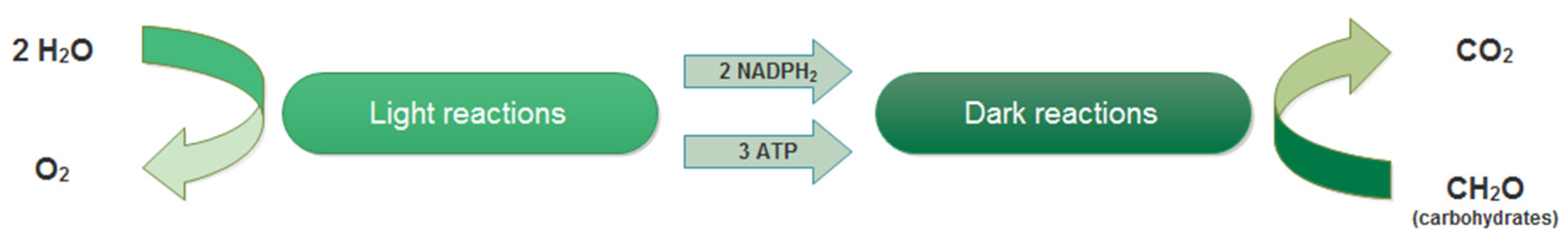
2.2.1. Overview

Oxygenic photosynthesis is performed by microorganisms such as microalgae and cyanobacteria. Cyanobacteria are prokaryotic cells in which there is no compartmentalization in organelles. In them, the genetic material is accumulated in the nucleoid, and the photosynthetic reactions occur inside a chromoplast, peripherical and close to the cell wall. On the other hand, microalgae are eukaryotic microorganisms that contain chloroplasts as organelles responsible for photosynthesis.
Photosynthesis is a process that converts solar energy and inorganic compounds in chemical energy and organic compounds. This process is essential for life on Earth, and all living beings are somehow dependent on it. The process is composed by two series of reactions, as shown in Figure 2: the light and the dark reactions. During the light reactions, the light energy is transformed in chemical energy in the form of two molecules of NADPH2 (nicotinamide adenine dinucleotide phosphate), which is a cofactor in anabolic reactions as a reducing agent, and three molecules of ATPs, providing energy for the dark reactions [25]. Water is used as a donor of electrons, and oxygen is generated as a side-product. On the other hand, dark reactions involve the fixation of atmospheric CO2 in organic compounds.
In Figure 3, the arrangement of photosystems is shown. While light is harvested by photosystems II (PSII) and photosystem I (PSI), two electrons are extracted from water, which is in turn reduced in oxygen. The electrons are transported through a series of electrons carriers, including plastoquinone (PQ), cytochrome b6/f (Cytbf) and plastocyanin (PC), to PSI, where they are used to produce NADPH. Simultaneously, protons (H+) are transported into the thylakoid lumen, and a pH gradient is generated. This gradient allows ATP synthase to work [26].

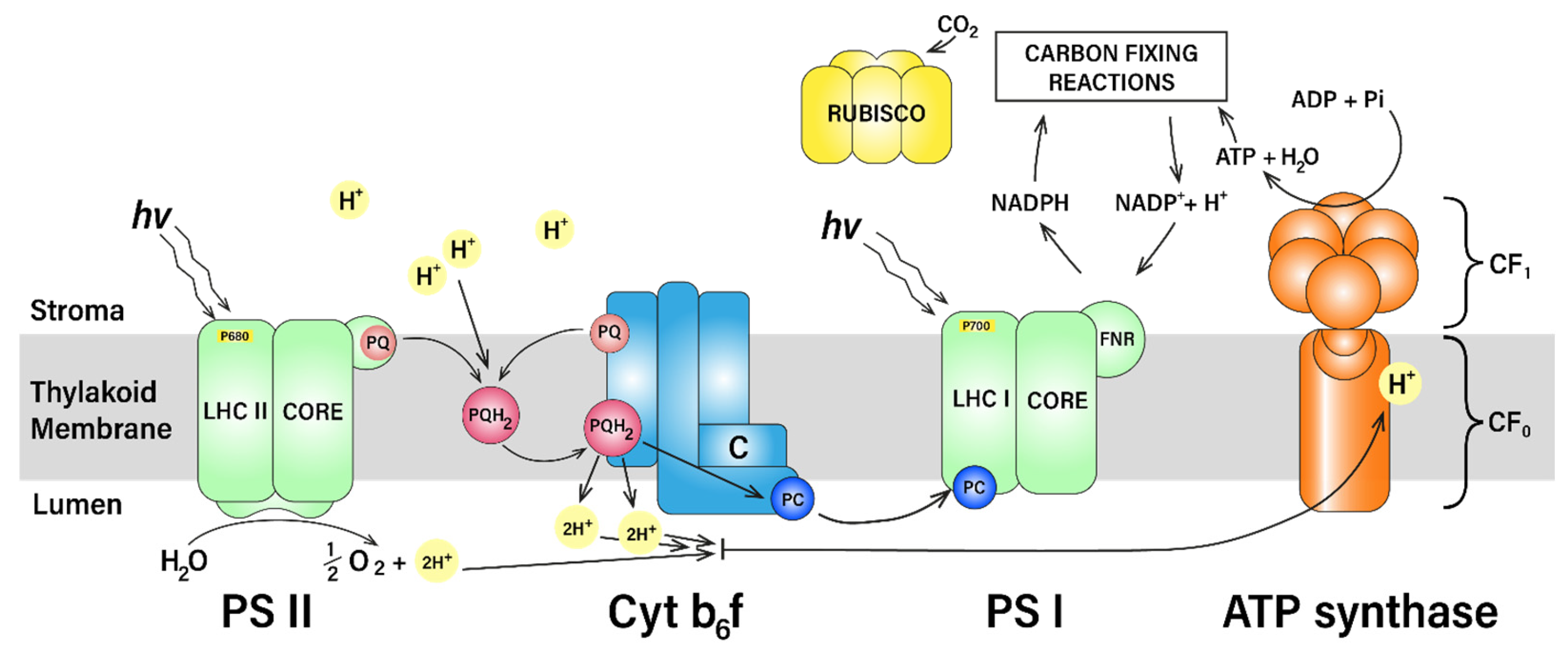
2.2.2. Light Reactions: Photochemical Process

The light reactions of photosynthesis occur in photosystems II and I (PSII and PSI), contained in the chloroplast. The chloroplast is constituted by an outer and an inner membrane that surround the stroma. Embedded in the stroma, there are grana, which are stacks of membranes called thylakoids. Between grana, there are connections of membranes called stromal lamellae, as shown in Figure 4.
Antenna systems are molecular complexes with the primary function of light harvesting and energy transfer to the photosynthetic reaction centers PSII and PSI. Light-harvesting complex II (LHC II) provides energy to PSII, and a genetically and biochemically diverse group called light-harvesting complex I (LHC I) serves PSI [27].
PSII is a macro-complex of subunits embedded in the grana, while PSI and ATPase are located in stromal lamellae. As all cellular organelles, this structure can vary; in fact, the thylakoid membrane is flexible and can reorganize PSII in order to respond to environmental stresses. PSII is a multisubunit complex contained in the chloroplast of cyanobacteria, algae and higher plants. It harvests light and converts it in chemical energy employed to oxidize water and reduce plastoquinone (PQ) [28]. PSII is composed by a reaction center (RC) that is conserved, plus some LHCs or antennas. These are variable in number and quality depending on the organism and on the environmental condition.
The RC always contains six Chl a and two pheophytins (pheophytins are the first electron carrier intermediate in the electron transfer pathway of PSII in plants), and it is contained in a core complex, which includes two antenna complexes: CP43 and CP47. They carry 13 and 16 Chl a and several β-carotene molecules. The antenna complexes are not excessively close to the reaction center because, during charge separation, pigments are oxidated; antenna complexes need to be close enough to transmit the energy from the photosynthetic pigments of the antennas to the reaction center, where the energy is processed. Within the core, there are around 20 subunits; the ratio between pigments and protein is quite low [29]. The LHC complexes commonly contain Chl a and b pigments together with xanthophylls lutein, violaxanthin and neoxanthin, responsible for light absorption and Photosynthetic Electron Energy Transfer (PET). In the context of overall photosynthesis, PSII is where the separation of charges begins; the primary donor is P680, and then electron flows through pheophytin to plastoquinone PQ, as shown in Figure 3.
Between PSII and PSI, there is an intermediate cytochrome b6/f complex; the electron flow through them is assisted by two mobile carriers: plastoquinone (PQ) from PSII to cytochrome b6/f complex and plastocyanin (PC) from cytochrome b6/f complex to PSI. Plastoquinone also translocates two protons from the stroma to the lumen.
PSI is the second complex responsible for light absorption in the photosynthetic process. It has the main role of oxidating plastocyanin (PC) and reducing ferredoxin (Fe). It contains a core with about 100 Chl a, and generally there are, similarly than in PSII, outer antennas of the LHCI multigenic family. The PSI is the highest efficiency quantum converter in nature [30], and the ratio between the number of generated electrons and the absorbed photons is almost 1. Again, the peculiarity of these highly efficient photosystems consists of the presence of proteins that keep the pigments at the right distance to the reaction center. The core complex contains about 11–14 subunits and, in cyanobacteria, includes 96 Chl a and 22 β carotenes [31]. Pigments are generally connected to the two largest subunits, PsaA and PsaB. The primary PSI donor is P700, absorbing at 700 nm. PET in the PSI core is very fast (20–40 ps), despite the presence of chlorophyll red forms that absorb at lower energies and are slower. These chlorophylls, at the same time, broaden the absorption spectrum. In higher plants, antenna complex is made of four LHCa complexes (LHCa 1-4), organized in two dimers. All the members of the LHC multigenic family are able to switch from a “light-harvesting” state to a “quenched” state, involved in photoprotection when excess energy needs to be dissipated [32]. In plants, the super complex PSI-LHCI traps the light in about 50 ps. When PSI is excited, part of the LHCII population rearranges to PSI to form a PSI-LHCI-LHCII super complex. This is a short-term acclimation mechanism to keep the excitation balance between the two photosystems in the case of rapid changes in light.
Thanks to the electron transport and water oxidation, a proton concentration gradient within the thylakoid membrane is generated. The resulting proton electrochemical potential is exploited by ATP synthase to obtain ATP from adenosine diphosphate (ADP) and pyrophosphate (Pi) (Figure 3). ATP synthase or ATPase is regulated by the pH level in the lumen and is composed of two multisubunit subcomplexes, CF0 and CF1.
The CF0 subunit functions as a proton channel, and the flux of protons through it powers the CF1 subunit, which forms a ring-like structure containing catalytic sites for ATP synthesis. Approximately four protons must pass through to produce one ATP molecule [25]. The reaction conducted by ATPase is called photophosphorylation and can be expressed as follows:
2 N A D P + 2 H 2 O + 3 A D P + 3 P i l i g h t   e n e r g y , c h l o r o p h y l l 2 N A D P H 2 + 3 A T P + O 2

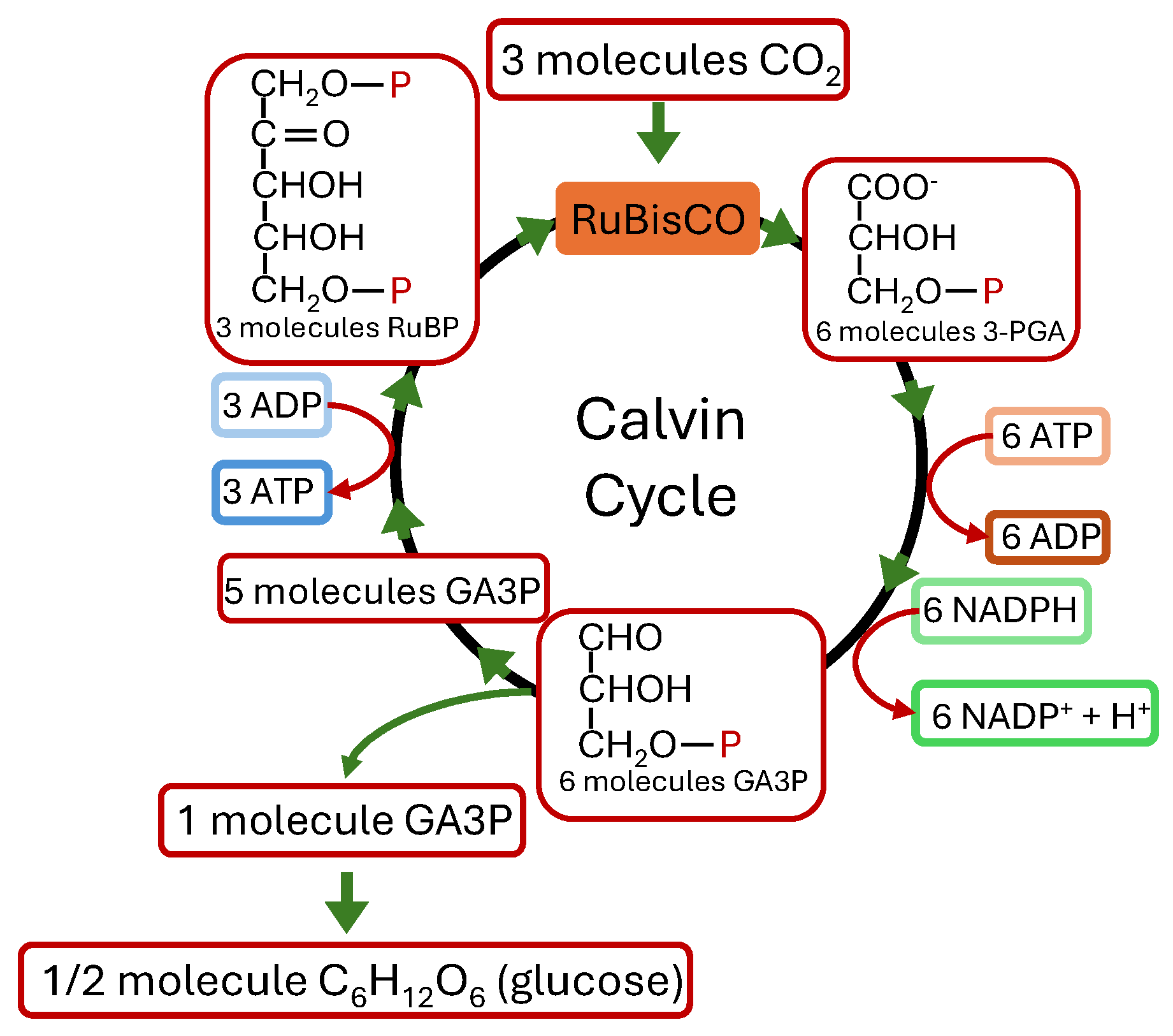
2.2.3. Dark Reactions: Carbon Fixation

Carbon assimilation, also called the Benson–Calvin Cycle, is one of the dark reactions of photosynthesis. In order to fix one CO2 molecule, two NADPH2 molecules and three of ATP are required, as expressed in the following equation:
C O 2 + 2 H + + 4 e 2   N A D P H 2 , 3   A T P , e n z y m e s C H 2 O + H 2 O
The fixation of CO2 occurs in four phases:
  • The carboxylation phase, where CO2 is inserted to the 5-carbon sugar ribulose bisphosphate (ribulose-bis-P) to form phosphoglycerate (glycerate-P). This reaction is catalyzed by the enzyme ribulose bisphosphate carboxylase/oxygenase (Rubisco);
  • The reduction phase, where the phosphoglycerate is converted to 3-carbon products by its phosphorylation (involving ATP) and the subsequent reduction (involving NADPH2);
  • The regeneration phase, where ribulose phosphate is regenerated for further CO2 fixation;
  • The production phase, in which carbohydrates but also fatty acids, amino acids and organic acids are produced.
The process is shown in Figure 5.
Another dark reaction that competes with carboxylation is photorespiration, in which organic carbon is converted into CO2 without any metabolic benefit. This process is influenced by the relative concentrations of oxygen and CO2: a high O2/CO2 ratio promotes photorespiration, while a low ratio favors carboxylation [25].

2.2.4. Alternative Sinks for Electrons

Several other electron sinks, diverse from Benson–Calvin cycle, are present in the chloroplast. They are metabolic pathways and/or signaling networks that require energy and may apparently act in opposition to carbon assimilation. Besides the already described linear electron transfer mode (LEF), in which the ferredoxin transfers electrons to FNR (ferredoxin NADP + reductase) to produce NADPH, the photosynthetic apparatus can also perform Cyclic Electron Transport (CEF), in which ferredoxin transfers electrons back to the plastoquinone pool, causing a cyclic electron flow around PSI. This causes proton translocations, the acidification of the lumen and consequently ATP formation. These two processes are modulated together to match the photochemical reactions to the Calvin–Benson cycle; in fact, the assimilation of CO2 in the Benson–Calvin cycle requires ATP and NADPH in a 3:2 ratio. However, this ratio is not fully met by linear electron flow (LEF), which produces a pH gradient that is insufficient for generating the necessary amount of ATP. Furthermore, CEF is adopted also in other cases such as low CO2, high light or drought [26].
In the case of an over-reduction in electron transport, to prevent electrons from stalling, another alternative cycle operates, called the Mehler reaction. This is a pseudo-cyclic electron flow from water to PSI with molecular oxygen serving as alternative electron acceptor. From this process, a superoxide is formed and converted by superoxide dismutase and catalase to water and oxygen. This water–water cycle restores the redox levels into the photosynthetic membranes [26].
Another pathway involves the chlororespiratory chain, which participates in electron transfer reactions by transferring electrons from stromal reductants to molecular oxygen via the plastoquinone (PQ) pool. It involves a NADP dehydrogenase complex and a plastid quinol terminal oxidase called PTOX [33].
Photorespiration serves as another sink for excess electrons due to the consumption of NADPH and ATP [26]. In photorespiration, organic carbon is converted into CO2 without any metabolic gain [34]. It is estimated that more than 20% of total electron flux through RuBisCo may fuel O2 reduction, and it has been for a long time seen as a wasteful side-product of carbon assimilation. Today, it is generally assumed that, since this process competes with CO2 assimilation, it represents a modulation of the relative rate of O2 and CO2 reduction for maintaining a redox homeostasis, especially in CO2 starvation [27]. It has also been demonstrated that the photorespiratory pathway may play an indicative role in the protection of plants against photoinhibition [33].

2.2.5. Plastoquinone Oxidation State as a Regulation of Photosynthesis

PSI and PSII have a slightly different pigment composition, and they can be differently excited depending on the nature of light. The difference in electron transport chains is balanced by state transitions; in fact, the plastoquinone pool can be reduced or oxidated depending on which photosystem is more excited between PSI and PSII. If plastoquinone is reduced, a kinase is activated that phosphorylates LHCII; this causes the migration of LHCII to PSI and regulates the excitation energy between PSI and PSII. The redox state of plastoquinone is, therefore, central for the electron transfer regulation and can be affected by factors such as wavelength shifts, light intensity, ATP/ADP ratio, CO2 and PTOX concentration and the operation of the Benson–Calvin cycle. Changes in plastoquinone pool redox state can also induce a change in chloroplast and nuclear gene expression. Furthermore, plastoquinone is, as already mentioned, a mobile electron carrier from PSII to cytochrome b6/f complex. Its diffusion can act as a regulatory limiting step in the electron flow. It was proposed that it moves quickly in some microdomains located in the proximity of PSII active centers and diffuses slowly if it migrates through long distances. The creation of these microdomains is, therefore, a regulative factor, and it was also suggested that the phosphorylation/dephosphorylation of LHCII and PSII plays a role in this regulation [26].
The plastoquinone pool oxidation state can be affected also by the trans-thylakoid lumen pH. In fact, most of the electron transfer downregulation pathways consists of a feedback mechanism involving the two main products of photosynthesis: NADPH and the ∆pH in the lumen. The latter has a direct effect on PQ oxidation at the luminal site of the cytochrome b6/f complex. This control has the role of tight coupling between electron and proton transfer during PQH2oxidation.

2.2.6. Regulation Through CO2 Availability

Unlike terrestrial plants, which thrive in an environment rich in CO2, microalgae in aquatic habitats rely on dissolved CO2, which is present at low concentrations and primarily in the form of bicarbonate (HCO3). To overcome this limitation, microalgae have developed Carbon-Concentrating Mechanisms (CCMs), which locally increase CO2 availability for photosynthesis.
CCMs rely on specialized structures, such as the pyrenoid in algae (located in the chloroplast) or carboxysomes in cyanobacteria (found in the cytoplasm). The microalgal cell membrane is permeable to gases, allowing CO2 diffusion, but acts as a barrier to ions such as bicarbonate. For this reason, there are bicarbonate transporters that allow the uptake and accumulation of HCO3 inside the cell. In microalgae, bicarbonate is transported into the chloroplast near the pyrenoid, where the enzyme carbonic anhydrase catalyzes its conversion into CO2, leading to a local increase in CO2 concentration around Rubisco, which subsequently utilizes it in the Calvin cycle [35].
Although CCMs vary among different microalgal species, their general functioning remains similar. The pyrenoid plays a crucial role in this process by limiting CO2 diffusion out of the cell and promoting its immediate utilization by Rubisco. These metabolic adaptations enable microalgae to maintain high photosynthetic efficiency even in CO2 limited aquatic environments while simultaneously reducing photorespiration [36].

3. Light Quality Adjustments for Enhanced Photosynthesis

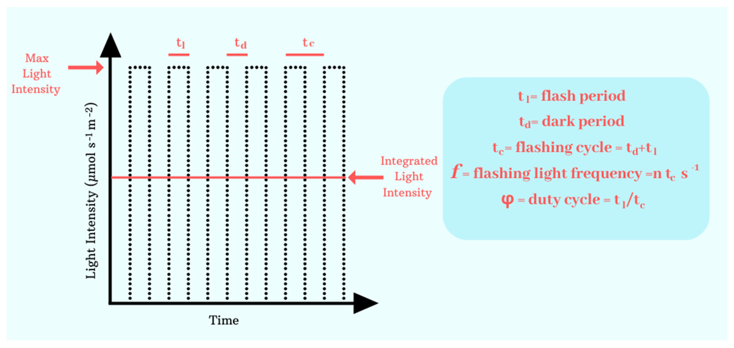
3.1. Application of Flashing Lights

Flashing light (FL) consists of providing illumination by dense packs of high-intense light spaced with short dark periods, differently than in continuous light (CL). It is the repetition of light (tl) and dark (td) periods in an approximately rectangular waveform (Figure 6). The sum of a light and a dark period is defined as flashing cycle (tc) (Figure 6). How often a flashing cycle repeats per second (s−1) is indicated by the flashing light frequency (f). For instance, a frequency of 50 Hz means that light and dark periods change 50 times per second. The proportion of the light period to the flashing cycle is defined as the duty cycle (DC or ϕ). For example, a duty cycle of 0.05 indicates that the light is on for only 5% of the whole flashing cycle, while 95% of the time the light is turned off. The averaged light intensity (Ia) measured under flashing light is the average obtained during a flashing cycle, which is composed by an instantaneous light intensity Il emitted during the light flash period (tl) and no light emission (e.g., Id = 0 µmol s−1 m2) during the dark phase td. For instance, when the averaged light intensity is Ia = 300 µmol s−1 m−2 and the duty cycle is DC = 0.05, then the instantaneous light intensity becomes Il = 6000 µmol s−1 m−2. As seen previously, photosynthesis involves a sequence of rapid light-dependent reactions followed by slower biochemical processes. The timescale of these slower biochemical reactions regulates the overall turnover rate of photosynthesis. The idea that inspires the application of intermittent light on microalgal culture is that of matching the rates of photochemical reactions. In fact, with flashing lights correctly tuned, the dark phase would align with the moment at which photosynthetic centers are closed and unable to collect further energy. The flash period, instead, would match with the harvesting light kinetics performed by the photosystems. This should lead to an enhanced photosynthetic, and possibly biomass, yields compared to continuous light, by spending the same amount of energy [37].
Flashing lights can be applied to microalgal cultures using a rotating disk and incandescent lamps [38,39,40] or relying on illumination cycles based on the movement of the cells inside the photobioreactors, which undergo light–dark cycles as they the approach or move away from the light source [41]. Although this approach is reported in the literature, it has yielded unclear results and has been surpassed by other approaches [42]. Alternatively, the use of regulated LEDs for microalgal cultures, preferably with short light paths to minimize self-shading, has been proposed due to the numerous advantages of LEDs [43]. In this context, it is worth remembering the importance of having a short light-path when testing a specific kind of illumination on a microalgal culture, as showed before [40,44]. Even before, flashing lights were proposed as a strategy to increase the photon penetration depth into a cell-dense culture; in fact, flashing lights would increase photon penetration depth because of the high light intensity [45].
The first attempts to obtain the “flashing light effect”, namely, the described increase in photosynthetic yield, were tried in the last century by Philips and Myers, who observed an increase in growth rate in Chlorella when the flash time approached the hundredth of a second, while no effects were observed under shorter flash times [38]. Oppositely, other works showed an increase in photosynthetic yields with increasing light/dark frequencies [39,46].
Xue et al. showed that while increasing light intensity, the increase in frequency increased also the maximum growth factor of Spirulina platensis grown under intermittent lighting [40]. In other cases, by using Dunaliella salina grown with a duty cycle of 50% and an average light intensity of 400 µmol s−1 m−2, none of the frequency tested increased the growth factor nor the high-value compound accumulation compared to continuous light [47]. The results of the mentioned studies are, therefore, not straightforward and sometimes contradictory.
Although the desired effect of intermittent lights is that of increasing the photosynthetic yield and consequently the amount of biomass obtained, a great number of research studies highlighted a different effect: a shift in the biochemical composition of microalgal biomass, often connected to a decrease in the net biomass yield. For example, Kim et al. obtained a significative increase in astaxanthin concentration in Haematococcus pluvialis when cultivated under a flashing light of 65.6 µE m−2 s−1 and a frequency of 3.49 Hz [48]. What presumably happens is that the excess energy and electron transfer obtained, thanks to the flashing effect, is not directed towards the carbon fixation pathways but flows to different routes, causing, for example, the accumulation of carotenoids, proteins and fatty acids, especially of the omega-3 series. This was shown by Lima et al. [49] who studied the effect of the frequency of 5, 50 and 500 Hz on three different microalgal species (Nannochloropsis gaditana, Tetraselmis chuii and Koliella antarctica) with a constant duty cycle of 5% and an average light intensity of 300 µmol m−2 s−1. The growth performance of the culture grown under a flashing light was lower compared to continuous light, while the content in carotenoids, proteins and lipids was significantly improved. Furthermore, a transcriptomics study on Nannochloropsis gaditana grown in the same conditions [50] showed that low-frequency flashing lights caused the dissipation of excess energy produced through the violaxanthin cycle. The same growing conditions were applied also to Diacronema lutheri and Tetraselmis striata, obtaining a similar accumulation of high-value compounds [51]. Xi et al. showed an increase in the growth of Dunaliella salina cultured under a flashing light with a duty cycle of 50% and an average light intensity of 400 µmol m−2 s−1 compared to a continuous light. The condition that showed the lower growth (50 Hz) was the same, which led to an increase in carotenoid content, indicating that somehow the growth and the carotenoid content are inversely related [52].
It is interesting to note the slightly different approach of Borella et al., who analyzed the effect of illumination times on growth increase. They applied flashing lights of different wavelengths to Arthrospira maxima grown in continuous-flow reactors: this guaranteed the acclimation of the cells to the light condition, which is not assured in batch cultivation. It is worth noting that for different average light intensities, there is a different range of frequencies, which allows one to obtain an increase in concentration compared to the control [53].
Lu et al. applied an experimental design according to the Taguchi Orthogonal Array to assess the effect of flashing parameters on photosynthetic bacteria features; they observed, for example, that biomass concentration increase is obtained in conditions of high light intensities, frequencies and duty cycles, while carotenoid concentration is obtained under high frequencies and low duty cycles [54]. In a more recent study, Levasseur et al. showed that Chlorella vulgaris grown in acclimated and isoactinic conditions did not improve the growth and the photosynthetic yield compared to continuous light under different average light intensities and different frequencies at a fixed duty cycle of 50%, while in some cases, the content in carotenoids did improve [55].
According to the described findings, we can conclude that when flashing lights are tuned for decreasing the violaxanthin cycle, the excess energy of electron transfer is used for enhancing the Calvin cycle and increasing biomass yields [56]; otherwise, if flashing lights enhance the violaxanthin cycle, the excess energy is used for increasing the concentration of compounds such as carotenoids, fatty acids and proteins [50]. Anyway, flashing lights can be a powerful instrument that can be tuned in order to obtain an enhanced growth or a shift in the biochemical composition of the microalgal biomass, depending on the producer’s requirements.

3.2. Tailoring the Wavelength

The influence of light wavelength on microalgae has been studied for years. It has been demonstrated that wavelength can influence various aspects, including photosynthetic activity, growth performance, biomass production and its biochemical composition. When studying the wavelength effect, the main challenge derives from the different effect obtained in different species. Physiological processes depend on the different interaction of light with the molecular constituent of the microalgal cell. Each species is characterized by the presence of different photosynthetic molecules, and this leads to different responses. Photosynthesis begins with the absorption of light by pigments that absorb light at specific wavelengths. Depending on the wavelength they receive, different metabolic processes are activated, leading to various effects on the microalga. Furthermore, it has been observed that microalgae can adapt to different lighting conditions by modifying the quantitative composition of their pigments, thereby optimizing the use of available light [57,58].
The preferred wavelengths for various photosynthetic pigments are well known. Among chlorophylls, chlorophyll-a absorbs in the ranges 380–470 nm (blue) and 600–680 nm (red), chlorophyll-b in the 410–480 nm (blue) and 600–685 nm (red), and chlorophyll-c absorbs blue light at 450 nm, while chlorophyll-d and chlorophyll-f absorb in the 700–750 nm range (far-red).
Among accessory pigments, carotenoids such as β-carotene absorb in the 400–500 nm range (blue and blue-green), while xanthophylls (lutein, fucoxanthin, astaxanthin, violaxanthin, zeaxanthin, etc.) absorb in the 400–550 nm range (violet, blue-green and green). Among phycobiliproteins, phycocyanin absorbs in the 600–640 nm range (orange-red), phycoerythrin absorbs between 480 and 570 nm (blue-green, green and yellow) and allophycocyanin absorbs between 620 and 660 nm (orange-red).
Microalgae can be classified into four major groups characterized by different types of pigments. Based on this, it can generally be said that green algae prefer a blue and red spectrum and cyanobacteria prefer red and yellow light, while red algae and diatoms prefer blue and green light [17,18,19].
Several studies in the literature have investigated the effects of wavelength on the growth and biochemical composition of microalgae. However, standardizing these effects is challenging due to the influence of additional experimental parameters, such as agitation type, growth medium, light intensity, and temperature, among others. These factors also play a crucial role in determining microalgal growth and biochemical composition.
Table 1 summarizes the growth conditions adopted in the various studies analyzed, specifically focusing on the effect of wavelength. For simplicity, microalgal groups have been categorized into green algae, diatoms, red algae, and cyanobacteria.
As previously mentioned, each group is characterized by the presence of specific pigments that facilitate the absorption of specific wavelengths. However, the correlation between wavelength and its effects in microalgae is highly variable. Not only do different groups exhibit distinct responses, but even within the same group, species react in diverse, and sometimes even contradictory, ways. Surprisingly, variations have also been observed within the same species. Furthermore, many studies have employed mixed light in contrast to monochromatic light.

3.2.1. Green Algae

Regarding green algae, it has been shown that photosynthetic efficiency is enhanced under blue and red lights. This is because their primary photosynthetic pigments, chlorophyll a and chlorophyll b, reflect green light, which gives these algae their characteristic coloration. Red light, indeed, is believed to be efficient in photosystem II (PSII) activation, which plays a crucial role in electron transport during photosynthesis. On the other hand, blue light, due to its higher photon energy, is expected to stimulate the production of accessory pigments that provide protection against oxidative stress. Nevertheless, a portion of green light is still utilized through accessory pigments [19,81]. Several studies have confirmed that red and blue light promote growth rate and/or biomass production, while green light has been associated with reduced growth rate and biomass accumulation [69,72,75]. Other studies have indicated that red or red-orange light is the most effective for microalgal growth [59,62,64,65,68,70,75] and biomass production [66,69]. However, two studies reported that red light had the least impact on growth [67] and biomass production [76], compared to other wavelengths. Regarding blue light, it was found to be optimal for optimizing growth rate in Nannochloropsis sp. [71]. However, in other studies, blue light was among the least effective wavelengths for promoting cellular growth and biomass production [62,65,70]. Some studies utilizing mixed light demonstrated that certain combinations of wavelengths were more effective in stimulating growth than monochromatic light [63,76]. On the other hand, for Scenedesmus quadricauda [74], no significant effect was observed in response to wavelength variation.
The analysis of these studies highlights that even within the same microalgal species, responses to wavelengths can differ on the base of growth conditions. For instance, in Chlamydomonas reinhardtii [59,60,61,62], biomass production was enhanced by multiple wavelengths, including red, red-orange, blue and yellow light. In contrast, for Chlorella vulgaris [59,65,66], red light was generally the most effective, except in [67], which employed mixed lighting.
As previously mentioned, multiple factors influence these responses. For example, in [60], biomass production was stimulated by red light at 24 °C and blue light at 32 °C. Similarly, in [61], biomass production was enhanced by blue light under low light intensity, while red-orange and white-yellow light were more effective under high light intensity.
Across various studies, growth rate and biomass production were optimized under different wavelengths. In [62], red light promoted growth rate, whereas yellow light enhanced biomass production. In [64], red light increased growth rate but resulted in the lowest biomass production, which was instead optimized by white light. In [67], orange light promoted growth rate, whereas violet light increased biomass production. In [73], both blue and red light enhanced growth rate, but blue light was the least effective for biomass production, which was instead optimized by white light.
It has been supposed that red light may intensify cell division, leading to the formation of smaller cells, whereas blue light might inhibit the division of small cells, promoting the formation of larger cells. This mechanism could ultimately influence total biomass accumulation [19].
Regarding biochemical composition, no specific trends were observed concerning wavelength. For lipid production, red and red-orange light were the most effective [60,61,64,72,75], while blue light was optimal in [71,72] and white light in [65]. Interestingly, the authors of [66,69] reported that Chlorella vulgaris and Golenkinia SDEC-16 utilized green light most efficiently for lipid production. However, no significant effects of wavelength on lipid production were observed for Tetraselmis lutua [76].
For pigment production, red light was found to be the most effective in [59,64,67], while white light was more efficient in [65,73]. In Chlamydomonas reinhardtii and Dunaliella salina [59], no difference was observed between red and white light. Blue light favored pigment production in Scenedesmus rubescens [73] and Scenedesmus quadricauda [74]. In studies using mixed light, the most effective combinations were red–blue [70] and blue–green [76].
Protein production was generally promoted by blue light [61,65] and white light [61,64,72], although one study indicated that green light was the most effective [72].
Finally, for carbohydrate production, three studies identified red light as the most effective [61,64,65], while blue light also yielded positive results in [72].

3.2.2. Diatoms

In regards of diatoms, it has been suggested that their pigment composition promotes blue and green light absorption. This is partly because diatoms commonly inhabit deeper water layers, where blue and green wavelengths penetrate more efficiently than red light, which has lower photon energy and, thus, is more readily absorbed by water [19,81].
The analyzed studies confirm that blue light enhances biomass production in Pavlova lutheri [66] and growth rate in Phaeodactylum tricornutum [77]. Conversely, green light was more effective in promoting growth rate in Synedra [78], where blue light was found to be the least effective.
Regarding biochemical composition, yellow light promoted lipid production in Pavlova lutheri [66], whereas blue light favored pigment production in Phaeodactylum tricornutum [77], with red light specifically enhancing violaxanthin production.

3.2.3. Red Algae

Red algae contain phycobiliproteins as accessory pigments, which suggests a preference for blue and green light. This preference is primarily due to their habitat as red algae typically grow in deeper aquatic environments, where red light is rapidly absorbed by water and thus does not penetrate efficiently [19,81].
Analysis of relevant studies reveals notable findings. In the study on Porphyridium cruentum [66], green light promoted biomass production, which was consistent with expectations. Similarly, in the study on Porphyridium purpureum [79], green light was found to be beneficial for biomass accumulation; however, in the latter case, blue light had a negative effect on biomass production compared to other wavelengths. Conversely, when studying Galdieria sulphuraria [79], red light was the most effective wavelength for biomass production.
Regarding biochemical composition, red light stimulated phycobiliprotein production in Galdieria sulphuraria and Porphyridium purpureum [79], whereas green and blue light had a negative impact on phycobiliprotein accumulation. For Porphyridium cruentum [66], red light was also found to enhance lipid production. These findings suggest that red light plays a significant role in shaping the biochemical composition of red algae.

3.2.4. Cyanobacteria

Cyanobacteria, based on their pigment composition, are expected to preferentially absorb red and yellow light. This is also because cyanobacteria often live in shallow waters, where red light is abundant and readily absorbed by these organisms [19,81].
This was confirmed in the study on Microcystis [78]; red and white light promoted growth rate more effectively than other wavelengths. In the case of Spirulina platensis [59], red light was more effective than white light in enhancing growth rate, though no significant difference was observed in pigment production. However, another study on Spirulina platensis [80] reported contrasting results, indicating that blue light was more effective in stimulating both biomass production and protein synthesis.
In a study on Gloeothece membranacea [67], violet light was the most effective in promoting growth rate, while green light enhanced pigment production.
In conclusion, the analysis of the existing studies suggests that it is not possible to establish a uniform trend regarding the influence of wavelength on microalgae. The responses observed vary significantly depending on the species, growth conditions and other experimental parameters, leading to inconsistencies across different studies. It is worth noting that, besides the already analyzed culture conditions, which prevent the effects of wavelength from being observed unambiguously, the optical path of the light inside the culture vessel should also be analyzed. In fact, similar to the flashing light, it is impossible to study the effect of a light feature on microalgal growth if significant self-shading effects occur. For this reason, it is essential to continue research to optimize the use of light wavelength in microalgal cultivation.

4. Photobioreactor Design for Optimized Light Utilization

The previously mentioned studies, which highlighted an effect on the obtained biomass through the application of specific light features, were applied to the design of photobioreactors to achieve the same effect. In this sense, there are three categories of photobioreactors: those in which the light gradients are limited to allow the optimal distribution of light within the photobioreactor; photobioreactors, which imitate the flashing effects with the use of special geometries or specific elements in the design; photobioreactors in which the light wavelength is modified with the use of filters. This section analyzes these categories.

4.1. Strategies to Minimize Light Gradients

Light attenuation creates three light-dependent zones: photolimitation, photosaturation and photoinhibition. Photolimitation occurs when light intensity is too low to support the maximum rate of photosynthesis. In this regime, photosynthesis is limited by the availability of photons. On the other hand, photosaturation occurs when light intensity reaches a level where the photosynthetic machinery is operating at its maximum capacity. Beyond this point, increasing light intensity does not increase the photosynthetic rate. When light intensity is so high that it causes damage to the photosynthetic apparatus, photoinhibition occurs. Photoinhibition is indeed associated with a reduction in the photosynthetic rate [82].
The main goal of this category of photobioreactors is to reduce the light gradients within the photobioreactor. In recent years, two different strategies have emerged. One of the main approaches is to modify the reactor geometry in a way that the light path is minimized to reduce the impact of self-shading [6]. Consequently, these geometries are also characterized by a high surface-to-volume ratio (SVR) [83,84,85]. If the light path is too short, the system is more susceptible to photoinhibition and thus to a reduction in the photosynthetic efficiency, even at low light intensities [6,8]. Indeed, an excessive increase in the surface-to-volume ratio (SVR) could also be detrimental for areal productivity, as in [6,8,83,86]. Furthermore, in some types of reactors, a shorter light path could also increase the chances of biofouling formation, as shown by González-Camejo et al., which reduced the light path of a membrane photobioreactor from 25 cm to 10 cm, with an increase in areal biomass productivity and photosynthetic ratio. However, the authors reported an increase in the biofouling rate with the shorter light path [86]. Recently, Venancio et al. evaluated the performances of two thin-layer cascade systems with two different SVR values of 60 m−1 and 80 m−1, respectively. The results showed that the thin-layer cascade with the highest SVR reached a greater biomass concentration and volume daily productivity. However, the authors reported a lower photosynthetic efficiency in the same reactor, probably due to photoinhibition. Furthermore, the two photobioreactors showed a similar areal productivity, reporting again the necessity to find specific surface to volume ratios to get the best operating conditions [83]. Chuka-ogwude et al. made a deep optimization of the geometry in an inclined thin-layer photobioreactor with microalgae growing in anaerobic digestate. In such conditions of high turbidity, light attenuation is even more problematic. However, the authors report that the best operating conditions were achieved by just increasing the reactor depth from 0.005 m to 0.011 m. These results highlight the necessary trade-off between higher biomass growth rates and areal productivity [87]. Small-scale photobioreactors could also give a better understanding of the impact of the light path. Saccardo et al. worked with small-scale photobioreactors with different thickness, revealing that a decrease in light path value from 35 to 15 mm increased the biomass concentration. However, for lower values, photo inhibition became more significant, causing a reduction in biomass concentration [6]. A revision of traditional photobioreactor’s geometries is another option for decreasing light gradients. A vertical photobioreactor with “oval-shaped tubes” has been designed to limit the self-shading [88]. Hijazi et al. proposed a “cactus-like” cylindrical photobioreactor (Figure 7); this geometry showed a 29% improvement in light penetration and double micro-algal productivity compared to a traditional cylindrical photobioreactor due to the lower light path [89]. A Fibonacci-type tubular photobioreactor (Figure 8) has been developed for outdoor applications, with a low SVR compared to vertically arranged tubular photobioreactors [90,91].
However, excessive sun irradiance could also be a limitation for outdoor photobioreactors. As an attempt to solve it, Chin-On et al. designed a novel V-shaped photobioreactor (Figure 9) for tropical areas of the world. In this type of photobioreactor, incident light is diluted due to its refraction on the reactor surfaces. Light dilution, paired also with a lower light loss, brought a 39% increase in biomass productivity compared to the reference flat photobioreactor [92].
The other main approach for minimizing light gradients could be achieved by implementing light distribution systems instead of varying the reactor geometry. A light distribution system could be spatial or temporal [87]. Spatial light distribution systems can better distribute the light inside the reactors, for example, with the help of internal light waveguides [84], light conducting frameworks [93], or reflectors to illuminate more uniformly the photobioreactor [8,94]. In this sense, an innovative LED arrangement could also uniformly illuminate the photobioreactor [95].
Temporal light distribution systems are instead based on acting on the reactor mixing to achieve a proper enhanced light/dark cycle frequency. In this way, the light regime could improve, and photoinhibition could be avoided [6,8,87,96,97]. This is achieved by adding static mixers/buffers to improve mixing performances [96]. However, it could also cause an increase in complexity and/or a higher energy consumption [94]. To ensure lighting in the photolimited zones of the photobioreactor, light-guiding materials, like optical fibers or waveguides, could be used [84,97,98]. This approach works for indoor and outdoor applications. Even more advanced technologies, like solar tracking devices, wireless light emitters, or plasmonic light scattering, have been investigated. However, in this case, industrial applications are not always certain, and economic evaluations are still necessary [98].
For example, Sivakaminathan et al. introduced light-transmitting guides in high-rate ponds with a 3.9 folder increase in areal productivity and a 2-fold higher photosynthetic efficiency compared to the control [84].
Light intensity management and light source configuration also play an important role in achieving homogeneous illumination. Introducing internal illumination could be an option to pursue this objective [7]. Alternatively, a system with three interconnected LEDs has been proposed; it had an automatic system able to regulate the LED intensity to compensate for light gradients in the photobioreactor [99]. The combination of internal illumination and light reflectors also showed high potential. In fact, the synergistic use of internal illumination and reflective surfaces causes a redistribution of light intensity in the reactor, obtaining a more constant light dispersion. In such photobioreactors, reflector’s position, geometry and material are fundamental parameters that impact the photobioreactor efficiency [8,94]. Also, the use of mirrors, in an internal illuminated flat plate photobioreactor, led to higher microalgae growth rate, nutrient removal and bioproducts synthesis compared to other photobioreactor configurations [94]. Lastly, Porto et al. evaluated the use of various reflectors, with different shapes and materials, to illuminate a tubular photobioreactor. The results indicated the highest performances in the tubular photobioreactor, assembled with parabolic collectors with the highest specular reflectance, guaranteeing a more homogeneous light distribution. However, the authors reported that the impact of the material’s performance decreases as the reflector’s geometry becomes simpler. Moreover, the performance gains achieved using parabolic-shaped deflectors may not justify the increased complexity and capital costs required for their implementation [8].

4.2. Implementing Effective Dark/Light Cycles

As already mentioned, algae growing within a photobioreactor may experiment illumination cycles when approaching or moving away from the light source. For this reason, some photobioreactor designs aimed at exploiting this mechanism were proposed. Some works suggested to obtain the flashing light effect through the use of additional elements, such as baffles or deflectors. Zhang et al. added flow deflectors and wing baffle to a raceway pond and obtained a reduction in dead zone and a decrease in the period (consequently, an increase in the frequency), together with an increase in the obtained biomass of Chlorella sp. [100]. Similar effects were obtained by Cheng et al. by the addition of up-down chute baffles to a raceway pond [101]. Yang et al. proposed the addition of horizontal tubes and triangular prism baffle in a flat-plate photobioreactor, which led also in this case to a decreased dark–light cycle period. In this case, the biomass of Chlorella mutant PY-ZU1 increased by 70% [102]. In another case, optical fibers were used as an inner light source to apply a flashing light to the microalgal culture of Spirulina platensis and Scenedesmus dimorphus inside different kinds of photobioreactors. The results demonstrated that it could produce uniformed light/dark frequencies over 10 Hz, and microalgae productivity increased by 43% and 38% [103].

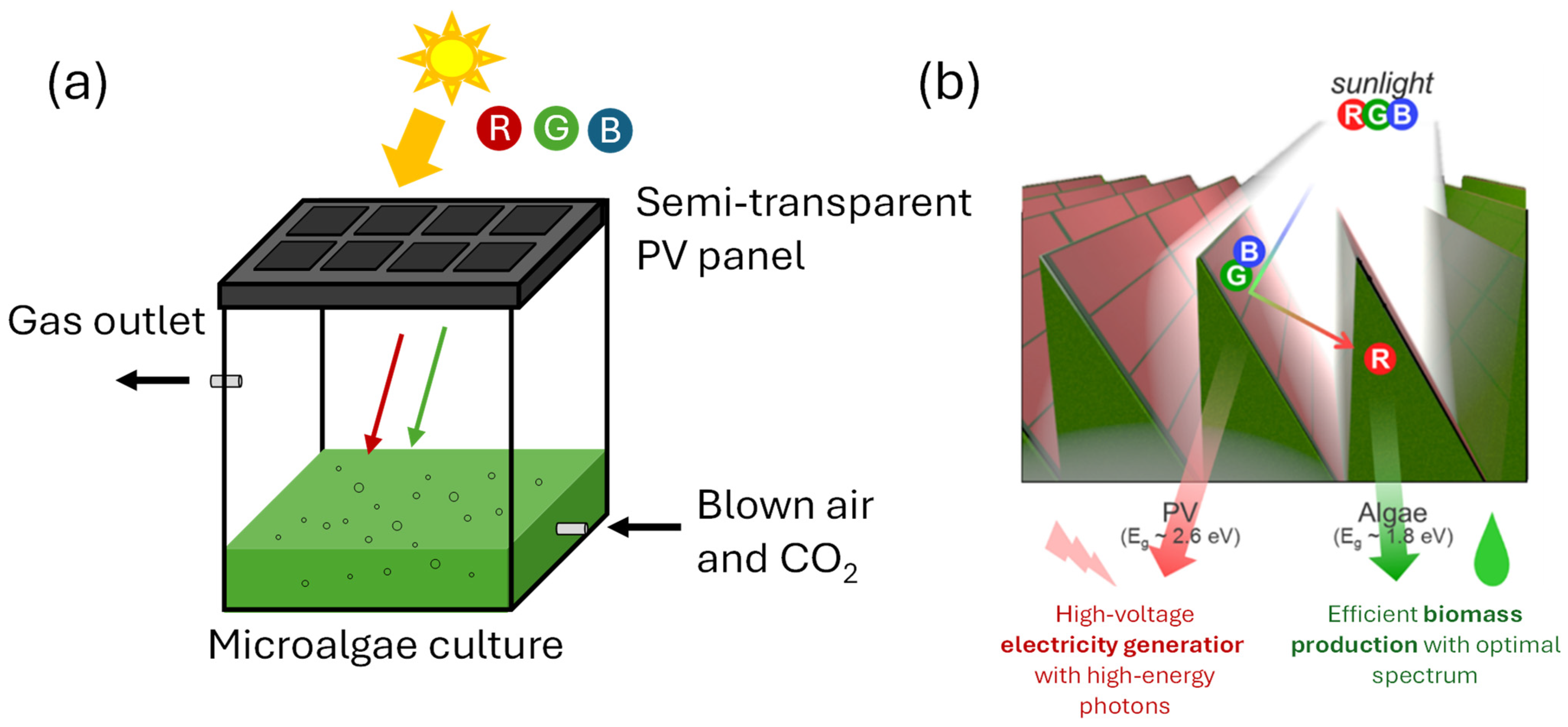
4.3. Optimizing Light Spectrum Through the Application of Filters in Photobioreactors

Solar energy is challenging to manage due to seasonal intensity variations and the presence of harmful wavelengths like infrared and near-infrared [104,105], which are not absorbed. Additionally, blue photons may be inefficiently utilized during photosynthesis due to significant energy loss as heat in the photosystem [106]. Filters for sunlight light are considered a method to efficiently manage the entire sunlight spectrum and mimic, in other words, the laboratory optimal conditions in a scale-up process [107].
Three different types of filters for optimizing solar radiation use were identified [108]: glass absorption filters, reflective/thin-film filters and thermochromic filters. These filters selectively make the PAR region of the solar spectrum available to algae. Glass absorption filters allow specific wavelengths to pass while absorbing unwanted ones. Reflective filters, in contrast, permit desired wavelengths to pass and reflect the rest, offering greater accuracy and efficiency in isolating narrow spectral regions compared to glass absorption filters. Thermochromic filters adjust light transmission or reflection based on atmospheric temperature [104,109,110,111]. Following the results of Balbuena-Ortega et al., in photobioreactors inoculated with microalgae consortia for the treatment of wastewaters, the physiological microalgal parameters (such as chlorophyll and carotenoid production), showed correlations with the different light wavelengths. Green filters led to a higher chlorophyll production; conversely, the blue filters boosted the pigment accumulation. Moreover, green and blue filters produced more pigments in lower radiation conditions; meanwhile, red and blue light were shown to have a key role in photosynthetic efficiency and in microalgae growth [107].
An interesting application of a particular kind of solar filters is represented by semi-transparent photovoltaic panels placed on the photobioreactor surface. Semi-transparent photovoltaic panels can absorb specific wavelengths to generate electricity while allowing other wavelengths to pass through, supporting photosynthetic microalgae growth [112]. In this way, the system can benefit from the coproduction of biomass and electricity but also from the partial shadowing effect that increases the microalgae growth during hotter periods, reducing the thermal regulation energy request [113,114]. Innovative cultivation techniques that integrate solar energy sharing between photovoltaic systems and microalgae cultivation are gaining interest, with different approaches already documented. One of these involves partially covering microalgae bioreactors with opaque photovoltaic cells, permitting some light to reach the algae culture. Crystalline-silicon-based photovoltaic panels (single-crystalline silicon or multi-crystalline silicon) are the most commonly used due to their high solar light conversion efficiency. Studies have demonstrated the feasibility of this approach in various settings [115]. Besides the silicon-based panels, copper indium-diselenide and amorphous silicon were proposed in the literature, demonstrating how these technologies, provide a greater carbon credit revenue opportunity for microalgae producers [116].
Barbera et al. used semi-transparent dye-sensitized solar cells to partially cover a photobioreactor for the cultivation of a Scenedesmus obliquus strain [117]. They found that at low incident light intensity (220 µmol m−2 s−1), biomass concentration decreases significantly, leading to a drop in productivity of about 45% compared to the control. On the contrary, increasing the light intensity (615 µmol m−2 s−1) resulted in higher biomass concentration and productivity due to the increased energy input, with the control PBR achieving a dry weight of 4.37 ± 0.22 g L−1.
The calculated energy conversion efficiency highest value was found at 220 µmol m−2 s−1 light intensity, about 40% higher than the control value. In a similar study, Damergi et al. used a particular system for the growth of Chlorella vulgaris, filtering the solar light with different dye-sensitized solar cells (DSCs) to red and green (see Figure 10a) [115]. A maximum cell growth rate of μ = 0.86 day−1 was found with the DSC-red solar cells as filters, compared to a normal glass control growth rate of about μ = 0.51 day−1. At the same time, a maximum value of chlorophyll-a of about 11 μg/mg was reached in green and red conditions, compared to a value of 9.6 μg/mg obtained with the control system. The maximum carotenoid concentration in the biomass was also obtained in red conditions (1.11 μg/mg, versus 0.8 μg/mg in the control case).
In another work, using a similar experimental layout (Figure 10b), the authors employed spectrally selective photovoltaic cells and demonstrated the potential to achieve 20.3 g/m2/day of algal biomass production and 0.22 kWh/m2/day of electricity generation by utilizing multiple band gaps within a single system under an illumination of 7.2 kWh/m2/day [106].
Figure 10. Scheme of lab-scale photobioreactors with dye-sensitized solar cells as filter [106,115]. Scheme of a single module photobioreactor (a) and of different connected modules (b).
Processes 13 01159 g010
Research in both technologies is undergoing rapid expansion, driven by significant advancements in industrial applications. The integration of photovoltaic systems and photobioreactors has already demonstrated remarkable potential for improving overall efficiency. Continuous improvements, such as enhancing energy conversion rates, optimizing operational parameters, and reducing costs, are being recorded. These advancements underline the growing feasibility of combined systems for large-scale implementation. Future research will focus on optimization these systems, exploring innovative materials and configurations, and addressing scalability challenges to unlock their full potential and ensure long-term sustainability in industrial applications.

5. Conclusions

In this work, the effects of light quality adjustments on microalgal culture were assessed. After an initial section that explored the photosynthetic mechanisms and their regulation, two specific light features were investigated: illumination provided by dense packs of high-intense light spaced with short dark periods (flashing or intermittent light) and the application of specific wavelengths. The effects of both approaches were analyzed in terms of increase in biomass production and shift in composition, referring to a large and representative part of the existing literature. The results showed that the effect of flashing light on microalgal culture may vary depending on the culture conditions and the employed microalgae. However, it is possible to state that when flashing lights are regulated for decreasing the violaxanthin cycle, the excess energy of electron transfer is used for enhancing the Calvin cycle and increasing biomass yields; otherwise, if flashing lights enhance the violaxanthin cycle, the excess energy is used for increasing the concentration of compounds such as carotenoids, fatty acids and proteins. For what concerns wavelength shift, there are several parameters that influence its effect such as the light intensity, the agitation type, growth medium, light intensity and temperature. Due to the interplay of these parameters, it is very complex to generalize its impact.
In the final section of the review, studies applying these types of lighting in photobioreactor design were analyzed. Three categories of photobioreactors were examined: (1) reactors designed to minimize light gradients, (2) reactors where the geometry produces a flashing light effect on the cells and (3) reactors that use filters to obtain a shift in the sunlight wavelength. In conclusion, this work can serve as a reference for selecting operational parameters in microalgal cultivation and photobioreactor design to achieve specific objectives, such as enhancing biomass production or accumulating high-value compounds.

Author Contributions

Conceptualization, S.L.; writing—original draft preparation, A.M., S.L., F.G. and A.C.; writing—review and editing, A.M., S.L., A.C. and F.S. All authors have read and agreed to the published version of the manuscript.

Funding

This work was realized with the cofounding of PRIN Progetti di Rilevante Interesse Nazionale 2022, under the title A Knowledge-Based Approach to Automatic Control and Optimisation of Photosynthetic Bioprocesses (PHOTOCONTROL), CUP B53D23005650006. The work of Alessandro Cosenza has been partially supported by the funding D26_PREMIO_SINGOLI_RIC_2023 from the Department of Engineering of the University of Palermo.

Conflicts of Interest

The authors declare no conflicts of interest.

References

  1. Hemaiswarya, S.; Raja, R.; Ravikumar, R.; Carvalho, I.S. Microalgae Taxonomy and Breeding. In Biofuel Crops: Production, Physiology and Genetics; CABI: Wallingford, UK, 2013; pp. 44–53. [Google Scholar] [CrossRef]
  2. Pruvost, J.; Cornet, J.-F.; Pilon, L. Large-Scale Production of Algal Biomass: Photobioreactors. In Algae Biotechnology: Products and Processes; Bux, F., Chisti, Y., Eds.; Springer: Berlin/Heidelberg, Germany, 2016; pp. 41–66. [Google Scholar]
  3. Legrand, J.; Artu, A.; Pruvost, J. A Review on Photobioreactor Design and Modelling for Microalgae Production. React. Chem. Eng. 2021, 6, 1134–1151. [Google Scholar] [CrossRef]
  4. Luzi, G.; McHardy, C. Modeling and Simulation of Photobioreactors with Computational Fluid Dynamics—A Comprehensive Review. Energies 2022, 15, 3966. [Google Scholar] [CrossRef]
  5. Khan, M.J.; Singh, N.; Mishra, S.; Ahirwar, A.; Bast, F.; Varjani, S.; Schoefs, B.; Marchand, J.; Rajendran, K.; Banu, J.R.; et al. Impact of Light on Microalgal Photosynthetic Microbial Fuel Cells and Removal of Pollutants by Nanoadsorbent Biopolymers: Updates, Challenges and Innovations. Chemosphere 2022, 288, 132589. [Google Scholar] [CrossRef] [PubMed]
  6. Saccardo, A.; Bezzo, F.; Sforza, E. Microalgae Growth in Ultra-Thin Steady-State Continuous Photobioreactors: Assessing Self-Shading Effects. Front. Bioeng. Biotechnol. 2022, 10, 977429. [Google Scholar] [CrossRef]
  7. Penloglou, G.; Pavlou, A.; Kiparissides, C. Recent Advancements in Photo-Bioreactors for Microalgae Cultivation: A Brief Overview. Processes 2024, 12, 1104. [Google Scholar] [CrossRef]
  8. Porto, B.; Silva, T.F.; Gonçalves, A.L.; Esteves, A.F.; de Souza, S.M.; de Souza, A.A.; Pires, J.C.; Vilar, V.J. Tubular Photobioreactors Illuminated with LEDs to Boost Microalgal Biomass Production. Chem. Eng. J. 2022, 435, 134747. [Google Scholar] [CrossRef]
  9. Lima, S.; Brucato, A.; Caputo, G.; Schembri, L.; Scargiali, F. Modelling Nannochloropsis Gaditana Growth in Reactors with Different Geometries, Determination of Kinetic Parameters and Biochemical Analysis in Response to Light Intensity. Appl. Sci. 2022, 12, 5776. [Google Scholar] [CrossRef]
  10. Singh, V.; Mishra, V. A Review on the Current Application of Light-Emitting Diodes for Microalgae Cultivation and Its Fiscal Analysis. Crit. Rev. Biotechnol. 2023, 43, 665–679. [Google Scholar] [CrossRef]
  11. Maltsev, Y.; Maltseva, K.; Kulikovskiy, M.; Maltseva, S. Influence of Light Conditions on Microalgae Growth and Content of Lipids, Carotenoids, and Fatty Acid Composition. Biology 2021, 10, 1060. [Google Scholar] [CrossRef]
  12. Baidya, A.; Akter, T.; Islam, M.R.; Shah, A.K.M.A.; Hossain, M.A.; Salam, M.A.; Paul, S.I. Effect of Different Wavelengths of LED Light on the Growth, Chlorophyll, β-Carotene Content and Proximate Composition of Chlorella ellipsoidea. Heliyon 2021, 7, e08525. [Google Scholar] [CrossRef]
  13. Borella, L.; Marchese, D.; Trivellin, N.; Sforza, E. Complementary Chromatic Adaptation as a Strategy to Increase Energy Conversion Efficiency of Microalgae-Cyanobacteria Consortia in Continuous LED Photobioreactors. Energy Convers. Manag. 2023, 294, 117549. [Google Scholar] [CrossRef]
  14. Carvalho, A.P.; Silva, S.O.; Baptista, J.M.; Malcata, F.X. Light Requirements in Microalgal Photobioreactors: An Overview of Biophotonic Aspects. Appl. Microbiol. Biotechnol. 2011, 89, 1275–1288. [Google Scholar] [CrossRef] [PubMed]
  15. Blanken, W.; Cuaresma, M.; Wijffels, R.H.; Janssen, M. Cultivation of Microalgae on Artificial Light Comes at a Cost. Algal Res. 2013, 2, 333–340. [Google Scholar] [CrossRef]
  16. Kim, Z.H.; Park, Y.S.; Ryu, Y.J.; Lee, C.G. Enhancing Biomass and Fatty Acid Productivity of Tetraselmis sp. in Bubble Column Photobioreactors by Modifying Light Quality Using Light Filters. Biotechnol. Bioprocess. Eng. 2017, 22, 397–404. [Google Scholar] [CrossRef]
  17. Ievina, B.; Romagnoli, F. Unveiling Underlying Factors for Optimizing Light Spectrum to Enhance Microalgae Growth. Bioresour. Technol. 2025, 418, 131980. [Google Scholar] [CrossRef]
  18. Schulze, P.S.C.; Barreira, L.A.; Pereira, H.G.C.; Perales, J.A.; Varela, J.C.S. Light Emitting Diodes (LEDs) Applied to Microalgal Production. Trends Biotechnol. 2014, 32, 422–430. [Google Scholar] [CrossRef]
  19. Aizpuru, A.; González-Sánchez, A. Traditional and New Trend Strategies to Enhance Pigment Contents in Microalgae. World J. Microbiol. Biotechnol. 2024, 40, 272. [Google Scholar] [CrossRef]
  20. Liu, J.; van Iersel, M.W. Photosynthetic Physiology of Blue, Green, and Red Light: Light Intensity Effects and Underlying Mechanisms. Front. Plant Sci. 2021, 12, 619987. [Google Scholar] [CrossRef]
  21. Gitelson, A.; Qiuang, H.; Richmond, A. Photic Volume in Photobioreactors Supporting Ultrahigh Population Densities of the Photoautotroph Spirulina platensis. Appl. Environ. Microbiol. 1996, 62, 1570–1573. [Google Scholar] [CrossRef]
  22. Wu, B.S.; Rufyikiri, A.S.; Orsat, V.; Lefsrud, M.G. Re-Interpreting the Photosynthetically Action Radiation (PAR) Curve in Plants. Plant Sci. 2019, 289, 110272. [Google Scholar] [CrossRef]
  23. Danziger, N.; Bernstein, N. Light Matters: Effect of Light Spectra on Cannabinoid Profile and Plant Development of Medical Cannabis (Cannabis sativa L.). Ind. Crops Prod. 2021, 164, 113351. [Google Scholar] [CrossRef]
  24. Lewicka, K.; Siemion, P.; Kurcok, P. Chemical Modifications of Starch: Microwave Effect. Int. J. Polym. Sci. 2015, 2015, 867697. [Google Scholar] [CrossRef]
  25. Richmond, A.; Hu, Q. Handbook of Microalgal Culture; Richmond, A., Hu, Q., Eds.; John Wiley & Sons, Ltd.: Oxford, UK, 2013; ISBN 9781118567166. [Google Scholar]
  26. Rochaix, J.-D. Regulation of Photosynthetic Electron Transport. Biochim. Biophys. Acta-Bioenerg. 2011, 1807, 878–886. [Google Scholar] [CrossRef] [PubMed]
  27. Eberhard, S.; Finazzi, G.; Wollman, F.-A. The Dynamics of Photosynthesis. Annu. Rev. Genet. 2008, 42, 463–515. [Google Scholar] [CrossRef]
  28. Drop, B.; Webber-Birungi, M.; Yadav, S.K.N.; Filipowicz-Szymanska, A.; Fusetti, F.; Boekema, E.J.; Croce, R. Light-Harvesting Complex II (LHCII) and Its Supramolecular Organization in Chlamydomonas reinhardtii. Biochim. Biophys. Acta-Bioenerg. 2014, 1837, 63–72. [Google Scholar] [CrossRef]
  29. Van Amerongen, H.; Croce, R. Light-Harvesting in Photosystem II. Photosynth. Res. 2013, 116, 251–263. [Google Scholar] [CrossRef]
  30. Nelson, N. Plant Photosystem I–The Most Efficient Nano-Photochemical Machine. J. Nanosci. Nanotechnol. 2009, 9, 1709–1713. [Google Scholar] [CrossRef]
  31. Fromme, P.; Jordan, P.; Krauß, N. Structure of Photosystem I. Biochim. Biophys. Acta-Bioenerg. 2001, 1507, 5–31. [Google Scholar] [CrossRef]
  32. Croce, R.; van Amerongen, H. Light-Harvesting in Photosystem I. Photosynth. Res. 2013, 116, 153–166. [Google Scholar] [CrossRef]
  33. Rumeau, D.; Peltier, G.; Cournac, L. Chlororespiration and Cyclic Electron Flow around PSI during Photosynthesis and Plant Stress Response. Plant Cell Environ. 2007, 30, 1041–1051. [Google Scholar] [CrossRef]
  34. Shi, X.; Bloom, A. Photorespiration: The Futile Cycle? Plants 2021, 10, 908. [Google Scholar] [CrossRef] [PubMed]
  35. Badger, M.R.; Andrews, T.J.; Whitney, S.M.; Ludwig, M.; Yellowlees, D.C.; Leggat, W.; Price, G.D. The Diversity and Coevolution of Rubisco, Plastids, Pyrenoids, and Chloroplast-Based CO2-Concentrating Mechanisms in Algae. Can. J. Bot. 1998, 76, 1052–1071. [Google Scholar] [CrossRef]
  36. Burlacot, A.; Peltier, G. Energy Crosstalk between Photosynthesis and the Algal CO2-Concentrating Mechanisms. Trends Plant Sci. 2023, 28, 795–807. [Google Scholar] [CrossRef] [PubMed]
  37. Schulze, P.S.C.; Guerra, R.; Pereira, H.; Schüler, L.M.; Varela, J.C.S. Flashing LEDs for Microalgal Production. Trends Biotechnol. 2017, 35, 1088–1101. [Google Scholar] [CrossRef] [PubMed]
  38. Phillips, J.N.; Myers, J. Growth Rate of Chlorella in Flashing Light. Plant Physiol. 1953, 29, 152–161. [Google Scholar] [CrossRef]
  39. Grobbelaar, J.U.; Nedbal, L.; Tichý, V. Influence of High Frequency Light/Dark Fluctuations on Photosynthetic Characteristics of Microalgae Photoacclimated to Different Light Intensities and Implications for Mass Algal Cultivation. J. Appl. Phycol. 1996, 8, 335–343. [Google Scholar] [CrossRef]
  40. Xue, S.; Su, Z.; Cong, W. Growth of Spirulina Platensis Enhanced under Intermittent Illumination. J. Biotechnol. 2011, 151, 271–277. [Google Scholar] [CrossRef]
  41. Gordon, J.M.; Polle, J.E.W. Ultrahigh Bioproductivity from Algae. Appl. Microbiol. Biotechnol. 2007, 76, 969–975. [Google Scholar] [CrossRef]
  42. Luzi, G.; McHardy, C.; Lindenberger, C.; Rauh, C.; Delgado, A. Comparison between Different Strategies for the Realization of Flashing-Light Effects–Pneumatic Mixing and Flashing Illumination. Algal Res. 2019, 38, 101404. [Google Scholar] [CrossRef]
  43. Kim, D.G.; Choi, Y.-E. Microalgae Cultivation Using LED Light. Korean Chem. Eng. Res. 2014, 52, 8–16. [Google Scholar] [CrossRef]
  44. Lima, S.; Grisafi, F.; Scargiali, F.; Caputo, G.; Brucato, A. Growing Microalgae in a “Quasi-Isoactinic” Photobioreactor. Chem. Eng. Trans. 2018, 64, 673–678. [Google Scholar] [CrossRef]
  45. Park, K.H.; Lee, C.G. Effectiveness of Flashing Light for Increasing Photosynthetic Efficiency of Microalgal Cultures over a Critical Cell Density. Biotechnol. Bioprocess Eng. 2001, 6, 189–193. [Google Scholar] [CrossRef]
  46. Matthijs, H.C.P.; Balke, H.; Van Hes, U.M.; Kroon, B.M.A.; Mur, L.R.; Binot, R.A. Application of Light-Emitting Diodes in Bioreactors: Flashing Light Effects and Energy Economy in Algal Culture (Chlorella pyrenoidosa). Biotechnol. Bioeng. 1996, 50, 98–107. [Google Scholar] [CrossRef]
  47. Combe, C.; Hartmann, P.; Rabouille, S.; Talec, A.; Bernard, O.; Sciandra, A. Long-Term Adaptive Response to High-Frequency Light Signals in the Unicellular Photosynthetic Eukaryote Dunaliella salina. Biotechnol. Bioeng. 2015, 112, 1111–1121. [Google Scholar] [CrossRef]
  48. Kim, Z.-H.; Kim, S.-H.; Lee, H.-S.; Lee, C.-G. Enhanced Production of Astaxanthin by Flashing Light Using Haematococcus pluvialis. Enzym. Microb. Technol. 2006, 39, 414–419. [Google Scholar] [CrossRef]
  49. Lima, S.; Schulze, P.S.C.; Schüler, L.M.; Rautenberger, R.; Morales-Sánchez, D.; Santos, T.F.; Pereira, H.; Varela, J.C.S.; Scargiali, F.; Wijffels, R.H.; et al. Flashing Light Emitting Diodes (LEDs) Induce Proteins, Polyunsaturated Fatty Acids and Pigments in Three Microalgae. J. Biotechnol. 2021, 325, 15–24. [Google Scholar] [CrossRef]
  50. Lima, S.; Lokesh, J.; Schulze, P.S.C.; Wijffels, R.H.; Kiron, V.; Scargiali, F.; Petters, S.; Bernstein, H.C.; Morales-Sánchez, D. Flashing Lights Affect the Photophysiology and Expression of Carotenoid and Lipid Synthesis Genes in Nannochloropsis gaditana. J. Biotechnol. 2022, 360, 171–181. [Google Scholar] [CrossRef]
  51. Schüler, L.M.; Walter, J.M.; Kato, H.; Suzuki, H.; Hulatt, C.J.; Rautenberger, R.; Navalho, S.; Schmid, B.; Varela, J.; Kiron, V.; et al. High-Value Compound Induction by Flashing Light in Diacronema lutheri and Tetraselmis striata CTP4. Bioresour. Technol. Rep. 2022, 19, 101158. [Google Scholar] [CrossRef]
  52. Xi, Y.; Bian, J.; Luo, G.; Kong, F.; Chi, Z. Enhanced β-Carotene Production in Dunaliella Salina under Relative High Flashing Light. Algal Res. 2022, 67, 102857. [Google Scholar] [CrossRef]
  53. Borella, L.; Diotto, D.; Barbera, E.; Fiorimonte, D.; Sforza, E.; Trivellin, N. Application of Flashing Blue-Red LED to Boost Microalgae Biomass Productivity and Energy Efficiency in Continuous Photobioreactors. Energy 2022, 259, 125087. [Google Scholar] [CrossRef]
  54. Lu, H.; Zhao, R.; Wang, C.; Zhang, G.; Chen, C.; Li, B.; Han, T. Exploration of Flashing Light Interaction Effect on Improving Biomass, Protein, and Pigments Production in Photosynthetic Bacteria Wastewater Treatment. J. Clean. Prod. 2022, 348, 131304. [Google Scholar] [CrossRef]
  55. Levasseur, W.; Perré, P.; Pozzobon, V. Chlorella vulgaris Acclimated Cultivation under Flashing Light: An in-Depth Investigation under Iso-Actinic Conditions. Algal Res. 2023, 70, 102976. [Google Scholar] [CrossRef]
  56. Abu-Ghosh, S.; Fixler, D.; Dubinsky, Z.; Solovchenko, A.; Zigman, M.; Yehoshua, Y.; Iluz, D. Flashing Light Enhancement of Photosynthesis and Growth Occurs When Photochemistry and Photoprotection Are Balanced in Dunaliella salina. Eur. J. Phycol. 2015, 50, 469–480. [Google Scholar] [CrossRef]
  57. Gatamaneni Loganathan, B.; Orsat, V.; Lefsrud, M.; Wu, B. Sen A Comprehensive Study on the Effect of Light Quality Imparted by Light-Emitting Diodes (LEDs) on the Physiological and Biochemical Properties of the Microalgal Consortia of Chlorella variabilis and Scenedesmus obliquus Cultivated in Dairy Wastewater. Bioprocess. Biosyst. Eng. 2020, 43, 1445–1455. [Google Scholar] [CrossRef]
  58. Ueno, Y.; Aikawa, S.; Kondo, A.; Akimoto, S. Adaptation of Light-Harvesting Functions of Unicellular Green Algae to Different Light Qualities. Photosynth. Res. 2019, 139, 145–154. [Google Scholar] [CrossRef]
  59. Detweiler, A.M.; Mioni, C.E.; Hellier, K.L.; Allen, J.J.; Carter, S.A.; Bebout, B.M.; Fleming, E.E.; Corrado, C.; Prufert-Bebout, L.E. Evaluation of Wavelength Selective Photovoltaic Panels on Microalgae Growth and Photosynthetic Efficiency. Algal Res. 2015, 9, 170–177. [Google Scholar] [CrossRef]
  60. Li, X.; Slavens, S.; Crunkleton, D.W.; Johannes, T.W. Interactive Effect of Light Quality and Temperature on Chlamydomonas Reinhardtii Growth Kinetics and Lipid Synthesis. Algal Res. 2021, 53, 102127. [Google Scholar] [CrossRef]
  61. Li, X.; Huff, J.; Crunkleton, D.W.; Johannes, T.W. Light Intensity and Spectral Quality Modulation for Improved Growth Kinetics and Biochemical Composition of Chlamydomonas reinhardtii. J. Biotechnol. 2023, 375, 28–39. [Google Scholar] [CrossRef]
  62. de Mooij, T.; de Vries, G.; Latsos, C.; Wijffels, R.H.; Janssen, M. Impact of Light Color on Photobioreactor Productivity. Algal Res. 2016, 15, 32–42. [Google Scholar] [CrossRef]
  63. Yan, C.; Zhu, L.; Wang, Y. Photosynthetic CO2 Uptake by Microalgae for Biogas Upgrading and Simultaneously Biogas Slurry Decontamination by Using of Microalgae Photobioreactor under Various Light Wavelengths, Light Intensities, and Photoperiods. Appl. Energy 2016, 178, 9–18. [Google Scholar] [CrossRef]
  64. Parveen, A.; Rawat, J.; Bhatnagar, P.; Gautam, P.; Kumar, S.; Upadhyay, S.; Vlaskin, M.S.; Anna, I.K.; Kumar, V.; Nanda, M. Enhanced Production of High-Value Compounds from Chlorella sorokiniana by Two-Stage Cultivation under Red Light and Salinity Stress. Biocatal. Agric. Biotechnol. 2024, 60, 103315. [Google Scholar] [CrossRef]
  65. Esteves, A.F.; Pardilhó, S.; Gonçalves, A.L.; Vilar, V.J.P.; Pires, J.C.M. Unravelling the Impact of Light Spectra on Microalgal Growth and Biochemical Composition Using Principal Component Analysis and Artificial Neural Network Models. Algal Res. 2025, 85, 103820. [Google Scholar] [CrossRef]
  66. Kim, S.H.; Sunwoo, I.Y.; Hong, H.J.; Awah, C.C.; Jeong, G.T.; Kim, S.K. Lipid and Unsaturated Fatty Acid Productions from Three Microalgae Using Nitrate and Light-Emitting Diodes with Complementary LED Wavelength in a Two-Phase Culture System. Bioprocess. Biosyst. Eng. 2019, 42, 1517–1526. [Google Scholar] [CrossRef] [PubMed]
  67. Mohsenpour, S.F.; Richards, B.; Willoughby, N. Spectral Conversion of Light for Enhanced Microalgae Growth Rates and Photosynthetic Pigment Production. Bioresour. Technol. 2012, 125, 75–81. [Google Scholar] [CrossRef]
  68. Hashmi, Z.; Zaini, J.; Abdullah, R.; Abu Bakar, M.S.; Bilad, M.R. Effects of Light Color on Growth, Nutrient Uptake, and Harvesting of the Indigenous Strain of Chlorococcum sp. Bioresour. Technol. Rep. 2024, 28, 101966. [Google Scholar] [CrossRef]
  69. Nie, C.; Jiang, L.; Yu, Z.; Yang, Z.; Hou, Q.; Pei, H. Campus Sewage Treatment by Golenkinia SDEC-16 and Biofuel Production under Monochromic Light. J. Chem. 2020, 2020, 5029535. [Google Scholar] [CrossRef]
  70. Pereira, S.; Otero, A. Haematococcus pluvialis Bioprocess Optimization: Effect of Light Quality, Temperature and Irradiance on Growth, Pigment Content and Photosynthetic Response. Algal Res. 2020, 51, 102027. [Google Scholar] [CrossRef]
  71. Teo, C.L.; Atta, M.; Bukhari, A.; Taisir, M.; Yusuf, A.M.; Idris, A. Enhancing Growth and Lipid Production of Marine Microalgae for Biodiesel Production via the Use of Different LED Wavelengths. Bioresour. Technol. 2014, 162, 38–44. [Google Scholar] [CrossRef]
  72. Abomohra, A.E.F.; Shang, H.; El-Sheekh, M.; Eladel, H.; Ebaid, R.; Wang, S.; Wang, Q. Night Illumination Using Monochromatic Light-Emitting Diodes for Enhanced Microalgal Growth and Biodiesel Production. Bioresour. Technol. 2019, 288, 121514. [Google Scholar] [CrossRef]
  73. Diaz-MacAdoo, D.; Nagai, S.; Mata, M.T.; Minei, R.; Ogura, A.; Riquelme, C. Comparative Analysis of Carotenoid Synthesis and Transcriptome of a Microalga Chlorophyta MCH-35, Potential Lutein Producer, in Response to Different Quality Light. Algal Res. 2023, 74, 103206. [Google Scholar] [CrossRef]
  74. Niizawa, I.; Heinrich, J.M.; Irazoqui, H.A. Modeling of the Influence of Light Quality on the Growth of Microalgae in a Laboratory Scale Photo-Bio-Reactor Irradiated by Arrangements of Blue and Red LEDs. Biochem. Eng. J. 2014, 90, 214–223. [Google Scholar] [CrossRef]
  75. Jin, Y.; Li, Y.; Qi, Y.; Wei, Q.; Yang, G.; Ma, X. A Modified Cultivation Strategy to Enhance Biomass Production and Lipid Accumulation of Tetradesmus Obliquus FACHB-14 with Copper Stress and Light Quality Induction. Bioresour. Technol. 2024, 400, 130677. [Google Scholar] [CrossRef] [PubMed]
  76. Gao, F.; Woolschot, S.; Cabanelas, I.T.D.; Wijffels, R.H.; Barbosa, M.J. Light Spectra as Triggers for Sorting Improved Strains of Tisochrysis lutea. Bioresour. Technol. 2021, 321, 124434. [Google Scholar] [CrossRef] [PubMed]
  77. Costa Shellenberger, B.; Jungandreas, A.; Jakob, T.; Weisheit, W.; Mittag, M.; Wilhelm, C. Blue Light Is Essential for High Light Acclimation and Photoprotection in the Diatom Phaeodactylum tricornutum. J. Exp. Bot. 2012, 63, 695–709. [Google Scholar] [CrossRef]
  78. Xu, L.; Pan, W.; Yang, G.; Tang, X.; Martin, R.M.; Liu, G.; Zhong, C. Impact of Light Quality on Freshwater Phytoplankton Community in Outdoor Mesocosms. Environ. Sci. Pollut. Res. 2021, 28, 58536–58548. [Google Scholar] [CrossRef]
  79. Baer, S.; Heining, M.; Schwerna, P.; Buchholz, R.; Hübner, H. Optimization of Spectral Light Quality for Growth and Product Formation in Different Microalgae Using a Continuous Photobioreactor. Algal Res. 2016, 14, 109–115. [Google Scholar] [CrossRef]
  80. Bahman, M.; Aghanoori, M.; Jalili, H.; Bozorg, A.; Danaee, S.; Bidhendi, M.E.; Amrane, A. Effect of Light Intensity and Wavelength on Nitrogen and Phosphate Removal from Municipal Wastewater by Microalgae under Semi-Batch Cultivation. Environ. Technol. 2022, 43, 1352–1358. [Google Scholar] [CrossRef]
  81. Barsanti, L.; Gualtieri, P. Algae: Anatomy, Biochemistry, and Biotechnology, 2nd ed.; CRC Press: Boca Raton, FL, USA, 2014; ISBN 9781439867334. [Google Scholar]
  82. Fu, J.; Huang, Y.; Liao, Q.; Xia, A.; Fu, Q.; Zhu, X. Photo-Bioreactor Design for Microalgae: A Review from the Aspect of CO2 Transfer and Conversion. Bioresour. Technol. 2019, 292, 121947. [Google Scholar] [CrossRef]
  83. Venancio, H.C.; Cella, H.; Lopes, R.G.; Derner, R.B. Surface-to-Volume Ratio Influence on the Growth of Scenedesmus Obliquus in a Thin-Layer Cascade System. J. Appl. Phycol. 2020, 32, 821–829. [Google Scholar] [CrossRef]
  84. Sivakaminathan, S.; Wolf, J.; Yarnold, J.; Roles, J.; Ross, I.L.; Stephens, E.; Henderson, G.; Hankamer, B. Light Guide Systems Enhance Microalgae Production Efficiency in Outdoor High Rate Ponds. Algal Res. 2020, 47, 101846. [Google Scholar] [CrossRef]
  85. Štěrbová, K.; Manoel, J.C.; Lakatos, G.E.; Grivalský, T.; Masojídek, J. Microalgae as an Aquaculture Feed Produced in a Short Light-Path Annular Column Photobioreactor. J. Appl. Phycol. 2023, 35, 603–611. [Google Scholar] [CrossRef]
  86. González-Camejo, J.; Aparicio, S.; Jiménez-Benítez, A.; Pachés, M.; Ruano, M.V.; Borrás, L.; Barat, R.; Seco, A. Improving Membrane Photobioreactor Performance by Reducing Light Path: Operating Conditions and Key Performance Indicators. Water Res. 2020, 172, 115518. [Google Scholar] [CrossRef] [PubMed]
  87. Chuka-ogwude, D.; Ogbonna, J.C.; Moheimani, N.R. Depth Optimization of Inclined Thin Layer Photobioreactor for Efficient Microalgae Cultivation in High Turbidity Digestate. Algal Res. 2021, 60, 102509. [Google Scholar] [CrossRef]
  88. Naď, M.; Brummer, V.; Lošák, P.; Máša, V.; Sukačová, K.; Tatarová, D.; Pernica, M.; Procházková, M. Waste-to-Energy Plants Flue Gas CO2 Mitigation Using a Novel Tubular Photobioreactor While Producing Chlorella algae. J. Clean. Prod. 2023, 385, 135721. [Google Scholar] [CrossRef]
  89. Hijazi, R.M.; Mounsef, J.R.; Kanaan, H.Y. Comparison of Light Intensity Effect on Microalgal Growth in Cactus-like and Cylindrical Photo Bioreactors. Processes 2024, 12, 1664. [Google Scholar] [CrossRef]
  90. Díaz, J.P.; Inostroza, C.; Acién, F.G. Yield and Production Cost of Chlorella Sp. Culture in a Fibonacci-Type Photobioreactor. Process Biochem. 2023, 129, 209–220. [Google Scholar] [CrossRef]
  91. Díaz, J.P.; Inostroza, C.; Acién Fernández, F.G. Fibonacci-Type Tubular Photobioreactor for the Production of Microalgae. Process Biochem. 2019, 86, 1–8. [Google Scholar] [CrossRef]
  92. Chin-On, R.C.; Barbosa, M.J.; Wijffels, R.H.; Janssen, M. A Novel V-Shaped Photobioreactor Design for Microalgae Cultivation at Low Latitudes: Modelling Biomass Productivities of Chlorella sorokiniana on Bonaire. Chem. Eng. J. 2022, 449, 137793. [Google Scholar] [CrossRef]
  93. Zeng, W.; Chen, K.; Huang, Y.; Xia, A.; Zhu, X.; Zhu, X.; Liao, Q. Three-Dimensional Porous Biofilm Photobioreactor with Light-Conducting Frameworks for High-Efficiency Microalgal Growth. Algal Res. 2023, 69, 102942. [Google Scholar] [CrossRef]
  94. Ahangar, A.K.; Yaqoubnejad, P.; Divsalar, K.; Mousavi, S.; Taghavijeloudar, M. Design a Novel Internally Illuminated Mirror Photobioreactor to Improve Microalgae Production through Homogeneous Light Distribution. Bioresour. Technol. 2023, 387, 129577. [Google Scholar] [CrossRef]
  95. Deprá, M.C.; Mérida, L.G.R.; de Menezes, C.R.; Zepka, L.Q.; Jacob-Lopes, E. A New Hybrid Photobioreactor Design for Microalgae Culture. Chem. Eng. Res. Des. 2019, 144, 1–10. [Google Scholar] [CrossRef]
  96. Wang, L.; Wang, Q.; Zhao, R.; Tao, Y.; Ying, K.Z.; Mao, X.Z. Novel Flat-Plate Photobioreactor with Inclined Baffles and Internal Structure Optimization to Improve Light Regime Performance. ACS Sustain. Chem. Eng. 2021, 9, 1550–1558. [Google Scholar] [CrossRef]
  97. Sun, Y.; Yu, G.; Xiao, G.; Duan, Z.; Dai, C.; Hu, J.; Wang, Y.; Yang, Y.; Jiang, X. Enhancing CO2 Photo-Biochemical Conversion in a Newly-Designed Attached Photobioreactor Characterized by Stacked Horizontal Planar Waveguide Modules. Sci. Total Environ. 2021, 760, 144041. [Google Scholar] [CrossRef] [PubMed]
  98. Nwoba, E.G.; Parlevliet, D.A.; Laird, D.W.; Alameh, K.; Moheimani, N.R. Light Management Technologies for Increasing Algal Photobioreactor Efficiency. Algal Res. 2019, 39, 101433. [Google Scholar] [CrossRef]
  99. Brzychczyk, B.; Hebda, T.; Fitas, J.; Giełżecki, J. The Follow-up Photobioreactor Illumination System for the Cultivation of Photosynthetic Microorganisms. Energies 2020, 13, 1143. [Google Scholar] [CrossRef]
  100. Zhang, Q.; Xue, S.; Yan, C.; Wu, X.; Wen, S.; Cong, W. Installation of Flow Deflectors and Wing Baffles to Reduce Dead Zone and Enhance Flashing Light Effect in an Open Raceway Pond. Bioresour. Technol. 2015, 198, 150–156. [Google Scholar] [CrossRef]
  101. Cheng, J.; Yang, Z.; Ye, Q.; Zhou, J.; Cen, K. Enhanced Flashing Light Effect with Up-down Chute Baffles to Improve Microalgal Growth in a Raceway Pond. Bioresour. Technol. 2015, 190, 29–35. [Google Scholar] [CrossRef]
  102. Yang, Z.; Cheng, J.; Xu, X.; Zhou, J.; Cen, K. Enhanced Solution Velocity between Dark and Light Areas with Horizontal Tubes and Triangular Prism Baffles to Improve Microalgal Growth in a Flat-Panel Photo-Bioreactor. Bioresour. Technol. 2016, 211, 519–526. [Google Scholar] [CrossRef]
  103. Xue, S.; Zhang, Q.; Wu, X.; Yan, C.; Cong, W. A Novel Photobioreactor Structure Using Optical Fibers as Inner Light Source to Fulfill Flashing Light Effects of Microalgae. Bioresour. Technol. 2013, 138, 141–147. [Google Scholar] [CrossRef]
  104. Sero, E.T.; Siziba, N.; Bunhu, T.; Shoko, R. Light Filtration Technology for Sustainable Microalgal Biomass Production. Biotechnol. Biotechnol. Equip. 2022, 36, 914–924. [Google Scholar] [CrossRef]
  105. Nwoba, E.G.; Parlevliet, D.A.; Laird, D.W.; Vadiveloo, A.; Alameh, K.; Moheimani, N.R. Can Solar Control Infrared Blocking Films Be Used to Replace Evaporative Cooling for Growth of Nannochloropsis sp. in Plate Photobioreactors? Algal Res. 2019, 39, 101441. [Google Scholar] [CrossRef]
  106. Cho, C.; Nam, K.; Kim, G.-Y.; Seo, Y.H.; Hwang, T.G.; Seo, J.-W.; Kim, J.P.; Han, J.-I.; Lee, J.-Y. Multi-Bandgap Solar Energy Conversion via Combination of Microalgal Photosynthesis and Spectrally Selective Photovoltaic Cell. Sci. Rep. 2019, 9, 18999. [Google Scholar] [CrossRef] [PubMed]
  107. Balbuena-Ortega, A.; Flores-Bahena, P.D.; Villa-Calderón, A.; del Río, J.A.; Arias, D.M. Impact of Light Spectrum on Outdoors Tubular Photobioreactors Used for Microalgae-Based Wastewater Treatment. J. Environ. Chem. Eng. 2024, 12, 114884. [Google Scholar] [CrossRef]
  108. Sero, E.T.; Siziba, N.; Bunhu, T.; Shoko, R.; Jonathan, E. Biophotonics for Improving Algal Photobioreactor Performance: A Review. Int. J. Energy Res. 2020, 44, 5071–5092. [Google Scholar] [CrossRef]
  109. Saeli, M.; Piccirillo, C.; Warwick, M.E.; Binions, R. Thermochromic Thin Films: Synthesis, Properties and Energy Consumption Modelling; Formatex Research Center: Badajoz, Spain, 2013. [Google Scholar]
  110. Kalyani, V.L.; Sharma, V. Different Types of Optical Filters and Their Realistic Application. J. Manag. Eng. Inf. Technol. 2016, 3, 2394–8124. [Google Scholar]
  111. Kamalisarvestani, M.; Saidur, R.; Mekhilef, S.; Javadi, F.S. Performance, Materials and Coating Technologies of Thermochromic Thin Films on Smart Windows. Renew. Sustain. Energy Rev. 2013, 26, 353–364. [Google Scholar] [CrossRef]
  112. Louveau, J.; Titica, M.; Moheimani, N.R.; Pruvost, J. Coupling Semi-Transparent PV PANELS with Thin-Film PBRs for Sustainable Microalgal Biomass Production. Chem. Eng. Res. Des. 2024, 205, 722–732. [Google Scholar] [CrossRef]
  113. Morales, M.; Hélias, A.; Bernard, O. Optimal Integration of Microalgae Production with Photovoltaic Panels: Environmental Impacts and Energy Balance. Biotechnol. Biofuels 2019, 12, 239. [Google Scholar] [CrossRef]
  114. Nwoba, E.G.; Parlevliet, D.A.; Laird, D.W.; Alameh, K.; Moheimani, N.R. Pilot-Scale Self-Cooling Microalgal Closed Photobioreactor for Biomass Production and Electricity Generation. Algal Res. 2020, 45, 101731. [Google Scholar] [CrossRef]
  115. Damergi, E.; Qin, P.; Sharma, S.; Nazeeruddin, M.K.; Ludwig, C. Enhancing Algae Biomass Production by Using Dye-Sensitized Solar Cells as Filters. ACS Sustain. Chem. Eng. 2021, 9, 14353–14364. [Google Scholar] [CrossRef]
  116. Rodrigues Dias, R.; Costa Deprá, M.; Basso Sartori, R.; Terezinha Schneider, A.; Queiroz Zepka, L.; Jacob-Lopes, E. Integrating Solar Cell Technologies in Microalgae Facilities: Environmental Sustainability Metrics and Indicators. Sustain. Energy Technol. Assess. 2023, 58, 103362. [Google Scholar] [CrossRef]
  117. Barbera, E.; Sforza, E.; Guidobaldi, A.; Di Carlo, A.; Bertucco, A. Integration of Dye-Sensitized Solar Cells (DSC) on Photobioreactors for Improved Photoconversion Efficiency in Microalgal Cultivation. Renew. Energy 2017, 109, 13–21. [Google Scholar] [CrossRef]
Figure 2. Major products of the light and dark reactions of photosynthesis. The light reactions produce oxygen, two molecules of NADPH2 and three molecules of ATP using the light energy and reducing water. NADPH2 and ATP are used in the dark reactions to fix atmospheric CO2 in organic matter. Reproduced from [25].
Processes 13 01159 g002
Figure 3. Arrangement of photosystems involved in the photosynthetic light reactions.
Processes 13 01159 g003
Figure 4. Chloroplast structure.
Processes 13 01159 g004
Figure 5. Calvin cycle. The process is divided into four phases: carboxylation phase, catalyzed from the enzyme Rubisco; reduction phase; regeneration phase; production phase.
Processes 13 01159 g005
Figure 6. Flashing light is the repetition of dark (td) and flash (tl) periods in an approximative rectangular waveform. A flash cycle (tc) is formed by the sum of dark and flash periods and the number of flash cycles in a second is the frequency (f) of the flashing light. The enlightened portion of the flashing cycle is the duty cycle (DC or ϕ).
Processes 13 01159 g006
Figure 7. The “cactus-like” cylindrical photobioreactor proposed by Hijazi et al. [89].
Processes 13 01159 g007
Figure 8. The Fibonacci-type tubular from Diaz et al. [90].
Processes 13 01159 g008
Figure 9. V-shaped photobioreactor by Chin-On et al. [92].
Processes 13 01159 g009
Table 1. Effect of different light wavelengths on the main groups of microalgae.
Groups of Microalgae (Phylum)MicroalgaeCultivation ConditionsType of LightingWavelengthGrowth Rate and Biomass ProductionBiochemical CompositionRef.
Green AlgaeBotryococcus
sudeticus (UTEX 2629)
Conical flask (250 mL)
ATCC BG11 + medium
Aerated agitation
Tav = 18.5 °C
LSC panelsWhite light
Red light (600–730 nm)
Red light slightly promoted growth rate compared to white lightRed light promoted pigment production[59]
Chlamydomonas
reinhardtii WT CC-125
Batch flask (250 mL)
TAP medium
6 days
Mechanical agitation
T = 24–32 °C
24:0 h light/dark cycle
105 ± 3 μmol/m2 s
LEDBlue light
White-Yellow light
Red-Orange light
Red-orange light was the most effective in promoting biomass production at 24 °C
Blue light was the most effective in promoting biomass production at 32 °C
Red light promoted lipid production[60]
Chlamydomonas
reinhardtii WT CC-125
Batch flask (250 mL)
TAP medium
7 days
Mechanical agitation
T = 28 ± 0.3 °C
45–305 μmol/m2 s
LEDBlue light
White-Yellow light
Red-Orange light
Blue light was the most effective in promoting biomass production at 35 μmol/m2 s
Red-orange and white-yellow light were the most effective in promoting biomass production at 305 μmol/m2 s
Red-orange light promoted lipid and carbohydrate production
Blue light and white-yellow light promoted protein production
[61]
Chlamydomonas
reinhardtii CC-1690
Flat-panel photobioreactors
Sueoka high salt medium
Aerated agitation
T = 25 °C
pH = 7
1500 μmol/m2 s
LEDWhite light
Blue light
Yellow light
Orange-Red light
Red light
Red light was the most effective in promoting growth rate
Yellow light was the most effective in promoting biomass production
Blue light was the least effective for growth rate and biomass production
[62]
Chlamydomonas
reinhardtii (CC 1690)
Conical flask (250 mL)
P49 medium
Aerated agitation
Tav = 18.5 °C
LSC panelsWhite light
Red light (600–730 nm)
Red light slightly promoted growth rate compared to white lightNo difference was found between the use of red light and white light in pigment production[59]
Chlorella sp.Transparent polyethylene bag
Biogas slurry
6 days
Manual agitation
T = 25 ± 0.5 °C
12:12 light/dark cycle
600 μmol/m2 s
LEDWhite light
Blue light (460 nm)
Red light (660 nm)
Mixed red–blue (2:8; 5:5; 8:2)
Red–blue light ratio (5:5) was the most effective in promoting growth rate [63]
Chlorella sorokiniana (UUIND6)Conical flask (250 mL)
BBM medium
7 days (stationary phase)
T = 25 °C
18:6 h light/dark cycle
300 μmol/m2 s
LEDWhite light
Blue light
Green light
Red light
Red light was the most effective in promoting growth rate but the least effective in biomass production
White light was the most effective in promoting biomass production
Green light was the least effective for growth rate
Red light promoted lipid, pigment and carbohydrate production
White light promoted protein production
Green light was the least effective for protein and carbohydrate production
[64]
Chlorella vulgarisBatch flask (1 L)
BG-11 medium
17 days (stationary growth phase)
Aerated agitation (+0.04% CO2)
T = 25 ± 2 °C
24:0 h light/dark cycle
71 ± 2 μmol/m2 s
LEDWhite light
Blue light (420–450 nm)
Green light (495–570 nm)
Orange light (590–625 nm)
Red light (620–700 nm)
Red light was the most effective in promoting growth rate
Blue light was the least effective for growth rate
White light promoted lipid and pigment production
Blue light promoted protein production
Red light promoted carbohydrate production
[65]
Chlorella vulgarisFlask (1 L)
Modified f/2 medium
14 days
Air bubble agitation
T = 20 °C
12:12 light/dark cycle
100 μmol/m2 s
LED/Fluorescence tubesWhite light
Purple light (400 nm)
Blue light (465 nm)
Green light (520 nm)
Yellow light (590 nm)
Red light (625 nm)
Red light was the most effective in promoting biomass productionGreen light promoted lipid production[66]
Chlorella vulgaris (ATCC-29498)Conical flask (250 mL)
ATCC 847 medium
Aerated agitation
Tav = 18.5 °C
LSC panelsWhite light
Red light (600–730 nm)
Red light slightly promoted growth rate compared to white lightRed light promoted pigment production[59]
Chlorella vulgaris (CCAP 211/79)Rectangular chambers
3n-BBM + V medium
14 days
T = 23 ± 2 °C
250 μmol/m2 s
Light filtersViolet light
Green light
Orange light
Red light
Orange light was the most effective in promoting growth rate
Violet light was the most effective in promoting biomass production
Red light was the least effective for growth rate
Red light promoted pigment production[67]
Chlorococcum sp.Batch flask (250 mL)
15 days
Aerated agitation
T = 23 ± 2 °C
24:0 light/dark cycle
104 μmol/m2 s (red and blue light)
150 μmol/m2 s (white light)
48 μmol/m2 s (violet light)
LEDWhite light (450 nm)
Blue light (388 nm)
Violet light (368 nm)
Red light (638 nm)
Red light was the most effective in promoting growth rate
Violet light was the least effective for growth rate
[68]
Dunaliella salina (CCAO 19/18)Raceway (50 L)
Instant Ocean Aquarium medium
18 days
Mechanical agitation
Tav = 18.5 °C
LSC panelsWhite light
Red light (600–730 nm)
Red light slightly promoted growth rate compared to white lightNo significant difference was found between the effects of red and white light on pigment production[59]
Golenkinia SDEC-16Batch flask (1 L)
Wastewater
7 days
Aerated agitation
Tav = 25 ± 2 °C
24:0 light/dark cycle
LEDBlue light (460 nm)
Green light (520 nm)
Red light (660 nm)
Red light and blue light, respectively, promoted biomass production
Green light was the least effective for growth rate
Green light was the most effective in promoting lipid production
Blue light was the least effective for lipid production
[69]
Haematococcus
pluvialis
Cylindrical photobioreactors (1 L)
OHM medium
4 days (late exponential phase)
Aerated agitation (+10% CO2)
T = 21 °C
pH = 7/7.5
12:12 h light/dark cycle
100 μmol/m2 s
LEDBlue light (450 nm)
Red light (660 nm)
Mixed red–blue ≈ 1:1; 1:4; 4:1
Red light was the most effective in promoting growth rate
Blue light was the least effective for growth rate
Mixed red–blue promoted pigment production[70]
Nannochloropsis sp.Batch flask (1 L)
Walne’s medium
14 days (late exponential phase)
T = 23 ± 0.5 °C
pH = 8 ± 0.2
24:0 h light/dark cycle
100 μmol/m2 s
LED/Fluorescence lampWhite fluorescence lamp
Blue light (457 nm)
Red light (660 nm)
Mix red–blue
Blue light was the most effective in promoting growth rateBlue light promoted lipid production
White light was the least effective in promoting lipid production
[71]
Scenedesmus
obliquus (SAG276-10)
Bubble photobioreactors column
Wastewater
14 days (exponential phase)
Air bubble agitation (+3% CO2 at aeration rate of 0.2 vvm)
25.0 ± 1.5 °C
LEDWhite light (400–700 nm)
Blue light (460 nm)
Green light (540 nm)
Red light (650 nm)
Blue light and red light, respectively, promoted biomass production [g/L], with blue light having a moderately greater effectBlue light and red light promoted lipids and carbohydrate production
White and green light promoted protein production
[72]
Scenedesmus
rubescens
Multi-Cultivator MC 1000-Mix equipment (100 mL)
UMA-5 medium
7 days
Air bubble agitation
24:0 light/dark cycle
80 μmol/m2 s
LEDWhite light (404–789 nm)
Blue light (453 nm)
Red light (633 nm)
Blue light and red light, respectively, promoted growth rate, with blue light having a moderately greater effect
White light promoted biomass production
Blue light was the least effective for biomass production
Blue light and white light promoted pigment production
Red light inhibited pigment production
[73]
Scenedesmus
quadricauda 276/21
Cylindrical PBR (2 L)
BBM medium
9 days
Aerated agitation
T = 25–28 °C
LEDBlue light (470 nm)
Red light (630 nm)
No effect of wavelength on biomass production was observedBlue light promoted pigment production[74]
Tetraselmis sp.Batch flask (1 L)
Walne’s medium
14 days (late exponential phase)
T = 23 ± 0.5 °C
pH = 8 ± 0.2
24:0 h light/dark cycle
100 μmol/m2 s
LED/Fluorescence lampWhite fluorescence lamp
Blue light (457 nm)
Red light (660 nm)
Mix red–blue
Blue light was the most effective in promoting growth rateBlue light promoted lipid production
White light was the least effective for lipid production
[71]
Tetradesmus obliquus FACHB-14Batch flask (1 L)
BG11 medium
9 days
Aerated agitation
T = 25 ± 1 °C
pH = 7.1 ± 0.1
68 μmol/m2 s
White light
Blue light
Green light
Red light (660 nm)
Red light and blue light, respectively, promoted growth rate and biomass production
Green light is the lowest promoter for growth rate and biomass production
Red light promoted lipid production[75]
Tetraselmis lutuaFlat panel photobioreactor (400 mL)
NutriBloom Plus medium
16 days
Air bubble agitation
pH = 8
18:6 light/dark cycle
50 μmol/m2 s
LEDBlue light
Green light
Red light
Red–reen–blue ≈ 1.6:1:1.3
Blue–red ≈ 1:5
Blue–green ≈ 6:5
Red–green–lue and blue light promoted biomass production
Red light was the least effective for biomass production
Blue–green light promoted pigment production
No effect of wavelength on lipid production was observed
[76]
DiatomsPavlova lutheriFlask (1 L)
Modified f/2 medium
14 days
Air bubble agitation
T = 20 °C
12:12 light/dark cycle
100 μmol/m2 s
LED/Fluorescence tubesWhite light
Purple light (400 nm)
Blue light (465 nm)
Green light (520 nm)
Yellow light (590 nm)
Red light (625 nm)
Blue light was the most effective in promoting biomass productionYellow light promoted lipid production[66]
Phaeodactylum
tricornutum (UTEX 646)
Air-lifted rectangular bioreactor
f/2 medium
7 days
T = 20 °C
14:10 light/dark cycle
120 μmol/m2 s (white light)
72 μmol/m2 s (blue light)
123 μmol/m2 s (red light)
LED/Fluorescence tubesWhite light
Blue light (469 ± 10 nm)
Red light (659 ± 11 nm)
Blue light was the most effective in promoting growth rate and biomass productionBlue light promoted pigment production, but, in particular, red light promoted violaxanthin production[77]
SynedraCylindrical plastic buckets (100 L)
Lake water medium (Meiliang Bay)
Mechanical agitation
25 days
Light filtersWhite light (400–700 nm)
Blue light (444 nm)
Green light (543 nm)
Red light (700 nm)
Green light was the most effective in promoting growth rate
Blue light was the least effective for growth rate
[78]
Red AlgaeGaldieria sulphurariaContinuos bubble column photobioreactors (700 mL)
Gross and Schnarrenberger medium
Air bubble agitation (+3% CO2 at aeration)
100 μmol/m2 s
LEDBlue light (490 nm)
Green light (525 m)
Red light (625 nm)
Red light was the most effective in promoting biomass production
Green light was the least effective for biomass production
Red light promoted phycobiliprotein production
Green light was the least effective in promoting phycobiliprotein production
[79]
Porphyridium
cruentum
Flask (1 L)
Modified f/2 medium
14 days
Air bubble agitation
T = 20 °C
12:12 light/dark cycle
100 μmol/m2 s
LED/Fluorescence tubesWhite light
Purple light (400 nm)
Blue light (465 nm)
Green light (520 nm)
Yellow light (590 nm)
Red light (625 nm)
Green light was the most effective in promoting biomass productionRed light promoted lipid production[66]
Porphyridium
purpureum
Continuous bubble column photobioreactors (700 mL)
Artificial seawater medium
Air bubble agitation (+3% CO2 at aeration)
100 μmol/m2 s
16 days
LEDBlue light (490 nm)
Green light (525 m)
Red light (625 nm)
Green light was the most effective in promoting biomass production
Blue light had a negative effect on biomass production
Red light promoted phycobiliprotein production
Blue light had a negative effect on phycobiliprotein production
[79]
CyanobacteriaGloeothece
membranacea
Rectangular chambers
3n-BBM + V medium
14 days
T = 23 ± 2 °C
150 μmol/m2 s
Light filtersViolet light
Green light
Orange light
Red light
Violet light was the most effective in promoting growth rateGreen light promoted pigment production[67]
MicrocystisCylindrical plastic buckets (100 L)
Lake water medium (Meiliang Bay)
Mechanical agitation
25 days
Light filtersWhite light (400–700 nm)
Blue light (444 nm)
Green light (543 nm)
Red light (700 nm)
White and red light promoted growth rate
Green light was the least effective for growth rate
[78]
Spirulina platensisOpen raceway pond (4 L)
Artificial wastewater
8 days
Mechanical agitation
pH = 10/11
12:12 light/dark cycle
90 μmol/m2 s
Light filtersWhite light
Blue light (470 nm)
Purple light (415 nm)
Red light (685 nm)
Blue light was the most effective in promoting biomass production
White light was the least effective for biomass production
Blue light promoted protein production[80]
Spirulina platensis (ATCC 2940)Conical flask (250 mL)
Spirulina medium
Aerated agitation
Tav = 18.5 °C
LSC panelsWhite light
Red light (600–730 nm)
Red light slightly promoted growth rate compared to white lightNo significant difference was found between the effects of red and white light on pigment production[59]
Disclaimer/Publisher’s Note: The statements, opinions and data contained in all publications are solely those of the individual author(s) and contributor(s) and not of MDPI and/or the editor(s). MDPI and/or the editor(s) disclaim responsibility for any injury to people or property resulting from any ideas, methods, instructions or products referred to in the content.

