Article

Potential Natural Antioxidant and Anti-Inflammatory Properties of Carthamus caeruleus L. Root Aqueous Extract: An In Vitro Evaluation

1
Laboratory of Analytical Biochemistry & Biotechnology Research, Faculty of Biological Sciences and Agricultural Sciences, University Mouloud Mammeri, Tizi-Ouzou 15000, Algeria
2
Laboratory of Natural Resources, Faculty of Sciences, University Mouloud Mammeri, Tizi-Ouzou 15000, Algeria
3
Laboratoire de Maitrise Des Energies Renouvelables, Faculté des Sciences de la Nature et de la Vie, Université de Bejaia, Bejaia 06000, Algeria
4
Marine Biodiversity Laboratory, National Institute of Marine Sciences and Technology (INSTM), University of Carthage, Tunis 1054, Tunisia
5
cE3c—Center for Ecology, Evolution and Environmental Changes & CHANGE—Global Change and Sustainability Institute, Faculdade de Ciências da Universidade de Lisboa Campo Grande, 1749-016 Lisboa, Portugal
*
Authors to whom correspondence should be addressed.
Processes 2025, 13(3), 878; https://doi.org/10.3390/pr13030878
Submission received: 31 January 2025 / Revised: 3 March 2025 / Accepted: 6 March 2025 / Published: 17 March 2025

Abstract

:
Carthamus caeruleus L. is traditionally used in Algerian medicine, particularly for burn treatment, but its therapeutic potential remains insufficiently studied. This study aimed to evaluate the antioxidant and anti-inflammatory properties of the root aqueous extract, and to perform phytochemical characterization to identify its bioactive compounds. Phytochemical analysis was conducted using spectrophotometry and reverse-phase high-performance liquid chromatography (RP-HPLC). The antioxidant potential was assessed through various assays, including ferric reducing antioxidant power (FRAP), total antioxidant capacity (TAC), DPPH radical scavenging, hydroxyl radical scavenging, ferrous ion chelation, and hydrogen peroxide decomposition. Anti-inflammatory activity was evaluated using membrane stabilization, protein denaturation, and membrane peroxidation assays. The extract exhibited moderate levels of polyphenols, flavonoids, and condensed tannins, quantified as 21.19 ± 0.37 mg GAE/g, 0.72 ± 0.013 mg QE/g, and 27.28 ± 1.04 mg TAE/g of dry extract, respectively. RP-HPLC analysis identified 22 phytochemical compounds, primarily phenolic acids, flavonoids, and tannins, with orientin and vanillin as the major constituents. The extract demonstrated significant antioxidant activity, with moderate efficacy in TAC and FRAP assays (IC50 values of 5405.1 ± 4.42 and 1132.35 ± 4.97 µg/mL, respectively). Notable activities included DPPH and hydroxyl radical scavenging (34.43 ± 4.83 and 512.81 ± 9.46 µg/mL, respectively), ferrous ion chelation (2462.76 ± 1.38 µg/mL), lipid peroxidation inhibition (22.32 ± 3.31%), and hydrogen peroxide decomposition (263.93 ± 7.87 µg/mL). Additionally, the extract stabilized erythrocyte membranes under osmotic, thermal, and oxidative stress conditions (98.13 ± 0.15%, 70 ± 1.27%, and 89 ± 0.87%, respectively), inhibited ovalbumin denaturation (81.05 ± 2.2%), and protected against lipid peroxidation in brain homogenates (69.25 ± 0.89%). These findings support the traditional therapeutic applications of C. caeruleus and highlight its potential as a source of antioxidant and anti-inflammatory agents.

1. Introduction

The Global Burden of Disease report [1] highlights a concerning rise in both chronic and acute diseases worldwide, posing a substantial challenge to public health systems. Major non-communicable diseases, including cardiovascular disorders, malignancies, chronic respiratory conditions, diabetes mellitus, and severe infectious diseases, have emerged as significant global health concerns, impacting both individual well-being and healthcare infrastructure [2,3]. Current evidence indicates that oxidative stress and inflammatory processes are fundamental, interrelated pathophysiological mechanisms underlying these disease states [4,5]. These interconnected pathways serve as critical targets for therapeutic intervention in the management of chronic diseases.
Oxidative stress is a pathological condition characterized by an excessive accumulation of free radicals—highly reactive molecular species generated through normal cellular metabolism. These reactive molecules, distinguished by unpaired electrons in their outer orbitals, exhibit high chemical reactivity, seeking electron transfer to achieve molecular stability. Under physiological conditions, cellular homeostasis is maintained through a delicate balance between reactive oxygen species (ROS) and endogenous antioxidant defense mechanisms that neutralize their activity. However, disruption of this equilibrium, either due to excessive free radical production or impaired antioxidant capacity, results in oxidative stress. This imbalance leads to structural and functional alterations in essential cellular macromolecules, including proteins, lipids, carbohydrates, and nucleic acids [6].
Oxidative stress-induced cellular damage acts as a potent activator of inflammatory responses [7]. While inflammation is a fundamental protective mechanism, its chronic persistence—independent of the initial stimulus—can lead to detrimental pathological consequences [8]. The sustained interplay between oxidative stress and chronic inflammation fosters a pathogenic environment that contributes to the progression of various disease states [5].
Current therapeutic strategies for managing oxidative stress and inflammation primarily rely on synthetic pharmaceutical interventions [9]. Despite their efficacy, these conventional treatments present notable limitations, particularly adverse effects that may compromise their long-term clinical utility [8,10]. These challenges underscore the urgent need for alternative therapeutic strategies with improved efficacy and safety profiles to address these complex pathophysiological conditions.
Phytotherapeutic agents, which have been utilized throughout human history, represent a vast reservoir of bioactive metabolites with significant therapeutic potential, warranting extensive scientific investigation [4,11]. Numerous pharmacological studies have validated the therapeutic efficacy of plant-derived compounds, particularly their potent antioxidant and anti-inflammatory properties [12]. Compared to synthetic pharmaceuticals, natural compounds from botanical sources generally exhibit superior safety profiles, providing a significant advantage in therapeutic applications [13]. The historical precedence of ethnopharmacological applications, along with widespread patient acceptance, further enhances their therapeutic value [14]. Recognizing this potential, contemporary global health initiatives, including those led by the World Health Organization (WHO), are allocating substantial resources to explore and optimize the medicinal applications of plant-derived compounds [15].
Burn injuries induce an inflammatory response that is essential for the healing process. However, excessive inflammation during the early stages of wound healing can lead to disfiguring and problematic scars, often necessitating surgical intervention. Therefore, effective inflammation management is crucial to prevent such complications [16]. Additionally, burns trigger an inflammatory cascade mediated by various chemical molecules, including free radicals generated by heat exposure. The administration of antioxidants has been shown to mitigate this response, thereby promoting the healing process [17].
Given the detrimental effects of excessive inflammation and oxidative stress on burn injuries, natural remedies with antioxidant and anti-inflammatory properties—such as Carthamus tinctorius [18], Carthamus oxyacantha [19], and particularly Carthamus caeruleus [11]—are receiving increasing scientific interest. C. caeruleus L., a plant of significant relevance in Algerian traditional medicine, has long been utilized for the treatment of various dermatological conditions, particularly burn injuries [11,20]. Despite its established ethnopharmacological applications, scientific investigations into its therapeutic properties remain limited. This study provides new insights into the in vitro antioxidant and anti-inflammatory potential of C. caeruleus L. root aqueous extract, evaluating these properties for the first time through in vitro assays. Furthermore, phytochemical characterization will be conducted to elucidate the molecular basis underlying its traditional therapeutic applications.

2. Materiel and Methods

2.1. Chemicals and Instruments

The chemical products utilized in this study are: Folin–Ciocalteu reagent, sodium bicarbonate (Na2CO3), gallic acid, aluminum trichloride (AlCl3), quercetin, Folin–Denis, tannic acid, sulfuric acid, sodium phosphate, ammonium molybdate, ascorbic acid, potassium ferricyanide, trichloroacetic acid (TCA), iron chloride (FeCl3), 2.2-diphenyl-1-picrylhydrazyl (DPPH), butylated hydroxytoluene (BHT), thiobarbituric acid, linoleic acid, ethylenediamine tetraacetic acid (EDTA), ferrous chloride (FeCl2), ferrozine, iron sulfate (FeSO4), hydrogen peroxide, saponin, aspirin, phosphate-buffered saline (PBS), sodium chloride (NaCl), hypochlorous acid (HClO), ovalbumin, sodium dodecyl sulfate (SDS), and acetic acid. The absorbances in this study were measured using a MEDLINE MD 2000 UV-visible spectrophotometer (Medline Industries, Inc., Northfield, IL, USA).

2.2. Plant Collection

Roots of C. caeruleus L. were collected in May 2022 from Freha, Tizi-Ouzou, Algeria (36°45′8.42″ N; 4°18′55.80″ E). Botanical identification was carried out by Dr. Benghanem Nabil, a botanist at the National Superior School of Agronomy (ENSA), Algiers, Algeria. A sample was placed in the herbarium of Mouloud Mammeri University (UMMTO) under the reference number 2023/UMMTO/21. After cleaning, the roots were air-dried in the dark for 15 days, and then ground into a fine powder using an electric grinder (Siyo Lux Electro). The resulting powder was stored in opaque, hermetically sealed containers, and stored in a dry place at room temperature for later use.

2.3. Aqueous Extraction

Aqueous extraction was prepared by macerating 10 g of the powdered roots in 100 mL of distilled water (10%, w/v) under magnetic stirring at room temperature for 24 h. The macerate underwent coarse filtration through a sieve, followed by fine filtration using Whatman No. 1 paper. The filtrate was then lyophilized and stored in the dark.

2.4. Phytochemical Studies

2.4.1. Total Phenolic Content

Total polyphenol content was estimated using the Folin–Ciocalteu method, as described by Haddouchi et al. [21]. This technique relies on the oxidation of phenolic compounds by phosphomolybdic and phosphotungstic acids present in the Folin–Ciocalteu reagent, which results in a blue color upon reduction. In brief, 0.2 mL of the extract (1 mg/mL) was mixed with 1 mL of freshly diluted Folin–Ciocalteu reagent (1:10, v/v). After a 4 min incubation at room temperature in the dark, 0.8 mL of sodium carbonate solution (Na2CO3) (7.5%, w/v) was added. The mixture was then stirred and incubated under the same conditions for 45 min. Absorbance was measured at 760 nm, and results were expressed as milligrams of gallic acid equivalent per gram of dry extract (mg GAE/g) based on a gallic acid calibration curve.

2.4.2. Total Flavonoid Content

Total flavonoid content was quantified according to Akrout et al. [22] using aluminum trichloride (AlCl3), which forms a complex with flavonoids. A volume of 1 mL of the extract (10 mg/mL) was mixed with 1 mL of a 2% aluminum trichloride methanolic solution. Absorbance was measured after incubating for 15 min at 430 nm against a blank, where the extract was substituted with 1 mL of distilled water. Results were expressed as milligrams of quercetin equivalent per gram of dry extract (mg QE/g).

2.4.3. Condensed Tannins

The quantification of condensed tannins was performed using the Folin–Denis reagent, as described by Hmid et al. [23]. This method is based on the reduction of a mixture containing phosphomolybdic acid, sodium tungstate, and phosphoric acid in the presence of a saturated sodium carbonate solution, resulting in the formation of tungsten and molybdenum oxides, which impart a blue color.
A volume of 0.1 mL of the sample (10 mg/mL) was mixed with 0.75 mL of distilled water, 1 mL of Folin–Denis reagent, and 2 mL of saturated Na2CO3 solution. After incubating the mixture at room temperature for 30 min, the absorbance was measured at 760 nm. Results were reported in terms of milligrams of tannic acid equivalent per gram of dry extract (mg TAE/g).

2.4.4. RP-HPLC Analysis

Chromatographic analysis utilizing reverse-phase high-performance liquid chromatography (RP-HPLC) was conducted for the separation and identification of polyphenolic compounds in C. caeruleus L. root aqueous extract. This analysis employed an AGILENT 1100 series system equipped with a diode array detector (DAD G1315B), with varying wavelengths selected based on the specific molecules under investigation. The stationary phase of the C18 reverse-phase column (dimensions: 4.6 × 250 mm, particle size: 5 µm), was used for the separation. The mobile phase was a gradient composed of two solvents: acidified water (0.2% acetic acid, pH 3.1) and acetonitrile. The flow rate was set at 1.5 mL/min for 30 min at room temperature, beginning with 95% of acidified water and gradually increasing to 100% acetonitrile. Extract solutions were prepared in methanol at a concentration of 100 mg/mL, and were filtered through a 0.45 μm membrane filter before injection. The identification of phytochemical compounds in the extract was achieved by comparing their retention times with those of known reference molecules [24,25].

2.5. Antioxidant Activity

2.5.1. Total Antioxidant Capacity (TAC)

The total antioxidant capacity of the extract was assessed using the method outlined by Rao et al. [26]. This approach relies on the reduction of the molybdate ion Mo (VI) to Mo (V) by plant antioxidants, leading to the formation of a blue-green color.
To conduct the assay, 0.1 mL of the extract or a reference antioxidant (vitamin C) at concentrations ranging from 100 to 1000 μg/mL was mixed with 1 mL of a reagent solution consisting of 0.6 M sulfuric acid, 28 mM sodium phosphate, and 4 mM ammonium molybdate. The resulting mixture was incubated at 95 °C for 90 min, and absorbance was measured at a wavelength of 695 nm. The total antioxidant capacity was calculated by determining the median inhibitory concentration (IC50) from the linear regression curve plotted from the results graph.

2.5.2. Ferric Reducing Antioxidant Power (FRAP)

This test was conducted following the method of Moualek et al. [27], based on the reduction of ferric ions (Fe3⁺) from potassium ferricyanide to ferrous ions (Fe2⁺), resulting in the formation of a Prussian blue color. Consequently, the quantity of Fe2⁺ is determined by the formation of a Prussian blue complex.
For the test, 0.4 mL of the extract (100 and 1000 µg/mL) was combined with 0.4 mL of phosphate buffer (0.2 M, pH 6.6) and 0.4 mL of a 1% potassium ferricyanide solution. The mixture was incubated at 50 °C for 20 min, then 0.4 mL of 10% trichloroacetic acid was added to each tube. After centrifugation at 950× g for 10 min, 0.4 mL of the supernatant was transferred and mixed with 0.4 mL of distilled water and 0.16 mL of 0.1% FeCl3. Absorbance was measured at 700 nm, and the results were expressed as the IC50. Vitamin C was used as a positive control (10 to 100 µg/mL).

2.5.3. 2,2-Diphenyl-1-picrylhydrazyl (DPPH) Radical Scavenging Test

The extract antiradical activity was assessed using a DPPH radical, as described by Toubane et al. [11]. DPPH is a stable purple radical that changes to yellow upon reduction by the extract.
To perform the assay, 1.5 mL of the extract (100 to 1000 µg/mL) or vitamin C (1 to 10 µg/mL) was combined with 0.1 mL of a DPPH methanolic solution (0.8 mM). The control tube contained methanol, replacing the extract. The mixture was incubated for 30 min, and the absorbance was measured at 517 nm. The scavenging activity was calculated using the following equation. The IC50 was determined graphically.
S c a v e n g i n g   a c t i v i t y   ( % ) = [ ( A c A s ) / A c ] × 100
Ac: absorbance of the control (methanol); As: absorbance of the sample.

2.5.4. Inhibition of Lipid Peroxidation: Thiobarbituric Acid (TBA) Method

Lipid peroxidation inhibition was assessed according to Kizil et al. [28] using linoleic acid. At elevated temperatures (100 °C) and low pH, thiobarbituric acid forms a red complex with malondialdehyde (MDA), which is generated during the oxidation of linoleic acid. In brief, 2 mL of the extract (1 mg/mL) or ethanol (control) was combined with 2 mL of linoleic acid (2.5%) dissolved in ethanol, 4 mL of phosphate buffer (0.05 M, pH 7), and 2 mL of distilled water. After incubating at 40 °C for 30 min in the dark, 1 mL of this mixture was mixed with 2 mL of trichloroacetic acid (20%) and 2 mL of thiobarbituric acid aqueous solution (0.67%). The tubes were then incubated for 10 min at 100 °C, centrifuged at 950× g for 20 min, and cooled. The absorbance of the supernatant was recorded at 532 nm, and the lipid peroxidation inhibition percentage was determined using the following calculation:
P r o t e c t i o n ( % ) = ( A c A s ) / A c × 100
“Ac” is the absorbance of the control (ethanol) and “As” is the absorbance of the sample. Butyl hydroxy toluene (BHT) served as the positive control of lipid peroxidation inhibition (1 mg/mL).

2.5.5. Ferrous Ion-Chelating Activity

The method described by Nabavi et al. [29] assesses the ion-chelating capacity of C. caeruleus L. roots extract. In this technique, a complex is formed between ferrozine and free iron from ferrous chloride (ferrozine-Fe2⁺). Consequently, the inhibition of this complex formation indicates the chelation of ferrous ions by the extract, reflecting its antioxidant potential.
Briefly, in each tube, 0.1 mL of the extract or EDTA (ethylenediamine tetra-acetic acid) (as a standard) at various concentrations was combined with 0.05 mL of ferrous chloride (FeCl2, 2 mM) and 0.2 mL of ferrozine (5 mM). The mixture was incubated for 10 min at room temperature. A control tube was prepared by substituting distilled water for the extract. The absorbance was recorded at 562 nm, and the results were presented as a percentage of chelation using Equation (3). Finally, the IC50 was determined to evaluate the concentration at which half of the chelating activity is achieved.
C h e l a t i o n ( % ) = ( A c A s ) A c × 100
Ac: absorbance of the control (distilled water); As: absorbance of the sample.

2.5.6. Hydroxyl Radical Scavenging Assay

To assess the extract’s ability to scavenge hydroxyl radicals (OH), the method of Rajamanikandan et al. [30] was applied, which involves generating these radicals through the Fenton reaction. A total of 0.5 mL of the extract or vitamin C (200 to 2000 µg/mL) was added to 1 mL of FeSO4 (1.5 mM), 0.7 mL of hydrogen peroxide (6 mM), and 0.3 mL of sodium salicylate (20 mM). The mixture was incubated for 1 h at 37 °C, after which the absorbance was read at 560 nm. Radical scavenging percentage was determined using Equation (4), and the IC50 was determined graphically.
S c a v e n g i n g % = ( A c A s ) A c × 100
Ac: absorbance of the control (distilled water); As: absorbance of the sample.

2.5.7. Hydrogen Peroxide (H2O2) Decomposition

C. caeruleus L. root extract capacity to decompose hydrogen peroxide (H2O2) was evaluated according to the method of Serteser et al. [31]. In each tube, 3.4 mL of either the extract or vitamin C, dissolved in 0.1 M phosphate buffer (pH 7.4), was combined with 0.6 mL of a 40 mM hydrogen peroxide solution in the same buffer. In the control tube, the extract was substituted with phosphate buffer. After incubating for 10 min in the dark, the absorbance was read at 230 nm. H2O2 decomposition was calculated according to Equation (5), and the IC50 was graphically determined.
D e c o m p o s i t i o n   o f   H 2 O 2 ( % ) = ( A c A s ) A c × 100
Ac: absorbance of the control (distilled water); As: absorbance of the sample.

2.6. Erythrocyte Suspension Preparation

Venous blood collected from healthy human donors in heparinized tubes was used to prepare an erythrocyte suspension. The University Ethics Committee approved the experimental protocol for this study under approval number 2023/EC/UMMTO-01, in accordance with the Algerian legislation (NoEthi/UMMTO/02/QPR-2022). Tubes containing blood were centrifuged at 700× g/r 5 min/4 °C. The pellet obtained was washed three times with phosphate-buffered saline (PBS), which consisted of NaCl (150 mM), NaH2PO4 (1.9 mM), and Na2HPO4 (8.1 mM), adjusted to pH 7.4. After washing, the pellet was re-dispersed in PBS and kept at 4 °C for later analysis.

2.7. Analysis of the Extract’s Toxicity

The toxicity of the extract was assessed in vitro, as described by Haddouchi et al. [21]. An amount of 0.1 mL of the extract or saponine (1 to 8 mg/mL) was mixed with 1.9 mL of 5% erythrocyte suspension. Tubes were incubated for 1 h at 37 °C, and then centrifuged at 700× g/5 min/4 °C. Distilled water was employed as a positive control for hemolysis, with PBS serving as the negative control. The supernatant’s absorbance was read at 540 nm, and the hemolysis percentage was calculated using the following equation:
H e m o l y s i s   ( % ) = [ ( A s A c ) / A c + ] × 100
A s : absorbance of extract/saponin; A c : absorbance of negative control (PBS); A c + : absorbance of the positive control (ditilled water).

2.8. Anti-Inflammatory Activity

2.8.1. Protection Against Hypotonic Stress-Induced Hemolysis

The extract’s anti-inflammatory activity was assessed, in vitro, on erythrocytes, following the procedure outlined by Henneh et al. [32]. This test aims to determine the extract’s ability to stabilize erythrocyte membranes by preventing hemolysis induced by hypotonic stress. An amount of 1 mL of the extract (250 to 2000 µg/mL) was prepared in various concentrations of sodium chloride (NaCl: 0.9%, 0.7%, 0.5%, 0.3%, 0.1%), and 0.025 mL of the erythrocyte suspension was added. Then, the tubes were incubated for 30 min at 37 °C while stirring. A control containing distilled water instead of the extract was used. After incubation, tubes were centrifuged at 700× g/5 min/4 °C, and the supernatant was analyzed by measuring its absorbance at 540 nm. The protection percentage was determined using the following Equation (7):
P r o t e c t i o n   ( % ) = [ ( A c o n t r o l A s a m p l e ) / A c o n t r o l ] × 100
A s a m p l e : absorbance of extract at different concentrations of NaCl; A c o n t r o l : absorbance of negative control (distilled water).

2.8.2. Protection Against Heat Stress-Induced Hemolysis

The extract’s protective effect against heat-induced lysis was tested as described by Henneh et al. [32]. An amount of 1 mL of the extract or aspirin (a reference anti-inflammatory) dissolved in PBS (at concentrations ranging from 100 to 3000 µg/mL) was combined with 1 mL of a 2% erythrocyte suspension, then incubated at 56 °C for 30 min. A control was included, with PBS replacing the extract. The tubes were centrifuged after cooling at 700× g/5 min/4 °C, and then the supernatant’s absorbance was read at 560 nm. The percentage of protection against thermal denaturation was calculated according to Formula (7).

2.8.3. Protection Against Oxidative Stress-Induced Hemolysis

This study follows the protocol described by Suwalsky et al. [33] to test the extract ‘s ability to protect erythrocytes against oxidative stress induced by hypochlorous acid (HClO). An amount of 1 mL erythrocyte suspension (at 10%) was mixed with 1 mL of the extract or aspirin dissolved in PBS (100 to 2000 µg/mL). After incubation at 37 °C for 15 min, 0.05 mL of HClO (0.5 mM) was added, followed by a second incubation at the same temperature for 10 min. The mixture was centrifuged at 700× g/5 min/4 °C, and the supernatant’s absorbance was read at 540 nm. The percentage of protection was calculated according to Formula (7).

2.8.4. Inhibition of Ovalbumin Denaturation

The ability of the extract to prevent protein denaturation, such as in ovalbumin, is tested in this study, according to the protocol described by Ullah et al. [34]. A 5 mL reaction mixture was prepared, containing 0.2 mL ovalbumin (0.2%, w/v), 2.8 mL PBS (pH 6.4), and 2 mL extract or aspirin (from 20 to 2000 µg/mL). The control contained PBS instead of the extract. The mixture was heated to 72 °C for 5 min, then the absorbance was measured after cooling at 660 nm. The protection’s percentage against ovalbumin denaturation was calculated using Formula (7).

2.8.5. Assessment of Lipid Peroxidation in Animal Tissues Using the TBARS Method (Thiobarbituric Acid Reactive Substance)

The inhibition of lipid peroxidation was assessed as described by Sakat et al. [35], adapted for the analysis of thiobarbituric acid reactive substances (TBARS) in brain homogenate prepared according to Anoopkumar-Dukie et al. [36]. Wistar rats (130–160 g), provided by the Pasteur Institute of Algeria, were sacrificed in accordance with Algerian legislation (Law No. 12-235/2012) to prepare the brain homogenate. The collected brains were homogenized at a 10% (w/v) ratio with PBS (0.1 M, pH 7.4).
In each assay, 1 mL of the homogenate was added to 1 mL of the extract or butylhydroxytoluene (BHT) as a standard (200 to 4000 µg/mL), followed by 0.1 mL of FeSO4 (15 mM). After incubating for 30 min at 37 °C, 0.1 mL of the mixture (homogenate + extract/BHT) was mixed with 0.1 mL of SDS (8.1%, w/v), 0.75 mL of acetic acid (20%), and 0.75 mL of thiobarbituric acid (TBA, 0.8%). The final volume was adjusted to 2 mL with distilled water, and then heated 1 h at 95 °C. After cooling and centrifugation at 700× g for 10 min, the supernatant’s absorbance was recorded at 532 nm, and the protection percentage against peroxidation was calculated according to Formula (7).

2.9. Statistical Analysis

Each test was performed in triplicate, and the results are presented as the mean ± standard error of the mean (SEM). Statistical analysis was conducted using Student’s t-test with SPSS software (version 25), considering a p-value of <0.05 (p < 0.05) as statistically significant.

3. Results

3.1. Phytochemical Analysis

The present study revealed moderate quantities of polyphenols, flavonoids, and condensed tannins in root extract, measured at 21.19 ± 0.37 mg GAE/g, 0.72 ± 0.013 mg QE/g, and 27.28 ± 1.04 mg TAE/g dry extract, respectively. These concentrations were determined using linear regression equations derived from calibration curves of gallic acid, quercetin, and tannic acid, respectively.

3.2. RP-HPLC Profile

The RP-HPLC profile of C. caeruleus L. root aqueous extract is summarized in Table 1. The results identified 22 chemical compounds, demonstrating a wide range of active components, which may contribute to the extract’s potential therapeutic effects. As shown in Table 1, the identified biochemical molecules can be grouped into various categories, including phenolic acids and flavonoids (such as flavonols, flavanones, and flavones). Many of these compounds have been reported in the literature to exhibit antioxidant and/or anti-inflammatory activities when tested individually, as referenced in the table.

3.3. Antioxidant Activity

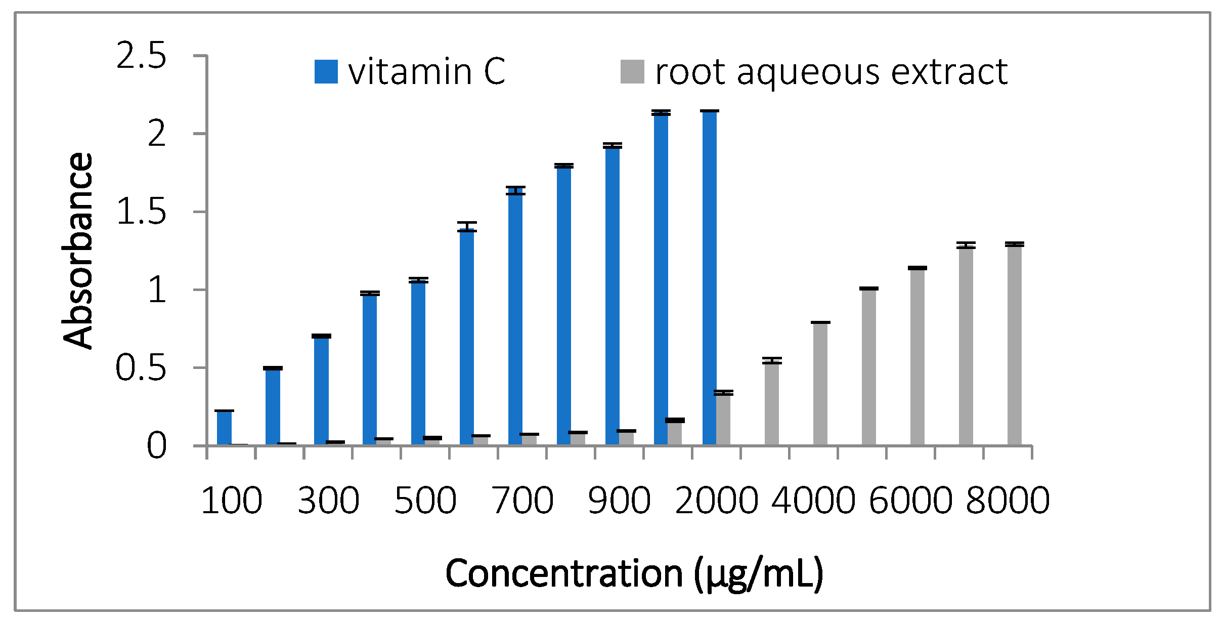
3.3.1. Total Antioxidant Capacity (TAC)

The total antioxidant capacity (TAC) of C. caeruleus L. root aqueous extract, measured using the phosphomolybdate method, is presented in Figure 1. The IC50 value, determined graphically, was calculated using the linear regression equation derived from the calibration curve. A low IC50 value (expressed in µg/mL) indicates high antioxidant activity. In this study, the reducing potential of the standard (471.34 ± 1.41 µg/mL) was significantly higher than that of the roots (5405.1 ± 4.42 µg/mL) (p < 0.05). A dose-dependent antioxidant activity was noted.

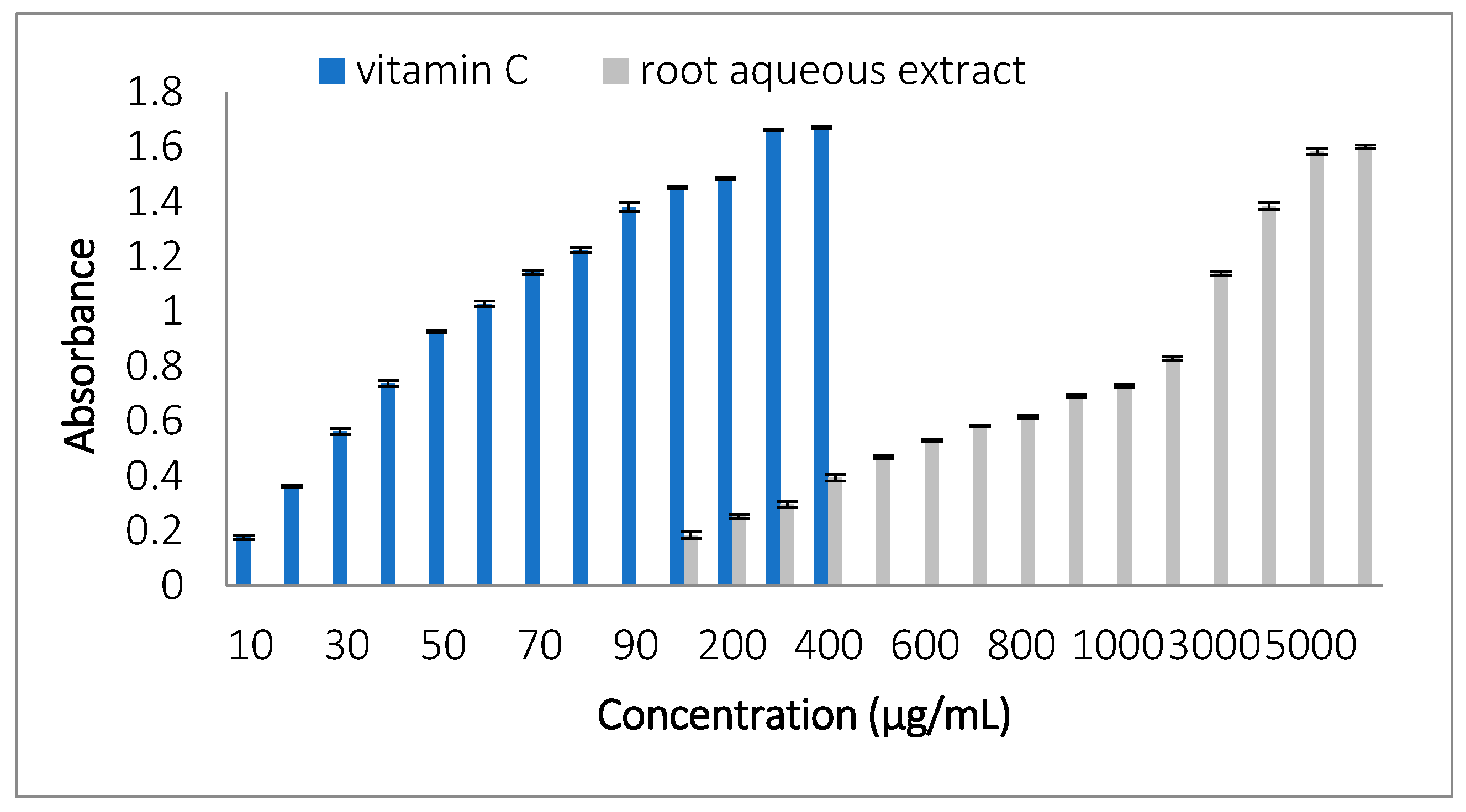
3.3.2. Ferric Reducing Antioxidant Power (FRAP)

To assess the antioxidant potential, the iron-reducing capacity of vitamin C and root extract was evaluated. Figure 2 displays the dose–response curves for both samples, with the IC50 value representing the concentration needed to reduce half of the iron in the assay. The IC50 values obtained were 50.47 ± 0.34 µg/mL for vitamin C and 1132.35 ± 4.97 µg/mL for the root extract. Vitamin C showed significantly higher antioxidant activity than the root extract, as evidenced by its lower IC50 value (p < 0.05).

3.3.3. 2,2-Diphenyl-1-picrylhydrazyl (DPPH) Radical Scavenging Test

The DPPH scavenging activity was evaluated, and the results are presented in Figure 3. The IC50 values obtained were 6.29 ± 0.04 µg/mL for vitamin C and 34.43 ± 4.83 µg/mL for the root extract. Figure 3 shows the dose–response curves for both samples. Vitamin C exhibited significantly higher scavenging activity compared to the root extract (p < 0.05).

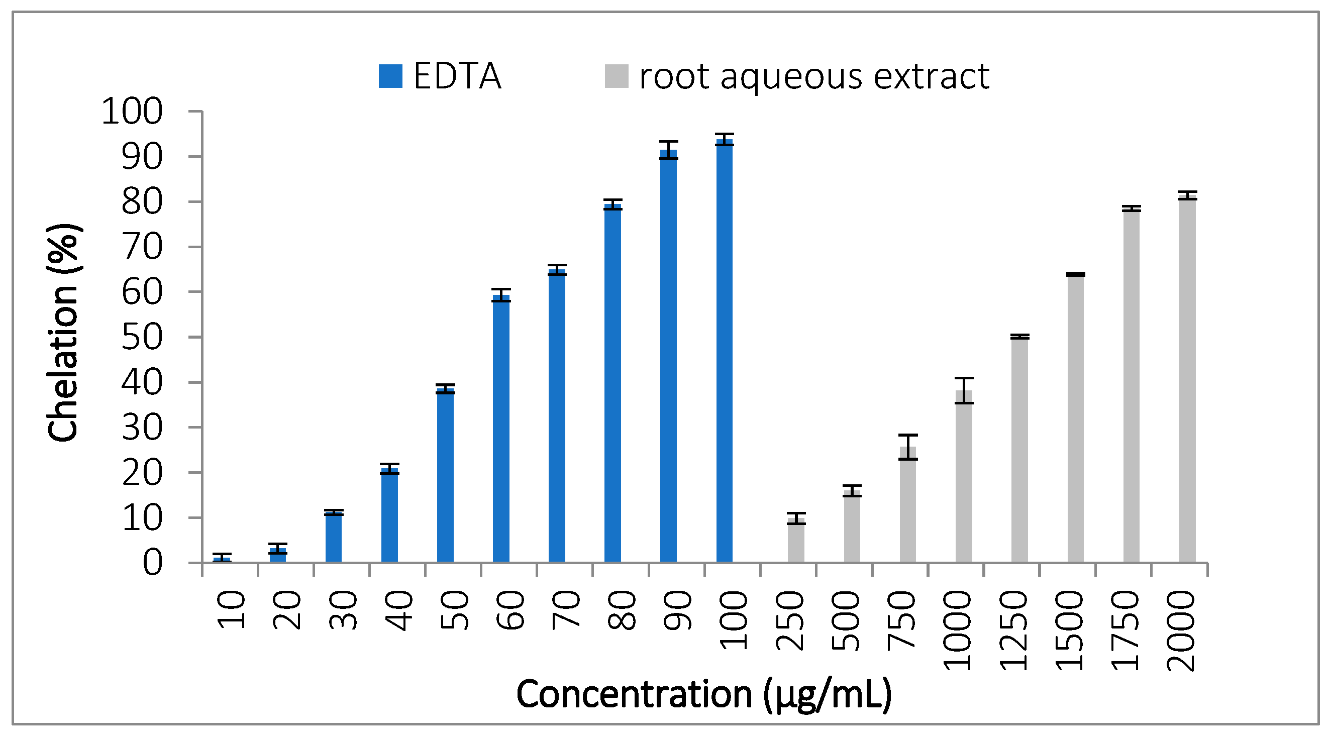
3.3.4. Ferrous Ion Chelating Activity

This test demonstrates the extract’s ability to chelate ferrous ions and inhibit the formation of the ferrozine–Fe2+ complex. The chelating activity of the root extract and EDTA is shown in Figure 4, with IC50 values of 2462.76 ± 1.38 µg/mL for the extract and 57.97 ± 1.29 µg/mL for EDTA. EDTA exhibited significantly higher chelating activity compared to the extract (p < 0.05).

3.3.5. Antioxidant Activity in the Linoleic Acid System: Thiobarbituric Acid (TBA) Method

Linoleic acid peroxidation inhibition by the extract was assessed using the thiobarbituric acid method. The results, presented in Figure 5, show the percentage of protection against lipid peroxidation. BHT (1 mg/mL) provided the highest protection (77.36 ± 1.88%), followed by the root extract (22.32 ± 3.31%) (p < 0.05).

3.3.6. Hydroxyl Radical Scavenging Assay

C. caeruleus L. root extract showed a higher hydroxyl radical (OH) scavenging capacity compared to vitamin C, as demonstrated in Figure 6. The IC50 value for root extract was 512.81 ± 9.46 µg/mL, indicating its ability to neutralize hydroxyl radicals effectively. In contrast, vitamin C exhibited a higher IC50 value of 715.92 ± 1.06 µg/mL (p < 0.05), showing weaker scavenging activity than the root extract. This study highlights, for the first time, the potential of C. caeruleus L. root extract to scavenge hydroxyl radicals, suggesting its significant antioxidant properties.

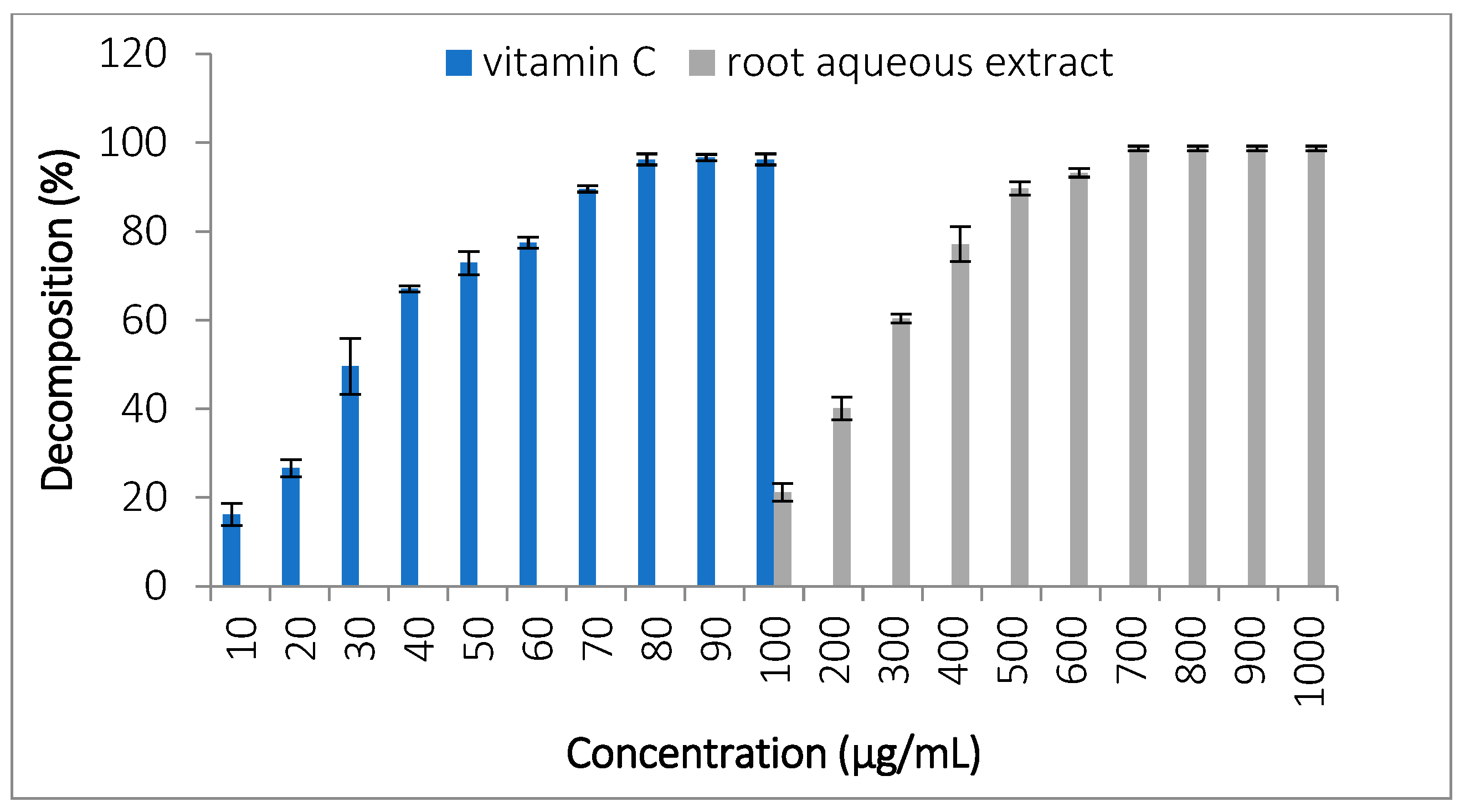
3.3.7. Hydrogen Peroxide (H2O2) Decomposition

This test assesses the ability of the extract to decompose H2O2 compared with a standard (vitamin C), as shown in Figure 7. A significant difference is recorded between the standard and the extract. Vitamin C exhibited the highest activity (IC50 of 37.84 ± 5.98 µg/mL), followed by roots (IC50 of 263.93 ± 7.87 µg/mL) (p < 0.05). This is the first study to document the ability of C. caeruleus L. root extract to decompose hydrogen peroxide.

3.4. Analysis of the Extract’s Toxicity

To properly evaluate the anti-inflammatory activity of an extract, it is essential to assess its safety profile to rule out cytotoxicity, avoid interference with anti-inflammatory assays, and confirm its suitability for pharmacological applications [21]. As shown in Figure 8, the results demonstrate that the C. caeruleus L. root extract, at concentrations ranging from 1 to 8 mg/mL, does not induce hemolysis in erythrocytes, with hemolysis percentages between 0.045 ± 0.12% and 0.59 ± 0.09%. In contrast, saponin caused significant hemolysis, with values ranging from 51.3 ± 0.37% to 98.88 ± 0.18% (p < 0.05).

3.5. Anti-Inflammatory Activity

The anti-inflammatory activity of C. caeruleus L. was evaluated through in vitro methods, as summarized in Table 2. This table shows the maximum activity of the extract, expressed as a percentage, compared to the standard in different assays. To our knowledge, this is the first in vitro analysis of the extract’s anti-inflammatory effects, demonstrating its potential in preventing inflammation-related cellular damage and protecting against protein denaturation.

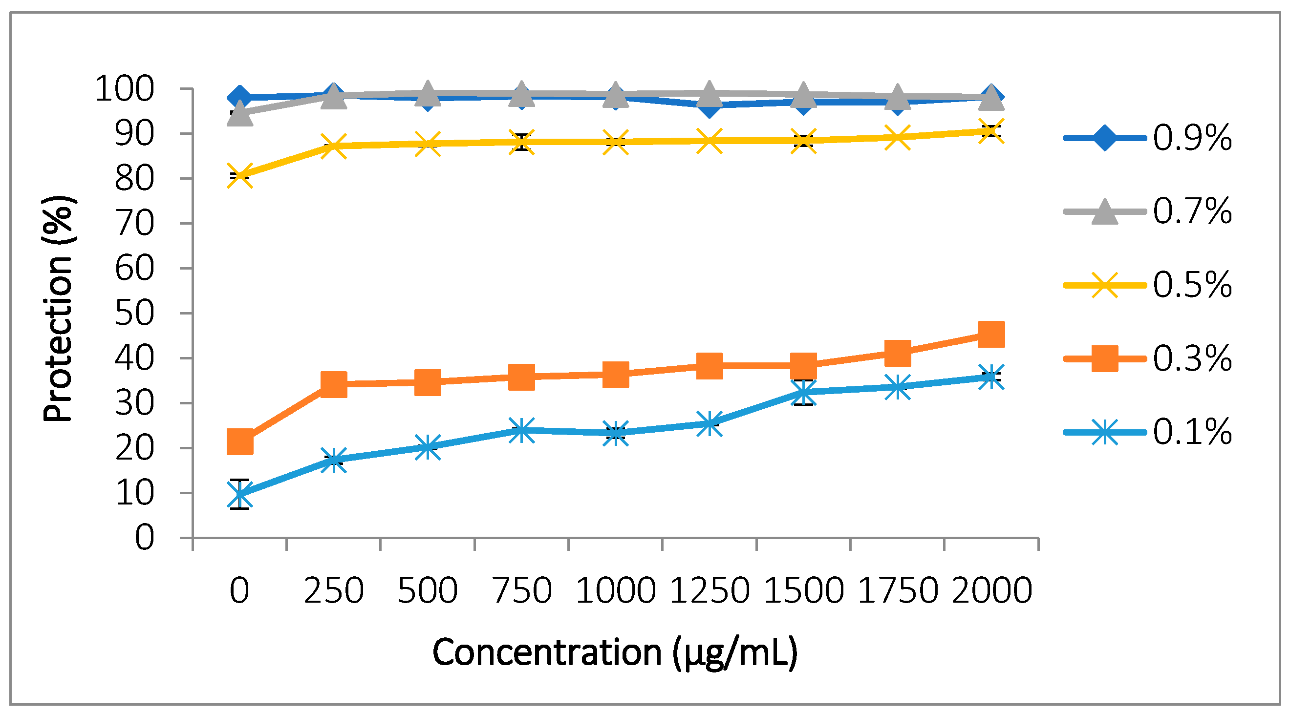
3.5.1. Protection Against Hemolysis Induced by Hypotonic Stress

Research on the stabilization of erythrocyte membranes aims to elucidate the anti-inflammatory mechanism of action. The results demonstrate that the extract effectively prevents membrane lysis induced by hypotonic stress (Table 2 and Figure 9). For the four hypotonic solutions tested (0.7%, 0.5%, 0.3%, and 0.1%), at a concentration of 2000 µg/mL, the extract demonstrated the following percentages of protection: 98.13 ± 0.15%, 90.59 ± 1.07%, 45.3 ± 0.89%, and 35.8 ± 0.75%, respectively. These values are compared with the protection percentage of the extract in the physiological medium of red blood cells (0.9% NaCl), which was 98.2 ± 0.13% at the same concentration. No significant differences were observed when comparing this value to that obtained for the 0.7% NaCl concentration in the presence of the extract. However, significant differences were noted with the other concentrations (0.5%, 0.3%, and 0.1%) compared to 0.9% NaCl (p < 0.05).

3.5.2. Protection Against Heat-Induced Hemolysis

In this study, the extract demonstrated significant erythrocyte protection, although this was significantly lower than that observed with aspirin at the maximum concentration of 3000 µg/mL (p < 0.05). Protection percentages reported for the extract ranged from 19.2 ± 0.2 to 70 ± 1.27% at concentrations from 100 to 3000 µg/mL. Aspirin protection percentages ranged from 40.77 ± 0.59 to 78.53 ± 0.51% over the same concentration range (Table 2, Figure 10).

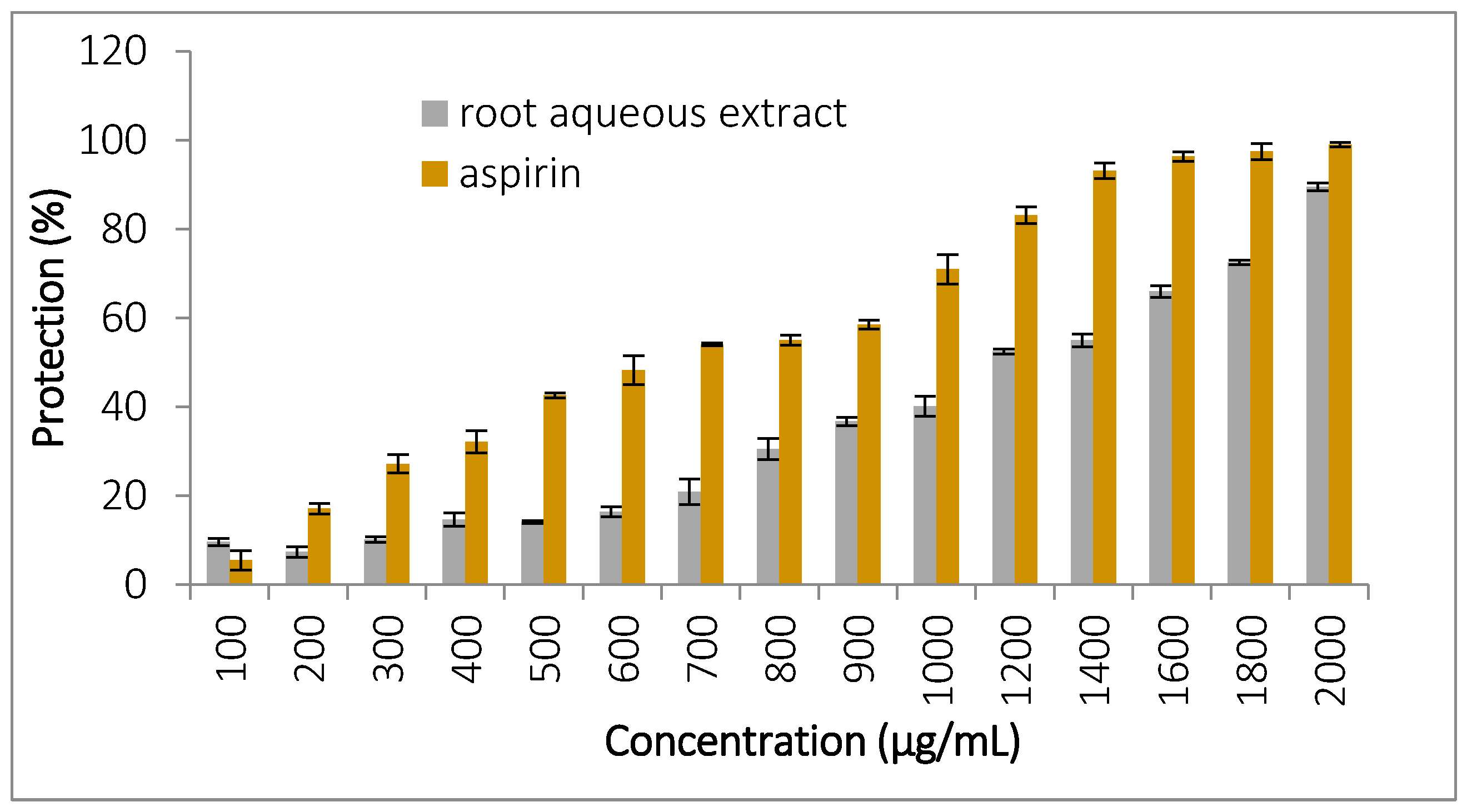
3.5.3. Protection Against Oxidative Stress-Induced Hemolysis

To evaluate the anti-inflammatory potential of the extract, oxidative stress was induced in erythrocytes using hypochlorous acid (HClO). The results revealed a notable protective effect, with protection percentages ranging from 8.66 ± 0.86% to 89 ± 0.87%, similar to those observed with aspirin (5.47 ± 2.16% to 99 ± 0.5%) across concentrations from 100 to 2000 µg/mL (Table 2, Figure 11). No difference was found between the extract and aspirin at the highest concentration of 2000 µg/mL (p < 0.05).

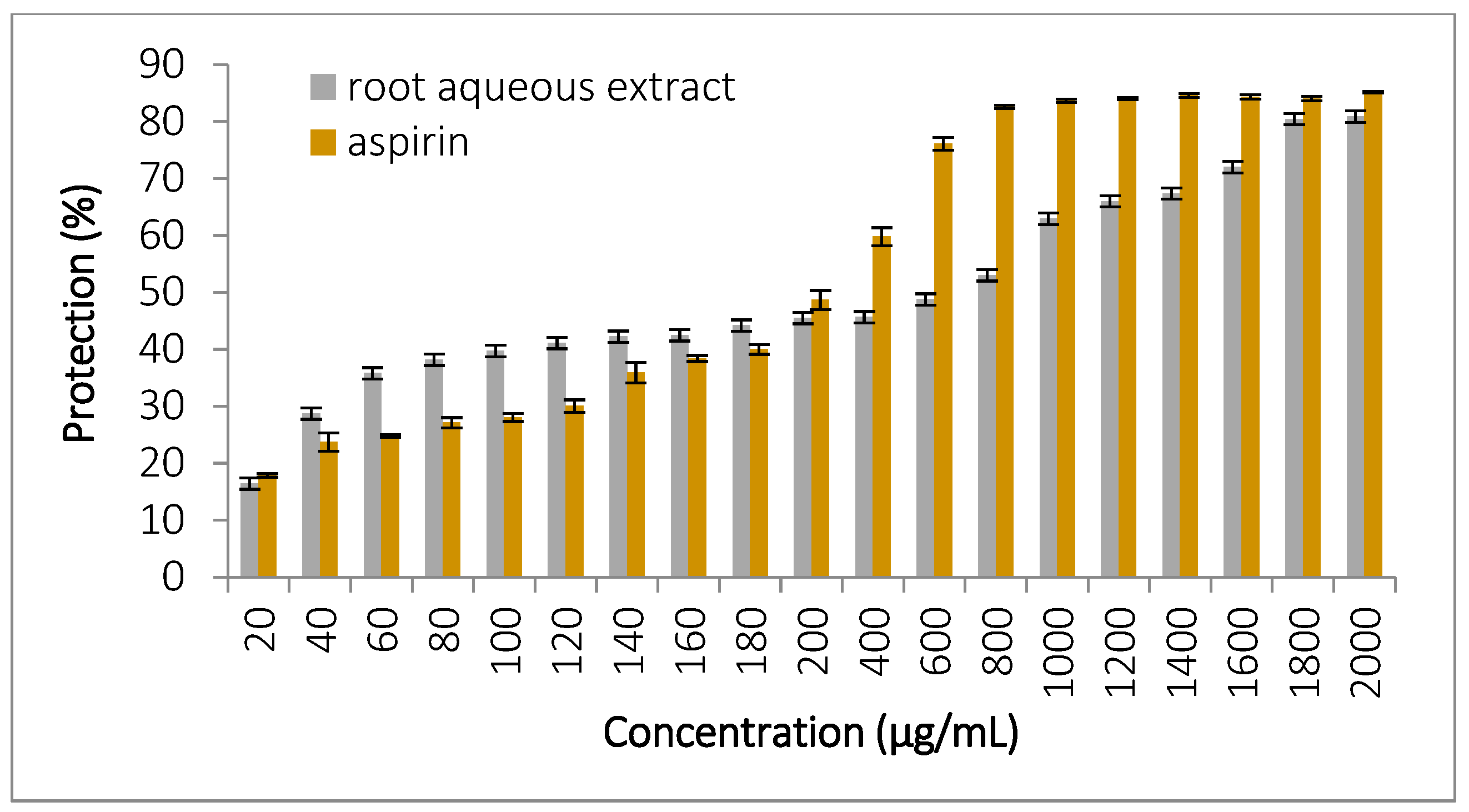
3.5.4. Inhibition of Ovalbumin Denaturation

Protein denaturation plays a key role in several inflammatory conditions, and the capacity of C. caeruleus L. to inhibit denaturation suggests an anti-inflammatory potential. In this study, the root extract effectively inhibited heat-induced ovalbumin denaturation at concentrations ranging from 20 to 2000 µg/mL, showing protection percentages from 16.46 ± 2.13% to 80.86 ± 2.23%. In comparison, aspirin demonstrated protection values between 17.86 ± 0.3% and 85.28 ± 0.11%, showing no significant difference when compared to the extract at 2000 µg/mL (Figure 12).

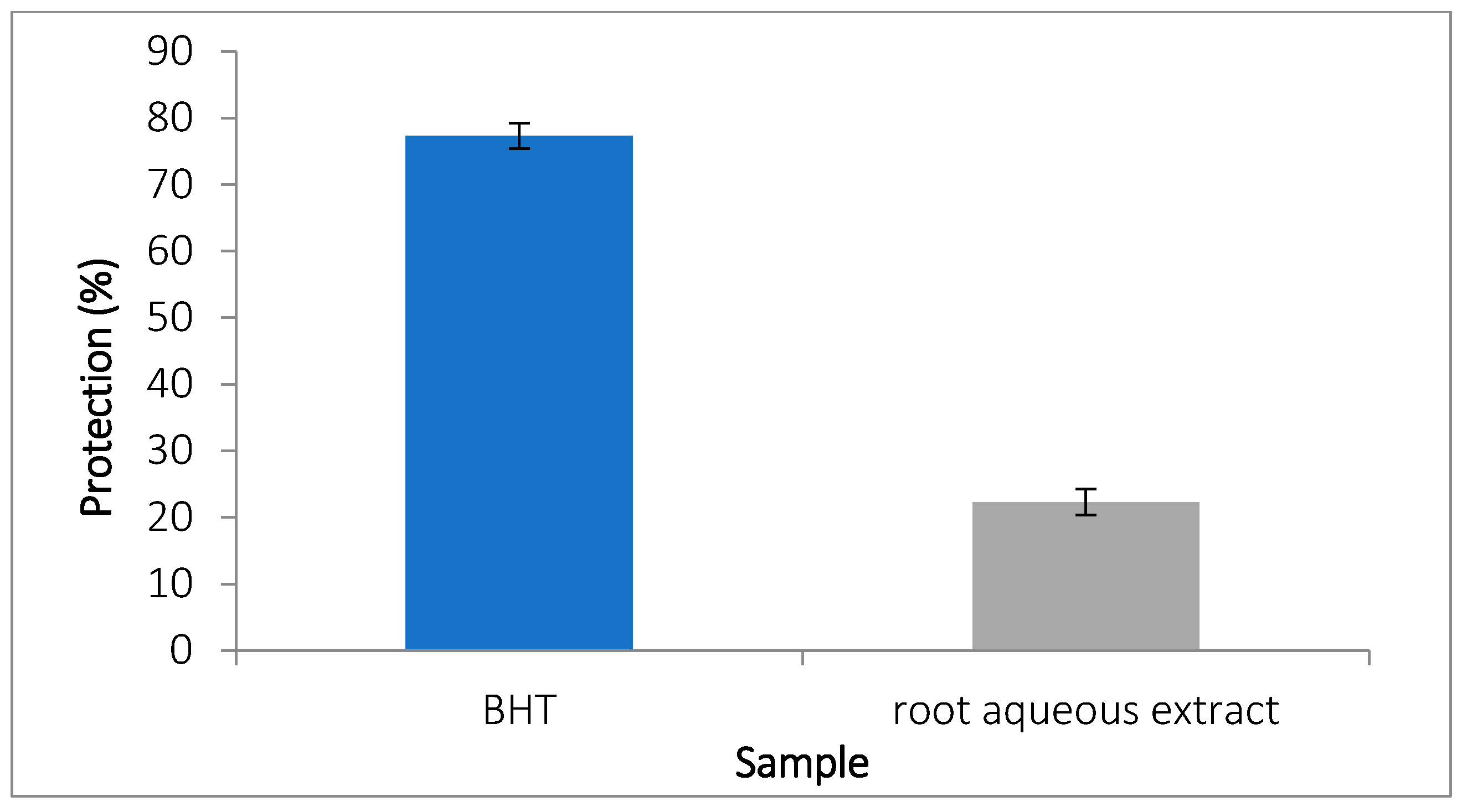
3.5.5. Assessment of Lipid Peroxidation in Animal Tissues Using the TBARS Method

The results of lipid peroxidation assays on brain homogenate (TBARS) are presented in Table 2 and Figure 13. Significant activity was observed for the extract (23.69 ± 0.61 to 69.25 ± 0.89%) compared with BHT (32.53 ± 1 to 77.2 ± 1.02%) for concentrations ranging from 200 to 4000 µg/mL (Figure 13). At a concentration of 4000 µg/mL, a significant difference was observed between the extract and BHT (p < 0.05).

4. Discussion

Polyphenols are a diverse group of bioactive compounds, with flavonoids, tannins, and phenolic acids being among the most widely studied [61]. These compounds are known for their significant antioxidant and anti-inflammatory activities, making them of particular interest in various medicinal applications. In this context, C. caeruleus L., a plant traditionally used in Algerian medicine, has garnered attention for its potential beneficial properties.
When comparing our results, which revealed moderate quantities of polyphenols in the C. caeruleus L. root aqueous extract (21.19 ± 0.37 mg GAE/g dry extract), Baghiani et al. [67] and Ouda et al. [68] reported lower polyphenol contents in C. caeruleus L. methanolic root extract, with values of 12.966 ± 0.727 and 13.08 ± 0.22 mg GAE/g extract, respectively. Both of these studies utilized methodologies similar to ours. Interestingly, while their studies indicated higher flavonoid contents, recorded at 2.231 ± 0.146 and 5.02 ± 0.55 mg QE/g extract, respectively, our results for flavonoids content were significantly lower, at 0.72 ± 0.013 mg QE/g dry extract. These discrepancies may be attributed to the polarity of the solvents during extract preparation [69]. Solvent selection plays a crucial role in determining the effectiveness of polyphenol and flavonoid extraction: polar solvents extract hydrophilic compounds, while non-polar solvents extract lipophilic substances. Variations in solvent polarity can thus lead to differing amounts of these compounds from the same plant material [70]. In this regard, Moussa et al. [71] stated that polyphenols in C. caeruleus L. tend to be hydrophilic, and that adding water to methanol during extraction increases their quantity. This could explain the lower polyphenol content observed in the methanolic root extracts (13.08 ± 0.22 mg GAE/g extract) obtained by Baghiani et al. [67] and Ouda et al. [68]. Conversely, the higher flavonoid levels observed in these studies could be due to the chemical properties of flavonoids, which are more soluble in methanol, as noted by Ali-Rachedi et al. [69], which may explain the higher quantities reported in these two studies.
The assessment of plant extracts’ antioxidant activity is a major area of research, with in vitro tests being the most frequently employed to estimate extract efficacy and facilitate rapid screening [72]. Indeed, low in vitro activity may reflect limited in vivo efficacy [73]. Although no single test can comprehensively summarize the total antioxidant activity of a plant due to its chemical complexity [67], various methods based on redox reactions have been developed. These methods involve electron or proton donation, as well as metal ion chelation [74]. This study employed various assays to assess the antioxidant capacity of C. caeruleus L. root aqueous extract.
The antioxidant activity of C. caeruleus L. is primarily attributed to its reducing capacity, a key mechanism in neutralizing free radicals. This capacity, which serves as an indicator of the extract’s antioxidant potential, is further supported by the results from the TAC and FRAP assays, which are essential for evaluating the reducing properties of antioxidant agents [75,76]. The observed activity in these assays is linked to the extract’s ability to reduce molybdate Mo(VI) ions to molybdenum Mo(V), or to convert iron into its ferrous form (Fe2⁺), through the electron transfer capacity of the polyphenols present in the extract [77,78,79]. The bioactive compounds in the extract, including tannic acid, quercetin [78], luteolin [55], and myricetin [80], are likely to play a central role in these reduction processes.
Our extract is also capable of neutralizing free radicals, as demonstrated by the DPPH and OH assays. In line with previous studies by Baghiani et al. [67] and Moussa et al. [71], who reported DPPH radical scavenging activity in C. caeruleus L. methanolic root extracts (187.38 ± 2.36 μg/mL and 1606 ± 0.05 μg/mL, respectively), our results confirm the significant antioxidant activity of the root extract. This variation in activity could be attributed to differences in extraction methods and solvent nature, both well-known factors influencing the type and quantity of polyphenolic compounds extracted [81]. Additionally, as noted by Habibou et al. [79], geographical variations may also contribute to differences in polyphenol content and antioxidant capacity. In this study, the observed antioxidant activity was further corroborated by the extract’s ability to donate hydrogen atoms and/or electrons, which led to the disappearance of the characteristic purple color of DPPH, and the formation of the reduced radical (DPPH-H) [82]. The presence of bioactive compounds might explain the observed antioxidant effect. For example, caffeic acid [83], luteolin [55], hesperidin [54], rutin [52], quercetin, myricetin [80], and p-coumaric acid [76] are known for their capacity to stabilize the DPPH radical.
Furthermore, the OH radical, one of the most reactive species responsible for the degradation of biological biomolecules, particularly lipids, was also effectively scavenged by the C. caeruleus L. root extract. This inhibitory effect against OH radicals could be attributed to the extract’s ability to neutralize the radical directly or to inhibit the Fenton reaction, which generates OH radicals, through hydrogen peroxide decomposition and/or metal ion chelation, particularly iron. Studies have shown that the ability of extracts to neutralize free radicals is closely related to their chemical composition. For example, Parhiz et al. [84], Nile et al. [85], and Vieira et al. [60] demonstrated, respectively, that compounds such as hesperidin, quercetin, and caffeine are effective in scavenging OH radicals. According to Papuc et al. [74], the phenolic hydroxyl group of flavonoids, particularly their structural position, plays a crucial role in scavenging OH radicals. The superior scavenging activity of our extract compared to vitamin C may be attributed to a synergistic effect among its bioactive compounds, enhancing the collective radical scavenging capacity and surpassing the individual effect of vitamin C.
As previously mentioned, our extract may inhibit OH radicals through ferrous ion chelation, and the results from the chelation assay with C. caeruleus L. further support this hypothesis. Ouda et al. [68] also demonstrated the ability of C. caeruleus L. root extract to chelate ferrous ions. The significant activity observed in this study can be related to the presence of polyphenols in the extract, which are known for their strong metal ion binding properties. Among these compounds, flavonoids are particularly important due to their hydroxyl and carbonyl groups, which enable them to effectively bind metal ions [75]. Notably, quercetin stands out as a potent chelator due to its hydroxyl functional groups, including 3′-OH and 4′-OH from the B-ring and 3-OH and 4-oxo from the C-ring, as well as 4-oxo and 5-OH [86].
As previously suggested, our extract has exhibited antioxidant potential by decomposing hydrogen peroxide, a molecule with indirect oxidizing properties that can be converted into water through electron and proton transfer by polyphenols [74]. Hydrogen peroxide plays a role in the degradation of biological molecules by generating OH radicals, which underscores the importance of evaluating H2O2 decomposition activity [87]. In this study, C. caeruleus L. root aqueous extract demonstrated significant antioxidant activity by effectively decomposing hydrogen peroxide. This activity is attributed to the presence of polyphenols, such as tannic acid, which facilitate the conversion of hydrogen peroxide into water through electron and proton transfer [74,88].
Finally, root extract demonstrated a strong inhibitory effect on lipid peroxidation in vitro, as shown by the TBA test. These findings are consistent with those of Baghiani et al. [67], who reported the inhibitory activity of C. caeruleus L. extract against linoleic acid oxidation, measured by the β-carotene method. Lipid peroxidation is a key model for assessing antioxidant potential, involving free radicals and metal ions that lead to the formation of malondialdehyde (MDA) [61,89]. In this study, the extract reduced the formation of peroxidation products, likely due to polyphenols such as quercetin, rutin, and tannic acid, which may act by chelating Fe2⁺ ions [37,74] or scavenging peroxyl (LOO) and alkoxy (LO) radicals through their conjugated cyclic structure and hydroxyl groups [9,50].
Polyphenols are renowned for their wide range of biological activities, which are largely attributed to the presence of their aromatic rings, hydroxyl groups, and specific configurations within the phenyl ring [90]. These compounds possess the ability to easily transfer protons and electrons during oxidative stress, a feature enhanced by the resonance stabilization provided by the aromatic ring [67]. Numerous studies have highlighted the relationship between structure and activity, demonstrating that the antioxidant properties of polyphenols are intricately linked to their chemical structures [50,52]. For example, C. caeruleus L. root extract contains tannic acid, which, with its hydrophobic core and hydrophilic outer shell, interacts effectively in various environments, neutralizing free radicals through its abundant hydroxyl groups [88]. Similarly, the ortho-dihydroxy configuration of rosmarinic acid has been shown to enhance its antioxidant activity [44]. Additionally, the presence of flavonoids such as quercetin, myricetin, hesperidin, and rutin likely contributes to the potent antioxidant activity of the extract. According to Borges-Bubols et al. [50] and Apak et al. [78], quercetin’s 3,4-dihydroxy structure in the phenolic B ring aids in metal ion chelation and its 2,3 double bonds in the C ring facilitate electron delocalization, while myricetin, with its three OH groups on the B ring, proves effective in lipid systems. Furthermore, the strong antioxidant properties of hesperidin [54] and rutin [52] can be attributed to the presence of a 3′-OH group. Likewise, luteolin’s 3′,4′-dihydroxy structure on the phenolic B ring likely plays a key role in its antioxidant effects [55].
Lysozymes, which are produced by neutrophils during inflammation, contribute to the perpetuation of inflammation and tissue damage. Limiting their release by stabilizing lysosomal membranes is crucial to preventing such harm. Similarly to lysosomal membranes, erythrocyte membranes can be used to assess the extract’s capacity to prevent lysozyme release [91]. Human erythrocytes serve as an important model for evaluating the anti-inflammatory potential of extracts. The release of hemoglobin following lysis allows for the measurement of hemolytic power spectrophotometrically, reflecting membrane integrity [92]. While little is known about the anti-inflammatory effects of phytochemicals, extracts may prevent erythrocyte hemolysis by stabilizing membrane integrity [35] and mitigating oxidative damage [39]. Investigating the extract’s ability to stabilize erythrocyte membranes during lysis induced by osmotic stress [91], heat [93], or oxidative stress with HClO [33] provides a measure of its in vitro anti-inflammatory activity.
The presence of a hypotonic solution surrounding erythrocytes leads to an excessive accumulation of fluid within these cells, resulting in membrane rupture [94]. The activity of C. caeruleus L. in preventing this hemolysis thus highlights its ability to maintain membrane integrity, likely due to the presence of phytochemicals including flavonoids, alkaloids, and tannins [95]. While the exact mechanism of this protection remains unclear, AlSaffar et al. [96] suggested that the deformability and lysis of erythrocytes might be related to their calcium content. Indeed, intracellular calcium plays a crucial role in cellular function, but its concentration must be kept low to preserve cell integrity. An excessive accumulation of intracellular Ca2⁺ leads to irreversible changes in the membrane, causing the damage and destruction of erythrocytes. Similarly, it has been demonstrated that free radicals alter the structure of phospholipids by increasing intracellular calcium levels [97]. In this context, it can be suggested that C. caeruleus L. extract may protect erythrocytes by modulating calcium influx into erythrocytes and maintaining calcium concentration at low levels.
Thermal stress is a primary factor contributing to inflammation and cellular damage, leading to morphological changes and membrane lysis [98]. This hyperthermia induces protein unfolding, including membrane proteins, which leads to their aggregation and irreversible damage if not effectively managed [99]. In this context, several studies report that certain plant extracts can inhibit heat-induced protein denaturation [28]. Consequently, it may be inferred that C. caeruleus L. extract may protect cells against thermal stress by inhibiting the denaturation of membrane proteins, which, consequently, would explain their ability to prevent erythrocyte hemolysis. Various studies have highlighted that phytochemicals, like flavonoids, tannins, and alkaloids present in this extract, could be responsible for this capacity to stabilize membranes [100]. Consequently, these results could support the traditional use of this plant in treating skin burns, as shown by ethnobotanical investigations [101,102].
Hypochlorous acid (HClO) is a powerful natural oxidant generated by neutrophils and monocytes during inflammation which is responsible for tissue damage. While the exact mechanism of action of this oxidant remains unclear, research has shown that the primary site of action is the cell membrane, inducing cell lysis [33]. The protective activity of C. caeruleus L. root aqueous extract appears to be closely linked to its chemical composition. Previous research, such as that conducted by Suwalsky et al. [103], demonstrated that polyphenols present in the extracts are capable of diffusing through and being distributed within the cell membranes, inhibiting the diffusion of free radicals. This mechanism contributes to membrane protection and the extract’s anti-inflammatory effect. Furthermore, An et al. [56] also revealed that quercetin, vitexin, orientin, and luteolin protect red blood cells against H2O2-induced toxicity. These findings support the observations made by Suwalsky et al. [33], who, through electron microscopy analysis, showed that resveratrol neutralizes the oxidative effects of HClO and prevents modifications in human erythrocytes’ forms. According to their conclusions, the insertion of resveratrol molecules into the lipid bilayer of cell membranes could disrupt the insertion of HClO, thus reducing its deleterious effects. They suggested that this protection results from the hydrogen atoms on its three hydroxyl groups, which can neutralize reactive oxidants and prevent them from attacking the cell membrane before they can penetrate.
The inflammatory response can also arise from protein denaturation [93]. Heat, a physiological response to inflammation, can act as a denaturant, causing conformational changes in proteins that disrupt their structure and biological functionality [104]. Non-steroidal anti-inflammatory drugs (NSAIDs) counter this effect by stabilizing proteins through specific binding interactions, preventing conformational changes that lead to denaturation and preserving their function under thermal stress [105]. In light of this, studying heat-induced protein denaturation or lipid peroxidation provides a useful model for evaluating the anti-inflammatory activity of extracts [93]. C. caeruleus L.’s capacity to inhibit ovalbumin denaturation, for instance, is likely due to the presence of phenolic compounds that interact with proteins, stabilizing them by complexing with ovalbumin. Similar findings have been reported for other plant extracts, such as Rosa canina, which inhibits bovine serum albumin (BSA) denaturation by binding to aliphatic regions around lysine residues [106].
In inflammation, the overproduction of free radicals can lead to cell damage and lipid peroxidation, which subsequently destroy membranes and exacerbate the inflammatory response [35]. Cell membranes, especially those of neurons, contain a large amount of polyunsaturated fatty acids [107]. According to numerous pharmacological studies, polyunsaturated fatty acids, being the molecules most sensitive to peroxidation, affect the biochemical characteristics of membranes, such as fluidity and permeability [68]. The TBARS assay measures the products of this peroxidation, thus providing insights into the degree of oxidative damage within membranes. Various cell types have been used as sources of the fatty acids—liver, spleen [35], red blood cells [108], and the brain—which were utilized in our study [36]. The measurement of malondialdehyde, a product of lipid peroxidation, in animal tissues is a method for assessing the extent of this reaction [36]. In this study, the extract’s ability to prevent lipid peroxidation in brain homogenate can be attributed to its chemical composition. Louerrad et al. [94] demonstrated that flavonoids can inhibit lipid peroxidation of erythrocyte membranes by chelating iron, which prevents the formation of free radicals and, consequently, protects polyunsaturated fatty acids from damage. For example, quercetin is effective against H2O2-induced lipid peroxidation in erythrocyte membranes [51]. Other research has revealed that ferulic acid prevents peroxidation through its role in neutralizing free radicals, targeting hydroxyl and peroxyl radicals [39]. In this context, further studies suggested that the observed activity may result from the capacity of polyphenols to insert into membranes and restrict their fluidity, thereby limiting the diffusion of free radicals that could induce lipid peroxidation [103].
Finally, this study further supports the safety profile of Carthamus caeruleus, which exhibits significant antioxidant and anti-inflammatory activities, as described in previous sections. A prior in vivo toxicity assessment conducted on Wistar rats demonstrated no signs of toxicity when treated with root aqueous extract at doses of 300, 500, and 1000 mg/kg body weight over a 24 h observation period [68]. These findings are consistent with those of Dahmani et al. [109], who also investigated the toxicity of C. caeruleus L. root methanolic extract.

5. Conclusions

C. caeruleus L. root aqueous extract demonstrates significant antioxidant and anti-inflammatory activities, primarily attributed to its rich phytochemical composition. Its antioxidant effects are linked to its ability to reduce and chelate metal ions, especially iron, which are involved in generating harmful radicals, like hydroxyl and pyroxyl. Additionally, the extract directly neutralizes free radicals, such as OH and DPPH, or decomposes hydrogen peroxide. Furthermore, the anti-inflammatory effects of the extract can be attributed not only to its antioxidant potential, but also to the ability of its chemical compounds to integrate into membranes, stabilizing their integrity and providing protection against protein denaturation and membrane lipid peroxidation. In light of these results, this study provides novel perspectives on the antioxidant and anti-inflammatory potential of C. caeruleus L., reinforcing its traditional use and efficacy against burns. However, further studies are recommended to identify new bioactive molecules and evaluate the beneficial potential of this plant in vivo.

Supplementary Materials

The following supporting information can be downloaded at: https://www.mdpi.com/article/10.3390/pr13030878/s1, Figure S1: Gallic Acid Calibration Curve; Figure S2: Quercetin Calibration Curve; Figure S3: Tannic Acid Calibration Curve; Figure S4: Chromatogram of C. caeruleus L. root aqueous extract at 230.4 nm; Figure S5: Chromatogram of C. caeruleus L. root aqueous extract at 255.4 nm: Figure S6: Chromatogram of C. caeruleus L. root aqueous extract at 280.4 nm; Figure S7: Chromatogram of C. caeruleus L. root aqueous extract at 300.4 nm; Figure S8: Chromatogram of C. caeruleus L. root aqueous extract at 355.4 nm; Figure S9: Spectrum of the mixture of reference standards.

Author Contributions

Writing—original draft, Y.B.; conceptualization, I.M. and H.S.; methodology, Y.B., I.M., H.A.I. and K.H.; data curation, Y.B.; Formal analysis, Y.B., B.S. and S.S.; validation, K.H. and L.T.; investigation, Y.B.; visualization, I.M., H.S., K.H. and E.-h.N.; supervision and validation E.-h.N., C.C. and K.H. All authors have read and agreed to the published version of the manuscript.

Funding

This research was funded by (i) Portuguese funds from Fundação para a Ciência e a Tecnologia through the project UIDB/00329/2020 (DOI 10.54499/UIDB/00329/2020).

Data Availability Statement

The original contributions presented in this study are included in the article/Supplementary Materials. Further inquiries can be directed to the corresponding authors.

Acknowledgments

The authors would like to express their gratitude to the Algerian Ministry of Higher Education and to Benghanem Nabil for his precious help during the research.

Conflicts of Interest

The authors declare no conflicts of interest.

Abbreviations

A: absorbance; ANOVA, analysis of variance; BHT, butyl hydroxy toluene; DAD, diode array detector; DPPH, 2,2-diphenyl-1-picrylhydrazil; EDTA, ethylenediamine tetraacetic acid; FRAP, ferric reducing antioxidant power; GAE, gallic acid equivalents; H2O2, hydrogen peroxide; HClO: hypochlorous acid; HPLC, high-performance liquid chromatography; IC50, inhibition concentration 50%; OH•, hydroxyl radical; QE, quercetin equivalents; RT, retention time; SEM, standard error of mean; TAC, total antioxidant capacity; TAE, tannic acid equivalents; TBA, thiobarbituric acid; TBARS, thiobarbituric acid reactive substance.

References

  1. GBD. Disease and Injury Incidence and Prevalence Collaborators. Global, Regional, and National Incidence, Prevalence, and Years Lived with Disability for 354 Diseases and Injuries for 195 Countries and Territories, 1990–2017: A Systematic Analysis for the Global Burden of Disease Study 2017; University of Leicester: Leicester, UK, 2017. [Google Scholar]
  2. Abegunde, D.O.; Mathers, C.D.; Adam, T.; Ortegon, M.; Strong, K. The burden and costs of chronic diseases in low-income and middle-income countries. Lancet 2007, 370, 1929–1938. [Google Scholar] [CrossRef]
  3. Lucero-Prisno, D.E., III; Shomuyiwa, D.O.; Kouwenhoven, M.B.N.; Dorji, T.; Odey, G.O.; Miranda, A.V.; Ogunkola, I.O.; Adebisi, Y.A.; Huang, J.; Xu, L.; et al. Top 10 public health challenges to track in 2023: Shifting focus beyond a global pandemic. Public Health Chall. 2023, 2, e86. [Google Scholar] [CrossRef]
  4. Alkhateeb, H.H. Evaluation of antidiabetic, antioxidant, and antilipidemic potential of natural dietary product prepared from Cyphostemma digitatum in rats’ model of diabetes. J. Herbmed Pharmacol. 2022, 11, 197–203. [Google Scholar] [CrossRef]
  5. Jasemi, S.V.; Khazaei, H.; Fakhri, S.; Mohammadi-Noori, E.; Farzaei, M.H. Naringenin improves ovalbumin-induced allergic asthma in rats through antioxidant and anti-inflammatory effects. Based Complement. Altern. Med. 2022, 1, 9110798. [Google Scholar] [CrossRef] [PubMed]
  6. Migdal, C.; Serres, M. Espèces réactives de l’oxygène et stress oxydant. Médecine/Sci. 2011, 27, 405–412. [Google Scholar] [CrossRef] [PubMed]
  7. Biswas, S.K. Does the interdependence between oxidative stress and inflammation explain the antioxidant paradox? Oxidative Med. Cell. Longev. 2016, 2016, 5698931. [Google Scholar] [CrossRef]
  8. Mobashar, A.; Shabbir, A.; Shahzad, M.; Gobe, G. Preclinical Rodent Models of Arthritis and Acute Inflammation Indicate Immunomodulatory and Anti-Inflammatory Properties of Juglans Regia Extracts. Evid. Based Complement. Altern. Med. 2022, 2022, 1695701. [Google Scholar] [CrossRef] [PubMed]
  9. Kizil, G.; Kizil, M.; Ceken, B.; Yavuz, M.; Demir, H. Protective ability of ethanol extracts of Hypericum scabrum L. and Hypericum Retusum Aucher Against Protein Oxidation DNA Damage. Int. J. Food Prop. 2011, 14, 926–940. [Google Scholar] [CrossRef]
  10. Asadi, M. Antioxidant and antimicrobial activities in the different extracts of Caspian saffron, Crocus caspius Fisch & CA Mey. Ex Hohen. Casp. J. Environ. Sci. 2016, 14, 331–338. [Google Scholar]
  11. Toubane, A.; Rezzoug, S.A.; Besombes, C.; Daoud, K. Optimization of Accelerated Solvent Extraction of Cart. Caeruleus L. Eval. Antioxid. Anti-Inflamm. Act. Extr. Ind. Crops Prod. 2017, 97, 620–631. [Google Scholar]
  12. Shakya, A.K. Medicinal plants: Future source of new drugs. Int. J. Herb. Med. 2016, 4, 59–64. [Google Scholar]
  13. Nguyen, T.L.A.; Bhattacharya, D. Antimicrobial activity of quercetin: An approach to its mechanistic principle. Molecule 2022, 27, 2494. [Google Scholar] [CrossRef]
  14. Gorniak, I.; Bartoszewski, R.; Króliczewski, J. Comprehensive review of antimicrobial activities of plant flavonoids. Phytochem. Rev. 2019, 18, 241–272. [Google Scholar] [CrossRef]
  15. Ganeshpurkar, A.; Saluja, A. The pharmacological potential of hesperidin. Indian J. Biochem. Biophys. (IJBB) 2019, 56, 287–300. [Google Scholar]
  16. Strudwick, X.L.; Cowin, A.J. The Role of the Inflammatory Response in Burn Injury. In Hot Topics in Burn Injuries; InTech: London, UK, 2018. [Google Scholar]
  17. Sahib, A.S.; Al-Jawad, F.H.; Al-Kaisy, A.A. Burns, endothelial dysfunction, and oxidative stress: The role of antioxidants. Ann. Burn. Fire Disasters 2009, 22, 6. [Google Scholar]
  18. Khémiri, I.; Essghaier, B.; Sadfi-Zouaoui, N.; Bitri, L. Antioxidant and antimicrobial potentials of seed oil from Carthamus tinctorius L. in the management of skin injuries. Oxidative Med. Cell. Longev. 2020, 2020, 4103418. [Google Scholar] [CrossRef]
  19. Khalil, H.E.; Al Ahmed, A. Phytochemical Analysis and Free Radical Scavenging Activity of Carthamus oxyacantha growing in Saudi Arabia: A Comparative Study. Int. J. Pharm. Sci. Rev. Res. 2017, 45, 51–55. [Google Scholar]
  20. Saffidine, K.; Sahli, F.; Mihoub, Z.M. Antimicrobial activity of an Algerian medicinal plant: Cart. Caeruleus L. Pharmacogn. Commun. 2013, 3, 71–76. [Google Scholar]
  21. Haddouchi, F.; Chaouche, T.M.; Halla, N. Phytochemical screening, antioxidant activities and hemolytic power of four Saharan plants from Algeria. Phytothérapie 2016, 16, 1–9. [Google Scholar]
  22. Akrout, A.; Gonzalez, L.A.; El Jani, H.; Madrid, P.C. Antioxidant and antitumor activities of Artemisia campestris and Thymelaea hirsuta from southern Tunisia. Food Chem. Toxicol. 2011, 49, 342–347. [Google Scholar] [CrossRef]
  23. Hmid, I.; Hanine, H.; Elothmani, D.; Oukabli, A. The physicochemical characteristics of Morrocan pomegranate and evaluation of the antioxidant activity for their juices. J. Saudi Soc. Agric. Sci. 2018, 17, 302–309. [Google Scholar]
  24. Jurišić Grubešić, R.; Srečnik, G.; Kremer, D.; Vuković Rodríguez, J.; Nikolić, T.; Vladimir-Knežević, S. Simultaneous RP-HPLC-DAD separation, and determination of flavonoids and phenolic acids in Plantago L. species. Chem. Biodivers. 2013, 10, 1305–1316. [Google Scholar] [CrossRef]
  25. Trinel, M.; Dubois, C.; Burger, P.; Plainfossé, H.; Azoulay, S.; Verger-Dubois, G.; Fernandez, X. Phytochemical Investigation of an Ostrya carpinifolia L. Extract: An Effective Anti-Pollution Cosmetic Active Ingredient. Chem. Biodivers. 2024, 22, e202402139. [Google Scholar] [CrossRef] [PubMed]
  26. Rao, A.S.; Reddy, S.G.; Babu, P.P.; Reddy, A.R. The antioxidant and antiproliferative activities of methanolic extracts from Njavara rice bran. BMC Complement. Altern. Med. 2010, 10, 4. [Google Scholar] [CrossRef] [PubMed]
  27. Moualek, I.; Benarab, K.; Belounis, Y.; Houali, K. Anti-Inflammatory and Antioxidant Activities of Cow’s Milk Supplemented with Aqueous Extract of Malva Sylvestris. Eurasia Proc. Health Environ. Life Sci. 2023, 11, 1–11. [Google Scholar] [CrossRef]
  28. Kızıl, G.; Kızıl, M.; Yavuz, M.; Emen, S.; Hakimoğlu, F. Antioxidant Activities of Ethanol Extracts of Hypericum triquetrifolium. and Hypericum Scabroides. Pharm. Biol. 2008, 46, 231–242. [Google Scholar] [CrossRef]
  29. Nabavi, S.F.; Nabavi, S.M.; Hellio, C.; Alinezhad, H.; Zare, M.; Azimi, R.; Baharfar, R. Antioxidant and antihemolytic activities of methanol extract of Hyssopus angustifolius. J. Appl. Bot. Food Qual. 2012, 85, 198–201. [Google Scholar]
  30. Rajamanikandan, S.; Sindhu, T.; Durgapriya, D.; Sophia, D.; Ragavendran, P.; Gopalakrishnan, V.K. Radical scavenging and antioxidant activity of ethanolic extract of Mollugo nudicaulis by in vitro assays. Indian J. Pharm. Educ. Res. 2011, 45, 310–316. [Google Scholar]
  31. Serteser, A.; Kargıoğlu, M.; Gök, V.; Bağci, Y.; Özcan, M.M.; Arslan, D. Antioxidant properties of some plants growing wild in Turkey. Grasas Y Aceites 2009, 60, 147–154. [Google Scholar] [CrossRef]
  32. Henneh, I.T.; Ameyaw, E.O.; Biney, R.P.; Armah, F.A.; Obese, E.; Konjah, D.; Eric, T.O. Ziziphus abyssinica hydro-ethanolic root bark extract attenuates acute inflammation, possibly through membrane stabilization and inhibition of protein denaturation and neutrophil degranulation. Front. Pharmacol. 2018, 29, 81–94. [Google Scholar]
  33. Suwalsky, M.; Villena, F.; Gallardo, M.J. In vitro protective effects of resveratrol against oxidative damage in human erythrocytes. Biochim. Biophys. Acta (BBA)-Biomembr. 2015, 1848, 76–82. [Google Scholar] [CrossRef] [PubMed]
  34. Ullah, H.A.; Zaman, S.; Juhara, F.; Akter, L.; Tareq, S.M.; Masum, E.H.; Bhattacharjee, R. Evaluation of antinociceptive, In-vivo & In-vitro anti-inflammatory activity of ethanolic extract of Curcuma zedoaria rhizome. BMC Complement. Altern. Med. 2014, 14, 1–12. [Google Scholar]
  35. Sakat, S.; Juvekar, A.R.; Gambhire, M.N. In vitro antioxidant and anti-inflammatory activity of methanol extract of Oxalis corniculata Linn. Int. J. Pharm. Pharm. Sci. 2010, 2, 146–155. [Google Scholar]
  36. Anoopkumar-Dukie, S.; Walker, R.B.; Daya, S. A sensitive and reliable method for the detection of lipid peroxidation in biological tissues. J. Pharm. Pharmacol. 2001, 53, 263–266. [Google Scholar] [CrossRef] [PubMed]
  37. Razzaghi-Asl, N.; Garrido, J.; Khazraei, H.; Borges, F.; Firuzi, O. Antioxidant properties of hydroxycinnamic acids: A review of structure-activity relationships. Curr. Med. Chem. 2013, 20, 4436–4450. [Google Scholar] [CrossRef]
  38. Heng, Y.Z.; Zhou, Y.; Guo, R.; Fu, Z.M.; Chen, D.F. Structure-antioxidant activity relationship of ferulic acid derivatives: Effect of ester groups at the end of the carbon side chain. Lwt 2020, 120, 108932. [Google Scholar]
  39. Sompong, W.; Cheng, H.; Adisakwattana, S. Protective effects of ferulic acid on high glucose-induced protein glycation, lipid peroxidation, and membrane ion pump activity in human erythrocytes. PLoS ONE 2015, 10, e0129495. [Google Scholar] [CrossRef]
  40. Khadem, S.; Marles, R.J. Monocyclic phenolic acids; hydroxy-and polyhydroxybenzoic acids: Occurrence and recent bioactivity studies. Molecules 2010, 15, 7985–8005. [Google Scholar] [CrossRef]
  41. Martinka Maksymiak, M.; Zięba, M.; Orchel, A.; Musiał-Kulik, M.; Kowalczuk, M.; Adamus, G. Bioactive (co) oligoesters as potential delivery systems of p-anisic acid for cosmetic purposes. Materials 2020, 13, 41–53. [Google Scholar] [CrossRef]
  42. Boz, H. p-Coumaric acid in cereals: Presence, antioxidant, and antimicrobial effects. Int. J. Food Sci. Technol. 2015, 50, 2323–2328. [Google Scholar] [CrossRef]
  43. Roychoudhury, S.; Sinha, B.; Choudhury, B.P.; Jha, N.K.; Palit, P.; Kundu, S.; Kesari, K.K. Scavenging properties of plant-derived natural biomolecule para-coumaric acid in the prevention of oxidative stress-induced diseases. Antioxidants 2021, 10, 1205. [Google Scholar] [CrossRef] [PubMed]
  44. Adomako-Bonsu, A.G.; Chan, S.L.; Pratten, M.; Fry, J.R. Antioxidant activity of rosmarinic acid and its principal metabolites in chemical and cellular systems: Importance of physico-chemical characteristics. Toxicol. Vitr. 2017, 40, 248–255. [Google Scholar] [CrossRef] [PubMed]
  45. Luo, C.; Zou, L.; Sun, H.; Peng, J.; Gao, C.; Bao, L.; Sun, S. A review of the anti-inflammatory effects of rosmarinic acid on inflammatory diseases. Front. Pharmacol. 2020, 11, 153. [Google Scholar] [CrossRef]
  46. Kaczmarek, B. Tannic acid with antiviral and antibacterial activity as a promising component of biomaterials-A minireview. Materials 2020, 13, 3224. [Google Scholar] [CrossRef]
  47. Jing, W.; Xiaolan, C.; Yu, C.; Feng, Q.; Haifeng, Y. Pharmacological effects and mechanisms of tannic acid. Biomed. Pharmacother. 2022, 154, 113561. [Google Scholar] [CrossRef]
  48. Olszowy-Tomczyk, M.; Wianowska, D. Antioxidant Properties of Selected Flavonoids in Binary Mixtures-Considerations on Myricetin, Kaempferol and Quercetin. Int. J. Mol. Sci. 2023, 24, 10070. [Google Scholar] [CrossRef]
  49. Imran, M.; Saeed, F.; Hussain, G.; Imran, A.; Mehmood, Z.; Gondal, T.A.; Islam, S. Myricetin: A comprehensive review on its biological potentials. Food Sci. Nutr. 2021, 9, 5854–5868. [Google Scholar] [CrossRef] [PubMed]
  50. Borges-Bubols, G.; da Rocha Vianna, D.; Medina-Remon, A.; Von Poser, G.; Maria Lamuela-Raventos, R.; Lucia Eifler-Lima, V.; Cristina Garcia, S. The antioxidant activity of coumarins and flavonoids. Mini Rev. Med. Chem. 2013, 13, 318–334. [Google Scholar]
  51. Remigante, A.; Spinelli, S.; Straface, E.; Gambardella, L.; Caruso, D.; Falliti, G.; Morabito, R. Antioxidant activity of quercetin in an H2O2-induced oxidative stress model in red blood cells: Functional role of Band 3 protein. Int. J. Mol. Sci. 2022, 23, 10991. [Google Scholar] [CrossRef]
  52. Choi, S.S.; Park, H.R.; Lee, K.A. comparative study of rutin and rutin glycoside: Antioxidant activity, anti-inflammatory effect, effect on platelet aggregation and blood coagulation. Antioxidants 2021, 10, 1696. [Google Scholar] [CrossRef]
  53. Tejada, S.; Pinya, S.; Martorell, M.; Capó, X.; Tur, J.A.; Pons, A.; Sureda, A. Potential anti-inflammatory effects of hesperidin from the genus citrus. Curr. Med. Chem. 2018, 25, 4929–4945. [Google Scholar] [CrossRef] [PubMed]
  54. Choi, S.S.; Lee, S.H.; Lee, K.A. A comparative study of hesperetin, hesperidin, and hesperidin glucoside: Antioxidant, anti-inflammatory, and antibacterial activities in vitro. Antioxidants 2022, 11, 1618. [Google Scholar] [CrossRef]
  55. Ahmadi, S.M.; Farhoosh, R.; Sharif, A.; Rezaie, M. Structure antioxidant activity relationships of luteolin and catechin. J. Food Sci. 2020, 85, 298–305. [Google Scholar] [CrossRef] [PubMed]
  56. An, F.; Wang, S.; Yuan, D.; Gong, Y.; Wang, S. Attenuation of oxidative stress of erythrocytes by plant-derived flavonoids, orientin, and luteolin. Evid. Based Complement. Altern. Med. 2016, 2016, 3401269. [Google Scholar] [CrossRef] [PubMed]
  57. Praveena, R.; Sadasivam, K.; Deepha, V.; Sivakumar, R. Antioxidant potential of orientin: A combined experimental and DFT approach. J. Mol. Struct. 2014, 1061, 114–123. [Google Scholar] [CrossRef]
  58. Babaei, F.; Moafizad, A.; Darvishvand, Z.; Mirzababaei, M.; Hosseinzadeh, H.; Nassiri-Asl, M. Review of the effects of vitexin in oxidative stress-related diseases. Food Sci. Nutr. 2020, 8, 2569–2580. [Google Scholar] [CrossRef]
  59. Milekn, M.; Młodecki, Ł.; Dżugan, M. Caffeine content and antioxidant activity of various brews of specialty grade coffee. Acta Sci. Pol. Technol. Aliment. 2021, 20, 179–188. [Google Scholar]
  60. Vieira, A.J.; Gaspar, E.M.; Santos, P.M. Mechanisms of potential antioxidant activity of caffeine. Radiat. Phys. Chem. 2020, 174, 108968. [Google Scholar] [CrossRef]
  61. Hennebelle, T.; Sahpaz, S.; Bailleul, F. Polyphénols végétaux, sources, utilisations et potentiel dans la lutte contre le stress oxydatif. Phytothérapie 2004, 2, 3–6. [Google Scholar] [CrossRef]
  62. Bountagkidou, O.G.; Ordoudi, S.A.; Tsimidou, M.Z. Structure–antioxidant activity relationship study of natural hydroxybenzaldehydes using in vitro assays. Food Res. Int. 2010, 43, 2014–2019. [Google Scholar] [CrossRef]
  63. Kovacic, P.; Somanathan, R. Resorcinols, flavonoids, and stilbene phenols: Redox, radicals, and physiological effects. In Systems Biology of Free Radicals and Antioxidants; Springer: Berlin/Heidelberg, Germany, 2014. [Google Scholar]
  64. Rahen, M.; De Tran, Q.; Negar, S.S.; Khadija, A.P.; Pranoy, S.; Sukumar, B.; Shamima, A. In vitro anti-inflammatory resorcinol derivatives and their in silico analysis. Can Tho Univ. J. Sci. 2020, 12, 80–84. [Google Scholar]
  65. Tai, A.; Sawano, T.; Yazama, F.; Ito, H. Evaluation of antioxidant activity of vanillin by using multiple antioxidant assays. Biochim. Biophys. Acta (BBA) Gen. Subj. 2011, 1810, 170–177. [Google Scholar] [CrossRef] [PubMed]
  66. Costantini, E.; Sinjari, B.; Falasca, K.; Reale, M.; Caputi, S.; Jagarlapodii, S.; Murmura, G. Assessment of the vanillin anti-inflammatory and regenerative potentials in inflamed primary human gingival fibroblast. Mediat. Inflamm. 2021, 2021, 5562340. [Google Scholar] [CrossRef]
  67. Baghiani, A.; Boumerfeg, S.; Belkhiri, F.; Khennouf, S.; Charef, N.; Harzallah, D.; Arrar, L.; Mosaad Attia, A.W. Antioxidant and radical scavenging properties of Carthamus caeruleus L extracts grow wild in Algeria flora. Comun. Sci. 2009, 1, 128–136. [Google Scholar]
  68. Ouda, A.N.; Fatiha, M.; Sadia, M.; Zohra, S.F.; Noureddine, D. In vivo Anti-inflammatory Activity of Aqueous Extract of Carthamus caeruleus L Rhizome Against Carrageenan-Induced Inflammation in Mice. Jordan J. Biol. Sci. 2021, 14, 529–535. [Google Scholar]
  69. Ali-Rachedi, F.; Meraghni, S.; Touaibia, N.; Mesbah, S. Analyse quantitative des composés phénoliques d’une endémique algérienne Scabiosa atropurpurea sub. Maritima L. Bull. La Société R. Des Sci. Liège 2018, 87, 13–21. [Google Scholar] [CrossRef]
  70. Fuad, F.M.; Nadzir, M.M.; Harun, A. Hydrophilic natural deep eutectic solvent: A review on physicochemical properties and extractability of bioactive compounds. J. Mol. Liq. 2021, 339, 116923. [Google Scholar] [CrossRef]
  71. Moussa, H.; Dahmoune, F.; Hentabli, M.; Remini, H.; Mouni, L. Optimization of ultrasound-assisted extraction of phenolic-saponin content from Carthamus caeruleus L. rhizome and predictive model based on support vector regression optimized by dragonfly algorithm. Chemom. Intell. Lab. Syst. 2022, 222, 104493. [Google Scholar] [CrossRef]
  72. Vuolo, M.M.; Lima, V.S.; Junior, M.R.M. Phenolic compounds: Structure, classification, and antioxidant power. In Bioactive Compounds; Woodhead Publishing: Cambridge, UK, 2019; pp. 33–50. [Google Scholar]
  73. Karagoz, A.; Artun, F.T.; Özcan, G.; Melikoğlu, G.; Anıl, S.; Kültür, Ş.; Sütlüpınar, N. In vitro evaluation of antioxidant activity of some plant methanol extracts. Biotechnol. Biotechnol. Equip. 2015, 29, 1184–1189. [Google Scholar] [CrossRef]
  74. Papuc, C.; Goran, G.V.; Predescu, C.N.; Nicorescu, V.; Stefan, G. Plant polyphenols as antioxidant and antibacterial agents for shelf-life extension of meat and meat products: Classification, structures, sources, and action mechanisms. Compr. Rev. Food Sci. Food Saf. 2017, 16, 1243–1268. [Google Scholar] [CrossRef]
  75. Cavia-Saiz, M.; Busto, M.D.; Pilar-Izquierdo, M.C.; Ortega, N.; Perez-Mateos, M.; Muñiz, P. Antioxidant properties, radical scavenging activity and biomolecule protection capacity of flavonoid naringenin and its glycoside naringin: A comparative study. J. Sci. Food Agric. 2010, 90, 1238–1244. [Google Scholar] [CrossRef]
  76. Kiliç, I.; Yeşiloğlu, Y. Spectroscopic studies on the antioxidant activity of p-coumaric acid. Spectrochim. Acta Part A: Mol. Biomol. Spectrosc. 2013, 115, 719–724. [Google Scholar] [CrossRef]
  77. Bidie, A.P.; N’guessan, B.B.; Yapo, A.F.; N’Guessan, J.D.; Djaman, A.J. Activités antioxydantes de dix plantes medicinales de la pharmacopée ivoirienne. Sci. Nat. 2011, 8, 1–12. [Google Scholar]
  78. Apak, R.; Özyürek, M.; Güçlü, K.; Çapanoğlu, E. Antioxidant activity/capacity measurement. 1. Classification, physicochemical principles, mechanisms, and electron transfer (ET)-based assays. J. Agric. Food Chem. 2016, 64, 997–1027. [Google Scholar] [CrossRef] [PubMed]
  79. Habibou, H.H.; Idrissa, M.; Ikhiri Khalid, P.; Benjamin, O. Activité Antioxydante des Extraits Méthanoliques de Differents Organes de Detarium microcarpum Guill. & Perr. Eur. Sci. J. 2019, 15, 1857–7881. [Google Scholar]
  80. Hidalgo, M.; Sánchez-Moreno, C.; De Pascual-Teresa, S. Flavonoid–flavonoid interaction and its effect on their antioxidant activity. Food Chem. 2010, 121, 691–696. [Google Scholar] [CrossRef]
  81. Bouzid, W.; Yahia, M.; Abdeddaim, M.; Aberkane, M.C.; Ayachi, A. Évaluation de l’activité antioxydante et antimicrobienne des extraits de l’aubépine monogyne. Leban. Sci. J. 2011, 12, 59–69. [Google Scholar]
  82. Akar, Z.; Küçük, M.; Doğan, H. A new colorimetric DPPH• scavenging activity method with no need for a spectrophotometer applied on synthetic and natural antioxidants and medicinal herbs. J. Enzym. Inhib. Med. Chem. 2017, 32, 640–647. [Google Scholar] [CrossRef]
  83. Masek, A.; Chrzescijanska, E.; Latos, M. Determination of antioxidant activity of caffeic acid and p-coumaric acid by using electrochemical and spectrophotometric assays. Int. J. Electrochem. Sci. 2016, 11, 10644–10658. [Google Scholar] [CrossRef]
  84. Parhiz, H.; Roohbakhsh, A.; Soltani, F.; Rezaee, R.; Iranshahi, M. Antioxidant and anti-inflammatory properties of the citrus flavonoids hesperidin and hesperetin: An updated review of their molecular mechanisms and experimental models. Phytother. Res. 2015, 29, 323–331. [Google Scholar] [CrossRef]
  85. Nile, S.H.; Nile, A.S.; Keum, Y.S.; Sharma, K. Utilization of quercetin and quercetin glycosides from onion (Allium cepa L.) solid waste as an antioxidant, urease, and xanthine oxidase inhibitors. Food Chem. 2017, 235, 119–126. [Google Scholar] [CrossRef] [PubMed]
  86. Ghedadba, N.; Hambaba, L.; Ayachi, A.; Aberkane, M.C.; Bousselsela, H.; Oueld-Mokhtar, S.M. Polyphenols content, antioxidant and antimicrobial activities of leaf extracts of Marrubium deserti de Noé ex Coss. Phytothérapie 2015, 13, 118–129. [Google Scholar] [CrossRef]
  87. Ozyurek, M.; Bektaşoğlu, B.; Güçlü, K.; Güngör, N.; Apak, R. A novel hydrogen peroxide scavenging assay of phenolics and flavonoids using cupric reducing antioxidant capacity (CUPRAC) methodology. J. Food Compos. Anal. 2010, 23, 689–698. [Google Scholar] [CrossRef]
  88. Gülçin, İ.; Huyut, Z.; Elmastaş, M.; Aboul-Enein, H.Y. Radical scavenging and antioxidant activity of tannic acid. Arab. J. Chem. 2010, 3, 43–53. [Google Scholar] [CrossRef]
  89. Shalaby, E.A.; Shanab, S.M. Antioxidant compounds, assays of determination, and mode of action. Afr. J. Pharm. Pharmacol. 2013, 7, 528–539. [Google Scholar] [CrossRef]
  90. Sidoryk, K.; Jaromin, A.; Filipczak, N.; Cmoch, P.; Cybulski, M. Synthesis and antioxidant activity of caffeic acid derivatives. Molecules 2018, 23, 2199. [Google Scholar] [CrossRef]
  91. Kumari, C.S.; Yasmin, N.; Hussain, M.R.; Babuselvam, M. Invitro anti-inflammatory and anti-arthritic property of Rhizopora mucronata leaves. Int. J. Pharma Sci. Res. 2015, 6, 482–485. [Google Scholar]
  92. Freitas, M.V.; Rita de Cássia, M.N.; Da Costa Huss, J.C.; De Souza, T.M.T.; Costa, J.O.; Firmino, C.B.; Penha-Silva, N. Influence of aqueous crude extracts of medicinal plants on the osmotic stability of human erythrocytes. Toxicol. Vitr. 2008, 22, 219–224. [Google Scholar] [CrossRef]
  93. Ansari, P.; Shofiul, A.J.; Sumonto, S.; Kallol, K.M.; Tasnim, T.; Sanjeeda, S.B. Potential investigation of anti-inflammatory and anti-oxidative properties of ethanolic extract of Ixora nigricans leaves. IJPR 2015, 5, 104–111. [Google Scholar]
  94. Louerrad, Y.; Haddi, R.; Kaid, H.M. Etude de la peroxydation lipidique chez une plante médicinale Haloxylon scoparium Pomel. J. Biores. Val. 2016, 1, 28–33. [Google Scholar]
  95. Padmanabhan, P.; Jangle, S.N. Evaluation of in-vitro anti-inflammatory activity of herbal preparation, a combination of four medicinal plants. Int. J. Basic Appl. Med. Sci. 2012, 2, 109–116. [Google Scholar]
  96. AlSaffar, R.M.; Rashid, S.; Ahmad, S.B.; Rehman, M.U.; Hussain, I.; Parvaiz Ahmad, S.; Ganaie, M.A. D-limonene (5 (one-methyl-four-[1-methylethenyl]) cyclohexane) diminishes CCl4-induced cardiac toxicity by alleviating oxidative stress, inflammatory and cardiac markers. Redox Rep. 2022, 27, 92–99. [Google Scholar] [CrossRef]
  97. Orlov, S.N.; Aksentsev, S.L.; Kotelevtsev, S.V. Extracellular calcium is required for the maintenance of plasma membrane integrity in nucleated cells. Cell Calcium 2005, 38, 53–57. [Google Scholar] [CrossRef]
  98. Iba, T.; Sawada, T.; Kondo, Y.; Kondo, K.; Levy, J.H. Morphological changes in blood cells in a rat model of heatstroke: A pilot study. J. Clin. Med. 2022, 11, 4821. [Google Scholar] [CrossRef]
  99. Kampinga, H.H. Cell biological effects of hyperthermia alone or combined with radiation or drugs: A short introduction to newcomers in the field. Int. J. Hyperth. 2006, 22, 191–196. [Google Scholar] [CrossRef] [PubMed]
  100. Martins, C.A.F.; Campos, M.L.; Irioda, A.C.; Stremel, D.P.; Trindade, A.C.L.B.; Pontarolo, R. Anti-inflammatory effect of Malva sylvestris, Sida cordifolia, and Pelargonium graveolens is related to inhibition of prostanoid production. Molecules 2017, 22, 1883. [Google Scholar] [CrossRef] [PubMed]
  101. Meddour, R.; Meddour-Sahar, O. Medicinal plants and their traditional uses in Kabylia (Tizi Ouzou, Algeria). Arab. J. Med. Aromat. Plants 2015, 1, 137–151. [Google Scholar]
  102. Meddour, R.; Sahar, O.; Abdoune, N.; Dermouche, M. Quantitative ethnobotanical investigation of medicinal plants used by local population in the rural municipalities of Haizer and El Asnam, province of Bouira, Northern Algeria. Mediterr. Bot. 2022, 43, e71190. [Google Scholar] [CrossRef]
  103. Suwalsky, M.; Orellana, P.; Avello, M.; Villena, F. Protective effect of Ugni molinae Turcz against oxidative damage of human erythrocytes. Food Chem. Toxicol. 2007, 45, 130–135. [Google Scholar] [CrossRef]
  104. Balcao, V.M.; Vila, M.M. Structural and functional stabilization of protein entities: State-of-the-art. Adv. Drug Deliv. Rev. 2015, 93, 25–41. [Google Scholar] [CrossRef]
  105. Saso, L.; Valentini, G.; Casini, M.L.; Grippa, E.; Gatto, M.T.; Leone, M.G.; Silvestrini, B. Inhibition of heat-induced denaturation of albumin by nonsteroidal antiinflammatory drugs (NSAIDs): Pharmacological implications. Arch. Pharmacal Res. 2001, 24, 150–158. [Google Scholar] [CrossRef] [PubMed]
  106. Fetni, S.; Bertella, N. Composés bioactifs: Etude in vitro des propriétés anti-inflammatoires de l’extrait méthanolique des fruits de Rosa canina L. (rosacees). Nutr. Santé 2020, 9, 117–125. [Google Scholar] [CrossRef]
  107. Lavialle, M.; Layé, S. Acides gras poly-insaturés (omega 3, omega 6) et fonctionnement du système nerveux central. Innov. Agron. 2010, 10, 25–42. [Google Scholar]
  108. Pazzini, C.E.F.; Colpo, A.C.; Poetini, M.R.; Pires, C.F.; de Camargo, V.B.; Mendez, A.S.L.; Folmer, V. Effects of red wine tannat on oxidative stress induced by glucose and fructose in erythrocytes in vitro. Int. J. Med. Sci. 2015, 12, 478–486. [Google Scholar] [CrossRef]
  109. Dahmani, M.M.; Laoufi, R.; Selama, O.; Arab, K. Gas chromatography coupled to mass spectrometry characterization, anti-inflammatory effect, wound-healing potential, and hair growth-promoting activity of Algerian Carthamus caeruleus L. (Asteraceae). Indian J. Pharmacol. 2018, 50, 123–129. [Google Scholar] [CrossRef]
Figure 1. Total antioxidant capacity of vitamin C and C. caeruleus L. root aqueous extract.
Processes 13 00878 g001
Figure 2. Ferric reducing power of vitamin C and C. caeruleus L. root aqueous extract.
Processes 13 00878 g002
Figure 3. DPPH scavenging effect of vitamin C and C. caeruleus L. root aqueous extract.
Processes 13 00878 g003
Figure 4. Chelating activity of EDTA and C. caeruleus L. root aqueous extract.
Processes 13 00878 g004
Figure 5. Inhibition of lipid peroxidation of BHT and C. caeruleus L. root aqueous extract.
Processes 13 00878 g005
Figure 6. Hydroxyl radical scavenging effect of vitamin C and C. caeruleus L. root aqueous extract.
Processes 13 00878 g006
Figure 7. Hydrogen peroxide decomposition activity of vitamin C and C. caeruleus L. root aqueous extract.
Processes 13 00878 g007
Figure 8. Hemolytic effect of C. caeruleus L. root aqueous extract.
Processes 13 00878 g008
Figure 9. C. caeruleus L. root aqueous extract’s protection against hypotonicity-induced hemolysis.
Processes 13 00878 g009
Figure 10. C. caeruleus L. root aqueous extract protection against heat-induced hemolysis.
Processes 13 00878 g010
Figure 11. C. caeruleus L. root aqueous extract protection against hypochlorous acid-induced hemolysis.
Processes 13 00878 g011
Figure 12. C. caeruleus L. root aqueous extract protection against ovalbumin denaturation.
Processes 13 00878 g012
Figure 13. C. caeruleus L. root aqueous extract protection against lipid peroxidation in animal tissues.
Processes 13 00878 g013
Table 1. Molecules identified by RP-HPLC in C. caeruleus L root aqueous extract and their classification.
ClassComponentsReferences for ClassificationRetention Time (min)Area (%)Biological Activity/
References
Phenolic acids2,3-dimethyl cinnamic acid[37]14.7601.5895ND
3,4,5-Trimethoxy benzoic acid[37] 10.9082.1882ND
Dihydroxycinnamic acid[37]7.2562.1875ND
Ferulic acid[37]9.2502.8991Antioxidant [38]
Anti-inflammatory [39]
Isovanillic acid[40]7.5454.1639Antioxidant [40]
m-Anisic acid[41]12.1690.9167ND
p-Coumaric acid[42]9.6162.0014Antioxidant [42]
Anti-inflammatory [43]
Rosmarinic acid[44]10.1963.0362Antioxidant [44]
Anti-inflammatory [45]
Tannic acid[46]3.5382.4771Antioxidant [47]
Anti-inflammatory [47]
FlavonoidsFlavonolsMyricetin[48] 11.2551.2076Antioxidant [48]
Anti-inflammatory [49]
Quercetin[48]12.9321.6805Antioxidant [50]
Anti-inflammatory [51]
Rutin[52] 8.8322.5702Antioxidant [52]
Anti-inflammatory [52]
FlavanonesHesperidin[53]15.5790.4340Antioxidant [54]
Anti-inflammatory [54]
FlavonesLuteolin[55] 12.6581.2398Antioxidant [55]
Anti-inflammatory [56]
Orientin[57] 8.2604.9447Antioxidant [57]
Anti-inflammatory [56]
Vitexin[58] 7.0242.9931Antioxidant [58]
Anti-inflammatory [58]
OtherCaffeine (alkaloids)[59]6.3701.5039Antioxidant [60]
Coumarin[61]11.7841.4114Antioxidant [50]
Anti-inflammatory [50]
Hydroxy-quinone (quinone)[61]3.8953.3714ND
P-hydroxybenzaldehyde[62]7.8922.7577ND
Resorcinol[63]5.1731.4332Antioxidant [63]
Anti-inflammatory [64]
VanillinND8.4154.5950Antioxidant [65]
Anti-inflammatory [66]
ND (not defined).
Table 2. Results of C. caeruleus L. root aqueous extract’s anti-inflammatory activity.
TestsConcentration (µg/mL)Maximal Protection (%)
ExtractStandard
Hypotonicity-induced hemolysis at different concentrations of NaCl0.7%200098.13 ± 0.15 a98.2 ± 0.13 a
0.5%200090.59 ± 1.07 a98.2 ± 0.13 b
0.3%200045.3 ± 0.89 a98.2 ± 0.13 b
0.1%200035.8 ± 0.75 a98.2 ± 0.13 b
Heat-induced hemolysis300070 ± 1.27 a78.53 ± 0.51 b
HClO-induced hemolysis200089 ± 0.87 a99 ± 0.5 b
Albumin denaturation200081.05 ± 2.2 a85.28 ± 0.11 a
TBARS400069.25 ± 0.89 a77.2 ± 1.02 b
Values represent mean ± SEM. Significant differences are indicated by different superscript letters within the same row (p < 0.05). HClO: hypochlorous acid. TBARS: thiobarbituric acid reactive substance.
Disclaimer/Publisher’s Note: The statements, opinions and data contained in all publications are solely those of the individual author(s) and contributor(s) and not of MDPI and/or the editor(s). MDPI and/or the editor(s) disclaim responsibility for any injury to people or property resulting from any ideas, methods, instructions or products referred to in the content.

