Article

Antimicrobial Effect of Boswellia serrata Resin’s Methanolic Extracts Against Skin Infection Pathogens

by
Petar Todorović
1,
Maja Krstić Ristivojević
2,
Marko Jović
3,
Đurđa Ivković
3,
Jasmina Nestorović Živković
4,
Uroš Gašić
4,
Ivica Dimkić
5,
Ivana Stojiljković
6 and
Petar Ristivojević
1,*
1
Department of Analytical Chemistry, University of Belgrade-Faculty of Chemistry, Studentski trg 12-16, 11158 Belgrade, Serbia
2
Department of Biochemistry, University of Belgrade-Faculty of Chemistry, Studentski trg 12-16, 11158 Belgrade, Serbia
3
Innovation Centre of the Faculty of Chemistry Ltd., University of Belgrade, Studentski trg 12-16, 11158 Belgrade, Serbia
4
Department of Plant Physiology, Institute for Biological Research “Siniša Stanković”, National Institute of Republic of Serbia, University of Belgrade, Bulevar despota Stefana 142, 11108 Belgrade, Serbia
5
University of Belgrade-Faculty of Biology, Studentski trg 16, 11158 Belgrade, Serbia
6
University of Belgrade-Faculty of Forestry, Kneza Višeslava 1, 11030 Belgrade, Serbia
*
Author to whom correspondence should be addressed.
Processes 2025, 13(3), 850; https://doi.org/10.3390/pr13030850
Submission received: 1 January 2025 / Revised: 5 March 2025 / Accepted: 11 March 2025 / Published: 14 March 2025

Abstract

:
Frankincense resin (Boswellia serrata), native to arid regions of India, the Middle East, and parts of Africa, has been highly valued for its medicinal properties. This study evaluated the antimicrobial potential of methanolic extracts of Boswellia serrata resin against Staphylococcus aureus, Pseudomonas aeruginosa, and Listeria monocytogenes. High-performance thin-layer chromatography (HPTLC) coupled with bioautography identified bioactive zones, while Liquid Chromatography–Mass Spectrometry (LC-MS) quantified the phenolic and terpenoid compounds. The cytotoxicity was assessed on HaCaT human keratinocyte cells to evaluate the safety for dermatological applications. The results demonstrated significant antibacterial activity, particularly against S. aureus and L. monocytogenes. The bioautograms revealed that samples from central and southern Serbia showed the highest antimicrobial effect against the tested bacterial strains. The active compounds included 11-keto-β-boswellic acid (up to 3733.96 μg/g), gallic acid (110.93 μg/g), and naringenin (53.13 μg/g). Cytotoxicity assays confirmed non-toxic effects at 10 µg/mL, with sample 6 enhancing the keratinocyte viability by 137%, while higher concentrations (50 µg/mL) showed variable cytotoxicity. These findings highlight the potential of B. serrata resin as a natural antimicrobial agent, particularly against antibiotic-resistant pathogens. Its therapeutic applicability in pharmaceutical and cosmetic formulations is promising provided that dosing ensures a balance between efficacy and safety.

1. Introduction

Boswellia serrata Roxb., a member of the Burseraceae family, is a branching tree indigenous to India, the Middle East, and North Africa that thrives in arid and desert regions. Its resin, commonly referred to as frankincense, olibanum, or kundur (Unani system of medicine) has been harvested for centuries by incising the tree bark during dry seasons, and traditional Arab, Ayurvedic, and Chinese medicine utilize its oleo gum resin for therapeutic purposes [1,2,3,4]. The significance of the Boswellia species spans many ancient civilizations, and its enduring popularity in traditional and contemporary medicine underscores its timeless value. The Ebers Papyrus from Egypt and Ayurvedic texts, like Charaka samhita and Astangahrdaya samhita, document their use for treating inflammation and respiratory infections [5,6].
Frankincense’s chemical composition includes 5–9% essential oil, varying depending on the biological source. It also contains 65–85% alcohol-soluble resin and approximately 20% water-soluble gum, which is a mix of heteropolysaccharides, polysaccharides, and polymeric substances [7]. The resin is particularly rich in triterpenoids, specifically pentacyclic and tetracyclic triterpenes, which are responsible for its diverse pharmacological effects. Pentacyclic triterpenes, such as 11-keto-β boswellic acid (KBA), 3-O-acetyl-11-keto-β-boswellic acid (AKBA), α- and β-boswellic acids (α- and β-BAs), and their acetylated derivatives, are recognized as potential therapeutic agents for several disorders [8,9,10].
Extensive research has highlighted the significant therapeutic potential of B. serrata, particularly in its ability to modulate inflammation and combat microbial pathogens [11]. Its pharmacological effects are largely attributed to the inhibition of 5-lipoxygenase, an enzyme involved in leukotriene biosynthesis, thereby reducing inflammation [12]. Subsequent research demonstrated the resin’s effectiveness in treating chronic inflammatory diseases, like colitis, asthma, and arthritis, as well as its potential to reduce cerebral edema, intestinal inflammation, and exhibit anticancer properties [9,13]. Additionally, boswellic acids have demonstrated potential antimicrobial activities against a broad spectrum of multi-resistant bacterial strains and biofilm-forming microorganisms, which helps them adhere to surfaces (like medical devices or tissues) and resist immune responses and antibiotic treatment [9,14,15]. Raja and co-workers evaluated the antimicrobial activity of boswellic acids against 112 pathogenic bacterial isolates, including ATCC strains. AKBA, the most potent, was further tested in time-kill studies and postantibiotic effect and biofilm susceptibility assays [16]. Obistioiu and co-workers examined the chemical composition and antioxidant, anti-inflammatory, and antimicrobial properties of a commercial Boswellia essential oil containing Boswellia carteri, B. sacra, B. papryfera, and B. frereana. Gram-positive bacteria were more sensitive to investigated extracts, while Pseudomonas aeruginosa and Haemophilus influenzae showed varied responses [17].
These properties make B. serrata resin a promising candidate for developing new antimicrobial agents, particularly against antibiotic-resistant opportunistic pathogenic bacteria. However, conventional microbiological methods, such asdisk diffusion and dilution techniques, have a key limitation—they assess the overall synergistic effect of all components in complex samples but fail to provide specific information on the activity of individual components [18]. Due to the complex composition of B. serrata resin, planar chromatography combined with biological detection, known as bioautography, offers a valuable approach to elucidating its bioactivity. Bioautography, widely recognized as an ideal screening method, includes chromatographic separation of bioactive compounds, followed by the application of enzyme solutions, radical agents, or microbial suspensions onto a chromatogram [17]. Planar chromatography, particularly in its advanced form as high-performance thin-layer chromatography (HPTLC), is a robust technique for separating components within complex matrices. Its open-layer stationary phase allows for the evaporation of the mobile phase and direct application of biological agents onto the plate, facilitating direct-effect analysis [19]. Due to its simplicity, efficiency, and minimal equipment requirements, HPTLC–bioautography has gained recognition as an effective tool for detecting microbial inhibitors. Furthermore, following the targeted identification of bioactive compounds, this method can be integrated with structural elucidation techniques, such as Fourier-transform infrared spectroscopy (FTIR), mass spectrometry (MS), and nuclear magnetic resonance (NMR) [20].
The primary aim of this study was to evaluate the antimicrobial activities of methanolic extracts from a commercial sample of monk’s frankincense (B. serrata) using an innovative combination of high-performance thin-layer chromatography (HPTLC) and bioautography. This approach allowed for the precise localization of bioactive compounds directly on the HPTLC chromatograms. The skin antimicrobial activity was assessed against three clinically relevant bacterial strains—Staphylococcus aureus, Pseudomonas aeruginosa, and Listeria monocytogenes—which were chosen for their significance as opportunistic pathogens and for their resistance to conventional antibiotics. This study uniquely integrated HPTLC and bioautography to identify active antimicrobial components within the complex resin matrix, offering valuable insights for the development of novel antibacterial agents [21,22]. HPTLC–bioautography was employed to evaluate β-boswellic acid and 11-keto-β-boswellic acid as constituents with significant contributions to the antimicrobial activity. Additionally, Liquid Chromatography–Mass Spectrometry (LC-MS) quantified fourteen metabolites in the investigated samples. Furthermore, cytotoxicity assays using HaCaT cell lines demonstrated that the extracts were non-toxic at lower concentrations, which further supported their potential for therapeutic applications. This study reaffirmed the historical and contemporary significance of B. serrata as a medicinal resource and offered an innovative approach to evaluating the bioactivity of its metabolites.

2. Materials and Methods

2.1. Chemicals and Reagents

Commercial suppliers provided all chemicals and reagents used in this study. Methanol (MeOH), ethanol, n-hexane, ethyl-acetate, glacial acetic acid, sulfuric acid, sodium dihydrogen phosphate, sodium chloride, sodium carbonate, and glass HPTLC plates (Silica Gel 60, F254) were purchased from Merck (Darmstadt, Germany). 3-(4,5-Dimethylthiazol-2-yl)-2,5-diphenyltetrazolium bromide (MTT) and p-anisaldehyde were purchased from Sigma Aldrich Chemie GmbH (Steinheim, Germany). Nutrient agar slants were provided from Lab M (Bury, UK), and Tripton LP0042 and yeast extract LP0021 were obtained from Oxoid LTD (Basingstoke, UK). Analytical standards (≥95.0%) β-boswellic acid, 11-keto-β-boswellic acid, gallic acid, protocatechuic acid, chlorogenic acid, syringic acid, p-coumaric acid, ferulic acid, sinapic acid, kaempferol 3-O-glucoside, luteolin, quercetin, naringenin, and kaempferol were supplied by Sigma Aldrich Chemie GmbH (Steinheim, Germany).

2.2. Samples Collection and Preparation

The B. serrata commercial samples were collected from various religious buildings in the Balkans and the Middle East (Table 1). To prepare the B. serrata extract, precisely 1 g of sample was measured into an Erlenmeyer flask and dissolved in 10 mL of MeOH. Secondary metabolites were extracted using ultrasonic extraction for 30 min at 40 °C. Methanol, a polar and volatile solvent, efficiently extracts bioactive compounds, like boswellic acids from B. serrata, ensuring high-quality extracts with minimal impurities for reproducible experiments [23,24]. The contents of the flask were then filtered, and the solvent excess was removed using a vacuum rotary evaporator (IKA-Werke, Breisgau, Germany). The residue was subsequently dissolved in a specific volume of MeOH to prepare the extracts with a 10 mg/mL concentration, which were used for further analysis.

2.3. High-Performance Thin-Layer Chromatography

2.3.1. Chemical Profile

B. serrata resin extracts concentration 10 mg/mL were applied in a volume of 2 µL as 8 mm wide zones using a Linomat 5 (CAMAG, Muttenz, Switzerland) onto 20 × 10 cm HPTLC glass silica gel 60 F254 plate (Merck, Darmstadt, Germany). Bands were applied 8 mm from the lower edge with a minimum distance of 15 mm from each side. Plate development was carried out up to a distance of 70 mm in a saturated 20 cm × 10 cm Twin Trough Chamber (CAMAG). The mobile phase consisted of a mixture of n-hexane/ethyl acetate/glacial acetic acid in a volume ratio of 16:5:1 (v/v/v) [25]. After the development, the HPTLC chromatograms were dried with a stream of warm air for 5 min and subsequently immersed in a p-anisaldehyde/sulfuric acid (ASA) reagent using a Chromatogram Immersion Device 3 (CAMAG) for 1 s. The ASA reagent was freshly prepared by dissolving 0.5 mL of p-anisaldehyde in a solution that consisted of 20 mL of acetic acid and 170 mL of methanol, followed by the addition of 10 mL of sulfuric acid. After the immersion, the plate was heated at 110 °C until zone visualization. The chromatogram was scanned, and the resulting image was saved in the TIFF for further analysis.

2.3.2. Bioautographic Assays

HPTLC bioautographic assays were performed against P. aeruginosa ATCC 15692, S. aureus ATCC 6538, and L. monocytogenes ATCC 19114. The standard bacterial strains were cultured on nutrient agar plates for 24 h at 37 °C. A single colony from the grown culture was inoculated in 10 mL of Luria Bertani (LB) broth and subjected to a seventeen-hour incubation period in an Orbital Shaker-Incubator ES-20 (BioSan, Riga, Latvia) at 37 °C and 220 rpm. The resulting dense inocula were mixed with fresh LB medium at a 1:1000 ratio, and incubation continued as described. Bacterial growth was monitored periodically by measuring the optical density (OD) at 600 nm using a CINTRA 6 ultraviolet (UV)–visible (vis) spectrophotometer (GBC Scientific Equipment Ltd., Dandenong, Australia). Once OD values of ~0.5 for P. aeruginosa, ~0.6 for S. aureus, and ~0.3 for L. monocytogenes were reached, the developed HPTLC chromatograms were immersed in the bacterial suspension for a few seconds and transferred to a plastic box lined with a moist filter paper, thermostated at 37 °C. After 1.5 h of incubation, the plates were immersed for a few seconds in a thermostated solution of MTT, which was prepared by the dissolution of 200 mg of MTT in 200 mL of sterile phosphate buffer solution (0.1 M, pH 7.2) and further incubated under the same conditions until active zones became visible. The chromatograms were scanned and saved as images in the TIFF for further analysis.

2.4. LC-MS

The quantification of phenolic compounds and 11-keto-β-boswellic acid in B. serrata resin extracts was performed using a Thermo Scientific™ Vanquish™ Core HPLC system coupled to the Orbitrap Exploris 120 mass spectrometer (San Jose, CA, USA). Separation was achieved on a Hypersil Gold C18 column (San Jose, CA, USA) (50 × 2.1 mm, 1.9 μm) with a mobile phase that consisted of 0.1% formic acid in water (A) and acetonitrile + 0.1% formic acid (B) at a flow rate of 0.3 mL/min, using a 5 μL injection volume. The other LC-MS parameters were the same as previously published by [26]. Calibration standards were prepared by diluting a 10 mg/mL stock solution in methanol to achieve concentrations between 0.025 and 1.000 mg/L. The calibration curve (R2 > 0.9998) was generated by correlating peak areas with standard concentrations, and phenolic compound quantification was conducted using the external standard method. The results are expressed as µg of the corresponding phenolic compound or 11-keto-β-boswellic acid per g of the dry extract.

2.5. Cytotoxicity Assay

2.5.1. Cell Culture

The HaCaT immortalized keratinocyte cell line (cultured human keratinocyte cells) was utilized to assess the frankincense extracts’ potential skin cytotoxic effects. Following a standardized methodology, the HaCaT cells were propagated in T-75 culture flasks that contained 12 mL of Dulbecco’s Modified Eagle Medium (DMEM), enriched with 10% (v/v) fetal bovine serum (FBS), 1% (v/v) penicillin, 1% (v/v) streptomycin, and 1% (v/v) glutamine to ensure optimal growth conditions. To mimic physiological conditions, the cultures were maintained in a controlled environment at 37 °C with a 5% CO2 atmosphere. Approximately 10,000 cells were plated per well in a microtiter plate to evaluate the cell viability, where each contained 0.18 mL of complete growth medium. After 24 h of incubation to allow for cell adhesion, 20 μL of extracts prepared in complete DMEM were introduced into the respective wells [27].

2.5.2. MTT Assay Protocol

After 24 h of the extracts’ incubation, an MTT solution (5 mg/mL in phosphate-buffered saline (PBS)) was prepared, sterilized via cold filtration through a 0.22 μm pore filter (Thermo Fisher Scientific, Waltham, MA, USA), and stored in the dark. Before application, the solution was warmed to 37 °C. Under sterile conditions, 20 μL of the pre-warmed MTT solution was added to each microtiter plate well. The plate was then incubated for an additional 2 h at 37 °C in a 5% CO2 atmosphere. After the incubation, the medium was carefully removed, leaving the formazan crystals formed by viable cells at the bottom of each well. To solubilize the crystals, 150 μL of DMSO was added to each well, and the plate was gently shaken on a vortex for 10 min. The absorbance was then measured at 570 and 640 nm using an ELISA reader (BioTek Synergy™ LX Multi-Mode Microplate Reader, Agilent Technologies, Inc., Headquarters, Santa Clara, CA, USA). The cell viability was calculated by subtracting A640 from A570, and the percentage of viable cells was determined using the following formula:
Cell   viability   = A s a m p l e A n e g ·   c o n t r o l A p o s · c o n t r o l A n e g ·   c o n t r o l     × 100 %
where Asample represents the absorbance of the sample’s well, Aneg·control represents the absorbance of the well with the cell medium, and Apos·control represents the absorbance of the well that contained the untreated cells.

3. Results and Discussion

3.1. HPTLC Profiling

Distinct HPTLC profiles of the extracts were obtained using a mobile phase of n-hexane/ethyl acetate/acetic acid (16:5:1, v/v/v) optimized to achieve the maximum separation of compounds from B. serrata. Derivatization with the ASA reagent produces HPTLC fingerprints that display bands of various colors under white light [19]. ASA is a commonly used reagent for natural products, renowned for its high sensitivity to various functional groups, particularly nucleophilic ones, and is extensively applied in detecting terpenoids, steroids, phenols, and higher alcohols [28]. The color variations, spanning from pale gray and blue to shades of violet, rose-red, and green, serve as valuable indicators for distinguishing different components [29]. The presence of identical zones with varying intensities was noted across all B. serrata samples. Dark purple bands in the upper part of the chromatogram are characteristic of triterpenes and phytosterols, blue colors are typical for monoterpenes and monoterpene alcohols [30], and brown spots for diterpenes [31], whereas gray bands are typical of terpene esters and steroids [31] (Figure 1a). A zone at RF = 0.56 was identified in all samples, which may indicate the presence of β-boswellic acid. Samples 10, 12, and 15 exhibited the richest terpenoid profiles, while samples 11 and 14 displayed the weakest terpenoid compositions. At RF = 0.64, the most intense blue zones were observed on the chromatogram, where they appeared in all samples except for 14 and 11, with weaker intensities noted in samples 2 and 7, which could indicate the presence of monoterpenes or monoterpene alcohols [30], as well as in the profile for sample 11 with RF = 0.81. The literature corroborates this finding, highlighting that B. serrata is renowned for its high monoterpene content, particularly α-thujene, along with other terpenes, such as myrcene, sabinene, and α-pinene [32]. Furthermore, studies suggest that the composition of the essential oil varies based on the geographical origin of the incense, indicating that the same Boswellia species can exhibit differences in the monoterpene profile of its essential oil [33,34]. A common feature of samples 8, 9, 12, and 15 was the presence of prominent grey zones at RF = 0.75, suggesting the potential presence of terpene esters or steroids [31], with sample 12 exhibiting the most intense coloration. The highly intense brown zone observed in sample 10 at RF = 0.85 may suggest the presence of diterpenes [31] in this sample. Based on the literature data, frankincense is a source of diterpene derivatives, of which membrane derivatives, which are associated with the numerous healing properties of frankincense, receive special attention [35].

3.2. HPTLC Bioassay Profiling

The active zones on the bioautograms were identified through the use of bioautographic assays, which enabled the detection of biologically active compounds. The applied bioassays are based on the activity of oxidoreductases present in viable bacterial cells, which reduce the yellow MTT to the purple formazan [24]. Zones on the chromatogram corresponding to antibacterial compounds in the extracts appear as faint yellow spots against a purple background [18]. The identification of single bands using the MTT colorimetric bioassay was employed to assess the antibacterial activity of the tested extracts against the strains P. aeruginosa, S. aureus, and L. monocytogenes. Various pathogenic microorganisms responsible for skin infections include human papillomavirus (HPV), varicella zoster virus (VZV), Candida albicans, and Leishmania species. However, bacterial pathogens, such as those tested in this study, often act as secondary or co-infecting agents, exacerbating infections caused by these primary pathogens. Among the strains tested, P. aeruginosa is particularly significant due to its association with persistent and chronic skin infections, highlighting its clinical relevance in the assessment of antibacterial treatments [36,37]. S. aureus is a prominent hospital- and community-acquired pathogen responsible for a range of infections, from mild skin and soft tissue infections to infectious endocarditis. This microorganism has a notable ability to develop antibiotic resistance, with the most well-known strain being methicillin-resistant S. aureus (MRSA) [38]. P. aeruginosa is one of the most virulent hospital pathogens, primarily due to its high mortality rate, pronounced intrinsic resistance to a broad spectrum of antibiotics, and its ability to form biofilms on invasive medical devices [39]. Skin involvement is frequently observed, arising either from direct inoculation at the site or as a secondary spread to the skin following bloodstream infections [40]. L. monocytogenes is a foodborne pathogen and the causative agent of human listeriosis, a rare but potentially severe disease in pregnant women, the elderly, and immunocompromised individuals [41]. Cutaneous listeriosis typically presents with mild fever and numerous papulopustular skin lesions, from which the causative organism can be recovered. The condition bears a resemblance to the rash observed in newborns with early-onset systemic listeriosis [42]. The treatment of infections caused by these strains represents an increasing challenge due to the rise in resistance to conventional antibiotics. In addition, all three bacteria are common pathogens due to their environmental resilience, opportunistic nature, ability to form biofilms, potential for causing invasive infections, and the presence of various virulence factors. Their antibiotic resistance and ability to cause serious infections make them critical concerns in both healthcare and environmental contexts.
The obtained HPTLC chromatograms showed a greater number of active zones against L. monocytogenes. Intense zones near the front of the mobile phase suggest strong antibacterial activity from the lipophilic components within the extracts; this aligns with the results of the HPTLC profile, which showed high RF values at 0.75, 0.81, and 0.85, potentially indicating the antimicrobial potential of these compounds [43,44]. The integration and disruption of lipid bilayers in microbial cells render them more susceptible to the action of terpenoid compounds and their analogs due to the compounds’ lipophilic nature. This interaction increases the permeability of microbial cell membranes, disrupting critical cellular transport processes and ultimately resulting in bacterial cell death [45]. Based on the intensity of the antibacterial zones, samples 2 and 11 displayed somewhat lower activity against P. aeruginosa compared with the other samples, while the samples against S. aureus and L. monocytogenes exhibited significant antimicrobial potential, with sample 1 showing the most pronounced activity. A noticeable difference in the antibacterial activity of lipophilic compounds was observed between Gram-positive and Gram-negative bacteria. The lack of antibacterial activity against the Gram-negative bacteria can be attributed to the presence of a lipophilic outer membrane. This outer layer, primarily composed of lipopolysaccharide molecules, forms a hydrophilic permeability barrier that protects Gram-negative bacteria from the effects of highly hydrophobic compounds [46,47]. This could potentially explain the resistance of Gram-negative bacteria to lipophilic compounds. The bioassays of P. aeruginosa and S. aureus showed active zones at RF = 0.44 and RF = 0.42, respectively, which could indicate the antibacterial effect of 11-keto-β-boswellic acid. In the case of P. aeruginosa and L. monocytogenes, active zones were present at RF = 0.58 and RF = 0.56, indicating the antibacterial effect of β-boswellic acid, although this activity was somewhat weaker in samples 3, 4, and 11 on the P. aeruginosa assay and in samples 3 and 13 on the L. monocytogenes assay. It is worth noting that in the case of L. monocytogenes, very intense zones with RF = 0.64 were observed, which corresponded to the zones on the HPTLC profile. These findings could indicate the antimicrobial potential of monoterpenes or monoterpene alcohols [43,44]. The presence of highly active zones, specifically with RF = 0.53 for P. aeruginosa and RF = 0.47 for S. aureus, is particularly noteworthy. The results regarding the antimicrobial activity of B. serrata align with the literature data for the specified bacterial strains [48,49,50], where the greater activity against Gram-positive bacteria compared with Gram-negative bacteria has also been confirmed in the literature [51]. Upon analyzing the three bioautograms, it can be concluded that extracts 10, 12, and 15 exhibited the most active zones against the tested bacterial strains, indicating their highest potential in inhibiting bacterial growth.
This study uniquely analyzed commercially available frankincense resin from South-Eastern Europe, a region previously unexplored in this context. It was the first to apply a bioautographic approach to assess antibacterial properties by extending beyond traditional microbiological methods. Notably, it included L. monocytogenes in the assay and provided an in-depth fingerprint analysis of methanolic extracts, which linked the chemical composition to bioactivity and offered new insights into the phytochemical constituents and antibacterial effects.

3.3. LC-MS Analysis

Using LC-MS analysis, a total of 13 phenolic compounds and 11-keto-β-boswellic acid were identified, quantified in analyzed extracts, and expressed as μg of correspoding compounds per gram of dry extract (Table 2). The predominant phenolic acids in the frankincense extracts were p-coumaric acid, protocatechuic acid, and gallic acid, while naringenin was identified as the primary flavonoid. Sample 14 exhibited the highest concentration of p-coumaric acid (115.54 ± 2.63 μg/g) and a notable amount of protocatechuic acid (14.43 ± 0.27 μg/g). On the other hand, sample 2 contained the highest levels of protocatechuic acid (280.53 ± 5.33 μg/g), alongside a significant amount of p-coumaric acid (43.46 ± 0.99 μg/g). Gallic acid was abundant in samples 6 (110.93 ± 1.76 μg/g), 8 (91.34 ± 1.45 μg/g), and 15 (75.08 ± 1.19 μg/g). The naringenin content was particularly high in sample 6 (36.86 ± 0.50 μg/g), with sample 8 exhibiting the highest naringenin concentration overall (53.13 ± 0.72 μg/g). Naringenin was also identified in B. serrata samples by Alshafei et al., although the quantified concentration (5.16 μg/g) was notably lower than the levels determined in the present study. Furthermore, p-coumaric acid was not detected in their analysis [52]. The substantial differences in the extraction and quantification of phenolic compounds can likely be attributed to the use of water as the extraction solvent, which is known to be less efficient than methanol, as corroborated by previous studies [53,54]. It is important to highlight that the phenolic compounds present in frankincense [55] also exhibited notable antimicrobial potential against the bacterial strains investigated in this study [56,57,58]. It can be observed that noticeable amounts of 11-keto-β-boswellic acid were present in every sample, with the highest concentrations found in sample 2 (3.73 ± 0.19 mg/g), sample 12 (3.35 ± 0.17 mg/g), and sample 8 (3.27 ± 0.16 mg/g).

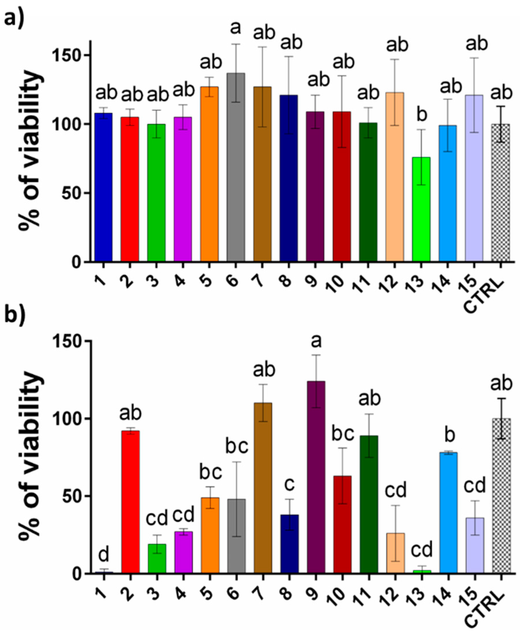
3.4. Cytotoxicity Assessment of Extracts

Keratinocytes, the predominant cells in the epidermis, are integral to the skin’s regenerative and protective functions. They facilitate re-epithelialization, maintain moisture, and form a barrier against microorganisms and UV radiation [59]. To ensure the safe incorporation of frankincense extracts into cosmetic and pharmaceutical products, it is imperative to identify non-toxic concentrations suitable for skin cells. We evaluated 10 and 50 µg/mL extract concentrations. According to the ISO 10993-5 standards, a cell viability below 70% relative to the untreated control indicates cytotoxic potential [60].
Our cytotoxicity analysis revealed that all the extracts were non-toxic at 10 µg/mL, with several promoting low-to-moderate keratinocyte proliferation (>100% viability, Figure 2a). Specifically, sample 6 exhibited a notable increase in cell proliferation (137 ± 21%) compared with the untreated cells (100 ± 13%), suggesting its potential usage in skin regeneration formulations. Other extracts, such as samples 5 (127 ± 7%), 7 (127 ± 29%), 8 (121 ± 28%), and 12 (123 ± 24%), also demonstrated notable proliferative effects. Sample 13 exhibited the most pronounced reduction in cell viability (76 ± 20%), a statistically significant decrease compared with the other samples.
At a concentration of 50 µg/mL, samples 1 (1 ± 2%) and 13 (2 ± 3%) exhibited cytotoxicity toward the cells (Figure 2b). Samples 3, 4, 5, 6, 12, and 15 demonstrated pronounced cytotoxic effects, with the cell viability reduced to below 30%. In contrast, sample 2 maintained cell viability with 92 ± 1% viable cells, while samples 11 (89 ± 14%) and 14 (78 ± 1%) were also non-toxic at this concentration. Notably, but not statistically significant, sample 9 enhanced the keratinocyte proliferation and achieved 124 ± 17% viability. Additionally, it seems that sample 7 exhibited a modest proliferative effect, with a viability of 110 ± 12%, which was statistically distinct from most other samples.
Although B. serrata resin extracts are generally considered non-toxic and are commonly incorporated into cosmetic formulations, our cytotoxicity assessments using HaCaT cells underscore the necessity for precise dosing. Certain extracts elicited toxic responses, even at relatively low concentrations. Our findings indicate that frankincense extracts promote regenerative effects at 10 µg/mL, whereas they tend to be generally cytotoxic at 50 µg/mL. Notably, sample 6 exhibited proliferative properties at the lower concentration, and sample 9 at a higher concentration. Sample 7 demonstrated proliferative effects at both tested concentrations, suggesting their potential utility in dermatological applications.

4. Conclusions

This study highlighted the significant antimicrobial effect of methanolic extracts of B. serrata resin, emphasizing its promising role in combating antibiotic-resistant pathogens. Utilizing HPTLC bioautographic assays, the extracts demonstrated potent activity against S. aureus; P. aeruginosa; and, notably, L. monocytogenes, marking the first evaluation of this pathogen with B. serrata resin. While HPTLC has been widely used for fingerprinting natural products, its combination with bioautography was particularly novel for the antibacterial screening of B. serrata resin. The identification of active zones and their correlation with specific boswellic acids—predominantly 11-keto-β-boswellic acid—demonstrated the resin’s efficacy in targeting pathogenic bacteria. The most active zones on the bioautograms indicate that the samples collected from central and southern Serbia (10, 12, and 15) showed the strongest antimicrobial effects against the tested bacterial strains. The LC-MS analysis identified sample 2 as the richest in bioactive components, including 11-keto-β-boswellic acid, which likely contributed to its high antimicrobial activity. Additionally, sample 6 exhibited a notable 137% enhancement of keratinocyte proliferation at lower concentrations, indicating its potential in regenerative dermatological applications. Sample 9 showed cytocompatibility, even at higher concentrations, further supporting the resin’s therapeutic versatility. These findings underline the innovative integration of advanced analytical methods, such as HPTLC bioautography, with traditional cytotoxicity assays, paving the way for the development of natural, effective antibacterial agents. Future research should explore the clinical applications of B. serrata extracts, ensuring their safety and efficacy in pharmaceutical and cosmetic formulations.

Author Contributions

Conceptualization, P.R.; Methodology, P.T., P.R., M.K.R. and U.G.; Software, P.T., M.K.R., U.G. and J.N.Ž.; Validation, M.K.R., U.G., I.S. and P.R.; Formal analysis P.T., J.N.Ž., M.K.R., Đ.I., M.J., I.S. and I.D.; Investigation, P.T., M.J., Đ.I., M.K.R. and J.N.Ž.; Resources, P.R. and I.D.; Data curation, P.T., M.K.R., P.R. and U.G.; Writing—original draft preparation, P.T., M.J., Đ.I. and I.S.; Writing—review and editing, P.R., M.K.R., I.S., U.G. and I.D.; Visualization, P.T.; Supervision, M.K.R., I.S. and P.R.; Project administration, P.R.; Funding acquisition, P.R. and I.D. All authors have read and agreed to the published version of the manuscript.

Funding

This work was supported by The Science Fund of the Republic of Serbia, Serbian Science and Diaspora Collaboration Program, No. 6389927, and the Ministry of Science, Technological Development and Innovation of Republic of Serbia, contract numbers: 451-03-66/2024-03/200168, 451-03-66/2024-03/200288, 451-03-65/2024-03/200169, and 451-03-66/2024-03/200007.

Data Availability Statement

The original contributions presented in this study are included in the article. Further inquiries can be directed to the corresponding author.

Conflicts of Interest

Authors Marko Jović and Đurđa Ivković were employed by the company Innovation Centre of the Faculty of Chemistry Ltd. The remaining authors declare that the research was conducted in the absence of any commercial or financial relationships that could be construed as a potential conflict of interest. The company had no role in the design of the study; in the collection, analyses, or interpretation of data; in the writing of the manuscript, or in the decision to publish the results.

References

  1. Warrier, K.C.S.; Muthupandiyan, S. Boswellia serrata Roxb. Ex Coleb.: A Threatened Tree. Int. J. Plant Soil Sci. 2022, 527–534. [Google Scholar] [CrossRef]
  2. Tironi de Castilho, A.L.T.; de Liori Teixeira, L.d.L.; Costa Lima, V.A.C.; Pesse, V.B.; Rozza, A.L. Chapter 3—Boswellia serrata. In Herbs, Spices and Their Roles in Nutraceuticals and Functional Foods; Amalraj, A., Kuttappan, S., Varma, A.C.K., Matharu, A., Eds.; Academic Press: Cambridge, MA, USA, 2023; pp. 31–40. [Google Scholar] [CrossRef]
  3. Alam, T.; Alam Khan, S.A.; Najam, L. Chemistry, Biological Activities, and Uses of Resin of Boswellia serrata Roxb. In Gums, Resins and Latexes of Plant Origin: Chemistry, Biological Activities and Uses; Murthy, H.N., Ed.; Springer International Publishing: Cham, Switzerland, 2022; pp. 1–43. [Google Scholar] [CrossRef]
  4. Hafeel, M.; Rizwana, A.A. Medicinal uses of Boswellia serrata Roxb (Kundur) with special reference to its ulcer healing property. J. Pharmacogn. Phytochem. 2021, 10, 34–37. [Google Scholar] [CrossRef]
  5. Hamidpour, R.; Hamidpour, S.; Hamidpour, M.; Shahlari, M. Frankincense (乳香 Rǔ Xiāng; Boswellia Species): From the Selection of Traditional Applications to the Novel Phytotherapy for the Prevention and Treatment of Serious Diseases. J. Tradit. Complement. Med. 2013, 3, 221–226. [Google Scholar] [CrossRef] [PubMed]
  6. Sultana, A.; Rahman, K.U.; Padmaja, A.R.; Rahman, S.U. Boswellia serrata Roxb. a Traditional Herb with Versatile Pharma-cological Activity: A Review. Int. J. Pharm. Sci. Res. 2013, 4, 2106–2117. [Google Scholar] [CrossRef]
  7. Al-Yasiry, A.R.M.; Kiczorowska, B. Frankincense—Therapeutic properties. Postepy Hig. I Med. Doswiadczalnej 2016, 70, 380–391. [Google Scholar] [CrossRef] [PubMed]
  8. Qurishi, Y.; Hamid, A.; Zargar, M.A.; Singh, S.K.; Saxena, A.K. Potential role of natural molecules in health and disease: Im-portance of boswellic acid. J. Med. Plants Res. 2010, 4, 2778–2785. [Google Scholar]
  9. Gupta, M.; Verma, S.K.; Singh, S.; Trivedi, L.; Rout, P.K.; Vasudev, P.G.; Luqman, S.; Darokar, M.P.; Bhakuni, R.S.; Misra, L. Anti-proliferative and antibacterial activity of oleo-gum-resin of Boswellia serrata extract and its isolate 3-hydroxy-11-keto-β-boswellic acid. J. Herb. Med. 2022, 32, 100546. [Google Scholar] [CrossRef]
  10. Gupta, M.; Singh, S.; Kurmi, A.; Luqman, S.; Saikia, D.; Thomas, M.; Rout, P.K. Correlation of boswellic acids with antiproliferative, antioxidant and antimicrobial activities of topographically collected Boswellia serrata oleo-gum-resin. Phytomed. Plus 2022, 2, 100289. [Google Scholar] [CrossRef]
  11. Kaushik, S.; Roy, A.; Purabiya, P.; Satapathy, T. Anti Bacterial and In Vitro Anti-inflammatory Potential of Boswellia seratta: A Potent Bioactive Compound. Res. J. Pharm. Technol. 2020, 13, 1079. [Google Scholar] [CrossRef]
  12. Siemoneit, U.; Pergola, C.; Jazzar, B.; Northoff, H.; Skarke, C.; Jauch, J.; Werz, O. On the interference of boswellic acids with 5-lipoxygenase: Mechanistic studies in vitro and pharmacological relevance. Eur. J. Pharmacol. 2009, 606, 246–254. [Google Scholar] [CrossRef]
  13. Singh, G.B.; Atal, C.K. Pharmacology of an extract of salai guggal ex-Boswellia serrata, a new non-steroidal anti-inflammatory agent. Inflamm. Res. 1986, 18, 407–412. [Google Scholar] [CrossRef]
  14. Al-Kharousi, Z.S.; Mothershaw, A.S.; Nzeako, B. Antimicrobial Activity of Frankincense (Boswellia sacra) Oil and Smoke against Pathogenic and Airborne Microbes. Foods 2023, 12, 3442. [Google Scholar] [CrossRef]
  15. Jaroš, P.; Timkina, E.; Michailidu, J.; Maršík, D.; Kulišová, M.; Kolouchová, I.; Demnerová, K. Boswellic Acids as Effective Antibacterial Antibiofilm Agents. Molecules 2022, 27, 3795. [Google Scholar] [CrossRef] [PubMed]
  16. Raja, A.F.; Ali, F.; Khan, I.A.; Shawl, A.S.; Arora, D.S.; Shah, B.A.; Taneja, S.C. Antistaphylococcal and biofilm inhibitory activities of acetyl-11-keto-β-boswellic acid from Boswellia serrata. BMC Microbiol. 2011, 11, 54. [Google Scholar] [CrossRef]
  17. Obiștioiu, D.; Hulea, A.; Cocan, I.; Alexa, E.; Negrea, M.; Popescu, I.; Herman, V.; Imbrea, I.M.; Heghedus-Mindru, G.; Suleiman, M.A.; et al. Boswellia Essential Oil: Natural Antioxidant as an Effective Antimicrobial and Anti-Inflammatory Agent. Antioxidants 2023, 12, 1807. [Google Scholar] [CrossRef] [PubMed]
  18. Hossain, T.J. Methods for screening and evaluation of antimicrobial activity: A review of protocols, advantages, and limitations. Eur. J. Microbiol. Immunol. 2024, 14, 97–115. [Google Scholar] [CrossRef] [PubMed]
  19. Agatonovic-Kustrin, S.; Wong, S.; Dolzhenko, A.V.; Gegechkori, V.; Ku, H.; Tucci, J.; Morton, D.W. Evaluation of bioactive compounds from Ficus carica L. leaf extracts via high-performance thin-layer chromatography combined with effect-directed analysis. J. Chromatogr. A 2023, 1706, 464241. [Google Scholar] [CrossRef]
  20. Choma, I.M.; Jesionek, W. TLC-Direct Bioautography as a High Throughput Method for Detection of Antimicrobials in Plants. Separations 2015, 2, 225–238. [Google Scholar] [CrossRef]
  21. Ahmad, I.; Husain, F.M.; Maheshwari, M.; Zahin, M. Medicinal Plants and Phytocompounds: A Potential Source of Novel Antibiofilm Agents. In Antibiofilm Agents: From Diagnosis to Treatment and Prevention; Springer: Berlin/Heidelberg, Germany, 2014; pp. 205–232. [Google Scholar] [CrossRef]
  22. Deza, M.A.; Araujo, M.; Garrido, M.J. Inactivation of Escherichia coli, Listeria monocytogenes, Pseudomonas aeruginosa and Staphylococcus aureus on stainless steel and glass surfaces by neutral electrolysed water. Lett. Appl. Microbiol. 2005, 40, 341–346. [Google Scholar] [CrossRef]
  23. Ranjbarnejad, T.; Saidijam, M.; Moradkhani, S.; Najafi, R. Methanolic extract of Boswellia serrata exhibits anti-cancer activities by targeting microsomal prostaglandin E synthase-1 in human colon cancer cells. Prostaglandins Other Lipid Mediat. 2017, 131, 1–8. [Google Scholar] [CrossRef]
  24. Alharbi, S.A.; Asad, M.; Abdelsalam, K.E.A.; Ibrahim, M.A.; Chandy, S. Beneficial Effect of Methanolic Extract of Frankincense (Boswellia sacra) on Testis Mediated through Suppression of Oxidative Stress and Apoptosis. Molecules 2022, 27, 4699. [Google Scholar] [CrossRef]
  25. CBS. Analytical Tasks-Efficiently Solved by HPTLC; CAMAG: Pratteln, Switzerland, 2010. [Google Scholar]
  26. Stojković, D.; Gašić, U.; Uba, A.I.; Zengin, G.; Rajaković, M.; Stevanović, M.; Drakulić, D. Chemical profiling of Anthriscus cerefolium (L.) Hoffm., biological potential of the herbal extract, molecular modeling and KEGG pathway analysis. Fitoterapia 2024, 177, 106115. [Google Scholar] [CrossRef]
  27. Ivkovic, D.; Cvijetic, I.; Radoicic, A.; Stojkovic-Filipovic, J.; Trifkovic, J.; Krstic Ristivojevic, M.; Ristivojevic, P. NADES-Based Extracts of Selected Medicinal Herbs as Promising Formulations for Cosmetic Usage. Processes 2024, 12, 992. [Google Scholar] [CrossRef]
  28. Agatonovic-Kustrin, S.; Wong, S.; Dolzhenko, A.V.; Gegechkori, V.; Morton, D.W. Bioassay-guided detection, identification and assessment of antibacterial and anti-inflammatory compounds from olive tree flower extracts by high-performance thin-layer chromatography linked to spectroscopy. J. Pharm. Biomed. Anal. 2024, 239, 115912. [Google Scholar] [CrossRef]
  29. Rocamora, C.R.; Ramasamy, K.; Lim, S.M.; Majeed, A.B.A.; Agatonovic-Kustrin, S. HPTLC based approach for bioassay-guided evaluation of antidiabetic and neuroprotective effects of eight essential oils of the Lamiaceae family plants. J. Pharm. Biomed. Anal. 2020, 178, 112909. [Google Scholar] [CrossRef] [PubMed]
  30. Agatonovic-Kustrin, S.; Wong, S.; Dolzhenko, A.V.; Gegechkori, V.; Ku, H.; Tan, W.K.; Morton, D.W. Effect directed analysis of bioactive compounds in leaf extracts from two Salvia species by High-performance thin-layer chromatography. J. Pharm. Biomed. Anal. 2023, 227, 115308. [Google Scholar] [CrossRef] [PubMed]
  31. Paul, M.; Brüning, G.; Bergmann, J.; Jauch, J. A Thin-layer Chromatography Method for the Identification of Three Different Olibanum Resins (Boswellia serrata, Boswellia papyrifera and Boswellia carterii, respectively, Boswellia sacra). Phytochem. Anal. 2012, 23, 184–189. [Google Scholar] [CrossRef]
  32. Gupta, M.; Rout, P.K.; Misra, L.N.; Gupta, P.; Singh, N.; Darokar, M.P.; Saikia, D.; Singh, S.C.; Bhakuni, R.S. Chemical composition and bioactivity of Boswellia serrata Roxb. essential oil in relation to geographical variation. Plant Biosyst.—Int. J. Deal. All Asp. Plant Biol. 2017, 151, 623–629. [Google Scholar] [CrossRef]
  33. Suhail, M.M.; Wu, W.; Cao, A.; Mondalek, F.G.; Fung, K.-M.; Shih, P.-T.; Fang, Y.-T.; Woolley, C.; Young, G.; Lin, H.-K. Boswellia sacra essential oil induces tumor cell-specific apoptosis and suppresses tumor aggressiveness in cultured human breast cancer cells. BMC Complement. Altern. Med. 2011, 11, 129. [Google Scholar] [CrossRef]
  34. DeCarlo, A.; Agieb, S.; Johnson, S.; Satyal, P.; Setzer, W.N. Inter-Tree Variation in the Chemical Composition of Boswellia papyrifera Oleo-Gum-Resin. Nat. Prod. Commun. 2022, 17, 1934578X221117411. [Google Scholar] [CrossRef]
  35. Hussain, H.; Rashan, L.; Hassan, U.; Abbas, M.; Hakkim, F.L.; Green, I.R. Frankincense diterpenes as a bio-source for drug discovery. Expert Opin. Drug Discov. 2022, 17, 513–529. [Google Scholar] [CrossRef]
  36. Wu, D.C.; Chan, W.W.; Metelitsa, A.I.; Fiorillo, L.; Lin, A.N. Pseudomonas Skin Infection. Am. J. Clin. Dermatol. 2011, 12, 157–169. [Google Scholar] [CrossRef]
  37. Alkheeqani, B.; Khassaf, A. Detection and Pathogenicity Features of Pseudomonas Aeruginosa in Patients with Skin Infection. Univ. Thi-Qar J. Sci. 2024, 11, 23–29. [Google Scholar] [CrossRef]
  38. Guo, Y.; Song, G.; Sun, M.; Wang, J.; Wang, Y. Prevalence and Therapies of Antibiotic-Resistance in Staphylococcus aureus. Front. Cell. Infect. Microbiol. 2020, 10, 107. [Google Scholar] [CrossRef]
  39. Fady, M.; Irie, Y.; Aljowaie, R.M.; Almutairi, S.M. Effect of β-sitosterol on PEL and PSL of Pseudomonas aeruginosa. J. King Saud Univ. Sci. 2024, 36, 103400. [Google Scholar] [CrossRef]
  40. Spernovasilis, N.; Psichogiou, M.; Poulakou, G. Skin manifestations of Pseudomonas aeruginosa infections. Curr. Opin. Infect. Dis. 2021, 34, 72–79. [Google Scholar] [CrossRef] [PubMed]
  41. Gul, A.; Pewe, L.L.; Willems, P.; Mayer, R.; Thery, F.; Asselman, C.; Aernout, I.; Verbeke, R.; Eggermont, D.; Van Moortel, L.; et al. Immunopeptidomics Mapping of Listeria monocytogenes T Cell Epitopes in Mice. Mol. Cell. Proteom. 2024, 23, 100829. [Google Scholar] [CrossRef] [PubMed]
  42. Schlech, W.F., III. Epidemiology and Clinical Manifestations of Listeria monocytogenes Infection. Microbiol. Spectr. 2019, 7, 10-1128. [Google Scholar] [CrossRef]
  43. Garcia, R.; Alves, E.S.S.; Santos, M.P.; Glória Viégas Aquije, M.F.; Fernandes, A.A.R.; Santos, R.B.D.; Ventura, J.A.; Fernandes, P.M.B. Antimicrobial Activity and Potential use of Monoterpenes as Tropical Fruits Preservatives. Braz. J. Microbiol. 2008, 39, 163–168. [Google Scholar] [CrossRef]
  44. Bassolé, I.H.N.; Lamien-Meda, A.; Bayala, B.; Tirogo, S.; Franz, C.; Novak, J.; Nebié, R.C.; Dicko, M.H. Composition and antimicrobial activities of Lippia multiflora Moldenke, Mentha x piperita L. and Ocimum basilicum L. Essential oils and their major monoterpene alcohols alone and in combination. Molecules 2010, 15, 7825–7839. [Google Scholar] [CrossRef]
  45. Dhar, P.; Chan, P.; Cohen, D.T.; Khawam, F.; Gibbons, S.; Snyder-Leiby, T.; Dickstein, E.; Rai, P.K.; Watal, G. Synthesis, Antimicrobial Evaluation, and Structure–Activity Relationship of α-Pinene Derivatives. J. Agric. Food Chem. 2014, 62, 3548–3552. [Google Scholar] [CrossRef]
  46. Hancock, R.E. The bacterial outer membrane as a drug barrier. Trends Microbiol. 1997, 5, 37–42. [Google Scholar] [CrossRef] [PubMed]
  47. Helander, I.M.; Alakomi, H.-L.; Latva-Kala, K.; Mattila-Sandholm, T.; Pol, I.; Smid, E.J.; Gorris, L.G.M.; von Wright, A. Characterization of the Action of Selected Essential Oil Components on Gram-Negative Bacteria. J. Agric. Food Chem. 1998, 46, 3590–3595. [Google Scholar] [CrossRef]
  48. Vakayil, R.; Murugesan, K.; Gnanasekaran, A.; Radhakrishnan, A.; Ranjith, M.; Bakthavatchalam, P.; Velmurugan, P.; Sivasubramanian, K.; Arumugam, N.; Almansour, A.I.; et al. Determination of phyto-moieties from the traditional therapeutic plant Boswellia serrata Roxb. extract against nosocomial pathogens: In vitro and silico approaches. J. Herb. Med. 2024, 43, 100823. [Google Scholar] [CrossRef]
  49. Bhutada, S.A.; Farhan, M.M.; Dahikar, S.B. Preliminary phytochemical screening and antibacterial activity of resins of Boswellia serrata Roxb. J. Pharmacogn. Phytochem. 2017, 6, 182–185. [Google Scholar]
  50. Balgoon, M.J.; Alghamdi, A.M. Biochemical Assessment of Boswellic Acid Enrich-Frankincense Extract and its Antioxidant, Antibacterial, Anticancer and Anti-inflammatory Potential in Ameliorating the GlycerolToxicity in Rats. Pak. Veter. J. 2024, 44, 1023–1032. [Google Scholar] [CrossRef]
  51. Xavier, S. Study of Antibacterial Activity of Resins of Boswellia serrata Roxb ex Colebr., Commiphora mukul (Hooks ex-Stocks) engl., Gardenia resinifera roth. and Shorea robusta gaertn. Int. J. Pharm. Pharm. Sci. 2016, 8, 29–31. [Google Scholar]
  52. Alshafei, M.M.; Mabrouk, A.M.; Hanafi, E.M.; Ramadan, M.M.; Korany, R.M.; Kassem, S.S.; Mohammed, D.M. Prophylactic supplementation of microencapsulated Boswellia serrata and probiotic bacteria in metabolic syndrome rats. Food Biosci. 2023, 51, 102325. [Google Scholar] [CrossRef]
  53. Osorio-Tobón, J.F. Recent advances and comparisons of conventional and alternative extraction techniques of phenolic compounds. J. Food Sci. Technol. 2020, 57, 4299–4315. [Google Scholar] [CrossRef]
  54. Reis, S.F.; Rai, D.K.; Abu-Ghannam, N. Water at room temperature as a solvent for the extraction of apple pomace phenolic compounds. Food Chem. 2012, 135, 1991–1998. [Google Scholar] [CrossRef]
  55. Sultan, F.I. Phytochemical Analysis and Antibacterial Activities of Frankincense of Boswellia serrata. Plant Arch. 2020, 20, 5219–5226. [Google Scholar]
  56. Plyuta, V.; Zaitseva, J.; Lobakova, E.; Zagoskina, N.; Kuznetsov, A.; Khmel, I. Effect of plant phenolic compounds on biofilm formation by Pseudomonas aeruginosa. APMIS 2013, 121, 1073–1081. [Google Scholar] [CrossRef] [PubMed]
  57. Zamuz, S.; Munekata, P.E.; Dzuvor, C.K.; Zhang, W.; Sant’Ana, A.S.; Lorenzo, J.M. The role of phenolic compounds against Listeria monocytogenes in food. A review. Trends Food Sci. Technol. 2021, 110, 385–392. [Google Scholar] [CrossRef]
  58. Miklasińska-Majdanik, M.; Kępa, M.; Wojtyczka, R.D.; Idzik, D.; Wąsik, T.J. Phenolic Compounds Diminish Antibiotic Resistance of Staphylococcus aureus Clinical Strains. Int. J. Environ. Res. Public Health 2018, 15, 2321. [Google Scholar] [CrossRef] [PubMed]
  59. Piipponen, M.; Li, D.; Landén, N.X. The Immune Functions of Keratinocytes in Skin Wound Healing. Int. J. Mol. Sci. 2020, 21, 8790. [Google Scholar] [CrossRef]
  60. ISO 10993-5:2009; Biological Evaluation of Medical Devices—Part 5: Tests For In Vitro Cytotoxicity. Tanzania Bureau of Standards: Dar es Salaam, Tanzania, 2009.
Figure 1. (a) HPTLC profile visualized under white light; (b) MTT antimicrobial assay against L. monocytogenes; (c) MTT antimicrobial assay against P. aeruginosa; (d) MTT antimicrobial assay against S. aureus. Mobile phase, n-hexane/ethyl acetate/glacial acetic acid (16:5:1 v/v/v).
Processes 13 00850 g001
Figure 2. Percentages (%) of HaCaT cells’ viabilities following incubation with frankincense extracts at (a) 10 µg/mL and (b) 50 µg/mL concentrations. Statistically significant differences (p < 0.05) are indicated by different letters on the graph, as determined using Tukey’s multiple comparisons test (ordinary one-way ANOVA).
Processes 13 00850 g002
Table 1. The number of extracts, geographical region of monk’s frankincense, and the name of the place from which the investigated B. serrata commercial samples were obtained.
Extract NumberGeographical Region of Monk’s FrankincenseName of the Church/Monastery
1Subotica, SerbiaSt. George’s church
2Golubac, SerbiaMonastery Tumane
3Kuršumlija, SerbiaSt. Nichola’s church
4Novi Sad, SerbiaSt. George’s church
5Valjevo, SerbiaChurch of the Holy Mother of God
6Banja Luka, Bosnia and HerzegovinaSt. Peter and Paul’s church
7Bethlehem, PalestineChurch of the Nativity
8Mionica, SerbiaSt. Nichola’s church
9Koceljeva, SerbiaSt. George’s church
11Kotor, MontenegroSt. Luka’s church
12Kruševac, SerbiaChurch of the Holy Mother of God
13Pljevlje, MontenegroChurch of St. James
14Mount Atos, GreeceMonastery Hilandar
15Vranje, SerbiaSt. George’s church
16Standard mixture
Table 2. The quantified phenols identified in the frankincense extracts and GT are presented as µg/g ± SD. Each measurement was conducted in triplicate. NF indicates “not found”.
µg/gGAPCACaffeic AcidCGASAClp-CAFASAK-3-O-GLTQCTNARKOKBA (mg/g)
113.13 ± 0.2128.80 ± 0.551.53 ± 0.043.36 ± 0.223.74 ± 0.105.50 ± 0.133.81 ± 0.105.27 ± 0.45NFNFNF12.45 ± 0.17NF2.76 ± 0.14
2NF280.53 ± 5.33NFNF5.37 ± 0.1543.46 ± 0.999.97 ± 0.266.46 ± 0.55NFNFNF5.77 ± 0.08NF3.73 ± 0.19
36.03 ± 0.108.84 ± 0.17NFNF3.76 ± 0.1014.82 ± 0.343.85 ± 0.104.59 ± 0.39NFNFNF12.96 ± 0.17NF2.1 ± 0.1
410.63 ± 0.1715.77 ± 0.301.62 ± 0.053.54 ± 0.233.72 ± 0.105.74 ± 0.138.32 ± 0.225.12 ± 0.44NFNFNF12.44 ± 0.17NF2.86 ± 0.14
513.13 ± 0.21NF1.72 ± 0.053.72 ± 0.243.75 ± 0.1016.15 ± 0.3710.54 ± 0.28NF5.94 ± 0.077.32 ± 0.24NF22.00 ± 0.3013.00 ± 0.441.84 ± 0.09
6110.93 ± 1.76NF1.68 ± 0.0519.05 ± 1.233.70 ± 0.109.29 ± 0.2110.61 ± 0.284.51 ± 0.388.35 ± 0.098.09 ± 0.27NF36.86 ± 0.5014.84 ± 0.502.07 ± 0.10
756.27 ± 0.899.35 ± 0.181.64 ± 0.0510.39 ± 0.673.76 ± 0.1017.10 ± 0.394.64 ± 0.124.64 ± 0.396.41 ± 0.077.32 ± 0.24NF20.35 ± 0.2712.65 ± 0.432.18 ± 0.11
891.34 ± 1.459.61 ± 0.181.59 ± 0.048.79 ± 0.573.74 ± 0.1010.05 ± 0.236.61 ± 0.184.52 ± 0.385.38 ± 0.06NFNF53.13 ± 0.7215.28 ± 0.523.27 ± 0.16
96.26 ± 0.10NF1.53 ± 0.04NFNF8.04 ± 0.183.85 ± 0.10NFNFNFNF11.20 ± 0.1510.69 ± 0.362.50 ± 0.12
1026.50 ± 0.4212.17 ± 0.231.54 ± 0.045.89 ± 0.383.70 ± 0.1010.06 ± 0.235.31 ± 0.144.53 ± 0.395.24 ± 0.067.65 ± 0.26NF19.39 ± 0.2612.02 ± 0.412.43 ± 0.12
11NF19.48 ± 0.3726.18 ± 0.74NF3.69 ± 0.10NF11.06 ± 0.294.43 ± 0.384.35 ± 0.057.34 ± 0.2524.89 ± 0.753.27 ± 0.04NF1.84 ± 0.09
12NF27.44 ± 0.521.64 ± 0.05NF3.75 ± 0.1011.13 ± 0.256.56 ± 0.174.91 ± 0.42NFNFNF11.80 ± 0.16NF3.35 ± 0.17
1311.15 ± 0.189.43 ± 0.181.54 ± 0.043.15 ± 0.203.72 ± 0.1010.17 ± 0.236.06 ± 0.164.45 ± 0.38NF7.38 ± 0.25NF11.25 ± 0.1511.56 ± 0.392.30 ± 0.12
14NF14.43 ± 0.271.52 ± 0.04NFNF115.54 ± 2.639.13 ± 0.244.59 ± 0.39NFNFNF2.44 ± 0.03NF1.66 ± 0.08
1575.08 ± 1.1918.90 ± 0.36NFNF3.78 ± 0.1014.44 ± 0.338.91 ± 0.245.07 ± 0.43NFNFNF13.97 ± 0.1911.27 ± 0.382.82 ± 0.14
1693.05 ± 1.4816.65 ± 0.321.57 ± 0.0410.34 ± 0.673.75 ± 0.107.88 ± 0.1819.40 ± 0.515.00 ± 0.439.43 ± 0.108.38 ± 0.2839.12 ± 1.194.37 ± 0.0610.84 ± 0.370.59 ± 0.03
GA—gallic acid; PCA—protocatechuic acid; CGA—chlorogenic acid; SACl—syringic acid; p-CA—p-coumaric acid; FA—ferulic acid; SA—sinapic acid; K-3-O-G—kaempferol 3-O-glucoside; LT—luteolin; QCT—quercetin; NAR—naringenin; KO—kaempferol; KBA—11-keto-β-boswellic acid.
Disclaimer/Publisher’s Note: The statements, opinions and data contained in all publications are solely those of the individual author(s) and contributor(s) and not of MDPI and/or the editor(s). MDPI and/or the editor(s) disclaim responsibility for any injury to people or property resulting from any ideas, methods, instructions or products referred to in the content.

