Article

Freeze-Dried Liposomes as Carriers of Eugenia pyriformis Cambess Phytoactives for Cosmetic Applications

by
Gabriela Alves Silva
,
Letícia Kakuda
and
Wanderley Pereira Oliveira
*
School of Pharmaceutical Sciences of Ribeirão Preto, University of São Paulo, Av. Café s/n, Bloco Q, Ribeirão Preto 14040-903, SP, Brazil
*
Author to whom correspondence should be addressed.
Processes 2025, 13(3), 693; https://doi.org/10.3390/pr13030693
Submission received: 28 January 2025 / Revised: 24 February 2025 / Accepted: 25 February 2025 / Published: 28 February 2025
(This article belongs to the Section Pharmaceutical Processes)

Abstract

:
The demand for phytoactives in cosmetics is growing due to their potential as safer and sustainable alternatives to synthetic compounds. The fruit pulp of Eugenia pyriformis Cambess (uvaia), a species native to the Atlantic Forest, is rich in phenolic compounds and ascorbic acid, with high antioxidant activity, making it a promising active ingredient for cosmetic applications, particularly in skin anti-aging formulations. This study aimed to extract bioactives from uvaia fruit, evaluate their antioxidant properties, and develop freeze-dried liposomes to enhance their stability and physicochemical characteristics. Uvaia pulp was freeze-dried and extracted via dynamic maceration using water (EX.AQ) and 70% ethanol (EX.ET). EX.ET exhibited the highest polyphenol content (16.44 ± 0.95 mg GAE/g dry basis) and antioxidant activity (IC50 = 122.09 ± 5.28 µg/mL). Liposomes containing EX.ET (F1-EX) and blank liposomes (F1-B) were prepared using an adapted ethanol injection method and freeze-dried with two cryoprotectant mixtures: A1 (trehalose, mannitol, and colloidal silicon dioxide) and P1 (whey protein and colloidal silicon dioxide). F1-EX liposomes had a larger particle size (232.50 ± 2.72 nm) than F1-B (143.90 ± 2.80 nm), with similar polydispersity (PdI < 0.30) and zeta potential (>30 mV). The encapsulation efficiency of F1-EX reached 58.8 ± 0.5%. Freeze-dried liposomes showed low water activity (0.068–0.340) and moisture content (2.71–3.58%), while cryoprotectant A1 resulted in a lower PdI (0.144 ± 0.039) and higher zeta potential (−39.00 ± 0.44). These results demonstrate that uvaia is a valuable source of bioactive substances, with encapsulation and drying technologies enhancing their stability and functionality, making them suitable for potential applications in the pharmaceutical and cosmetic sectors. Furthermore, this approach supports the sustainable use of natural resources and contributes to preserving Brazil’s biodiversity.

1. Introduction

Skin aging is a multifactorial process influenced by intrinsic and extrinsic factors, leading to noticeable changes in appearance [1]. While chronological aging is a key component, it is not the sole determinant. Lifestyle choices and environmental factors collectively called the exposome—such as air pollution, solar radiation, and smoking—accelerate skin aging by generating free radicals. This process affects physical appearance and self-esteem, driving the strong demand in the cosmetics market for products designed to delay the signs of aging [2].
In this context, extracts derived from flora biodiversity offer significant potential as innovative active ingredients for cosmetic products. These natural resources demonstrate proven efficacy and align with sustainability goals by encouraging responsible use of biodiversity. Brazil’s rich and unique flora is a valuable source of novel compounds among such resources. Specifically, Eugenia pyriformis Cambess, commonly known as uvaia, is native to the Atlantic Forest and predominantly found in the southern states of Paraná, Rio Grande do Sul, and Santa Catarina. Uvaia has been traditionally utilized by small farmers and local communities, highlighting its cultural and economic importance [3]. The uvaia fruit contains high levels of phenolic compounds, especially gallic acid and quercetin, in addition to ascorbic acid, which contributes to its potent antioxidant properties [4]. These attributes position uvaia as a promising, yet underutilized, raw material for the cosmetic industry, allowing the alignment of sustainability with the valorization of Brazilian biodiversity [5,6,7]. However, uvaia fruit has a short post-harvest shelf life because of its high respiratory rates and soft texture, making it highly susceptible to mechanical damage [7]. The freeze-drying process is one of the most effective post-harvest technologies to address the perishability of fruits [8]. By reducing water activity, freeze-drying minimizes susceptibility to degradation reactions and extends the storage period while maintaining fruit quality [9].
Plant-based active ingredients, such as those derived from uvaia, often face challenges related to their physicochemical properties, which hinder their application in more complex formulations. Encapsulation processes provide a viable solution by enabling controlled release, enhancing biocompatibility, and increasing the functionality and value of plant-derived components [10]. Liposomes, as lipid-based encapsulation systems, comprise phospholipid bilayers that form spherical vesicles capable of encapsulating polar and non-polar substances [11,12].
Despite their versatility, liposomes are inherently unstable and prone to the fusion, aggregation, and leakage of encapsulated bioactive compounds. The freeze-drying process addresses these limitations by removing the aqueous content, thus preserving liposome integrity and functionality [13,14]. By reducing water activity, freeze-drying and other drying processes minimize the susceptibility to degradation reactions and extend the storage period while maintaining product quality [15].
This study aimed to obtain and characterize uvaia pulp extract, develop liposomes, and produce freeze-dried liposomes incorporating this extract for potential cosmetic applications. By combining encapsulation and freeze-drying, this research introduces an innovative, sustainable, plant-based active ingredient while enhancing the value of an underutilized natural resource. This approach promotes the responsible use of Brazilian biodiversity and aligns with sustainability principles, supporting advancements in the cosmetic industry and contributing to environmental sustainability and efficient resource management.

2. Materials and Methods

This section presents the experimental methodology used to develop the freeze-dried liposomes loaded with the uvaia fruit pulp bioactives, from the harvesting of the uvaia’s fruit, pre-processing, freeze-drying, extraction, liposomes production and characterization, freeze-drying the liposomes, and reconstitution.

2.1. Pre-Processing of Uvaia Fruits

Uvaia fruits with yellow or yellow-orange coloration, corresponding to the fully mature state, were manually harvested from the Ribeirão Preto Campus of the University of São Paulo (21°09′40.86″ S, 47°51′34.71″ W). The source tree was identified (Figure 1), and a voucher specimen was deposited in the herbarium of the Department of Biology, FFCLRP-USP, under Registration No. 18612. The project was registered in the National System for the Management of Genetic Heritage and Associated Traditional Knowledge (SisGen) under registration number AFCEBCE, as required for research involving Brazilian biodiversity.
The harvested fruits were washed, and the seeds were manually removed. The pulp was then blended using a Britânia Black 600W blender (Britânia, Paraná, Brazil) with the addition of Milli-Q® water. The resulting mixture was homogenized with an Ultra-Turrax® homogenizer (T25 Digital, IKA Werke, Staufen, Germany) at 24,000 rpm for 3 min and filtered through a fine-mesh sieve. The filtered pulp was characterized for its solid content using a moisture analyzer balance (Model MA-35, Sartorius, Göttingen, Germany).

2.2. Freeze-Drying of Uvaia Pulp

Due to its short post-harvest shelf life, uvaia pulp was freeze-dried to extend its conservation period [16,17]. For this process, 5% (w/w) colloidal silicon dioxide (Aerosil 200®) was added as a cryoprotectant relative to the pulp’s solid content. The preparations were frozen at −80 °C for 24 h before being freeze-dried for 72 h [13,18]. The freeze-drying was performed using a lyophilizer (Model SNL108B, Thermo Fisher Scientific, Waltham, MA, USA) equipped with a 1.5 L micromodule lyophilizer unit, stainless steel condenser, 1/4 hp compressor (0.30 kW power), ultra-vacuum pump (LyoPump VLP195FD), freeze-drying containers, and independent valves. The freeze-dried pulp’s water activity was determined using an Aqua Lab 4TEV water activity meter (Decagon Devices, Pullman, WA, USA), and the moisture content was measured via Karl Fischer titration (Model 743, Metrohm, Herisau, Switzerland).

2.3. Extraction of Bioactive Constituents from Freeze-Dried Uvaia Fruit Pulp

Bioactive compounds were extracted from the freeze-dried uvaia pulp using two solvent systems: water (EX.AQ) and 70% (v/v) ethanol (EX.ET). The extractions were carried out at a 1:30 ratio (w/v) of freeze-dried pulp to solvent using dynamic maceration at 45 °C for 60 min in an ultra-thermostatic bath (MA-184, Marconi, São Paulo, Brazil). This extraction time was selected to prevent an excessively long maceration process, thereby minimizing the risk of degradation or oxidation of the bioactive compounds in the extract. The resulting extracts were vacuum-filtered through a ceramic Buchner funnel with a paper filter into an Erlenmeyer flask coupled to a vacuum pump (Prismatec, São Paulo, Brazil). The filtered extractive solutions were then characterized for solid content, total polyphenol content, ascorbic acid concentration, and antioxidant activity using the DPPH method.

2.4. Characterization of Uvaia Pulp Extracts

The uvaia pulp extracts’ bioactive properties were characterized by quantifying the total polyphenol content, ascorbic acid concentration, and antioxidant activity. Detailed methodologies for each analysis are provided below.

2.4.1. Total Polyphenol Content

The total polyphenol content was determined using the Folin–Denis method, previously validated [13,19]. Aqueous extract solutions (100 µL/mL) were prepared, and 0.5 mL of each solution was mixed with 0.5 mL of Folin–Denis reagent and 4 mL of sodium carbonate. After vortex homogenization, the mixture was incubated for 2 min at room temperature before absorbance was read at 750 nm in a UV–Vis spectrophotometer (HP model 8453, Hewlett-Packard, Palo Alto, CA, USA) in triplicate. The calibration curve was prepared using gallic acid, and the results were expressed in mg gallic acid equivalent per g of dry matter and µg gallic acid equivalent per mL of extract for each extract.

2.4.2. Ascorbic Acid Content

Vitamin C was quantified using high-performance liquid chromatography (HPLC), as Bresolin and Hubinger (2014) described [20]. The analysis was performed on a Shimadzu Prominence LC-20A series with an LC-6A double pump (Shimadzu Corporation, Kyoto, Japan) with diode array detection (DAD). The analyses were performed under the following chromatographic conditions: 20 μL sample injection volume, C18 column, 1 mL/min phosphate buffer mobile phase at pH 2.5, with a 15-min run time, and detection at 254 nm. Solutions of each extracted sample were prepared using 3% metaphosphoric acid, homogenized in an ultrasound bath for 10 min, and then filtered using 0.45 µm syringe filters (Millex, Darmstadt, Germany). The calibration curve was prepared using ascorbic acid, and the results were expressed in mg ascorbic acid (AA) per mL of extract.

2.4.3. Antioxidant Activity (DPPH Assay)

The antioxidant activity of the extracts was assessed using the DPPH method, as described by Fernandes et al. (2014) [21]. First, the blank was prepared by adding 1 mL of 0.1 M acetate buffer pH 5.5 and 1.5 mL of absolute ethanol to a test tube. Next, the positive control was prepared in triplicate by adding 1 mL of 0.1 M acetate buffer pH 5.5, 1 mL of absolute ethanol, and 0.5 mL of 250 µM DPPH solution prepared with absolute ethanol incubated for 20 min, this reaction time being necessary for the stabilization of the sample color, and absorbance was read at 517 nm in an UV–Vis spectrophotometer (HP model 8453, Hewlett-Packard, Palo Alto, CA, USA) [22]. Six concentrations were prepared for the samples to obtain six points, including four purple and two yellow points. Then, 1 mL of 0.1 M acetate buffer pH 5.5, 1 mL of absolute ethanol, and 0.5 mL of 250 µM DPPH solution were added. The percentage of DPPH inhibition was calculated from the absorbance data using Equation (1):
%   I n h i b i t i o n = A 0 A S A 0 × 100
In Equation (1), A0 is the absorbance of the positive control, and AS is the absorbance of the sample. The IC50 values, representing the concentration required to reduce the initial DPPH absorbance by 50%, were determined by plotting the percentage inhibition against the extract concentration and fitting the data to a dose–response equation using non-linear regression (GraphPad Prism® software, Version 8.0.1, 2018) [23]. Based on the IC50 results and the initial concentration of DPPH in the reaction medium, the EC50 values, expressed in grams of extract per gram of DPPH, were also determined.

2.5. Liposome Preparation and Characterization

2.5.1. Liposome Production

To formulate liposomes loaded with bioactives from the uvaia pulp, the ethanol content in the EX.ET extract (70% ethanolic extract) was first reduced using a rotary vacuum evaporator (Fisatom, São Paulo, Brazil), removing approximately two-thirds of the initial solvent volume. This concentration step ensured that the extract was suitable for incorporation into liposomes. The solid content of the concentrated extract solution was determined using a moisture analyzer balance (Model MA-35, Sartorius, Göttingen, Germany).
Liposomes were then prepared using a modified ethanol injection method based on the procedure described by Jaafar-Maalej et al. (2010) [24]. The steps of this process, from the extraction of bioactives to the preparation of liposomes, are outlined in the flowchart below (Figure 2).
The liposome formulations were adapted from Kakuda, Maia Campos, and Oliveira (2024) and Bankole et al. (2020) [13,15]. The components included cholesterol (Sigma-Aldrich—St. Louis, MO, USA), hydrogenated soybean phosphatidylcholine (Phospholipon® 90H, Lipoid—Ludwigshafen, Germany), absolute ethanol, Milli-Q® water, and the concentrated uvaia hydroalcoholic extract. Two formulations were developed: liposomes without the active substance (blank—F1-B) and liposomes containing 1% (w/w), relative to the solid content of the uvaia hydroalcoholic extract (F1-EX), as summarized in Table 1.
The aqueous phase, comprising Milli-Q® water and the uvaia extract, was heated to 70 °C in a heating bath (Fisatom, São Paulo, Brazil). Simultaneously, the oil phase—containing cholesterol, hydrogenated soybean phosphatidylcholine, and absolute ethanol—was heated until the lipids melted. The oil phase was injected into the aqueous phase at a constant flow rate of 4.1 mL/min using a peristaltic pump (Model 77201, Masterflex L/S Easy-Load, Gelsenkirchen, Germany). Following injection, the absolute ethanol was evaporated through magnetic homogenization for 2 h at room temperature.
The formulations were subjected to high-shear and ultrasonic processing to reduce the particle size further and enhance homogeneity. First, the liposomes were homogenized using an Ultra-Turrax® (T18 Basic, IKA Werke, Staufen, Germany) at 22,000 rpm for 5 min, followed by the ultrasonication performed with a VibraCell VCX 740 (Sonics and Materials, Newtown, CT, USA) equipped with a 13 mm probe operating at 45% amplitude.
The number of ultrasonication cycles was determined through preliminary assays, which evaluated the effect of the cycles on the particle size and polydispersity index for formulations F1-B and F1-EX. Aliquots were collected after each cycle to determine the optimal conditions. The study assessed up to six cycles, each lasting 5 min with 2-min intervals. During ultrasonication, the formulations were maintained in an ice bath to prevent overheating [15].

2.5.2. Liposome Characterization

The resulting liposome formulations were allowed to rest for 24 h to stabilize before being characterized by determining their particle size, polydispersity index (PdI), zeta potential, and encapsulation efficiency.
The particle size and PdI of the liposome formulations were measured using dynamic light scattering (DLS), which analyzes the velocity of particles under Brownian motion [24,25]. The zeta potential was determined through microelectrophoresis, which calculates the movement of dispersed particles in an electric field to assess the surface charge of the nanocarriers [25,26].
For both analyses, the liposome formulations were diluted to a 1:500 (v/v) ratio with Milli-Q® water and homogenized using a magnetic stirrer for 30 min to ensure uniform dispersion [27]. The samples were kept under agitation until analysis. For particle size and PdI measurements, 1000 μL of the diluted solution was transferred into a disposable cuvette and analyzed in triplicate using the Zetasizer Malvern Nano-ZS90 (Malvern Panalytical, Malvern, UK) in Size mode at 25 °C [15]. For zeta potential measurements, 750 μL of the diluted solution was placed in a disposable capillary cell and analyzed in triplicate using the same instrument in the zeta potential mode at 25 °C [15].
Encapsulation efficiency was assessed based on the polyphenol content in the liposome formulations containing uvaia extract. The formulations were centrifuged using an Eppendorf 5430 R centrifuge (Eppendorf, Hamburg, Germany) at 7100× g and 25 °C for 20 min. Centrifugal filters (Millipore Amicon® Ultra-15) with a pore size of 10 kDa were used to separate non-encapsulated polyphenols in the filtrate.
After centrifugation, the filtrate was diluted to a 1:5 (v/v) ratio, and the total polyphenol content was determined using the previously described method (Section 2.4.1). The encapsulation efficiency (EE) was then calculated using Equation (2):
E E ( % ) = G A E T G A E N E G A E T × 100
In Equation (2), EE represents the encapsulation efficiency, expressed as a percentage. GAET denotes the theoretical total polyphenol content in the liposomes, referring to the polyphenol content determined for EX.ET according to Section 2.4.1. GAENE refers to the total polyphenol content found in the diluted filtrate. Both values are expressed in mg of gallic acid equivalent (GAE) per 100 g. The difference between GAET and GAENE corresponds to the polyphenol content successfully encapsulated within the liposomes.

2.6. Production and Characterization of Freeze-Dried Liposomes

The empty liposomes (F1-B) and liposomes containing uvaia extract (F1-EX) were subjected to freeze-drying with the addition of cryoprotectants. Two cryoprotectant combinations were evaluated: the first combination (A1) consisted of trehalose (28%), mannitol (10%), and colloidal silicon dioxide (Aerosil 200®) (2%), while the second combination (P1) included whey protein (18%) and colloidal silicon dioxide (Aerosil 200®) (2%).
For each combination, a concentrated cryoprotectant solution was prepared in Milli-Q® water and stirred magnetically for 2 h. Two concentrations of the cryoprotectant solution, 5% and 10% (relative to the total solids content of the liposomes), were tested. The preparations were homogenized for 90 min using a magnetic stirrer, and aliquots were collected for characterization before freeze-drying. The samples were frozen at −80 °C for 24 h, freeze-dried for 72 h, and then placed in the freeze-dryer. The freeze-dried samples were characterized by determining the water activity and moisture content by the procedure described in Section 2.2.
To evaluate the particle size, polydispersity index (PdI), and zeta potential, the freeze-dried samples were reconstituted by redispersing the powder in Milli-Q® water, using the initial total solids content as a reference. The characteristics of the reconstituted formulations were compared with the original formulations to assess the impact of the freeze-drying process on the liposome properties. The solutions were stirred for 60 min using a magnetic stirrer (Model RT 15, IKA Werke, Staufen, Germany), as Bankole et al. (2020) described [13].
The reconstituted liposome formulations were filtered using 1.20 µm syringe filters (GVS, Bologna, Italy). This pore size, approximately three times larger than the previously measured particle size, was chosen to remove large cryoprotectant particles that might remain in the reconstituted formulations. This filtration step minimized potential errors during analysis [28,29,30]. The filtered formulations were then characterized using the Zetasizer Malvern Nano-ZS90 (Malvern Panalytical, Malvern, UK) following the methodologies described in Section 2.5.2.

2.7. Statistical Analysis

Statistical analyses were performed using GraphPad Prism® software (version 8.0.1, 2018). The Shapiro–Wilk test was used to assess the normality of the data. For data with a normal distribution, one-way ANOVA followed by Tukey’s post-test was applied. The Kruskal–Wallis test with Dunn’s post-test was employed for non-normal distribution data. A significance level of p ≤ 0.05 was considered significant for all analyses. The results are presented as the mean ± standard deviation (SD).

3. Results and Discussion

3.1. Characterization of Uvaia Pulp Extract

The processed uvaia pulp exhibited a solid content of 11.14 ± 1.36%. Uvaia is known for its short post-harvest shelf life due to its soft texture, susceptibility to mechanical damage, and high moisture content, which contribute to rapid perishability and limit its availability for consumption and processing [7,9]. Hence, freeze-drying was employed as a preservation technique, effectively reducing the moisture content and water activity and minimizing microbial growth and degradation [8].
After drying, the uvaia pulp achieved a water activity of 0.261 ± 0.004 and a moisture content of 5.30 ± 0.21%. These values indicate a significantly reduced risk of microbial contamination and extended shelf life, since, ideally, dry products should have a water activity below 0.5 and a moisture content below 10%, which prevents degradation reactions and microbial growth [8]. Similarly, Darniadi et al. (2021) highlighted the effectiveness of freeze-drying for durian (Durio zibethinus) fruits, demonstrating its ability to extend preservation and retain the potential for off-season applications [17].
The hydroalcoholic extract (EX.ET) demonstrated a higher solids content (2.81 ± 0.23%) compared to the aqueous extract (EX.AQ) (2.52 ± 0.03%), yet without statistically significant differences (p ≥ 0.05). However, it can be inferred that the higher solid content for EX.ET is related to the greater extraction capacity of 70% ethanol.
Table 2 shows that the total polyphenol content in EX.ET was significantly higher (p ≤ 0.05) than in EX.AQ. This difference reflects the preferential solubility of phenolic compounds in 70% ethanol due to its moderate polarity and higher affinity for hydroxyl substituents, which enhances the extraction of these secondary metabolites, making it an optimal choice for maximizing bioactive compound recovery from plant matrices [31]. Among the phenolic compounds isolated from uvaia are flavonoids such as quercetin, rutin, and kaempferol, as well as phenolic acids such as gallic acid and chlorogenic acid. In this context, gallic acid and quercetin stand out due to their higher concentrations. Therefore, the polyphenol content in the extracts was analyzed based on the determination of gallic acid equivalents [4,32]. According to Rufino et al. (2010), the polyphenol content on a dry basis is classified as low (<1000 mg GAE/100 g), medium (1000–5000 mg GAE/100 g), or high (>5000 mg GAE/100 g) [33]. Based on this classification, EX.AQ exhibited a low polyphenol content (767 mg GAE/100 g), while EX.ET exhibiting a medium content (1644 mg GAE/100 g).
These findings align with previous studies reporting total polyphenol levels of 127 mg GAE/100 g (fresh basis) and 1930 mg GAE/100 g (dry basis) for freeze-dried uvaia pulp [33]. However, these values are lower than those observed by Farias et al. (2020), who reported 4936 ± 0.24 mg GAE/100 g (dry basis) for hydroalcoholic extracts of uvaia pulp [34]. Differences in the polyphenol content between studies, as well as in the contents of other bioactive compounds, such as vitamin C itself, can be attributed to environmental and genetic factors that influence the production of secondary metabolites. That is, the secondary metabolites are synthesized as adaptive responses to environmental stressors such as temperature, humidity, light intensity, water availability, and soil nutrients [35]. These ecological variables directly impact phytochemical biosynthesis, leading to variations in the content [36]. Standardizing bioactive compound levels in plant-derived raw materials remains a critical challenge for their industrial application.
The aqueous extract of uvaia pulp exhibited a higher vitamin C content compared to the hydroalcoholic extract, though the difference was not statistically significant (p ≥ 0.05) (Table 2). Both extracts demonstrated promising potential as organic vitamin C sources. Compared to the vitamin C content of Brazil’s most widely produced fruit, the orange, the uvaia extracts contained higher levels on a fresh weight basis. For instance, the aqueous extract (EX.AQ) surpassed both the Seville orange (34.7 mg/100 g fresh weight) and the sweet orange (43.5 mg/100 g fresh weight) [37,38]. These results highlight the antioxidant potential of uvaia extracts, demonstrating their viability as plant-based raw materials for incorporation into cosmetic formulations.
Vitamin C (L-ascorbic acid) is a water-soluble antioxidant capable of participating in redox reactions, effectively neutralizing reactive oxygen species generated by skin exposure to the exposome [39]. Additionally, vitamin C plays a key role in treating hyperpigmentation and promoting skin regeneration by enhancing keratinocyte differentiation and contributing to dermal–epidermal cohesion, thereby mitigating skin alterations associated with aging [40]. Furthermore, vitamin C supports the absorption of iron, calcium, and folic acid; hence, a deficiency in this nutrient may result in immunosuppression [38]. It also benefits from concomitant antioxidant substances in the extract, further enhancing its application potential in cosmetic and nutraceutical products.
The EX.ET extract exhibited superior antioxidant activity, with a statistically significant difference (p ≤ 0.05), as evidenced by its lower IC50 value (Table 2). This value reflects the minimum concentration of antioxidants required to inhibit 50% of a given concentration of free radicals [23]. The enhanced antioxidant activity of EX.ET is likely attributed to its higher polyphenol content, a class of compounds well recognized for their potent antioxidant properties. Moreover, literature reports indicate that the contribution of phenolic compounds to antioxidant activity is more significant compared to that of vitamin C, which explains why, even though EX.AQ exhibited the highest ascorbic acid content, it demonstrated the lowest antioxidant activity [41]. However, it is important to emphasize that the combined presence of different bioactive compounds leads to a synergistic effect that enhances antioxidant activity, as it can both protect cells against oxidation and amplify antioxidant mechanisms [1,41,42]. These interactions highlight the multifaceted potential of uvaia extracts.
The EC50 value (Table 2), which represents the quantity of bioactive uvaia extract necessary to reduce 50% of the initial radical concentration, was consistent with values reported in the literature [23,33]. The results reported here support the antioxidant capacity of the EX.ET extract and highlight its potential for applications in cosmetic and nutraceutical formulations. For these reasons, EX.ET was selected to develop liposomes.

3.2. Liposome Preparation and Characterization

Preliminary assays on ultrasound homogenization evaluated its effectiveness in reducing the particle size and polydispersity index (Figure 3 and Figure 4). For the F1-EX formulations, processing with the Ultra-Turrax® resulted in a significant increase in particle size and PdI (p ≤ 0.05). However, after ultrasonication treatment, a reduction in both metrics was observed, with a statistically significant difference (p ≤ 0.05). This effect can be attributed to the greater efficiency of ultrasonication in breaking aggregates and dispersing particles due to its superior control of amplitude and pressure compared to the Ultra-Turrax [43,44]. Additionally, the results showed a reduction in particle size and PdI with processing, but without statistically significant differences (p ≥ 0.05), after two ultrasonication cycles for the uvaia extract formulation (F1-EX). However, a statistically significant (p ≤ 0.05) reduction in particle size was observed between the first and second ultrasound cycles for F1-EX liposomes, highlighting the importance of ultrasonication homogenization. Furthermore, PdI values above 0.3 were observed for F1-EX when only one ultrasound cycle was applied, indicating a less uniform particle size distribution [45,46].
Two processing cycles were sufficient to achieve an appropriate particle size, particularly for cosmetic applications, where the typical size range of nanoparticulate systems is between 100 and 300 nm [15,47,48]. Based on these findings, liposome samples processed up to the second ultrasonication cycle were selected for subsequent characterization steps. Limiting the process to two cycles is more economically viable, as it reduces energy consumption during formulation processing without compromising the desired properties.

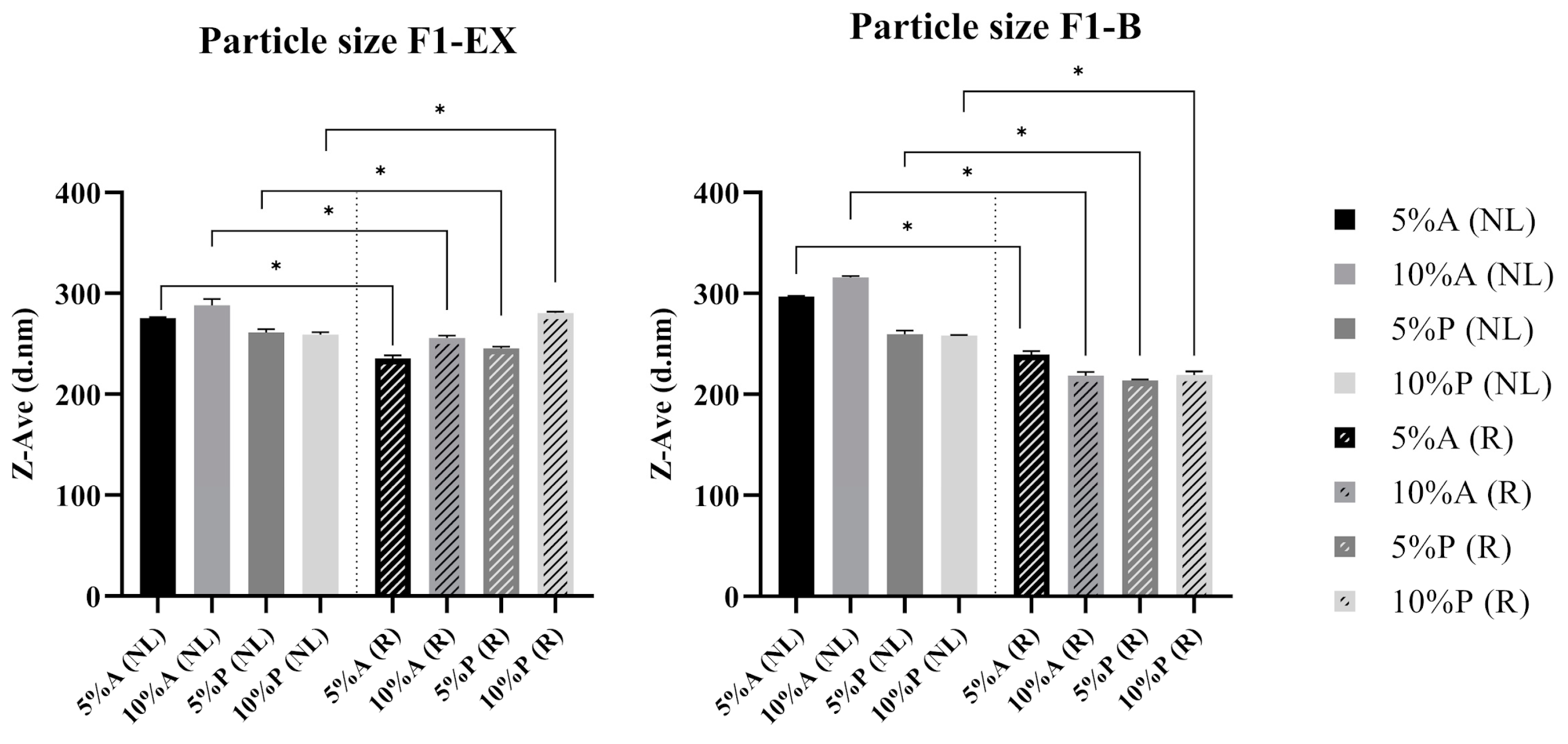
3.3. Impact of Homogenization on the Physicochemical Properties of Liposomes

The homogenization process improved the physical characteristics and encapsulation efficiency of liposomes. F1-B and F1-EX liposomes, after two ultrasonication cycles, showed particle sizes of 143.90 ± 2.80 nm and 232.5 ± 2.72 nm, respectively, with the addition of uvaia extract, resulting in a significant (p ≤ 0.05) increase in particle size. The PdI values for F1-B and F1-EX, after two ultrasonication cycles, were 0.249 ± 0.016 and 0.219 ± 0.027, respectively, with no statistically significant differences (p ≥ 0.05) between formulations. These results are consistent with Danaei et al. (2018), who reported that PdI values ≤ 0.3 indicate a homogeneous vesicle population [49].
The zeta potential measurements, which reflect the surface charge of nanocarriers and the repulsive forces between lipid particles, were −31.40 ± 0.90 mV for F1-B and −29.40 ± 0.10 mV for F1-EX [50]. Although there was a statistically significant difference (p ≤ 0.05), both formulations demonstrated sufficient stability, since, as described by Laouini et al. (2012), stable systems exhibit zeta potential values greater than 30 mV (in modulus) due to the higher particle repulsion, which reduces aggregation or coalescence phenomena, favoring the maintenance of small and homogeneous particle sizes [51].
The encapsulation efficiency analysis demonstrated that ultrasound homogenization improved the retention of uvaia bioactives in liposomes. F1-EX achieved close to 50% encapsulation efficiencies when only high-shear homogenization (Ultra-Turrax) was applied. The addition of ultrasound further increased the encapsulation efficiency, with the highest value recorded for F1-EX C2 at 58.8 ± 0.5% m/m (Figure 5), despite no statistically significant variations (p ≥ 0.05). These results demonstrate the effective encapsulation of hydrophilic substances like polyphenols, as they surpass the 50% threshold considered ideal for such compounds [52]. Based on these findings, it can be concluded that two ultrasound cycles are needed for producing liposomes, as they achieve the desired particle size, polydispersity index, zeta potential, and encapsulation efficiency.

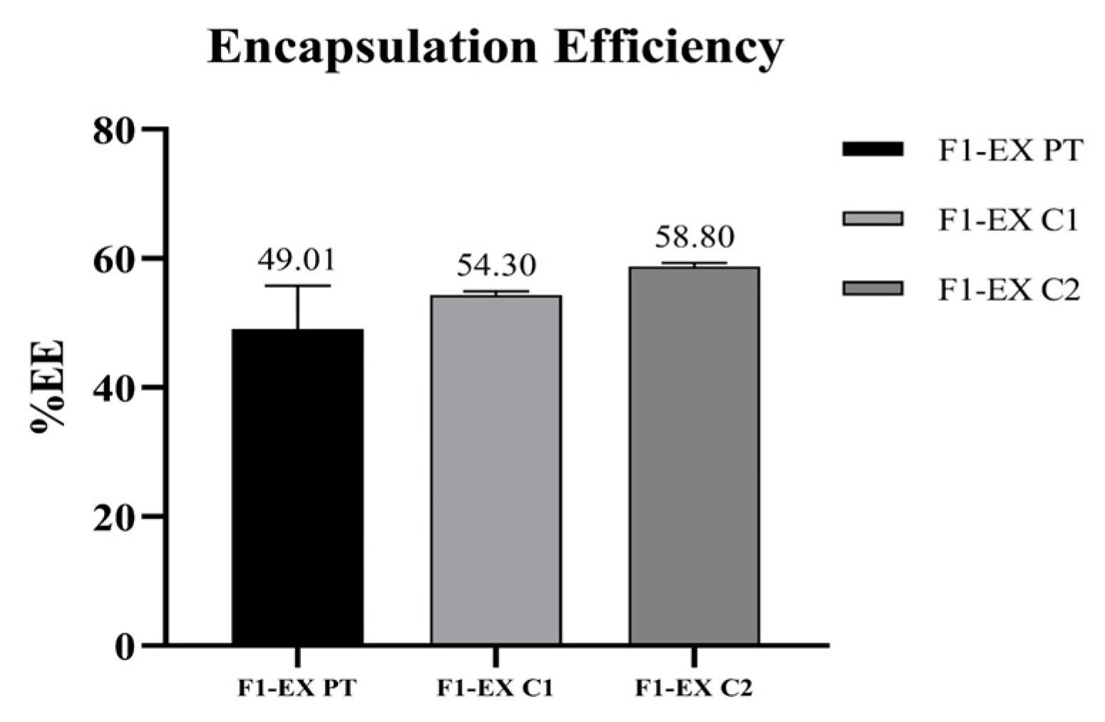
3.4. Production and Characterization of Freeze-Dried Liposomes

The freeze-dried F1-EX liposomes appeared yellow, while the F1-B formulations were white, regardless of the cryoprotectant composition (Figure 6). These visual differences are attributed to the presence of uvaia extract in the F1-EX formulation.
The water activity values for the freeze-dried liposomes with A1 or P1 adjuvants ranged from 0.068 ± 0.006 to 0.340 ± 0.038. Maintaining the water activity below 0.5 is crucial to prevent microbial growth in dry products [53]. The values observed in this study indicate a low probability of microbial proliferation. However, for lipid-containing formulations such as freeze-dried liposomes, water activity values below 0.2 may increase the risk of lipid peroxidation [54]. This risk can be mitigated by optimizing the freeze-drying process, such as reducing the drying time, to achieve ideal water activity levels that minimize the oxidation risks.
The moisture content, which is closely linked to water activity, also plays a vital role in the stability of freeze-dried liposomes. Excessive moisture can accelerate degradation reactions and microbial growth. The liposomes’ freeze-dried moisture content ranged from 2.71 ± 0.014% to 3.58 ± 0.42%, remaining well below the 10% threshold recommended for ensuring physicochemical and microbiological stability [19,55]. These values suggest that the freeze-dried liposomes are stable for long-term storage.
Regarding particle size, redispersed liposomes showed a statistically significant reduction (p ≤ 0.05) compared to their pre-freeze-drying counterparts for both F1-EX and F1-B formulations (Figure 7). The particle size of redispersed liposomes ranged from 213.83 to 280.43 nm, which is within the optimal range for cosmetic applications (100–300 nm) [15,47,48]. Statistical analysis revealed significant differences between redispersed liposomes (R) and non-freeze-dried liposomes (NL), with p-values ranging from <0.0184 to <0.0001 across different formulations. These results indicate that the freeze-drying process influenced particle size.
The zeta potential of the liposome formulations (Figure 8), which reflects the electrophoretic stability of disperse systems, was above or close to 30 mV (in modulus) for all formulations, both before freeze-drying and after redispersion. Statistically significant variations (p ≤ 0.05) were observed after redispersion for both F1-EX and F1-B formulations. The cryoprotectant mixture A1 produced higher zeta potential values (in the modulus) than P1 at equivalent concentrations, indicating improved stability. A significant difference (p ≤ 0.05) was observed between F1-EX 10%A and F1-EX 10%P.
The polydispersity index (PdI) values also showed significant differences (p ≤ 0.05) before and after freeze-drying and redispersion for the F1-B 10%A, F1-EX 5%A, and F1-EX 10%A formulations. Formulations containing sugar-based cryoprotectants exhibited lower PdI values than those with uvaia extract, indicating a more uniform particle size distribution. For example, F1-EX 5%A had a lower PdI than F1-EX 5%P, and F1-EX 10%A exhibited a lower PdI than F1-EX 10%P, suggesting that the A1 cryoprotectant improved size uniformity.

4. Conclusions

This study demonstrated the antioxidant potential of uvaia pulp extracts, with hydroalcoholic extracts exhibiting a higher polyphenol content and significant levels of vitamin C. The interaction between these bioactive compounds enhanced the antioxidant activity, as confirmed by the DPPH assay, highlighting uvaia as a valuable source of natural antioxidants. The bioactive compounds were successfully encapsulated in liposomes, ensuring their stability, and further processed into freeze-dried formulations with cryoprotectants. This approach improved their preservation and suitability for cosmetic applications. These findings demonstrate the potential of underutilized species like uvaia as raw materials for innovative and sustainable products, contributing to the responsible use of natural resources and the valorization of plant biodiversity.
Studies for the identification of other phenolic compounds by HPLC, as well as the evaluation of the antioxidant activity of liposomes and freeze-dried liposomes combined with cryoprotectants, are ongoing. Furthermore, investigations are being conducted on the in vitro safety of the formulations and the clinical efficacy of cosmetic formulations containing freeze-dried liposomes.

Author Contributions

G.A.S.: investigation, formal analysis, data curation, manuscript writing, validation, and visualization. L.K.: validation, manuscript reviewing and editing, and visualization. W.P.O.: validation, resources, project supervision, project administration, funding acquisition, manuscript reviewing and editing, and visualization. All authors have read and agreed to the published version of the manuscript.

Funding

This research was funded by the São Paulo State Research Foundation—FAPESP, grant numbers 2024/09270-5, 2018/26069-0, and 2021/08152-0.

Data Availability Statement

The data presented in this study are available upon request from the corresponding author.

Conflicts of Interest

The authors declare no conflicts of interest.

References

  1. Silva, B.D.L.D.A.; Vasconcelos, M.A.D.S.; Batista, K.S.; Batista, F.R.D.C.; Cavalcante, H.C.; Toscano, L.D.L.T.; Aquino, J.D.S. Hepatoprotective, lipid-lowering and antioxidant effects of mangaba powder (Hancornia speciosa) administered to rats fed a high-fat diet. Foods 2024, 13, 3773. [Google Scholar] [CrossRef] [PubMed]
  2. Guest, P.C. (Ed.) Reviews on Biomarker Studies in Aging and Anti-Aging Research; Springer Nature: Cham, Switzerland, 2019; Volume 1178. [Google Scholar]
  3. Silva, N.A.D.; Rodrigues, E.; Mercadante, A.Z.; de Rosso, V.V. Phenolic compounds and carotenoids from four fruits native from the Brazilian Atlantic forest. J. Agric. Food Chem. 2014, 62, 5072–5084. [Google Scholar] [CrossRef] [PubMed]
  4. Haminiuk, C.W.I.; Plata-Oviedo, M.S.V.; de Mattos, G.; Carpes, S.T.; Branco, I.G. Extraction and quantification of phenolic acids and flavonols from Eugenia pyriformis using different solvents. J. Food Sci. Technol. 2014, 51, 2862–2866. [Google Scholar] [CrossRef] [PubMed]
  5. Silva, A.P.G.; Sganzerla, W.G.; Jacomino, A.P.; da Silva, E.P.; Xiao, J.; Simal-Gandara, J. Chemical composition, bioactive compounds, and perspectives for the industrial formulation of health products from uvaia (Eugenia pyriformis Cambess–Myrtaceae): A comprehensive review. J. Food Compos. Anal. 2022, 109, 104500. [Google Scholar] [CrossRef]
  6. Jacomino, A.P.; da Silva, A.P.; de Freitas, T.P.; de Paula Morais, V.S. Uvaia—Eugenia pyriformis Cambess. In Exotic Fruits; Academic Press: London, UK, 2018; pp. 435–438. [Google Scholar]
  7. Krumreich, F.; D’Avila, R.F.; Freda, S.A.; Chim, J.F.; Chaves, F.C. Análises físico-químicas e estabilidade de compostos bioativos presentes em polpa de uvaia em pó obtidos por métodos de secagem e adição de maltodextrina e goma arábica. Rev. Thema 2016, 13, 4–17. [Google Scholar] [CrossRef]
  8. Oliveira, E.N.A.; da Costa Santos, D.; de Sousa, F.C.; Martins, J.N.; de Oliveira, S.P.A. Obtenção de ubaia desidratada pelo processo de liofilização. Rev. Bras. De Tecnol. Agroindustrial 2010, 4, 235–242. [Google Scholar] [CrossRef]
  9. Chong, C.H.; Law, C.L. Drying of Exotic Fruits. In Vegetables and Fruits, 2nd ed.; Academic Press: Cambridge, MA, USA, 2011; pp. 1–42. [Google Scholar]
  10. Yang, S.; Liu, L.; Han, J.; Tang, Y. Encapsulating plant ingredients for dermocosmetic application: An updated review of delivery systems and characterization techniques. Int. J. Cosmet. Sci. 2020, 42, 16–28. [Google Scholar] [CrossRef]
  11. Silva, G.S.; Jange, C.G.; Rocha, J.S.; Chaves, M.A.; Pinho, S.C. Characterisation of curcumin-loaded proliposomes produced by coating of micronised sucrose and hydration of phospholipid powders to obtain multilamellar liposomes. Int. J. Food Sci. Technol. 2017, 52, 772–780. [Google Scholar] [CrossRef]
  12. Rudzińska, M.; Grygier, A.; Knight, G.; Kmiecik, D. Liposomes as Carriers of Bioactive Compounds in Human Nutrition. Foods 2024, 13, 1814. [Google Scholar] [CrossRef] [PubMed]
  13. Bankole, V.O.; Osungunna, M.O.; Souza, C.R.F.; Salvador, S.L.; Oliveira, W.P. Spray-dried proliposomes: An innovative method for encapsulation of Rosmarinus officinalis L. polyphenols. AAPS PharmSciTech 2020, 21, 1–17. [Google Scholar] [CrossRef]
  14. Hua, Z.Z.; Li, B.G.; Liu, Z.J.; Sun, D.W. Freeze-drying of liposomes with cryoprotectants and its effect on retention rate of encapsulated ftorafur and vitamin A. Dry. Technol. 2003, 21, 1491–1505. [Google Scholar] [CrossRef]
  15. Kakuda, L.; Maia Campos, P.M.; Oliveira, W.P. Development and Efficacy Evaluation of Innovative Cosmetic Formulations with Caryocar brasiliense Fruit Pulp Oil Encapsulated in Freeze-Dried Liposomes. Pharmaceutics 2024, 16, 595. [Google Scholar] [CrossRef] [PubMed]
  16. Oliveira, W.P. (Ed.) Phytotechnology: A Sustainable Platform for the Development of Herbal Products; CRC Press: Boca Raton, FL, USA, 2021. [Google Scholar]
  17. Darniadi, S.; Handoko, D.D.; Sunarmani, S.; Widowati, S. Determination of shelf-life using accelerated shelf-life testing (Aslt) method and characterization of the flavour components of freeze-dried durian (Durio zibethinus) products. Food Res. 2021, 5, 98–106. [Google Scholar] [CrossRef]
  18. Baldim, I.; Souza, C.R.F.; Oliveira, W.P. Encapsulation of Essential Oils in Lipid-Based Nanosystems. In Phytotechnology; CRC Press: Boca Raton, FL, USA, 2021; pp. 197–230. [Google Scholar]
  19. Zampiér, M.N. Desenvolvimento, Padronização e Avaliação Biológica de Extratos Nebulizados de Dalbergia ecastaphyllum. Ph.D. Thesis, Universidade de São Paulo, São Paulo, Brazil, 2012. [Google Scholar]
  20. Bresolin, J.D.; Hubinger, S.Z. Metodologia para determinação de ácido ascórbico em sucos de citrus utilizando cromatografia líquida de alta eficiência. In Proceedings of the Simpósio Nacional de Instrumentação Agropecuária, São Carlos, Brazil, 18–21 November 2014; pp. 497–500. [Google Scholar]
  21. Fernandes, M.R.V.; Dias, A.L.T.; Carvalho, R.R.; Souza, C.R.F.; Oliveira, W.P.D. Antioxidant and antimicrobial activities of Psidium guajava L. spray dried extracts. Ind. Crops Prod. 2014, 60, 39–44. [Google Scholar] [CrossRef]
  22. Silveira, A.C.d.; Kassuia, Y.S.; Domahovski, R.C.; Lazzarotto, M. Método de DPPH adaptado: Uma ferramenta para analisar atividade antioxidante de polpa de frutos da erva-mate de forma rápida e reprodutível. Embrapa Florestas. Comun. Técnico 2018, 421, 11. Available online: https://ainfo.cnptia.embrapa.br/digital/bitstream/item/188244/1/CT-421-1642-final.pdf (accessed on 24 February 2025).
  23. Magalhães, W.L.E.; De Matos, M.; Lourençon, T. Metodologia Científica: Determinação da Capacidade Antioxidante de Lignina Pela Captura do Radical Livre DPPH; Embrapa Florestas: Colombo, Brazil, 2018. [Google Scholar]
  24. Jaafar-Maalej, C.; Diab, R.; Andrieu, V.; Elaissari, A.; Fessi, H. Ethanol injection method for hydrophilic and lipophilic drug-loaded liposome preparation. J. Liposome Res. 2010, 20, 228–243. [Google Scholar] [CrossRef]
  25. Baldim, I.; Tonani, L.; Kress, M.R.v.Z.; Oliveira, W.P. Lippia sidoides essential oil encapsulated in lipid nanosystem as an anti-Candida agent. Ind. Crops Prod. 2019, 127, 73–81. [Google Scholar] [CrossRef]
  26. Bhattacharjee, S. DLS and zeta potential—What they are and what they are not? J. Control. Release 2016, 235, 337–351. [Google Scholar] [CrossRef] [PubMed]
  27. Almeida, L.F.; Gil, G.A.; Moraes, L.N.; Furtado, F.B.; Kakuda, L.; Grotto, R.M.T.; Oliveira, W.P. Nanostructured lipid carriers loaded with essential oils: A strategy against SARS-CoV-2. J. Microencapsul. 2024, 41, 284–295. [Google Scholar] [CrossRef] [PubMed]
  28. Farrell, E.; Brousseau, J.L. Guide for DLS sample preparation. Brookhaven Instrum. 2014, 1, 1–3. [Google Scholar]
  29. Malvern. User Manual: Zetasizer Nano; Malvern: Malvern, UK, 2013. [Google Scholar]
  30. ISO 14887; Sample Preparation—Dispersing Procedures for Powders in Liquids. International Organization for Standardization: Geneva, Switzerland, 2000.
  31. Rockenbach, I.I.; Silva, G.L.D.; Rodrigues, E.; Kuskoski, E.M.; Fett, R. Influência do solvente no conteúdo total de polifenóis, antocianinas e atividade antioxidante de extratos de bagaço de uva (Vitis vinifera) variedades Tannat e Ancelota. Food Sci. Technol. 2008, 28, 238–244. [Google Scholar] [CrossRef]
  32. Rodrigues, L.M.; Romanini, E.B.; Silva, E.; Pilau, E.J.; Da Costa, S.C.; Madrona, G.S. Uvaia (Eugenia pyriformis Cambess) residue as a source of antioxidants: An approach to ecofriendly extraction. LWT 2021, 138, 110785. [Google Scholar] [CrossRef]
  33. Rufino, M.D.S.M.; Alves, R.E.; de Brito, E.S.; Pérez-Jiménez, J.; Saura-Calixto, F.; Mancini-Filho, J. Bioactive compounds and antioxidant capacities of 18 non-traditional tropical fruits from Brazil. Food Chem. 2010, 121, 996–1002. [Google Scholar] [CrossRef]
  34. Farias, P.D.; de Araujo, F.F.; Neri-Numa, I.A.; Dias-Audibert, F.L.; Delafiori, J.; Catharino, R.R.; Pastore, G.M. Distribution of nutrients and functional potential in fractions of Eugenia pyriformis: An underutilized native Brazilian fruit. Food Res. Int. 2020, 137, 109522. [Google Scholar] [CrossRef] [PubMed]
  35. Zlatić, N.; Jakovljević, D.; Stanković, M. Temporal, plant part, and interpopulation variability of secondary metabolites and antioxidant activity of Inula helenium L. Plants 2019, 8, 179. [Google Scholar] [CrossRef] [PubMed]
  36. Carrubba, A.; Lazzara, S.; Giovino, A.; Ruberto, G.; Napoli, E. Content variability of bioactive secondary metabolites in Hypericum perforatum L. Phytochem. Lett. 2021, 46, 71–78. [Google Scholar] [CrossRef]
  37. Abrafrutas. Quais São as Frutas Mais Produzidas No Brasil? Available online: https://abrafrutas.org/2023/04/quais-sao-as-frutas-mais-produzidas-no-brasil/ (accessed on 15 January 2025).
  38. NEPA—UNICAMP. Tabela Brasileira de Composição de Alimentos; NEPA—Unicamp: Campinas, Brazil, 2011. [Google Scholar]
  39. Caritá, A.C.; Fonseca-Santos, B.; Shultz, J.D.; Michniak-Kohn, B.; Chorilli, M.; Leonardi, G.R. Vitamin C: One compound, several uses. Advances for delivery, efficiency and stability. Nanomed. Nanotechnol. Biol. Med. 2020, 24, 102117. [Google Scholar] [CrossRef] [PubMed]
  40. Rattanawiwatpong, P.; Wanitphakdeedecha, R.; Bumrungpert, A.; Maiprasert, M. Anti-aging and brightening effects of a topical treatment containing vitamin C, vitamin E, and raspberry leaf cell culture extract: A split-face, randomized controlled trial. J. Cosmet. Dermatol. 2020, 19, 671–676. [Google Scholar] [CrossRef] [PubMed]
  41. Contreras-Calderón, J.; Calderón-Jaimes, L.; Guerra-Hernández, E.; García-Villanova, B. Antioxidant capacity, phenolic content and vitamin C in pulp, peel and seed from 24 exotic fruits from Colombia. Food Res. Int. 2011, 44, 2047–2053. [Google Scholar] [CrossRef]
  42. Jagdeo, J.; Kurtti, A.; Hernandez, S.; Akers, N.; Peterson, S. Novel Vitamin C and E and Green Tea Polyphenols Combination Serum Improves Photoaged Facial Skin. J. Drugs Dermatol. JDD 2021, 20, 996–1003. [Google Scholar] [CrossRef]
  43. Cassol, G.S. Estudo da Aplicação do Ultrassom e de um Agitador de alta Velocidade no Desenvolvimento de Protetores Solares. Bachelor’s Thesis, Universidade Federal do Rio Grande do Sul, Porto Alegre, Brazil, 2016. [Google Scholar]
  44. Hielscher, T. Ultrasonic Production of Nano-Size Dispersions and Emulsions. In Proceedings of the ENS 2005 Conference, Paris, France, 14–16 December 2005; pp. 138–143. [Google Scholar]
  45. Chirayil, C.J.; Joy, J.; Mathew, L. Instrumental techniques for the characterization of nanocarriers. In Thermal and Rheological Measurement Techniques for Nanomaterials Characterization; Elsevier: Amsterdam, The Netherlands, 2017; pp. 1–36. [Google Scholar]
  46. Anton, N.; Vandamme, T.F.; Ubrich, N. Nano-emulsions and nanocapsules by the PIT method: An investigation on the role of the temperature cycling on the emulsion phase inversion. Int. J. Pharm. 2007, 344, 44–52. [Google Scholar] [CrossRef] [PubMed]
  47. Souza, C.; de Freitas, L.A.P.; Maia Campos, P.M.B.G. Topical formulation containing beeswax-based nanocarriers improved in vivo skin barrier function. AAPS PharmSciTech 2017, 18, 2505–2516. [Google Scholar] [CrossRef] [PubMed]
  48. Filon, F.L.; Mauro, M.; Adami, G.; Bovenzi, M.; Crosera, M. Nanocarriers skin absorption: New aspects for a safety profile evaluation. Regul. Toxicol. Pharmacol. 2015, 72, 310–322. [Google Scholar] [CrossRef] [PubMed]
  49. Danaei, M.; Dehghankhold, M.; Ataei, S.; Hasanzadeh Davarani, F.; Javanmard, R.; Dokhani, A.; Khorasani, S.; Mozafari, M.R. Impact of particle size and polydispersity index on the clinical applications of lipidic nanocarrier systems. Pharmaceutics 2018, 10, 57. [Google Scholar] [CrossRef] [PubMed]
  50. Baldim, I. Desenvolvimento Tecnológico e Secagem de Sistemas Lipídicos Incorporando óleo Essencial de Lippia Sidoides. Ph.D. Thesis, Universidade de São Paulo, Ribeirão Preto, Brazil, 2017. [Google Scholar]
  51. Laouini, A.; Jaafar-Maalej, C.; Limayem-Blouza, I.; Sfar, S.; Charcosset, C.; Fessi, H. Preparation, characterization and applications of liposomes: State of the art. J. Colloid Sci. Biotechnol. 2012, 1, 147–168. [Google Scholar] [CrossRef]
  52. Tang, Y.; Zhou, A.; Zhou, S.; Ruan, J.; Qian, C.; Wu, C.; Ye, L. Preparation of VC nanoliposomes by high pressure homogenization: Process optimization and evaluation of efficacy, transdermal absorption, and stability. Heliyon 2024, 10, e29516. [Google Scholar] [CrossRef]
  53. Oliveira, W.P.; Freitas, L.A.P.; Freire, J.T. Drying of pharmaceutical products. In Transport Phenomena in Particulate Systems; Freire, J.T., Silveira, A.M., Ferreira, M.C., Eds.; Bentham Science: Sharjah, United Arab Emirates, 2012; pp. 148–171. [Google Scholar]
  54. Labuza, T.P.; Altunakar, B. Water activity prediction and moisture sorption isotherms. In Water Activity in Foods: Fundamentals and Applications; CRC Press: Boca Raton, FL, USA, 2020; pp. 161–205. [Google Scholar]
  55. Chaves, M.A.; Pinho, S.C. Unpurified soybean lecithins impact on the chemistry of proliposomes and liposome dispersions encapsulating vitamin D3. Food Biosci. 2020, 37, 100700. [Google Scholar] [CrossRef]
Figure 1. Voucher specimen of the uvaia tree (personal archive).
Processes 13 00693 g001
Figure 2. Flowchart of the liposome production protocol.
Processes 13 00693 g002
Figure 3. Results for the particle size (left, in blue) and polydispersity index (right, in red) for blank liposomes (F1-B) without Ultra-Turrax (ST), after Ultra-Turrax (PT), and based on the number of ultrasonication cycles (C).
Processes 13 00693 g003
Figure 4. Results for the particle size (left, in blue) and polydispersity index (right, in red) for liposomes containing uvaia pulp hydroalcoholic extract (F1-EX) without Ultra-Turrax (ST), after Ultra-Turrax (PT), and based on the number of ultrasonication cycles (C).
Processes 13 00693 g004
Figure 5. Encapsulation efficiency of liposomes containing uvaia pulp hydroalcoholic extract (F1-EX) following high-shear homogenization with Ultra-Turrax (PT), after one ultrasonication cycle (C1), and after two ultrasonication cycles (C2).
Processes 13 00693 g005
Figure 6. Visual comparison of freeze-dried blank liposomes (F1-B) and liposomes containing uvaia pulp hydroalcoholic extract (F1-EX).
Processes 13 00693 g006
Figure 7. Particle size of blank liposomes (F1-B) and liposomes containing uvaia pulp hydroalcoholic extract (F1-EX) with cryoprotectants before freeze-drying (NL) and after redispersion (R). * Indicates a statistically significant difference between redispersed liposomes (R) and non-freeze-dried liposomes (NL) (p ≤ 0.05).
Processes 13 00693 g007
Figure 8. Zeta potential of blank liposomes (F1-B) and liposomes containing uvaia pulp hydroalcoholic extract (F1-EX) with cryoprotectants before freeze-drying (NL) and after redispersion (R). * Indicates a statistically significant difference between redispersed liposomes (R) and non-freeze-dried liposomes (NL) (p ≤ 0.05).
Processes 13 00693 g008
Table 1. Components of the F1-B (blank) and F1-EX (with uvaia extract) liposome formulations.
ComponentsConcentration (%)
F1-BF1-EX
Phospholipon® 90H55
Cholesterol11
Absolute ethanol2828
Uvaia pulp extract-1
Milli-Q® water (q.s.p.)100100
Table 2. Total polyphenols, vitamin C, and antioxidant activity of uvaia fruit extracts.
SampleBioactive Concentration
Total PolyphenolsVitamin C (AA)Antioxidant Activity
GAEeb (µg/mL)GAEdb (mg/g)GAEfb (mg/g)AAeb (µg/mL)AAdb (mg/g)AAfb (mg/g)IC50 (µg/mL)EC50
(g Extract/100 g DPPH)
EX.AQ255.67 ± 5.60 *7.67 ± 0.170.85 ± 0.02139.01 ± 1.594.17 ± 0.050.46 ± 0.01219.87 ± 6.00 *3.69
EX.ET548.09 ± 31.70 *16.44 ± 0.951.83 ± 0.11127.46 ± 7.503.82 ± 0.230.43 ± 0.03122.09 ± 5.28 *2.05
* Indicates a significant difference between the aqueous extract (EX.AQ) and the hydroalcoholic extract (EX.ET) (p < 0.01). Subscripts eb, db, and fb represent extract, dry, and fresh pulp bases, respectively.
Disclaimer/Publisher’s Note: The statements, opinions and data contained in all publications are solely those of the individual author(s) and contributor(s) and not of MDPI and/or the editor(s). MDPI and/or the editor(s) disclaim responsibility for any injury to people or property resulting from any ideas, methods, instructions or products referred to in the content.

