Article

CTAB-Assisted Formation of Hierarchical Porosity in Cu-BDC-NH2 Metal–Organic Frameworks and Its Enhanced Peroxidase-like Catalysis for Xanthine Sensing

1
School of Environmental and Biological Engineering, Nanjing University of Science and Technology, Nanjing 210094, China
2
Department of Biotechnology Engineering, Ben-Gurion University of the Negev, Beer-Sheva 84105, Israel
*
Author to whom correspondence should be addressed.
These authors contribute equally to this work.
Processes 2025, 13(2), 387; https://doi.org/10.3390/pr13020387
Submission received: 3 January 2025 / Revised: 26 January 2025 / Accepted: 27 January 2025 / Published: 31 January 2025

Abstract

:
A novel porous metal-organic framework (MOF), pCu-BDC-NH2, with hierarchical porosity was synthesized using cetyltrimethylammonium bromide (CTAB) as a pore-generation agent. In addition to its common functions including structure-directing ligands or soft micelle templates, the judicious use of CTAB effectively modulated pore architecture in Cu-BDC-NH2 MOFs. With additional mesopores generated during the synthesis process, the intrinsic MOF scaffolds further obtained pore hierarchies and interconnectivity, enabling efficient substrate access to the active metal centers, and thus significantly facilitated catalytic performance. As a proof of concept, we applied the finely engineered porous MOF pCu-BDC-NH2 in a cascaded enzymatic system for xanthine sensing. This colorimetric biosensor exhibited a low detection limit of 0.11 μM, and a wide linear range of 1–120 μM. Furthermore, the sensor demonstrated exceptional stability, reproducibility, and was independent of interferences. Our simple yet effective method may find broader applications in tailoring pore architecture, enabling finer engineered structures to improve catalytic activities of nanomaterials.

1. Introduction

Metal-organic frameworks (MOFs) are a class of crystalline materials built up of metal-based secondary building units (SBU, ions or clusters) [1] connected by multivalent organic linkers via coordination interactions [2]. Their unique features including high surface areas, tunable pore sizes, and functional ligand diversities, offer a wide range of applications like gas adsorption, separation, catalysis, sensing and biomedical applications [3,4]. Recently, proprietary MOFs [5,6] exhibited remarkable versatility enabling selective mass transportation, cascaded catalysis and control release [7,8]. Their tunability introduced either through a one-pot synthesis process or post-synthetic modification (PSM) [9] allows for hierarchical and complex nanoscale architectures, thus ensuring a more homogeneous-like catalytic environment due to the efficient exposure of active sites, while maintaining the recyclability like heterogeneous catalysts [10].
The pore sizes of MOFs are typically smaller than 2 nm, inherited from the short organic ligands and arranged topology [11]. As a result, the diffusion of the reactants is hindered, especially in cases of large MOFs crystals. Therefore, not all catalytic sites are adequately exposed, decreasing overall catalytic efficiency and reaction rates [12]. Utilizing longer ligands are limited by commercial availability, undesirable interpenetrated structures or weak stability [13]. Alternatively, the fabrication of mesostructures to interconnect the original chambers or channels of MOFs turns out to be an effective strategy [14,15]. Template-assisted pore engineering is able to tailor the “defects” in MOFs [16]. Sacrificial templates, e.g., surfactants, nanoparticles, and even metal–organic assemblers, are incorporated during the fabrication process of MOFs [17], and then the temporarily occupied cavities are released through the removal of the templates. Recently, Chen et al. used HKUST-1 MOFs as sacrificial templates to prepare a second microporous MOFs of MIL-100(Fe) through the spontaneous ion substitution and hydrolysis process, resulting in better enzyme loading and synergetic degradation capacity of bisphenol A [18]. Tuo et al. synthesized a hierarchically porous Zr-MOF by using Pluronic F127 (PEO106PPO70PEO106) as a soft template in an emulsion system. After grafting phytic acid, the functionalized MOF exhibited impressive adsorption capacity and kinetics for Uranium [19]. Xu’s group investigated the self-assembly processes of MOF nanocrystals to obtain highly porous morphologies via amino-acid modulation, providing abundant moieties for downstream functionalization with more efficient and selective adsorption abilities [20]. Therefore, employing simple methods to interfere with, regulate nanoscopic processes of crystal growth, or grain assembly, can yield defect-engineered MOFs with hierarchical internal porosity, thereby enhancing overall catalytic performance.
Herein, we systematically explore a strategy to tune the hierarchical porosity in Cu-BDC-NH2 MOFs by controlling the synthesis process with CTAB. During the crystal growth, CTAB, a cationic surfactant, occupies certain copper sites, while its long-chain molecular structure fills the adjacent spaces [21]. This results in the formation of a primary mesoporous network once CTAB is removed. Coupled with the inherent microporosity of the MOFs and their high-density, uniformly distributed copper paddle-wheel catalytic centers, the carefully engineered hierarchical pore structure optimizes accessibility of active sites while promoting substrate diffusion. This strategy addresses the common limitations observed in larger MOF crystals, thereby enhancing the catalytic performance. Based on the well-tuned hierarchical porous MOF pCu-BDC-NH2, a model xanthine sensor is evaluated for free radical transfer, detection sensitivity, and potential for advanced sensing applications.

2. Materials and Methods

2.1. Instrumentation and Methods

The graphical morphologies of the synthesized materials were obtained using an FEI Quanta250 Field Emission Scanning Electron Microscope (FESEM) (FEI, Houston, TX, USA). FT-IR spectra were measured using a TENSOR Model 27 spectrometer (Bruker Co., Ltd., Natick, MA, USA). X-ray diffraction (XRD) patterns were collected using a Bruker D8 X-ray diffractometer (Bruker, Karlsruhe, Germany) with Cu Kα radiation (λ = 1.5406 Å) at 40 kV and 40 mA. X-ray photoelectron spectroscopy (XPS) measurements were performed on a Thermo ESCALAB 250XI (Thermo Electron Corporation, Waltham, MA, USA) equipped with a monochromatic Al Kα X-ray source. The surface area of the samples was determined by nitrogen adsorption–desorption isotherms at 77 K using an Autosorb-iQ-C instrument (Quantachrome Instruments, Boynton, FL, USA), and the BET surface area was calculated from the data. Ultraviolet-visible (UV-Vis) absorption spectra were collected with an Allsheng Feyond-A300 spectrophotometer (Allsheng, Hangzhou, China).

2.2. Materials

Copper(II) nitrate trihydrate (Cu(NO3)2·3H2O, AR, 99%), 2-aminobenzene-1,4-dicarboxylic acid (BDC-NH2, C8H7NO4), dimethylformamide (DMF, C3H7NO), ethanol (C2H5OH, AR, 99%), cetyltrimethylammonium bromide (CTAB, C19H42BrN) were purchased from Sinopharm Chemical Reagent Co., Ltd. All other chemicals were of analytical grade and used without further purification. Deionized water (DI) was used throughout the experiment.

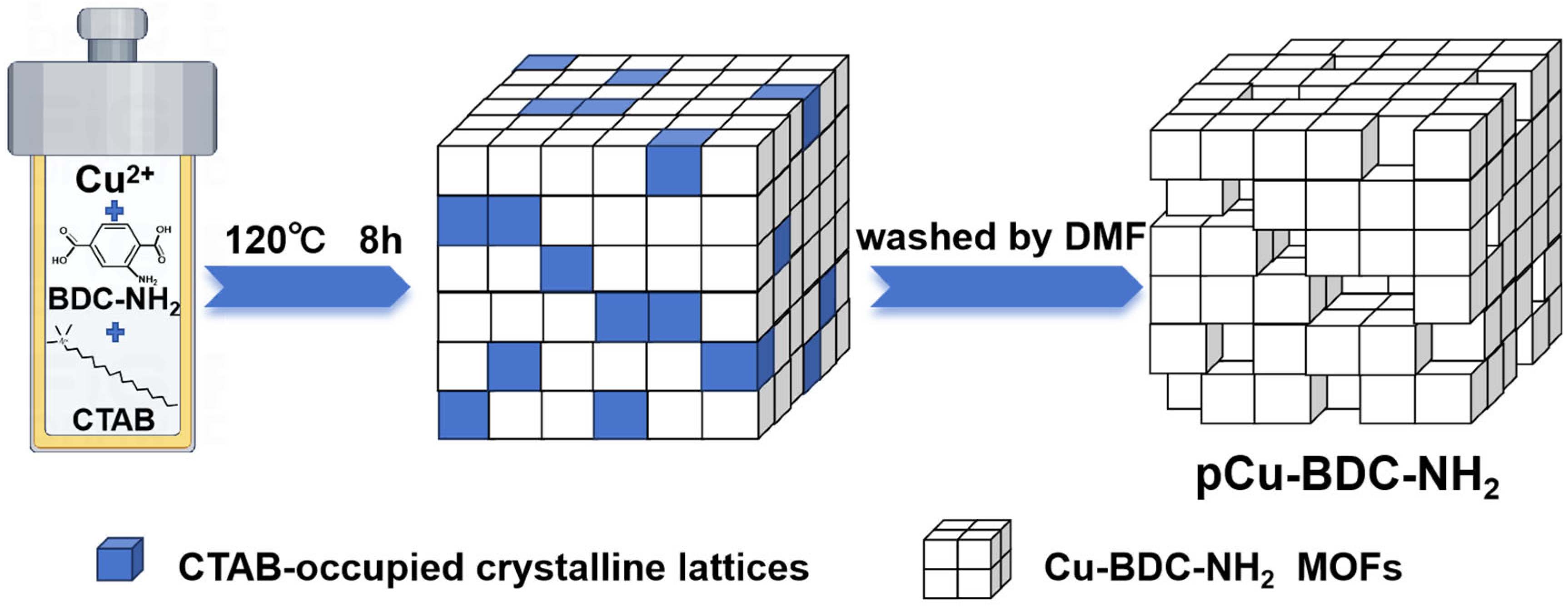
2.3. Synthesis of the Porous MOFs

During MOF synthesis, varying concentrations of CTAB were added to the system to investigate their effect on pore formation. At first, 0.0289 g (10 mmol) Cu(NO3)2·3H2O was dissolved in 4 mL of DMF solution, then separately, 0.0065 g (3 mmol) BDC-NH2 and different amounts of CTAB (0 mmol, 1 mmol, 2 mmol, 3 mmol, 4 mmol) were added to 8 mL of 1:1 (v/v) DMF and ethanol, and ultrasonicated for 30 min to facilitate ionic pre-organization. Thereafter, the aforementioned two solutions were mixed, stirred and transferred to a 20 mL PTFE reaction vessel with a tightened stainless steel liner. The solvothermal reaction was carried out at 120 °C for 8 h to promote crystal growth. At the end of the reaction, the mixture was centrifuged (13,000 rpm, 5 min) to separate out the solidified product. The product was then washed three times with ultra-pure water to remove solvents and impurities. Finally, the rinsed solids were freeze-dried under vacuum at −50 °C for 10 h. The various collected Cu-BDC-NH2 MOFs were respectively named Cu-BDC-NH2, Cu-BDC-NH2-C1, pCu-BDC-NH2, Cu-BDC-NH2-C3, and Cu-BDC-NH2-C4, depending on the amount of CTAB added.

2.4. Peroxidase-like Activity of Cu-BDC-NH2 MOFs

MOF-mediated oxidation of TMB was performed by mixing 120 μL of 1 mg/mL Cu-BDC-NH2 MOF suspension (dispersed in 100 mM NaAc-HAc buffer via ultrasound), 1 mL of TMB solution (4 mg TMB dissolved in 1 mL DMF, and then diluted with 9 mL NaAc-HAc buffer), and 50 μL of H2O2 solution with concentrations ranging from 1 nM to 200 μM. The different mixtures were incubated at 40 °C for 9 min. The maximum absorbance of the TMB oxidation products was measured at 652 nm.

2.5. Xanthine Concentration Sensing

At room temperature, 50 μL of xanthine solutions at different concentrations (1–600 μM) were mixed with 150 μL of xanthine oxidase solution (1.0 U/mL) and incubated for 30 min. The incubated solution was then mixed with 120 μL of 1 mg/mL Cu-BDC-NH2 MOF suspension, and added to 1 mL of TMB solution. After mixing, the reaction was incubated at 40 °C for 9 min, with the resulting oxTMB measured at 652 nm absorbance.

3. Results and Discussion

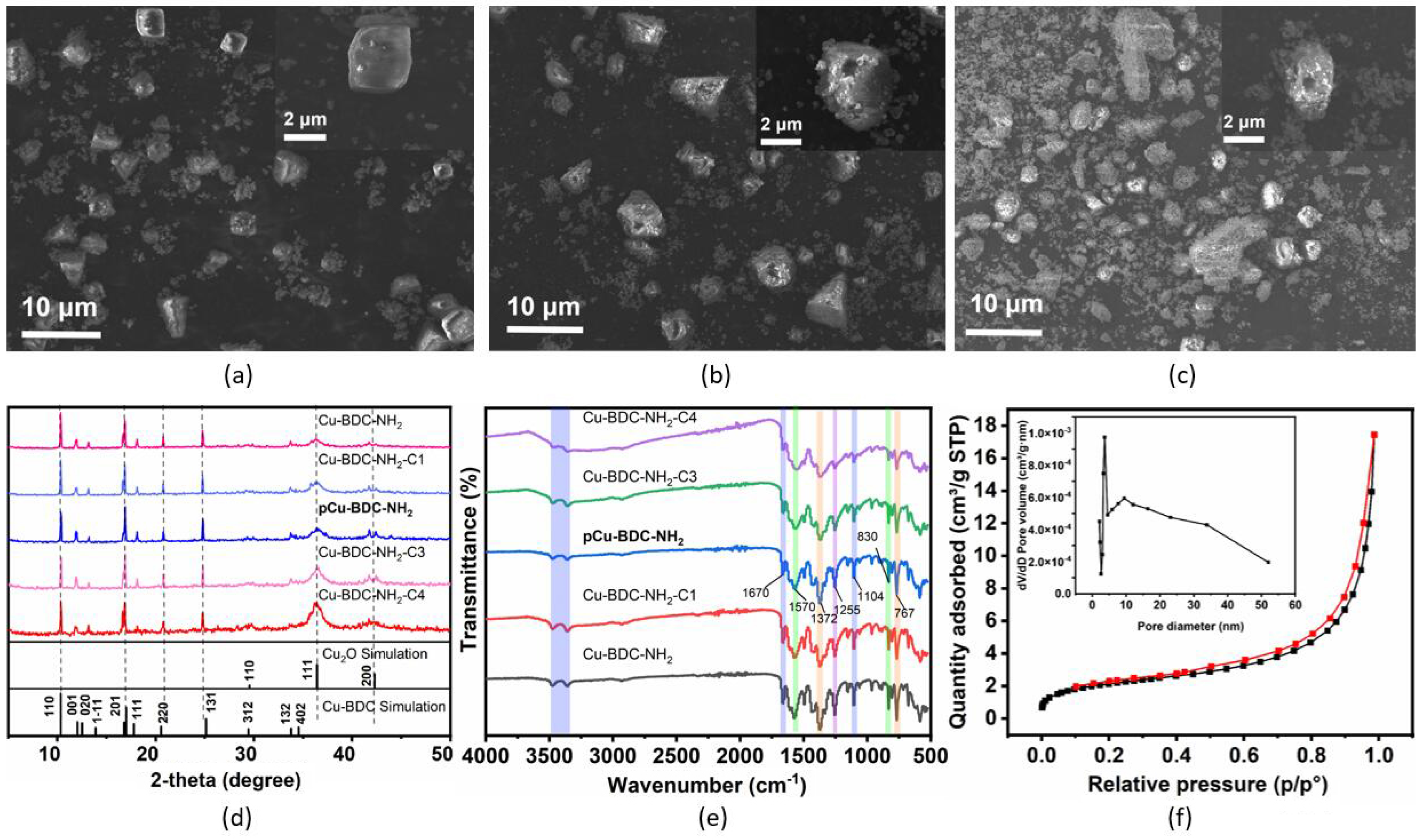
3.1. Characterization of Cu-BDC-NH2 MOFs

Cu-BDC-NH2 MOFs, featuring Cu(II) as the metal core and BDC-NH2 as the bridging ligand, are anticipated to be composed of the specific structural motifs in which two 5-coordinate Cu(II) ions are connected in a paddle-wheel type configuration. The porous Cu-BDC-NH2 MOFs could be synthesized through defect engineering using CTAB as a cationic surfactant to electrostatically interact with BDC-NH2 during the crystallization process [22,23]. After the removal of CTAB, its occupied space would be released, resulting in the formation of hierarchical porosity in the MOF crystals, as illustrated in Scheme 1. This hypothesis relies on the stability of the primary MOF in the presence of the different amount of pore-generating agents.
Therefore, The SEM images revealed the morphological features of three selected MOFs under different CTAB conditions. In the absence of CTAB (Figure 1a), Cu-BDC-NH2 exhibited smooth, defect-free cubic crystals with diameters of approximately 2 μm. This is attributed to the regular hydrogen bonding and the π-π stacking between the two-dimensional planes of Cu-BDC-NH2, which promotes the stacking of MOF layers along the [201] direction [24], and the degree of stacking is closely related to synthesis conditions [25]. Compared to Cu-BDC (CCDC 687690), the amino groups on the ligand BDC-NH2 enhanced the tendency for 2D MOF stacking [26,27]. SEM images of pCu-BDC-NH2 (CTAB 0.02 mmol, Figure 1b) clearly showed pore formation after eluting CTAB, with minimal impact on the overall morphology. On the other hand, when excessive CTAB (0.04 mmol) was added, the resulting product Cu-BDC-NH2-C4 (Figure 1c) experienced collapse of the crystalline blocks, generating large amounts of fragments.
XRD data for 5 samples with different CTAB contents during the synthesis process was depicted in Figure 1d. Diffraction peaks were present at 10.34°, 11.9°, 13.16°, 16.8°, 18.18°, 20.8°, 24.8°, 28.4°, 33.8° and 41.7°, corresponding to the Miller indices 110, 020, 11 − 1, −201, 111, 220, 131, 31 − 2, −402, and 51 − 2, respectively. These peaks closely aligned with the theoretical PXRD pattern of Cu-BDC as documented in the Cambridge Crystallographic Data Centre with the reference number 687690 and were consistent with the work of Gupta et al. [28]. Some lattice parameters of Cu-BDC-NH2 were found to differ slightly from those previously published for Cu-BDC, which was attributed to the substitution of BDC with BDC-NH2. Additionally, the peak at 36.3° was attributed to Cu2O and its broad pattern indicating the nanoscale degree of crystallization, which was a byproduct derived from the synthesis of Cu MOFs [29].
Based on the SEM images and XRD results, we concluded that addition of CTAB did not affect crystal growth at the nanoscopic scale. However, at a more macroscopic scale, the morphology and pore structure of the products were influenced by the CTAB-assisted modulation: (1) without CTAB, MOF crystals grew stably and aged, forming a characteristic cubic structure; (2) with a moderate amount of CTAB, distinct cavities appeared within the cubic particles, but the integrity of the particle structure was maintained; and (3) with an excessive amount of CTAB, significant collapse of the MOF particles occurred, resulting in smaller crystal grains, and remaining particles adopted increased irregularity.

3.2. Hierarchical Porosity in pCu-BDC-NH2

According to XRD spectra, the composition of the main chemical bonds in the Cu-BDC-NH2 MOF samples should be maintained. In the wavenumber region from 4000 to 500 cm−1 (Figure 1e), the presence of a Cu-O stretching vibration was indicated by the distinctive and strong absorption band at 582 cm−1 [30]. This observation served as strong evidence for the successful formation of a metal-oxygen bond between the carboxylate group of BDC-NH2 and the Cu(II) ion, aligning with the proposed theoretical framework [28]. The characteristic peaks observed at 1104, 1150, 1491, and 1570 cm−1 correspond to the aromatic ring vibration signal, and the bands at 1609 cm−1 and 1372 cm−1 were attributed to the carboxylate ligand (−COO) in BDC-NH2. Two distinct peaks at 3478 cm−1 (asymmetric) and 3350 cm−1 (symmetric) were attributed to the N-H stretching vibrations of the NH2-BDC ligands, accompanied by the band at 1255 cm−1 for the aromatic C-N stretching [31], whereas the band at 1670 cm−1 was assigned to the stretching vibration of the carbonyl group in DMF, which weakly coordinated along the z-axis of the copper-based paddlewheel secondary building units (SBU) and could be detached during the subsequent catalytic studies.
In the FT-IR spectra, completeness of functional groups in the MOF structure was confirmed, indicating that the dominant material remained was Cu-BDC-NH2 MOFs. Samples with or without CTAB displayed matched spectra, suggesting the complete removal of washed CTAB. Therefore, the spaces previously occupied by CTAB during synthesis were likely released. To confirm the presence of a mesoporous structure in pCu-BDC-NH2, N2-sorption measurements were conducted. Figure 1f depicted the N2 adsorption-desorption isotherm of pCu-BDC-NH2 particles, which exhibited characteristics intermediate between type I (typical of microporous materials) and type IV (typical of mesoporous materials). A distinct mesoporous hysteresis loop in the N2 isotherm was apparent. Pore size distribution of pCu-BDC-NH2 was different from those of Cu-BDC-NH2 and Cu-BDC-NH2-C4, with significantly higher abundance of pores in the 3 to 5 nm range, as shown in Figure 1f and Figure S1. These findings indicated that synthesized pCu-BDC-NH2 material possessed both mesoporous and microporous structures.

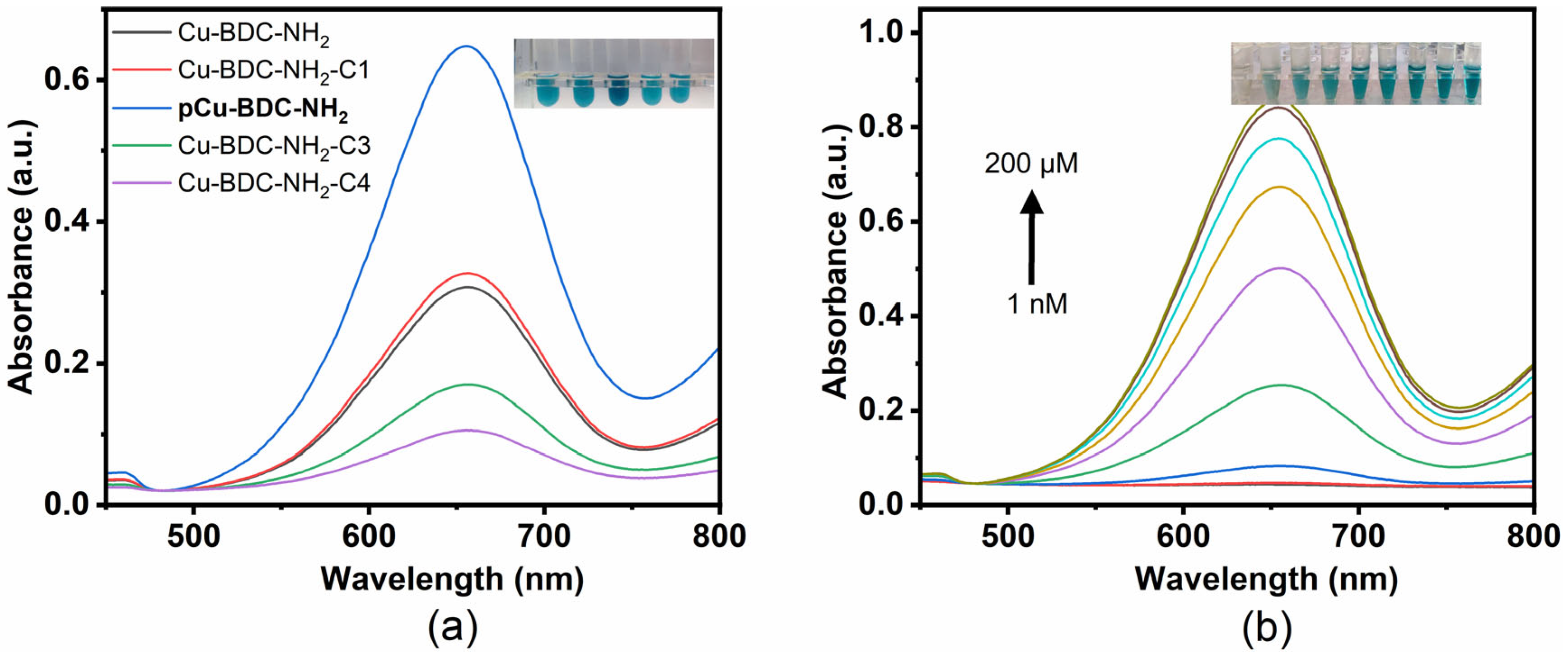
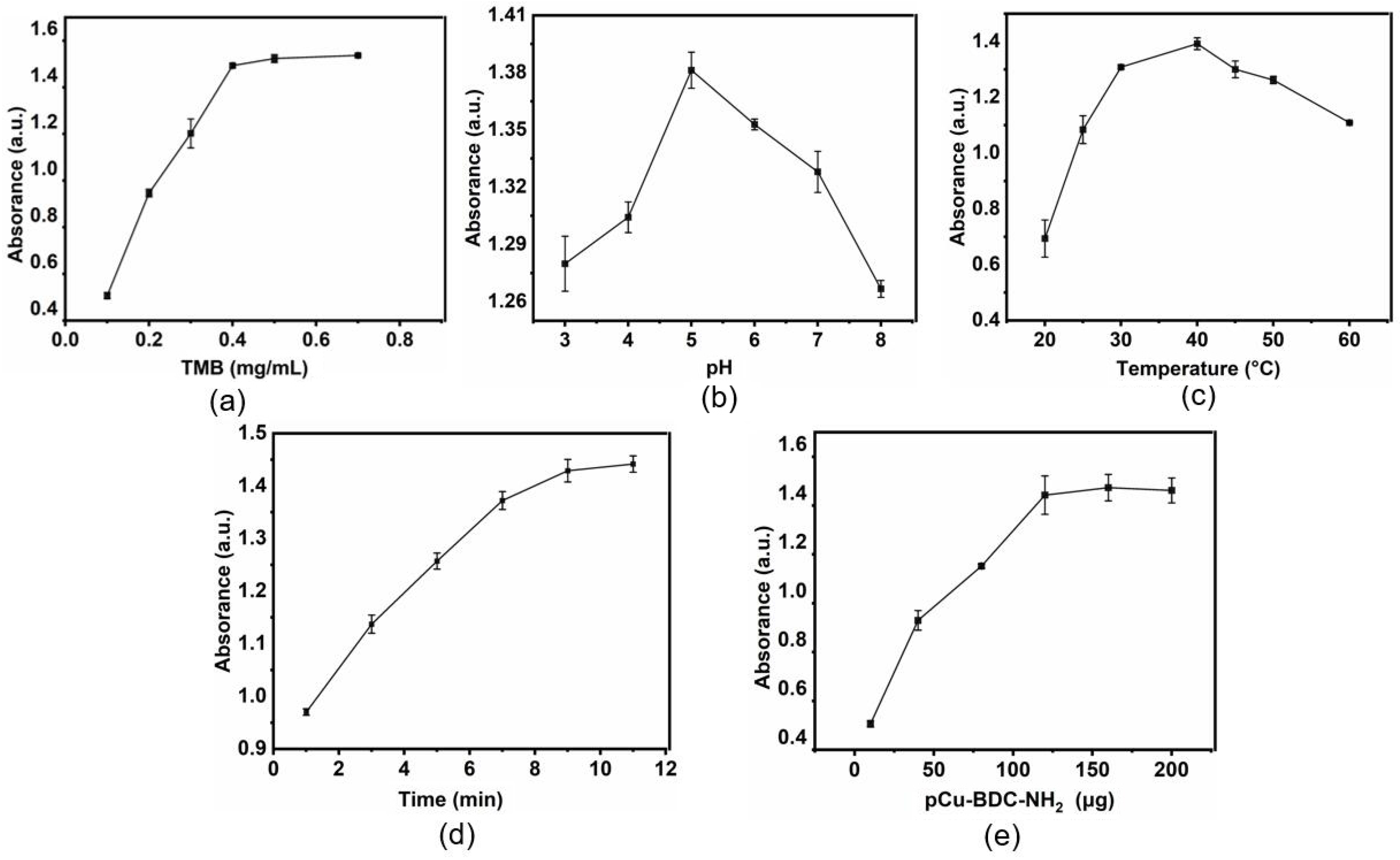
3.3. Synergetic Enhancement of Catalysis

In order to analyze the mechanism and potential enhancement in catalysis, the chemical composition of some crucial atoms should be determined. Full-scan XPS survey spectra of Cu-BDC-NH2, pCu-BDC-NH2, and Cu-BDC-NH2-C4 exhibited similar characteristic peaks to C, O, N, and Cu at their respective binding energies (Figure S2a–c). However, significant differences emerged when the high-resolution spectra of the Cu 2p and O 1s orbitals were analyzed. All the Cu 2p spectra in Figure 2 presented two clear peaks at binding energies of 933 and 960 eV, corresponding to the Cu 2p3/2 and Cu 2p1/2 levels, respectively, accompanied by several satellite peaks. The presence of Cu(II) was confirmed by the observation of four shake-up satellite peaks, resulting from p → d hybridization in the d9 configuration, within the energy ranges of 940–945 eV and 958–964 eV. After deconvolution, it can be observed that the peak at 933 eV decomposed into two peaks at 932.7 and 934.7 eV, which correspond to Cu(I) and Cu(II), respectively. Noticeably, a significantly higher proportion of Cu(I) was observed in the pCu-BDC-NH2 sample (Figure 2a) compared to samples Cu-BDC-NH2 (Figure 2b) and Cu-BDC-NH2-C4 (Figure 2c). This could be attributed to the specific heterogeneous interface formed by CTAB and the surrounding solvents environment that facilitate the reduction of Cu(II). In contrast, the excessive concentration of CTAB in Cu-BDC-NH2-C4 disrupted the MOF structure, reducing the Cu(I) portion. This outcome could affect the enhancement of peroxidase-like activity of the Cu-BDC-NH2 MOFs, because Cu(I) is known to facilitate the breakdown of H2O2 and the production of HO•, which is then followed by the alternative and cyclic transformation between Cu(II) and Cu(I) [32]. Additionally, the O 1s spectrum was deconvoluted into three peaks (Figure 2d), corresponding to C=O, C-O, and Cu-O groups, respectively. The presence of Cu-O bonds is also one of the powerful indications of the successful synthesis of Cu MOF. The N 1s (Figure S3a–c) and C 1s (Figure S3e–f) spectra showed similar states of these elements, regardless of CTAB addition, which was consistent with our expectations.
The catalytic centers of Cu-BDC-NH2 MOFs were located on the paddle-wheel structured SBUs, whereas in defect-free MOF cubic crystals, the majority of these centers were buried within the crystal structure. The hierarchical pore system combines both mesopores and micropores enabling greater access to catalytic sites, with reasonable pore interconnections facilitating reactant diffusion and transport. Moreover, the Cu(I) species in the pCu-BDC-NH2 sample, either in the MOF scaffold or in the massively generated Cu2O nanoparticles, were able to provide a parallel route for the generation of reactive oxygen radicals during the catalytic decomposition of H2O2, as follows:
Main   route :   p C u I I - B D C - N H 2 + H 2 O 2 p C u I - B D C - N H 2 + H O O + H +
Synergistic   enhancement :   C u ( I ) 2 O + 2 H 2 O 2 C u ( I I ) 2 O + 2 H O + 2 O H -
Cyclic   catalysis :   p C u I - B D C - N H 2 + H 2 O 2 p C u I I - B D C - N H 2 + H O + O H -
C u ( I I ) 2 O + 2 H 2 O 2 C u ( I ) 2 O + 2 H O O + 2 H +
Among them, Equation (1) is the rate-limiting step in the classical Cu-Fenton reaction [33], while the presence of Cu(I) in the samples provides the possibility for rapid reaction in the catalytic process.

3.4. pCu-BDC-NH2 Enzyme Mimicry

To assess the peroxidase-like activity of pCu-BDC-NH2, oxidation of 3,3′,5,5′-tetramethylbenzidine (TMB) in the presence of H2O2 was optically detected by quantifying the characteristic absorption peak of the blue oxTMB located at 652 nm in the UV-vis spectrum [34,35], with a reaction process involving reactive oxygen species (ROS) radicals as follows:
TMB + HO oxTMB + H 2 O
TMB + HOO oxTMB + H 2 O
Colorimetric experiments illustrated hierarchically porous MOF pCu-BDC-NH2, exhibiting superior performance in the TMB-H2O2 reaction, yielding a pronounced blue color compared to other experimental groups (Figure 3a). pCu-BDC-NH2 composite material exhibited enhanced peroxidase mimicry, attributed to the high surface area of the CTAB-mediated hierarchical porous structure inside the MOF crystals and the concomitant active Cu(I) oxide species formed during the synthesis process.
To thoroughly evaluate the performance of pCu-BDC-NH2 in the enzyme-mimicking catalytic reactions, a H2O2-TMB gradient oxidation assay model was employed. As the H2O2 concentration increased from 1 nM to 200 μM, the absorption peak intensity at 652 nm gradually intensified. Digital photographs taken during the experiment further demonstrated the progressive deepening of the solution color (from light blue to dark blue), corresponding to the increase in H2O2 concentration. This indicated that the system possessed good sensitivity and resolution in detecting low concentrations of H2O2.

3.5. pCu-BDC-NH2 Xanthine Colorimetric Monitoring

H2O2 is involved in a wide range of biological functions, and xanthine oxidation by Xanthine oxidase (XOD) to produce H2O2 [36] was used as a proof of concept. Our pCu-BDC-NH2 would catalyze these cascaded reactions into a measurable colored product. Prior to conducting the sensing studies, the experimental conditions were optimized, as shown in Figure 4, taking into account the concentration of TMB, pH, temperature, time, and the amount of pCu-BDC-NH2, in order to maximize the catalytic activity of pCu-BDC-NH2. First, the effect of TMB concentration was shown in Figure 4a. The absorbance at 652 nm increased linearly and reached maximum at 0.4 mg/mL. In term of pH, the reaction activity increased from pH 3.0 to pH 5, then decreased (Figure 4b). In Figure 4c, we found that the reaction temperature was also a significant factor, and the optimum temperature was 40 °C. As for the incubation time, 9 min (Figure 4d) was most efficient. The best additive amount for the pCu-BDC-NH₂ catalyst was tested to be 120 μg, as shown in Figure 4e. In summary, the optimized conditions for the colorimetric system were as follows: TMB concentration of 0.4 mg/mL, pH 5.0, reaction temperature of 40 °C, reaction time of 9 min, and catalyst weight of 120 μg. These conditions were employed in subsequent experiments to ensure consistent and maximal catalytic performance.
Xanthine can be successfully quantified based on the serial catalysis strategy, as illustrated in Figure 5a. With the increase of the concentration of xanthine in solution, the concomitant rise in absorbance at 652 nm occurred as shown in Figure 5b. In this experiment, a good linear relationship between absorbance and xanthine concentration is shown (Figure 5c), with two characteristic ranges of 1 to 120 μM, and 120 to 600 μM. Thanks to the enhanced catalytic ability, our experimental results can be identified by the naked eye without the need of instrumental assistance. By employing the 3σ method [37], the limit of detection (LOD) for xanthine by this strategy was calculated to be 0.11 μM, significantly lower than the concentrations reported in previous studies (Table 1). Therefore, applying the novel pCu-BDC-NH2 catalyst as the signal transducer, a visual and straightforward detection of the target molecule was achieved.
To further validate the application potential of the pCu-BDC-NH2 material as a xanthine sensor, we evaluated its selectivity and stability. Experiments were conducted under conditions where various possible interferents, such as alanine, glucose, Mg2+, Na+, and K+, were added to ensure that the response to xanthine was not significantly affected. As shown in Figure 6a, no significant changes in the colorimetric signal were observed, with the absorbance intensity at 652 nm remaining stable, while common ions or small molecules in complex sample environments did not interfere with the accuracy and reliability of the detection. To assess the long-term stability of the sensor, as depicted in Figure 6b, a 60-day dry storage performance at room temperature was done. Samples were taken every 5 days for xanthine detection experiments, recording changes in the colorimetric response. The results showed that throughout the testing period, the catalytic performance and color intensity of the system did not exhibit significant decay, and the absorbance values at 652 nm remained highly consistent. This fully demonstrates the chemical stability and catalytic activity durability of the material under mid-term storage conditions.

4. Conclusions

We investigated a strategy to creat novel MOFs including hierarchical porosities by simply incorporating the surfactant CTAB into the solvothermal synthesis system, followed by its subsequent elution. The physicochemical properties of MOFs at varying concentrations of surfactants were characterized, pointing to pCu-BDC-NH2 MOF as the one with superior catalytic performance. Thanks to increased accessibility of the active metal centers to substrate molecules, significantly improving the overall catalytic activity. As a proof of concept, a xanthine detection biosensor was subsequently tested, enabling a simple, rapid, and quantitative detection by colorimetry with TMB. This sensor achieved a detection limit of 0.11 μM and exhibited high specificity even in the presence of a high concentration of potential interferents. We believe that this copper MOF material as well as its synthesis strategy and excellent peroxidase-like activity would promote the development of other types of biosensors.

Supplementary Materials

The following supporting information can be downloaded at: https://www.mdpi.com/article/10.3390/pr13020387/s1, Figure S1: N2 sorption isotherms at 77 K of (a) Cu-BDC-NH2 and (b) Cu-BDC-NH2-C4 samples, insets were the pore size distributions; Figure S2: XPS full-survey scan of (a) pCu-BDC-NH2, (b) Cu-BDC-NH2 and (c) Cu-BDC-NH2-C4, high-resolution XPS spectrum of the O1s orbital of (d) Cu-BDC-NH2 and (e) Cu-BDC-NH2-C4; Figure S3: High-resolution XPS response of N1s of (a)Cu-BDC-NH2, (b)pCu-BDC-NH2, and (c) Cu-BDC-NH2-C4, and the response of C1s of (d) Cu-BDC-NH2, (e) pCu-BDC-NH2, and (f) Cu-BDC-NH2-C4.

Author Contributions

Conceptualization, C.T. and J.L.; methodology, C.T. and F.Z.; formal analysis, C.T. and J.H.; investigation, J.H. and Y.G.; data curation, R.X. and C.T.; writing—original draft preparation, J.H.; writing—review and editing, J.L. and R.S.M.; funding acquisition, J.L. All authors have read and agreed to the published version of the manuscript.

Funding

The authors greatly acknowledge the financial support from National Natural Science Foundation of China (No. 62001224), Natural Science Foundation of Jiangsu Province (No. BK20190457), the 69th batch of China Postdoctoral Science Foundation (No. 2021M691600), “Overseas Academic Partnership Program” of Nanjing University of Science and Technology (2019), and Sino-French International Research Network “New Nanostructured Materials and Biomaterials for Renewable Electrical Energy Sources”.

Data Availability Statement

Data are available on request from the authors.

Conflicts of Interest

The authors declare that the research was conducted in the absence of any commercial or financial relationships that could be construed as a potential conflicts of interest.

References

  1. Tranchemontagne, D.J.; Mendoza-Cortés, J.L.; O’Keeffe, M.; Yaghi, O.M. Secondary Building Units, Nets and Bonding in The Chemistry of Metal–Organic Frameworks. Chem. Soc. Rev. 2009, 38, 1257–1283. [Google Scholar] [CrossRef] [PubMed]
  2. Zhou, H.-C.; Long, J.; Yaghi, O.M. Introduction to Metal–Organic Frameworks. Chem. Rev. 2012, 112, 673–674. [Google Scholar] [CrossRef] [PubMed]
  3. Furukawa, H.; Cordova, K.E.; O’Keeffe, M.; Yaghi, O.M. The Chemistry and Applications of Metal-Organic Frameworks. Science 2013, 341, 1230444. [Google Scholar] [CrossRef] [PubMed]
  4. Vaitsis, C.; Sourkouni, G.; Argirusis, C. Metal Organic Frameworks (MOFs) and ultrasound: A review. Ultrason. Sonochem. 2019, 52, 106–119. [Google Scholar] [CrossRef]
  5. Sun, K.; Qian, Y.; Jiang, H.-L. Metal-Organic Frameworks for Photocatalytic Water Splitting and CO2 Reduction. Angew. Chem. Int. Ed. 2023, 62, e202217565. [Google Scholar] [CrossRef]
  6. Ding, B.; Chen, H.; Tan, J.; Meng, Q.; Zheng, P.; Ma, P.A.; Lin, J. ZIF-8 Nanoparticles Evoke Pyroptosis for High-Efficiency Cancer Immunotherapy. Angew. Chem. Int. Ed. 2023, 62, e202215307. [Google Scholar] [CrossRef]
  7. Rao, R.; Ma, S.; Gao, B.; Bi, F.; Chen, Y.; Yang, Y.; Liu, N.; Wu, M.; Zhang, X. Recent advances of metal-organic framework-based and derivative materials in the heterogeneous catalytic removal of volatile organic compounds. J. Colloid Interface Sci. 2023, 636, 55–72. [Google Scholar] [CrossRef]
  8. Fu, H.; Ma, H.; Zhao, S. Structural Control of Copper-Based MOF Catalysts for Electroreduction of CO2: A Review. Processes 2024, 12, 2205. [Google Scholar] [CrossRef]
  9. Yin, Z.; Wan, S.; Yang, J.; Kurmoo, M.; Zeng, M.-H. Recent advances in post-synthetic modification of metal–organic frameworks: New types and tandem reactions. Coord. Chem. Rev. 2019, 378, 500–512. [Google Scholar] [CrossRef]
  10. Zhu, L.; Liu, X.Q.; Jiang, H.L.; Sun, L.B. Metal-Organic Frameworks for Heterogeneous Basic Catalysis. Chem. Rev. 2017, 117, 8129–8176. [Google Scholar] [CrossRef]
  11. Furukawa, H.; Ko, N.; Go, Y.B.; Aratani, N.; Choi, S.B.; Choi, E.; Yazaydin, A.Ö.; Snurr, R.Q.; O’Keeffe, M.; Kim, J.; et al. Ultrahigh Porosity in Metal-Organic Frameworks. Science 2010, 329, 424–428. [Google Scholar] [CrossRef] [PubMed]
  12. Kang, Y.-S.; Lu, Y.; Chen, K.; Zhao, Y.; Wang, P.; Sun, W.-Y. Metal–organic frameworks with catalytic centers: From synthesis to catalytic application. Coord. Chem. Rev. 2019, 378, 262–280. [Google Scholar] [CrossRef]
  13. Deng, H.; Grunder, S.; Cordova, K.E.; Valente, C.; Furukawa, H.; Hmadeh, M.; Gándara, F.; Whalley, A.C.; Liu, Z.; Asahina, S.; et al. Large-Pore Apertures in a Series of Metal-Organic Frameworks. Science 2012, 336, 1018–1023. [Google Scholar] [CrossRef] [PubMed]
  14. Connolly, B.M.; Aragones-Anglada, M.; Gandara-Loe, J.; Danaf, N.A.; Lamb, D.C.; Mehta, J.P.; Vulpe, D.; Wuttke, S.; Silvestre-Albero, J.; Moghadam, P.Z.; et al. Tuning porosity in macroscopic monolithic metal-organic frameworks for exceptional natural gas storage. Nat. Commun. 2019, 10, 2345. [Google Scholar] [CrossRef]
  15. Yang, X.-Y.; Chen, L.-H.; Li, Y.; Rooke, J.C.; Sanchez, C.; Su, B.-L. Hierarchically porous materials: Synthesis strategies and structure design. Chem. Soc. Rev. 2017, 46, 481–558. [Google Scholar] [CrossRef]
  16. Fang, Z.; Bueken, B.; De Vos, D.E.; Fischer, R.A. Defect-Engineered Metal–Organic Frameworks. Angew. Chem. Int. Ed. 2015, 54, 7234–7254. [Google Scholar] [CrossRef]
  17. Furukawa, S.; Reboul, J.; Diring, S.; Sumida, K.; Kitagawa, S. Structuring of metal–organic frameworks at the mesoscopic/macroscopic scale. Chem. Soc. Rev. 2014, 43, 5700–5734. [Google Scholar] [CrossRef]
  18. Chen, G.; Song, R.; Wang, H.; Huang, L.; Wang, L.; Lei, J. Construction of hierarchical porous MIL-100(Fe) for horseradish peroxidase immobilization and its degradation performance of bisphenol A. J. Clean. Prod. 2024, 481, 144178. [Google Scholar] [CrossRef]
  19. Tuo, K.; Li, J.; Li, Y.; Liang, C.; Shao, C.; Hou, W.; Fan, C.; Li, Z.; Pu, S.; Chen, Z.; et al. Phytic Acid Functionalized Hierarchical Porous Metal-Organic Framework Microspheres for Efficient Extraction of Uranium from Seawater. Small 2024, 21, 2407272. [Google Scholar] [CrossRef]
  20. Ma, L.; Huang, C.; Yao, Y.; Fu, M.; Han, F.; Li, Q.; Wu, M.; Zhang, H.; Xu, L.; Ma, H. Self-assembled MOF microspheres with hierarchical porous structure for efficient uranium adsorption. Sep. Purif. Technol. 2023, 314, 123526. [Google Scholar] [CrossRef]
  21. Zhang, X.; Yang, Y.; Song, L.; Chen, J.; Yang, Y.; Wang, Y. Enhanced adsorption performance of gaseous toluene on defective UiO-66 metal organic framework: Equilibrium and kinetic studies. J. Hazard. Mater. 2019, 365, 597–605. [Google Scholar] [CrossRef] [PubMed]
  22. Saravanan, K.; Tyagi, B.; Shukla, R.S.; Bajaj, H.C. Esterification of palmitic acid with methanol over template-assisted mesoporous sulfated zirconia solid acid catalyst. Appl. Catal. B Environ. 2015, 172, 108–115. [Google Scholar] [CrossRef]
  23. Zhang, X.; Xiong, B.; Li, J.; Qian, L.; Liu, L.; Liu, Z.; Fang, P.; He, C. Dependence of dye molecules adsorption behaviors on pore characteristics of mesostructured MOFs fabricated by surfactant template. ACS Appl. Mater. Interfaces 2019, 11, 31441–31451. [Google Scholar] [CrossRef] [PubMed]
  24. Wang, S.; Wang, T.; Zheng, H.; Fan, F.; Gu, Z.; He, W.; Zhang, B.; Shao, L.; Chen, H.; Li, Y.; et al. Fabrication of mesoporous MOF nanosheets via surfactant-template method for C–S coupling reactions. Microporous Mesoporous Mater. 2020, 303, 110254. [Google Scholar] [CrossRef]
  25. Amali, R.K.A.; Lim, H.N.; Ibrahim, I.; Zainal, Z.; Ahmad, S.A.A. Silver nanoparticles-loaded copper (II)-terephthalate framework nanocomposite as a screen-printed carbon electrode modifier for amperometric nitrate detection. J. Electroanal. Chem. 2022, 918, 116440. [Google Scholar] [CrossRef]
  26. Rodenas, T.; Luz, I.; Prieto, G.; Seoane, B.; Miro, H.; Corma, A.; Kapteijn, F.; Llabrés i Xamena, F.X.; Gascon, J. Metal–organic framework nanosheets in polymer composite materials for gas separation. Nat. Mater. 2015, 14, 48–55. [Google Scholar] [CrossRef]
  27. Carrington, E.J.; McAnally, C.A.; Fletcher, A.J.; Thompson, S.P.; Warren, M.; Brammer, L. Solvent-switchable continuous-breathing behaviour in a diamondoid metal–organic framework and its influence on CO2 versus CH4 selectivity. Nat. Chem. 2017, 9, 882–889. [Google Scholar] [CrossRef]
  28. Gupta, N.K.; Kim, S.; Bae, J.; Soo Kim, K. Fabrication of Cu(BDC)0.5(BDC-NH2)0.5 metal-organic framework for superior H2S removal at room temperature. Chem. Eng. J. 2021, 411, 128536. [Google Scholar] [CrossRef]
  29. Zhang, M.; Chen, Z.; Wang, Y.; Zhang, J.; Zheng, X.; Rao, D.; Han, X.; Zhong, C.; Hu, W.; Deng, Y. Enhanced light harvesting and electron-hole separation for efficient photocatalytic hydrogen evolution over Cu7S4-enwrapped Cu2O nanocubes. Appl. Catal. B 2019, 246, 202–210. [Google Scholar] [CrossRef]
  30. Nayak, A.; Viegas, S.; Dasari, H.; Sundarabal, N. Cu-BDC and Cu2O Derived from Cu-BDC for the Removal and Oxidation of Asphaltenes: A Comparative Study. ACS Omega 2022, 7, 34966–34973. [Google Scholar] [CrossRef]
  31. Keypour, H.; Kouhdareh, J.; Rabiei, K.; Karakaya, İ.; Karimi-Nami, R.; Alavinia, S. Pd nanoparticles decorated on a porous Co(BDC-NH2) MOF as an effective heterogeneous catalyst for dye reduction. Nanoscale Adv. 2023, 5, 5570–5579. [Google Scholar] [CrossRef] [PubMed]
  32. Sun, Y.; Tian, P.; Ding, D.; Yang, Z.; Wang, W.; Xin, H.; Xu, J.; Han, Y.-F. Revealing the active species of Cu-based catalysts for heterogeneous Fenton reaction. Appl. Catal. B 2019, 258, 117985. [Google Scholar] [CrossRef]
  33. Li, S.; Liang, L.; Tian, L.; Wu, J.; Zhu, Y.; Qin, Y.; Zhao, S.; Ye, F. Enhanced peroxidase-like activity of MOF nanozymes by co-catalysis for colorimetric detection of cholesterol. J. Mater. Chem. B 2023, 11, 7913–7919. [Google Scholar] [CrossRef] [PubMed]
  34. Hou, C.; Chen, F.; Cheng, D.; Zou, S.; Wang, J.; Shen, M.; Wang, Y. MOFs-derived Cu/Carbon membrane as bi-functional catalyst: Improving catalytic activity in detection and degradation of PFOA via regulation of reactive Cu species. Chem. Eng. J. 2024, 481, 148467. [Google Scholar] [CrossRef]
  35. Liu, H.; Ding, Y.-N.; Yang, B.; Liu, Z.; Zhang, X.; Liu, Q. Iron Doped CuSn(OH)6 Microspheres as a Peroxidase-Mimicking Artificial Enzyme for H2O2 Colorimetric Detection. ACS Sustain. Chem. Eng. 2018, 6, 14383–14393. [Google Scholar] [CrossRef]
  36. Sun, Q.; Yu, W.; Gong, M.; Ma, J.; Liu, G.; Mei, T.; Luo, X. Xanthine oxidase immobilized cellulose membrane-based colorimetric biosensor for screening and detecting the bioactivity of xanthine oxidase inhibitors. Int. J. Biol. Macromol. 2024, 275, 133450. [Google Scholar] [CrossRef]
  37. Bagheri, N.; Khataee, A.; Hassanzadeh, J.; Habibi, B. Visual detection of peroxide-based explosives using novel mimetic Ag nanoparticle/ZnMOF nanocomposite. J. Hazard. Mater. 2018, 360, 233–242. [Google Scholar] [CrossRef]
  38. Lu, M.; Wang, Z.; Xie, W.; Zhang, Z.; Su, L.; Chen, Z.; Xiong, Y. Cu-MOF derived CuO@g-C3N4 nanozyme for cascade catalytic colorimetric sensing. Anal. Bioanal. Chem. 2023, 415, 5949–5960. [Google Scholar] [CrossRef]
  39. Wang, X.; Lin, Z.-Z.; Hong, C.-Y.; Huang, Z.-Y. Colorimetric detection of hypoxanthine in aquatic products based on the enzyme mimic of cobalt-doped carbon nitride. New J. Chem. 2021, 45, 18307–18314. [Google Scholar] [CrossRef]
  40. Li, Z.; Liu, X.; Liang, X.-H.; Zhong, J.; Guo, L.; Fu, F. Colorimetric determination of xanthine in urine based on peroxidase-like activity of WO3 nanosheets. Talanta 2019, 204, 278–284. [Google Scholar] [CrossRef]
  41. Wu, X.; Chen, T.; Wang, J.; Yang, G. Few-layered MoSe2 nanosheets as an efficient peroxidase nanozyme for highly sensitive colorimetric detection of H2O2 and xanthine. J. Mater. Chem. B 2018, 6, 105–111. [Google Scholar] [CrossRef]
  42. Li, Z.; Cao, L.; Sui, J.; Wang, L.; Lin, H.; Wang, K. Bimetallic Fe/Ni metal organic framework-based hypoxanthine biosensor for early monitoring of freshness changes of aquatic products. Food Chem. 2024, 447, 138902. [Google Scholar] [CrossRef]
Scheme 1. Pore engineering strategy in the Cu-BDC-NH2 MOFs.
Processes 13 00387 sch001
Figure 1. SEM images of (a) Cu-BDC-NH2, (b) pCu-BDC-NH2, and (c) Cu-BDC-NH2-C4, with zoomed-in MOF particles in insets. (d) X-ray diffraction patterns and (e) FTIR spectra of Cu-BDC MOF series. (f) N2 sorption isotherms at 77 K of the pCu-BDC-NH2 sample, with the inset showing the pore size distribution.
Processes 13 00387 g001
Figure 2. High-resolution XPS spectra of the Cu 2p orbital for the (a) pCu-BDC-NH2, (b) Cu-BDC-NH2 and (c) Cu-BDC-NH2-C4 MOF samples, and the spectrum of O 1s orbital for (d) pCu-BDC-NH2.
Processes 13 00387 g002
Figure 3. (a) UV-vis spectra and digital photos (inset) showing the cascade reaction catalyzed by Cu-BDC-NH2 MOFs. (b) UV-vis spectra and digital photos (inset) showing the cascade reaction with varying H2O2 concentrations (1 nM, 10 nM, 100 nM, 1 μM, 10 μM, 50 μM, 100 μM, 150 μM and 200 μM).
Processes 13 00387 g003
Figure 4. The effect of (a) TMB concentration, (b) pH, (c) temperature, (d) reaction time and (e) catalyst concentration for the cascaded oxidation of TMB.
Processes 13 00387 g004
Figure 5. (a) Mechanism of xanthine sensing transduced by pCu-BDC-NH2. (b) Absorption spectra of the TMB reaction solution in the presence of different concentrations of xanthine (1, 5, 10, 25, 40, 60, 80, 100, 150, 200, 400 and 600 μM). Inset was the corresponding photo of each solution. (c) The calibration of the absorbance measurements at 652 nm in the presence of different concentrations of xanthine. Inset was the calibration result of the low concentration region.
Processes 13 00387 g005
Figure 6. (a) Selectivity and (b) mid-term stability tests of the pCu-BDC-NH2-based xanthine sensing. The concentration of xanthine was 80 μM, and the dense concentration of potential interferents was at 480 μM.
Processes 13 00387 g006
Table 1. Comparison of pCu-BDC-NH2 and other reported enzyme-mimic catalysts for the detection of xanthine.
NanozymeLinear Range (μM)LOD (μM)Ref.
CuO@g-C3N4XOD1–1200.20[38]
Co-doped-g-C3N47–4505.40[39]
WO3 nanosheets25–2001.24[40]
MoSe2nanosheets10–3201.96[41]
Fe3Ni7MOF3–701.39[42]
pCu-BDC-NH21–1200.11our work
Disclaimer/Publisher’s Note: The statements, opinions and data contained in all publications are solely those of the individual author(s) and contributor(s) and not of MDPI and/or the editor(s). MDPI and/or the editor(s) disclaim responsibility for any injury to people or property resulting from any ideas, methods, instructions or products referred to in the content.

