Article

Modification of Cotton with Chitosan: Deposition of Copper(II) Sulfate by Complexation Copper Ions

by
Małgorzata Świerczyńska
1,2,
Zdzisława Mrozińska
1,
Michał Juszczak
1,3,
Katarzyna Woźniak
3 and
Marcin H. Kudzin
1,*
1
Łukasiewicz Research Network—Łódź Institute of Technology, Marii Sklodowskiej-Curie 19/27, 90-570 Lodz, Poland
2
Institute of Polymer and Dye Technology, Faculty of Chemistry, Lodz University of Technology, Stefanowskiego 16, 90-537 Lodz, Poland
3
Department of Molecular Genetics, Faculty of Biology and Environmental Protection, University of Lodz, Pomorska 141/143, 90-236 Lodz, Poland
*
Author to whom correspondence should be addressed.
Processes 2024, 12(12), 2772; https://doi.org/10.3390/pr12122772
Submission received: 10 October 2024 / Revised: 20 November 2024 / Accepted: 29 November 2024 / Published: 5 December 2024
(This article belongs to the Special Issue Biomaterial Applications in Polymer Processing and Drug Design)

Abstract

:
This study introduces a novel approach for enhancing the functional properties of cotton fibers through complexation of copper sulfate, and subsequent combination with chitosan (COT-CuSO4-CTS). Our preliminary investigations focused on the development composites as candidate materials for functional coatings with antimicrobial properties. The materials were thoroughly characterized via scanning electron microscopy (SEM) and optical microscopy, providing insights into their structural features and composition. The findings show that the modified cotton materials exhibit potent antimicrobial activity. Specifically, the COT-CuSO4 and COT-CuSO4-CTS samples demonstrated zones of inhibition against both Gram-positive Staphylococcus aureus and Gram-negative Escherichia coli, confirming their ability to reduce microbial growth significantly. The incorporation of a chitosan layer significantly enhanced the Ultraviolet Protection Factor (UPF) of the cotton fabric from 3.37 to over 50, indicating exceptional UV shielding capabilities, while copper(II) oxide treatment provided a moderate UPF value of 14.56. Blood compatibility studies further revealed that COT-CuSO4 and COT-CuSO4-CTS fabrics influence coagulation parameters, with a marked prolongation in activated partial thromboplastin time (aPTT) and prothrombin time (PT) compared to untreated cotton. This anticoagulant effect is primarily linked to the presence of copper, although the addition of chitosan modulates this response, slightly reducing clotting times compared to COT-CuSO4 alone. Cytotoxicity and genotoxicity assessments using Peripheral Blood Mononuclear (PBM) cells indicated that untreated cotton was non-toxic and non-genotoxic. However, COT-CuSO4 and COT-CuSO4-CTS fabrics displayed a reduction in cell viability and induced DNA damage, highlighting their potential cytotoxic and genotoxic effects. Notably, COT-CuSO4-CTS showed lower cytotoxicity and genotoxicity than COT-CuSO4-CTS, suggesting that chitosan reduces the overall cytotoxic and genotoxic potential of the composite. Furthermore, plasmid DNA relaxation assays indicated that COT-CuSO4 and COT-CuSO4-CTS interact with DNA, with COT-CuSO4 exhibiting a stronger interaction than COT-CuSO4-CTS, consistent with the findings on PBM cells.

1. Introduction

Cotton (COT) (in accordance with our earlier reports with COT—cotton (CTS—chitosan and CTN—chitin) [1,2], a cellulose-based natural fiber [3,4,5], is widely recognized as a leading material in the textile industry, especially within garment production. Its unique properties, such as softness, adaptability, high moisture absorbency, and breathability, play a crucial role in its broad application and sustained high demand in the sector [6,7]. The polymer chains in cotton fibers consist of several hundred to over a thousand D-glucose units linked by β(1→4) glycosidic bonds. These glucose units contain numerous hydroxyl groups, which are the primary contributors to cotton’s high capacity for moisture absorption [8,9]. The increasing concern over infectious diseases has led to a rising demand for medical protective textiles, where antimicrobial efficacy is essential. Although cotton fabric is preferred for its cost-effectiveness, it is susceptible to bacterial colonization, which diminishes its durability and increases the risk of infection [10,11,12]. To address these limitations, surface modification of cotton fibers has become a prominent modern strategy in the textile industry for imparting antimicrobial properties. In recent years, chitosan has garnered substantial attention for its broad applications in textiles, drawing widespread interest from researchers focused on enhancing the functional properties of cotton [13].
Chitosan (CTS) is a polysaccharide derived from chitin, a biopolymer predominantly composed of N-acetyl-D-glucosamine (GlcNAc) and D-glucosamine (GlcN) units [14,15,16,17]. Its structure, characterized by hydroxyl and amine groups, enables various physical and chemical modifications, enhancing its capacity to form networks [18,19,20,21]. Due to its cationic properties, chitosan has attracted substantial interest for its biological activity, with research showing that it exhibits notable antimicrobial properties [22,23,24,25,26]. Integrating chitosan into cotton fibers significantly improves their resistance to microbial contamination. Additionally, the surface modification of cotton with chitosan affects various physical properties, such as softness, texture, and durability, by creating a protective coating on the fiber surface [27]. Research has shown that chitosan is an excellent binder for immobilizing antibacterial agents [28], with a strong ability to chelate a wide variety of metal ions [29,30,31].
Cotton textiles treated with metallic copper salts, Cu2O, and CuO, exhibit strong antiviral and antibacterial properties [32,33,34,35,36]. Nanoparticles, due to their high surface-to-volume ratio, offer numerous active sites that facilitate antibacterial mechanisms [37]. Our research focused on copper oxide (CuO) nanoparticles, which exhibit strong antibacterial activity and offer superior cost-efficiency compared to silver nanoparticles, making them a more favorable alternative [38]. Additionally, CuO nanoparticles exhibit antiviral and antifungal properties, are skin-friendly, and do not trigger allergic reactions [39].
Similarly to solid copper oxides (CuO or Cu2O), soluble copper salts exhibit and distinguish antibacterial activity [40,41,42,43,44,45,46,47,48,49]. Copper complexes have become crucial in medical applications due to their potential as antimicrobial agents. The chelation of copper ions by chitosan has attracted significant attention, particularly for its enhanced antibacterial properties [50].
As part of our research program dedicated to biologically active polymer–metal materials [51,52,53,54,55,56,57,58,59,60], we present the preliminary results on the synthesis of cotton–copper sulfate chelate and a cotton–copper sulfate–chitosan composite and its biochemical exploration.
PBM cells serve as a valuable model for investigating the biological impact of new chemical compounds [61,62]. Given their accessibility and ease of isolation, they are frequently used in research. Evaluating cell viability is an essential component of such studies, and the resazurin reduction assay is a popular choice for this purpose [63,64]. Single Cell Gel electrophoresis (SSGE), also known as comet assay, is a well-established technique employed for detecting DNA damage at the single-cell level [65,66]. The plasmid relaxation assay offers a means to examine the direct interaction between a compound and DNA by analyzing alterations in DNA conformation [67].
Medical textiles with enhanced antimicrobial properties are of significant interest due to their potential applications in wound care, surgical drapes, and other healthcare-related fields. In this study, we present preliminary results concerning the development of a bio-material based on cotton, copper(II) oxide (COT-CuSO4), and chitosan complexed with copper ions (COT-CuSO4-CTS), which may be applied in the production of antimicrobial coatings. Copper, in both its ionic and metallic forms, is known for its rapid antimicrobial action, mitigating the risk of resistance development that may occur with other antiseptic agents, such as silver [68]. Additionally, copper is naturally present in living organisms, which distinguishes it from alternative metals. To characterize the COT-CuSO4 and COT-CuSO4-CTS samples, scanning electron microscopy (SEM) and optical microscopy were employed. Further investigations included biological assessments of the materials by measuring activated partial thromboplastin time (aPTT) and prothrombin time (PT) to analyze their effects on blood coagulation, with potential biomedical applications in mind. Antimicrobial efficacy tests were also conducted to evaluate the material’s activity against the fungal strain Chaetomium globosum and bacteria: the Gram-positive Staphylococcus aureus and the Gram-negative Escherichia coli, aiming to determine the material’s optimal activity range and its ability to inhibit or eliminate microorganisms without significantly damaging cells. The next components of biological studies include the determination of cell viability and DNA damage in PBM cells after incubation with cotton post-incubation mixtures. Analysis of the ability to direct the interaction with plasmid DNA was performed by the plasmid relaxation assay.
This study introduces a technique for fiber modification through the biopolymer chelating of copper(II) salts. The proven efficacy of this approach suggests potential applications in the creation of bio-composites with a sustainable and environmentally friendly profile. Chitosan, as a biocompatible and biodegradable material, adds an ecological benefit to the process. This method enables controlled and effective fiber modification, opening new avenues in areas such as functional textiles and biomedical applications.

2. Materials and Methods

2.1. Materials

The experimental material consisted of medical-grade cotton with a plain weave structure and a weight of 200 g/m2, supplied by Andropol S.A., Andrychów, Poland. Fabric samples, cut to dimensions of 5.0 cm × 5.0 cm, were used in their untreated form without any preliminary modifications. The synthesis process employed copper(II) sulfate pentahydrate (CuSO4×5H2O; purity ≥ 98.0%) and acetic acid (AcOH; purity ≥99.7%), both sourced from Sigma-Aldrich. Low molecular weight chitosan, also obtained from Sigma-Aldrich (Darmstadt, Germany), with a degree of deacetylation between 75% and 85% and a molecular weight in the range of 50,000–190,000 Da (dependent on viscosity), was incorporated to maintain the required concentration of copper ions throughout the procedure. This molecular weight range was chosen to provide the cotton substrate with sufficient structural integrity and mechanical stability. All reagents used were of analytical grade and were applied without further purification.
The bacterial strains Escherichia coli (ATCC 25922) and Staphylococcus aureus (ATCC 6538), as well as the fungal strain Chaetomium globosum (ATCC 6205), were procured from Microbiologics, located in St. Cloud, MN, USA. Human plasma, lyophilized for stability, along with coagulation testing reagents, including Dia-PTT, Dia-PT, and a 0.025 M calcium chloride (CaCl2) solution, were sourced from Diagon Kft, Budapest, Hungary. These products were prepared in accordance with the manufacturers’ protocols and analyzed using K-3002 OPTIC coagulation analyzers manufactured by KSELMED®, Grudziądz, Poland.
Resazurin sodium salt, LMP and NMP agarose, and molecular pure water were purchased from Sigma-Aldrich (St. Louis, MO, USA).

2.2. Methods

2.2.1. COT-CuSO4 and COT-CuSO4-CTS Sample Preparation

1. Synthesis of COT-CuO Samples
Cotton fibers were immersed in a saturated copper(II) sulfate (CuSO4) solution, for approximately 30 min, allowing copper ions to adsorb onto the fiber surfaces (COT→COT-CuSO4(H2O)X). The COT-CuSO4(H2O)X fibers underwent a drying process in a laboratory oven at 160 °C for 10 min. During this drying step, COT-CuSO4(H2O)X converted into COT-CuSO4, in which copper ions were more firmly complexed by cellulose hydroxy functions, forming a stable coating.
Preparation of COT-CuSO4-CTS Sample
The COT-CuSO4 fibers were immersed in 50 mL of chitosan solution (a 2% (w/v) CTS concentration in 4% AcOH) and kept at temperatures between 60 and 80 °C for 30 min (COT-CuSO4→COT-CuSO4-CTS). After the impregnation stage, the samples were dried in a laboratory dryer at 160 °C for 10 min.

2.2.2. Copper Concentration Analysis

The copper concentration in the composite samples was analyzed using a Magnum II microwave mineralizer (Ertec, Wrocław, Poland), a specialized single-module system for controlled sample digestion. This digestion process took place in a sealed chamber, allowing for precise control over both temperature and pressure. To ensure complete digestion, each sample (0.5 g) was treated with 2.5 mL of concentrated nitric acid (65% HNO3) and 2.5 mL of hydrogen peroxide (H2O2). After digestion, copper content was quantified using an ICP-MS 7900 spectrometer (Agilent Technologies, Santa Clara, USA). For accuracy, each sample underwent duplicate analysis, with the final copper concentration reported as the mean of the two measurements.

2.2.3. Optical and SEM Examination Characterization

Optical imaging was conducted using a VHX-7000N digital microscope (Keyence, Osaka, Japan), while SEM analysis was performed with a Phenom ProX G6 scanning electron microscope (Thermo Fisher Scientific, Waltham, MA, USA). SEM imaging was carried out under low vacuum conditions (60 Pa) at an accelerating voltage of 15 keV. An energy-dispersive X-ray spectroscopy (EDS) system from Oxford Instruments (Abingdon, UK) was also integrated with the SEM to enable detailed elemental analysis.

2.2.4. Evaluating UV Protection and Transmission of Fabrics

The Ultraviolet Protection Factor (UPF) of the samples was evaluated following thePN-EN ISO 13758-1+A1:2007 [69] standard by quantifying the UV radiation absorbed by the fabric, using a Jasco 440 V spectrophotometer. To assess changes in physical properties, particularly transmittance (%T), a Jasco V-550 UV-VIS double-beam spectrophotometer (Jasco, Tokyo, Japan) with an integrating sphere was employed. Measurements were conducted across a wavelength range of 200 to 800 nm, allowing for a thorough analysis of the material’s optical properties throughout the modification process.

2.2.5. Coagulation Parameters: aPTT and PT Measurement

Lyophilized human plasma was dissolved using deionized water after thawing to prepare it for experimental use. A test sample was created by combining 1 mg of the dissolved plasma with 200 µL of fresh human plasma. The mixture was subjected to centrifugation and subsequently incubated at 37 °C for 15 min. Activated Partial Thromboplastin Time (aPTT) was assessed utilizing the Dia-PTT reagent, a preparation that includes kaolin, cephalin, and a 0.025 M calcium chloride (CaCl2) solution. For this analysis, 50 µL of the plasma sample was combined with 50 µL of the Dia-PTT reagent in a K-3002 OPTIC coagulation analyzer, prestabilized at 37 °C. Following a 3 min incubation period, 50 µL of 0.025 M CaCl2 was introduced to initiate coagulation, enabling the measurement of aPTT.
The Prothrombin Time (PT) assay was conducted by incubating 100 µL of plasma at 37 °C for 2 min. To activate clot formation, 100 µL of a Dia-PTT suspension containing rabbit-brain-derived thromboplastin, calcium ions, and a preservative was added. Prior to each use, the thromboplastin suspension was thoroughly homogenized to ensure consistent experimental conditions and reliable results.

2.2.6. Antimicrobial Activity

The antimicrobial performance of COT-CuSO4 and/or COT-CuSO4-CTS was assessed following the guidelines outlined in the PN-EN ISO 20645:2006 standard [70]. This evaluation was conducted against two bacterial strains: the Gram-negative Escherichia coli (ATCC 25922) and the Gram-positive Staphylococcus aureus (ATCC 6538). The analysis employed the agar diffusion technique, utilizing Mueller–Hinton agar plates as the culture medium. Sterilized Mueller–Hinton agar was prepared in Petri dishes and inoculated with bacterial suspensions. Material samples were then placed on the agar surfaces, and the plates were incubated at 37 °C for 24 h. The diameters of inhibition zones surrounding each sample were measured to assess antibacterial activity.
The antifungal efficacy of the tested materials was evaluated in accordance with the PN-EN 14119:2005 standard [71], utilizing Chaetomium globosum (ATCC 6205) and Aspergillus niger (ATCC 6275) as reference strains. Material specimens were carefully placed onto agar media pre-contaminated with fungal cultures and incubated at 29 °C for 14 days. Post-incubation, the contact zone between the material and the agar surface was analyzed for signs of fungal proliferation, and any inhibition zones observed were recorded. To confirm the consistency of the findings, the procedure was repeated twice for each sample.

2.2.7. PBM Cells

Peripheral blood mononuclear (PBM) cells were isolated from buffy coat preparations obtained from healthy, non-smoking donors at the Blood Bank in Lodz, Poland. Fresh blood was diluted 1:1 with phosphate-buffered saline and subjected to density gradient centrifugation using LymphoSep. The isolated PBM cells were washed three times with 1% phosphate-buffered saline and resuspended in RPMI 1640 medium. The study protocol was approved by the Ethics Committee of the University of Lodz (17/KBBN-UŁ/III/2019).
To investigate the impact of COT composite materials on the viability and DNA integrity of PBM cells, fabric fragments measuring 1 cm2 were incubated in 3 mL of RPMI 1640 medium at 37 °C in a humidified atmosphere containing 5% CO2 for 24 h. The resulting post-incubation mixtures were filtered through a 0.2 µm filter to achieve an aseptic condition. The filtered mixtures were then added to PBM cells at a 1:1 ratio to analyze their effects on cell viability and DNA damage.

2.2.8. Cell Viability by Resazurin Assay

Cell viability was assessed using the resazurin reduction assay, as described by O’Brien et al. [72]. PBM cells were seeded at a density of 5 × 10⁴ cells per well and exposed to cotton post-incubation mixtures. Cells were cultured for 24 h at 37 °C in a humidified incubator with 5% CO2. Resazurin reagent was added to each well, and the plates were incubated for an additional 2 h. Fluorescence was measured using a microplate reader Synergy HT (BioTek Instruments, Winooski, VT, USA) with excitation and emission wavelengths of 530 nm and 590 nm, respectively. The effect of the treatment was quantified by comparing the fluorescence of the treated cells to the fluorescence of the untreated control cells.

2.2.9. DNA Damage by the Comet Assay

PBM cells were seeded at a density of 7.5 × 10⁴ cells/well and treated with cotton post-incubation mixtures. Cells were incubated for 24 h at 37 °C in a humidified atmosphere containing 5% CO2. A negative control, consisting of RPMI1640 medium alone, and a positive control, involving treatment with 25 μM hydrogen peroxide for 15 min on ice, were included. Following treatment, the cells were washed with PBS and resuspended in fresh RPMI medium.
The comet assay was performed under alkaline conditions, adhering to the methodology outlined by Tokarz et al. [73]. Freshly prepared cells were suspended in low-melting-point agarose and layered onto microscope slides pre-coated with normal-melting-point agarose. Cells were then lysed in a high-salt, alkaline solution to release DNA. Subsequently, the slides were immersed in a highly alkaline solution to facilitate DNA unwinding. Electrophoresis was carried out under alkaline conditions to separate damaged DNA fragments from intact DNA. Finally, the slides were stained with DAPI to visualize the comet-like structures formed by the migrated DNA.
Comet assays were performed using an Eclipse fluorescence microscope equipped with a COHU 4910 video camera and a UV-1 A filter block. Images of comets were captured at 200× magnification and analyzed using the Lucia-Comet v. 6.0 software. Fifty comets were randomly selected from each sample, and the mean percentage of DNA in the comet tail was employed as an index of DNA damage.

2.2.10. Plasmid Relaxation Assay

To assess the influence of cotton composite materials (COT-CuSO4 and/or COT-CuSO4-CTS) on plasmid DNA, 1 cm2 cotton fabric segments were incubated in molecular-grade water under controlled conditions (37 °C, 5% CO2) for 24 h. Post-incubation mixtures were sterilized by filtration (0.2 µm) and subsequently mixed with pUC19 plasmid (isolated from DH5α E. coli) in a 1:1 ratio. The native supercoiled (CCC) form and the linear (L) form (generated by PstI digestion) of the plasmid were subjected to agarose gel electrophoresis following a 24 h incubation period with the cotton composite mixtures.
pUC19 plasmid was extracted from DH5α E. coli cells using the AxyPrep Plasmid Miniprep Kit, adhering to the manufacturer’s protocol. The quantity and quality of the isolated plasmid were determined by measuring the A260/A280 ratio and analyzing it via gel electrophoresis. The native supercoiled (CCC) form of pUC19 was enzymatically digested with PstI to induce a linear (L) form. Topological differences between the CCC and L forms account for their distinct electrophoretic mobility. The plasmid, at a concentration of 50 ng/µL, was incubated with cotton composite post-incubation mixtures for a period of 24 h. Subsequently, the samples were subjected to 1% agarose gel electrophoresis, stained with ethidium bromide, visualized under UV light (302 nm), and analyzed using GeneTools 4.3.9.0 software. A 1 kb DNA ladder was included as a molecular weight marker.

2.2.11. Statistical Analysis

Data are presented as mean ± standard deviation (SD), with the exception of comet assay data, which are presented as the mean ± standard error of the mean (SEM). Statistical analysis was conducted using a two-tailed Student’s t-test. Differences were deemed statistically significant when the p-value was less than 0.05.

3. Results

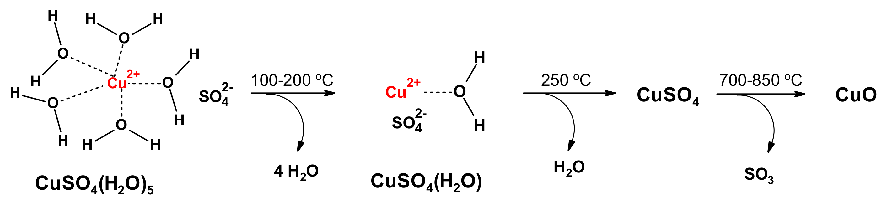
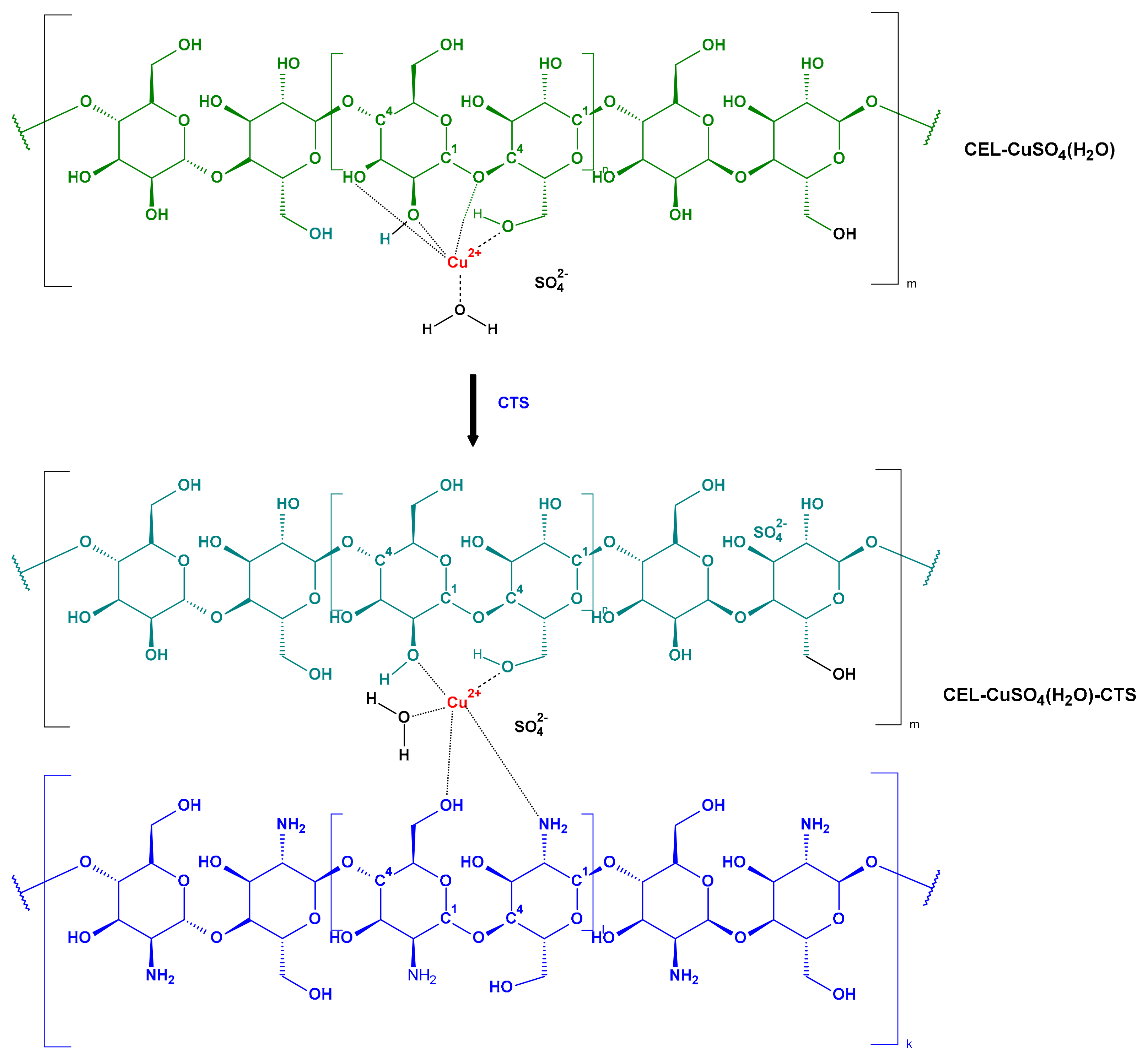
3.1. Preparation of Composite Materials

In this study, two copper-modified cotton materials, designated as COT-CuSO4 and/or COT-CuSO4/CTS, were developed. The samples COT-CuSO4 were obtained by chemisorption of copper sulfate on cotton cellulose (COT→COT-CuSO4⋅5H2O), followed by gradual thermal dehydration of intermediary COT-CuSO4⋅5H2O to the more stable form COT-CuSO4⋅H2O (COT-CuSO4⋅5H2O→COT-CuSO4⋅H2O.
The samples COT-CuSO4⋅H2O treated with chitosan form COT-CuSO4⋅H2O/CTS composites.

Preparation of COT-CuSO4

Taking into account the thermal instability of copper sulfate hydrates (Figure 1) [74], and also that water competes with hydroxyls of cellulose in copper chelation, we assumed that the stability of the cellulose–copper cation should increase after thermal exposure of the COT-CuSO4⋅5H2O material.
The putative reaction schemes of preparation of both composite materials are proposed on Figure 2 and Figure 3, respectively.
The results of copper concentration in the COT-CuSO4 and COT-CuSO4/CTS materials are presented in Table 1.

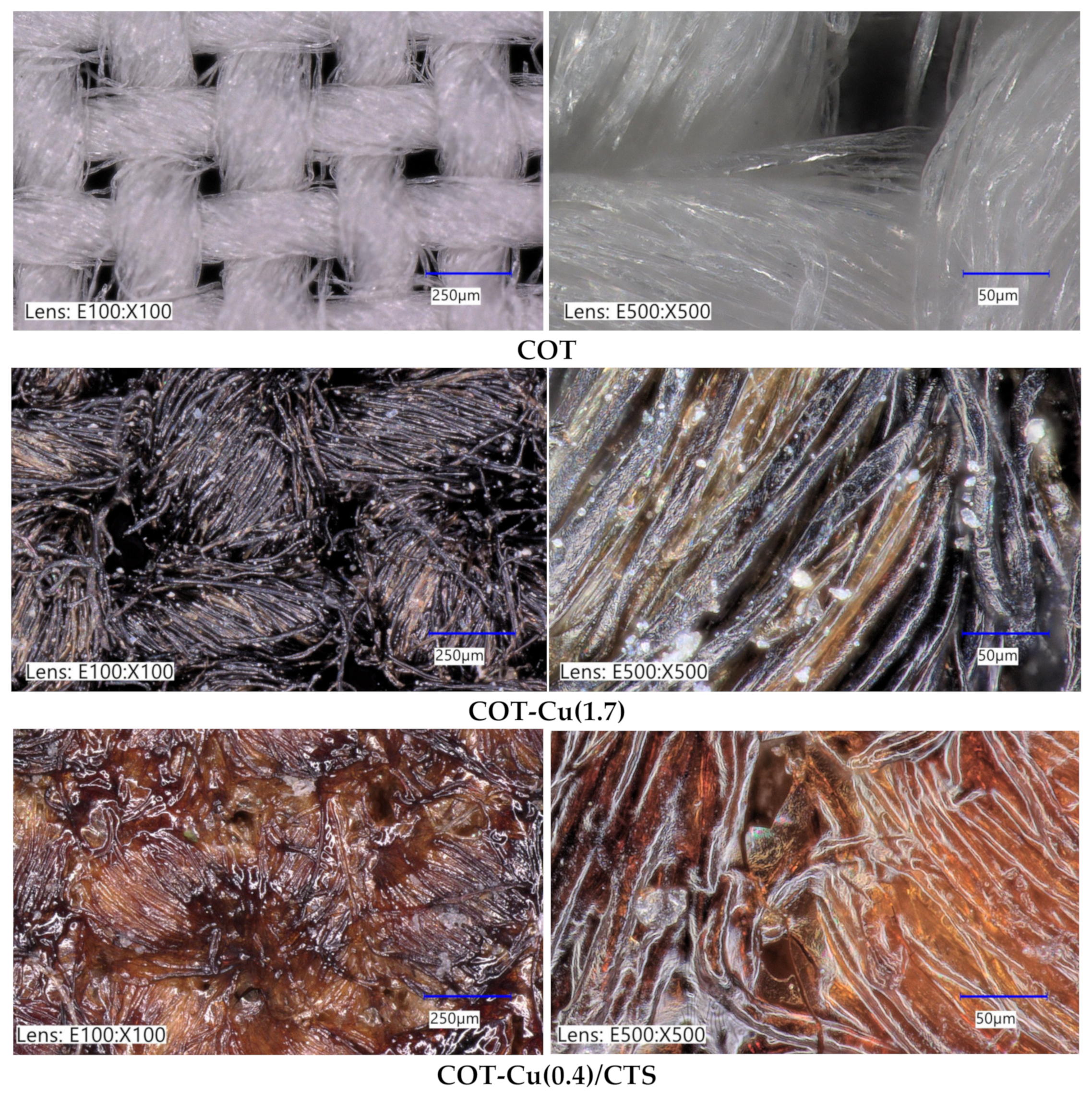
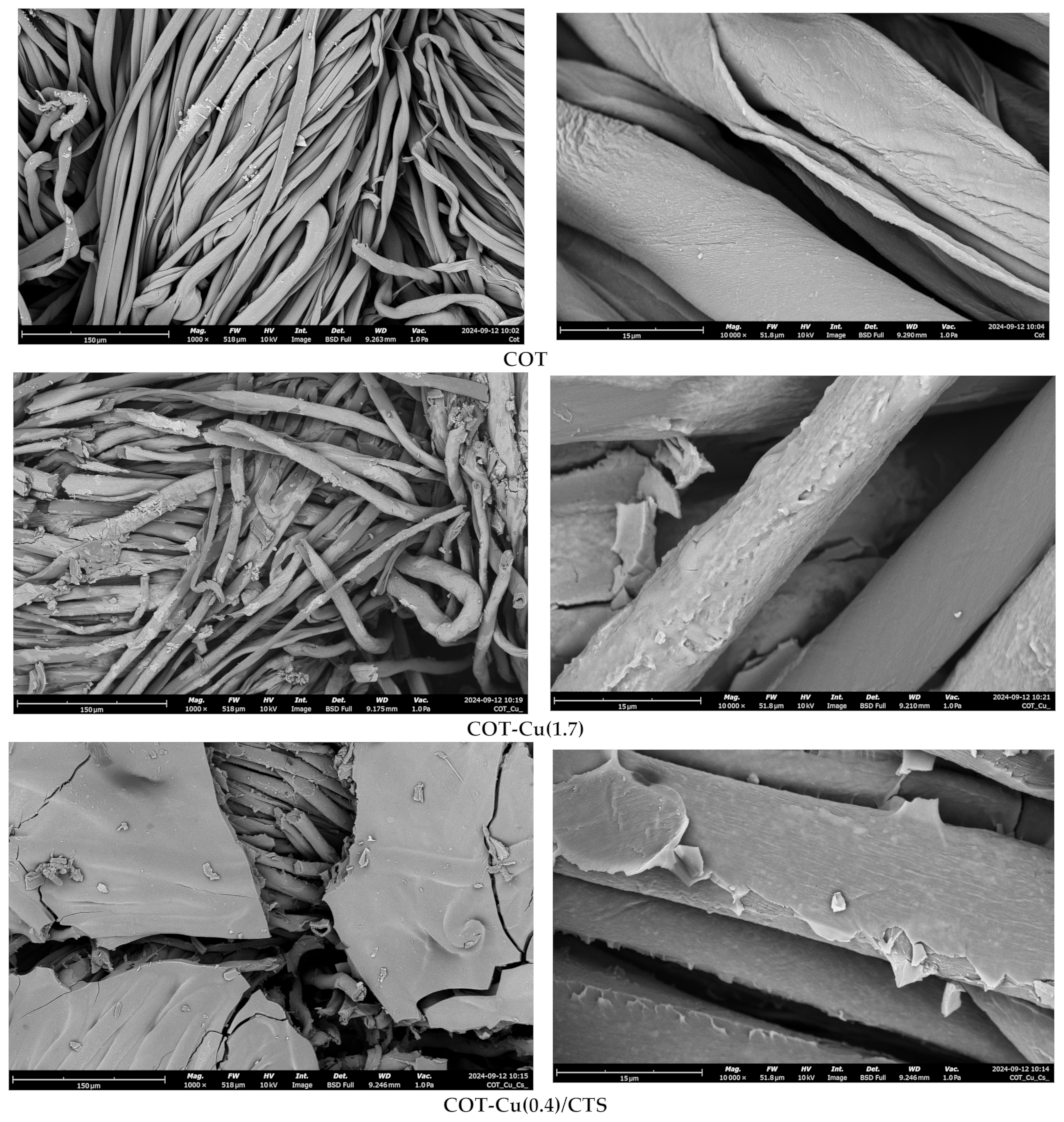
3.2. Optical and SEM Analysis

Microscopic examination of the COT, COT-Cu(1.7), and COT-Cu(0.4)/CTS samples reveals significant morphological differences, resulting from the chemical and thermal treatments applied to the materials. The images in Figure 4 illustrate these changes.
In the initial set of images, the cotton appears in its unaltered state. At lower magnification, the structure of the cotton fibers is clearly visible, displaying smooth, evenly arranged fibers characteristic of pure cotton. At higher magnification, the fibers exhibit clear, slender bands without additional surface features, indicating that the cotton has not undergone any modifications. The absence of surface alterations or impregnation confirms the material’s purity, retaining the typical structure of untreated cotton.
The introduction of chitosan leads to significant morphological alterations in the cotton fibers, forming a thin coating that contributes to a more complex structural configuration.
Analysis of the scanning electron microscopy (SEM) images reveals marked structural changes, highlighting the impact of the applied chemical and thermal modifications. The observed variations in fiber morphology across these samples directly result from these specific treatments, as illustrated in Figure 5.
The SEM image of the untreated cotton sample reveals fibers with a predominantly smooth and uniform surface, illustrating the orderly structure characteristic of raw cotton. The fibers appear densely packed, aligning with the expected traits of unmodified cotton. At higher magnification, finer surface details are observable, yet the fibers maintain a smooth texture with no evident chemical or structural alterations. The well-organized, smooth appearance confirms the material’s untreated state, with no visible deposits or signs of chemical modification or thermal degradation. The SEM images of the COT-Cu(1.7) fibers display notable structural modifications, characterized by a visible surface coating. The fibers exhibit a rough texture with numerous microscopic formations. The SEM images of COT-Cu(0.4)/CTS fibers reveal the presence of a distinct chitosan layer, serving as a stabilizing matrix for the fibers. This coating forms a smooth, uniform layer that adheres closely to the cotton surface, as observed at the highest magnifications. The chitosan layer enhances the durability and stability of the fibers, acting as a protective barrier that reduces fiber degradation while stabilizing the copper deposits. The incorporation of chitosan into COT-Cu(1.7) (COT-Cu(1.7)→COT-Cu(0.4)/CTS) fibers was specifically intended to stabilize copper(II) oxide and enhance the cotton’s mechanical and chemical resilience. As a natural polysaccharide with chelating properties, chitosan effectively complexes copper ions, forming a thin yet robust layer on the fiber surface.

3.3. EDS Analysis

Energy-dispersive X-ray spectroscopy (EDS) analysis highlights differences in the chemical composition of the tested materials, directly resulting from the distinct chemical and thermal modifications applied to each sample.
Table 2 presents EDS data showing key differences in the chemical composition of untreated and modified cotton samples. The untreated cotton sample (COT) is primarily composed of carbon and oxygen, with atomic concentrations of 41.81% and 58.19%, respectively. This composition aligns with the structure of cellulose, the main component of cotton fibers. The homogeneity of untreated cotton and the absence of additional elements suggest limited antimicrobial activity due to the lack of functional additives.
The EDS data confirm the presence of copper and sulfur, signifying their successful integration into the fiber matrix.
The COT-Cu(0.4)/CTS sample, which includes chitosan, displays further changes in chemical composition. Chitosan, a natural polysaccharide, increases the carbon content and introduces nitrogen (5.67%) into the fiber matrix, forming a protective coating around the cotton fibers. This layer not only modifies the chemical profile of the material but also reduces sulfur and copper levels on the fiber surface. Additionally, chitosan’s chelation of copper ions likely regulates copper release, contributing to enhanced stability and durability of the material’s antimicrobial properties.

3.4. UV-VIS Analysis and UV Protection Assessment

Protecting the skin from solar radiation is essential to reducing the detrimental impact of ultraviolet (UV) exposure. UV radiation, spanning UVA and UVB wavelengths, accelerates skin aging, triggers oxidative stress, and induces direct DNA damage within epidermal cells. Cumulatively, these effects contribute to various skin disorders, with the most serious being skin carcinogenesis, including melanoma as its most aggressive form [78]. Utilizing specialized materials, such as UV-protective textiles, enhances the skin’s natural defense by providing a physical barrier that significantly limits the penetration of harmful rays into deeper skin layers.
The ultraviolet protection factor (UPF) quantifies a fabric’s effectiveness in shielding against UV radiation, as outlined in the PN-EN 13758-1:2007 standard [69]. In this assessment, fabrics were subjected to UVA and UVB radiation. The results demonstrate that fabric color significantly influences UPF values, with darker shades—such as black, blue, and dark green—absorbing more UV radiation and thus exhibiting higher UPF ratings [79]. A higher UPF index corresponds to enhanced protection, as presented in Table 3.
Table 4 presents the UVA, UVB, and UPF values obtained from tests on COT-Cu(1.7), COT-Cu(0.4)/CTS, and pure cotton (COT), in accordance with the PN-EN ISO 13758-1+A1:2007 standard [69], along with a comparison to other materials exhibiting UV protective properties. The results demonstrate that the addition of a chitosan layer increased the UPF value from 3.37 to >50. Notably, the Cot-Cu sample exhibited moderate UPF values (UPF = 14.56), while untreated medical cotton displayed significantly lower UPF values of 3.37. The UPF values were determined based on transmission measurements within the range of λ = 290–400 nm. According to the standard, a UPF rating of ≥40 signifies excellent protection, indicating that the fabric blocks 97.5% of UV radiation.

3.5. Results of Biological Activity of Materials

3.5.1. Activity Against Microorganisms

Considering the critical importance of safety and hygiene in various material applications, including protective clothing, medical, and sanitary equipment, it is essential to assess the effectiveness of materials in eliminating microorganisms. Table 5 presents the average microbial inhibition zone values for the tested materials (COT, COT-Cu(1.7), and COT-Cu(0.4)/CTS) along with comparative data for other materials demonstrating antimicrobial activity. The results clearly indicate that untreated cotton (Cot) does not exhibit any antimicrobial properties against the tested microorganisms. No inhibition zones were observed, suggesting that the untreated cotton is ineffective in preventing microbial growth.
The COT-Cu(1.7) material exhibited a inhibition zone, highlighting the efficacy of copper in imparting both antibacterial and antifungal properties. The modified cotton demonstrated moderate antimicrobial activity, with inhibition zones measuring approximately 3 mm for both E. coli (Gram-negative) and S. aureus (Gram-positive) bacteria (Figure 6). In comparison, the inhibition zones for the fungi A. niger and Ch. globosum were slightly smaller, around 2 mm. Copper, known for its antimicrobial properties, likely interferes with critical cellular processes in microorganisms, leading to the suppression of their growth.
The incorporation of a chitosan layer (Cot-Cu(0.4)/Cs) did not result in a significant enhancement of antimicrobial activity. While chitosan is recognized for its antimicrobial properties and could theoretically potentiate the effect of copper, its impact in this formulation appears to be minimal. The reduced antimicrobial efficacy may stem from the differing mechanisms of action between copper and chitosan. Copper primarily disrupts cellular functions by damaging cell membranes and interfering with key enzymatic processes [90]. Conversely, chitosan forms a protective coating, which may limit the direct interaction between microorganisms and copper, thereby reducing microbial exposure to copper ions. This interaction could explain the observed decrease in the overall antimicrobial effectiveness of copper in the presence of chitosan.

3.5.2. Evaluation of Activated aPTT and PT

The parameters aPTT and PT are fundamental for evaluating the impact of materials on the coagulation system, offering critical insights into the interactions between biomaterials and blood. Figure S3 illustrates the influence of the tested materials on aPTT time and PT time. Table 5 presents results for the materials under study (COT, COT-Cu(1.7), and COT-Cu(0.4)/CTS), alongside comparative data from other materials known to affect coagulation processes. Analysis of these results reveals distinct differences in the coagulation properties of the materials following various modification processes. These modifications directly influence blood coagulation parameters, including activated partial thromboplastin time (aPTT) and prothrombin time (PT), highlighting the variable impact of each treatment on coagulation activity.
The base material, pure medical cotton (Cot), displays an average aPTT of 34.92 s and PT of 13.53 s, values that align closely with physiological norms, reflecting stable material characteristics. The low standard deviation for both parameters (0.38 for aPTT and 0.3 for PT) indicates minimal variability and high reproducibility, underscoring the consistency of these results. Pure cotton does not significantly affect the clotting process, affirming its biocompatibility and suitability for applications involving blood contact. Control sample (C) results exhibit slightly lower mean aPTT and PT values compared to pure cotton, though variability within this group does not substantially impact the observed properties, confirming consistent material behavior across tests.
The COT-Cu(1.7) material notably prolongs clotting time, with an average aPTT of 71.30 s, exceeding twice the value observed for unmodified cotton. Likewise, PT is elevated, reaching an average of 16.33 s. The COT-Cu(0.4)/CTS material, featuring an additional chitosan coating, exhibited an intermediate aPTT value of 40.65 s, positioning it between that of unmodified cotton and Cot-Cu. However, aPTT remains extended compared to untreated cotton, suggesting that while chitosan moderates copper’s impact, it does not fully eliminate its influence on coagulation. The PT value of 14.27 s reflects a similarly intermediate effect, underscoring the limited but persistent impact of the combined chitosan and copper modification on clotting dynamics (Table 6).

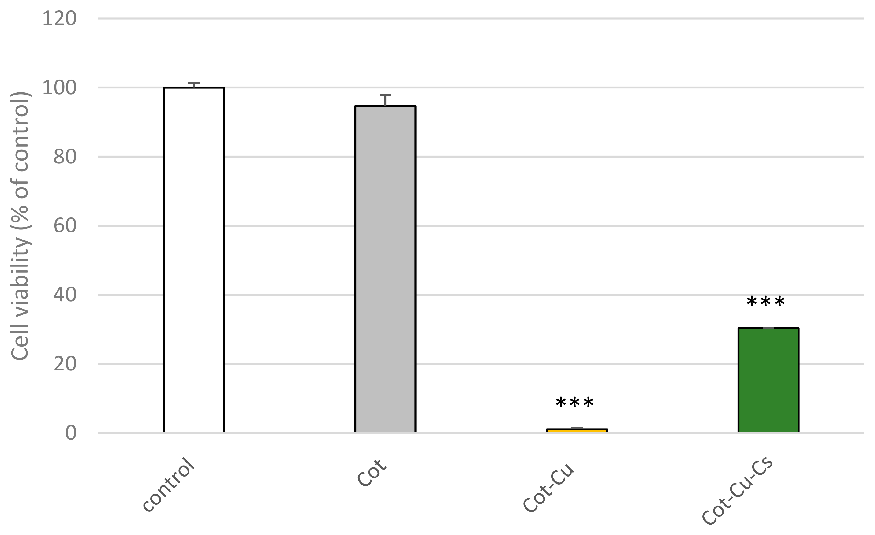
3.5.3. Effect of Cotton Samples on the Viability of PBM Cells

We employed the resazurin reduction assay to assess cell viability following incubation with post-incubation mixtures of Cot materials. This assay utilizes an indicator dye to monitor oxidation-reduction reactions, which primarily take place in the mitochondria of living cells. The non-fluorescent dark blue dye, resazurin, is reduced by metabolically active cells, transforming into a fluorescent pink form at 570 nm and a fluorescent red form at neutral pH (resorufin). We demonstrated that exposure to post-incubation mixtures of cotton affects the cell viability of PBM cells (Figure 7). Cotton alone did not decrease cell viability, and the results are similar to the control. However, in the case of COT-Cu(1.7) and COT-Cu(0.4)/CTS, we noticed a decrease in cell viability to 5% in the case of Cot-Cu and to about 30% in the case of COT-Cu(0.4)/CTS. These results suggest cytotoxic potential in both cotton fabrics containing copper. Our findings suggest that copper, ubiquitously existing in textile materials, induces a Fenton reaction [93]. During the chemical process, extremely reactive hydroxyl radicals may be formed, exhibiting a destructive capacity towards biomolecules such as deoxyribonucleic acid, protein, or lipids [94,95].

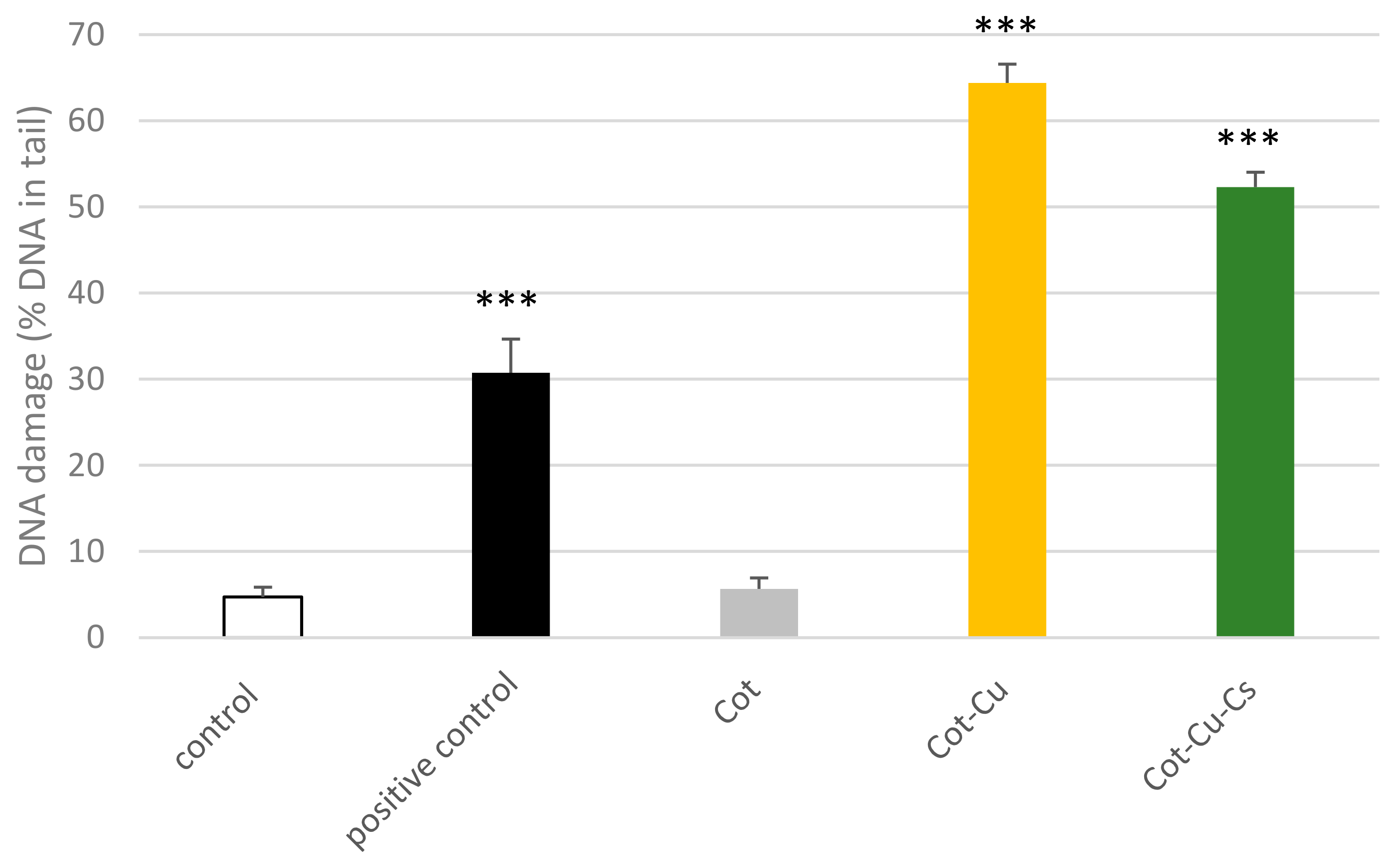
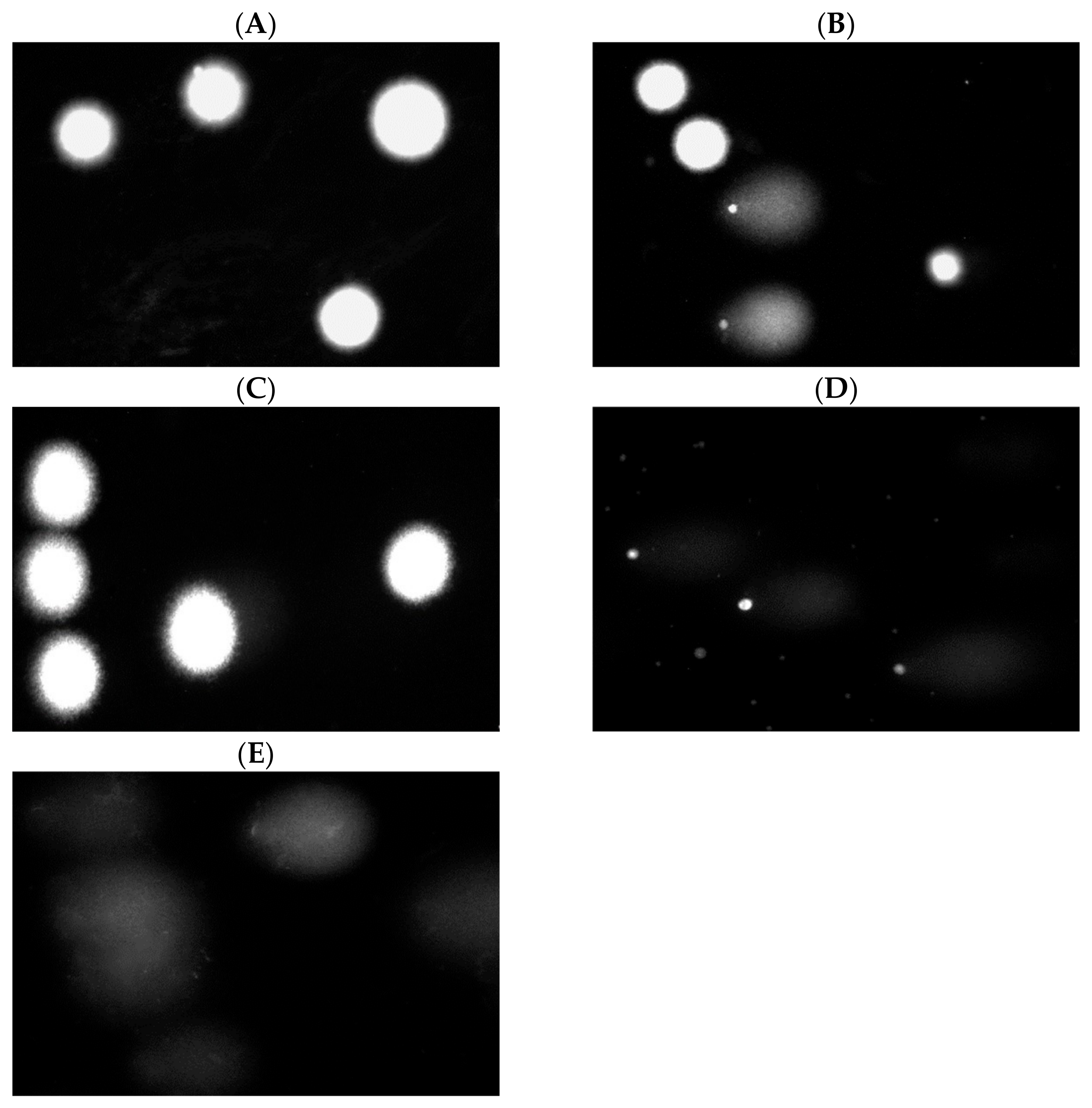
3.5.4. Effect of Cotton Samples on DNA Damage

The alkaline version of the comet assay is a straightforward and highly sensitive technique for assessing DNA damage, including single- and double-strand breaks, as well as alkali-labile sites in live cells [96]. We observed significant DNA damage in PBM cells treated with 25 µM H2O2 (positive control). We did not observe DNA damage in the case of cotton alone. Both COT-Cu(1.7) and COT-Cu(0.4)/CTS samples exhibit a high potential for the induction of DNA damage in PBM cells (Figure 8). Our results confirm images of cell nuclei from the fluorescence microscope (Figure 9). Copper can cause DNA damage [97,98]. The main genotoxic effects seen with COT-Cu(1.7) and COT-Cu(0.4)/CTS indicate the release of copper to the culture medium and induction of DNA damage.

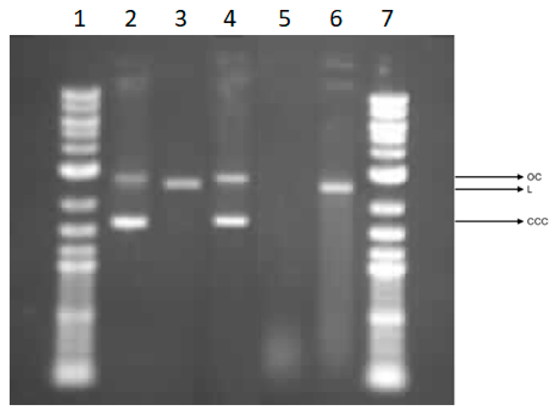
3.5.5. Effect of Cotton Samples on Plasmid DNA

We explored the possibility of direct interaction between COT post-incubation mixtures and plasmid DNA. To facilitate this analysis, we utilized a plasmid relaxation assay (Figure 10). Electrophoretic mobility shift assay (EMSA) results indicated that the isolated pUC19 plasmid from DH5α E. coli predominantly exists in a supercoiled (CCC) form. A small amount of the OC (open circular) form of the plasmid is also visible in the sample (Figure 10, line 2). Overnight treatment at 37 °C with restrictase PstI led to a linear form (L) of the plasmid (Figure 10, line 3). We incubated plasmid DNA with post-incubation mixtures of cotton materials for 24 h at 37 °C. Cotton alone did not interact with plasmid DNA and the results are similar to the negative control (Figure 10, line 4). COT-Cu(1.7) induces severe DNA damage leading to the degradation of plasmid DNA to smear (Figure 10, line 5). COT-Cu(0.4)/CTS) induced a linear form of plasmid and smear, however, to a lesser extent than COT-Cu(1.7) (Figure 10, line 6). Studies have demonstrated that copper, particularly Cu2+, can interact with DNA [99,100]. Moreover, copper ions are capable of generating ROS via the Fenton reaction, leading to the production of highly reactive hydroxyl radicals [101].

4. Conclusions

  • Enhanced Antimicrobial Properties: The modification of medical cotton fibers with copper sulfate and chitosan, followed by thermal processing, enhances the material’s antimicrobial effectiveness, positioning it as a promising candidate for advanced medical textiles. This modified cotton is highly suitable for healthcare and hygiene applications due to its improved functional properties.
  • Chemical and Structural Analysis: Detailed analyses of the chemical composition and structural properties of the modified fibers were conducted, employing methods such as flame atomic absorption spectrometry (FAAS); SEM; EDS, etc.
  • Impact on Blood Coagulation: Thia study thoroughly examined the biochemical characteristics of the modified material, particularly regarding its effects on hemostatic processes. Blood coagulation effects were evaluated through measurements of activated partial thromboplastin time (aPTT) and prothrombin time (PT), offering insights into the modified cotton’s interactions with blood plasma clotting mechanisms. Thermal reduction of copper(II) sulfate on the cotton surface to copper(II) oxide resulted in notable changes in blood clotting, specifically extending both aPTT and PT times due to the anticoagulant properties of copper. The addition of a chitosan layer on the modified cotton surface reduced the anticoagulant effect, though a prolongation of clotting time persisted when compared to unmodified cotton.
  • Cytotoxic and Genotoxic Effects: Pure cotton did not reduce cell viability, cause DNA damage, or interact with plasmid DNA, demonstrating a benign profile for cellular integrity. In contrast, copper-modified cotton COT-Cu(1.7) and copper–chitosan-modified cotton (COT-Cu(0.4)/CTS) showed potential to induce cyto- and genotoxicity in peripheral blood mononuclear (PBM) cells, with COT-Cu(0.4)/CTS exhibiting lower cytotoxic and genotoxic effects than COT-Cu(1.7). COT-Cu(0.4)/CTS fabrics thus demonstrate decreased cyto- and genotoxic potential, suggesting chitosan may mitigate some cytotoxic properties associated with copper modification.
These findings collectively underscore the potential of copper ions and chitosan-modified cotton as a functional biomaterial, particularly in medical applications requiring the precise control of antimicrobial activity and blood clotting properties.

Supplementary Materials

The following supporting information can be downloaded at: https://www.mdpi.com/article/10.3390/pr12122772/s1, Figure S1: Energy dispersive X-ray spectroscopy (EDS) diagrams are from three separate and independent runs. Selected representative results from these runs were selected for detailed analysis and are presented here; Figure S2: Comparison of transmittance spectra (%T) of Cot; Cot-Cu and Cot-Cu-Cs, recorded in the range: λ = 200–800 nm; Figure S3: Influence of tested materials on (a) aPTT time; (b) PT time. Results are presented as mean (×), range (bars), median (line), and interquartile range (box). The study included three experimental runs, with significant findings selected for detailed analysis.

Author Contributions

M.Ś. developed the concept and designed experiments, performed experiments, analyzed data, and wrote the paper; Z.M. performed experiments and analyzed data; M.J. performed experiments and analyzed data; K.W. analyzed data; M.H.K. developed the concept and designed experiments, analyzed data, and wrote the paper. All authors have read and agreed to the published version of the manuscript.

Funding

This research was partly carried out within the National Science Centre (Poland), project M-ERA.NET 2022, No. 2022/04/Y/ST4/00157.

Data Availability Statement

The original contributions presented in the study are included in the article, further inquiries can be directed to the corresponding author.

Acknowledgments

In memory of the Krystyna Pietrucha.

Conflicts of Interest

The authors declare no conflicts of interest.

References

  1. Kudzin, M.H.; Giełdowska, M.; Król, P.; Sobanska, Z. Preparation of Cotton–Zinc Composites by Magnetron Sputtering Metallization and Evaluation of their Antimicrobial Properties and Cytotoxicity. Materials 2022, 15, 2746. [Google Scholar] [CrossRef] [PubMed]
  2. Kudzin, M.H.; Giełdowska, M.; Krata, A.A.; Sulak, E.; Urbaniak, P.; Drabowicz, J. Phosphorylation of Chitosan (Chitin) Surface with PCl3. Phosphorus Sulfur Silicon Relat. Elem. 2022, 197, 625–629. [Google Scholar] [CrossRef]
  3. Klemm, D.; Heublein, B.; Fink, H.P.; Bohn, A. Cellulose: Fascinating biopolymer and sustainable raw material. Angew. Chem. Int. Ed. 2005, 44, 3358–3393. [Google Scholar] [CrossRef]
  4. Klemm, D.; Kramer, F.; Moritz, S.; Lindstrom, T.; Ankerfors, M.; Gray, D.; Dorris, A. Nanocelluloses: A new Family of Nature-Based Materials. Angew. Chem. Int. Ed. 2011, 50, 5438–5466. [Google Scholar] [CrossRef]
  5. Rojas, O.J. (Ed.) Cellulose Chemistry and Properties: Fibers, Nanocelluloses and Advanced Materials; Part of the book series: Advances in Polymer Science; Springer: Berlin/Heidelberg, Germany, 2016; Volume 271, 341p. [Google Scholar] [CrossRef]
  6. Mahbubul Bashar, M.; Khan, M.A. An Overview on Surface Modification of Cotton Fiber for Apparel Use. J. Polym. Environ. 2013, 21, 181–190. [Google Scholar] [CrossRef]
  7. Ahmed, F.; Mondal, M.d.I.H. 1—Introduction to Natural Fibres and Textiles. In Fundamentals of Natural Fibres and Textiles; Mondal, M.d.I.H., Ed.; The Textile Institute Book Series; Woodhead Publishing: Cambridge, UK, 2021; pp. 1–32. ISBN 978-0-12-821483-1. [Google Scholar]
  8. Lam, Y.-L.; Kan, C.-W.; Yuen, C.-W.M. Developments in Functional Finishing of Cotton Fibres—Wrinkle-Resistant, Flame-Retardant and Antimicrobial Treatments. Text. Prog. 2012, 44, 175–249. [Google Scholar] [CrossRef]
  9. Bhuiyan, M.A.R.; Shaid, A.; Khan, M.A. Cationization of Cotton Fiber by Chitosan and Its Dyeing with Reactive Dye without Salt. Chem. Mater. Eng. Cease Publ. 2014, 2, 96–100. [Google Scholar] [CrossRef]
  10. Gao, D.; Li, X.; Li, Y.; Lyu, B.; Ren, J.; Ma, J. Long-Acting Antibacterial Activity on the Cotton Fabric. Cellulose 2021, 28, 1221–1240. [Google Scholar] [CrossRef]
  11. Borkow, G.; Gabbay, J. Biocidal Textiles Can Help Fight Nosocomial Infections. Med. Hypotheses 2008, 70, 990–994. [Google Scholar] [CrossRef]
  12. Halepoto, H.; Gong, T.; Memon, H. A Bibliometric Analysis of Antibacterial Textiles. Sustainability 2022, 14, 11424. [Google Scholar] [CrossRef]
  13. Rahman Bhuiyan, M.A.; Hossain, M.A.; Zakaria, M.; Islam, M.N.; Zulhash Uddin, M. Chitosan Coated Cotton Fiber: Physical and Antimicrobial Properties for Apparel Use. J. Polym. Environ. 2017, 25, 334–342. [Google Scholar] [CrossRef]
  14. Harugade, A.; Sherje, A.P.; Pethe, A. Chitosan: A Review on Properties, Biological Activities and Recent Progress in Biomedical Applications. React. Funct. Polym. 2023, 191, 105634. [Google Scholar] [CrossRef]
  15. Desai, N.; Rana, D.; Salave, S.; Gupta, R.; Patel, P.; Karunakaran, B.; Sharma, A.; Giri, J.; Benival, D.; Kommineni, N. Chitosan: A Potential Biopolymer in Drug Delivery and Biomedical Applications. Pharmaceutics 2023, 15, 1313. [Google Scholar] [CrossRef] [PubMed]
  16. Ahmed, M.E.; Mohamed, M.I.; Ahmed, H.Y.; Elaasser, M.M.; Kandile, N.G. Fabrication and Characterization of Unique Sustain Modified Chitosan Nanoparticles for Biomedical Applications. Sci. Rep. 2024, 14, 13869. [Google Scholar] [CrossRef]
  17. Mawazi, S.M.; Kumar, M.; Ahmad, N.; Ge, Y.; Mahmood, S. Recent Applications of Chitosan and Its Derivatives in Antibacterial, Anticancer, Wound Healing, and Tissue Engineering Fields. Polymers 2024, 16, 1351. [Google Scholar] [CrossRef]
  18. Aranaz, I.; Alcántara, A.R.; Civera, M.C.; Arias, C.; Elorza, B.; Heras Caballero, A.; Acosta, N. Chitosan: An Overview of Its Properties and Applications. Polymers 2021, 13, 3256. [Google Scholar] [CrossRef]
  19. Li, B.; Elango, J.; Wu, W. Recent Advancement of Molecular Structure and Biomaterial Function of Chitosan from Marine Organisms for Pharmaceutical and Nutraceutical Application. Appl. Sci. 2020, 10, 4719. [Google Scholar] [CrossRef]
  20. Doyo, A.N.; Kumar, R.; Barakat, M.A. Recent Advances in Cellulose, Chitosan, and Alginate Based Biopolymeric Composites for Adsorption of Heavy Metals from Wastewater. J. Taiwan Inst. Chem. Eng. 2023, 151, 105095. [Google Scholar] [CrossRef]
  21. Alves, N.M.; Mano, J.F. Chitosan Derivatives Obtained by Chemical Modifications for Biomedical and Environmental Applications. Int. J. Biol. Macromol. 2008, 43, 401–414. [Google Scholar] [CrossRef]
  22. Kou, S.; Peters, L.; Mucalo, M. Chitosan: A Review of Molecular Structure, Bioactivities and Interactions with the Human Body and Micro-Organisms. Carbohydr. Polym. 2022, 282, 119132. [Google Scholar] [CrossRef]
  23. da Silva, D.J.; Ferreira, R.R.; da S. Ferreira, G.; Barbosa, R.F.S.; Marciano, J.S.; Camani, P.H.; Souza, A.G.; Rosa, D.S. Multifunctional Cotton Fabrics with Novel Antibacterial Coatings Based on Chitosan Nanocapsules and Polyacrylate. J. Coat. Technol. Res. 2023, 20, 1541–1555. [Google Scholar] [CrossRef] [PubMed]
  24. Li, J.; Zhuang, S. Antibacterial Activity of Chitosan and Its Derivatives and Their Interaction Mechanism with Bacteria: Current State and Perspectives. Eur. Polym. J. 2020, 138, 109984. [Google Scholar] [CrossRef]
  25. Ke, C.-L.; Deng, F.-S.; Chuang, C.-Y.; Lin, C.-H. Antimicrobial Actions and Applications of Chitosan. Polymers 2021, 13, 904. [Google Scholar] [CrossRef] [PubMed]
  26. Yilmaz Atay, H. Antibacterial Activity of Chitosan-Based Systems. In Functional Chitosan: Drug Delivery and Biomedical Applications Chitosan; Springer: Berlin/Heidelberg, Germany, 2020; pp. 457–489. [Google Scholar] [CrossRef]
  27. Liu, X.D.; Nishi, N.; Tokura, S.; Sakairi, N. Chitosan Coated Cotton Fiber: Preparation and Physical Properties. Carbohydr. Polym. 2001, 44, 233–238. [Google Scholar] [CrossRef]
  28. Xiao, Y.; Shen, G.; Zheng, W.; Fu, J.; Fu, F.; Hu, X.; Jin, Z.; Liu, X. Remarkable Durability of the Antibacterial Function Achieved via a Coordination Effect of Cu(II) Ion and Chitosan Grafted on Cotton Fibers. Cellulose 2022, 29, 1003–1015. [Google Scholar] [CrossRef]
  29. Verma, C.; Quraishi, M.A. Chelation Capability of Chitosan and Chitosan Derivatives: Recent Developments in Sustainable Corrosion Inhibition and Metal Decontamination Applications. Curr. Res. Green Sustain. 2021, 4, 100184. [Google Scholar] [CrossRef]
  30. Wang, W.; Xue, C.; Mao, X. Chitosan: Structural Modification, Biological Activity and Application. Int. J. Biol. Macromol. 2020, 164, 4532–4546. [Google Scholar] [CrossRef]
  31. Gamage, A.; Jayasinghe, N.; Thiviya, P.; Wasana, M.L.D.; Merah, O.; Madhujith, T.; Koduru, J.R. Recent Application Prospects of Chitosan Based Composites for the Metal Contaminated Wastewater Treatment. Polymers 2023, 15, 1453. [Google Scholar] [CrossRef]
  32. Hassabo, A.G.; El-Naggar, M.E.; Mohamed, A.L.; Hebeish, A.A. Development of Multifunctional Modified Cotton Fabric with Tri-Component Nanoparticles of Silver, Copper and Zinc Oxide. Carbohydr. Polym. 2019, 210, 144–156. [Google Scholar] [CrossRef]
  33. Xu, Q.; Ke, X.; Ge, N.; Shen, L.; Zhang, Y.; Fu, F.; Liu, X. Preparation of Copper Nanoparticles Coated Cotton Fabrics with Durable Antibacterial Properties. Fibers Polym. 2018, 19, 1004–1013. [Google Scholar] [CrossRef]
  34. Anita, S.; Ramachandran, T.; Rajendran, R.; Koushik, C.; Mahalakshmi, M. A Study of the Antimicrobial Property of Encapsulated Copper Oxide Nanoparticles on Cotton Fabric. Text. Res. J. 2011, 81, 1081–1088. [Google Scholar] [CrossRef]
  35. Qian, J.; Dong, Q.; Chun, K.; Zhu, D.; Zhang, X.; Mao, Y.; Culver, J.N.; Tai, S.; German, J.R.; Dean, D.P.; et al. Highly Stable, Antiviral, Antibacterial Cotton Textiles via Molecular Engineering. Nat. Nanotechnol. 2023, 18, 168–176. [Google Scholar] [CrossRef] [PubMed]
  36. Devaraji, M.; Thanikachalam, P.V.; Elumalai, K. The potential of Copper Oxide Nanoparticles in Nanomedicine: A Comprehensive Review. Biotechnol. Notes 2024, 5, 80–99. [Google Scholar] [CrossRef] [PubMed]
  37. Parham, S.; Wicaksono, D.H.B.; Bagherbaigi, S.; Lee, S.L.; Nur, H. Antimicrobial Treatment of Different Metal Oxide Nanoparticles: A Critical Review. J. Chin. Chem. Soc. 2016, 63, 385–393. [Google Scholar] [CrossRef]
  38. Morones, J.R.; Elechiguerra, J.L.; Camacho, A.; Holt, K.; Kouri, J.B.; Ramírez, J.T.; Yacaman, M.J. The Bactericidal Effect of Silver Nanoparticles. Nanotechnology 2005, 16, 2346. [Google Scholar] [CrossRef]
  39. Agrawal, N.; Low, P.S.; Tan, J.S.J.; Fong, E.W.M.; Lai, Y.; Chen, Z. Durable Easy-Cleaning and Antibacterial Cotton Fabrics Using Fluorine-Free Silane Coupling Agents and CuO Nanoparticles. Nano Mater. Sci. 2020, 2, 281–291. [Google Scholar] [CrossRef]
  40. Belcastro, S.; Staffolani, N.; Pugliese, M.; D’Alo, F. An in-vitro Study of the Antimicrobial Activity of Copper and Zinc Salts on Pure and Mixed Microbial Cultures. Minerva Stomatol. 1994, 43, 393–396. [Google Scholar]
  41. Ghoranneviss, M.; Shahidi, S. Effect of Various Metallic Salts on Antibacterial Activity and Physical Properties of Cotton Fabrics. J. Ind. Text. 2013, 42, 193–203. [Google Scholar] [CrossRef]
  42. Ning, C.; Wang, X.; Li, L.; Zhu, Y.; Li, M.; Yu, P.; Zhou, L.; Zhou, Z.; Chen, J.; Tan, G.; et al. Concentration Ranges of Antibacterial Cations for Showing the Highest Antibacterial Efficacy but the Least Cytotoxicity Against Mammalian Cells: Implications for a New Antibacterial Mechanism. Chem. Res. Toxicol. 2015, 28, 1815–1822. [Google Scholar] [CrossRef]
  43. Cheknev, S.B.; Vostrova, E.I.; Apresova, M.A.; Piskovskaya, L.S.; Vostrov, A.V. Deceleration of Bacterial Growth in Staphylococcus Aureus and Pseudomonas Aeruginosa Cultures in the Presence of Copper and Zinc Cations. Zh. Mikrobiol. Epidemiol. Immunobiol. 2015, 2, 9–17, PMID: 26016338. [Google Scholar] [PubMed]
  44. Cheknev, S.V.; Vostrova, E.L.; Sarycheva, M.A.; Vostrov, A.V. Inhibition of Growth of Bacteria in Staphylococcus Aureus and Pseudomonasaeruginosa Cultures by Copper and Zinc Cations, Applied at Physiological Concentrations. Zh. Mikrobiol. Epidemiol. Immunobiol. 2016, 3, 9–18, PMID: 30695447. [Google Scholar] [PubMed]
  45. Febré, N.; Silva, V.; Báez, A.; Palza, H.; Delgado, K.; Aburto, I.; Silva, V. Antibacterial Activity of Copper Salts Against Microorganisms Isolated from Chronic Infected Wounds. Rev. Medica Chile 2016, 144, 1523–1530. [Google Scholar] [CrossRef] [PubMed]
  46. Fowler, L.; Engqvist, H.; Öhman-Mägi, C. Effect of Copper Ion Concentration on Bacteria and Cells. Materials 2019, 12, 3798. [Google Scholar] [CrossRef] [PubMed]
  47. Benhalima, L.; Amri, S.; Bensouilah, M.; Ouzrout, R. Antibacterial Effect of Copper Sulfate against Multi-Drug Resistant Nosocomial Pathogens Isolated from Clinical Samples. Pak. J. Med. Sci. 2019, 35, 1322–1328. [Google Scholar] [CrossRef]
  48. Phan, D.N.; Dorjjugder, N.; Saito, Y.; Qamar Khan, M.Q.; Ullah, A.; Bie, X.; Taguchi, G.; Kim, I.S. Antibacterial Mechanisms of Various Copper Species Incorporated in Polymeric Nanofibers against Bacteria. Mater. Today Commun. 2020, 25, 101377. [Google Scholar] [CrossRef]
  49. Popov, S.; Saphier, O.; Popov, M.; Shenker, M.; Entus, S.; Shotland, Y.; Saphier, M. Factors Enhancing the Antibacterial Effect of Monovalent Copper Ions. Curr. Microbiol. 2020, 77, 361–368. [Google Scholar] [CrossRef]
  50. Pereira, F.S.; de Souza, G.G.; Moraes, P.G.P.; Barroso, R.P.; Lanfredi, S.; Gomes, H.M.; Costa-Filho, A.J.; González, E.R.P. Study of Chitosans Interaction with Cu(II) from the Corresponding Sulfate and Chloride Salts. Cellulose 2015, 22, 2391–2407. [Google Scholar] [CrossRef]
  51. Kudzin, M.H.; Boguń, M.; Mrozińska, Z.; Kaczmarek, A. Physical Properties, Chemical Analysis, and Evaluation of Antimicrobial Response of New Polylactide/Alginate/Copper Composite Materials. Mar. Drugs 2020, 18, 660. [Google Scholar] [CrossRef]
  52. Kudzin, M.H.; Kaczmarek, A.; Mrozińska, Z.; Olczyk, J. Deposition of Copper on Polyester Knitwear Fibers by a Magnetron Sputtering System. Physical Properties and Evaluation of Antimicrobial Response of New Multi-Functional Composite Materials. Appl. Sci. 2020, 10, 6990. [Google Scholar] [CrossRef]
  53. Kudzin, M.H.; Mrozińska, Z.; Kaczmarek, A.; Lisiak-Kucińska, A. Deposition of Copper on Poly(Lactide) Non-Woven Fabrics by Magnetron Sputtering-Fabrication of New Multi-Functional, Antimicrobial Composite Materials. Materials 2020, 13, 3971. [Google Scholar] [CrossRef]
  54. Kudzin, M.H.; Giełdowska, M.; Mrozińska, Z.; Boguń, M. Poly(Lactic acid)/zinc/alginate Complex Material: Preparation and Antimicrobial Properties. Antibiotics 2021, 10, 1327. [Google Scholar] [CrossRef] [PubMed]
  55. Mrozińska, Z.; Ponczek, M.; Kaczmarek, A.; Boguń, M.; Sulak, E.; Kudzin, M.H. Blood Coagulation Activities of Cotton–Alginate–Copper Composites. Mar. Drugs 2023, 21, 625. [Google Scholar] [CrossRef] [PubMed]
  56. Mrozińska, Z.; Świerczyńska, M.; Juszczak, M.; Woźniak, K.; Kudzin, M.H. Poly(Lactide) Nonwoven Fabric with Iron Coating and its Biological Properties. Coatings 2024, 14, 1050. [Google Scholar] [CrossRef]
  57. Mrozińska, Z.; Kudzin, M.H.; Ponczek, M.B.; Kaczmarek, A.; Król, P.; Lisiak-Kucińska, A.; Żyłła, R.; Walawska, A. Biochemical Approach to Poly(Lactide)–Copper Composite—Impact on Blood Coagulation Processes. Materials 2024, 17, 608. [Google Scholar] [CrossRef]
  58. Mrozińska, Z.; Kaczmarek, A.; Świerczyńska, M.; Juszczak, M.; Kudzin, M.H. Biochemical Behavior, Influence on Cell DNA Condition, and Microbiological Properties of Wool and Wool–Copper Materials. Materials 2024, 17, 2878. [Google Scholar] [CrossRef]
  59. Świerczyńska, M.; Mrozińska, Z.; Lisiak-Kucińska, A.; Walawska, A.; Kudzin, M.H. Biochemical Evaluation and Structural Characteristics of Copper Coating Cellulose Nonwovens Prepared by Magnetron Sputtering Technology. Coatings 2024, 14, 843. [Google Scholar] [CrossRef]
  60. Świerczyńska, M.; Mrozińska, Z.; Juszczak, J.; Woźniak, K.; Kudzin, M.H. Preparation and Biochemical Activity of Copper-Coated Cellulose Nonwoven Fabric via Magnetron Sputtering and Alginate-Calcium Ion Complexation. Mar. Drugs 2024, 22, 436. [Google Scholar] [CrossRef]
  61. Sen, P.; Kemppainen, E.; Orešič, M. Perspectives on Systems Modeling of Human Peripheral Blood Mononuclear Cells. Front. Mol. Biosci. 2018, 4, 96. [Google Scholar] [CrossRef]
  62. Miquelestorena-Standley, E.; da Silva, A.V.V.; Monnier, M.; Chadet, S.; Piollet, M.; Héraud, A.; Lemoine, R.; Bochaton, T.; Derumeaux, G.; Roger, S.; et al. Human Peripheral Blood Mononuclear Cells Display a Temporal Evolving Inflammatory Profile after Myocardial Infarction and Modify Myocardial Fibroblasts Phenotype. Sci. Rep. 2023, 13, 16745. [Google Scholar] [CrossRef]
  63. Riss, T.L.; Moravec, R.A.; Niles, A.L.; Duellman, S.; Benink, H.A.; Worzella, T.J.; Minor, L. Cell Viability Assays. In Assay Guidance Manual; Markossian, S., Grossman, A., Arkin, M., Auld, D., Austin, C., Baell, J., Brimacombe, K., Chung, T.D.Y., Coussens, N.P., Dahlin, J.L., et al., Eds.; Eli Lilly & Company and the National Center for Advancing Translational Sciences: Bethesda, MD, USA, 2004. [Google Scholar]
  64. Vieira-da-Silva, B.; Castanho, M.A.R.B. Resazurin Reduction-Based Assays Revisited: Guidelines for Accurate Reporting of Relative Differences on Metabolic Status. Molecules 2023, 28, 2283. [Google Scholar] [CrossRef]
  65. Phillips, D.H.; Arlt, V.M. Genotoxicity: Damage to DNA and Its Consequences. In Molecular, Clinical and Environmental Toxicology: Volume 1: Molecular Toxicology; Luch, A., Ed.; Birkhäuser: Basel, Switzerland, 2009; pp. 87–110. ISBN 978-3-7643-8336-7. [Google Scholar]
  66. Rojas, E.; Lopez, M.C.; Valverde, M. Single Cell Gel Electrophoresis Assay: Methodology and Applications. J. Chromatogr. B Biomed. Appl. 1999, 722, 225–254. [Google Scholar] [CrossRef] [PubMed]
  67. Sengupta, S.; Ganguly, A.; Roy, A.; BoseDasgupta, S.; D’Annessa, I.; Desideri, A.; Majumder, H.K. ATP Independent Type IB Topoisomerase of Leishmania Donovani Is Stimulated by ATP: An Insight into the Functional Mechanism. Nucleic Acids Res. 2011, 39, 3295–3309. [Google Scholar] [CrossRef] [PubMed]
  68. Vincent, M.; Hartemann, P.; Engels-Deutsch, M. Antimicrobial Applications of Copper. Int. J. Hyg. Environ. Health 2016, 219, 585–591. [Google Scholar] [CrossRef] [PubMed]
  69. PN-EN ISO 13758-1+A1:2007; Textiles—Protective Properties against UV Radiation—Part 1: Test Method for Flat Textile Products. International Organization for Standardization: Geneva, Switzerland, 2007.
  70. EN ISO 20645:2006; Textile Fabrics. Determination of Antibacterial Activity—Agar Diffusion Plate Test. International Organization for Standardization: Geneva, Switzerland, 2006.
  71. EN 14119: 2005 Point 10.5 (B2); Testing of Textiles. Evaluation of the Action of Microfungi. Visual Method. International Organization for Standardization: Geneva, Switzerland, 2005.
  72. O’Brien, J.; Wilson, I.; Orton, T.; Pognan, F. Investigation of the Alamar Blue (Resazurin) Fluorescent Dye for the Assessment of Mammalian Cell Cytotoxicity. Eur. J. Biochem. 2000, 267, 5421–5426. [Google Scholar] [CrossRef]
  73. Tokarz, P.; Piastowska-Ciesielska, A.W.; Kaarniranta, K.; Blasiak, J. All-Trans Retinoic Acid Modulates DNA Damage Response and the Expression of the VEGF-A and MKI67 Genes in ARPE-19 Cells Subjected to Oxidative Stress. Int. J. Mol. Sci. 2016, 17, 898. [Google Scholar] [CrossRef]
  74. Gadalla, A.M. Kinetics of Thermal Decomposition of CuSO4·5H2O to CuO. Int. J. Chem. Kinet. 1984, 16, 655–668. [Google Scholar] [CrossRef]
  75. Földvári, M. Handbook of Thermogravimetric System of Minerals and Its Use in Geological Practice; Geological Institute of Hungary (=Magyar Állami Földtani Intézet): Budapest, Hungary, 2011; ISBN 978-963-671-288-4. [Google Scholar]
  76. Pasquarello, A.; Petri, I.; Salmon, P.S.; Parisel, O.; Car, R.; Tóth, É; Powell, D.H.; Fischer, H.E.; Helm, L.; Merbach, A.E. First Solvation Shell of the Cu(II) Aqua Ion: Evidence for Fivefold Coordination. Science 2001, 291, 856–859. [Google Scholar] [CrossRef]
  77. McNaught, A.D. Nomenclature of carbohydrates (IUPAC Recommendations 1996). Pure Appl. Chem. 1996, 68, 1919–2008. [Google Scholar] [CrossRef]
  78. D’Orazio, J.; Jarrett, S.; Amaro-Ortiz, A.; Scott, T. UV Radiation and the Skin. Int. J. Mol. Sci. 2013, 14, 12222–12248. [Google Scholar] [CrossRef]
  79. Kibria, G.; Repon, M.R.; Hossain, M.F.; Islam, T.; Jalil, M.A.; Aljabri, M.D.; Rahman, M.M. UV-Blocking Cotton Fabric Design for Comfortable Summer Wears: Factors, Durability and Nanomaterials. Cellulose 2022, 29, 7555–7585. [Google Scholar] [CrossRef]
  80. Tran Thi, V.H.; Lee, B.-K. Development of Multifunctional Self-Cleaning and UV Blocking Cotton Fabric with Modification of Photoactive ZnO Coating via Microwave Method. J. Photochem. Photobiol. A Chem. 2017, 338, 13–22. [Google Scholar] [CrossRef]
  81. Irfan, M.; Naz, M.Y.; Saleem, M.; Tanawush, M.; Głowacz, A.; Glowacz, W.; Rahman, S.; Mahnashi, M.H.; Alqahtani, Y.S.; Alyami, B.A.; et al. Statistical Study of Nonthermal Plasma-Assisted ZnO Coating of Cotton Fabric through Ultrasonic-Assisted Green Synthesis for Improved Self-Cleaning and Antimicrobial Properties. Materials 2021, 14, 6998. [Google Scholar] [CrossRef] [PubMed]
  82. Zhang, S.; Fang, K.; Liu, X.; Qiao, X.; Wang, J. Enhancement UV-Resistance and Hydrophobic of Cotton Fabrics through Membrane Formation by Cationic Colored Nanospheres. Surf. Interfaces 2023, 37, 102701. [Google Scholar] [CrossRef]
  83. Rana, M.; Hao, B.; Mu, L.; Chen, L.; Ma, P.-C. Development of Multi-Functional Cotton Fabrics with Ag/AgBr–TiO2 Nanocomposite Coating. Compos. Sci. Technol. 2016, 122, 104–112. [Google Scholar] [CrossRef]
  84. Bharadiya, P.; Mahajan, C.; Sharma, A.K.; Mishra, S. Enriched Mechanical, UV Shielding and Flame Retarding Properties of Cotton Fabric Coated with Graphite Nano Platelets Filled Polyaniline–Gum Arabic Nanocomposites. Cellulose 2019, 26, 8135–8151. [Google Scholar] [CrossRef]
  85. Biliuta, G.; Bostănaru-Iliescu, A.-C.; Mareș, M.; Pavlov-Enescu, C.; Năstasă, V.; Burduniuc, O.; Coseri, S. Antibacterial and Antifungal Silver Nanoparticles with Tunable Size Embedded in Various Cellulose-Based Matrices. Molecules 2022, 27, 6680. [Google Scholar] [CrossRef]
  86. Jayaramudu, T.; Varaprasad, K.; Pyarasani, R.D.; Reddy, K.K.; Kumar, K.D.; Akbari-Fakhrabadi, A.; Mangalaraja, R.V.; Amalraj, J. Chitosan Capped Copper Oxide/Copper Nanoparticles Encapsulated Microbial Resistant Nanocomposite Films. Int. J. Biol. Macromol. 2019, 128, 499–508. [Google Scholar] [CrossRef]
  87. Bukhari, S.I.; Hamed, M.M.; Al-Agamy, M.H.; Gazwi, H.S.S.; Radwan, H.H.; Youssif, A.M. Biosynthesis of Copper Oxide Nanoparticles Using Streptomyces MHM38 and Its Biological Applications. J. Nanomater. 2021, 2021, 6693302. [Google Scholar] [CrossRef]
  88. Jose, B.; Thomas, F. Photocatalytic Degradation Of Methylene Blue Using Iron Oxide Nanoparticles Synthesized Using Annona Muricata Leaf Extract. Int. J. Pharm. Pharm. Sci. 2020, 12, 46–51. [Google Scholar] [CrossRef]
  89. Bezza, F.A.; Tichapondwa, S.M.; Chirwa, E.M.N. Fabrication of Monodispersed Copper Oxide Nanoparticles with Potential Application as Antimicrobial Agents. Sci. Rep. 2020, 10, 16680. [Google Scholar] [CrossRef]
  90. Springer, C.; Humayun, D.; Skouta, R. Cuproptosis: Unraveling the Mechanisms of Copper-Induced Cell Death and its Implication in Cancer Therapy. Cancers 2024, 16, 647. [Google Scholar] [CrossRef] [PubMed]
  91. Wang, C.; Guo, J.; Liu, Q.; Zeng, X.; Liu, Y.; Deng, Y.; Lin, Y.; Wu, X.; Deng, H.; Chen, L.; et al. The Characterization and Analysis of the Compound Hemostatic Cotton Based on Ca2+/Poly (Vinyl Alcohol)/Soluble Starch-Fish Skin Collagen. Int. J. Biol. Macromol. 2024, 262, 130084. [Google Scholar] [CrossRef] [PubMed]
  92. Li, T.-T.; Lou, C.-W.; Chen, A.-P.; Lee, M.-C.; Ho, T.-F.; Chen, Y.-S.; Lin, J.-H. Highly Absorbent Antibacterial Hemostatic Dressing for Healing Severe Hemorrhagic Wounds. Materials 2016, 9, 793. [Google Scholar] [CrossRef] [PubMed]
  93. Toyama, T.; Kobayashi, M.; Rubiy‵atno; Morikawa, M.; Mori, K. Sulfamethoxazole Removal and Fuel-Feedstock Biomass Production from Wastewater in a Phyto-Fenton Process Using Duckweed Culture. Chemosphere 2024, 361, 142592. [Google Scholar] [CrossRef]
  94. Fernando, P.D.S.M.; Ko, D.O.; Piao, M.J.; Kang, K.A.; Herath, H.M.U.L.; Hyun, J.W. Protective Effect of Luteolin against Oxidative Stress-mediated Cell Injury via Enhancing Antioxidant Systems. Mol. Med. Rep. 2024, 30, 121. [Google Scholar] [CrossRef]
  95. Huang, Y.; Liu, H.; Zhao, Y.; Chen, H.; Li, Q.; Li, X.; Hua, S.; Cao, D.; Chang, Y. Disrupting Redox Homeostasis for Tumor Therapy Based on PDT/Chemo/Ferroptosis Therapeutic Hybrid Liposomes. RSC Adv. 2024, 14, 20152–20162. [Google Scholar] [CrossRef]
  96. Singh, N.P.; McCoy, M.T.; Tice, R.R.; Schneider, E.L. A Simple Technique for Quantitation of Low Levels of DNA Damage in Individual Cells. Exp. Cell Res. 1988, 175, 184–191. [Google Scholar] [CrossRef]
  97. Linder, M.C. The Relationship of Copper to DNA Damage and Damage Prevention in Humans. Mutat. Res. Mol. Mech. Mutagen. 2012, 733, 83–91. [Google Scholar] [CrossRef]
  98. Song, Y.; Xie, X.; Liu, Y.; Zhu, Z.; Sun, L. Nanoscale Study of DNA–Cu2+ Interactions by Liquid-Cell Electron Microscopy. ACS Omega 2023, 8, 26325–26331. [Google Scholar] [CrossRef]
  99. Jing, M.; Liu, Y.; Song, W.; Yan, Y.; Yan, W.; Liu, R. Oxidative Damage Induced by Copper in Mouse Primary Hepatocytes by Single-Cell Analysis. Environ. Sci. Pollut. Res. 2016, 23, 1335–1343. [Google Scholar] [CrossRef]
  100. Erxleben, A. Interactions of Copper Complexes with Nucleic Acids. Coord. Chem. Rev. 2018, 360, 92–121. [Google Scholar] [CrossRef]
  101. de Souza, Í.P.; de P. Machado, B.; de Carvalho, A.B.; Binatti, I.; Krambrock, K.; Molphy, Z.; Kellett, A.; Pereira-Maia, E.C.; Silva-Caldeira, P.P. Exploring the DNA Binding, Oxidative Cleavage, and Cytotoxic Properties of New Ternary Copper(II) Compounds Containing 4-Aminoantipyrine and N,N-Heterocyclic Co-Ligands. J. Mol. Struct. 2019, 1178, 18–28. [Google Scholar] [CrossRef]
Figure 1. Thermal dehydration of copper sulphate penta-hydrate according to Földvári, 2011 [75]. (The structure of CuSO4⋅5H2O in aqueous solution exists as a mixture of square pyramidal and a trigonal bipyramidal configurations [76]).
Processes 12 02772 g001
Figure 2. The putative reactions of copper chelation and complex-dehydration. (Structures both cellulose and chitosan are presented using Mills projections. The hydrogen atoms are omitted) [77]. In the text instead of CEL-CuSO4⋅(H2O)n and CEL-CuSO4(H2O)n-CTS, the more technical names COT-CuSO4 and COT-CuSO4-CTS will be applied).
Processes 12 02772 g002
Figure 3. The putative reactions of CEL-CuSO4(H2O) with chitosan. (Structures both cellulose and chitosan are presented using Mills projections. The hydrogen atoms are omitted [77]. In the text instead of CEL-CuSO4(H2O)n and CEL-CuSO4(H2O)n-CTS, the more technical names COT-Cu and COT-Cu/CTS will be applied).
Processes 12 02772 g003
Figure 4. Optical microscopy images were obtained for the analyzed samples: COT, COT-Cu(1.7), and COT-Cu(0.4)/CTS. The investigation was carried out across three distinct and independent experimental runs. A selection of representative results from these runs was extracted and presented for detailed analysis.
Processes 12 02772 g004
Figure 5. The SEM analysis of the samples COT, COT-Cu(1.7), and COT-Cu(0.4)/CTS. This investigation was carried out through three separate and independent experimental trials. From these trials, a subset of representative results was selected and presented for detailed analysis.
Processes 12 02772 g005
Figure 6. Results of antimicrobial activity of COT, COT-Cu(1.7), and COT-Cu(0.4)/CTS materials on selected bacterial strains. The images provided are for illustrative purposes only.
Processes 12 02772 g006
Figure 7. Viability of PBM cells after 24 h incubation with Cotton Fabric (COT, COT-Cu(1.7), COT-Cu(0.4)/CTS post-incubation mixtures. Data represent the mean ± SD of six replicates. *** p < 0.001.
Processes 12 02772 g007
Figure 8. Effect of Cotton Fabric (COT, COT-Cu(1.7), COT-Cu(0.4)/CTS) post-incubation mixtures on DNA damage after 24 h incubation of PBM cells. Results are presented as mean result from six repeats. Error bars denote SEM; *** p < 0.001.
Processes 12 02772 g008
Figure 9. Effect of medium (A), 25 µM hydrogen peroxide, (B); COT (C); COT-Cu(1.7) (D); COT-Cu(0.4)/CTS) (E), post-incubation mixtures on DNA damage after 24 h incubation of PBM cells.
Processes 12 02772 g009
Figure 10. Plasmid relaxation assay. pUC19 plasmid was incubated for 24 h (37 °C) with Cotton Fabric post-incubation mixtures (COT, COT-Cu(1.7), COT-Cu(0.4)/CTS) then was separated on a 1% agarose gel, stained with ethidium bromide and visualized in UV light. Line 1—DNA ladder; line 2—pUC19 plasmid (the supercoiled form, CCC); line 3—pUC19 plasmid incubated with restrictase PstI (the linear form, L); lines 4–6—pUC19 plasmid incubated with (COT, COT-Cu(1.7), COT-Cu(0.4)/CTS), respectively; line 7—DNA ladder. OC—the open circular form of plasmid DNA.
Processes 12 02772 g010
Table 1. Findings on the copper concentration in COT, COT-CuSO4, and COT-CuSO4/CTS.
SampleCu ConcentrationSample Code
[mg/kg] ± SDg/kgMol/kg a
COT11 ± 0.00.0110.0002 bCOT
COT-CuSO4107.1 × 103 ± 6.0 × 103107.11.685COT-Cu(1.7)
COT-CuSO4/CTS24.4 × 103 ± 1.6 × 10324.40.384COT-Cu(0.4)/CTS
a Molar mass of copper 63.5460 g/mol. b Level negligible.
Table 2. Elemental composition analysis of samples using energy-dispersive X-ray spectroscopy (EDS).
SamplesSurface Elements Determined
CarbonOxygenCopperSulfurNitrogen
A.C.W.C.A.C.W.C.A.C.W.C.A.C.W.C.A.C.W.C.
COT41.8135.0458.1964.96
COT-Cu(1.7)27.6917.4257.8148.455.9219.728.5814.41
COT-Cu(0.4)/CTS34.9224.2850.4946.754.2915.784.638.595.674.60
A.C.—Atomic Concentration: Atomic percentage is calculated by dividing the number of atoms of an element by the total atoms in the sample, multiplied by 100. W.C.—Weight Concentration: Weight percentage is determined by dividing the element’s weight by the total weight of all elements in the sample, multiplied by 100. Concentration values are rounded to two decimal places.
Table 3. Table UPF rating and protection grades [80].
Protection CategoryGoodVery GoodExcellent
UPF rating [%]15–2425–3940–50, 50+
UV radiation blocked93.3–95.996.0–97.497.5 to 98+
Table 4. UPF measurements were conducted on the test samples, including Cot-Cu, Cot-Cu-Cs, and medical cotton (Cot) as the control, and the results were compared with those of other UV-protective materials.
Sample NameUV TransmittanceUPF [-]Lit
UV A [%]UV B [%]
COT *33.5626.673.37This work
COT-Cu(1.7) *6.496.7614.56
COT-Cu(0.4)/CTS *0.150.10>50
COT-ZnO7.432.3633.56[81]
CPNP(R218)0.31 g/L4.353.3028.79[82]
0.62 g/L4.083.0630.97
1.25 g/L3.312.8733.88
2.50 g/L2.952.5937.85
Silane cotton1 layer/2 layer *33.5036.72.8/14.1 *[83]
NC-coated cotton18.2023.04.6/41.9 *
GnP/PANI–GA1% wt. %2.122.1150[84]
3% wt. %1.741.73>50
5% wt. %1.951.97>50
* Measurements made in the range from λmin = 290 nm to λmax = 400 nm with a step of Δλ = 5 nm.
Table 5. Results of antibacterial effectiveness of tested materials (COT, COT-Cu(1.7), and COT-Cu(0.4)/CTS) and comparison of their microbicidal activity with other antibacterial materials.
POLYM-
METAL(n)(conc.) or
METAL(n)(conc.)NCs
Diameter of Inhibition Zones (±SD) [mm]Lit.
BacteriaFungi
Gram-PositiveGram-Negative
S. aureusB. subtilisE. coliP. aerugin.A. nigerCh. glob.
COTa,b0 0 00This work
COT-Cu(2+)(1.7) a,b3 ± 0.5 3 ± 0.5 2 ± 0.52 ± 0.5
COT-Cu(+2)(0.4)/CTS a,b3 ± 0.5 2.5 ± 0.5 2 ± 0.52 ± 0.5
CNW-Cu(0)(0.2) a1 2 12[59]
CNW-Cu(0)(0.4) a2 3 32
MC-AgNPs2 7 [85]
HPC-AgNPs8 10
CA-AgNPs10 8
EC-AgNPs9 11
COT-ZnO3 3 [81]
CH-CuO 16 [86]
CH-Cu 14
CuONPs 18.0 [87]
Fe2O3 NPs (10 µg) 12 [88]
Fe2O3 NPs (20 µg) 14
Ciprofloxacin (30 µg) 18
Cu2O NPs (1 mg/mL) 5.30 4.5 [89]
Cu2O NPs (1 mg/mL) 9.8 9.7
Abbreviation: Polymers: COT—cotton, CNW—Cellulose Nonwoven; HPC—Hydroxy-Propyl Cellulose; MC—Methyl Cellulose; EC—Ethyl Cellulose; CA—cellulose acetate; Bacteria and Fungi: S. aureusStaphylococcus aureus; B. subtilisBaccillus subtilis; E. coliEschericia coli; P. aerugin.Pseudomonas aeruginosa; A. nigerAspergillus niger; Ch. glob.Chaetomium globosum; a determined according to PN-EN ISO 20,645:2006 [70] or PN-EN 14119:2005 [71] standards. Results are presented as mean results from three repeats. b Concentration of inoculum [CFU/mL]: E. coli: = 1.5 × 10⁸; S. aureus: = 1.3 × 10⁸; A. niger: 1.8 × 10⁶; C. globosum: 2.1 × 10⁶.
Table 6. The influence of other metal-coated materials on blood-clotting processes.
POLYM-
METAL(n)(conc.)
Metal ConcentrationCoagulation TestsLit
aPTTPT
g/kgmol/kg
COT-Cu(2+)(1.7) a107.11.68571.30 ± 0.2/16.33 ± 0.4This work
COT-Cu(2+)(0.4)-CTS a24.40.38440.65 ± 0.2514.27 ± 0.3
PVA-Ca(2+)(0.47)/SS-FSC b18.650.4740.26 ± 3.6626[91]
PVA-Ca(2+)(0.47)/SS-FSC c18.650.4735.34 ± 2.9925
TCP nonwoven d 50.914.3[92]
PLA-Fe(3+)(0.04) e2.230.044314[53]
PLA-Fe(3+)(0.34) e19.00.344813.6
PLA-Fe(3+)(0.08) e4.460.084213.8
PLA-Fe(3+)(0.51) e28.50.514614
CNW-Cu(0)(0.15) f9.590.1541.215.6[60]
CNW-Cu(0)(0.41) f26.130.4150.416.0
CNW-Cu(0)(0.23) f14.350.2343.215.9
CNW-Cu(0)(0.44) f28.110.4452.116.2
a Standard sample: Cot aPTT: 34.92 ± 0.38 s; PT: 13.53 ± 0.3 s and control aPTT: 32.5 ± 1.3 s; PT: 12.97 ± 0.4 s. b Standard sample: 2 mg PVA/SS-FSC aPTT: 60.25 ± 3.56 s; PT: ≈ 27 s. c Standard sample: 4 mg PVA/SS-FSC aPTT: 58.75 ± 4.13 s; PT: 13.53 ± 0.3 s and control aPTT: ≈ 25 s. d Standard sample: Plasma APTT: 52.3 s; PT: 14.85 s. e Standard sample: PLA aPTT: ≈ 38 s; PT: ≈ 14 s and control aPTT: 39 s; PT: ≈ 13.9 s. f Standard sample: CNW aPTT: ≈ 38.4 s; PT: ≈ 14.1 s and control aPTT: 37.8 s; PT: ≈ 13.9 s.
Disclaimer/Publisher’s Note: The statements, opinions and data contained in all publications are solely those of the individual author(s) and contributor(s) and not of MDPI and/or the editor(s). MDPI and/or the editor(s) disclaim responsibility for any injury to people or property resulting from any ideas, methods, instructions or products referred to in the content.

