Article

Antimicrobial Activity of Silver Nanoparticles Stabilized by Liposoluble Extract of Artemisia terrae-albae

by
Moldyr A. Dyusebaeva
1,†,
Dmitriy A. Berillo
2,*,†,
Alya E. Berganayeva
3,
Gulzat E. Berganayeva
1,
Nailya A. Ibragimova
3,
Saltanat M. Jumabayeva
3,
Nurbolat Z. Kudaibergenov
1,
Fatima M. Kanapiyeva
1,
Aray A. Kirgizbayeva
4 and
Gulzira K. Vassilina
1
1
Faculty of Chemistry and Chemical Technology, Al-Farabi Kazakh National University, 71 Al-Farabi Ave., Almaty 050042, Kazakhstan
2
Department of Chemistry and Biochemical Engineering, Satbayev University, Satbayev av. 22, Almaty 050013, Kazakhstan
3
Scientific Center for Anti-Infectious Drugs, 75A Al-Farabi Ave., Almaty 050060, Kazakhstan
4
Department of Biochemistry, Asfendiyarov Kazakh National Medical University, 94 Tole bi Ave., Almaty 050012, Kazakhstan
*
Author to whom correspondence should be addressed.
These authors contributed equally to this work.
Processes 2023, 11(10), 3041; https://doi.org/10.3390/pr11103041
Submission received: 15 September 2023 / Revised: 16 October 2023 / Accepted: 19 October 2023 / Published: 23 October 2023
(This article belongs to the Special Issue Green Extraction, Synthesis and Application of Bioactive Components)

Abstract

:
The global spread of multi-resistant pathogenic microorganisms has significantly complicated the treatment of chronic wounds. The development of novel drugs requires a substantial time investment. Hybrid materials such as nanoparticles stabilized by plant extracts are considered the best for creating efficient antiseptic substances. This paper is the first to discuss quantitative and qualitative analyses of the phytochemical constituents of the medicinal plant Artemisia terrae-albae, collected in Kazakhstan. The antimicrobial activity of the extracts, as well as of silver nanoparticles (AgNPs) stabilized by Artemisia terrae-albae extract, were evaluated. AgNPs were characterized by an average size of 82 nm or larger with a negative surface charge. TEM analysis of the obtained suspension showed a nonuniform structure of particles synthesized at a low concentration of ethyl acetate solvent in water. The SPR peak of AgNPs@Art aq. extract was detected at 420 nm, while any clear SPR peak was observed for AgNPs@Art ethylacetate extract. Diluted Artemisia terrae-albae extracts did not exhibit pronounced antimicrobial activity due to the poor solubility of compounds in water. Nevertheless, the AgNPs@Art aq. and AgNPs@Art EtAc. extracts possessed antimicrobial activity against the Pseudomonas aeruginosa ATCC 9027, Staphylococcus aureus ATCC 6538, Escherichia coli (ATCC 8739), and Candida albicans ATCC 10231 strains.

1. Introduction

The genus Artemisia is one of the largest and most extensively distributed genera within the Astraceae (Compositae) family. This genus is heterogeneous and consists of over 500 distinct species primarily found in temperate regions across Europe, Asia, and North America. These species include perennial, biennial, and annual herbs as well as small shrubs. The genus Artemisa is one of the largest and widely distributed genera in the Astraceae (Compositae) family [1,2]. The name ‘Artemisia’ is derived from the Greek word ‘Artemis,’ which is employed for women’s diseases in folk medicine and corresponds to Diana, a Greek goddess [3].
Artemisia spp. contains various chemical lipophilic and hydrophilic compounds including lactones, terpenoids (e.g., myrcene, germacrene D, camphor, chamazulene), flavonoids, and flavonoid glycosides [4,5,6,7,8].
In traditional folk medicine, Artemisia has been utilized for its medicinal properties, including its roles as fever reducer, antiseptic, anthelmintic (used against parasitic worms), tonic (to promote health and vitality), diuretic (to increase urine production), and for addressing abdominal pain [9].
Substances isolated from Artemisia exhibit antibacterial, anti-inflammatory, hepatoprotective, antidepressant, antioxidant [7,10,11], antimicrobial, antiviral [4,12,13], antistress, hepatoprotective, antispasmodic, antimalarial, and antipyretic properties as well as for the treatment of chronic fever [14].
In review 29 Artemisia spp. located in Pakistan are discussed; the authors conclude that essential oils and artemisinin are compounds utilized to treat malaria; cancer; stomach, kidney, liver, spleen, and other bacterial fungal diseases; and helminhts parasites, etc. [15].
Earlier very promising antibacterial and anticancer results were obtained for silver nanoparticles (AgNPs) stabilized by Artemisia quttensis Podlech aerial parts extract. The AgNPs@A. quttensis produced a cytotoxicity effect on HT29 and HEK293 cells [16]. According to another study, AgNPs@Artemisia oliveriana extract efficient was for the most part more resistant to the gram-positive bacteria than to the gram-negative bacteria, producing a shown significant cellular toxicity on the A549 cell line [17]. AgNPs@ Artemisia kopetdaghensis extract with average size of 22 and 18 nm was potent against K. pneumoniae and S. aureus, respectively. Moreover, it proved to be anti-proliferatively active for the human hepatocellular cancer cell line [18]. 46-nm AgNPs@Artemisia absinthium extract was antimicrobially resistant to all tested bacterial strains compared to the pure extracts [19]. Similar biological activity results were observed for AgNPs@Callendula off. and the Callendula off. extract [20].
AgNPs@Artemisia annua were efficient for three species with minimum inhibitory concentration (MIC) in the range of 80–120 mg/L. Thus, Candida glabrata was significantly sensitive to AgNPs, followed by Candida tropicalis and C. albicans [21]. AgNPs@Artemisia annua of less than 50 nm produced an antiplasmodial effect on parasitized red blood cells in a AgNPs concentration range of 0.6 to 7.5 mg/L [22]. AgNPs @Artemisia vulgaris extract with an average size of 7 nm was applied for optical sensor development. It was selective and highly sensitive to Hg2+ ions and may be utilized to produce sensors for environment monitoring [23]. Moreover, AgNPs@Artemisia capillaris particles with an SPR of 534–543 nm which were dependent on extract concentration particles with an average size of 17  ±  5.47~30  ±  9.8 nm were successfully tested in a catalytic redaction [24]. AgNPs@Artemisia ciniformis (A. ciniformis) leaf extract with a size in the range of 4–14 nm inhibited the proliferation of AGS human gastric carcinoma cells [25]. AgNPs@Artemisia monosperma triggered a significant decrease in MCF-7 cell viability by 13–86%, with an IC50 value 32 mg/L [26]. AgNPs@Artemisia herba-alba plant extract possessed spherical particles of 9.7 to 36.7 nm and showed an efficiency for the pest Spodoptera littoralis larvae [27].
The present study investigates the chemical ingredients for the aerial part of the medicinal plant Artemisia terrae-albae grown in the Almaty region of Kazakhstan and the use of the extract as a reducing and stabilizing agent of AgNPs. The extraction method, eluent composition, and other conditions can significantly influence the chemical composition of the extract. Additionally, factors such as the timing of plant collection and the location of cultivation can impact the content of biologically active compounds. Therefore, it is necessary to assess the activity of both the extract and the AgNPs obtained under specific conditions. Antimicrobial activity of AgNPs and their physico-chemical characterization are discussed.

2. Materials and Methods

Petroleum ether 99%, chloroform 99%, ethyl acetate 99%, and butanol 99% were purchased from Sigma-Aldrich (Saint Louis, MO, USA). Ethanol 96% was produced by Talgar ethanol factory (Talgar, Kazakhstan); AgNO3 99% was acquired from Labrpharma (Almaty, Kazakhstan). The materials for the antimicrobial experiments, including culture media and specific compounds such as nutrient broth, nutrient agar, and tryptic soy broth, were bought from Himedia, India. Additionally, lecithin (containing a minimum of 60% phosphatidylcholine, with specific attributes: negative for Escherichia coli, iodine number between 60 and 70, maximum peroxide number of 3.0, maximum 3% lysophosphatidylcholine, and maximum 20% non-polar lipids, in accordance with distilled water standards per GOST R 58144-2018) and tween-80 (with a molecular weight of 1310 g/mol) were acquired from AppliChem, Germany. The test strains used, including Pseudomonas aeruginosa ATCC 9027, Staphylococcus aureus ATCC6538, Escherichia coli (ATCC 8739), and Candida albicans ATCC 10231, were sensitive strains obtained from the Republican Collection of Microorganisms (RCM) in Astana, Kazakhstan.

2.1. Plant Material

The aerial part of the A. terrae-albae plant was collected in June 2023 in the Alakolsky district (GPS coordinates 46.221455, 81.237623) of the Almaty region. The air-dried aerial part of A. terrae-albae was cut into small pieces and stored at room temperature until future use.

2.2. Extraction

The dried A. terrae-albae plant material, weighing 1.7 kg, was ground into a fine powder and subjected to three consecutive ethanol extractions (each lasting seven days) at room temperature, using a total of 15 L of ethanol. After removing the solvent through vacuum evaporation, 80.30 g of the resulting residue was combined and suspended in water. This mixture was then sequentially partitioned using petroleum ether, chloroform, ethyl acetate (EtAc), and butanol to obtain distinct extracts. The process yielded specific extracts as follows: petroleum ether extract (5.2 g), chloroform extract (25.8 g), EtAc extract (16.2 g), and butanol extract (5.4 g). The quantitative and qualitative compositions of bioactive compounds in the aerial part of the plant were determined using established methods as previously described [4].

2.3. AgNPs Stabilised by A. terrae-albae Extract Synthesis

6.6 g of ethanolic extract of A. terrae-albae dissolved in water 1 to 40 was mixed with 13.2 g of 1.5 mM AgNO3 aqueous solution. The pH of the reaction mixture was 4.6 and was adjusted to 9.8 by adding a few drops of 10% ammonia solution. Instantly after pH adjustment the solution darkened. The SPR peak was observed at 445 nm.
To 0.061 g of the ethyl acetate extract of A. terrae-albae was added 2.75 mL of 8% solution of ethyl acetate in water. Then, the 45-fold-diluted plant extract solution was mixed with 5.5 mL of 1.5 mM AgNO3 aqueous solution, and the pH was adjusted by 10% ammonia solution to 9.4 Some separation of organic phase was observed. The color of the AgNPs@EtAc Artemisia extract ×45 became dark grey. After a 30 min incubation at room temperature in the dark place, the solution was treated in an ultrasonic bath for 2 min at conventional temperature and the UV-vis was recorded.
Analogously to the above description, 0.06 g of the ethyl acetate extract of A. terrae-albae was diluted 60 times by 8% solution of ethyl acetate in water. The obtained extract was mixed in a ratio 1 to 2 with 1.5 mM silver nitrate solution, and pH of the solution was adjusted to 9.4 with 10% ammonia solution. The final suspension of AgNPs@ Artemisia EtAc extract ×60 appeared to be homogeneous compared to AgNPs@ Artemisia EtAc extract ×45. Non-phase separation was observed. AgNP suspension was prepared using the standard method [20].
An etalon suspension of AgNPs stabilized with citrate was created using a well-established method. In summary, 90 mL of a 1 mM AgNO3 solution was mixed with 5 mL of a 10 mM sodium citrate solution at room temperature. Next, 10 mL (equivalent to 7 mg) of a freshly prepared sodium borohydrate solution was added with continuous stirring for several hours. The resulting AgNPs suspension was then stored at +4 °C for future use [20].

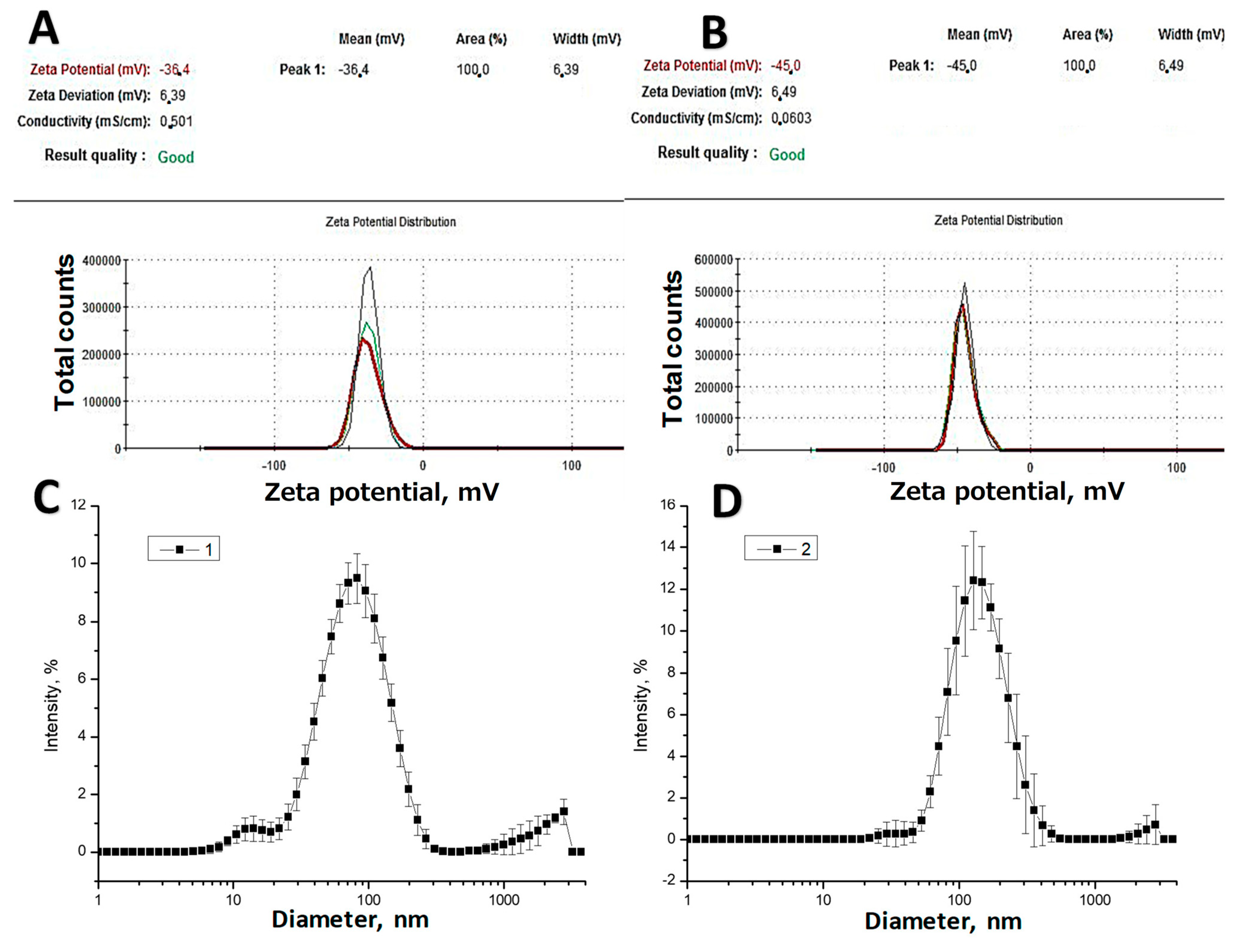
2.4. Zeta Potential Measurement and Size Distribution

An examination of the zeta potential was conducted using the Malvern Zetasizer Nano ZS90 instrument (United Kingdom). This analysis aimed to investigate the stability and dimensions of the produced AgNPs. Zetasizer systems employ electrophoretic (ELS) and dynamic light scattering (DLS) techniques, offering insights into particle mobility, charge, and size in dispersive systems, spanning from nanometers to micrometers [28]. The following configuration involved running 5 scans for each sample run, with a total of 3 runs conducted. The entire experiment was replicated at least 6 times. DLS analysis was executed using the Zetasizer 3000 instrument from Malvern Instruments. The size distribution was established by performing 10 scans for each spectrogram run, with a minimum of 10 runs recorded. The error bars were generated using standard the zeta potential software.

2.5. TEM

A JEOL JEM-1400 plus microscope was used to characterize the AgNPs stabilized by the plant extract, which was dispersed on the carbon coated copper grid, mesh size 200, and left it dry. The images were taken at the following settings with electron acceleration 80 kV and magnification 20,000, 50,000, 120,000 and 400,000 times. The TEM images were analysed using Image J area analysis and following a calculation of the average diameter.

2.6. Assessment of Antibacterial Effects of Artemisia Extracts

To assess the antibacterial properties, we employed a method involving double serial dilutions in an isotonic solution, followed by inoculation onto solid nutritional agar to evaluate the survival of the bacteria after being exposed to antimicrobial agents. The microorganism suspensions were prepared using isotonic saline solution, resulting in an absorbance of 0.056 at a wavelength of 625 nm, which indicated antimicrobial agents. The microorganism suspensions were prepared using isotonic saline solution, resulting in an absorbance of 0.056 at a wavelength of 625 nm, indicating a cell concentration of approximately 8 × 10⁸ CFU/mL. Subsequently, serial dilutions ranging from 1:1 to 1:2048, with a ratio of water extract Artemisia extract ×40: isotonic solution, were created using a Repeater M4 (Eppendorf) in a standard 96 well plate for ELISA. As a result, the final cell concentration was anticipated to be approximately 2.6 × 10⁸ CFU/mL. These preparations were then kept at room temperature for 24 h, after which they were plated onto agar broth plates and kept at 37 °C overnight [20]. The growth of bacteria was checked visually (Figure 6).

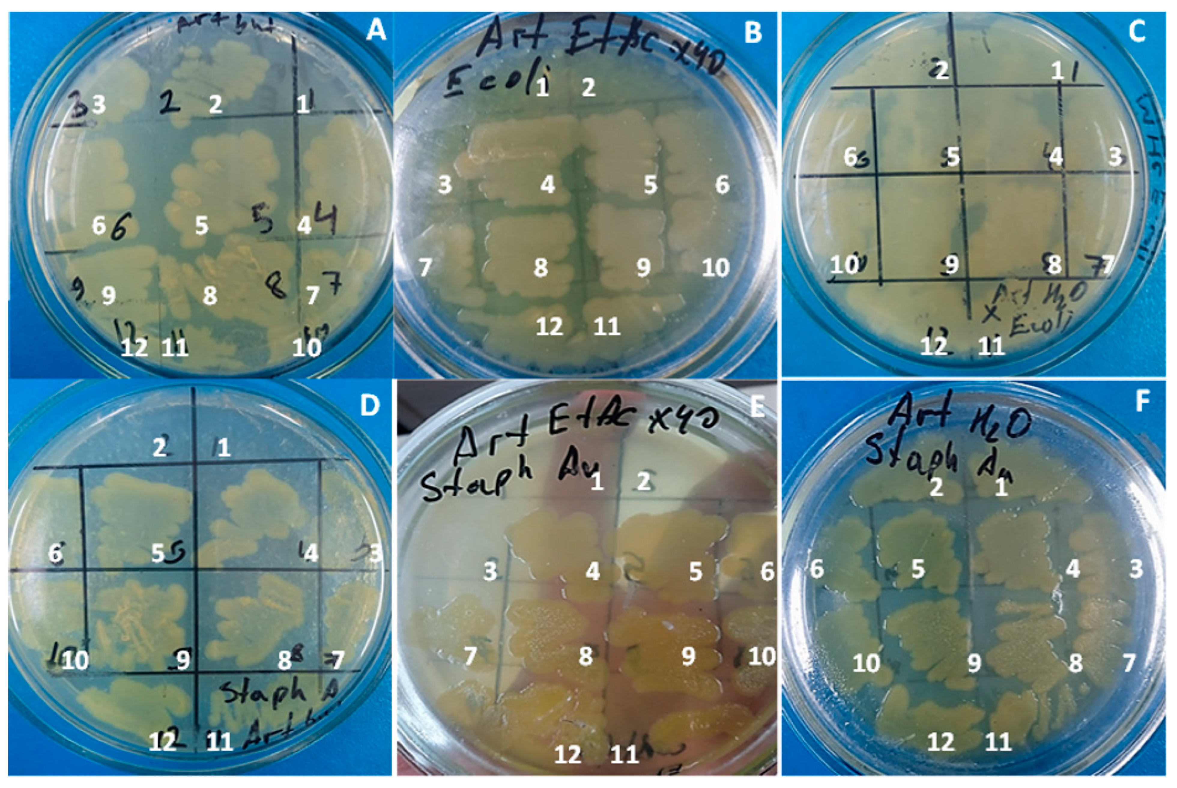
2.7. Antimicrobial Activity of Plant Extract and AgNPs

The preparation of test strain suspensions involved using 18–24 h cultures of the test strains. In this process, an aliquot of the respective test strain was introduced into a test tube containing 5–6 mL of 1/50 nutrient broth using a sterile loop. The contents were thoroughly mixed to achieve homogeneity. To determine the cell concentration, optical density was measured ensitometrically and found to be 1.2 units, following the McFarland standard. This measurement corresponded to a cell content of 6 × 10⁸ CFU/mL for bacterial suspensions and 6 × 10⁶ CFU/mL for yeast cell suspensions. To create working concentrations, the stock suspension was subjected to 10-fold serial dilutions; a final inoculum of 6 × 10⁵ CFU/mL was reached for each test strain. 0.4 mL of the obtained suspension of the corresponding strain was applied on the hole in the surface of agar. Bacterial growth was evaluated after incubation for 24 h at 30 °C [29].

3. Results and Discussion

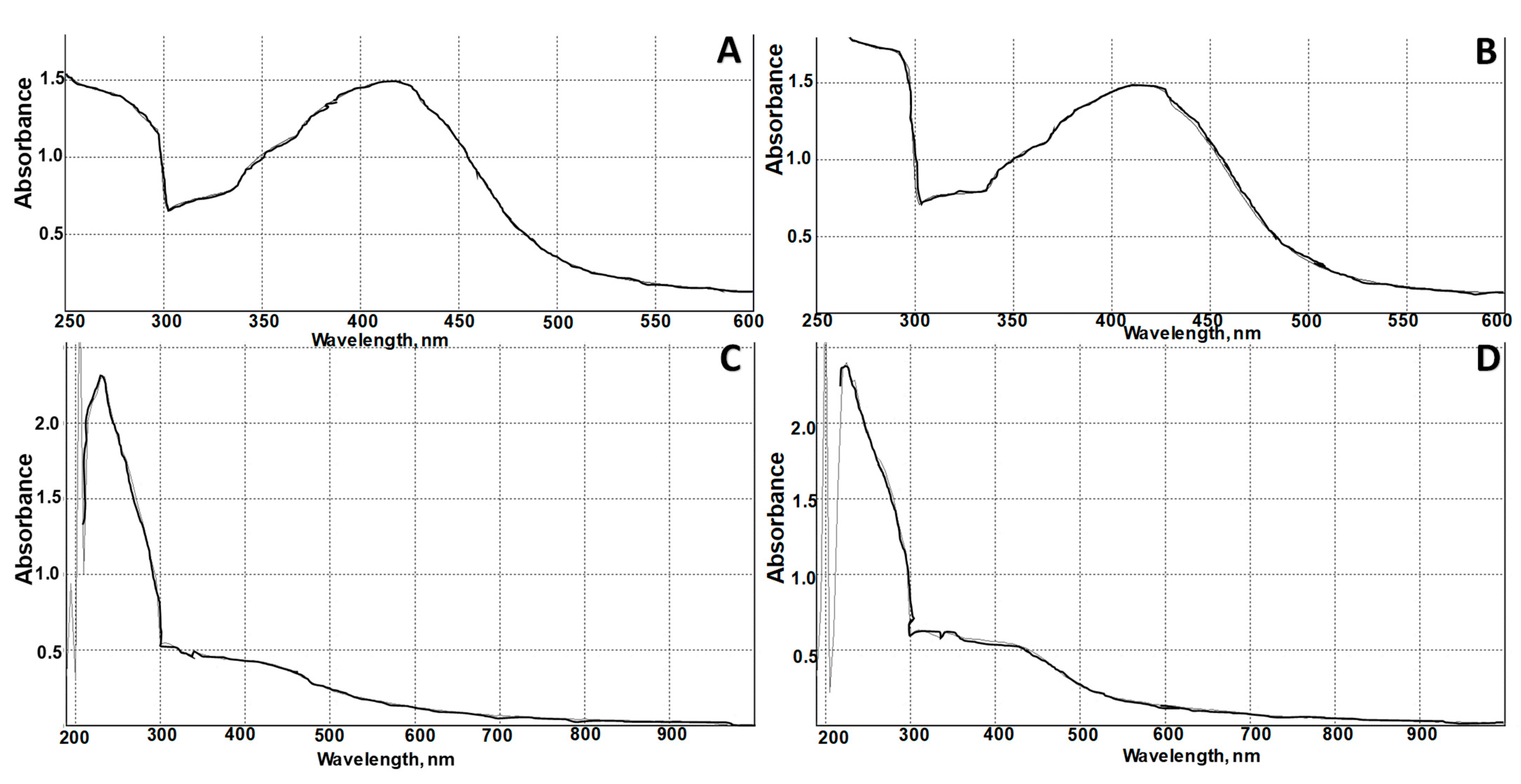
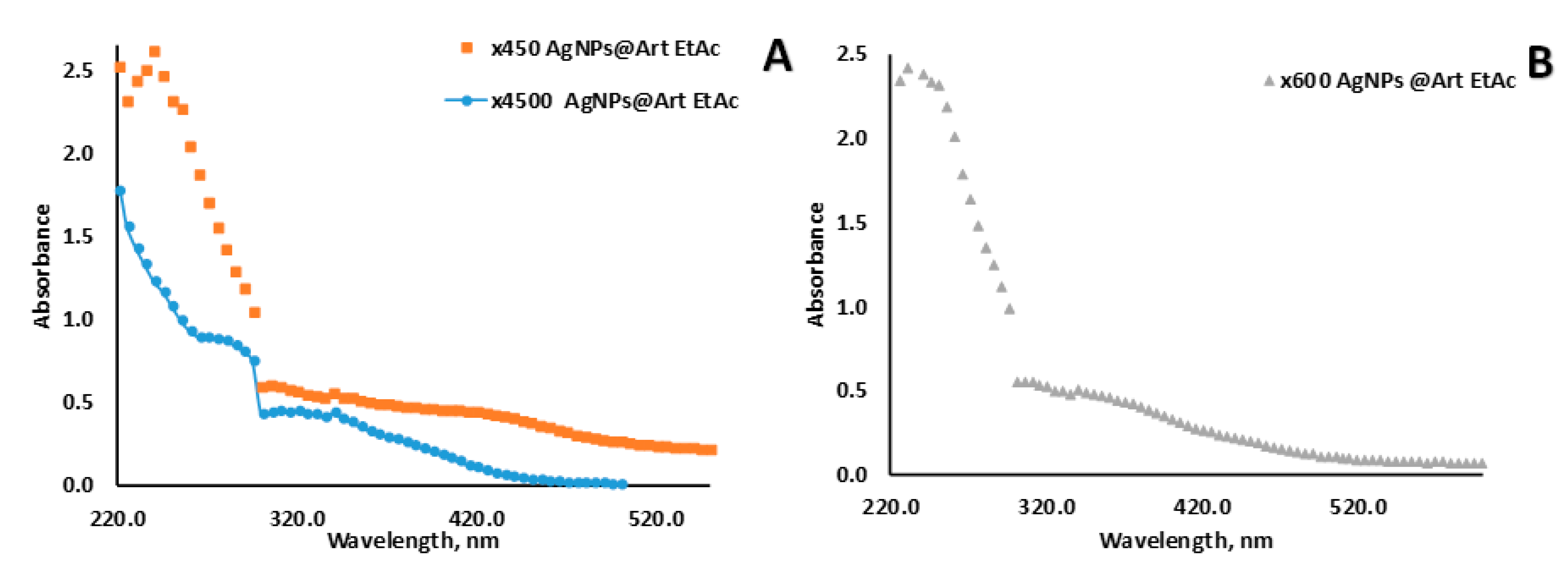
3.1. AgNPs Synthesis and Characterization

Recently, great attention has been focused on the utilization of plant extracts for AgNPs synthesis and its following usage as antimicrobial agent. There are a few studies related to Artemisia species (Artemisia quttensis, Artemisia oliveriana, Artemisia kopetdaghensis, Artemisia absinthium, Artemisia annua) extracts with AgNPs [16,17,18,19,21]. However, the role of A. terrae-albae extract in AgNP formation has not previously been studied.
The surface Plasmon resonance SPR peak of AgNPs@Art H2O at 420 nm confirmed the spontaneous formation of AgNPs (Figure 1A). In order to check the reduction of all silver phytocomplexes to AgNPs, the reaction mixture was additionally treated in an ultrasonic bath for 2 min in standard conditions. Enhancement of the AgNPs SPR peak after the additional treatment, which indicates the completion of the redox reaction, was not observed. Previously, the mechanism of AgNP stabilization by plant extract and the influence of various trigger parameters (UV or microwave irradiation, pH, temperature) on AgNP inducing have been discussed [20]. The research focused on examining the production of silver nanoparticles with sizes ranging from 35 to 60 nanometers, which were created using an aqueous extract derived from Eclipta prostrata and subjected to ultrasonic treatment. The resulting AgNPs displayed activity against the fourth-stage larvae of two disease-carrying mosquitoes: Culex quinquefasciatus say, which transmits filariasis, and Anopheles subpictus Grassi, responsible for spreading malaria [30]. On the other hand, AgNPs@Art EtAc extract ×45 and AgNPs@Art EtAc extract ×60 did not have a clear SPR peak, because of the wide size distribution of the AgNPs (Figure 2). Nonetheless, TEM and light scattering indicated the presence of AgNPs in samples (Figure 3 and Figure 4).
In the formation of AgNPs, phenolic groups of flavonoids participate in the reduction of Ag ions, which in turn become oxidized to quinone functionality [31]. We propose that additionally to the abovementioned, other functional groups such as aldehyde groups of Vanillin or other aromatic aldehyde may participate in the reduction process and following stabilization of AgNPs via carboxyl groups and chelate formation with oxygen and nitrogen containing functional groups.
AgNPs@EtAc Art extract ×45 was characterized by size in the range of 80–120 nm, while in AgNPs@EtAc Art extract ×60 the main fraction ranged from 80–150 nm (Figure 3C,D). Interestingly, that dilution factor of the plant extract also effects the surface charge of AgNPs, most probably due to a change in the ethylacetate in the solution. The zeta potential of AgNPs@EtAc Art extract ×45 and AgNPs@EtAc Art extract ×60 was −36 and −45 mV, respectively. This indicates that molecules chemosorbed on the AgNPs have a negative charge. Previously, the zeta potential of AgNPs-Calendula off. possessed a similar value—38—and had good aggregative stability within 9 months of storage at 4 °C [20]. Adoni et al. have demonstrated that AgNPs@Artemisia Annua L. aq. extract obtained from leaves is characterized by a particle size within the range of 5–20 nm and a zeta potential of −26 millivolts [32]. MF Baran’s study found that AgNPs stabilized with Artemisia Absinthium exhibited a maximum absorbance for a SPR peak of 449 nm. Additionally, X-ray analysis revealed that these nanoparticles had a size of approximately 14.6 nanometers [33]. Another study reported that distinctive SPR bands of AgNPs were detected at around 430 nm; these were attributed to AgNPs synthesized using Artemisia argyi leaf extract and having an average size of approximately 77.6 nm [34].
The size distribution data obtained by TEM analysis of the AgNPs agree well with the evaluation of the hydrodynamic diameter of the particles analyzed by using light scattering (Figure 3, Figure 4 and Figure S1). As expected, a significant dilution of EtAc extract with water decreases the solubility of the lipophilic compounds and their aggregation due to hydrophobic interactions. This causes the decline of the soluble molecules in water, which may participate in the stabilisation of AgNPs resulting in the formation of larger particles AgNPs@EtAc Artemisia extract ×60. The shape of the AgNPs@EtAc Artemisia extract ×40 is mostly spherical, whereas AgNPs@EtAc Artemisia ×60 is characterized by more a randomized shape (triangle) and includes tetrahedral shape particles. Moreover, AgNPs@EtAc Artemisia extract ×60 has less aggregative stability, which leads to their aggregation (Figure 4D–F). According to TEM images analysis the main fraction of AgNPs@EtAc Artemisia extract ×60 was in the range of 13–34 nm, whereas the size of AgNPs@EtAc Art extract ×40 varied in the range of 18–33 nm (Figure 5).

3.2. Antimicrobial Activity of AgNPs and Extracts

The screening of various dilutions of Artemisia extract did not illustrate the antimicrobial activity against E-coli and S. Aurous (Figure S2). Previous research conducted by several research groups has extensively elucidated the antimicrobial mechanisms of AgNPs [35]. It was revealed that the primary mechanism involves the generation of reactive oxygen species that disrupt the bacterial cell wall. However, we currently lack the necessary equipment for such a comprehensive investigation. The diluted solutions of extracts of Artemisia (butanol extract ×40; ethylacetate extract ×40 and aqueous extract ×20) were not effective for the bacteria inhibition of E. coli (ATCC 8739 and S. Aureus (ATCC 25923) (Figure 6). The contact time of bacterial suspension with AgNPs was over 24 h. The evaluation of AgNPs stabilized by the Artemisia terrae-albae extract aerial part was performed by agar diffusion.
AgNPs citrate and AgNPs@EtAc extract ×60 illustrated comparable antimicrobial activity against Escherichia coli (ATCC 8739) Pseudomonas aeruginosa ATCC 9027 C. albicans (ATCC 10281)(Table 1).
AgNPs citrate did not manifest activity S. aureus (ATCC 6538), but it was effective against other strains (Table 1 and Figure 7). Artemisia EtAc extract diluted ×15 times and Artemisia EtAc extract diluted ×8 times possessed a similar moderate level of efficiency for Escherichia coli (ATCC 8739). AgNPs@EtAc extract ×60 was active against all the studied strains, and the highest sensitivity was against E. coli and C.Albicans. Artemisia EtAc extract with dilutions 8, 12, 15, and 16 exhibited activity to C.Albicans. Artemisia herba-alba in combination with AgNPs exhibited significant larvicidal toxicity against mosquitoes. The LC50 (a lethal concentration with 50% mortality) of AgNPs against Indian strains was 9.7 mg/L for Anopheles stephensi, 10.7 mg/L for Aedes aegypti, and 11.4 mg/L for Culex quinquefasciatus. In the case of Saudi Arabian strains, the LC50 for AgNPs was 33.6 mg/L for Aedes aegypti and 38 g/L for Culex pipiens. In adult mosquito experiments, the Artemisia herba-alba extract alone indicated LC50 values ranging from 293 to 450 mg/L, while the LC50 values for AgNPs possessed activity from 8 to 27.4 g/L. This suggests that AgNPs significantly enhanced the adulticidal activity against mosquitoes compared to the A. herba-alba extract alone [36]. AgNPs synthesized with Artemisia tournefortiana Rchb ethanolic extract had an average size of 23 ± 14.8 nm. Their impact on in vitro cytotoxicity was assessed using normal human embryonic kidney (HEK293) cells and human colon adenocarcinoma cancer (HT29) cells [37]. AgNPs @Artemisia absinthium L. (2 mM) with a plant extract (10 mg/mL) at various ratios had a significant impact on the size, stability, and yield of AgNPs. When the extract-to- AgNO3 ratio was 6:4 v/v, it resulted in the highest conversion efficiency of AgNO3 to AgNPs, producing particles with an average size of less than 100 nm [38]. AgNPs stabilized by Artemisia afra have an average size of approximately 30.7 nm. These AgNPs were synthesized within 30 min at a reaction temperature of 80–90 °C using an aqueous leaf extract of Artemisia afra. The synthesized AgNPs exhibited SPR in the wavelength range of 423–438 nm [39]. Hu et al. reported that AgNPs stabilised by Artemisia argyi leaf extract exhibited a strong antimicrobial activity of 1 μg/mL for Acinetobacter baumannii, and 2 mg/L for Staphylococcus aureus, Escherichia coli, and Candida albicans. Additionally, the minimum biofilm eradication concentration at 50% for these AgNPs against these strains was 2 μg/mL for Acinetobacter baumannii, 4 μg/mL for Staphylococcus aureus, 0.5 μg/mL for Escherichia coli, and 2 μg/mL for Candida albicans [34].

4. Conclusions

Several Artemisia terrae-albae extracts were obtained. The appropriate aqueous and ethylacetate extracts were acceptable for the following evaluation of antimicrobial activity. Moreover, the extracts were utilized to reduce silver ions to AgNPs as well as to serve as a stabilizing agent. The zetta potential measurement of the AgNPs had a negative surface charge. The decrease of the ethylacetate extract solution in water for the Art @EtAc extract led to less stable AgNPs and following aggregation with average size in the range of 80–150 nm. Using a diluted ethylacetate solution allowed dissolving the lipophilic compounds and allowing for participation in the AgNP stabilization. Moreover, the preliminary attempt of the ethanolic solution of the Artemisia antimicrobial test illustrated activity only for the high content of ethanol, and it was unclear what is responsible for the activity. Then, EtAc allows the observation of the true values of antimicrobial activity. The dissolution of poorly soluble ethanolic and EtAc Artemisia terrae-albae extracts in water using a method of serial dilutions ×40, ×80, ×160 etc. did not reveal activity of E. coli (ATCC 8739) and S. aureus (ATCC 25923); however, it was difficult and unpractical to prepare a solution at higher concentration. EtAc Art extract diluted 15 and 8 times demonstrated comparable moderate levels of activity against Escherichia coli (ATCC 8739). AgNPs@EtAc extract ×60 exhibited activity against all the strains studied, with the highest sensitivity observed against E. coli and C. Albicans. Additionally, Artemisia terrae-albae EtAc extract at dilutions 8, 12, 15, and 16 exhibited activity against C. Albicans.

Supplementary Materials

The following supporting information can be downloaded at: https://www.mdpi.com/article/10.3390/pr11103041/s1, Figure S1: TEM photograph: (A–D) AgNPs@EtAc Art extract ×40; Figure S2: Bacterial growth on agar plate after exposure to Art extract obtained using different solvents: (A) Art butanol extract ×40 in 50% ethanol against of E. coli (ATCC 8739); (B) Art EtAc x40 in 50% ethanol against of E. coli (ATCC 8739); (C) Art aq. extract ×20 against of E. coli (ATCC 8739); (D) Art butanol extract ×40 in 50% ethanol; (E) Art EtAc extract ×40 in 50% ethanol against of S. aureus (ATCC 25923); (F) Art aq. extract ×20 against of S. aureus (ATCC 25923).

Author Contributions

Conceptualization, D.A.B.; Methodology, F.M.K.; Formal analysis, M.A.D., D.A.B., S.M.J., F.M.K., A.A.K. and G.K.V.; Investigation, D.A.B., A.E.B., G.E.B., N.A.I., S.M.J. and N.Z.K.; Resources, M.A.D., A.E.B., G.E.B., N.Z.K. and G.K.V.; Data curation, M.A.D., N.A.I. and A.A.K.; Writing—original draft, D.A.B.; Writing—review & editing, D.A.B. and M.A.D.; Project administration, G.K.V.; Funding acquisition, M.A.D., A.A.K. and G.K.V. All authors have read and agreed to the published version of the manuscript.

Funding

This study was conducted by the MES RK for 2021–2023 with a period of implementation of 36 months on the topic: “Phytochemical Profiles and Development of Medicinal Plants Targeting Skin Diseases”, IRN AP09057982. The APC was funded by the MES RK IRN AP09057982.

Institutional Review Board Statement

Not applicable.

Informed Consent Statement

Not applicable.

Data Availability Statement

Not applicable.

Conflicts of Interest

The authors declare no conflict of interest.

References

  1. Nigam, M.; Atanassova, M.; Mishra, A.P.; Pezzani, R.; Devkota, H.P.; Plygun, S.; Salehi, B.; Setzer, W.N.; Sharifi-Rad, J. Bioactive compounds and health benefits of Artemisia species. Nat. Prod. Commun. 2019, 14, 1934578–19850354. [Google Scholar] [CrossRef]
  2. Bora, K.S.; Sharma, A. The genus Artemisia: A comprehensive review. Pharm. Biol. 2011, 49, 101–109. [Google Scholar] [CrossRef] [PubMed]
  3. Siwan, D.; Nandave, D.; Nandave, M. Artemisia vulgaris Linn: An updated review on its multiple biological activities. Future J. Pharm. Sci. 2022, 8, 47. [Google Scholar] [CrossRef]
  4. Zhurinov, M.; Berillo, D.; Bazhykova, K.B.; Rakhimov, K.D.; Bekezhanova, T. An Estimation of the Antiviral Activity and Toxicity of Biologically Active Substances Obtained from the Raw Materials of Artemisia cina Berg. In Vitro and In Vivo. Molecules 2023, 28, 5413. [Google Scholar] [CrossRef] [PubMed]
  5. Janaćković, P.; Rajčević, N.; Gavrilović, M.; Novaković, J.; Giweli, A.; Stešević, D.; Marin, P.D. Essential oil composition of five Artemisia (Compositae) species in regards to chemophenetics. Biochem. Syst. Ecol. 2019, 87, 103960. [Google Scholar] [CrossRef]
  6. Ranjbar, M.; Naghavi, M.R.; Alizadeh, H. Chemical composition of the essential oils of Artemisia species from Iran: A comparative study using multivariate statistical analysis. J. Essent. Oil Res. 2020, 32, 361–371. [Google Scholar] [CrossRef]
  7. Ksibi, N.; Saada, M.; Yeddes, W.; Limam, H.; Tammar, S.; Wannes, W.A.; Labidi, N.; Hessini, K.; Dakhlaoui, S.; Frouja, O.; et al. Phytochemical Profile, Antioxidant and Antibacterial Activities of Artemisia absinthium L. Collected from Tunisian Regions. J. Mex. Chem. Soc. 2022, 66, 312–329. [Google Scholar] [CrossRef]
  8. Batiha, G.E.-S.; Olatunde, A.; El-Mleeh, A.; Hetta, H.F.; Al-Rejaie, S.; Alghamdi, S.; Zahoor, M.; Magdy Beshbishy, A.; Murata, T.; Zaragoza-Bastida, A.; et al. Bioactive compounds, pharmacological actions, and pharmacokinetics of wormwood (Artemisia absinthium). Antibiotics 2020, 9, 353. [Google Scholar] [CrossRef]
  9. Ahamad, J.; Mir, S.R.; Amin, S. A pharmacognostic review on Artemisia absinthium. Int. Res. J. Pharm. 2019, 10, 25–31. [Google Scholar] [CrossRef]
  10. Bordean, M.-E.; Muste, S.; Marțiș, G.S.; Mureșan, V.; Buican, B.C. Health effects of wormwood (Artemisia absinthium L.): From antioxidant to nutraceutical. J. Agroalim. Proc. Technol. 2021, 27, 211–218. [Google Scholar]
  11. Minda, D.; Ghiulai, R.; Banciu, C.D.; Pavel, I.Z.; Danciu, C.; Racoviceanu, R.; Soica, C.; Budu, O.D.; Muntean, D.; Diaconeasa, Z.; et al. Phytochemical profile; antioxidant; wound healing potential of three Artemisia species: In vitro and in ovo evaluation. Appl. Sci. 2022, 12, 1359. [Google Scholar] [CrossRef]
  12. Xiao, J.; Liu, P.; Hu, Y.; Liu, T.; Guo, Y.; Sun, P.; Zheng, J.; Ren, Z.; Wang, Y. Antiviral activities of Artemisia vulgaris L. extract against herpes simplex virus. Chin. Med. 2023, 18, 21. [Google Scholar] [CrossRef] [PubMed]
  13. Liu, P.; Zhong, L.; Xiao, J.; Hu, Y.; Liu, T.; Ren, Z.; Wang, Y.; Zheng, K. Ethanol extract from Artemisia argyi leaves inhibits HSV-1 infection by destroying the viral envelope. Virol. J. 2023, 20, 8. [Google Scholar] [CrossRef] [PubMed]
  14. Hbika, A.; Daoudi, N.E.; Bouyanzer, A.; Bouhrim, M.; Mohti, H.; Loukili, E.H.; Mechchate, H.; Al-Salahi, R.; Nasr, F.A.; Bnouham, M.; et al. Artemisia absinthium L. Aqueous and ethyl acetate extracts: Antioxidant effect and potential activity in vitro and in vivo against pancreatic α-amylase and intestinal α-glucosidase. Pharmaceutics 2022, 14, 481. [Google Scholar] [CrossRef]
  15. Zeb, S.; Ali, A.; Zaman, W.; Zeb, S.; Ali, S.; Ullah, F.; Shakoor, A. Pharmacology, taxonomy and phytochemistry of the genus Artemisia specifically from Pakistan: A comprehensive review. Pharm. Biomed. Res. 2019, 4, 1–12. [Google Scholar] [CrossRef]
  16. Ghanbar, F.; Mirzaie, A.; Ashrafi, F.; Noorbazargan, H.; Dalirsaber Jalali, M.; Salehi, S.; Sadat Shandiz, S.A. Antioxidant, antibacterial and anticancer properties of phyto-synthesised Artemisia quttensis Podlech extract mediated AgNPs. IET Nanobiotechnol. 2017, 11, 485–492. [Google Scholar] [CrossRef]
  17. Fard, N.N.; Noorbazargan, H.; Mirzaie, A.; Ch, M.H.; Moghimiyan, Z.; Rahimi, A. Biogenic synthesis of AgNPs using Artemisia oliveriana extract and their biological activities for an effective treatment of lung cancer. Artif. Cells Nanomed. Biotechnol. 2018, 46 (Suppl. S3), 1047–1058. [Google Scholar] [CrossRef]
  18. Taghavizadeh Yazdi, M.E.; Darroudi, M.; Amiri, M.S.; Hosseini, H.A.; Nourbakhsh, F.; Mashreghi, M.; Farjadi, M.; Mousavi Kouhi, S.M.; Mousavi, S.H. Anticancer, antimicrobial, and dye degradation activity of biosynthesised silver nanoparticle using Artemisia kopetdaghensis. Micro Nano Lett. 2020, 15, 1046–1050. [Google Scholar] [CrossRef]
  19. Balciunaitiene, A.; Viskelis, P.; Viskelis, J.; Streimikyte, P.; Liaudanskas, M.; Bartkiene, E.; Zavistanaviciute, P.; Zokaityte, E.; Starkute, V.; Ruzauskas, M.; et al. Green synthesis of silver nanoparticles using extract of Artemisia absinthium L., Humulus lupulus L. and Thymus vulgaris L., physico-chemical characterization, antimicrobial and antioxidant activity. Processes 2021, 9, 1304. [Google Scholar] [CrossRef]
  20. Zhangabay, Z.; Berillo, D. Antimicrobial and antioxidant activity of AgNPs stabilized with Calendula officinalis flower extract. Results Surf. Interfaces 2023, 11, 100109. [Google Scholar] [CrossRef]
  21. Khatoon, N.; Sharma, Y.; Sardar, M.; Manzoor, N. Mode of action and anti-Candida activity of Artemisia annua mediated-synthesized silver nanoparticles. J. Mycol. Médicale 2019, 29, 201–209. [Google Scholar] [CrossRef] [PubMed]
  22. Avitabile, E.; Senes, N.; D’avino, C.; Tsamesidis, I.; Pinna, A.; Medici, S.; Pantaleo, A. The potential antimalarial efficacy of hemocompatible silver nanoparticles from Artemisia species against P. falciparum parasite. PLoS ONE 2020, 15, e0238532. [Google Scholar] [CrossRef] [PubMed]
  23. Adhikari, A.; Lamichhane, L.; Adhikari, A.; Gyawali, G.; Acharya, D.; Baral, E.R.; Chhetri, K. Green Synthesis of Silver Nanoparticles Using Artemisia vulgaris Extract and Its Application toward Catalytic and Metal-Sensing Activity. Inorganics 2022, 10, 113. [Google Scholar] [CrossRef]
  24. Lim, S.H.; Ahn, E.-Y.; Park, Y. Green synthesis and catalytic activity of gold nanoparticles synthesized by Artemisia capillaris water extract. Nanoscale Res. Lett. 2016, 11, 474. [Google Scholar] [CrossRef] [PubMed]
  25. Aslany, S.; Tafvizi, F.; Naseh, V. Characterization and evaluation of cytotoxic and apoptotic effects of green synthesis of silver nanoparticles using Artemisia Ciniformis on human gastric adenocarcinoma. Mater. Today Commun. 2020, 24, 101011. [Google Scholar] [CrossRef]
  26. Al-Sheddi, E.S.; Alsohaibani, N.; bin Rshoud, N.; Al-Oqail, M.M.; Al-Massarani, S.M.; Farshori, N.N.; Malik, T.; Al-Khedhairy, A.A.; Siddiqui, M.A. Anticancer efficacy of green synthesized silver nanoparticles from Artemisia monosperma against human breast cancer cells. S. Afr. J. Bot. 2023, 160, 123–131. [Google Scholar] [CrossRef]
  27. El-Ashmouny, R.S.; Rady, M.H.; Merdan, B.A.; El-Sheikh, T.A.A.; Hassan, R.E.; E El Gohary, E.G. Larvicidal and pathological effects of green synthesized silver nanoparticles from Artemisia herba-alba against Spodoptera littoralis through feeding and contact application. Egypt. J. Basic Appl. Sci. 2022, 9, 239–253. [Google Scholar] [CrossRef]
  28. Berillo, D.; Arysbek, A. Chitosan Glutaraldegyde Cryogels for Wastewater Treatment and Extraction of Silver Nanoparticles. Processes 2023, 11, 1661. [Google Scholar] [CrossRef]
  29. Berillo, D.; Zakiruly, O.; Tolepova, A.; Baimenov, A.; Tauanov, Z. Antimicrobial activity of polymer with triclosan Bulletin of KazNU. Eurasian J. Ecol. 2023, 76, 56–70. [Google Scholar] [CrossRef]
  30. Rajakumar, G.; Rahuman, A.A. Larvicidal activity of synthesized silver nanoparticles using Eclipta prostrata leaf extract against filariasis and malaria vectors. Acta Trop. 2011, 118, 196–203. [Google Scholar] [CrossRef]
  31. Chahardoli, A.; Karimi, N.; Fattahi, A. Nigella arvensis leaf extract mediated green synthesis of silver nanoparticles: Their characteristic properties and biological efficacy. Adv. Powder Technol. 2018, 29, 202–210. [Google Scholar] [CrossRef]
  32. Adoni, M.; Yadam, M.; Gaddam, S.A.; Rayalacheruvu, U.; Kotakadi, V.S. Antimicrobial, antioxidant, and dye degradation properties of biosynthesized silver nanoparticles from Artemisia annua L. Lett. Appl. NanoBioSci. 2020, 10, 1981–1992. [Google Scholar]
  33. Baran, M.F. Synthesis and antimicrobial applications of silver nanoparticles from Artemisia absinthium plant. Biol. Chem. Res. 2019, 6, 96–103. [Google Scholar]
  34. Hu, L.; Yang, X.; Yin, J.; Rong, X.; Huang, X.; Yu, P.; He, Z.; Liu, Y. Combination of AgNPs and domiphen is antimicrobial against biofilms of common pathogens. Int. J. Nanomed. 2021, 7181–7194. [Google Scholar] [CrossRef] [PubMed]
  35. Xu, H.; Qu, F.; Xu, H.; Lai, W.; Andrew Wang, Y.; Aguilar, Z.P.; Wei, H. Role of reactive oxygen species in the antibacterial mechanism of silver nanoparticles on Escherichia coli O157: H7. Biometals 2012, 25, 45–53. [Google Scholar] [CrossRef]
  36. Aziz, A.T.; Alshehri, M.A.; Panneerselvam, C.; Murugan, K.; Trivedi, S.; Mahyoub, J.A.; Hassan, M.M.; Maggi, F.; Sut, S.; Dall’Acqua, S.; et al. The desert wormwood (Artemisia herba-alba)–From Arabian folk medicine to a source of green and effective nanoinsecticides against mosquito vectors. J. Photochem. Photobiol. B Biol. 2018, 180, 225–234. [Google Scholar] [CrossRef]
  37. Baghbani-Arani, F.; Movagharnia, R.; Sharifian, A.; Salehi, S.; Shandiz, S.A.S. Photo-catalytic, anti-bacterial, and anti-cancer properties of phyto-mediated synthesis of silver nanoparticles from Artemisia tournefortiana Rchb extract. J. Photochem. Photobiol. B Biol. 2017, 173, 640–649. [Google Scholar] [CrossRef]
  38. Ali, M.; Kim, B.; Belfield, K.D.; Norman, D.; Brennan, M.; Ali, G.S. Green synthesis and characterization of silver nanoparticles using Artemisia absinthium aqueous extract—A comprehensive study. Mater. Sci. Eng. C 2016, 58, 359–365. [Google Scholar] [CrossRef]
  39. Elemike, E.E.; Onwudiwe, D.C.; Ekennia, A.C.; Jordaan, A. Synthesis and characterisation of silver nanoparticles using leaf extract of Artemisia afra and their in vitro antimicrobial and antioxidant activities. IET Nanobiotechnol. 2018, 12, 722–726. [Google Scholar] [CrossRef]
Figure 1. UV-vis spectra of AgNPs stabilized by the Art aqueous extract: (A) before ultrasonic treatment Art H2O ×40 + AgNO3 and (B) after ultrasonic treatment Art aq. extract ×40; (C) Art aq. extract ×60 before ultrasonic treatment; (D) Art aq. extract ×60 after ultrasonic treatment. Suspensions were additionally diluted 10-fold by water before spectra recording.
Processes 11 03041 g001
Figure 2. (A) UV-vis spectra of AgNPs@Art EtAc extract ×45 and (B) AgNPs@Art EtAc extract ×60 additionally diluted 10 and 100 times by distilled water.
Processes 11 03041 g002
Figure 3. Zeta potential of AgNPs: (A) AgNPs@ Art EtAc extract ×45; (B) AgNPs@EtAc Art extract ×60; AgNPs size distribution: (C) AgNPs@EtAc Art extract ×45; (D) AgNPs@EtAc Art extract ×60, n = 3.
Processes 11 03041 g003
Figure 4. TEM photograph at different magnifications: (AC) AgNPs@EtAc Art extract ×40; (DF) AgNPs@EtAc Art extract ×60.
Processes 11 03041 g004
Figure 5. Size distribution of AgNPs using Image J TEM images analysis: (A) AgNPs@EtAc Art extract dilution ×40; (B) AgNPs@EtAc Art extract dilution ×60.
Processes 11 03041 g005
Figure 6. Bacterial growth on agar plate [n = 3] after exposure to different dilutions Art extract obtained using different solvents: (A) Art butanol extract ×40 in 50% ethanol against of E. coli (ATCC 8739); (B) Art EtAc ×40 in 50% ethanol against of E. coli (ATCC 8739); (C) Art aq. extract ×20 against of E. coli (ATCC 8739); (D) Art butanol extract ×40 in 50% ethanol; (E) Art EtAc extract ×40 in 50% ethanol against of S. aureus (ATCC 25923); (F) Art aq. extract ×20 against of S. aureus (ATCC 25923).
Processes 11 03041 g006
Figure 7. Growth of bacterial strain after exposure to different dilutions plant extract s: (1) control blank; (2) Art EtAc extract ×15; (3) AgNPs@Art EtAc extract ×45; (4) AgNPs@Art EtAc extract ×45; (5) Art EtAc extract ×8; (6) Art EtAc extract ×12; (7) Art EtAc extract ×16; against (AC) C. albicans (ATCC 10281); (D,E) Pseudomonas aeruginosa ATCC 9027; (G,H) S. aureus (ATCC 6538) (F,K) Escherichia coli (ATCC 8739).
Processes 11 03041 g007
Table 1. Zone of bacterial growth inhibition under action of AgNPs and Artemisia plant extract at different dilution factor.
N of SampleS. aureus (ATCC 6538)Escherichia coli (ATCC 8739)Pseudomonas aeruginosa ATCC 9027C. albicans (ATCC 10281)
AgNPs citrate
etalon suspension
015 ± 312 ± 218 ± 3
EtAc extract ×1511 ± 212 ± 212 ± 212 ± 2
AgNPs@EtAc extract ×4511 ± 3011 ± 211 ± 2
AgNPs@EtAc extract ×6012 ± 320 ± 211 ± 220 ± 3
EtAc extract ×8011 ± 2011 ± 2
EtAc extract ×120012 ± 311 ± 2
EtAc extract ×1600 11 ± 2
Disclaimer/Publisher’s Note: The statements, opinions and data contained in all publications are solely those of the individual author(s) and contributor(s) and not of MDPI and/or the editor(s). MDPI and/or the editor(s) disclaim responsibility for any injury to people or property resulting from any ideas, methods, instructions or products referred to in the content.

