Article

Anti-Oxidative and Anti-Diabetic Effects of Electrolyzed Weakly Alkaline Reduced Water on Renal Proximal Tubular Epithelial Cells

1
Department of Convergence Medicine, Wonju College of Medicine, Yonsei University, Wonju 26426, Korea
2
Department of Global Medical Science, Wonju College of Medicine, Yonsei University, Wonju 26426, Korea
3
Department of Biology, College of Science, University of the Philippines Baguio, Baguio 2600, Philippines
4
NUGA MEDICAL Co., Ltd., 9, Bongeunsa-ro 68-gil, Gangnam-gu, Seoul 06153, Korea
5
Department of Internal Medicine, Division of Hematology-Oncology, Wonju Severance Christian Hospital, Wonju 26426, Korea
*
Author to whom correspondence should be addressed.
These authors contributed equally to this study as first authors.
Processes 2022, 10(10), 2025; https://doi.org/10.3390/pr10102025
Submission received: 11 August 2022 / Revised: 23 September 2022 / Accepted: 4 October 2022 / Published: 7 October 2022
(This article belongs to the Section Biological Processes and Systems)

Abstract

:
Diabetes, a chronic metabolic disorder, affects glucose consumption in the body and leads to severe kidney damage, known as diabetic nephropathy, where oxidative stress (OS) plays a vital role in disease pathogenesis. Electrolyzed weakly alkaline reduced water (EWARW) is known to have anti-oxidative functions in vitro due to its bioactive properties. However, there is an increasing need for adequate research on EWARW-specific therapies to minimize the negative consequences of hyperglycemia-induced OS in the kidney. This study aimed to determine the anti-oxidative and anti-diabetic effects of EWARW at pH 8.5 on human kidney-2 (HK-2) cells. Here, HK-2 cells were treated with 71.68 mM glucose to induce OS and then treated with 10% tap water (TW)_7.7, electrolyzed alkaline reduced water (EARW)_9.5, or EARW_8.5 for 24 h. After treatment, cell viability, OS and apoptotic markers, glucose uptake, adenosine triphosphate (ATP) content, and inflammatory markers were assessed to determine the effect of EARW_8.5. Our results revealed that EARW_8.5 rescued cell viability in comparison to the high glucose (HG) only and TW_7.7 groups. In addition, EARW_8.5 was effective in reducing OS markers (ROS, NO, and CAT), apoptotic markers (cleaved caspase-3), mitogen-activated protein kinase (MAPK) markers (p-JNK and p-p38), diabetic markers (glucose uptake), and inflammatory markers (p-STAT1, p-STAT3, IL-1β, and IL-6). Furthermore, we found EARW_8.5 treatment enhanced GPx and ATP content in HG-induced HK-2 cells compared to the HG-only and TW_7.7 groups. Collectively, EARW_8.5 stabilized antioxidant defenses and modulated inflammation, improving cellular metabolism and rescuing HG-induced harmful effects in HK-2 cells.

1. Introduction

Drinking water plays an important role in the body by regulating body temperature, transportation of nutrients, and waste removal [1]. According to the Environmental Protection Agency guidelines, it is suggested that the pH range of regular drinking water for individuals should range between pH 6.5 to 8.5 [2]. Similarly, Korean Food and Drug Safety guidelines distinguish drinking water according to its pH range, with regular water having a pH range of 5.8 to 8.5 and alkaline-reduced water (ARW) as available water with a pH range of 8.6 to 10.0. The ARW generator was approved as a household medical device (grade II), and it was recommended not to exceed a fixed amount per day for drinking [3]. Electrolyzed alkaline reduced water (EARW), formed by the electrolysis of water, has beneficial characteristics such as being rich in molecular hydrogen, alkaline pH, negative oxidation-reduction potential (ORP), and comparatively high mineral content. Recently, the consumption of EARW has increased owing to its various health benefits [4]. The application of EARW removes free radicals and protects cells from oxidative stress (OS) [3]. However, reviewing the studies on EARW, the majority of the research has been conducted in the pH range of 9.5–10.5 [5,6,7,8]. Relatively few studies have investigated the influence of electrolyzed weakly ARW (EWARW) at pH 8.5 or less [3], especially in metabolic diseases.
Numerous diseases, particularly those related to metabolic problems, have been linked to OS. Inflammation, apoptosis, and DNA damage can be caused by excessive reactive oxygen species (ROS) production. With the formation and accumulation of ROS in cells and tissues, the balance of biological systems to detoxify ROS byproducts can be disrupted [9]. Previous studies have shown that EARW reduces OS by balancing unpaired electrons and reducing free radicals. Additionally, EARW improves human health by activating antioxidant enzymes, including glutathione peroxidase (GPx) and catalase (CAT) [3,10]. Moreover, ARW exerts diverse biological effects on cellular functions. It enhances proximal tubular cell function by balancing the levels of various OS, apoptotic, and inflammatory markers [8]. One study has shown that high glucose (HG) induction in renal human kidney-2 (HK-2) cells activate the phosphorylated p38 mitogen-activated protein kinase (MAPK) and c-Jun N-terminal kinase (JNK) pathways, and cell apoptosis increases as a result of OS [11,12]. The MAPK signaling pathway also plays a role in inflammation, facilitating the progression of diabetic nephropathy (DN) [13].
Recently, the effects of EARW have attracted substantial interest from researchers in a variety of metabolic syndromes and OS-related disorders owing to its powerful anti-oxidative and ROS-scavenging properties [14,15]. OS can disrupt mitochondrial activity in cells, affecting cell metabolism and resulting in excessive ROS accumulation and adenosine triphosphate (ATP) depletion [16]. Additionally, the Janus kinase/signal transducer and activator of transcription (JAK/STAT) signaling system are involved in homeostasis, and dysregulation of this signaling pathway leads to the development of diabetes and obesity [17].
Recent studies also found that STAT activation promotes renal fibrosis, and pharmacological inhibition of this STAT protein can ameliorate the development of chronic kidney disease [18,19]. As STAT activation is an important mechanism by which hyperglycemia causes renal damage, modification of this system may reduce the renal and vascular consequences of diabetes [20]. Moreover, the STAT protein, a downregulated signaling protein in the JAK/STAT pathway, plays an important role in the intracellular signaling mechanisms involved in inflammation and cell survival [21]. The key pathogenic modification in DN is caused by inflammatory responses involving various pro-inflammatory cytokines such as interleukin (IL)-1β and IL-6 [8,22].
In recent years, it has been suggested that EARW might positively affect metabolic conditions, including diabetes and chronic kidney disease [23,24]. Diabetes is a chronic metabolic disease characterized by hyperglycemia that leads to microvascular damage and organ dysfunction, such as kidney failure [25]. It affects approximately 500 million people worldwide, expected to rise around 25% by 2030 and 51% by 2045 [26]. DN is the most frequent complication of diabetes, and OS plays a significant role in its pathogenesis. It is becoming increasingly common in developed nations [27,28]. OS and inflammation play important roles in developing diabetic complications [8]. A previous study on EARW has shown that it has anti-diabetic effects by reducing blood sugar levels and modulating total cholesterol and triglycerides in OLETF rats [29]. The most prevalent complication of diabetes is DN, which is a consequence of HG levels due to diabetes [30].
The kidney is one of the main organs prominently affected by diabetes [31]. Hence, in the current study, we used the kidney’s HK-2 proximal tubular epithelial cell line, which preserves the functional properties of the proximal tubular epithelium through Na+ dependent/phlorizin-sensitive sugar transport [32]. Here, we hypothesized that EWARW at pH 8.5 may reduce OS by neutralizing free radicals, thus preventing diabetes and its complications, such as DN, by minimizing cellular damage. This reduction in OS may improve the ability of the cells to maintain glucose homeostasis. Therefore, we investigated the anti-oxidative and anti-diabetic effects of EARW (pH 8.5) on HG-induced HK-2 cells.

2. Materials and Methods

2.1. Cells and Chemicals

The HK-2 cell line used in this study was obtained from a cell line bank (Seoul National University Cancer Institute, Seoul, Korea). Briefly, HK-2 cells were cultured in a cell culture plate containing Roswell Park Memorial Institute (RPMI)-1640 medium. The method of preparing the RPMI-1640 media involved various processes. Firstly, 100 mL distilled water was mixed with RPMI-1640 medium powder and dissolved with 2 gm sodium bicarbonate. This resulting medium was labelled as 10X-Stock. To prepare control, HG only, HG+TW_7.7, HG+EARW_9.5 and HG+EARW_8.5 media, the 10X-Stock was reconstituted with distilled water (NC, HG only), tap water with pH 7.7 (HG+TW_7.7), EARW with pH 9.5 (HG+EARW_9.5), and EARW with pH 8.5 (HG+EARW_8.5) groups to prepare 1X-RPMI media. After preparation, these media were mixed properly and filtered. The HK-2 cells used in this experiment were cultured in 10X-Stock with distilled water supplemented with 1% antibiotic-antimycotic by Gibco (Life Technologies Corporation, Grand Island, NY, USA) and 10% fetal bovine serum (HyClone Laboratories, GE Healthcare Life Sciences, South Logan, UT, USA) with 5% CO2 at 37 °C. These culture components were added to prepare the culture medium, and the cell line culture plates were incubated at 37 °C at 5% CO2 in a humidified atmosphere. OS was induced using HG (Welgene Inc., Daegu, Korea).

2.2. Properties of Experimental Waters

EARW_8.5 was used as the experimental water in this study and was generated using an electrolyzing device equipped with a dual electrolysis system and manufactured by Hanumul Co., Ltd. (EP-5001; Hanumul Co., Ltd., Goyang-si, Korea). As an experimental control, we used tap water (TW) with pH 7.7 and EARW with pH 9.5. The properties of TW_7.7, EARW_9.5, and EARW_8.5 were measured using the following devices: pH (HM-31P, TOA DKK, Tokyo, Japan), H2 content (MARK-509, Hydrogen Meter, Nizhny Novgorod, Russia), total dissolved solids (TDS) (BOMEX, Beijing, China), and oxidation-reduction potential (ORP) (RM-30P, TOA DKK, Tokyo, Japan). The resulting characteristics of TW_7.7, EARW_9.5, and EARW_8.5 are mentioned in Table 1.

2.3. Experimental Design

HK-2 cells were thawed and sub-cultured at 37 °C with 5% CO2. HG (71.68 mM) was used to induce OS in HK-2 cells, which were treated with different amounts of experimental water for 24 h. The five different groups used in this study were as follows: normal control (NC) group, HG induction only (HG only) as a non-treated (NT) group, HG induction and TW_7.7 treatment (HG + TW_7.7) as a negative control experimental water group, HG induction and EARW_ 9.5 treatment (HG+EARW_9.5) as a positive control experimental water group, and HG induction and EARW_ 8.5 treatment (HG + EARW_ 8.5) as an experimental water group. Briefly, HK-2 cells were separated, counted, and seeded in a specific culture plate for 24 h. The cells were then washed with 1X-phosphate buffer saline (1X-PBS) and incubated with HG medium (71.68 mM) for 24 h. They were then treated and incubated with 10% TW_7.7, EARW_ 9.5, or EARW_ 8.5 for 24 h. After treatment, the cells were washed thrice with 1X-PBS and used for further experiments.

2.4. Cell Viability Assay

In this study, Cell Counting Kit-8 (CCK-8) reagent from Quanti-MAX™ (Seoul, Korea) was used to assess cell viability according to the manufacturer’s instructions. Briefly, HK-2 cells were seeded in a 96-well plate in RPMI-1640 medium at a density of 2000 cells/well. The plate was then incubated for 24 h at 37 °C with 5% CO2. After washing with 1X-PBS, the cells were induced with HG and treated with different experimental water for 24 h in RPMI-1640 medium. After treatment, the cells were washed thrice with 1X-PBS, and 10 μL of CCK-8 reagent with RPMI-1640 medium was added to each well. The cells were incubated for 3 h at 37 °C. Optical density (OD) was measured at 380 nm using a SpectraMax® ABS Plus absorbance microplate reader (Molecular Devices, San Jose, CA, USA).

2.5. In-Cell Enzyme-Linked Immunosorbent Assay (ELISA)

In a 96-well plate, an in-cell ELISA was performed to detect the levels of cleaved caspase-3, p-STAT1, and p-STAT3 in HK-2 cells using the appropriate antibodies. Briefly, the cells were fixed using 8% paraformaldehyde in a microplate. Following incubation, cells were permeabilized with a solution and blocked. The cells were then treated with antibodies against cleaved caspase-3, p-STAT1, and p-STAT3 (Cell Signaling Technology, Danvers, MA, USA) and kept overnight at 4 °C. They were then washed and incubated with secondary antibodies for 2 h the next day. Finally, proteins were detected in HRP-labeled microplate wells and measured at 450 nm using a SpectraMax® ABS Plus microplate reader (Molecular Devices, San Jose, CA, USA).

2.6. Quantification of Total ROS and Nitric Oxide (NO) Assay

OS was measured in HK-2 cells using 2′-7′-dichloro-dihydro-fluorescein diacetate (DCFHDA) reagent for ROS detection (Abcam, Cambridge, MA, USA), according to the manufacturer’s instructions. Briefly, a 96-well plate containing 10 μL sample and 100 μL of 20 μM DCFHDA was added to the wells and incubated for 30 min at 37 °C. Subsequently, a DTX 880 multi-mode microplate reader (Beckman Coulter Inc., Brea, CA, USA) was used to measure ROS absorbance at 488 nm excitation/525 nm emission.
Additionally, the NO level in HK-2 cells was analyzed using Griess reagent (Promega Corp., Madison, WI, USA), following the manufacturer’s protocol. Briefly, 50 μL of samples and 50 μL of sulfanilamide were added to 96-well plates and incubated for 10 min at room temperature (RT). After incubation time, 50 μL N-(1-naphthyl)ethylenediamine dihydrochloride solution was added and incubated at RT for an additional 10 min. OD was measured using SpectraMax® ABS Plus absorbance microplate reader (Molecular Devices, San Jose, CA, USA) at 520 nm.

2.7. Assessment of Endogenous Antioxidant Assay

An antioxidant assay was used to assess enzymatic activity in HK-2 cells. CAT (Biomax Co., Ltd., Seoul, Korea) and GPx (Biovision Inc., Milpitas, CA, USA) were used for this experiment. Briefly, HK-2 cells were cultured in culture plates (1 × 10⁶) and washed with 1X-PBS after 24 h. Cell lysates were then collected after treatment using a specific assay buffer. After that, the cell lysates were centrifuged, and the supernatant was extracted for further experiments according to the manufacturer’s protocol. CAT and GPx activities in the cell lysates were quantified using SpectraMax® ABS Plus absorbance microplate reader (Molecular Devices, San Jose, CA, USA). The absorbance of CAT was measured at 560 nm, and GPx was measured at 340 nm.

2.8. Assessment of Glucose Uptake Assay

In this study, we performed a glucose uptake assay (Abcam, Seoul, Korea) according to the manufacturer’s guidelines. Briefly, HK-2 cells were serum-starved and washed with Krebs–Ringer Phosphate HEPES buffer. The cells were extracted using an extraction buffer and heated at 85 °C for 40 min. The obtained samples were then used for further investigation. Finally, the data were collected and analyzed after measuring the absorbance at 412 nm using a spectrophotometer (Molecular Devices, San Jose, CA, USA).

2.9. Analysis of ATP Concentration

ATP was determined using a colorimetric assay kit (Biomax, Seoul, Korea), according to the manufacturer’s guidelines. Briefly, 1 × 10⁶ cells were seeded in a culture plate overnight at 37 °C and treated with HG and different experimental waters. After washing with 1X-PBS, the cells were harvested, and the supernatant was extracted. In addition, 50 μL of the sample, standard, and reaction mix were added to a 96-well plate. The plates were incubated for 30 min at RT. Data were obtained by measuring absorbance at 570 nm using a spectrophotometer (Molecular Devices, San Jose, CA, USA).

2.10. Western Blot Analysis

The samples were harvested from different experimental groups of HK-2 cells using 1X-radioimmunoprecipitation assay buffer (1X-RIPA) following the standard guidelines. The harvested samples were then centrifuged, and the supernatants were collected. After protein estimation and normalization, equal amounts of samples were loaded onto 12% sodium dodecyl sulphate-polyacrylamide gel electrophoresis (SDS-PAGE) at 100 V for a specific time interval. The gels were then transferred onto nitrocellulose membranes for 2 h, after which the membranes were blocked using a blocking solution (Takara Bio Inc., Kusatsu, Japan) for 2 h at RT. The membranes were then incubated with specific primary antibodies against β-actin, p-JNK, p-p38, p-STAT1, and p-STAT3 (1:1000) (Cell Signaling Technology, Danvers, MA, USA) overnight. The next day, after washing, membranes were incubated with specific secondary antibodies at RT for 2 h. Specific protein bands were visualized using enhanced chemiluminescence (ECL) (AmershamTM Cytiva, Little Chalfont, UK) in a UVP Biospectrum 600 Imaging System (UVP, LLC, Upland, CA, USA). Finally, ImageJ software (Bio-Rad, Hercules, CA, USA) was used for analysis.

2.11. Cytokines Analysis

The Bio-Plex Pro Human Cytokine Assay Kit (NovoBiotechnology Co. Ltd., Beijing, China) was used to examine cytokines, including IL-1β and IL-6, in the supernatant obtained from HK-2 cells as described in the manufacturer’s protocol. In brief, cells were induced with 71.68 mM glucose and then treated with TW_7.7, EARW_9.5, and EARW_8.5. After treatment, the sample (supernatant) was obtained, and various concentrations of the standard were prepared to compare the samples. After the addition of reagents in a 96-well plate according to the manufacturer’s instructions, the plates were used for reading, and data were collected.

2.12. Statistical Analysis

The quantified data are presented as mean ± standard error. Data analysis was conducted using GraphPad Prism (version 5.0; GraphPad, San Diego, CA, USA). Statistical significance was set at p < 0.05. Furthermore, the study used a one-way analysis of variance (ANOVA) followed by multiple comparison tests (Tukey’s post-hoc test) to assess the mean values of different experimental groups.

3. Results

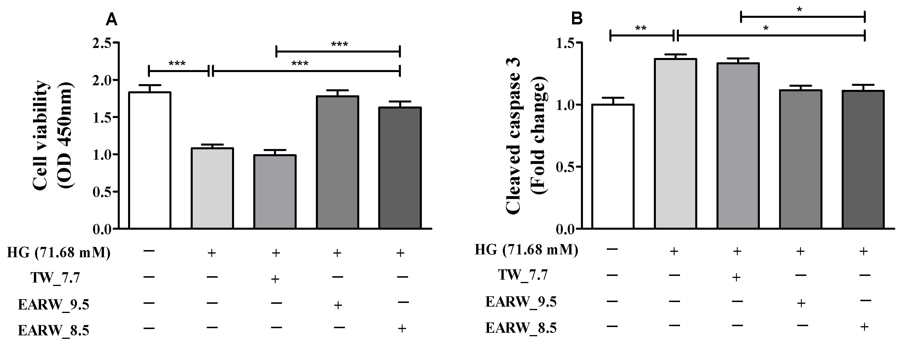
3.1. Cell Viability and Cleaved Caspase-3 Level in HG-Induced HK-2 Cells Are Alleviated by EWARW

To study the effects of EARW_8.5 on HG-induced HK-2 cells, we induced the cells with HG (71.68 mM), which is obtained from HK-2 cell cytotoxicity (IC50) and treated them for 24 h [8] with TW_7.7, EARW_9.5, and EARW_8.5. Compared with the NC group, the viability of cells treated with HG was considerably reduced (p < 0.001). Similarly, a significant increase in cell viability was observed in cells treated with HG+EARW_8.5 (p < 0.001) compared to the HG-only and HG+TW_7.7-treated group (Figure 1A). Similarly, we found that after HG induction, levels of cleaved caspase-3, which is involved in programmed cell death, increased significantly (p < 0.01) compared with the NC group. However, treatment with EARW_8.5 significantly (p < 0.05) reduced the level of cleaved caspase-3 compared with the HG-only and HG+TW_7.7-treated group (Figure 1B).

3.2. Increase in OS in HG-Induced HK-2 Cells Is Suppressed by EWARW

To illustrate the antioxidant potential of EARW_8.5 against OS, ROS and NO levels were determined in HG-induced HK-2 cells. In particular, HG induction significantly increased cellular ROS and NO levels compared to those in the NC group (both p < 0.001). The HG+TW_7.7 group showed a significant increase in cellular ROS (p < 0.001) and NO levels (p < 0.01) (Figure 2A,B). However, this increase in ROS levels was reduced after EARW_8.5 treatment (p < 0.001) compared to the HG-only and HG+TW_7.7 groups (Figure 2A). Similarly, the level of NO significantly decreased after treatment with EARW_8.5 (p < 0.05) compared to the HG-only and HG+TW_7.7 group (p < 0.01), as shown in Figure 2B. Moreover, we chose CAT and GPx among the cellular antioxidant mechanisms to examine the effect of EARW_8.5 on these enzyme activities under OS. We found that CAT activity was significantly reduced in the HG+EARW_8.5 group compared to the HG-only (p < 0.01) and HG+TW_7.7 (p < 0.01) groups (Figure 2C). In contrast, we found that GPx activity was markedly enhanced in the HG+EARW group compared to the HG-only (p < 0.01) and HG+ TW_7.7 (p < 0.001) groups (Figure 2D).

3.3. EWARW Attenuates the Increase in p-JNK and p-p38 MAPK Proteins in HG-Induced HK-2 Cells

To analyze the effects of EARW_8.5 on HG-induced HK-2 cells, we induced OS in HK-2 cells using 71.68 mM glucose and then treated them for 24 h with TW_7.7, EARW_9.5, or EARW_8.5. Western blotting and its relative concentrations were used to analyze the p-JNK and p-p38 proteins. Here, we found that p-JNK (p < 0.001) and p-p38 (p < 0.001) MAPK proteins were significantly elevated in the HG-only group compared to the NC group (Figure 3). However, these increases in MAPK signaling proteins were markedly reduced after EARW_8.5 treatment (both p < 0.001). Similarly, the EARW_8.5-treated group also showed a significant decline in p-JNK (p < 0.01) and p-p38 (p < 0.001) compared to the TW_7.7-treated group (Figure 3A,B).

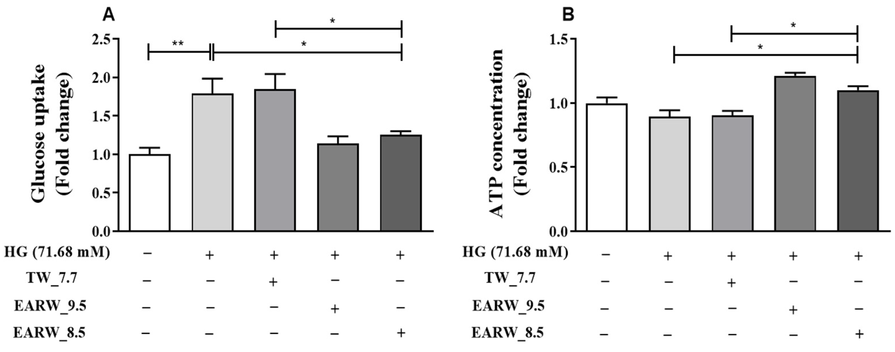
3.4. EWARW Improves Glucose Uptake and ATP Concentration in HG-Induced HK-2 Cells

We measured glucose uptake levels and ATP concentrations to determine the effect of EARW_8.5 treatment on HG-induced HK-2 cells. Our results showed a significant increase in glucose uptake after HG induction (p < 0.01) in the HG-only group compared to the NC group, whereas the HG+EARW_8.5 treatment group showed a significant reduction (p < 0.05) in glucose uptake compared to the HG-only and HG + TW_7.7 groups (Figure 4A). In contrast, we found that ATP concentration was significantly increased in the HG + EARW_8.5 treatment group as compared to the HG-only (p < 0.05) and HG + TW_7.7 (p < 0.05) groups (Figure 4B).

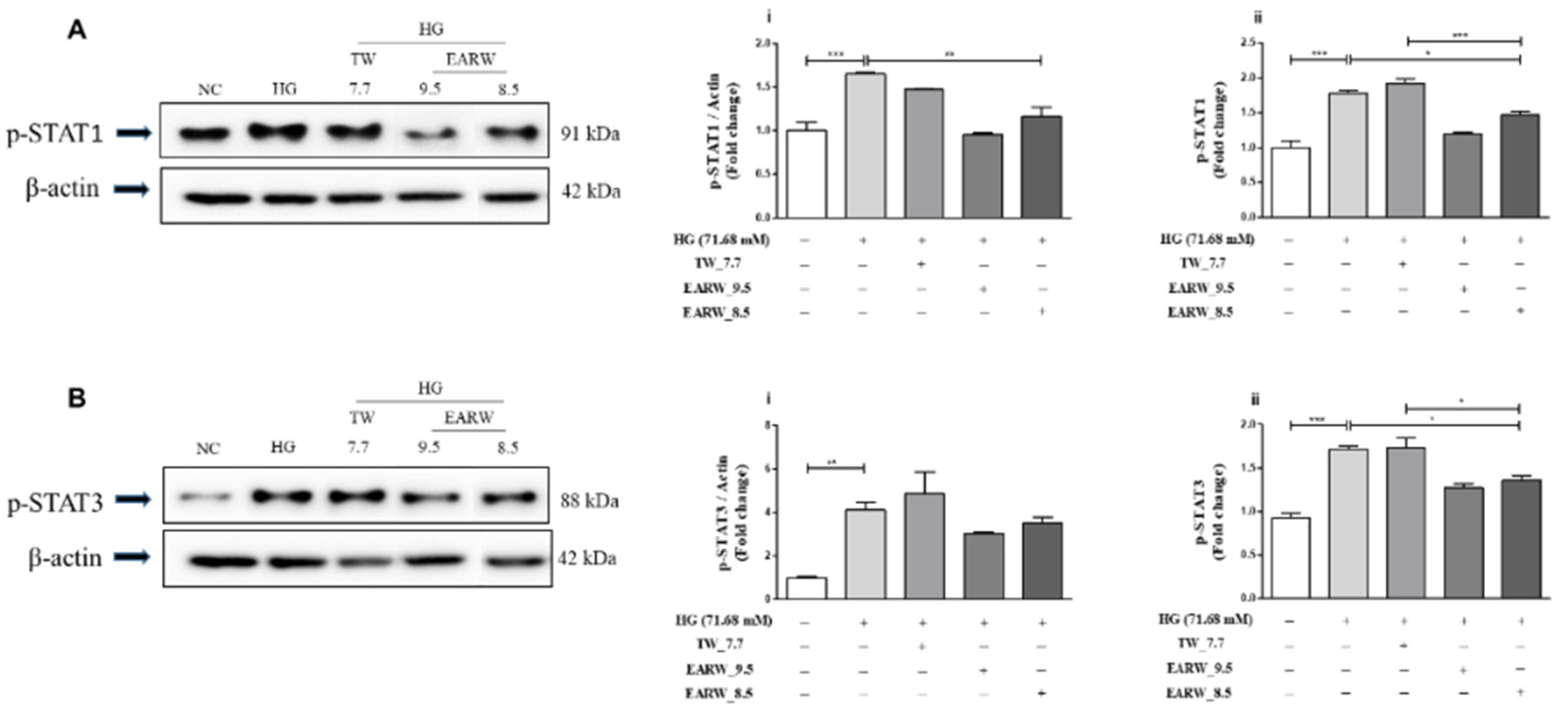
3.5. EWARW Rescued STAT Proteins in HG-Induced HK-2 Cells

After HG induction, we observed increased expression of p-STAT1 and p-STAT3 proteins in HK-2 cells. In particular, we observed a significant increase in the protein levels of p-STAT1 (p < 0.001) following HG induction in the HG-only group compared to the NC group in both the Western blot relative intensity and in-cell ELISA data. However, this expression was significantly reduced after treatment with EARW_8.5 as observed in Western blotting (p < 0.01) and in-cell ELISA (p < 0.05) compared to the HG-only group (Figure 5A). Additionally, our in-cell ELISA results for p-STAT1 in the EARW_8.5-treated group showed a significant reduction (p < 0.001) compared to the TW_7.7-treated group (Figure 5Aii). Similarly, we found significantly increased expression of p-STAT3 in the HG-only group compared to the NC group in HK-2 cells by Western blot relative intensity (p < 0.01) and in-cell ELISA (p < 0.001). Furthermore, we observed that treatment with EARW_8.5 reduced the relative expression of p-STAT3 in the Western blot data, but without any significant difference compared to the HG-only and TW_7.7-treated groups. Moreover, the in-cell ELISA results showed significant downregulation of p-STAT3 after HG+EARW_8.5 treatment (p < 0.05) compared to both the HG-only and TW_7.7-treated groups (Figure 5B).

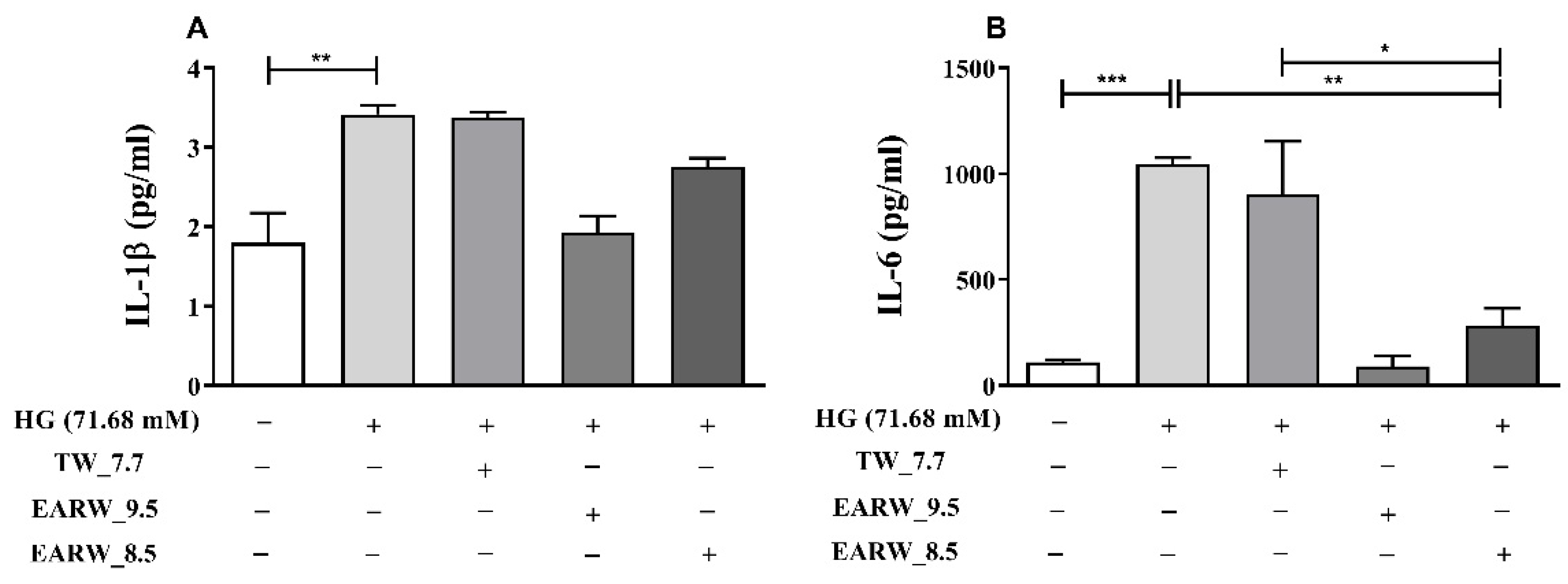
3.6. EWARW Rescues Pro-inflammatory Cytokines in HG-Induced HK-2 Cells

We used HG-treated HK-2 cells to investigate the effects of EARW_8.5 in vitro. As shown in Figure 6, HK-2 cells were exposed to 71.68 mM glucose and then treated with TW_7.7, EARW_9.5, or EARW_8.5 in specified groups for 24 h. Interestingly, IL-1β (p < 0.01) and IL-6 (p < 0.001) in the HG-induced group increased significantly compared to the NC group. Specifically, treatment with HG+EARW_8.5 reduced the level of IL-1β in comparison with the HG-only group, but the difference was not significant. However, the level of IL-6 in the HG + EARW_8.5 (p < 0.01) and HG + TW_7.7 (p < 0.05) groups decreased significantly compared to that in the HG-only group (Figure 6A,B).

4. Discussion

This study investigated the anti-oxidative and anti-diabetic effects of EARW_8.5 in HG-induced HK-2 cells. Several studies have demonstrated the beneficial effects of ARW against OS and diabetes in vivo and in vitro [3,8,29]. However, the collective anti-oxidative, anti-inflammatory, and anti-diabetic effects of EWARW on HG-induced HK-2 cells have not been extensively explored. Towards this, only a few studies have examined the effects of EWARW at pH 8.5 [3]; however, the majority of the studies investigated the effects of EARW at pH ranging from 9.5 to 10.5 [5,6,7]. In this study, we investigated and compared the effects of EARW_8.5 against HG-induced nephrotoxicity using HK-2 cells as a diabetic in vitro model. To the best of our knowledge, this is the first study to explore the effects of EARW_8.5 on HG-induced HK-2 cells. This cell line is frequently used to evaluate cellular responses during in vitro nephrotoxicity [32,33].
Interestingly, we found that EARW_8.5 regulated the growth of HK-2 cells cultured in HG media. One study showed that the activity of cleaved caspase-3, which is involved in apoptosis, is altered under diabetic conditions in proximal tubular cells [34]. Thus, in this experiment, we attempted to explore whether reduced cell viability is associated with cell apoptosis and whether EARW_8.5 can reduce cleaved caspase-3 activity. Our results demonstrated that HG-induced HK-2 cells showed decreased cell viability caused by apoptosis, as shown by the significant cleaved caspase-3 activation. Importantly, treatment with EARW_8.5 inhibited the enhanced onset of apoptosis (Figure 1). Thus, in this study, EARW_8.5 was efficient in countering the decreased cell viability caused by apoptosis.
Next, we assessed ROS and NO levels to determine the effect of EARW_8.5 on OS caused by HG induction in HK-2 cells. OS influences the progression of diabetes-related complications [35]. At low concentrations, ROS and reactive nitrogen species (RNS) are considered the primary signaling regulators and essential for normal cellular activity [36]. Excessive production of ROS and NO can cause the cellular redox state to become unstable, leading to irreversible or permanent cellular damage and apoptosis [37,38]. Here, we found that EARW_8.5 effectively reduces the excessive production of ROS and NO in HG-induced HK-2 cells.
Similarly, we also attempted to explore the role of EARW_8.5 on the activity of antioxidant enzymes such as CAT and GPx in HK-2 cells after HG induction. CAT and GPx help cells are free from free radicals, directly or indirectly [39,40]. CAT directly converts hydrogen peroxide (H2O2) (an oxygen-derived free radical) into water and oxygen, whereas GPx catalyzes the decomposition of H2O2 [41]. Moreover, CAT and GPx are widely recognized antioxidants that reduce OS indicators such as ROS and NO in the kidneys and other cell lines, as mentioned in previous studies [8,42,43]. Interestingly, our results showed that with HG-induced OS, EARW_8.5 treatment efficiently boosted antioxidant enzyme activities, including CAT and GPx (Figure 2). These findings suggest that by enhancing antioxidant enzyme activities, EARW_8.5 is efficient in mitigating the excessive production of harmful OS markers.
Additionally, to better understand the anti-oxidative effect of EARW_8.5, we focused on the p-JNK and p-p38 proteins of the MAPK pathway. Multiple investigations have shown that MAPK proteins are necessary for transforming extracellular signals into cellular reactions. JNK and p38 proteins are part of the MAPK signaling pathway, which is activated in response to various cellular stresses in renal diseases [8,42,44,45]. Moreover, the downstream proteins of the MAPK pathway, p-p38 and p-JNK, are essential for cellular reactions and OS [46,47]. However, research has also shown that ARW contributes to an increase in the endogenous antioxidants that defend cells against OS [3]. In this study, we induced HK-2 cells with HG and observed a significant increase in the p-JNK and p-p38 levels. However, after treatment with EARW_8.5, we found a significant reduction in their levels (Figure 3). This indicates that EARW_8.5 is effective in reducing MAPK proteins by enhancing endogenous antioxidants.
Moreover, we investigated glucose uptake and ATP concentration to confirm the hypothesis that EARW_8.5 regulates metabolism by regulating diabetes-associated nephropathy. In proximal tubular cells, HG treatment promotes mitochondrial stress and apoptosis [48,49]. One study explained that HG levels in the cells reduced mitochondrial fusion proteins, increasing mitochondrial fission proteins and cellular apoptosis. HG concentration-mediated cellular stress is linked to glucose uptake and ATP production [16]. Similarly, we found that HG induction in HK-2 cells increased glucose uptake. However, the ATP concentration in the cells showed a decreasing trend but not significantly. This demonstrates that besides the increased glucose uptake in the cells of the HG-only group, the production of ATP was slightly reduced. Hence, glucose cannot be metabolized properly, and the resulting increased glucose in the cytosol exacerbates OS.
In contrast, as expected, the increase in glucose uptake was reduced after the administration of EARW_8.5, and the ATP content was also significantly increased compared to the HG-only group. These results show that glucose in the cytosol is healthily metabolized and converted to ATP after EARW_8.5 treatment (Figure 4). Correspondingly, our results indicate that the physiological properties of EARW_8.5 in HK-2 cells help reduce the detrimental effect of diabetes and restore renal function by stabilizing the glucose uptake levels with an upsurge in ATP concentration.
Furthermore, we demonstrated that EARW_8.5 treatment inhibits the renal expression of STAT proteins that are implicated in the inflammatory response after HG induction in HK-2 cells. Hyperglycemia promotes renal dysfunction linked with diabetes by regulating a diverse range of genes involved in cell growth and inflammation through the JAK/STAT proteins [50,51,52]. The phosphorylation of downstream transcription factors, such as STAT1 and STAT3, is mediated by JAK [53]. Multiple studies have shown that STAT activation leads to the development of chronic inflammation, in which HG causes kidney damage in vivo and in vitro [20,54]. Thus, STAT protein members could be a potential molecular target for anti-inflammatory therapy [55]. We found that EARW_8.5 reduced p-STAT1 and p-STAT3 activation in HG-induced HK-2 cells (Figure 5). This finding indicates that modulation of this channel with EARW_8.5 can be used to rescue diabetes-related kidney disease.
Finally, as the STAT pathway also regulates cell proliferation and activation, it is a crucial intracellular mechanism for cytokines and other stimuli [20]. Several studies have reported that abnormal inflammation by HG can result in the modulation of pro-inflammatory cytokines, such as IL-1β and IL-6, in HK-2 cells, which can lead to renal damage [8,22,56,57,58]. It has also been reported that EARW can be used to rescue the cytokines that are elevated by OS [59]. Thus, we also investigated the impact of EARW_8.5 on pro-inflammatory cytokines in HG-induced HK-2 cells. We observed that IL-1β and IL-6 levels increased after HG induction. Treatment with EARW_8.5 reduced these changes in the cytokine levels. Among these cytokines, IL-6 levels were drastically upregulated after HG induction, an initial marker of acute inflammation [60] (Figure 6). This evidence supports the beneficial role of EARW_8.5 in inflammation linked with OS generated by HG induction in HK-2 cells.
However, the present study had some limitations. This study utilized proximal tubular epithelial cells, an HK-2 cell line, to investigate the anti-diabetic potential of EARW_8.5 in vitro. Further mechanistic research studies are needed in different kidney cells in relation to diabetes-associated nephropathy dysfunction. A detailed mechanistic investigation of the underlying processes and metabolic investigation, including cytosolic and mitochondrial pathways, is needed for better understanding, as EARW_8.5 reflects multiple effects on renal cells linked to anti-diabetic, anti-oxidative, and anti-inflammatory actions. In conclusion, although various studies on diabetes have been performed to determine the effect of EARW, to the best of our knowledge, this is the first study to explore the anti-diabetic, anti-oxidative, and anti-inflammatory effects of EARW_8.5 on renal HK-2 cells. Moreover, we found that EARW_8.5 had a regulatory impact on various parameters, including cell survival, OS, metabolism, and inflammation, compared to TW. Our results demonstrate that EARW_8.5 regulates and rescues the detrimental effects of diabetes by improving metabolism and alleviating OS and inflammation. Hence, with its therapeutic properties, this study supports the use of EARW_8.5 as an adjuvant treatment to treat diabetic patients with renal problems.

Author Contributions

S.S.: Writing—original draft preparation, Writing—review and editing, Methodology, Investigation. K.-J.L.: Conceptualization, Writing—review and editing, Methodology, Supervision. J.B.: Writing—review and editing, Methodology, Investigation, Data curation. T.T.T.: Investigation. J.M.A.: Investigation. M.H.R.: Investigation. K.V.: Investigation. A.-N.S.: Investigation. S.H.C.: Methodology, Investigation. C.-S.K.: Conceptualization, writing—review and editing, Methodology, Data curation. Y.K.: Conceptualization, Writing—review and editing, Methodology, Supervision. All authors have read and agreed to the published version of the manuscript.

Funding

This study received no external funding.

Institutional Review Board Statement

Not applicable.

Informed Consent Statement

Not applicable.

Data Availability Statement

The data presented in this study are available in the article (tables and figures).

Acknowledgments

The authors would like to thank Ailyn Fadriquela (Yonsei University) for critically reading and reviewing the manuscript.

Conflicts of Interest

The authors declare no conflict of interest.

References

  1. Jéquier, E.; Constant, F. Water as an essential nutrient: The physiological basis of hydration. Eur. J. Clin. Nutr. 2010, 64, 115–123. [Google Scholar] [CrossRef] [Green Version]
  2. Islam, R.; Faysal, S.M.; Amin, R.; Juliana, F.M.; Islam, M.J.; Alam, J.; Hossain, M.N.; Asaduzzaman, M. Assessment of ph and total dissolved substances (tds) in the commercially available bottled drinking water. IOSR J. Nurs. Health Sci. 2017, 6, 35–40. [Google Scholar] [CrossRef]
  3. Trinh, T.T.; Fadriquela, A.; Bajgai, J.; Sharma, S.; Rahman, M.H.; Goh, S.H.; Khang, S.S.; Khang, W.R.; Kim, C.S.; Lee, K.J. Anti-oxidative effect of weak alkaline reduced water in raw 264.7 murine macrophage cells. Processes 2021, 9, 2062. [Google Scholar] [CrossRef]
  4. Tanaka, Y.; Saihara, Y.; Izumotani, K.; Nakamura, H. Daily ingestion of alkaline electrolyzed water containing hydrogen influences human health, including gastrointestinal symptoms. Med. Gas Res. 2018, 8, 160–166. [Google Scholar] [CrossRef] [PubMed]
  5. Park, S.K.; Qi, X.F.; Song, S.B.; Kim, D.H.; Teng, Y.C.; Yoon, Y.S.; Kim, K.Y.; Li, J.H.; Jin, D.; Lee, K.J. Electrolyzed-reduced water inhibits acute ethanol-induced hangovers in sprague-dawley rats. Biomed. Res. 2009, 30, 263–269. [Google Scholar] [CrossRef] [PubMed] [Green Version]
  6. Magro, M.; Corain, L.; Ferro, S.; Baratella, D.; Bonaiuto, E.; Terzo, M.; Corraducci, V.; Salmaso, L.; Vianello, F. Alkaline water and longevity: A murine study. Evid. Based Complement. Alternat. Med. 2016, 2016, 3084126. [Google Scholar] [CrossRef] [Green Version]
  7. Logozzi, M.; Mizzoni, D.; Di Raimo, R.; Andreotti, M.; Macchia, D.; Spada, M.; Fais, S. In vivo antiaging effects of alkaline water supplementation. J. Enzym. Inhib. Med. Chem. 2020, 35, 657–664. [Google Scholar] [CrossRef] [Green Version]
  8. Sharma, S.; Bajgai, J.; Antonio, J.M.; Fadriquela, A.; Trinh, T.T.; Rahman, M.H.; Vira, K.; Sofian, A.N.; Kim, C.S.; Lee, K.J. Anti-hyperglycemic effect of magnesium-enhanced alkaline-reduced water on high glucose-induced oxidative stress in renal tubular epithelial cells. Processes 2022, 10, 919. [Google Scholar] [CrossRef]
  9. Zia, A.; Farkhondeh, T.; Pourbagher-Shahri, A.M.; Samarghandian, S. Role of oxidative stress and inflammatory factors in diabetic kidney disease. Curr. Mol. Med. 2022, 22, 37–49. [Google Scholar] [CrossRef]
  10. Trinh, T.T.; Fadriquela, A.; Lee, K.J.; Bajgai, J.; Sharma, S.; Rahman, M.H.; Kim, C.S.; Youn, S.H.; Jeon, H.T. Development of alkaline reduced water using high-temperature-roasted mineral salt and its antioxidative effect in raw 264.7 murine macrophage cell line. Processes 2021, 9, 1928. [Google Scholar] [CrossRef]
  11. Chen, P.; Yuan, Y.; Zhang, T.; Xu, B.; Gao, Q.; Guan, T. Pentosan polysulfate ameliorates apoptosis and inflammation by suppressing activation of the p38 mapk pathway in high glucosetreated hk2 cells. Int. J. Mol. Med. 2018, 41, 908–914. [Google Scholar] [CrossRef] [PubMed] [Green Version]
  12. Pan, Y.; Huang, Y.; Wang, Z.; Fang, Q.; Sun, Y.; Tong, C.; Peng, K.; Wang, Y.; Miao, L.; Cai, L.; et al. Inhibition of mapk-mediated ace expression by compound c66 prevents stz-induced diabetic nephropathy. J. Cell. Mol. Med. 2014, 18, 231–241. [Google Scholar] [CrossRef] [PubMed]
  13. Hong, Z.; Hong, Z.; Wu, D.; Nie, H. Specific mapk inhibitors prevent hyperglycemia-induced renal diseases in type 1 diabetic mouse model. Mol. Cell. Biochem. 2016, 419, 1–9. [Google Scholar] [CrossRef]
  14. Delos Reyes, F.S.L.G.; Mamaril, A.C.C.; Matias, T.J.P.; Tronco, M.K.V.; Samson, G.R.; Javier, N.D.; Fadriquela, A.; Antonio, J.M.; Sajo, M.E.J.V. The search for the elixir of life: On the therapeutic potential of alkaline reduced water in metabolic syndromes. Processes 2021, 9, 1876. [Google Scholar] [CrossRef]
  15. Bajgai, J.; Kim, C.S.; Rahman, M.H.; Jeong, E.S.; Jang, H.Y.; Kim, K.E.; Choi, J.; Cho, I.; Lee, K.J.; Lee, M. Effects of alkaline-reduced water on gastrointestinal diseases. Processes 2022, 10, 87. [Google Scholar] [CrossRef]
  16. Lee, W.C.; Chau, Y.Y.; Ng, H.Y.; Chen, C.H.; Wang, P.W.; Liou, C.W.; Lin, T.K.; Chen, J.B. Empagliflozin protects hk-2 cells from high glucose-mediated injuries via a mitochondrial mechanism. Cells 2019, 8, 1085. [Google Scholar] [CrossRef] [PubMed] [Green Version]
  17. Gurzov, E.N.; Stanley, W.J.; Pappas, E.G.; Thomas, H.E.; Gough, D.J. The jak/stat pathway in obesity and diabetes. FEBS J. 2016, 283, 3002–3015. [Google Scholar] [CrossRef] [PubMed] [Green Version]
  18. Jiao, B.; An, C.; Du, H.; Tran, M.; Wang, P.; Zhou, D.; Wang, Y. STAT6 Deficiency Attenuates Myeloid Fibroblast Activation and Macrophage Polarization in Experimental Folic Acid Nephropathy. Cells 2021, 10, 3057. [Google Scholar] [CrossRef] [PubMed]
  19. Jiao, B.; An, C.; Tran, M.; Du, H.; Wang, P.; Zhou, D.; Wang, Y. Pharmacological inhibition of STAT6 ameliorates myeloid fibroblast activation and alternative macrophage polarization in renal fibrosis. Front. Immunol. 2021, 12. [Google Scholar] [CrossRef]
  20. Ortiz-Munoz, G.; Lopez-Parra, V.; Lopez-Franco, O.; Fernandez-Vizarra, P.; Mallavia, B.; Flores, C.; Sanz, A.; Blanco, J.; Mezzano, S.; Ortiz, A.; et al. Suppressors of cytokine signaling abrogate diabetic nephropathy. J. Am. Soc. Nephrol. 2010, 21, 763–772. [Google Scholar] [CrossRef] [PubMed]
  21. Chen, B.; Jia, Y.; Lu, D.; Sun, Z. Acute glucose fluctuation promotes in vitro intestinal epithelial cell apoptosis and inflammation via the nox4/ros/jak/stat3 signaling pathway. Exp. Ther. Med. 2021, 22, 688. [Google Scholar] [CrossRef]
  22. Lin, Y.; Zhong, L.; Li, H.; Xu, Y.; Li, X.; Zheng, D. Psoralen alleviates high glucose-induced hk-2 cell injury by inhibition of smad 2 signaling via upregulation of microrna 874. BMC Pharmacol. Toxicol. 2020, 21, 52. [Google Scholar] [CrossRef] [PubMed]
  23. Giap, L.K.; Muthuraman, A. Health benefits of alkaline ionized water. Rapp. De Pharm. 2020, 6, 616–621. [Google Scholar]
  24. Rubik, B. Studies and observations on the health effects of drinking electrolyzed-reduced alkaline water. In Water and Society; Pepper, D.W., Brebbia, C.A., Eds.; WIT Press: Southampton, UK, 2011; pp. 317–327. [Google Scholar]
  25. Balaji, R.; Duraisamy, R.; Kumar, M.P. Complications of diabetes mellitus: A review. Drug Invent. Today 2019, 12, 98–103. [Google Scholar]
  26. Saeedi, P.; Petersohn, I.; Salpea, P.; Malanda, B.; Karuranga, S.; Unwin, N.; Colagiuri, S.; Guariguata, L.; Motala, A.A.; Ogurtsova, K.; et al. Global and regional diabetes prevalence estimates for 2019 and projections for 2030 and 2045: Results from the International Diabetes Federation Diabetes Atlas, 9th edition. Diabetes Res. Clin. Pract. 2019, 157, 107843. [Google Scholar] [CrossRef] [PubMed] [Green Version]
  27. Aghadavod, E.; Khodadadi, S.; Baradaran, A.; Nasri, P.; Bahmani, M.; Rafieian-Kopaei, M. Role of oxidative stress and inflammatory factors in diabetic kidney disease. Iran. J. Kidney Dis. 2016, 10, 337–343. [Google Scholar]
  28. Cho, S.J.; Kang, K.A.; Piao, M.J.; Ryu, Y.S.; Fernando, P.; Zhen, A.X.; Hyun, Y.J.; Ahn, M.J.; Kang, H.K.; Hyun, J.W. 7, 8-dihydroxyflavone protects high glucose-damaged neuronal cells against oxidative stress. Biomol. Ther. 2019, 27, 85–91. [Google Scholar] [CrossRef]
  29. Jin, D.; Ryu, S.H.; Kim, H.W.; Yang, E.J.; Lim, S.J.; Ryang, Y.S.; Chung, C.H.; Park, S.K.; Lee, K.J. Anti-diabetic effect of alkaline-reduced water on oletf rats. Biosci. Biotechnol. Biochem. 2006, 70, 31–37. [Google Scholar] [CrossRef] [PubMed] [Green Version]
  30. John, S. Complication in diabetic nephropathy. Diabetes Metab. Syndr. 2016, 10, 247–249. [Google Scholar] [CrossRef] [PubMed]
  31. Sturm, G.; Kollerits, B.; Neyer, U.; Ritz, E.; Kronenberg, F.; MMKD Study Group. Uric acid as a risk factor for progression of non-diabetic chronic kidney disease? The mild to moderate kidney disease (mmkd) study. Exp. Gerontol. 2008, 43, 347–352. [Google Scholar] [CrossRef] [Green Version]
  32. Ryan, M.J.; Johnson, G.; Kirk, J.; Fuerstenberg, S.M.; Zager, R.A.; Torok-Storb, B. Hk-2: An immortalized proximal tubule epithelial cell line from normal adult human kidney. Kidney Int. 1994, 45, 48–57. [Google Scholar] [CrossRef] [Green Version]
  33. Mossoba, M.E.; Mapa, M.S.T.; Sprando, J.; Araujo, M.; Sprando, R.L. Evaluation of transporter expression in hk-2 cells after exposure to free and ester-bound 3-mcpd. Toxicol. Rep. 2021, 8, 436–442. [Google Scholar] [CrossRef]
  34. Zhang, X.Q.; Dong, J.J.; Cai, T.; Shen, X.; Zhou, X.J.; Liao, L. High glucose induces apoptosis via upregulation of bim expression in proximal tubule epithelial cells. Oncotarget 2017, 8, 24119–24129. [Google Scholar] [CrossRef]
  35. Kowluru, R.A.; Chan, P.S. Oxidative stress and diabetic retinopathy. Exp. Diabetes Res. 2007, 2007, 43603. [Google Scholar] [CrossRef] [Green Version]
  36. Gharib, B.; Hanna, S.; Abdallahi, O.M.; Lepidi, H.; Gardette, B.; De Reggi, M. Anti-inflammatory properties of molecular hydrogen: Investigation on parasite-induced liver inflammation. C. R. Acad. Sci. III 2001, 324, 719–724. [Google Scholar] [CrossRef]
  37. Wang, M.; Cao, X.; Luan, C.; Li, Z. Hydrogen sulfide attenuates hydrogen peroxide-induced injury in human lung epithelial a549 cells. Int. J. Mol. Sci. 2019, 20, 3975. [Google Scholar] [CrossRef] [PubMed] [Green Version]
  38. Lubos, E.; Handy, D.E.; Loscalzo, J. Role of oxidative stress and nitric oxide in atherothrombosis. Front. Biosci. 2008, 13, 5323–5344. [Google Scholar] [CrossRef] [Green Version]
  39. Liou, G.Y.; Storz, P. Reactive oxygen species in cancer. Free Radic. Res. 2010, 44, 479–496. [Google Scholar] [CrossRef] [Green Version]
  40. Heck, D.E.; Shakarjian, M.; Kim, H.D.; Laskin, J.D.; Vetrano, A.M. Mechanisms of oxidant generation by catalase. Ann. N. Y. Acad. Sci. 2010, 1203, 120–125. [Google Scholar] [CrossRef] [Green Version]
  41. Jeon, G.; Kim, C.; Cho, U.M.; Hwang, E.T.; Hwang, H.S.; Min, J. Melanin-decolorizing activity of antioxidant enzymes, glutathione peroxidase, thiol peroxidase, and catalase. Mol. Biotechnol. 2021, 63, 150–155. [Google Scholar] [CrossRef]
  42. You, I.S.; Sharma, S.; Fadriquela, A.; Bajgai, J.; Thi, T.T.; Rahman, M.H.; Sung, J.; Kwon, H.U.; Lee, S.Y.; Kim, C.S.; et al. Antioxidant properties of hydrogen gas attenuates oxidative stress in airway epithelial cells. Molecules 2021, 26, 6375. [Google Scholar] [CrossRef] [PubMed]
  43. Ye, J.; Li, J.; Yu, Y.; Wei, Q.; Deng, W.; Yu, L. L-carnitine attenuates oxidant injury in hk-2 cells via ros-mitochondria pathway. Regul. Pept. 2010, 161, 58–66. [Google Scholar] [CrossRef]
  44. Lv, Z.M.; Wang, Q.; Wan, Q.; Lin, J.G.; Hu, M.S.; Liu, Y.X.; Wang, R. The role of the p38 mapk signaling pathway in high glucose-induced epithelial-mesenchymal transition of cultured human renal tubular epithelial cells. PLoS ONE 2011, 6, e22806. [Google Scholar] [CrossRef]
  45. Lv, L.; Zhang, J.; Tian, F.; Li, X.; Li, D.; Yu, X. Arbutin protects hk-2 cells against high glucose-induced apoptosis and autophagy by up-regulating microrna-27a. Artif. Cells Nanomed. Biotechnol. 2019, 47, 2940–2947. [Google Scholar] [CrossRef] [Green Version]
  46. Son, Y.; Cheong, Y.K.; Kim, N.H.; Chung, H.T.; Kang, D.G.; Pae, H.O. Mitogen-activated protein kinases and reactive oxygen species: How can ros activate mapk pathways? J. Signal Transduct. 2011, 2011, 792639. [Google Scholar] [CrossRef] [Green Version]
  47. Behl, T.; Rana, T.; Alotaibi, G.H.; Shamsuzzaman, M.; Naqvi, M.; Sehgal, A.; Singh, S.; Sharma, N.; Almoshari, Y.; Abdellatif, A.A.H.; et al. Polyphenols inhibiting mapk signalling pathway mediated oxidative stress and inflammation in depression. Biomed. Pharmacother. 2022, 146, 112545. [Google Scholar] [CrossRef] [PubMed]
  48. Lee, W.C.; Chiu, C.H.; Chen, J.B.; Chen, C.H.; Chang, H.W. Mitochondrial fission increases apoptosis and decreases autophagy in renal proximal tubular epithelial cells treated with high glucose. DNA Cell Biol. 2016, 35, 657–665. [Google Scholar] [CrossRef] [PubMed]
  49. Zhan, M.; Usman, I.M.; Sun, L.; Kanwar, Y.S. Disruption of renal tubular mitochondrial quality control by myo-inositol oxygenase in diabetic kidney disease. J. Am. Soc. Nephrol. 2015, 26, 1304–1321. [Google Scholar] [CrossRef] [Green Version]
  50. Brosius, F.C. New insights into the mechanisms of fibrosis and sclerosis in diabetic nephropathy. Rev. Endocr. Metab. Disord. 2008, 9, 245–254. [Google Scholar] [CrossRef] [PubMed] [Green Version]
  51. Marrero, M.B.; Banes-Berceli, A.K.; Stern, D.M.; Eaton, D.C. Role of the jak/stat signaling pathway in diabetic nephropathy. Am. J. Physiol. Renal Physiol. 2006, 290, F762–F768. [Google Scholar] [CrossRef]
  52. Lu, T.C.; Wang, Z.H.; Feng, X.; Chuang, P.Y.; Fang, W.; Shen, Y.; Levy, D.E.; Xiong, H.; Chen, N.; He, J.C. Knockdown of stat3 activity in vivo prevents diabetic glomerulopathy. Kidney Int. 2009, 76, 63–71. [Google Scholar] [CrossRef] [Green Version]
  53. Butturini, E.; Carcereri de Prati, A.; Mariotto, S. Redox regulation of stat1 and stat3 signaling. Int. J. Mol. Sci. 2020, 21, 7034. [Google Scholar] [CrossRef]
  54. Chen, D.; Liu, Y.; Chen, J.; Lin, H.; Guo, H.; Wu, Y.; Xu, Y.; Zhou, Y.; Zhou, W.; Lu, R.; et al. Jak/stat pathway promotes the progression of diabetic kidney disease via autophagy in podocytes. Eur. J. Pharmacol. 2021, 902, 174121. [Google Scholar] [CrossRef]
  55. de Prati, A.C.; Ciampa, A.R.; Cavalieri, E.; Zaffini, R.; Darra, E.; Menegazzi, M.; Suzuki, H.; Mariotto, S. Stat1 as a new molecular target of anti-inflammatory treatment. Curr. Med. Chem. 2005, 12, 1819–1828. [Google Scholar] [CrossRef]
  56. Zhou, J.; Zhang, S.; Sun, X.; Lou, Y.; Yu, J. Hyperoside protects hk-2 cells against high glucose-induced apoptosis and inflammation via the mir-499a-5p/nrip1 pathway. Pathol. Oncol. Res. 2021, 27, 629829. [Google Scholar] [CrossRef]
  57. Xiang, E.; Han, B.; Zhang, Q.; Rao, W.; Wang, Z.F.; Chang, C.; Zhang, Y.Q.; Tu, C.S.; Li, C.Y.; Wu, D.C. Human umbilical cord-derived mesenchymal stem cells prevent the progression of early diabetic nephropathy through inhibiting inflammation and fibrosis. Stem Cell Res. Ther. 2020, 11, 1–14. [Google Scholar] [CrossRef]
  58. Karmacharya, U.; Regmi, S.C.; Awasthi, B.P.; Chaudhary, P.; Kim, Y.E.; Lee, I.H.; Nam, T.g.; Kim, J.A.; Jeong, B.S. Synthesis and activity of n-(5-hydroxy-3, 4, 6-trimethylpyridin-2-yl) acetamide analogues as anticolitis agents via dual inhibition of tnf-α-and il-6-induced cell adhesions. Bioorg. Med. Chem. Lett. 2021, 43, 128059. [Google Scholar] [CrossRef]
  59. Rias, Y.A.; Kurniawan, A.L.; Chang, C.W.; Gordon, C.J.; Tsai, H.T. Synergistic effects of regular walking and alkaline electrolyzed water on decreasing inflammation and oxidative stress, and increasing quality of life in individuals with type 2 diabetes: A community based randomized controlled trial. Antioxidants 2020, 9, 946. [Google Scholar] [CrossRef]
  60. Gabay, C. Interleukin-6 and chronic inflammation. Arthritis Res. Ther. 2006, 8 (Suppl. 2), S3. [Google Scholar] [CrossRef] [PubMed]
Figure 1. Effect of EARW_8.5 on cell viability and cleaved caspase-3 in HG-induced HK-2 cells. (A) Cell viability of HK-2 cells was analyzed after induction with 71.68 mM glucose and treatment with 10% TW_7.7, EARW_9.5, or EARW_8.5 for 24 h. (B) Cleaved caspase-3 was analyzed by an enzyme-linked immunosorbent assay (ELISA). Values were calculated as mean ± standard error of mean (SEM). * p < 0.05, ** p < 0.01, and *** p < 0.001 indicate significant differences when tested with one-way analysis of variance (ANOVA).
Processes 10 02025 g001
Figure 2. OS markers, including ROS, NO, CAT, and GPx, were investigated to understand the effect of EARW_8.5 after HG-induction. HK-2 cells were induced with HG (71.68 mM) and treated with different experimental waters. After 24 h, (A) ROS, (B) NO, (C) CAT, and (D) GPx were analyzed. Values were calculated in mean ± SEM. * p < 0.05, ** p < 0.01, and *** p < 0.001 indicate significant differences when tested with ANOVA.
Processes 10 02025 g002
Figure 3. MAPK proteins p-JNK and p-p38 were analyzed to study the effect of EARW_8.5 after HG-induction. (A) Western blotting and its relative intensity for p-JNK were analyzed. (B) Western blot and its relative intensity for p-p38 were analyzed. Values were calculated in mean ± SEM. ** p < 0.01 and *** p < 0.001 indicate significant differences tested with ANOVA.
Processes 10 02025 g003
Figure 4. Effects of HG-induction on glucose uptake levels and ATP concentrations in HK-2 cells treated with TW_7.7, EARW_9.5, or EARW_8.5. HK-2 cells were induced with HG (71.68 mM) and then treated with various experimental waters. (A) After HG induction and treatment with different experimental waters, a glucose uptake assay was performed. (B) ATP levels with 10% TW_7.7, EARW_9.5, and EARW_8.5 were analyzed after HG induction. Data are presented as mean ± SEM. * p < 0.05 and ** p < 0.01 represent significant differences based on Tukey’s test and ANOVA test.
Processes 10 02025 g004
Figure 5. Inflammatory markers p-STAT1 and p-STAT3 were investigated to understand the effect of EARW_8.5 with different pH after HG induction. (A) (i) Western blot relative intensity and (ii) in-cell ELISA test for p-STAT1 were performed. (B) (i) Western blot fold change and (ii) in-cell ELISA test for p-STAT3 were performed. Values were calculated as mean ± SEM. * p < 0.05, ** p < 0.01, and *** p < 0.001 indicate significant differences tested with ANOVA.
Processes 10 02025 g005
Figure 6. Pro-inflammatory cytokine markers IL-1β and IL-6 were analyzed to investigate the effect of EARW_8.5 after HG-induction. Bio-Plex was used to perform and analyze the data for (A) IL-1β and (B) IL-6 with or without the treatment of EARW_8.5. Values were calculated as mean ± SEM. * p < 0.05, ** p < 0.01 and *** p < 0.001 indicate significant differences tested with ANOVA.
Processes 10 02025 g006
Table 1. Characteristics of the experimental waters.
WaterpHORP (mV)TDS (mg/L)H2 (ppb)
TW_7.77.7609630
EARW_9.59.5−39473225
EARW_8.58.5−6367150
TW—tap water; EARW—electrolyzed alkaline reduced water; ORP—oxidation-reduction potential; TDS—total dissolved solids; H2—molecular hydrogen.
Publisher’s Note: MDPI stays neutral with regard to jurisdictional claims in published maps and institutional affiliations.

