
Guided Growth of the Proximal Femur for the Management of the ‘Hip at Risk’ in Children with Cerebral Palsy—A Systematic Review


Abstract
:1. Introduction
2. Materials and Methods
2.1. Information Sources and Search Terms
2.2. Selection Process
2.3. Assessment of Quality and Bias
2.4. Outcome Measures and Statistics

| Reference | Bias Due to Confounding | Bias in Selection of Participants | Bias in Classification of Intervention | Bias due to Deviations from Intended Intervention | Bias Due to Missing Data | Bias in Measurement of Outcomes | Bias in Selection of the Reported Results | Overall Risk of Bias |
|---|---|---|---|---|---|---|---|---|
| [32] | Critical | Critical | Low | Low | No information | Serious | Moderate | Critical |
| [34] | Critical | Critical | Low | Low | No information | Serious | Moderate | Critical |
| [46] | Critical | Critical | Low | Low | Low | Critical | Moderate | Critical |
| [47] | Critical | Critical | Moderate | Low | Low | Critical | Moderate | Critical |
3. Results
3.1. Primary Outcomes
3.2. Quantitative Analysis
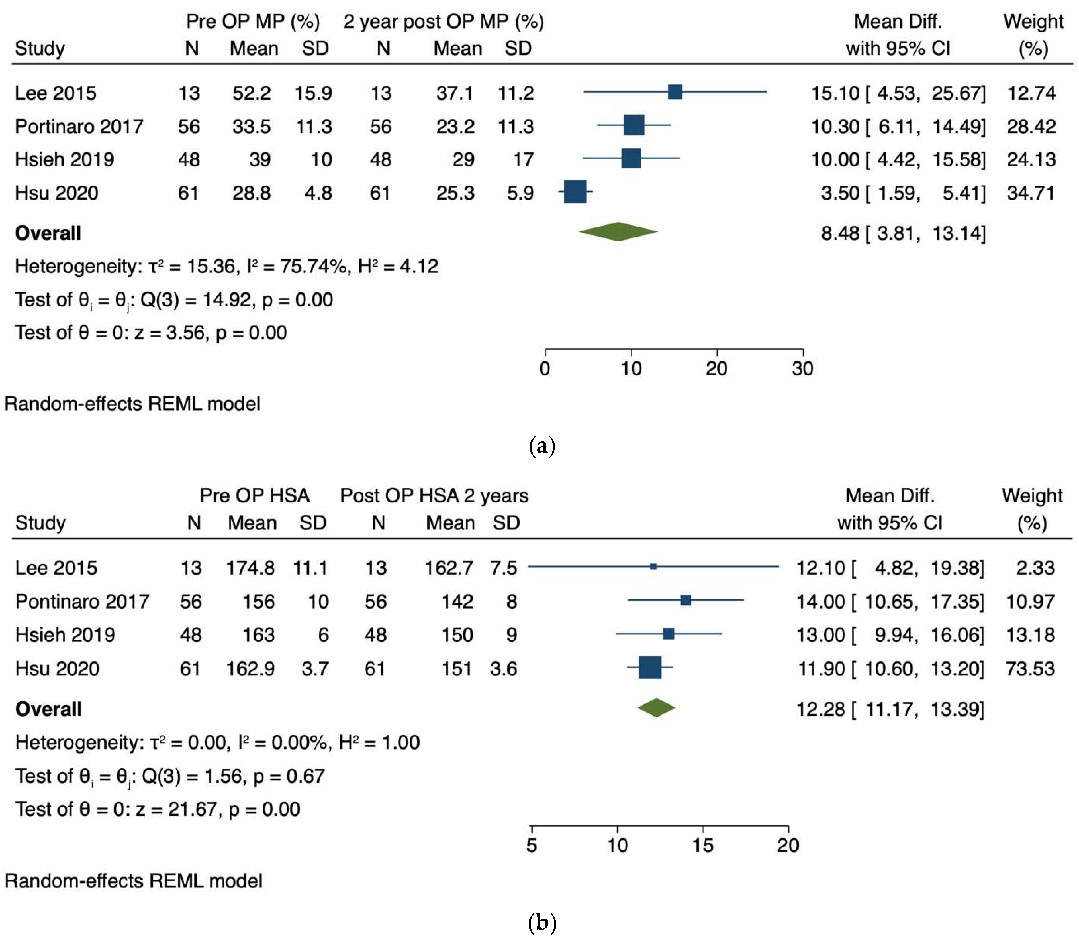
3.2.1. Migration Percentage
3.2.2. Head-Shaft Angle
3.2.3. Acetabular Index
3.3. Secondary Outcome-Reported Technical Considerations, Complications and Limitations of TMH-PF
4. Discussion
Author Contributions
Funding
Institutional Review Board Statement
Informed Consent Statement
Conflicts of Interest
References
- Lee, K.M.; Kang, J.Y.; Chung, C.Y.; Kwon, D.G.; Lee, S.H.; Choi, I.H.; Cho, T.; Yoo, W.J.; Park, M.S. Clinical relevance of valgus deformity of proximal femur in cerebral palsy. J. Pediatr. Orthop. 2010, 30, 720–725. [Google Scholar] [CrossRef] [PubMed]
- Robin, J.; Graham, H.K.; Selber, P.; Dobson, F.; Smith, K.; Baker, R. Proximal femoral geometry in cerebral palsy: A population-based cross-sectional study. J. Bone Jt. Surg. Br. 2008, 90, 1372–1379. [Google Scholar] [CrossRef] [PubMed] [Green Version]
- Gose, S.; Sakai, T.; Shibata, T.; Akiyama, K.; Yoshikawa, H.; Sugamoto, K. Verification of the Robin and Graham classification system of hip disease in cerebral palsy using three-dimensional computed tomography. Dev. Med. Child Neurol. 2011, 53, 1107–1112. [Google Scholar] [CrossRef]
- Graham, H.K.; Thomason, P.; Willoughby, K.; Hastings-Ison, T.; van Stralen, R.; Dala-Ali, B.; Wong, P.; Rutz, E. Musculoskeletal Pathology in Cerebral Palsy: A Classification System and Reliability Study. Children 2021, 8, 252. [Google Scholar] [CrossRef] [PubMed]
- Laplaza, F.J.; Root, L. Femoral anteversion and neck-shaft angles in hip instability in cerebral palsy. J. Pediatr. Orthop. 1994, 14, 719–723. [Google Scholar] [CrossRef]
- Bagg, M.R.; Farber, J.; Miller, F. Long-term follow-up of hip subluxation in cerebral palsy patients. J. Pediatr. Orthop. 1993, 13, 32–36. [Google Scholar] [CrossRef]
- Flynn, J.M.; Miller, F. Management of hip disorders in patients with cerebral palsy. J. Am. Acad. Orthop. Surg. 2002, 10, 198–209. [Google Scholar] [CrossRef]
- Gamble, J.G.; Rinsky, L.A.; Bleck, E.E. Established hip dislocations in children with cerebral palsy. Clin. Orthop. Relat. Res. 1990, 253, 90–99. [Google Scholar] [CrossRef]
- Ramstad, K.; Terjesen, T. Hip pain is more frequent in severe hip displacement: A population-based study of 77 children with cerebral palsy. J. Pediatr. Orthop. Part B 2016, 25, 217–221. [Google Scholar] [CrossRef]
- van der List, J.P.; Witbreuk, M.M.; Buizer, A.I.; van der Sluijs, J.A. The head-shaft angle of the hip in early childhood: A comparison of reference values for children with cerebral palsy and normally developing hips. Bone Jt. J. 2015, 97-B, 1291–1295. [Google Scholar] [CrossRef]
- Shrader, M.W.; Wimberly, L.; Thompson, R. Hip Surveillance in Children With Cerebral Palsy. J. Am. Acad Orthop. Surg. 2019, 27, 760–768. [Google Scholar] [CrossRef]
- Hägglund, G.; Lauge-Pedersen, H.; Wagner, P. Characteristics of children with hip displacement in cerebral palsy. BMC Musculoskelet. Disord. 2007, 8, 101. [Google Scholar] [CrossRef] [PubMed] [Green Version]
- Gordon, G.S.; Simkiss, D.E. A systematic review of the evidence for hip surveillance in children with cerebral palsy. J. Bone Jt. Surg. Br. 2006, 88, 1492–1496. [Google Scholar] [CrossRef] [PubMed] [Green Version]
- Larnert, P.; Risto, O.; Hägglund, G.; Wagner, P. Hip displacement in relation to age and gross motor function in children with cerebral palsy. J. Child Orthop. 2014, 8, 129–134. [Google Scholar] [CrossRef] [PubMed] [Green Version]
- Miller, F.; Bagg, M.R. Age and migration percentage as risk factors for progression in spastic hip disease. Dev. Med. Child Neurol. 1995, 37, 449–455. [Google Scholar] [CrossRef] [PubMed]
- Reimers, J. The stability of the hip in children. A radiological study of the results of muscle surgery in cerebral palsy. Acta Orthop. Scand. Suppl. 1980, 184, 1–100. [Google Scholar] [CrossRef]
- Gonnade, N.; Lokhande, V.; Ajij, M.; Gaur, A.; Shukla, K. Phenol Versus Botulinum Toxin A Injection in Ambulatory Cerebral Palsy Spastic Diplegia: A Comparative Study. J. Pediatr. Neurosci. 2017, 12, 338–343. [Google Scholar] [CrossRef]
- McNerney, N.P.; Mubarak, S.J.; Wenger, D.R. One-stage correction of the dysplastic hip in cerebral palsy with the San Diego acetabuloplasty: Results and complications in 104 hips. J. Pediatr. Orthop. 2000, 20, 93–103. [Google Scholar] [CrossRef]
- Inan, M.; Senaran, H.; Domzalski, M.; Littleton, A.; Dabney, K.; Miller, F. Unilateral versus bilateral peri-ilial pelvic osteotomies combined with proximal femoral osteotomies in children with cerebral palsy: Perioperative complications. J. Pediatr. Orthop. 2006, 26, 547–550. [Google Scholar] [CrossRef] [Green Version]
- Koch, A.; Jozwiak, M.; Idzior, M.; Molinska-Glura, M.; Szulc, A. Avascular necrosis as a complication of the treatment of dislocation of the hip in children with cerebral palsy. Bone Jt. J. 2015, 97-B, 270–276. [Google Scholar] [CrossRef]
- Dobson, F.; Boyd, R.N.; Parrott, J.; Nattrass, G.R.; Graham, H.K. Hip surveillance in children with cerebral palsy. Impact on the surgical management of spastic hip disease. J. Bone Jt. Surg. Br. 2002, 84, 720–726. [Google Scholar] [CrossRef]
- Métaizeau, J.P.; Wong-Chung, J.; Bertrand, H.; Pasquier, P. Percutaneous epiphysiodesis using transphyseal screws (PETS). J. Pediatr. Orthop. 1998, 18, 363–369. [Google Scholar] [CrossRef]
- Stevens, P.M.; Novais, E.N. Multilevel guided growth for hip and knee varus secondary to chondrodysplasia. J. Pediatr. Orthop. 2012, 32, 626–630. [Google Scholar] [CrossRef] [PubMed]
- Bouchard, M. Guided Growth: Novel Applications in the Hip, Knee, and Ankle. J. Pediatr. Orthop. 2017, 37, S32–S36. [Google Scholar] [CrossRef]
- Metaizeau, J.D.; Denis, D.; Louis, D. New femoral derotation technique based on guided growth in children. Orthop. Traumatol. Surg. Res. 2019, 105, 1175–1179. [Google Scholar] [CrossRef]
- Al-Aubaidi, Z.; Lundgaard, B.; Pedersen, N.W. Anterior distal femoral hemiepiphysiodesis in the treatment of fixed knee flexion contracture in neuromuscular patients. J. Child Orthop. 2012, 6, 313–318. [Google Scholar] [CrossRef] [Green Version]
- Klatt, J.; Stevens, P.M. Guided growth for fixed knee flexion deformity. J. Pediatr. Orthop. 2008, 28, 626–631. [Google Scholar] [CrossRef]
- Long, J.T.; Laron, D.; Garcia, M.C.; McCarthy, J.J. Screw Anterior Distal Femoral Hemiepiphysiodesis in Children With Cerebral Palsy and Knee Flexion Contractures: A Retrospective Case-control Study. J. Pediatr. Orthop. 2020, 40, e873–e879. [Google Scholar] [CrossRef]
- Rethlefsen, S.A.; Hanson, A.M.; Wren, T.A.L.; Abousamra, O.; Kay, R.M. Anterior distal femoral hemiepiphysiodesis with and without patellar tendon shortening for fixed knee flexion contractures in children with cerebral palsy. J. Child Orthop. 2020, 14, 415–420. [Google Scholar] [CrossRef]
- Wang, K.K.; Novacheck, T.F.; Rozumalski, A.; Georgiadis, A.G. Anterior Guided Growth of the Distal Femur for Knee Flexion Contracture: Clinical, Radiographic, and Motion Analysis Results. J. Pediatr. Orthop. 2019, 39, e360–e365. [Google Scholar] [CrossRef]
- Agus, H.; Önvural, B.; Kazimoglu, C.; Reisoglu, A.; Kalenderer, O. Medial percutaneous hemi-epiphysiodesis improves the valgus tilt of the femoral head in developmental dysplasia of the hip (DDH) type-II avascular necrosis. Acta Orthop. 2015, 86, 506–510. [Google Scholar] [CrossRef] [PubMed]
- Lee, W.-C.; Kao, H.-K.; Yang, W.-E.; Ho, P.-C.; Chang, C.-H. Guided Growth of the Proximal Femur for Hip Displacement in Children With Cerebral Palsy. J. Pediatr. Orthop. 2016, 36, 511–515. [Google Scholar] [CrossRef] [PubMed]
- McGillion, S.; Clarke, N.M. Lateral growth arrest of the proximal femoral physis: A new technique for serial radiological observation. J. Child Orthop. 2011, 5, 201–207. [Google Scholar] [CrossRef] [PubMed] [Green Version]
- Portinaro, N.; Turati, M.; Cometto, M.; Bigoni, M.; Davids, J.R.; Panou, A. Guided Growth of the Proximal Femur for the Management of Hip Dysplasia in Children With Cerebral Palsy. J. Pediatr. Orthop. 2019, 39, e622–e628. [Google Scholar] [CrossRef] [PubMed]
- Davids, J. Proximal Femur Guided Growth for the Management of Hip Dysplasia in Children with Cerebral Palsy. J. Pediatr. Orthop. Soc. N. Am. 2021, 3, e622–e628. [Google Scholar]
- McCarthy, J.J.; Noonan, K.J.; Nemke, B.; Markel, M. Guided growth of the proximal femur: A pilot study in the lamb model. J. Pediatr. Orthop. 2010, 30, 690–694. [Google Scholar] [CrossRef]
- Chang, C.H.; Chi, C.H.; Lee, Z.L. Progressive coxa vara by eccentric growth tethering in immature pigs. J. Pediatr. Orthop. Part B 2006, 15, 302–306. [Google Scholar] [CrossRef]
- Moher, D.; Shamseer, L.; Clarke, M.; Ghersi, D.; Liberati, A.; Petticrew, M.; Shekelle, P.; Stewart, L.A.; PRISMA-P Group. Preferred reporting items for systematic review and meta-analysis protocols (PRISMA-P) 2015 statement. Syst. Rev. 2015, 4, 1. [Google Scholar] [CrossRef] [Green Version]
- Bax, M.C. Terminology and classification of cerebral palsy. Dev. Med. Child Neurol. 1964, 6, 295–297. [Google Scholar] [CrossRef]
- Bax, M.; Goldstein, M.; Rosenbaum, P.; Leviton, A.; Paneth, N.; Dan, B.; Jacobsson, B.; Damiano, D.; Executive Committee for the Definition of Cerebral Palsy. Proposed definition and classification of cerebral palsy, April 2005. Dev. Med. Child Neurol. 2005, 47, 571–576. [Google Scholar] [CrossRef]
- Sterne, J.A.; Hernán, M.A.; Reeves, B.C.; Savović, J.; Berkman, N.D.; Viswanathan, M.; Henry, D.; Altman, D.G.; Ansari, M.T.; Boutron, I.; et al. ROBINS-I: A tool for assessing risk of bias in non-randomised studies of interventions. BMJ 2016, 355, i4919. [Google Scholar] [CrossRef] [PubMed] [Green Version]
- Higgins, J.; Green, S. Cochrane Handbook for Systematic Reviews of Interventions; Wiley-Blackwell: Oxford, UK, 2008. [Google Scholar]
- Clavien, P.A.; Strasberg, S.M. Severity grading of surgical complications. Ann. Surg. 2009, 250, 197–198. [Google Scholar] [CrossRef] [PubMed]
- Clavien, P.A.; Sanabria, J.R.; Strasberg, S.M. Proposed classification of complications of surgery with examples of utility in cholecystectomy. Surgery 1992, 111, 518–526. [Google Scholar] [PubMed]
- Sink, E.L.; Leunig, M.; Zaltz, I.; Gilbert, J.C.; Clohisy, J.; Group ANfCHOR. Reliability of a complication classification system for orthopaedic surgery. Clin. Orthop. Relat. Res. 2012, 470, 2220–2226. [Google Scholar] [CrossRef] [Green Version]
- Hsieh, H.-C.; Wang, T.-M.; Kuo, K.N.; Huang, S.-C.; Wu, K.-W. Guided Growth Improves Coxa Valga and Hip Subluxation in Children with Cerebral Palsy. Clin. Orthop. Relat. Res. 2019, 477, 2568–2576. [Google Scholar] [CrossRef]
- Hsu, P.-J.; Wu, K.-W.; Lee, C.-C.; Lin, S.-C.; Kuo, K.N.; Wang, T.-M. Does screw position matter for guided growth in cerebral palsy hips? Bone Jt. J. 2020, 102, 1242–1247. [Google Scholar] [CrossRef]
- Chou, P.C.; Huang, Y.C.; Hsueh, C.J.; Lin, J.G.; Chu, H.Y. Retrospective study using MRI to measure depths of acupuncture points in neck and shoulder region. BMJ Open 2015, 5, e007819. [Google Scholar] [CrossRef]
- Torode, I.P.; Young, J.L. Caput valgum associated with developmental dysplasia of the hip: Management by transphyseal screw fixation. J. Child Orthop. 2015, 9, 371–379. [Google Scholar] [CrossRef] [Green Version]
- Chang, C.H. Guided Growth of the Proximal Femur for Hip Displacement in Children with Cerebral Palsy: Response to the Readers’ Comments. J. Pediatr. Orthop. 2015, 35, e84. [Google Scholar] [CrossRef]
- Hermanson, M.; Hägglund, G.; Riad, J.; Rodby-Bousquet, E.; Wagner, P. Prediction of hip displacement in children with cerebral palsy: Development of the CPUP hip score. Bone Jt. J. 2015, 97-B, 1441–1444. [Google Scholar] [CrossRef]
- Kentish, M.; Wynter, M.; Snape, N.; Boyd, R. Five-year outcome of state-wide hip surveillance of children and adolescents with cerebral palsy. J. Pediatr. Rehabil. Med. 2011, 4, 205–217. [Google Scholar] [CrossRef] [PubMed]
- Terjesen, T. The natural history of hip development in cerebral palsy. Dev. Med. Child Neurol. 2012, 54, 951–957. [Google Scholar] [CrossRef] [PubMed]
- Rodda, J.; Graham, H.K. Classification of gait patterns in spastic hemiplegia and spastic diplegia: A basis for a management algorithm. Eur. J. Neurol. 2001, 8 (Suppl. S5), 98–108. [Google Scholar] [CrossRef] [PubMed]
- Wynter, M.; Gibson, N.; Willoughby, K.L.; Love, S.; Kentish, M.; Thomason, P.; Graham, H.K.; National Hip Surveillance Working Group. Australian hip surveillance guidelines for children with cerebral palsy: 5-year review. Dev. Med. Child Neurol. 2015, 57, 808–820. [Google Scholar] [CrossRef] [PubMed]
- Wordie, S.J.; Bugler, K.E.; Bessell, P.R.; Robb, J.E.; Gaston, M.S. Hip displacement in children with cerebral palsy. Bone Jt. J. 2021, 103-B, 411–414. [Google Scholar] [CrossRef] [PubMed]
- Cappello, T. Expanded Indications for Guided Growth in Pediatric Extremities. JPOSNA 2021, 3, 630. [Google Scholar]
- Peng, S.H.; Lee, W.C.; Kao, H.K.; Yang, W.E.; Chang, C.H. Guided growth for caput valgum in developmental dysplasia of the hip. J. Pediatr. Orthop. Part B 2018, 27, 485–490. [Google Scholar] [CrossRef]


| a. Study Characteristics. | ||||||||||
|---|---|---|---|---|---|---|---|---|---|---|
| Reference | Study Design | Time Frame for Inclusion | Number of Hips in Number of Patients | Age at Surgery | GMFCS Level | Mean Follow-Up | Method of Fixation | Concomitant Soft Tissue Releases | Concomitant Botox Injections | |
| [32] | Retrospective case series | January 2004–May 2012 | 13 hips in 9 patients | Mean 6.2 years (range 4.1–9.3 years) | IV V | 6 patients 3 patients | 45.6 months (range 24–96 months) | 7.0 mm partially threaded Synthes screw | 9/9 patients (common locations were psoas, adductor longus, gracilis and hamstrings) | 0/9 patients |
| [34] | Retrospective case series | January 2007–December 2010 | 56 hips in 28 patients | Mean 7.5 years (range 4–11 years) | III IV V | 7 patients 9 patients 12 patients | Not mentioned | 4.5 mm partially threaded titanium Synthes screw | 22/28 patients (bilateral distal hamstring lengthening | 3/28 patients (medial hamstrings and adductors) |
| [46] | Retrospective case series | January 2012–December 2016 | 48 hips in 24 patients | Mean 8 years (range 5–12 years) | I II III IV V | 3 patients 4 patients 7 patients 7 patients 3 patients | Mean 50 months (range 25–72 months) | 6.0 mm fully threaded Acutrak, Acumed screw / 7.0 stainless steel, partially threaded, Synthes screw | 24/48 hips 12/24 patients (adductor tenotomy) | 0/24 patients |
| [47] | Retrospective case series | July 2012–September 2017 | 61 hips in 32 patients | Group 1– Median age 7 years (interquartile range 6.5–9.0) Group 2– Median age 7.5 years (interquartile range 6.0–9.0) | I II III IV V | 4 patients 6 patients 10 patients 9 patients 3 patients | 6.0 mm fully threaded Acutrak, Acumed screw / 7.0 stainless steel, partially threaded, Synthes screw | Not described | Not described | |
| b. Reported Outcomes. | ||||||||||
| Reference | Preoperative Radiographic Measurements | Radiographic Measurements at 3 Months | Radiographic Measurements at 6 Months | Radiographic Measurements at 1 Year | Radiographic Measurements at 2 Years | Radiographic Measurements at Final Follow-Up | Number of Hips Growing Off Screws | Revision Surgery | ||
| [32] | Final follow-up at 5.8 years | |||||||||
| MP | 52.2% (range 36–83%) | 45.8% (p = 0.012) | 40.3% (p = 0.016 *) | 37.1% (p = 0.021 *) | 13/13 hips (100%) | |||||
| HSA | 173.3° | 166.4° (p < 0.001 *) | 162.7° (p = 0.15 *) | 157.2° | ||||||
| [34] | Final follow-up at 5 years | |||||||||
| MP | 33.5% (±11.29%) | 29.23% (p < 0.001) | 25.96% (p < 0.001 †) | 23.16% (p < 0.001†) | 9/56 hips 6/28 patients | 6 screw revisions 3 subsequent VDROs | ||||
| NSA | 156° (±10°) | 150° (p < 0.001) | 146° (p < 0.001 †) | 142° (p < 0.001 †) | ||||||
| AI | 23° (±6°) | 20° (p < 0.001) | 18° (p < 0.001 †) | 17° (p < 0.001 †) | ||||||
| [46] | Final follow-up at a mean of 50 months | |||||||||
| HSA | 163° (±6°) | 150° (p < 0.001 †) | ||||||||
| HEL | 10° (±4°) | 25° (p < 0.001 †) | ||||||||
| AI | 22° (±6°) | 19° (p < 0.001 †) | ||||||||
| MP | 39% (±10%) | 29% (p < 0.001 †) | ||||||||
| [47] | HSA | Group 1–163.6° Group 2–161.8° | Group 1–149.7° (p < 0.001 †) Group 2–153.1° (p < 0.001 †) | Group I– 16/37 hips Group 2– 4/24 hips | ||||||
| MP | Group 1–28.7% Group 2–29.0% | Group 1–23.8% (p < 0.001 †) Group 2–27.5% (p < 0.265 †) | ||||||||
| AI | Group 1–21.0° Group 2–21.2° | Group 1–19.4° (p < 0.001 †) Group 2–19.8° (p < 0.010 †) | ||||||||
| FAVA | Group 1–32.0° Group 2–31.2° | Group 1–24.3° (p < 0.001 †) Group 2–24.9° (p < 0.001 †) | ||||||||
Publisher’s Note: MDPI stays neutral with regard to jurisdictional claims in published maps and institutional affiliations. |
© 2022 by the authors. Licensee MDPI, Basel, Switzerland. This article is an open access article distributed under the terms and conditions of the Creative Commons Attribution (CC BY) license (https://creativecommons.org/licenses/by/4.0/).
Share and Cite
Lebe, M.; van Stralen, R.A.; Buddhdev, P. Guided Growth of the Proximal Femur for the Management of the ‘Hip at Risk’ in Children with Cerebral Palsy—A Systematic Review. Children 2022, 9, 609. https://doi.org/10.3390/children9050609
Lebe M, van Stralen RA, Buddhdev P. Guided Growth of the Proximal Femur for the Management of the ‘Hip at Risk’ in Children with Cerebral Palsy—A Systematic Review. Children. 2022; 9(5):609. https://doi.org/10.3390/children9050609
Chicago/Turabian StyleLebe, Moritz, Renée Anne van Stralen, and Pranai Buddhdev. 2022. "Guided Growth of the Proximal Femur for the Management of the ‘Hip at Risk’ in Children with Cerebral Palsy—A Systematic Review" Children 9, no. 5: 609. https://doi.org/10.3390/children9050609
APA StyleLebe, M., van Stralen, R. A., & Buddhdev, P. (2022). Guided Growth of the Proximal Femur for the Management of the ‘Hip at Risk’ in Children with Cerebral Palsy—A Systematic Review. Children, 9(5), 609. https://doi.org/10.3390/children9050609