Article

Seasonal Distribution, Virus-Specific Hematologic Profiles, and Predictors of Hospitalization in Children with Respiratory Viral Infections

by
Eda Özaydın
1,*,
Seher Açar Bilge
2,
Tuğçe Özbilgiç Demiröz
3,
Handan Akkuş Karabacak
2,
Elif Benderlioğlu
2 and
Aysun Yahsi
4
1
Department of General Pediatrics, Ankara Bilkent City Hospital, University of Health Sciences, Ankara 06800, Türkiye
2
Department of General Pediatrics, Ankara Bilkent City Hospital, Ministry of Health, Ankara 06800, Türkiye
3
Department of Pediatric Neurology, Gulhane Research and Training Hospital, University of Health Sciences, Ankara 06010, Türkiye
4
Department of Pediatric Infectious Diseases, Ankara Bilkent City Hospital, Ministry of Health, Ankara 06800, Türkiye
*
Author to whom correspondence should be addressed.
Children 2026, 13(8), 1040; https://doi.org/10.3390/children13081040
Submission received: 17 June 2026 / Revised: 23 July 2026 / Accepted: 31 July 2026 / Published: 4 August 2026
(This article belongs to the Section Pediatric Infectious Diseases)

Highlights

What are the main findings?
  • Distinct virus-specific hematologic profiles were identified, with the most pronounced alterations observed in adenovirus and influenza infections.
  • Respiratory viruses exhibited characteristic seasonal distribution and coinfection patterns, while younger age and elevated CRP were independent predictors of hospitalization.
What are the implications of the main findings?
  • Recognition of virus-specific hematologic profiles, together with seasonal circulation and coinfection patterns, may improve the clinical evaluation of children with respiratory viral infections.
  • Early assessment of age and CRP may support risk stratification and timely management of children at increased risk of hospitalization.

Abstract

Background: Respiratory viral infections are a major cause of morbidity in children worldwide; however, several aspects remain incompletely understood. In particular, hematological changes associated with specific viral pathogens are not well defined, and their potential diagnostic value remains uncertain. In addition, the seasonal distribution of respiratory viruses may vary between years and geographical regions Methods: This retrospective study analyzed pediatric patients presenting to the outpatient clinics at Ankara Bilkent City Hospital between September 2024 and May 2025 who underwent multiplex PCR testing for respiratory viruses. Demographic characteristics, viral etiology, hematological parameters and hospitalization status were analyzed. Results: A total of 1143 patients tested positive for at least one viral pathogen; 91.1% had monoinfection and 8.9% had coinfection. The most commonly detected pathogens were rhinovirus/enterovirus, influenza A and respiratory syncytial virus, with frequencies of 23.7%, 17.7% and 13.7% respectively. Viral positivity peaked during winter, with distinct seasonal distribution patterns across pathogens. Coinfection and comorbid conditions were not significantly associated with hospital admission. In multivariable analysis, younger age and elevated CRP levels were identified as independent predictors of hospitalization. Significant differences in hematological parameters were observed among respiratory viruses, particularly in adenovirus and influenza infections. Conclusions: Respiratory viruses may exhibit distinct seasonal and hematological patterns. Younger age and elevated CRP levels are associated with an increased risk of hospitalization. Routine hematological parameters may provide additional value in clinical evaluation. Further studies are needed to validate these findings.

1. Introduction

Acute respiratory infections (ARIs) are common respiratory diseases in childhood and remain a leading cause of morbidity, hospitalization, and mortality worldwide [1,2]. Respiratory viruses continuously evolve through changes in their molecular characteristics, and pathogens such as respiratory syncytial virus (RSV), influenza A virus (IAV), influenza B virus (IBV), rhinovirus/enterovirus (RV/EV), adenovirus (AdV), human parainfluenza virus (HPIV), human coronaviruses (HCoVs), human metapneumovirus (HMPV) account for the majority of seasonal respiratory infections in children [3]. In the northern hemisphere, these viruses exhibit characteristic seasonal patterns: influenza viruses and RSV peak during winter, rhinoviruses are more prevalent in spring and autumn, and HPIV types 1 and 3 circulate predominantly in winter and spring–summer, respectively, whereas HMPV, AdV and human bocavirus (HBoV) may circulate throughout the year [4,5]. The epidemiology of respiratory viral infections in children has changed substantially in the post-pandemic period. During the COVID-19 pandemic, the widespread implementation of non-pharmaceutical interventions, including masking, social distancing, school closures, and improved hand hygiene, markedly reduced the circulation of most respiratory viruses. Following the relaxation of these measures, however, several studies reported atypical seasonal patterns characterized by off-season outbreaks, altered timing of epidemics, and shifts in the predominance of circulating viruses, particularly RSV and influenza viruses. In contrast, RV and AdV circulation was less affected and resumed rapidly after restrictions were lifted [6,7]. These epidemiological changes have varied across geographical regions and seasons, underscoring the importance of continuous local surveillance to monitor evolving viral circulation and to support clinical diagnosis and public health planning in children.
Previous studies have investigated risk factors associated with hospitalization in respiratory tract infections. In a meta-analysis including 30 studies, several factors were identified as significant predictors of RSV-related acute lower respiratory infection (ALRI), including chronic diseases, younger age, viral coinfections and undernutrition. For influenza-related ALRI, chronic underlying conditions and age between 6 and 24 months were identified as important risk factors for poor outcomes. In SARS-CoV-2 (severe acute respiratory syndrome coronavirus 2)-associated ALRI, cardiovascular disease, immunosuppression, chronic kidney disease, diabetes and hypertension have been reported as major risk factors for mortality [8]. However, despite these findings, the evidence remains heterogeneous and insufficient to fully explain risk stratification across different populations.
Viral coinfections occur in approximately 10–20% of respiratory viral infections [9]. However, their clinical impact remains controversial, as some studies report an association with disease severity, whereas others find no significant effect on clinical outcomes [10,11,12]. Further research is needed to clarify their clinical relevance.
Hematologic and inflammatory parameters may reflect the host immune response during viral infections. Huang et al. reported significant differences in routine blood parameters among patients with COVID-19, IAV, and RSV early in the disease course suggesting their potential utility in differentiating these infections [13]. In addition, numerous studies in COVID-19 patients have demonstrated correlations between hematological parameters and disease severity [14,15,16]. Following the COVID-19 pandemic, several respiratory viruses, particularly RSV, have re-emerged with altered seasonality and, in some populations, increased disease severity. COVID-19’s effect was not limited to respiratory infectious diseases, but it affected other diseases, including urinary tract and gastrointestinal infections or menengitis. These epidemiological changes have been attributed to reduced population immunity after prolonged periods of limited viral exposure (“immunity gap” or “immune debt”) [17]. These changes may also influence host inflammatory and hematological responses. However, direct comparisons of virus-specific hematological profiles between the pre- and post-pandemic periods remain limited. Therefore, our study provides important data describing hematological responses to respiratory viral infections in children during the post-pandemic era.
The aim of this study was to evaluate virus-specific hematological parameters and predictors of hospitalization in children, and to characterize the seasonal distribution and coinfection patterns of common respiratory viruses in a large pediatric cohort.

2. Methods

2.1. Study Design, Population and Data Collection

This retrospective observational study was conducted at Ankara Bilkent City Hospital between September 2024 and May 2025. Medical records of pediatric patients who presented with symptoms of respiratory tract infection and underwent respiratory viral panel testing were reviewed. Age, gender, date and season of presentation, history of comorbid conditions (including chronic pulmonary disease, congenital heart disease, asthma/recurrent wheezing, neurologic disorders, and metabolic diseases), and laboratory parameters were retrospectively collected from the electronic medical records.
Children aged 1–72 months with a clinical diagnosis of upper or lower respiratory tract infection and an available respiratory multiplex PCR result were eligible for inclusion. Patients with underlying chronic hematologic or oncologic diseases, immunodeficiency, receipt of systemic corticosteroid or immunosuppressive therapy, or incomplete clinical or laboratory data were excluded. Other comorbid conditions were not considered exclusion criteria and were recorded as baseline clinical characteristics.

2.2. Laboratory Parameters

Laboratory data obtained at presentation included hemoglobin (Hb), white blood cell count (WBC), absolute neutrophil and lymphocyte counts (ANC, ALC), platelet count (Plt) and mean platelet volume (MPV), absolute eosinophil count (AEC), absolute monocyte count (AMC), neutrophil–lymphocyte ratio (NLR), delta neutrophil index (DNI), and C-reactive protein (CRP).
Nasopharyngeal swab samples were collected using standard procedures and transported to the microbiology laboratory. Viral pathogens were detected using a multiplex real-time PCR assay (Rotor-Gene Q, QIAGEN, Germantown, MD, USA). The panel included AdV, HBoV, HCoV, RV/EV, IAV, IBV, HPIV, RSV A/B, HMPV, Mycoplasma pneumoniae and SARS-CoV-2.
A single detected agent was classified as monoinfection, whereas detection of ≥2 viruses was classified as coinfection.

2.3. Seasonal Classification

Seasons were categorized as follows: autumn (September–November), winter (December–February), and spring (March–May).

2.4. Outcomes

Seasonal patterns of respiratory viral infections, independent predictors of hospitalization and virus-specific hematological profiles.

2.5. Statistical Analysis

The data obtained in this study were analyzed using the IBM SPSS Statistics software, version 23 (SPSS Inc., Armonk, New York, NY, USA, IBM Corp., USA). Since the sample size was greater than 30, normal distribution of the samples was assumed according to the Central Limit Theorem.
Descriptive statistics were presented as mean and standard deviation for continuous variables, and as frequency and percentage for categorical variables. For comparisons between groups, the Independent Samples t-Test was used for continuous variables that showed a normal distribution. The Pearson Chi-Square (χ2) test and Fisher’s Exact Test were applied for the comparison of categorical variables. For variables found to be significant in the Pearson Chi-Square analysis, pairwise comparisons were conducted using the post hoc Bonferroni test.
To determine the factors affecting hospitalization, both univariable and multivariable binary logistic regression analyses were performed. The model’s goodness of fit was evaluated using the Omnibus Tests of Model Coefficients, its explanatory power was assessed with Cox & Snell R2 and Nagelkerke R2, and its classification performance was evaluated with the Classification Table (overall accuracy rate). Relationships between variables were examined using a correlation matrix, and no multicollinearity was detected. For all analyses, a p-value < 0.050 was considered statistically significant.

2.6. Ethics

The study was consistent with the principles of the Declaration of Helsinki and was approved by the Ethics Committee of Ankara Bilkent City Hospital (Date: 16 April 2025, reference number: TABED 2-25-1083).

3. Results

General Characteristics of the Patients

This study included 1143 patients who tested positive for respiratory viruses. A single pathogen was detected in 1041 patients, dual pathogens in 96 patients, and three pathogens in six patients. There were 496 (43.4%) female and 647 (56.6%) male patients. The highest positivity rate was observed during the winter season (49.8%) and was subsequently detected in autumn and spring (29.8% and 20.4%, respectively). Underlying comorbid conditions were present in 71 patients (6.2%), including chronic pulmonary disease, congenital heart disease, asthma/recurrent wheezing, neurologic disorders, and metabolic diseases.
The most common pathogens were RV/EV (23.7%), followed by IAV (17.7%) and RSV A/B (13.7%) (Table 1). Other frequently identified agents included AdV (10.9%), seasonal coronaviruses (7.0%), and HPIV (6.0%). Among the 1143 patients evaluated, single viral infections constituted the majority (91.1%), whereas multiple infections were relatively uncommon (8.9%). Among coinfected patients, RV/EV was the most frequently detected pathogen, identified in 43 cases. The highest proportions of coinfection were observed in HBoV and AdV infections, whereas the lowest proportions were found in IAV, IBV, and SARS-CoV-2 infections.
Patients who required hospitalization were significantly younger than outpatients (25.08 ± 21.81 vs. 32.02 ± 22.05 months, p = 0.009) (Table 2). Hospitalized patients also had significantly lower hemoglobin levels (p = 0.008) and higher WBCs (p = 0.010). Although ANC, AMC and DNI tended to be higher in the hospitalized group, these differences did not reach statistical significance. Notably, CRP levels were significantly elevated in hospitalized patients (33.83 ± 66.27 vs. 14.96 ± 29.55 mg/L, p = 0.019).
Hospitalization rates were significantly higher among RSV-positive patients (26.7% vs. 14.1%) and lower among IAV-positive patients (6.7%) (p = 0.002). No statistically significant association was found between coinfections and hospitalization (p > 0.05).
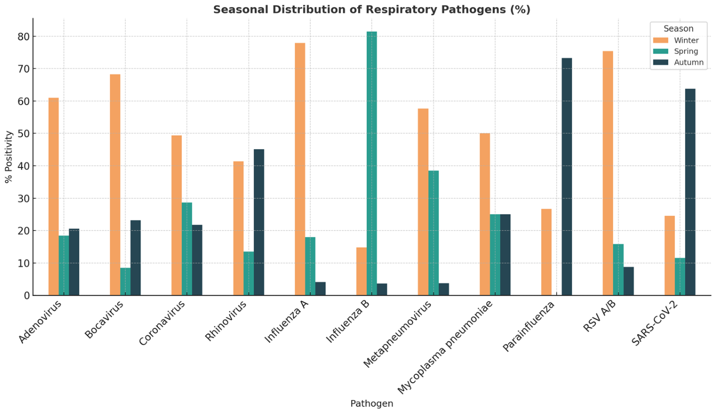
Figure 1 shows the percentage distribution of respiratory pathogens across winter, spring, and autumn seasons. RSV A/B, IAV, AdV, HBoV and HMPV demonstrated clear winter predominance. RV/EV and HPIV were most commonly detected in autumn, whereas IBV showed a peak in spring. SARS-CoV-2 positivity was highest in autumn and lowest in spring. These trends illustrate distinct seasonal circulation patterns among viral pathogens in early childhood.
Respiratory pathogens demonstrated significant seasonal variability (Figure 1). AdV and HBoV infections were significantly more common during winter, with positivity rates of 61% and 68.3%, respectively. In both pathogens, detection rates decreased markedly in spring (p = 0.018 and p = 0.002, respectively). IAV infections peaked in winter (77.9%), while RSV A/B positivity was highest in winter (75.4%). IBV infections showed a distinct seasonal peak in spring (81.5%) (p < 0.001). HMPV infections were predominantly detected in winter and spring, whereas only a small proportion of cases occurred in autumn (3.8%). RV/EV and HPIV were most frequently detected in autumn, with positivity rates of 45.1% and 73.3%, respectively. HPIV infections were not detected during spring (p < 0.001). SARS-CoV-2 infections were also significantly more common in autumn (63.8%) compared with other seasons (p < 0.001). M. pneumoniae infections were distributed relatively throughout the year, and no significant seasonal variation was observed (p = 0.741).
Univariable logistic regression analysis revealed that age, WBC, AMC, DNI, RSV A/B and RV/EV positivity are associated with hospitalization (Table 3). In univariable logistic regression, RV/EV positivity was associated with 1.7-fold higher odds of hospitalization, while RSV A/B positivity was associated with approximately 2.2-fold higher odds. In contrast, IAV positivity was associated with significantly lower odds of hospitalization (p = 0.007).
A multivariable logistic regression analysis identified younger age and higher CRP levels as independent predictors of hospital admission (Table 4). Younger age was independently associated with hospitalization (B = –0.018, p = 0.011, OR = 0.982, 95% CI: 0.968–0.996), indicating that each additional month of age decreased the odds of hospitalization by approximately 1.8%. Higher CRP levels were positively associated with hospital admission (B = 0.007, p = 0.015, OR = 1.007, 95% CI: 1.001–1.014), suggesting that each 1 mg/L increase in CRP increased the odds of hospitalization by about 0.7%.
Across the evaluated viral pathogens, distinct hematologic and inflammatory profiles were observed.
Table 5: AdV-positive patients were characterized by significantly increased WBC, ANC, ANC %, CRP and NLR, whereas eosinophil levels were reduced. HBoV infections were associated with younger age and higher eosinophil counts, without significant changes in other laboratory parameters. RV/EV-positive children exhibited significantly higher WBC, ANC, ALC, AEC and platelet counts compared with non-RV/EV patients.
In contrast, IAV infection was associated with significantly lower WBC, ANC, ALC, AMC, AEC and platelet counts, accompanied by higher NLR and older age. Similarly, IBV infection was characterized by lower WBC, ANC, ALC, and AEC levels, as well as decreased DNI, platelet counts, and CRP levels, together with increased MPV and older age. The highest mean age was observed among patients with IAV and IBV infections. HPIV infection was associated with younger age and significantly lower leukocyte, neutrophil, eosinophil, and NLR values. RSV-positive patients also demonstrated younger age, higher lymphocyte counts, lower NLR values, and reduced CRP levels.
SARS-CoV-2-positive patients showed significantly lower leukocyte and neutrophil parameters, reduced NLR and CRP levels, and younger age. Although monocyte counts were higher than those in the non-SARS-CoV-2 group, the difference was not statistically significant.
Patients with M. pneumoniae infection were significantly older than those without the infection; however, no significant differences in hematologic parameters were observed.
Comparison of respiratory virus positivity and laboratory findings between the two age groups revealed several age-related differences (Table 6). Rhinovirus/enterovirus (29.9% vs. 19.5%, p < 0.001), RSV A/B (17.1% vs. 11.5%, p = 0.010), and SARS-CoV-2 (7.8% vs. 3.2%, p = 0.001) were detected more frequently in children aged 1–36 months, whereas IVA (14.3% vs. 27.8%, p < 0.001), IVB (2.5% vs. 8.3%, p < 0.001), and M. pneumoniae (2.0% vs. 4.1%, p = 0.041) were more common in those aged 37–72 months. No significant age-related differences were observed in the positivity rates of AdV, HBoV, HMPV, or HPIV. Children aged 1–36 months had significantly higher WBC, ALC, AMC, AEC and platelet counts than those aged 37–72 months (all p < 0.001). In contrast, children aged 37–72 months had significantly higher ANC (p = 0.006), neutrophil percentage (p < 0.001), NLR (p < 0.001), and CRP levels (p = 0.029). DNI and MPV did not differ significantly between the two age groups.

4. Discussion

In this study, we investigated virus-specific variations in hematological parameters, age-related differences in respiratory pathogen distribution, independent predictors of hospitalization, and the seasonal distribution and coinfection patterns of common respiratory viruses. The most commonly identified pathogens were RV/EV, IAV, and RSV. RV/EV was the most frequently detected pathogen in coinfections, whereas AdV and HBoV showed the highest coinfection rates. Respiratory viruses demonstrated significant seasonal variability, peaking during the winter months. Age-stratified analysis showed that RV/EV, RSV, and SARS-CoV-2 infections were more common in younger children, whereas influenza viruses and M. pneumoniae predominated in older children. In univariable analyses, WBC, AMC, DNI, RSV positivity, and RV/EV positivity were significantly associated with hospital admission. Neither coinfections nor comorbid conditions were associated with hospitalization. Among a broad range of clinical and laboratory parameters, younger age and elevated CRP levels emerged as independent predictors of hospitalization. Distinct virus-specific hematological profiles were observed, with the most pronounced alterations occurring in adenovirus and influenza infections.
Numerous studies have investigated risk factors associated with hospitalization in general pediatric wards and pediatric intensive care units (PICUs). Prematurity, younger age and comorbid conditions have been identified as factors associated with an increased risk of hospital admission [18,19]. In our study, younger age and RSV positivity were associated with increased disease severity consistent with previous reports [20,21,22]. Influenza infections have also been reported to be associated with younger age and the presence of comorbidities [23]; however, in our cohort, influenza was associated with a lower risk of severe disease. The widespread use of antiviral therapy in our center and the inclusion of patients presenting to outpatient clinics may have contributed to reduced hospitalization rates. Moreover, the high proportion of otherwise healthy children without chronic conditions may have further caused the lower hospitalization rate. In contrast, RSV infection showed a stronger association with severe disease, highlighting the importance of effective RSV prevention strategies including maternal RSV vaccination during pregnancy or long-acting monoclonal antibodies such as nirsevimab or clesrovimab for infants entering their first RSV season.
Viral coinfections are frequently observed in children; however, whether specific viral interactions enhance or diminish the severity of respiratory disease remains controversial. In a meta-analysis by Goka, the evidence regarding the impact of coinfections on disease severity was inconclusive [10]. Similarly, in our study, no significant differences were observed between single infections and coinfections in terms of hospitalization rates. The frequency of coinfections was lower than that reported in the literature. This finding may be attributed to patient characteristics, as our cohort consisted of patients presenting to general pediatrics and infectious diseases clinics, with a relatively low hospitalization rate. This study demonstrated that SARS-CoV-2 and IAV were more commonly observed in monoinfections, consistent with previous data [24,25]. Maio et al. reported that HBoV exhibited the highest cocoinfectionate (87.8%), likely due to prolonged viral shedding, with viral DNA persisting for up to 3 months in outpatients and up to 1 year in hospitalized children after acute infection [26]. In our cohort, HBoV, AdV and RV/EV also demonstrated notable coinfection tendencies, in agreement with the literature. Additionally, the coinfection rate of M. pneumoniae was relatively high (28.1%). The study period coincided with the global resurgence of M. pneumoniae infections observed in 2024 following the COVID-19 pandemic. The increased circulation of M. pneumoniae, particularly among children, may have contributed to the high detection rate of M. pneumoniae and the frequent occurrence of viral coinfections observed in this study [27,28].
The rate of viral positivity varied significantly across seasons, with the highest rate observed in winter (49.8%). While this finding is generally consistent with the known seasonality of respiratory viruses, some differences compared with previous studies were noted. Zhu et al. [1] reported that AdV infections peaked in spring, whereas IAV and IBV in winter, and RSV in autumn. Zhao et al. found that IAV and IBV epidemics occurred predominantly in winter and spring, whereas AdV did not exhibit a distinct seasonal pattern and RSV peaked in winter [29]. Adenovirus infections occurred more frequently during winter in our cohort. Fang et al. also demonstrated a winter peak of AdV infections. These differences may reflect variations in geographical settings, population characteristics, and viral transmission patterns.
The COVID-19 pandemic has significantly altered the epidemiology of respiratory viruses, particularly in terms of seasonal distribution and peak incidence. Several studies from Türkiye have investigated these changes during and after the pandemic. Rhinovirus was the most frequently seen virus in these studies (excluding COVID-19) [3,30] The frequency of parainfluenza virus infections was found to be increased during the summer months, representing a novel finding [30]. In our cohort, parainfluenza infections were most frequently observed in autumn; however, the lack of summer data limits direct comparison. In another study conducted at our center, influenza virus was detected in 30.1% and HBoV in 28.3% of 1465 hospitalized patients [31]. These differences may be explained by variations in study design and population characteristics, as our study included outpatients, excluded emergency department cases, and had a relatively low prevalence of comorbidities. Despite changes in the epidemiology of respiratory viruses after the COVID-19 pandemic, rhinovirus has remained the most frequently detected pathogen. SARS-CoV-2, on the other hand, appears to be evolving toward an endemic pattern with increasing evidence of seasonal circulation; however, longer-term surveillance is required to confirm this transition.
We compared hematological parameters between virus-specific groups, including AdV vs. non-AdV and RSV vs. non-RSV infections, to explore their potential role as supportive diagnostic markers in the absence of molecular testing. Hematological parameters—including WBC, neutrophil, lymphocyte, platelet, and eosinophil counts—varied significantly among respiratory viral pathogens, supporting the concept that each virus may induce a distinct immuno-hematological profile.
Adenovirus infection was associated with the most pronounced inflammatory response, characterized by elevated CRP, WBC, and neutrophil counts, along with low eosinophil levels. These findings are consistent with previous reports indicating that AdV infections may mimic bacterial infections in terms of clinical, laboratory, and radiological features [32]. The highest WBC and ANC were observed in AdV and RV/EV infections.
Eosinopenia is often considered a marker of infection, although there is currently no universally accepted cutoff value The clinical utility of eosinopenia has been explored in various settings, including early neonatal sepsis, differentiation between bacterial and aseptic meningitis, and early diagnosis of COVID-19 infection [33,34,35]. Although the precise role of eosinophils in viral infections remains unclear, current evidence suggests that peripheral blood eosinophil count (EC) may serve as a predictive and prognostic biomarker for disease outcomes [36,37,38]. In the current study, we found no significant association between eosinophil levels and hospital admission but we observed low eosinophil levels in AdV-, IAV-, IBV-, and HPIV-positive patients. As key components of the innate immune system, eosinophils exert protective effects through their anti-infective and anti-inflammatory activities [39]. However, they can also contribute to pathological conditions such as asthma and atopic dermatitis. In our cohort, higher eosinophil levels were observed in RV/EV and HBoV infections. These findings are noteworthy, as these viruses have been implicated in the development of asthma and allergic diseases.
Low lymphocyte levels were observed in IAV and IBV patients consistent with the literature [40]. In contrast, RSV and RV/EV infections showed a lymphocyte-predominant profile and SARS-CoV-2 infection was associated with lower neutrophil levels and higher monocyte counts. Monocytosis has previously been identified as a potential marker of innate immune activation in pediatric COVID-19 [41]. In our study, elevated monocyte levels were also observed in hospitalized patients, suggesting a potential association with disease severity.
Age-stratified analysis confirmed the expected physiological differences in hematological parameters between younger and older children. However, the observed virus-associated hematological alterations could not be explained by age alone. Several respiratory viruses exhibited distinct hematologic profiles that differed from the expected age-related patterns. For example, RV/EV infection was associated with higher neutrophil, lymphocyte, and eosinophil counts despite occurring predominantly in younger children. Likewise, AdV, influenza A/B, and HPIV infections were consistently associated with eosinopenia, suggesting that reduced eosinophil counts reflect a virus-specific host immune response rather than physiological age-related variation. In contrast, SARS-CoV-2 infection was characterized by neutropenia, whereas AdV exhibited a pronounced inflammatory profile irrespective of age distribution. Collectively, these findings suggest that virus-specific host immune responses, rather than age alone, are important determinants of the hematological alterations observed in children with respiratory viral infections.
This study has several limitations. First, its retrospective design may have introduced selection bias and limited the availability of some clinical data. Second, the study was conducted in a single center, which may limit the generalizability of the findings. Third, the study population consisted of children initially presenting to the outpatient clinic. As only a relatively small proportion required hospitalization, the low number of hospitalized patients and the limited prevalence of severe disease and comorbid conditions may have affected the identification of hospitalization-related risk factors. In addition, the absence of summer data limited the evaluation of seasonal patterns for certain respiratory viruses, because the summer months were outside the predefined study period and were therefore unavailable at the time of data extraction. Despite these limitations, the study has several strengths, including a relatively large overall cohort and the comprehensive evaluation of multiple respiratory viruses.
In conclusion, respiratory viral infections in children exhibit distinct seasonal and hematological patterns. Age-related differences were observed in pathogen distribution, with RV/EV, RSV A/B, and SARS-CoV-2 detected more frequently in younger children, whereas IAV, IVB, and M. pneumoniae predominated in older children. Distinct virus-specific hematological profiles, particularly in AdV and influenza infections, suggest that routine hematological parameters may provide valuable insights into host immune responses and aid the clinical evaluation of children with respiratory viral infections. Although several hematological parameters were associated with hospitalization in the univariable analysis, only younger age and elevated CRP remained independent predictors indicating that most virus-specific hematological alterations are more likely to reflect pathogen-specific immune responses than disease severity. Routine hematological parameters may complement clinical assessment, particularly in settings where molecular diagnostics are not readily available. Further prospective, multicenter studies are needed to validate these findings and to elucidate the underlying biological mechanisms.


Author Contributions

E.Ö. designed the study, managed the data, and drafted the manuscript; S.A.B., T.Ö.D., H.A.K. and E.B. contributed to data collection and data management; A.Y. contributed to study design, supervised the project, and provided substantial scientific input. All authors have read and agreed to the published version of the manuscript.

Funding

This research received no external funding.

Institutional Review Board Statement

The study was consistent with the principles of the Declaration of Helsinki and was approved by the Ethics Committee of Ankara Bilkent City Hospital (date: 16 April 2025, reference number: TABED 2-25-1083).

Informed Consent Statement

Informed consent was waived because of the retrospective nature of the study.

Data Availability Statement

The data presented in this study are available on request from the corresponding author. The data are not publicly available due to restrictions, e.g., privacy.

Conflicts of Interest

The authors declare no conflicts of interest.

Abbreviations

ARIAcute respiratory infection
RSVRespiratory syncytial virus
IAVInfluenza A virus
IBVInfluenza B virus
RV/EVRhinovirus/Enterovirus
AdVHuman adenovirus
HPIVHuman parainfluenza virus
hCoVHuman coronaviruses
HMPVHuman metapneumovirus
hBoVHuman bocavirus
ALRIAcute lower respiratory infection
SARS-CoV-2Severe acute respiratory syndrome coronavirus 2
HbHemoglobin
WBCWhite blood cell count
ANCAbsolute neutrophil count
ALCAbsolute lymphocyte count
AMCAbsolute monocyte count
AECAbsolute eosinophil count
PltPlatelet count
MPVMean platelet volume
CRPC-reactive protein
PCRPolymerase chain reaction
PICUPediatric intensive care unit

References

  1. Zhu, G.; Xu, D.; Zhang, Y.; Wang, T.; Zhang, L.; Gu, W.; Shen, M. Epidemiological characteristics of four common respiratory viral infections in children. Virol. J. 2021, 18, 10. [Google Scholar] [CrossRef] [Scilit]
  2. Kurskaya, O.; Ryabichenko, T.; Leonova, N.; Shi, W.; Bi, H.; Sharshov, K.; Kazachkova, E.; Sobolev, I.; Prokopyeva, E.; Kartseva, T.; et al. Viral etiology of acute respiratory infections in hospitalized children in Novosibirsk City, Russia (2013–2017). PLoS ONE 2018, 13, e0200117. [Google Scholar] [CrossRef] [Scilit] [PubMed]
  3. Karbuz, A.; Aktaş, E.; Tutak, G.A.; İşançlı, D.K.; Kılıçaslan, Ö.; Kırmacı, Ç.; Emre, I.; Beşel, L.; Barış, A.; Arat, Z. The effects of measures taken during the COVID-19 pandemic on the seasonal dynamics of respiratory viruses in children. Turk. J. Pediatr. 2023, 65, 592–602. [Google Scholar] [CrossRef] [Scilit] [PubMed]
  4. Moriyama, M.; Hugentobler, W.J.; Iwasaki, A. Seasonality of respiratory viral infections. Annu. Rev. Virol. 2020, 7, 83–101. [Google Scholar] [CrossRef] [Scilit] [PubMed]
  5. Audi, A.; AlIbrahim, M.; Kaddoura, M.; Hijazi, G.; Yassine, H.M.; Zaraket, H. Seasonality of respiratory viral infections: Will COVID-19 follow suit? Front. Public Health 2020, 8, 567184. [Google Scholar] [CrossRef] [Scilit] [PubMed]
  6. Maglione, M.; Tipo, V.; Barbieri, E.; Ragucci, R.; Ciccarelli, A.S.; Esposito, C.; Carangelo, L.; Giannattasio, A. Changes in Respiratory Viruses’ Activity in Children During the COVID-19 Pandemic: A Systematic Review. J. Clin. Med. 2025, 14, 1387. [Google Scholar] [CrossRef] [Scilit] [PubMed]
  7. Chow, E.J.; Uyeki, T.M.; Chu, H.Y. The effects of the COVID-19 pandemic on community respiratory virus activity. Nat. Rev. Microbiol. 2022, 21, 195–210. [Google Scholar] [CrossRef] [Scilit] [PubMed]
  8. Trusinska, D.; Zin, S.T.; Sandoval, E.; Homaira, N.; Shi, T. Risk factors for poor outcomes in children hospitalized with virus-associated acute lower respiratory infections: A systematic review and meta-analysis. Pediatr. Infect. Dis. J. 2024, 43, 467–476. [Google Scholar] [CrossRef] [Scilit] [PubMed]
  9. Babawale, P.I.; Guerrero-Plata, A. Respiratory Viral Coinfections: Insights into Epidemiology, Immune Response, Pathology, and Clinical Outcomes. Pathogens 2024, 13, 316. [Google Scholar] [CrossRef] [Scilit] [PubMed]
  10. Goka, E.A.; Vallely, P.J.; Mutton, K.J.; Klapper, P.E. Single and multiple respiratory virus infections and severity of respiratory disease: A systematic review. Paediatr. Respir. Rev. 2014, 15, 363–370. [Google Scholar] [CrossRef] [Scilit] [PubMed]
  11. Scotta, M.C.; Chakr, V.C.; de Moura, A.; Becker, R.G.; de Souza, A.P.; Jones, M.H.; Pinto, L.A.; Sarria, E.E.; Pitrez, P.M.; Stein, R.T.; et al. Respiratory viral coinfection and disease severity in children: A systematic review and meta-analysis. J. Clin. Virol. 2016, 80, 45–56. [Google Scholar] [CrossRef] [Scilit] [PubMed]
  12. Asner, S.A.; Science, M.E.; Tran, D.; Smieja, M.; Merglen, A.; Mertz, D. Clinical Disease Severity of Respiratory Viral Co-Infection versus Single Viral Infection: A Systematic Review and Meta-Analysis. PLoS ONE 2014, 9, e99392. [Google Scholar] [CrossRef] [Scilit] [PubMed]
  13. Huang, L.; Ye, C.; Zhou, R.; Ji, Z. Diagnostic value of routine blood tests in differentiating between SARS-CoV-2, influenza A, and RSV infections in hospitalized children: A retrospective study. BMC Pediatr. 2024, 24, 328. [Google Scholar] [CrossRef] [Scilit] [PubMed]
  14. Jin, Q.; Ma, W.; Zhang, W.; Wang, H.; Geng, Y.; Geng, Y.; Zhang, Y.; Gao, D.; Zhou, J.; Li, L.; et al. Clinical and hematological characteristics of children infected with the omicron variant of SARS-CoV-2: Role of the combination of the neutrophil: Lymphocyte ratio and eosinophil count in distinguishing severe COVID-19. Front Pediatr. 2024, 12, 1305639. [Google Scholar] [CrossRef] [Scilit] [PubMed]
  15. Ötiken Arıkan, K.; Şahinkaya, Ş.; Böncüoğlu, E.; Kıymet, E.; Cem, E.; Akaslan Kara, A.; Bayram, N.; Devrim, İ. Can hematological findings of COVID-19 in pediatric patients guide physicians regarding clinical severity? Turk. J. Haematol. 2021, 38, 243–245. [Google Scholar] [CrossRef] [Scilit] [PubMed]
  16. Henry, B.M.; de Oliveira, M.H.S.; Benoit, S.; Plebani, M.; Lippi, G. Hematologic, biochemical and immune biomarker abnormalities associated with severe illness and mortality in coronavirus disease 2019 (COVID-19): A meta-analysis. Clin. Chem. Lab. Med. 2020, 58, 1021–1028. [Google Scholar] [CrossRef] [Scilit] [PubMed]
  17. Nygaard, U.; Holm, M.; Rabie, H.; Rytter, M. The pattern of childhood infections during and after the COVID-19 pandemic. Lancet Child Adolesc. Health 2024, 8, 910–920. [Google Scholar] [CrossRef] [Scilit] [PubMed]
  18. Kirolos, N.; Mtaweh, H.; Datta, R.R.; Farrar, D.S.; Seaton, C.; Bone, J.N.; Muttalib, F.; Kaziev, C.L.; Fortini, J.; Mahant, S.; et al. Risk factors for severe disease among children hospitalized with respiratory syncytial virus. JAMA Netw. Open 2025, 8, e254666. [Google Scholar] [CrossRef] [Scilit] [PubMed]
  19. Curns, A.T.; Rha, B.; Lively, J.Y.; Sahni, L.C.; Englund, J.A.; Weinberg, G.A.; Halasa, N.B.; Staat, M.A.; Selvarangan, R.; Michaels, M.; et al. Respiratory syncytial virus–associated hospitalizations among children <5 years old: 2016 to 2020. Pediatrics 2024, 153, e2023062574. [Google Scholar] [CrossRef] [Scilit] [PubMed]
  20. Adu-Gyamfi, C.; Asamoah, J.A.; Frimpong, J.O.; Larbi, R.; Ansah, R.O.; Aryeetey, S.N.A.; Gorman, R.; Acheampong, H.K.; Nyarko-Afriyie, E.; Hayford, M.; et al. Prevalence of common respiratory viruses in children: Insights from post-pandemic surveillance. BMC Infect. Dis. 2025, 25, 824. [Google Scholar] [CrossRef] [Scilit] [PubMed]
  21. Hasan, M.M.; Saha, K.K.; Yunus, R.M.; Alam, K. Prevalence of acute respiratory infections among children in India: Regional inequalities and risk factors. Matern. Child Health J. 2022, 26, 1594. [Google Scholar] [CrossRef] [Scilit] [PubMed]
  22. Sun, Y.P.; Qiang, H.S.; Lei, S.Y.; Zheng, X.Y.; Zhang, H.X.; Su, Y.Y.; Zheng, Z.-Z.; Zhang, J.; Lin, X.-Z.; Zhou, Y.-L. Epidemiological features, risk factors, and disease burden of respiratory viruses among hospitalized children with acute respiratory tract infections in xiamen, China. Jpn. J. Infect. Dis. 2022, 75, 537–542. [Google Scholar] [CrossRef] [Scilit] [PubMed]
  23. Dimopoulou, D.; Maltezou, H.C.; Neofytou, A.; Giannouchos, T.; Pantelia, E.; Dimitroglou, M.; Koziaki, G.; Makropoulou, M.; Tsolia, M.N. Characteristics of children with influenza admitted to a pediatric hospital in Greece during the 2022/2023 and 2023/2024 seasons. Eur. J. Pediatr. 2025, 184, 530. [Google Scholar] [CrossRef] [Scilit] [PubMed]
  24. Di Maio, V.C.; Scutari, R.; Forqué, L.; Colagrossi, L.; Coltella, L.; Ranno, S.; Linardos, G.; Gentile, L.; Galeno, E.; Vittucci, A.C.; et al. Presence and Significance of Multiple Respiratory Viral Infections in Children Admitted to a Tertiary Pediatric Hospital in Italy. Viruses 2024, 16, 750. [Google Scholar] [CrossRef] [Scilit] [PubMed]
  25. Weidmann, M.D.; Green, D.A.; Berry, G.J.; Wu, F. Assessing Respiratory Viral Exclusion and Affinity Interactions through Co-Infection Incidence in a Pediatric Population during the 2022 Resurgence of Influenza and RSV. Front. Cell. Infect. Microbiol. 2023, 13, 1208235. [Google Scholar] [CrossRef] [Scilit] [PubMed]
  26. Verbeke, V.; Reynders, M.; Floré, K.; Vandewal, W.; Debulpaep, S.; Sauer, K.; Cardoen, F.; Padalko, E. Human Bocavirus Infection in Belgian Children with Respiratory Tract Disease. Arch. Virol. 2019, 164, 2919–2930. [Google Scholar] [CrossRef] [Scilit] [PubMed]
  27. Meyer Sauteur, P.M.; Beeton, M.L. European Society of Clinical Microbiology and Infectious Diseases (ESCMID) Study Group for Mycoplasma and Chlamydia Infections (ESGMAC), and the ESGMAC Mycoplasma pneumoniae Surveillance (MAPS) study group. Mycoplasma pneumoniae: Delayed re-emergence after COVID-19 pandemic restrictions. Lancet Microbe 2024, 5, e100–e101. [Google Scholar] [CrossRef] [Scilit] [PubMed]
  28. de Groot, R.C.A.; Streng, B.M.M.; Bont, L.J.; Meyer Sauteur, P.M.; van Rossum, A.M.C. Resurgence of Mycoplasma pneumoniae infections in children: Emerging challenges and opportunities. Curr. Opin. Infect. Dis. 2025, 38, 468–476. [Google Scholar] [CrossRef] [Scilit] [PubMed]
  29. Zhao, X.; Zhu, X.; Wang, J.; Ye, C.; Zhao, S. The epidemiological analysis of respiratory virus infections in children in Hangzhou from 2019 to 2023. Virus Res. 2025, 355, 199558. [Google Scholar] [CrossRef] [Scilit] [PubMed]
  30. Bedir Demirdag, T.; Ozcicek, M.; Polat, M.; Kavas, F.C.; Demir, F.; Atay Unal, N.; Kara, N.; Gudeloglu, E.; Tezer, H.; Bozdayi, G.; et al. Effects of COVID-19 pandemic on epidemiological features of viral respiratory tract infections in children: A single-centre study. Epidemiol. Infect. 2024, 152, e128. [Google Scholar] [CrossRef] [Scilit] [PubMed]
  31. Kalayci, F.; Ozkaya Parlakay, A.; Yigit, M.; Güler, G.N.; Demircioglu Kalayci, B.; Cetin, A.N.; Yurteri, M.D.; Karakose, E.; Celebier, K.; Yilmaz, N.; et al. Increased risk during winter: Common respiratory viruses and clinical outcomes in hospitalized children. BMC Infect. Dis. 2025, 25, 563. [Google Scholar] [CrossRef] [Scilit] [PubMed]
  32. Bayhan, G.İ.; Gülleroğlu, N.B.; Çetin, S.; Erat, T.; Yıldız, S.; Özen, S.; Konca, H.K.; Yahşi, A.; Dinç, B. Radiographic findings of adenoviral pneumonia in children. Clin. Imaging 2024, 108, 110111. [Google Scholar] [CrossRef] [Scilit] [PubMed]
  33. Wilar, R. Diagnostic value of eosinopenia and neutrophil to lymphocyte ratio on early onset neonatal sepsis. Korean J. Pediatr. 2019, 62, 217–223. [Google Scholar] [CrossRef] [Scilit] [PubMed]
  34. Debray, A.; Nathanson, S.; Moulin, F.; Salomon, J.; Davido, B. Eosinopenia as a marker of diagnosis and prognostic to distinguish bacterial from aseptic meningitis in pediatrics. Eur. J. Clin. Microbiol. Infect. Dis. 2019, 38, 1821–1827. [Google Scholar] [CrossRef] [Scilit] [PubMed]
  35. Tanni, F.; Akker, E.; Zaman, M.M.; Figueroa, N.; Tharian, B.; Hupart, K.H. Eosinopenia and COVID-19. J. Osteopath. Med. 2020, 120, 504–508. [Google Scholar] [CrossRef] [Scilit] [PubMed]
  36. Xie, G.; Ding, F.; Han, L.; Yin, D.; Lu, H.; Zhang, M. The role of peripheral blood eosinophil counts in COVID-19 patients. Allergy Eur. J. Allergy Clin. Immunol. 2021, 76, 471–482. [Google Scholar] [CrossRef] [Scilit] [PubMed]
  37. Roca, E.; Ventura, L.; Zattra, C.M.; Lombardi, C.; Roca, E. Eosinopenia: An early, effective and relevant COVID-19 biomarker? QJM 2021, 114, 68–69. [Google Scholar] [CrossRef] [Scilit] [PubMed]
  38. Rosenberg, H.F.; Foster, P.S. Eosinophils and COVID-19: Diagnosis, prognosis, and vaccination strategies. Semin. Immunopathol. 2021, 43, 383–392. [Google Scholar] [CrossRef] [Scilit] [PubMed]
  39. Kanda, A.; Yasutaka, Y.; Van Bui, D.; Suzuki, K.; Sawada, S.; Kobayashi, Y.; Asako, M.; Iwai, H. Multiple Biological Aspects of Eosinophils in Host Defense, Eosinophil-Associated Diseases, Immunoregulation, and Homeostasis: Is Their Role Beneficial, Detrimental, Regulator, or Bystander? Biol. Pharm. Bull. 2020, 43, 20–30. [Google Scholar] [CrossRef] [Scilit] [PubMed]
  40. Asseri, A.A.; Al-Qahtani, S.M.; Alzaydani, I.A.; Al-Jarie, A.; Alyazidi, N.S.; Alrmelawi, A.A.; Alqahtani, A.M.; Alsulayyim, R.S.; Alzailaie, A.K.; Abdullah, D.M.; et al. Clinical and epidemiological characteristics of respiratory syncytial virus, SARS-CoV-2 and influenza paediatric viral respiratory infections in southwest Saudi Arabia. Ann. Med. 2025, 57, 2445791. [Google Scholar] [CrossRef] [Scilit] [PubMed]
  41. Sitthikarnkha, P.; Phunyaissaraporn, R.; Niamsanit, S.; Techasatian, L.; Saengnipanthkul, S.; Uppala, R. Clinical Characteristics and Outcomes of Pediatric COVID-19 Pneumonia Treated with Favipiravir in a Tertiary Care Center. Viruses 2024, 16, 946. [Google Scholar] [CrossRef] [Scilit] [PubMed]
Figure 1. Seasonal distribution of respiratory pathogens (%).
Children 13 01040 g001
Table 1. The number and percentage of respiratory viral infections.
VirusSingle N (%)Multiple N (%)Total N (%)
AdV97 (71.3%)39 (28.7%)136 (10.9%)
HBoV51 (62.2%)31 (37.8%)82 (6.6%)
HCoV75 (86.2%)12 (13.8%)87 (7.0%)
RV/EV254 (85.5%)43 (14.5%)297 (23.7%)
IAV202 (91.0%)20 (9.0%)222 (17.7%)
IBV52 (96.3%)2 (3.7%)54 (4.3%)
HMPV22 (84.6%)4 (15.4%)26 (2.1%)
M. pneumoniae23 (71.9%)9 (28.1%)32 (2.6%)
HPIV59 (78.7%)16 (21.3%)75 (6.0%)
RSV A/B141 (82.5%)30 (17.5%)171 (13.7%)
SARS-CoV-265 (94.2%)4 (5.8%)69 (5.5%)
Overall10412101251
Table 2. Comparison of hematologic and inflammatory parameters between hospitalized and non-hospitalized patients.
Hospitalization StateNumber of PatientsMeanStd. Deviationp
Age (month)N106832.0222.050.009
H7525.0821.81
Hb (g/dL)N47712.121.210.008
H7511.711.33
WBC (/mm3)N4778983.713805.090.010
H7510,585.335032.50
ANC (/mm3)N4774582.523863.330.079
H755519.074295.14
ANC %N47747.1318.870.800
H7547.7421.73
ALC (/mm3)N4773452.771933.750.392
H753661.472131.02
AMC (/mm3)N476609.12372.700.090
H75808.93998.15
AEC (/mm3)N476158.45190,530.328
H75183.07267.55
DNI %N4730.691.650.073
H751.202.32
Plt (/mm3)N475334,002119,2560.170
H75357,733140,699
MPV (fL)N4757.850.850.967
H757.841.02
NLRN4751.831.800.321
H752.102.29
CRP (mg/L)N39114.9629.550.019
H7333.8366.27
Abbreviation: Hemoglobin (Hb), white blood cell count (WBC), hematocrit (Hct), red blood cell count (RBC), mean corpuscular volume (MCV), absolute neutrophil count (ANC), absolute lymphocyte count (ALC), absolute monocyte count (AMC), absolute eosinophil count (AEC), platelet count, mean platelet volume (MPV), C-reactive protein (CRP), N: Non-hospitalized, H: Hospitalized.
Table 3. Univariable logistic regression analysis of factors associated with respiratory viral infections.
VariableBpExp(B)%95 GA (Exp(B))
Age (month)−0.0160.0090.9850.973–0.996
Hb (g/dL)−0.2680.0080.7650.628–0.933
WBC (/mm3)0.0000.0021.0001.000–1.000
CRP (mg/L)0.0090.0011.0091.004–1.014
AMC (/mm3)0.0010.0171.0011.000–1.001
DNI (%)0.1250.0261.1331.015–1.265
RSV A/B0.7920.0042.2081.287–3.789
IAV−1.2730.0070.2800.112–0.703
RV/EV0.5080.0431.6621.017–2.717
Table 4. Multivariable logistic regression analysis of factors associated with respiratory viral infections.
VariableBpExp(B)%95 GA (Exp(B))
Age (month)−0.0180.0110.9820.968–0.996
Hb (g/dL)−0.0490.6640.9520.763–1.189
WBC (/mm3)0.0000.2571.0001.000–1.000
CRP (mg/L)0.0070.0151.0071.001–1.014
AMC (/mm3)0.0000.3071.0001.000–1.001
DNI (%)0.0880.1591.0920.966–1.235
Constant−1.3400.3140.262
Table 5. Comparison of hematologic and inflammatory parameters across viral pathogens.
Viral EtiologyParameterNMean ± SDp
AdenovirusHb7311.72 ± 1.130.013
WBC7311,161.23 ± 4516.42<0.001
ANC736788.90 ± 3687.83<0.001
ANC (%)7357.89 ± 16.56<0.001
AEC7399.86 ± 118.87<0.001
NLR732.62 ± 1.88<0.001
CRP5946.53 ± 59.96<0.001
Human BocavirusAge (month)8229.51 ± 18.290.032
AEC44320.23 ± 312.400.001
Rhinovirus/EnterovirusAge (month)29727.19 ± 21.05<0.001
WBC14011,124.64 ± 4063.21<0.001
ANC1406139.64 ± 5734.07<0.001
ALC1404267.86 ± 2050.73<0.001
AEC140234.00 ± 234.81<0.001
Plt140388,128 ± 136,051<0.001
Influenza AAge (month)22240.24 ± 22.38<0.001
Hb10912.28 ± 1.070.037
WBC1096905.14 ± 3074.69<0.001
ANC1093645.41 ± 2481.75<0.001
ANC (%)10950.51 ± 20.080.045
ALC1092344.77 ± 1427.04<0.001
AMC109500.92 ± 278.570.002
AEC10980.46 ± 137.79<0.001
Plt109286,853 ± 98,009<0.001
NLR1092.29 ± 2.210.020
CRP9410.34 ± 14.09<0.001
Influenza BAge (month)5447.37 ± 22.07<0.001
HB2812.50 ± 0.820.008
WBC286130.00 ± 3106.28<0.001
ANC283010.71 ± 2248.440.019
ALC282400.71 ± 1414.090.003
AEC2874.29 ± 148.390.019
DNI280.35 ± 0.820.017
Plt28221,214.29 ± 76,440.90<0.001
MPV288.26 ± 1.090.010
CRP284.67 ± 7.13<0.001
Human Parainfluenza VirusAge (month)7526.57 ± 22.570.004
WBC367559.44 ± 3057.000.002
ANC363016.39 ± 2058.61<0.001
ANC (%)3639.18 ± 18.240.010
AEC3688.89 ± 98.82<0.001
DNI360.44 ± 0.890.048
NLR361.26 ± 1.590.047
RSV A/BAge (month)17126.85 ± 19.18<0.001
ALC743986.35 ± 2094.310.017
NLR731.39 ± 1.280.002
CRP6410.36 ± 15.370.002
SARS-CoV-2Age (month)6920.38 ± 21.20<0.001
WBC307225.00 ± 2558.00<0.001
ANC302433.00 ± 1660.83<0.001
ANC (%)3033.43 ± 18.72<0.001
DNI300.38 ± 0.970.042
NLR300.99 ± 0.89<0.001
CRP247.73 ± 10.29<0.001
Table 6. Age-specific respiratory virus positivity and laboratory findings.
Variable1–36 month37–72 monthp
Adenovirus81 (11.4)55 (12.6)0.573
Human Bocavirus55 (7.8)27 (6.2)0.347
Human Coronavirus48 (6.8)39 (9)0.206
Rhinovirus/Enterovirus212 (29.9)85 (19.5)<0.001
Influenza A101 (14.3)121 (27.8)<0.001
Influenza B18 (2.5)36 (8.3)<0.001
Human Metapneumovirus17 (2.4)9 (2.1)0.839
M. pneumoniae14 (2.0)18 (4.1)0.041
Human Parainfluenza Virus53 (7.5)22 (5.1)0.112
RSV A/B121 (17.1)50 (11.5)0.010
SARS-CoV-255 (7.8)14 (3.2)0.001
WBC (/mm3)9802.2 ± 4057.88365.3 ± 3832.5<0.001
ANC (/mm3)4315.5 ± 3736.65240.9 ± 41330.006
ANC (%)39.91 ± 18.0257.19 ± 16.18<0.001
ALC (/mm3)4286 ± 2036.22374.8 ± 1144.2<0.001
AMC (/mm3)707.8 ± 604.5538.4 ± 309.5<0.001
AEC (/mm3)199.6 ± 225.4109.7 ± 151.7<0.001
DNI (%)0.68 ± 1.760.86 ± 1.770.241
Plt (/mm3)358,291.5 ± 121,117.2307,784.5 ± 118,594.5<0.001
MPV (fL)7.87 ± 0.927.81 ± 0.800.410
NLR1.27 ± 1.332.67 ± 2.18<0.001
CRP (mg/L)14.48 ± 33.9722.61 ± 43.070.029
Disclaimer/Publisher’s Note: The statements, opinions and data contained in all publications are solely those of the individual author(s) and contributor(s) and not of MDPI and/or the editor(s). MDPI and/or the editor(s) disclaim responsibility for any injury to people or property resulting from any ideas, methods, instructions or products referred to in the content.