Left Atrial Volumes and Strains in Healthy Mid-Term Pregnancy—A Detailed Investigation from a Three-Dimensional Speckle-Tracking Echocardiographic MAGYAR-Preg Study

 ,
, 
Abstract
1. Introduction
2. Materials and Methods
2.1. Study Population
2.2. Two-Dimensional Doppler Echocardiography
2.3. Three-Dimensional Speckle-Tracking Echocardiography
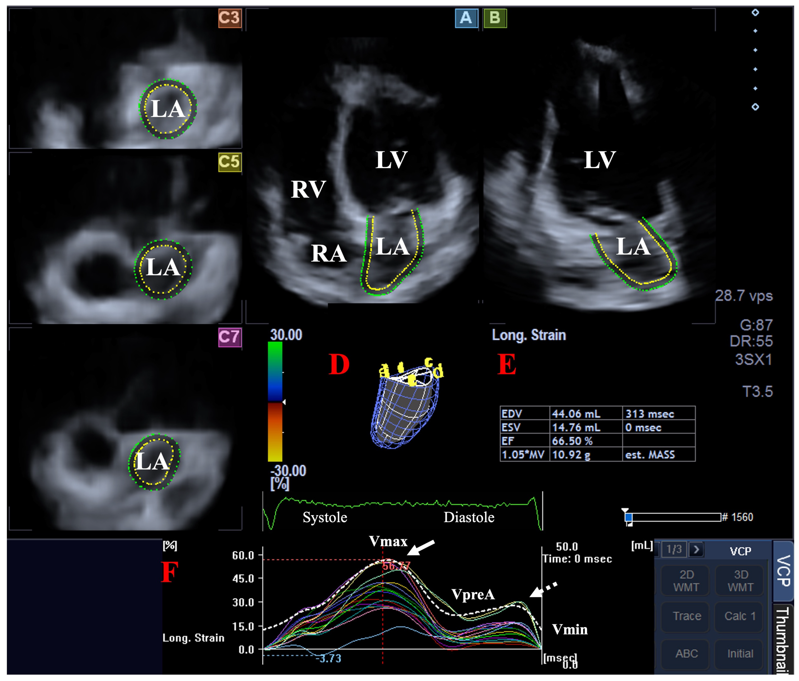
2.4. 3DSTE-Derived LA Quantifications
- Systolic reservoir function: represented by the LA total stroke volume (TASV = Vmax − Vmin) and the LA total emptying fraction (TAEF = TASV/Vmax).
- Early diastolic conduit function: characterized by the LA passive stroke volume (PASV = Vmax − VpreA) and the LA passive emptying fraction (PAEF = PASV/Vmax).
- Late diastolic booster pump function: defined by the LA active stroke volume (AASV = VpreA − Vmin) and the LA active emptying fraction (AAEF = AASV/VpreA).
2.5. Statistical Analysis
3. Results
3.1. Two-Dimensional Doppler Echocardiography
3.2. 3DSTE-Derived LA Volumes
3.3. 3DSTE-Derived LA Strains
3.4. Feasibility, Post Hoc Power and Multivariable Analyses
3.5. Interobserver and Intraobserver Variability
4. Discussion
4.1. Limitation Section
- A significant limitation is that the image quality produced by the 3DSTE tool used in this study is generally considered lower compared with traditional 2D echocardiography images. Due to its superior spatial and temporal resolution, 2D echocardiography maintains an advantage over 3DSTE in terms of image quality [32]. Additionally, the larger footprint of the 3DSTE probe complicates precise positioning. The multi-beat acquisition protocol (integrating six subvolumes) further introduces the risk of stitching artifacts and motion-related errors, potentially compromising the reliability of the data [11,12,13,14,15].
- The validation of 3DSTE-derived LA volumetric and strain analysis was beyond the scope of the present study, as it has already been validated, like against 2D echocardiography and volumetric 3D echocardiography in previous investigations [6,16]. Nevertheless, the scientific robustness of our findings could have been further strengthened by incorporating an alternative imaging modality for simultaneous internal validation.
- The evaluation was strictly focused on the LA; no functional or volumetric parameters of any other heart chambers were included in the analysis. The absence of LV strain or right heart evaluation limits the ability to interpret atrioventricular coupling and global cardiac adaptation.
- Due to the relatively small healthy cohort, this study may be statistically underpowered, leaving the possibility of Type II errors despite high measurement reproducibility. Consequently, these findings should be considered exploratory and require validation in larger, multi-center studies.
- The current study relies on a cross-sectional design rather than longitudinal follow-up of the same cohort, which limits our ability to definitively attribute the observed differences to pregnancy-related adaptations. This emphasizes the importance of prospective follow-up studies, which can be conducted relatively easily and within a manageable timeframe in a pregnant population.
4.2. Conclusions
Author Contributions
Funding
Institutional Review Board Statement
Informed Consent Statement
Data Availability Statement
Conflicts of Interest
References
- Regitz-Zagrosek, V.; Roos-Hesselink, J.W.; Bauersachs, J.; Blomström-Lundquist, C.; Cífková, R.; De Bonis, M.; Iung, B.; Johnson, M.R.; Kintscher, U.; Kranke, P.; et al. 2018 ESC Guidelines for the management of cardiovascular diseases during pregnancy. Eur. Heart J. 2018, 39, 3165–3241. [Google Scholar] [CrossRef]
- Hoit, B.D. Left atrial size and function: Role in prognosis. J. Am. Coll. Cardiol. 2014, 63, 493–505. [Google Scholar] [CrossRef]
- Blume, G.G.; Mcleod, C.J.; Barnes, M.E.; Seward, J.B.; Pellikka, P.A.; Bastiansen, P.M.; Tsang, T.S.M. Left atrial function: Physiology, assessment, and clinical implications. Eur. J. Echocardiogr. 2011, 12, 421–430. [Google Scholar] [CrossRef]
- Seward, J.B.; Hebl, V.B. Left atrial anatomy and physiology: Echo/Doppler assessment. Curr. Opin. Cardiol. 2014, 29, 403–407. [Google Scholar] [CrossRef] [PubMed]
- Domsik, P.; Kalapos, A.; Chadaide, S.; Sepp, R.; Hausinger, P.; Forster, T.; Nemes, A. Three-dimensional speckle tracking echocardiography allows detailed evaluation of left atrial function in hypertrophic cardiomyopathy-insights from the MAGYAR-Path Study. Echocardiography 2014, 31, 1245–1252. [Google Scholar] [CrossRef]
- Nemes, A.; Domsik, P.; Kalapos, A.; Lengyel, C.; Orosz, A.; Forster, T. Comparison of three-dimensional speckle tracking echocardiography and two-dimensional echocardiography for evaluation of left atrial size and function in healthy volunteers (results from the MAGYAR-Healthy Study). Echocardiography 2014, 31, 865–871. [Google Scholar] [PubMed]
- Nemes, A.; Kormányos, Á.; Domsik, P.; Kalapos, A.; Lengyel, C.; Forster, T. Normal reference values of three-dimensional speckle-tracking echocardiography-derived left atrial strain parameters (results from the MAGYAR-Healthy Study). Int. J. Cardiovasc. Imaging 2019, 35, 991–998. [Google Scholar] [PubMed]
- Nemes, A.; Kormányos, Á.; Domsik, P.; Kalapos, A.; Ambrus, N.; Lengyel, C. Normal reference values of left atrial volumes and volume-based functional properties using three-dimensional speckle-tracking echocardiography in healthy adults (Insights from the MAGYAR-Healthy Study). J. Clin. Ultrasound 2021, 49, 49–55. [Google Scholar] [CrossRef]
- Lang, R.M.; Badano, L.P.; Mor-Avi, V.; Afilalo, J.; Armstrong, A.; Ernande, L.; Flachskampf, F.A.; Foster, E.; Goldstein, S.A.; Kuznetsova, T.; et al. Recommendations for cardiac chamber quantification by echocardiography in adults: An update from the American Society of Echocardiography and the European Association of Cardiovascular Imaging. Eur. Heart J. Cardiovasc. Imaging 2015, 16, 233–270. [Google Scholar] [CrossRef]
- Anwar, A.M.; Soliman, O.I.I.; Geleijnse, M.L.; Nemes, A.; Vletter, W.B.; ten Cate, F.J. Assessment of left atrial volume and function by real-time three-dimensional echocardiography. Int. J. Cardiol. 2008, 123, 155–161. [Google Scholar] [CrossRef]
- Franke, A.; Kuhl, H.P. Second-generation real-time 3D echocardiography: A revolutionary new technology. MedicaMundi 2003, 47, 34–40. [Google Scholar]
- Ammar, K.A.; Paterick, T.E.; Khandheria, B.K.; Jan, M.F.; Kramer, C.; Umland, M.M.; Tercius, A.J.; Baratta, L.; Tajik, A.J. Myocardial mechanics: Understanding and applying three-dimensional speckle tracking echocardiography in clinical practice. Echocardiography 2012, 29, 861–872. [Google Scholar] [CrossRef]
- Urbano-Moral, J.A.; Patel, A.R.; Maron, M.S.; Arias-Godinez, J.A.; Pandian, N.G. Three-dimensional speckle-tracking echocardiography: Methodological aspects and clinical potential. Echocardiography 2012, 29, 997–1010. [Google Scholar] [CrossRef]
- Muraru, D.; Niero, A.; Rodriguez-Zanella, H.; Cherata, D.; Badano, L. Three-dimensional speckle-tracking echocardiography: Benefits and limitations of integrating myocardial mechanics with three-dimensional imaging. Cardiovasc. Diagn. Ther. 2018, 8, 101–117. [Google Scholar] [CrossRef] [PubMed]
- Gao, L.; Lin, Y.; Ji, M.; Wu, W.; Li, H.; Qian, M.; Zhang, L.; Xie, M.; Li, Y. Clinical Utility of Three-Dimensional Speckle-Tracking Echocardiography in Heart Failure. J. Clin. Med. 2022, 11, 6307. [Google Scholar] [CrossRef] [PubMed]
- Kleijn, S.A.; Aly, M.F.A.; Terwee, C.B.; van Rossum, A.C.; Kamp, O. Comparison between direct volumetric and speckle tracking methodologies for left ventricular and left atrial chamber quantification by three-dimensional echocardiography. Am. J. Cardiol. 2011, 108, 1038–1044. [Google Scholar] [CrossRef] [PubMed]
- Mochizuki, A.; Yuda, S.; Oi, Y.; Kawamukai, M.; Nishida, J.; Kouzu, H.; Muranaka, A.; Kokubu, N.; Shimoshige, S.; Hashimoto, A.; et al. Assessment of left atrial deformation and synchrony by three-dimensional speckle-tracking echocardiography: Comparative studies in healthy subjects and patients with atrial fibrillation. J. Am. Soc. Echocardiogr. 2013, 26, 165–174. [Google Scholar] [CrossRef]
- Javadi, N.; Bismee, N.N.; Abbas, M.T.; Scalia, I.G.; Pereyra, M.; Ali, N.B.; Esfahani, S.A.; Awad, K.; Farina, J.M.; Ayoub, C.; et al. Left Atrial Strain: State of the Art and Clinical Implications. J. Pers. Med. 2024, 14, 1093. [Google Scholar] [CrossRef]
- Pritchett, A.M.; Jacobsen, S.J.; Mahoney, D.W.; Rodeheffer, R.J.; Bailey, K.R.; Redfield, M.M. Left atrial volume as an index of left atrial size: A population-based study. J. Am. Coll. Cardiol. 2003, 41, 1036–1043. [Google Scholar] [CrossRef]
- Wang, X.-J.; Chen, L.-L.; Hong, M.-H.; Kong, L.-Y.; Xiang, W.; Fu, L.; Li, X.-W.; Liu, F. Dynamic change in maternal cardiac function during pregnancy. Front. Cardiovasc. Med. 2025, 12, 1577213. [Google Scholar] [CrossRef]
- Toncelli, L.; Pasquini, L.; Masini, G.; Orlandi, M.; Paci, G.; Mecacci, F.; Pedrizzetti, G.; Galanti, G. Difference in cardiac remodeling between female athletes and pregnant women: A case control study. Cardiovasc. Ultrasound 2022, 20, 10. [Google Scholar] [CrossRef]
- Rossi, A.; Cornette, J.; Johnson, M.R.; Karamermer, Y.; Springeling, T.; Opic, P.; Moelker, A.; Krestin, G.P.; Steegers, E.; Roos-Hesselink, J.; et al. Quantitative cardiovascular magnetic resonance in pregnant women: Cross-sectional analysis of physiological parameters throughout pregnancy and the impact of the supine position. J. Cardiovasc. Magn. Reson. 2011, 13, 31. [Google Scholar] [CrossRef]
- Mostafavi, A.; Feizian, M.; Fotook Kiaei, S.Z.; Tabatabaei, S.A. Dyspnea in pregnancy might be related to the incomplete physiological adaptation of the heart. J. Cardiovasc. Thorac. Res. 2022, 14, 228–233. [Google Scholar] [CrossRef] [PubMed]
- Naqvi, T.Z.; Narayanan, M.; Rafie, R.; Qamruddin, S.; Lee, M.S.; Girardo, M.E.; Daneshvar, S.; Wen, S.; Stek, A.M.; Elkayam, U. Cardiovascular Adaptation in Normal Pregnancy with 2D and 3D Echocardiography, Speckle Tracking, and Radial Artery Tonometry. JACC Adv. 2024, 3, 101360. [Google Scholar] [CrossRef]
- Dobrowolski, P.; Kosinski, P.; Prejbisz, A.; Szczepkowska, A.; Klisiewicz, A.; Januszewicz, M.; Wielgos, M.; Januszewicz, A.; Hoffman, P. Longitudinal changes in maternal left atrial volume index and uterine artery pulsatility indices in uncomplicated pregnancy. Am. J. Obstet. Gynecol. 2021, 224, 221.e1–221.e15. [Google Scholar] [CrossRef]
- Burlingame, J.M.; Yamasato, K.; Ahn, H.J.; Seto, T.; Wilson Tang, W.H. B-type natriuretic peptide and echocardiography reflect volume changes during pregnancy. J. Perinat. Med. 2017, 45, 577–583. [Google Scholar] [CrossRef][Green Version]
- Valensise, H.; Novelli, G.P.; Vasapollo, B.; Borzi, M.; Arduini, D.; Galante, A.; Romanini, C. Maternal cardiac systolic and diastolic function: Relationship with uteroplacental resistances. A Doppler and echocardiographic longitudinal study. Ultrasound Obstet. Gynecol. 2000, 15, 487–497. [Google Scholar] [CrossRef] [PubMed]
- Ando, T.; Kaur, R.; Holmes, A.A.; Brusati, A.; Fujikura, K.; Taub, C.C. Physiological adaptation of the left ventricle during the second and third trimesters of a healthy pregnancy: A speckle tracking echocardiography study. Am. J. Cardiovasc. Dis. 2015, 5, 119–126. [Google Scholar]
- Iloeje, U.N.; Jesurobo, D.; Mankwe, A.C.; Kweki, A.G.; Aiwujo, H.O.; Oladimeji, O.M.; Emenena, I.; Akpa, M.R.; Odia, O.J. Cardiac Dimensions in Normal Pregnancy: A Prospective Study. Cureus 2023, 15, e40868. [Google Scholar] [CrossRef] [PubMed]
- Tasar, O.; Kocabay, G.; Karagoz, A.; Karabay, A.K.; Karabay, C.Y.; Kalkan, S.; Kirma, C. Evaluation of Left Atrial Functions by 2-dimensional Speckle-Tracking Echocardiography During Healthy Pregnancy. J. Ultrasound Med. 2019, 38, 2981–2988. [Google Scholar] [CrossRef] [PubMed]
- Song, G.; Liu, J.; Ren, W.; Qiao, W.; Zhang, J.; Zhan, Y.; Bi, W. Reversible Changes of Left Atrial Function during Pregnancy Assessed by Two-Dimensional Speckle Tracking Echocardiography. PLoS ONE 2015, 10, e0125347. [Google Scholar] [CrossRef] [PubMed]
- Arslan, A.; Ilis, D.; Artac, I.; Karakayali, M.; Omar, T.; Erata, Y.; Karabag, Y.; Rencuzogullari, I. Relationship Between Left Atrial Coupling Index and Atrial High-Rate Episodes. Pacing Clin. Electrophysiol. 2025, 48, 813–820. [Google Scholar] [CrossRef] [PubMed]


| Non-Pregnant Healthy Cases (n = 43) | Pregnant Healthy Subjects (n = 19) | |
|---|---|---|
| LA diameter (mm) | 35.1 ± 4.0 | 33.3 ± 3.3 |
| LV end-diastolic diameter (mm) | 47.0 ± 3.6 | 48.4 ± 3.7 |
| LV end-diastolic volume (mL) | 96.0 ± 25.2 | 108.7 ± 14.9 |
| LV end-systolic diameter (mm) | 31.6 ± 3.1 | 28.6 ± 3.4 |
| LV end-systolic volume (mL) | 33.7 ± 8.0 | 30.6 ± 7.2 |
| Interventricular septum (mm) | 8.3 ± 1.3 | 9.0 ± 0.6 * |
| LV posterior wall (mm) | 8.7 ± 1.4 | 8.8 ± 0.7 |
| LV ejection fraction (%) | 65.6 ± 4.9 | 71.9 ± 6.2 * |
| E velocity (cm/s) | 86.9 ± 13.3 | 56.2 ± 15.4 * |
| A velocity (cm/s) | 62.0 ± 18.0 | 52.1 ± 11.7 * |
| Non-Pregnant Healthy Cases (n = 43) | Pregnant Healthy Subjects (n = 19) | |
|---|---|---|
| Calculated Volumes | ||
| Vmax (mL) | 34.9 ± 11.5 | 40.9 ± 6.5 * |
| Vmax/BSA | 20.4 ± 6.7 | 19.4 ± 6.2 |
| VpreA (mL) | 22.4 ± 7.1 | 23.2 ± 6.4 |
| VpreA/BSA | 13.1 ± 4.2 | 10.9 ± 2.8 |
| Vmin (mL) | 15.9 ± 5.7 | 16.2 ± 4.7 |
| Vmin/BSA | 9.3 ± 3.3 | 7.8 ± 2.0 |
| Stroke Volumes | ||
| TASV (mL) | 19.0 ± 7.9 | 24.7 ± 6.0 * |
| PASV (mL) | 12.5 ± 6.1 | 17.7 ± 6.3 * |
| AASV (mL) | 6.5 ± 3.5 | 7.0 ± 4.6 |
| Emptying fractions | ||
| TAEF (%) | 53.5 ± 12.0 | 60.2 ± 10.2 * |
| PAEF (%) | 35.0 ± 10.3 | 43.3 ± 12.5 * |
| AAEF (%) | 28.6 ± 13.1 | 29.2 ± 12.5 |
| Non-Pregnant Healthy Cases (n = 43) | Pregnant Healthy Subjects (n = 19) | |
|---|---|---|
| Global | ||
| RS (%) | −13.2 ± 8.5 | −15.6 ± 9.1 |
| CS (%) | 34.3 ± 15.5 | 39.4 ± 15.4 |
| LS (%) | 25.6 ± 9.1 | 41.5 ± 9.2 * |
| 3DS (%) | −5.7 ± 5.7 | −7.8 ± 4.8 |
| AS (%) | 68.1 ± 27.5 | 100.9 ± 35.4 * |
| Mean segmental | ||
| RS (%) | −17.6 ± 7.0 | −19.8 ± 8.1 |
| CS (%) | 38.2 ± 14.9 | 42.8 ± 14.5 |
| LS (%) | 28.9 ± 8.5 | 42.4 ± 9.0 * |
| 3DS (%) | −11.0 ± 5.1 | −12.2 ± 5.0 |
| AS (%) | 73.7 ± 27.5 | 103.8 ± 34.6 * |
| Regional | ||
| RS basal (%) | −18.1 ± 9.3 | −17.9 ± 7.5 |
| RS midatrial (%) | −17.6 ± 8.3 | −20.9 ± 9.8 |
| RS superior (%) | −17.5 ± 12.1 | −20.9 ± 11.0 |
| CS basal (%) | 40.8 ± 14.5 | 31.1 ± 10.4 * |
| CS midatrial (%) | 32.5 ± 12.6 | 40.4 ± 12.9 * |
| CS superior (%) | 42.5 ± 30.5 | 64.1 ± 30.4 * |
| LS basal (%) | 24.0 ± 12.3 | 31.0 ± 9.1 * |
| LS midatrial (%) | 34.5 ± 13.0 | 54.5 ± 9.8 * |
| LS superior (%) | 25.6 ± 14.8 | 41.5 ± 18.2 * |
| 3DS basal (%) | −11.9 ± 7.3 | −13.1 ± 5.7 |
| 3DS midatrial (%) | −10.3 ± 5.9 | −12.1 ± 6.5 |
| 3DS superior (%) | −11.0 ± 9.5 | −11.0 ± 6.5 |
| AS basal (%) | 63.9 ± 22.4 | 64.2 ± 19.5 |
| AS midatrial (%) | 71.8 ± 26.0 | 111.2 ± 29.0 * |
| AS superior (%) | 88.7 ± 78.0 | 152.2 ± 84.0 * |
| Non-Pregnant Healthy Cases (n = 43) | Pregnant Healthy Subjects (n = 19) | |
|---|---|---|
| Global | ||
| RS (%) | −4.2 ± 5.9 | −6.5 ± 5.6 |
| CS (%) | 11.9 ± 11.3 | 13.6 ± 8.9 |
| LS (%) | 9.1 ± 8.8 | 8.4 ± 7.3 |
| 3DS (%) | −2.4 ± 5.4 | −4.0 ± 4.3 |
| AS (%) | 20.8 ± 22.3 | 23.5 ± 19.1 |
| Mean segmental | ||
| RS (%) | −6.7 ± 4.9 | −8.2 ± 4.3 |
| CS (%) | 14.1 ± 9.5 | 17.1 ± 7.4 |
| LS (%) | 9.6 ± 5.5 | 10.9 ± 5.9 |
| 3DS (%) | −4.2 ± 4.6 | −5.9 ± 4.0 |
| AS (%) | 24.0 ± 16.6 | 30.9 ± 13.8 |
| Regional | ||
| RS basal (%) | −7.2 ± 6.3 | −8.1 ± 3.9 |
| RS midatrial (%) | −6.1 ± 5.0 | −8.3 ± 5.1 |
| RS superior (%) | −6.7 ± 7.8 | −8.1 ± 5.4 |
| CS basal %) | 14.3 ± 9.3 | 14.9 ± 7.8 |
| CS midatrial (%) | 12.1 ± 9.7 | 15.3 ± 8.5 |
| CS superior (%) | 16.8 ± 16.0 | 23.3 ± 12.8 |
| LS basal (%) | 7.3 ± 5.3 | 9.1 ± 4.0 |
| LS midatrial %) | 11.4 ± 8.4 | 11.8 ± 10.5 |
| LS superior (%) | 10.4 ± 9.2 | 12.3 ± 7.7 |
| 3DS basal (%) | −4.5 ± 5.7 | −6.0 ± 3.8 |
| 3DS midatrial (%) | −3.5 ± 4.6 | −5.8 ± 4.7 |
| 3DS superior %) | −4.9 ± 8.4 | −5.9 ± 5.2 |
| AS basal (%) | 20.3 ± 14.7 | 24.2 ± 12.0 |
| AS midatrial (%) | 23.8 ± 17.3 | 28.6 ± 21.1 |
| AS superior (%) | 29.9 ± 31.4 | 44.3 ± 27.3 |
Disclaimer/Publisher’s Note: The statements, opinions and data contained in all publications are solely those of the individual author(s) and contributor(s) and not of MDPI and/or the editor(s). MDPI and/or the editor(s) disclaim responsibility for any injury to people or property resulting from any ideas, methods, instructions or products referred to in the content. |
© 2026 by the authors. Licensee MDPI, Basel, Switzerland. This article is an open access article distributed under the terms and conditions of the Creative Commons Attribution (CC BY) license.
Share and Cite
Nemes, A.; Halcsik, R.; Kormányos, Á.; Gyenes, N.; Rajcsány, K.; Bordács, B.; Ambrus, N.; Nasiri, M.; Lengyel, C.; Novák, T. Left Atrial Volumes and Strains in Healthy Mid-Term Pregnancy—A Detailed Investigation from a Three-Dimensional Speckle-Tracking Echocardiographic MAGYAR-Preg Study. Biomedicines 2026, 14, 1225. https://doi.org/10.3390/biomedicines14061225
Nemes A, Halcsik R, Kormányos Á, Gyenes N, Rajcsány K, Bordács B, Ambrus N, Nasiri M, Lengyel C, Novák T. Left Atrial Volumes and Strains in Healthy Mid-Term Pregnancy—A Detailed Investigation from a Three-Dimensional Speckle-Tracking Echocardiographic MAGYAR-Preg Study. Biomedicines. 2026; 14(6):1225. https://doi.org/10.3390/biomedicines14061225
Chicago/Turabian StyleNemes, Attila, Renáta Halcsik, Árpád Kormányos, Nándor Gyenes, Kitti Rajcsány, Barbara Bordács, Nóra Ambrus, Mohammad Nasiri, Csaba Lengyel, and Tibor Novák. 2026. "Left Atrial Volumes and Strains in Healthy Mid-Term Pregnancy—A Detailed Investigation from a Three-Dimensional Speckle-Tracking Echocardiographic MAGYAR-Preg Study" Biomedicines 14, no. 6: 1225. https://doi.org/10.3390/biomedicines14061225
APA StyleNemes, A., Halcsik, R., Kormányos, Á., Gyenes, N., Rajcsány, K., Bordács, B., Ambrus, N., Nasiri, M., Lengyel, C., & Novák, T. (2026). Left Atrial Volumes and Strains in Healthy Mid-Term Pregnancy—A Detailed Investigation from a Three-Dimensional Speckle-Tracking Echocardiographic MAGYAR-Preg Study. Biomedicines, 14(6), 1225. https://doi.org/10.3390/biomedicines14061225