Review

The Vascular-Immune Hypothesis of Alzheimer’s Disease

by
Rashi I. Mehta
1,2,* and
Rupal I. Mehta
3,4,*
1
Department of Neuroradiology, Rockefeller Neuroscience Institute, West Virginia University, Morgantown, WV 26506, USA
2
Department of Neuroscience, Rockefeller Neuroscience Institute, West Virginia University, Morgantown, WV 26506, USA
3
Rush Alzheimer’s Disease Center, Rush University Medical Center, Chicago, IL 60612, USA
4
Department of Pathology, Rush University Medical Center, Chicago, IL 60612, USA
*
Authors to whom correspondence should be addressed.
Biomedicines 2023, 11(2), 408; https://doi.org/10.3390/biomedicines11020408

Submission received: 2 January 2023 / Revised: 26 January 2023 / Accepted: 27 January 2023 / Published: 30 January 2023
(This article belongs to the Special Issue Molecular Determinants of Neurodegenerative Diseases)

Abstract

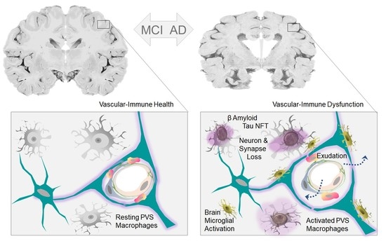
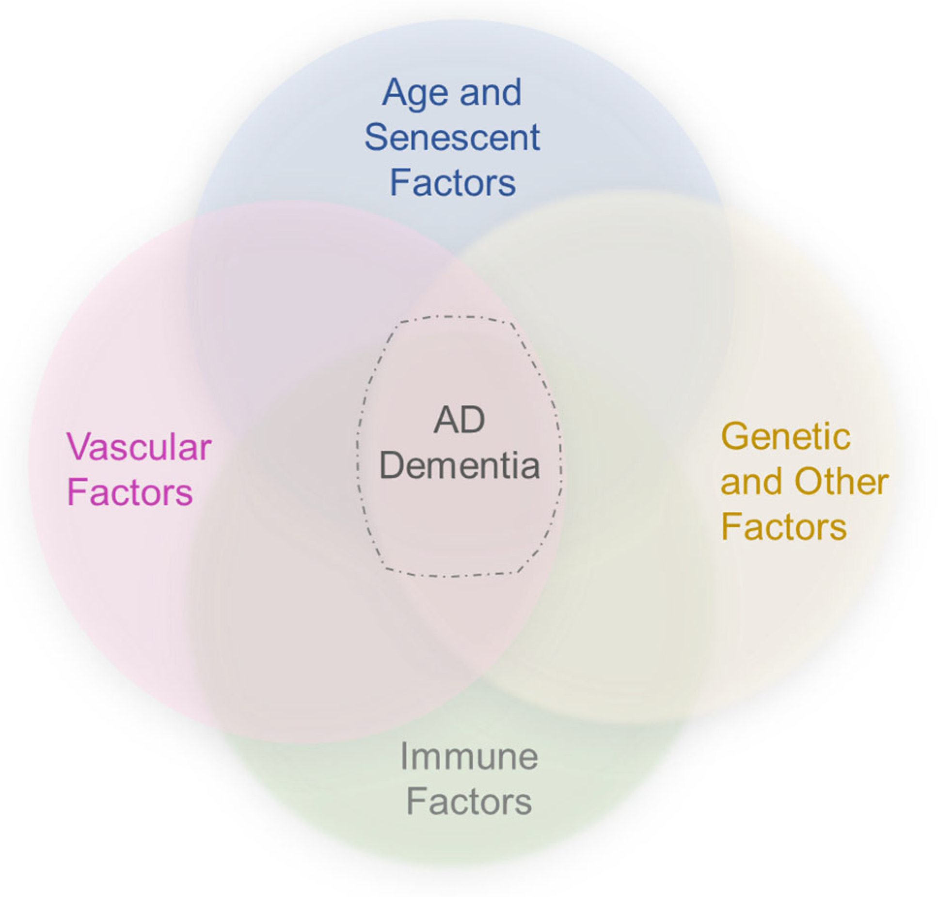
Alzheimer’s disease (AD) is a devastating and irreversible neurodegenerative disorder with unknown etiology. While its cause is unclear, a number of theories have been proposed to explain the pathogenesis of AD. In large part, these have centered around potential causes for intracerebral accumulation of beta-amyloid (βA) and tau aggregates. Yet, persons with AD dementia often exhibit autopsy evidence of mixed brain pathologies including a myriad of vascular changes, vascular brain injuries, complex brain inflammation, and mixed protein inclusions in addition to hallmark neuropathologic lesions of AD, namely insoluble βA plaques and neurofibrillary tangles (NFTs). Epidemiological data demonstrate that overlapping lesions diminish the βA plaque and NFT threshold necessary to precipitate clinical dementia. Moreover, a subset of persons who exhibit AD pathology remain resilient to disease while other persons with clinically-defined AD dementia do not exhibit AD-defining neuropathologic lesions. It is increasingly recognized that AD is a pathologically heterogeneous and biologically multifactorial disease with uncharacterized biologic phenomena involved in its genesis and progression. Here, we review the literature with regard to neuropathologic criteria and incipient AD changes, and discuss converging concepts regarding vascular and immune factors in AD.
Keywords: Alzheimer’s disease; beta-amyloid (βA); glymphatic–lymphatic system; mixed pathologies; neurovascular unit (NVU); perivascular unit (PVU); tau Alzheimer’s disease; beta-amyloid (βA); glymphatic–lymphatic system; mixed pathologies; neurovascular unit (NVU); perivascular unit (PVU); tau
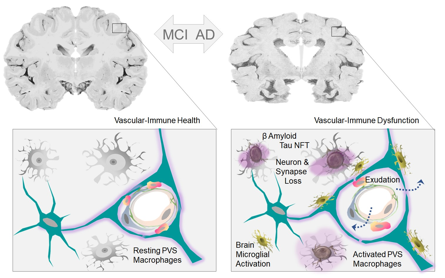
Graphical Abstract