Abstract
Stimuli-responsive materials based on renewable biopolymers are highly attractive for developing sustainable chemical sensors. Here, two spiropyran derivatives (SP1 and SP2) were synthesized and covalently grafted onto cellulose, yielding the functional materials Cel-SP1 and Cel-SP2. Cellulose was selected as a biocompatible, biodegradable, and renewable support able to provide a stable, hydrogen-bond-rich microenvironment for chromic responses. Raman spectroscopy confirmed successful esterification, while SEM-EDS analyses revealed preserved cellulose morphology and the incorporation of nitrogen-rich spiropyran moieties. Both materials exhibited pronounced solvatochromic and pH-dependent behaviors in the solid state. Diffuse reflectance measurements revealed distinct bathochromic or hypsochromic shifts depending on solvent polarity and specific solute–matrix interactions, with DMF and DMSO producing the strongest responses. Under acidic vapors, both materials generated new absorption bands consistent with the formation of protonated merocyanine species, whereas basic vapors promoted partial or full reversion to the spiropyran form. Cel-SP1 and Cel-SP2 also displayed solvent- and pH-dependent luminescence, with Cel-SP2 showing a markedly higher sensitivity to protonation. Prototype solvent strips and acid/base vapor indicators demonstrated fast, naked-eye, reversible chromic transitions. These results highlight spiropyran-modified cellulose as an effective, renewable platform for dual solvent and acid/base vapor sensing.
Keywords:
merocyanine; colorimetric sensors; naked eye; solvatochromic; acidochromic; emission; smart material 1. Introduction
In recent years, there has been increased interest in stimuli-responsive materials capable of altering their physical or chemical properties in response to external triggers like light, temperature, mechanical stress, and pH. These materials have captivated researchers due to their versatility and potential applications in various fields, including controlled release systems, adaptive shape memory materials, and sensors [1,2,3]. Among the molecules used to create such devices, spiropyrans stand out as a classical example, undergoing reversible structural changes, from the spiro (SP) isomer to the merocyanine (MC) form, through various stimuli such as light, pH variations, temperature shifts, presence of metal ions, solvent polarity, and others [4,5,6,7,8]. The versatility of spiropyrans drew researchers’ interest, making use of this class of molecules to functionalize various materials, including nanomaterials [9,10,11,12]. Extensive research has been conducted into the applications of spiropyran-based materials across a wide range of fields like bioimaging [13], data storage [14], controlled release [15], and sensors [16,17].
Marco et al. have developed acidochromic hydrogels functionalized with spiropyran derivatives for selective colorimetric detection of CO2 gas, capable of differentiating CO2 in aqueous media from other acid gases [18]. Raisch et al. reported a spiropyran-based copolymer made into fibers highly sensitive to directional mechanical stress; the aligned fibers produced a blue color only when perpendicularly stretched, with reversible properties [19]. In biological applications, Li et al. constructed a system in which mesoporous silica nanoparticles were anchored to a rhodamine-spiropyran probe, to monitor Cu2+ ions inside lysosomes [20]. Despite the growing literature, there is still a demand for the utilization of more biocompatible supports for spiropyrans.
Cellulose stands as the most abundant and commonly used natural polymer on Earth, renowned for its sustainability and ecological compatibility [21,22,23]. Cellulose has garnered significant attention in recent years owing to its remarkable properties and versatile applications in various fields, including materials science [24], biotechnology [25], and environmental engineering [26]. Comprised of glucose units, cellulose constitutes the primary structural component of plant cell walls, embodying a renewable and sustainable resource. Notably, its intrinsic features include reusability, non-toxicity, environmental friendliness, biocompatibility, and biodegradability, rendering it an interesting substitute for traditional polymers in modern applications [27,28,29]. This macromolecular polysaccharide has strong intra- and intermolecular hydrogen bonding and high crystallinity, thus exhibiting exceptional mechanical properties [30,31].
Studies have shown that cellulose can be utilized to create various sensor prototypes with outstanding performance. Among the most common media for cellulose display is paper. Sanjabi et al. reported on a chemosensor paper coated with nanocapsules containing spiropyran and leuco dye derivatives for ion sensing. This material demonstrated the capability to detect and differentiate heavy metal ions, with a limit of detection as low as 0.00023 µM of Pb2+ ions [32]. Moreover, sensing capabilities are not confined solely to metal ions. Ye et al. demonstrated that colorimetric pH sensing can also be achieved using cellulose nanocrystals functionalized with spiropyrans. Gradual color changes can be observed as the pH is modified in solution [33]. Despite these advancements, cellulose remains underrepresented in reports as a multi-stimulus responsive material.
Herein, two spiropyran derivatives (SP1 and SP2, Figure 1A,B) were synthesized, and their distinct chromic behaviors arising from SP-MC isomerization [34] were employed for sensing organic solvents, as well as acidic and basic environments, both in solution and in the vapor phase. In particular, the molecular design of SP1 incorporates a methoxy group (electron-donating moiety) in addition to the nitro group (electron-withdrawing moiety), which is intended to modulate the electronic distribution of the conjugated merocyanine system. This dual substituent strategy influences not only solvent–chromophore interactions governing solvatochromism, but also alters the local electronic environment of the phenolic oxygen in the ring-open form, thereby affecting protonation behavior and acidochromic response.
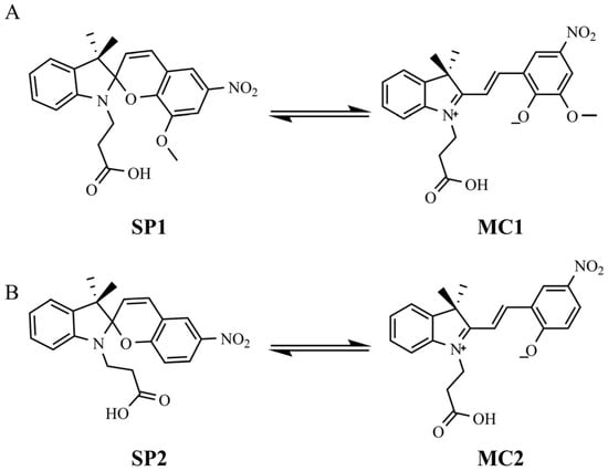
Figure 1.
Chemical structure of: (A) SP1 and MC1 and (B) SP2 and MC2.
To achieve this, the spiropyran derivatives were covalently immobilized onto a cellulose matrix, an environmentally friendly and renewable material, affording the functional materials Cel-SP1 and Cel-SP2 (Figure 2A and Figure 2B, respectively). These cellulose-based responsive systems were characterized in the solid state to validate their structural integrity. Furthermore, beyond the solvatochromic properties exhibited by the spiropyran derivatives in solution, the materials Cel-SP1 and Cel-SP2 were thoroughly investigated with respect to their solvatochromic and ionochromic responses by means of electronic absorption and emission spectroscopy, with these photophysical features correlated to the corresponding macroscopic color changes perceptible to the naked eye.
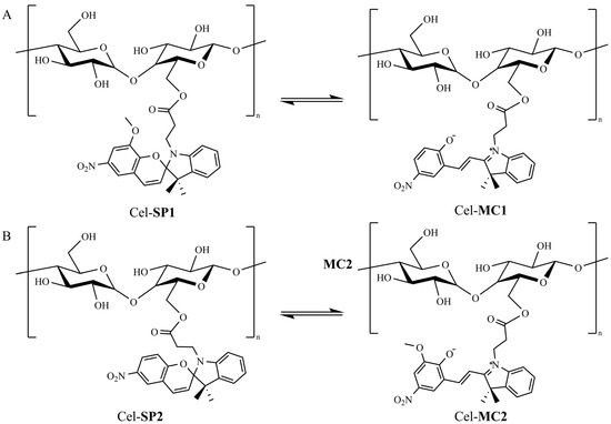
Figure 2.
Chemical structure of: (A) Cel-SP1 and Cel-MC1 and (B) Cel-SP2 and Cel-MC2.
2. Materials and Methods
2.1. Reagents and Solvents
The following reagents were acquired from Sigma-Aldrich (São Paulo, SP, Brazil): 2,3,3-trimethylindolenine (98%), 1,3,3-trimethyl-2-methyleneindoline (97%), 3-iodopropanoic acid (95%), 2-hydroxy-5-nitrobenzaldehyde (98%), 3-methoxy-5-nitrosalicylaldehyde (98%), 4-methylpiperidine (96%), dicyclohexylcarbodiimide (DCC), 4-N,N-dimethylaminopyridine (DMAP), microcrystalline cellulose, acetonitrile (ACN) (MS grade), methanol (MeOH) (≥99.8%), 2-butanone (MEK, ≥99.0%), diethyl ether (Et2O) anhydrous (≥99.0%), and dimethyl sulfoxide-d6 (DMSO-d6) (99.9%). The N,N-dimethylformamide (DMF) (>99.8%), dimethyl sulfoxide (DMSO) (99,5%) and acetone (ACE) (≥99.0%) were from Êxodo Científica, and the ammonium hydroxide (28%), sodium hydroxide (>97.0%), hydrochloric acid (37%), dichloromethane (CH2Cl2) (>99.5%), ethanol (EtOH) (99.5%), tetrahydrofuran (THF) (≥99.0%) and Isopropanol (iPOH) (>99.5%) were from Quimex. Poly(methyl methacrylate) (PMMA) (Mw ≈ 100 kDa) was supplied by Polysciences. All reagents were used without further purification.
2.2. Esterification of Cellulose
Synthesis of SP1 and SP2 followed a previously reported procedure [35]. Cellulose (1.0 g) was resuspended in 30 mL of THF and maintained in an ice bath. SP1 (0.270 g, 6.5 × 10⁻⁴ mol) for Cel-SP1 or SP2 (0.250 g, 6.5 × 10⁻⁴ mol) for Cel-SP2 was solubilized with DMAP (0.02 g, 1.6 × 10⁻⁴ mol) in 15 mL of THF and added to the cellulose suspension. The solution was kept in an ice bath with constant stirring for 30 min. After this period DCC (0.165 g, 8.0 × 10⁻⁴ mol) was added to the reaction solution. The ice bath was removed after a 2 h period, and the solution was maintained with constant stirring overnight. Both solids, Cel-SP1 and Cel-SP2, were filtered; washed with THF, EtOH, and distilled water; and dried under reduced pressure.
2.3. Solid-State Characterization
2.3.1. Raman Spectroscopy
The Raman spectra of cellulose, spiropyran derivatives (SP1 and SP2), and modified cellulose (Cel-SP1 and Cel-SP2) samples were collected on a MultiRam FT-Raman Bruker spectrometer equipped with a germanium detector using liquid nitrogen as the coolant and excited with a 1064 nm beam from an NdYAG laser. Each sample was placed into a small aluminum sample cup; the laser light, with a power of 150–350 mW, was introduced and focused on the sample, and the scattered radiation was collected at 180°. For each spectrum, an average of 512 scans were collected at a resolution of 4 cm−1 over the range of 4000–400 cm−1. Each spectrum was obtained at least twice for confirmation of wavenumber position and intensity, in order to avoid thermal degradation.
2.3.2. Scanning Electron Microscopy
Surface morphologies of the cellulose and modified cellulose (Cel-SP1 and Cel-SP2) samples were investigated using scanning electron microscopy (SEM). The unmodified cellulose was characterized by Phenom ProX, ThermoFischer Scientific (São Paulo, SP, Brazil). The SEM mode used was BSE (back-scattered electron detector), operated at 15 kV, without previous treatment, such as metal coating. The SEM for Cel-SP1 and Cel-SP2 was conducted using a Quanta 200 FEG-FEI (São Paulo, SP, Brazil) operated at an accelerating potential of 15.0 kV. In these two cases, prior to obtaining the SEM images, all of the samples were coated with a 5 nm-thick layer of gold using a sputter coater.
2.4. Solid-State UV-Visible Analysis
In addition to these analyses, diffuse reflectance spectra (DRS) were recorded using a UV-visible absorption spectrophotometer (PerkinElmer (São Paulo, SP, Brazil), Lambda 365, UV–Vis-DRS) in the range of 200 to 800 nm with an integrating sphere attachment and PTFE as a reflectance standard. Approximately 50 mg of modified cellulose powder (Cel-SP1 and Cel-SP2) were placed on the accessory holder, and measurements were obtained immediately after addition of 80 µL of each individual solvent (ACN, distilled water, CHCl3, CH2Cl2, DMF, DMSO, EtOH, iPOH, MeOH, and THF) on top of the powders. For the acid–base vapor assays, approximately 50 mg of the modified cellulose powder was deposited onto qualitative filter paper and positioned over the mouths of flasks containing either concentrated ammonium hydroxide or concentrated hydrochloric acid, with measurements obtained after 30 s of exposure.
2.5. Emission Spectroscopy of Spiropyran-Modified Celluloses (Cel-SP1 and Cel-SP2)
Photoluminescence experiments were performed by adding a small amount of each modified cellulose (Cel-SP1 and Cel-SP2) to each of the solvents (ACN, distilled water, CHCl3, CH2Cl2, DMF, DMSO, EtOH, iPOH, MeO, and THF) or aqueous solution at pH values ranging from 1 to 13 (aqueous solutions were prepared using HCl or NaOH to achieve the respective pH value). Experiments were carried out using quartz cuvettes with a path length of 10 mm and a volume of 1.5 mL, excited with a commercial diode laser λexc = 405 nm. All spectra were collected using an SR-2UVV240-25 Mini Spectrophotometer (São Paulo, SP, Brazil) with a range of 182 nm–908 nm, optical resolution of 1.33 nm, and aperture 25 μm. In addition, Cel-SP1 and Cel-SP2 were supported on top of poly(methyl methacrylate) (PMMA) films to obtain the emission spectra of both materials after their exposure to acid (HCl 37%) and base (NH4OH 28%) vapors.
3. Results and Discussion
The solvent- and acid/base vapor-sensing experiments presented herein are intended as a proof-of-concept to demonstrate the responsiveness of spiropyran-grafted cellulose materials (Cel-SP1 and Cel-SP2) in the solid state, establishing the feasibility of dual-mode qualitative sensing and clear naked-eye detectability, as well as material reusability. Such an approach is consistent with early-stage investigations of chromogenic sensing platforms, where visual responsiveness, reversibility, and functional stability are first validated. Within this framework, the observed behaviors of Cel-SP1 and Cel-SP2 compare favorably with previously reported spiropyran- and polymer-based vapor sensors in terms of simplicity of material preparation [4].
3.1. Raman Spectroscopy
Raman spectra for both spiropyran derivatives (SP1 and SP2) and their respective modified cellulose materials (Cel-SP1 and Cel-SP2) are depicted in Figure 3 (from 1800 to 0 cm−1); the full spectra can be found in Supplementary Material Figure S1. The main vibrational bands are discussed below, based on the literature [36,37,38]. For both derivatives, SP1 and SP2 (Figure 3C,E), several characteristic vibrational features can be identified. The NO2 asymmetric stretching band consistently appears in the 1575–1580 cm−1 region, while the more intense NO2 symmetric stretching mode is observed between 1332–1340 cm−1 for SP1 and SP2. In the 1600–1660 cm−1 interval, minor bands at approximately 1620 cm−1 and 1650 cm−1 are detectable, corresponding to C–H wagging and C–C stretching of aromatically substituted rings, respectively. Furthermore, a weak C–O (aryl) stretching vibration is systematically found at 1090 cm−1 in both derivatives’ spectra.
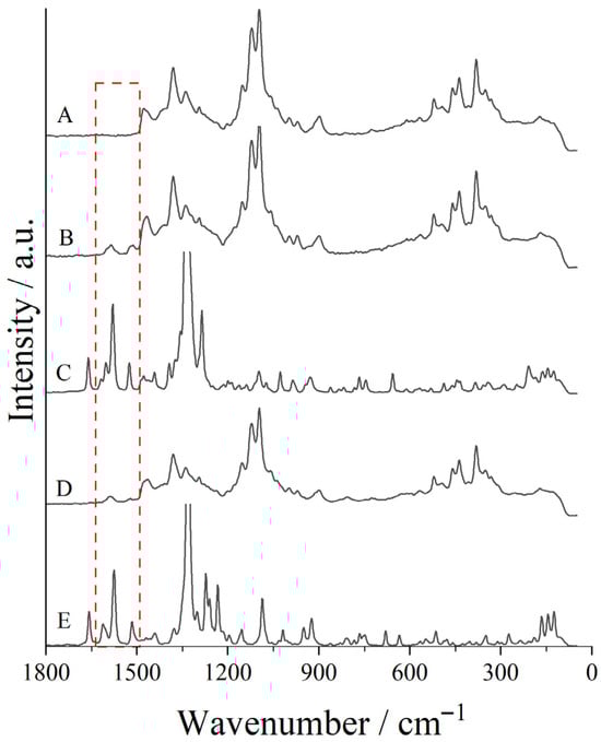
Figure 3.
Raman spectra of: (A) pure cellulose, (B) Cel-SP1, (C) SP1, (D) Cel-SP2, and (E) SP2 from 0 to 1800 cm–1.
For pure cellulose (Figure 3A), it is worth noting that most vibrational modes related to its structure are not present in the spiropyrans’ spectra. Several diagnostic vibrational features can be clearly distinguished. The CH2 deformation modes associated with scissoring motions give rise to a characteristic band in the 1450–1480 cm−1 region. The low-frequency band at 380 cm−1, commonly employed as an indicator of cellulose aggregation or crystallinity, is also present and originates from heavy-atom stretching within the C–C–C ring framework. Additional contributions appear near 1380 cm−1, corresponding to H–C–C, H–C–O, and H–O–C bending vibrations. The glycosidic linkage is evident through the bands at 912 and 1097 cm−1, which are assigned to the symmetric and asymmetric stretching modes of the C–O–C unit, respectively. All the vibrational modes discussed for pure cellulose are also present in the spiropyran-modified celluloses’ spectra (Figure 3B,D).
Specifically for Cel-SP1 and Cel-SP2, we highlight the 1500–1620 cm−1 region, in which are observed NO2 asymmetric stretching, C-H wagging, and C-C stretching of aromatic ring bands. These features are absent in the spectrum of pure cellulose, indicating contributions from the spiropyran moieties. Other potential changes are likely overlapped by the intense intrinsic cellulose bands or fall below the detection threshold due to low intensity. Nevertheless, the emergence of these new signals provides strong evidence for the successful chemical bonding of the spiropyran derivatives to the cellulose matrix.
3.2. Scanning Electron Microscopy
In order to identify whether the morphology of the cellulose was modified due to its esterification with both spiropyran derivatives, SEM analyses were carried out. The SEM for unmodified cellulose and its EDS mapping is presented in Supplementary Material Figure S2. This unmodified cellulose was previously washed with Milli-Q® water, ethanol, and acetone to remove any possible impurity. The unmodified microcrystalline cellulose displayed the characteristic fragmented and lamellar morphology typically associated with mechanically processed cellulose, comprising irregular platelet-like particles with folded surfaces and evident agglomeration. SEM analysis revealed particle sizes spanning from ten to several hundred micrometers, with rough surface topography that may enhance interfacial interactions in composite or suspension systems. Elemental mapping by EDS showed exclusive detection of carbon and oxygen distributed uniformly across the particle domains, with no measurable contributions from heavier elements. The SEM analysis for Cel-SP1 and Cel-SP2 is depicted in Figure 4. Both modified cellulose samples (Cel-SP1 and Cel-SP2) present similar morphology, and the EDS mapping depicted in Figure 5A,B, demonstrated the presence of nitrogen atoms, in addition to the carbon and oxygen, due to the presence of the spiropyran derivatives SP1 and SP2 in the modified material.
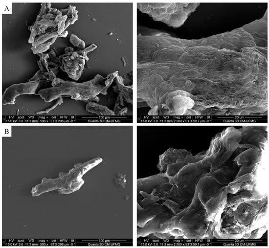
Figure 4.
SEM images of (A) Cel-SP1 and (B) Cel-SP2, at magnification of 500× and 2500×, respectively.
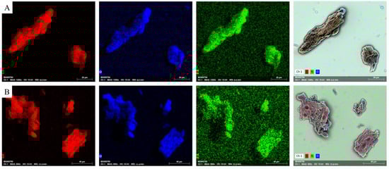
Figure 5.
EDS mapping for: (A) Cel-SP1 and (B) Cel-SP2 for (C, O, N atoms and merged image).
3.3. Solid-State UV-Visible Analysis
Solvent polarity and specific solute–solvent interactions exert a decisive influence on the photophysical response of spiropyran derivatives, and consequently on the colorimetric behavior of the modified cellulose sensors. In solution, spiropyrans exist in an equilibrium between the closed, non-conjugated SP form and the open, zwitterionic MC form. Shifts in this equilibrium strongly depend on the solvent environment. Polar and protic solvents stabilize the charge-separated MC species through dipolar and hydrogen-bonding interactions, producing bathochromic shifts in the absorption profile and enhanced visible coloration [39]. Conversely, nonpolar solvents favor the colorless SP form, yielding lower absorbance in the visible region [40]. Emission behavior follows a similar trend: The MC form typically exhibits solvent-dependent fluorescence with intensity and wavelength modulated by polarity, hydrogen bonding, and microviscosity [41]. These solvatochromic effects provide the basis for using spiropyran-modified cellulose as a colorimetric sensor, as environmental changes around the polymer matrix may translate into measurable data.
Solvatochromic behavior was assessed by UV-visible diffuse reflectance spectroscopy for both Cel-SP1 and Cel-SP2 in ten common solvents: ACN, H2O, CHCl3, CH2Cl2, DMF, DMSO, EtOH, iPOH, MeOH, and THF (Figure 6). For Cel-SP1, the visible-region band maxima ranged from 462 to 508 nm, with the dry control sample exhibiting a maximum at 466 nm. Cel-SP2 displayed slightly blue-shifted features, with band maxima between 439 and 470 nm and a control value of 457 nm. Among all tested media, DMSO and DMF produced the most pronounced bathochromic shifts for both materials, whereas MeOH and ACN induced the strongest hypsochromic shifts, though of smaller magnitude than the red shifts observed in the less polar aprotic solvents. It is also worth noting that both modified cellulose materials interacted poorly with water. The weak interaction of both Cel-SP1 and Cel-SP2 with water indicates that their chromic responses are only minimally affected by moisture, which is advantageous for practical sensing applications in humid environments where selectivity toward organic solvents or acid/base vapors must be preserved.
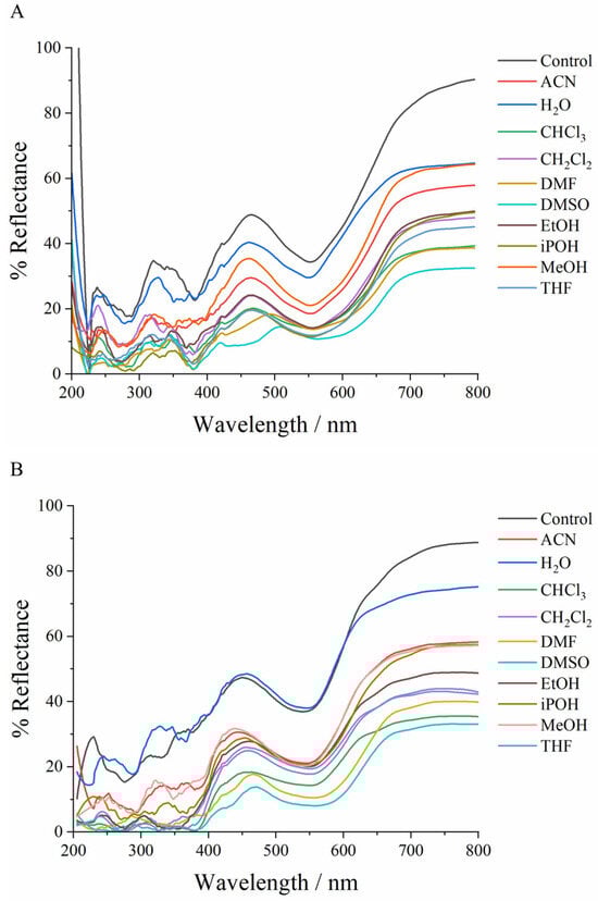
Figure 6.
Diffuse reflectance spectra of (A) Cel-SP1 and (B) Cel-SP2 in solvents.
Even though the behavior observed for the modified cellulose is similar to what is reported in the literature for spiropyran derivatives in solution [7,41], there is no linearity relating polarity (ET30) [42,43] and wavelength, which is not surprising for a solid cellulose matrix. Cellulose is a highly hydrogen-bonded, semicrystalline polymer with nanoscale pores and a network of hydroxyl-rich regions that selectively sorb and/or swell in different solvents; this produces local microenvironments whose polarity and specific interaction capability can differ substantially for each solvent [44].
Within this framework, the pronounced responses observed for DMSO and DMF can be rationalized by their exceptional ability to penetrate and swell microcrystalline cellulose, promoting partial chain separation and increasing access to solvatochromic spiropyran sites embedded within the matrix. These polar aprotic solvents act as strong hydrogen-bond acceptors and efficiently stabilize the MC form, even when their ET30 values alone would not predict the magnitude of the observed bathochromic shifts. By contrast, less penetrative solvents such as THF interact more superficially with the cellulose network, limiting MC stabilization despite moderate polarity. Thus, specific solvent–matrix and solvent–chromophore interactions—such as hydrogen-bond accepting ability, polarizability, and differential solvation of neutral versus zwitterionic MC species—cannot be captured by a single empirical polarity scale [45].
In order to verify the suitability to use the modified celluloses for the solvent detection, glass microtubes were filled with both Cel-SP1 and Cel-SP2, as schematically shown in Figure 7A (see Supplementary Material Figures S3 and S4). The color change due to the solvent elution is presented in Figure 7B under visible light irradiation and under UV emission in Figure 7C. Cel-SP1 exhibited color variations ranging from light to dark purple, whereas Cel-SP2 displayed shades of dark pink. Under UV irradiation, Cel-SP1 showed pronounced color differentiation for the first two solvents (left to right), clearly distinguishing them from the remaining samples and corroborating the trends observed in the solid-state UV-visible measurements. In contrast, Cel-SP2 exhibited a more uniform UV response, transitioning from dark to lighter glossy pink across the tested solvents, reflecting its reduced discrimination capability under excitation conditions.
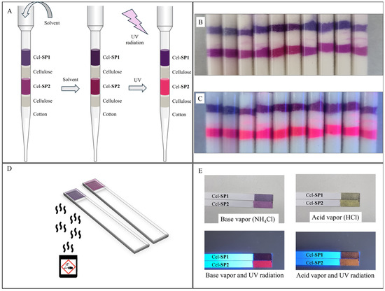
Figure 7.
(A) Schematic representation of the glass microtube with modified cellulose intercalated with the modified cellulose for the solvent detection, (B) color change based on solvent elution under visible light, (C) color change based on solvent elution under UV irradiation, (D) schematic representation of the pH test strip for base and acid vapors, and (E) color of the test strips after base and acid vapors under visible and UV light radiation. (Solvents tested in (B) and (C) from left to right: DMF, DMSO, ACN, THF, CHCl3, CH2Cl2, iPOH, H2O, EtOH, and MeOH).
Crucially, the pH-dependent color response of spiropyrans is not simply a binary switch between SP and MC form but involves a more complex equilibrium that includes a protonated merocyanine (MCH+) species [4]. Under acidic conditions, protonation of the MC yields MCH+, which strongly favors the ring-open, conjugated form; this leads to significant acidochromic shifts and pronounced visible absorption [46]. Depending on the substituted spiropyran and the strength (pKa) of the acid, protonation can promote spontaneous ring opening even in the dark, and different protonated isomers may be stable [47]. In more basic or neutral environments, deprotonation converts MCH+ back to MC, but the equilibrium between MC and SP (and their thermal kinetics) depends strongly on parameters such as the deprotonation constant (Ka) and the ring-closing vs. ring-opening rate constants [48].
For the acid–base vapor assays, the powdered samples of Cel-SP1 and Cel-SP2 were evenly distributed onto filter paper discs and positioned over the mouths of flasks containing concentrated acidic or basic solutions. Both materials exhibited visible color changes within seconds of exposure to the vapors. After approximately 30 s, the samples were transferred to a quartz holder for spectral acquisition. The corresponding diffuse reflectance spectra are presented in Figure 8. In both materials, exposure to acidic vapors produced marked changes in the spectral profile, with distinct bands emerging in the 595–620 nm region and around 530 nm, consistent with the formation of the more stable MCH+ species. In contrast, upon exposure to basic vapors, Cel-SP2 displayed no discernible bands within the analyzed region, suggesting near-complete reversion to the ring-closed SP form. Under the same conditions, Cel-SP1 retained its band at 500 nm, indicating that its SP ⇌ MC equilibrium does not fully shift back toward the colorless SP state.
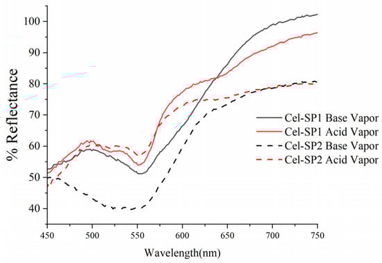
Figure 8.
Diffuse reflectance spectra of Cel-SP1 and Cel-SP2 after 30 s of exposure to acid (HCl) and base (NH4OH) vapors.
Following the solvent tests, pH-responsive strips were assembled using both materials (Cel-SP1 and Cel-SP2), as shown in Figure 7D. Upon exposure to acidic and basic vapors, respectively, the modified celluloses changed from pink to yellow, respectively; Figure 7E. This transition was fast and fully reversible, as demonstrated in Supplementary Video S1. Furthermore, Cel-SP1 exhibited detectable fluorescence under both acidic (HCl) and basic (NH4OH) conditions, although the emission was noticeably weaker in the presence of base. Cel-SP2 showed the same colorimetric reversibility but with less pronounced emissive changes.
3.4. Solid-State Emission Analysis
Across the tested solvents, Cel-SP1 emits in the ~645–695 nm window with systematic changes in peak position and shape depending on solvent, show in Figure 9A. In low-polarity, weakly interacting solvents (e.g., CHCl3, THF) Cel-SP1 is observed at its bluest positions (~645–655 nm) and presents relatively narrow, symmetric bands that are consistent with emission from a predominantly locally excited (LE) with limited charge separation. Increasing solvent polarity and hydrogen bonding ability produces a progressive red shift of the Cel-SP1 band (up to ~680–695 nm in highly polar aprotic solvents such as DMSO/DMF), followed with moderate broadening. This behavior is consistent with a degree of solvent-sensitive excited-state stabilization, whereby polar environments stabilize the excited dipole of Cel-SP1 and increase inhomogeneous broadening due to solvent relaxation effects. Nevertheless, Cel-SP1 appears to retain a comparatively limited charge-transfer character, particularly when contrasted with Cel-SP2 [47]. The relatively modest Stokes shifts and narrower bandwidths are therefore consistent with an excited state exhibiting restricted intramolecular charge-transfer (ICT) contribution rather than a fully developed ICT state.
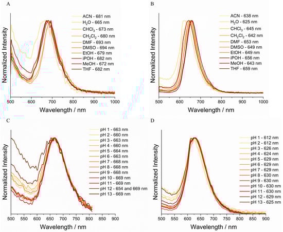
Figure 9.
Emission spectra for (A) Cel-SP1 and (B) Cel-SP2 in solvents in different solvents and (C) Cel-SP1 and (D) Cel-SP2 at different water solutions varying the pH from 1 to 13.
By contrast, Cel-SP2 is a consistently red-shifted relative to Cel-SP1, shown in Figure 9B, with emission maxima that are strongly sensitive to solvent polarity and proticity. In protic and highly polar solvents (MeOH, H2O, DMSO) Cel-SP2 shifts to longer wavelengths (≈665–695 nm) and displays broader, often asymmetric profiles, features that are consistent with enhanced stabilization of a more polar or charge-separated excited state. In less polar solvents Cel-SP2 shifts toward shorter wavelengths but remains red-shifted relative to Cel-SP1 in the corresponding solvent, suggesting intrinsically higher excited-state polarity or stronger solvent–solute interactions, such as hydrogen bonding or preferential solvation of charged resonance forms. The magnitude of the solvent-dependent shifts observed for Cel-SP2 is larger than for Cel-SP1, supporting the interpretation that Cel-SP2 accesses an excited state that experiences stronger solvation stabilization, which may involve a protonated or highly dipolar ICT-like state, although this assignment is phenomenological and based on steady-state emission data.
Both emissive species, Cel-SP1 and Cel-SP2, display pH-dependent behavior, but with markedly different sensitivities (Figure 9C,D). Cel-SP1 shows only moderate spectral shifts as a function of pH: At low pH, it appears slightly red-shifted and broader, and as pH increases its maximum undergoes only modest hypo- or bathochromic changes. These observations indicate that protonation influences the emissive properties of Cel-SP1 without inducing a major reorganization of its excited-state electronic structure. In contrast, Cel-SP2 is highly pH-responsive, exhibiting substantial red shifts and pronounced changes in spectra shape under acidic conditions, behavior that is consistent with stabilization of a more polar and/or protonated excited state. Under basic conditions, the emission shifts, narrows, or decreases in intensity, suggesting disruption of the interactions that stabilize this emissive species. Taken together, these systematic trends suggest that Cel-SP2 is more strongly affected by protonation equilibria than Cel-SP1, and its emission is consistent with a highly polar, resonance-stabilized, or ICT-like excited state whose properties depend sensitively on pH, as inferred from steady-state spectral observations rather than direct time-resolved or kinetic measurements.
Under UV excitation, which deposits ~2–3 eV of energy into the spiropyran chromophore, substantially exceeding the ~0.1–0.5 eV energy differences between protonation states in the ground state. Consequently, photoexcitation can access excited-state ring-opening and protonation pathways (photoacidochromism) that are not thermally available, temporarily leading to MC and protonated MC species even at pH values where the ground-state equilibrium would disfavor them, as shown in Figure 9C,D. This interpretation is supported qualitatively by the emission behavior observed when Cel-SP1 and Cel-SP2 are embedded in PMMA matrices and directly exposed to acid and base vapors (Figure 10A,B) [46,49,50].
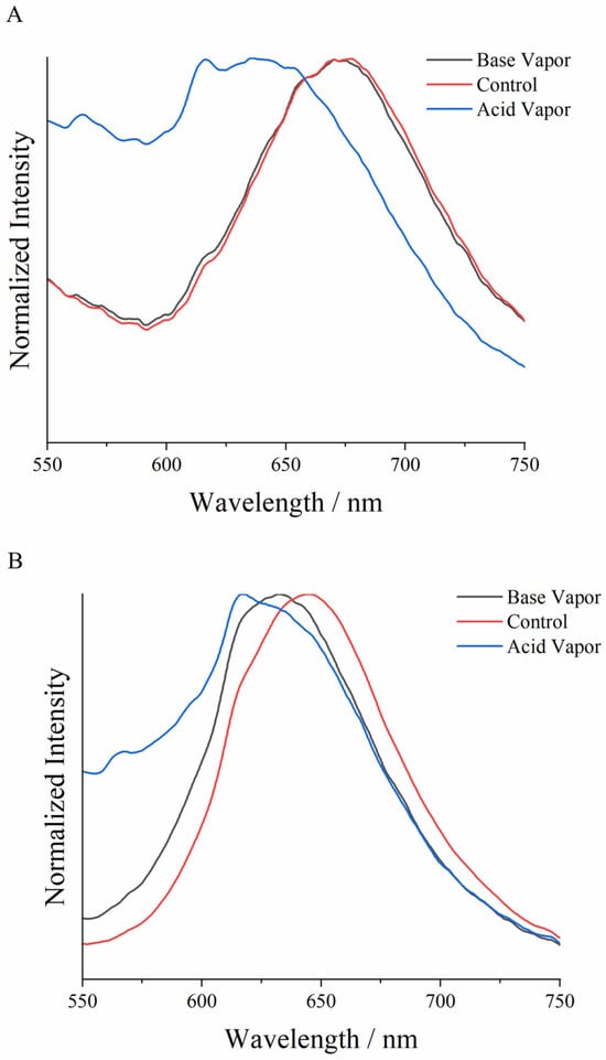
Figure 10.
Emission spectra for (A) Cel-SP1 and (B) Cel-SP2 directly exposed to acid and base vapors (red spectra refer to both modified cellulose without any previously influence).
4. Conclusions
Herein, spiropyran-modified celluloses Cel-SP1 and Cel-SP2 were successfully synthesized through covalent grafting and shown to retain both the photophysical versatility of spiropyrans and the sustainability benefits of cellulose. Structural characterization confirmed effective functionalization without compromising the morphology of the biopolymer matrix. The resulting materials exhibited strong solvatochromic responses and rapid, reversible acidochromic behavior in both solution-contact and vapor-phase conditions, enabling naked-eye detection of solvent environments and acidic or basic vapors. Emission analyses revealed that Cel-SP2 displays particularly high sensitivity to protonation, whereas Cel-SP1 maintains detectable fluorescence even under basic conditions. While solvent or pH responsiveness of spiropyran-modified cellulose are usually reported individually, combining Cel-SP1 and Cel-SP2 demonstrates the combined dual sensing of organic solvents and acid/base vapors in the solid state, correlated across absorption, emission, and naked-eye responses within the same renewable platform. The simple construction of microtube solvent indicators and pH-responsive strips demonstrates the practical applicability of these materials. Notably, the distinct solid-state solvatochromic behavior, reversible vapor-phase acidochromism, and differential fluorescence responses of Cel-SP1 and Cel-SP2 illustrate how subtle molecular design variations in the spiropyran structure translate into tunable sensing performance, rather than merely reproducing known spiropyran behavior. Overall, the combination of renewable cellulose with spiropyran derivatives provides an environmentally friendly platform for robust dual-mode chromic sensing, offering promising potential for low-cost chemical detection, smart packaging, and environmental monitoring.
Supplementary Materials
The following supporting information can be downloaded at: https://www.mdpi.com/article/10.3390/chemosensors14010017/s1, Figure S1: Raman spectra; Figure S2: SEM and EDS; Figure S3: Glass microtube; Figure S4: Glass microtube; Video S1: Vapor sensor.
Author Contributions
Conceptualization, D.D.S.d.S., R.S.N., F.B.M., and F.B.D.S.; Methodology, J.P.C.T., L.F.C.d.O., R.S.N., F.B.M., and F.B.D.S.; Validation, D.D.S.d.S.; Formal Analysis, D.D.S.d.S.; Investigation, J.P.C.T., L.F.C.d.O., H.S.B., F.A., R.S.N., F.B.M., and F.B.D.S.; Resources, J.P.C.T., L.F.C.d.O., H.S.B., F.A., R.S.N., F.B.M., and F.B.D.S.; Data Curation, F.B.D.S.; Writing—Original Draft Preparation, R.S.N., F.B.M., and F.B.D.S.; Writing—Review and Editing, D.D.S.d.S., J.P.C.T., L.F.C.d.O., H.S.B., F.A., R.S.N., F.B.M., and F.B.D.S.; Visualization, F.B.M. and F.B.D.S.; Supervision, F.B.M. and F.B.D.S.; Project Administration, F.B.D.S.; Funding Acquisition, J.P.C.T., L.F.C.d.O., H.S.B., F.A., R.S.N., F.B.M., and F.B.D.S. All authors have read and agreed to the published version of the manuscript.
Funding
This work was supported by Brazilian agencies: CNPq (grant number 303311/2024-0, 406853/2021-5, 303569/2022-0, and 383958/2025-3), FAPEMIG (grant numbers APQ-04537-22, RED-00045-23, APQ-00144-24, APQ-03079-23, and APQ-00887-23). Also (CEMASU) FAPESP-Funding (Process: 2021/11965-3) and (Process: 2017/50334-3). National Council of Scientific and Technological Development/CNPq (Grants: 309614/2021-0 and 302178/2025-2), National Institutes of Science and Technology (INCTs), INCT Polysaccharides (Grant: 406973/2022-9), INCT Circularity in Polymer Materials (Grant: 406925/2022-4/406925/2022-4), INCT-INFO (National Institute of Photonics). In addition to FINEP (0187/22–INFRASPEC), PETROBRAS (TC 0050.0121114.22.9), and CAPES.
Data Availability Statement
The original contributions presented in this study are included in the article/Supplementary Materials. Further inquiries can be directed to the corresponding authors.
Acknowledgments
The authors would like to acknowledge the Center of Microscopy at the Universidade Federal de Minas Gerais (http://www.microscopia.ufmg.br (access on 3 December 2025)) for providing the equipment and technical support for experiments involving electron microscopy.
Conflicts of Interest
The authors declare no conflicts of interest.
References
- Idumah, C.I.; Odera, R.S.; Ezeani, E.O.; Low, J.H.; Tanjung, F.A.; Damiri, F.; Luing, W.S. Construction, characterization, properties and multifunctional applications of stimuli-responsive shape memory polymeric nanoarchitectures: A review. Polym. Plast. Technol. Mater. 2023, 62, 1247–1272. [Google Scholar] [CrossRef]
- Png, Z.M.; Wang, C.-G.; Yeo, J.C.C.; Lee, J.J.C.; Surat’man, N.E.; Tan, Y.L.; Liu, H.; Wang, P.; Tan, B.H.; Xu, J.W.; et al. Stimuli-responsive structure–property switchable polymer materials. Mol. Syst. Des. Eng. 2023, 8, 1097–1129. [Google Scholar] [CrossRef]
- Ma, W.; Hua, D.; Xiong, R.; Huang, C. Bio-based stimuli-responsive materials for biomedical applications. Mater. Adv. 2023, 4, 458–475. [Google Scholar] [CrossRef]
- Miguez, F.B.; Moreira, O.B.O.; de Oliveira, M.A.L.; Denadai, Â.M.L.; de Oliveira, L.F.C.; De Sousa, F.B. Reversible electrospun fibers containing spiropyran for acid and base vapor sensing. J. Mater. Res. 2023, 38, 547–556. [Google Scholar] [CrossRef]
- Ahmed, S.A.; Okasha, R.M.; Khairou, K.S.; Afifi, T.H.; Mohamed, A.A.H.; Abd-El-Aziz, A.S. Design of thermochromic polynorbornene bearing spiropyran chromophore moieties: Synthesis, thermal behavior and Dielectric Barrier Discharge plasma treatment. Polymers 2017, 9, 630. [Google Scholar] [CrossRef] [PubMed]
- Li, Z.; Xiong, H.; Liang, H.; Chen, W.; Tian, Q.; Yan, M.; Su, H.; Royal, G. A New Spiropyran Hydrazone as an Unusual Colorimetric Sensor for Detection of Cu2+ and Cr3+ Based on Aggregation-Induced Enhancement Effects in Aqueous Solvent Mixtures. ChemistrySelect 2022, 7, e202201868. [Google Scholar] [CrossRef]
- Abdollahi, A.; Alinejad, Z.; Mahdavian, A.R. Facile and fast photosensing of polarity by stimuli-responsive materials based on spiropyran for reusable sensors: A physico-chemical study on the interactions. J. Mater. Chem. C Mater. 2017, 5, 6588–6600. [Google Scholar] [CrossRef]
- Rad, J.K.; Ghomi, A.R.; Karimipour, K.; Mahdavian, A.R. Progressive Readout Platform Based on Photoswitchable Polyacrylic Nanofibers Containing Spiropyran in Photopatterning with Instant Responsivity to Acid–Base Vapors. Macromolecules 2020, 53, 1613–1622. [Google Scholar] [CrossRef]
- Klajn, R. Spiropyran-based dynamic materials. Chem. Soc. Rev. 2014, 43, 148–184. [Google Scholar] [CrossRef]
- Rad, J.K.; Balzade, Z.; Mahdavian, A.R. Spiropyran-based advanced photoswitchable materials: A fascinating pathway to the future stimuli-responsive devices. J. Photochem. Photobiol. C Photochem. Rev. 2022, 51, 100487. [Google Scholar] [CrossRef]
- Fagan, A.; Bartkowski, M.; Giordani, S. Spiropyran-Based Drug Delivery Systems. Front. Chem. 2021, 9, 720087. [Google Scholar] [CrossRef] [PubMed]
- Marturano, V.; Kozlowska, J.; Bajek, A.; Giamberini, M.; Ambrogi, V.; Cerruti, P.; Garcia-Valls, R.; Montornes, J.M.; Tylkowski, B. Photo-triggered capsules based on lanthanide-doped upconverting nanoparticles for medical applications. Coord. Chem. Rev. 2019, 398, 213013. [Google Scholar] [CrossRef]
- Cong, Y.; Wang, X.; Zhu, S.; Liu, L.; Li, L. Spiropyran-Functionalized Gold Nanoclusters with Photochromic Ability for Light-Controlled Fluorescence Bioimaging. ACS Appl. Bio Mater. 2021, 4, 2790–2797. [Google Scholar] [CrossRef]
- Leinen, M.B.; Klein, P.; Sebastian, F.L.; Zorn, N.F.; Adamczyk, S.; Allard, S.; Scherf, U.; Zaumseil, J. Spiropyran-Functionalized Polymer–Carbon Nanotube Hybrids for Dynamic Optical Memory Devices and UV Sensors. Adv. Electron. Mater. 2020, 6, 2000717. [Google Scholar] [CrossRef]
- Zhang, R.; Min, Y.; Ji, P.; Zhou, G.; Yin, H.; Qi, D.; Deng, H.; Hua, Z.; Chen, T. Light and temperature dual stimuli-responsive micelles from carbamate-containing spiropyran-based amphiphilic block copolymers: Fabrication, responsiveness and controlled release behaviors. Eur. Polym. J. 2023, 200, 112493. [Google Scholar] [CrossRef]
- Jin, Y.; Petrescu, F.I.T.; Wang, Y.; Li, X.; Li, Y.; Shi, G. Spiropyran-Based Soft Substrate with SPR, Anti-Reflection and Anti-NRET for Enhanced Visualization/Fluorescence Dual Response to Metal Ions. Materials 2023, 16, 3746. [Google Scholar] [CrossRef]
- Kim, D.W.; Jang, H.G.; Lee, H.S.; Kim, J. Force-induced fluorescence spectrum shift of spiropyran-based polymer for mechano-response sensing. Sens. Actuators A Phys. 2023, 359, 114513. [Google Scholar] [CrossRef]
- Marco, A.; Guirado, G.; Sebastián, R.M.; Hernando, J. Spiropyran-based chromic hydrogels for CO2 absorption and detection. Front. Chem. 2023, 11, 1176661. [Google Scholar] [CrossRef]
- Raisch, M.; Genovese, D.; Zaccheroni, N.; Schmidt, S.B.; Focarete, M.L.; Sommer, M.; Gualandi, C. Highly Sensitive, Anisotropic, and Reversible Stress/Strain-Sensors from Mechanochromic Nanofiber Composites. Adv. Mater. 2018, 30, e1802813. [Google Scholar] [CrossRef]
- Li, Y.; Zhao, Y.; Chan, W.; Wang, Y.; You, Q.; Liu, C.; Zheng, J.; Li, J.; Yang, S.; Yang, R. Selective Tracking of Lysosomal Cu2+ Ions Using Simultaneous Target- and Location-Activated Fluorescent Nanoprobes. Anal. Chem. 2015, 87, 584–591. [Google Scholar] [CrossRef] [PubMed]
- Aziz, T.; Haq, F.; Farid, A.; Kiran, M.; Faisal, S.; Ullah, A.; Ullah, N.; Bokhari, A.; Mubashir, M.; Chuah, L.F.; et al. Challenges associated with cellulose composite material: Facet engineering and prospective. Environ. Res. 2023, 223, 115429. [Google Scholar] [CrossRef] [PubMed]
- Ferreira, F.V.; Souza, A.G.; Ajdary, R.; de Souza, L.P.; Lopes, J.H.; Correa, D.S.; Siqueira, G.; Barud, H.S.; Rosa, D.D.S.; Mattoso, L.H.C.; et al. Nanocellulose-based porous materials: Regulation and pathway to commercialization in regenerative medicine. Bioact. Mater. 2023, 29, 151–176. [Google Scholar] [CrossRef]
- Caicho-Caranqui, J.; Vivanco, G.; Egas, D.A.; Chuya-Sumba, C.; Guerrero, V.H.; Ramirez-Cando, L.; Reinoso, C.; De Sousa, F.B.; Leon, M.; Ochoa-Herrera, V.; et al. Non-modified cellulose fibers for toxic heavy metal adsorption from water. Adsorption 2025, 31, 18. [Google Scholar] [CrossRef]
- Wu, Z.; Chen, S.; Li, J.; Wang, B.; Jin, M.; Liang, Q.; Zhang, D.; Han, Z.; Deng, L.; Qu, X.; et al. Insights into Hierarchical Structure–Property–Application Relationships of Advanced Bacterial Cellulose Materials. Adv. Funct. Mater. 2023, 33, 2214327. [Google Scholar] [CrossRef]
- Akki, A.J.; Jain, P.; Kulkarni, R.; Rao, B.R.; Kulkarni, R.V.; Zameer, F.; Anjanapura, V.R.; Aminabhavi, T.M. Microbial biotechnology alchemy: Transforming bacterial cellulose into sensing disease—A review. Sens. Int. 2024, 5, 100277. [Google Scholar] [CrossRef]
- Rahman, A.; Wang, W.; Govindaraj, D.; Kang, S.; Vikesland, P.J. Recent advances in environmental science and engineering applications of cellulose nanocomposites. Crit. Rev. Environ. Sci. Technol. 2023, 53, 650–675. [Google Scholar] [CrossRef]
- Hossain, M.T.; Shahid, M.A.; Akter, S.; Ferdous, J.; Afroz, K.; Refat, K.R.I.; Faruk, O.; Jamal, M.S.I.; Uddin, M.N.; Samad, M.A.B. Cellulose and starch-based bioplastics: A review of advances and challenges for sustainability. Polym. Plast. Technol. Mater. 2024, 63, 1329–1349. [Google Scholar] [CrossRef]
- Simelane, N.P.; Olatunji, O.S.; John, M.J.; Andrew, J. Engineered transparent wood with cellulose matrix for glass applications: A review. Carbohydr. Polym. Technol. Appl. 2024, 7, 100487. [Google Scholar] [CrossRef]
- Las-Casas, B.; Arantes, V. From production to performance: Tailoring moisture and oxygen barrier of cellulose nanomaterials for sustainable applications—A review. Carbohydr. Polym. 2024, 334, 122012. [Google Scholar] [CrossRef] [PubMed]
- Parthasarathi, R.; Bellesia, G.; Chundawat, S.P.S.; Dale, B.E.; Langan, P.; Gnanakaran, S. Insights into Hydrogen Bonding and Stacking Interactions in Cellulose. J. Phys. Chem. A 2011, 115, 14191–14202. [Google Scholar] [CrossRef]
- Mokhena, T.C.; Sadiku, E.R.; Mochane, M.J.; Ray, S.S.; John, M.J.; Mtibe, A. Mechanical properties of cellulose nanofibril papers and their bionanocomposites: A review. Carbohydr. Polym. 2021, 273, 118507. [Google Scholar] [CrossRef]
- Sanjabi, S.; Rad, J.K.; Salehi-Mobarakeh, H.; Mahdavian, A.R. Dual-chromic cellulose paper modified with nanocapsules containing leuco dye and spiropyran derivatives: A colorimetric portable chemosensor for detection of some heavy metal cations. J. Environ. Chem. Eng. 2024, 12, 111724. [Google Scholar] [CrossRef]
- Ye, X.; Wang, A.; Zhang, D.; Zhou, P.; Zhu, P. Light and pH dual-responsive spiropyran-based cellulose nanocrystals. RSC Adv. 2023, 13, 11495–11502. [Google Scholar] [CrossRef]
- Mendonça, N.E.N.; Leão, C.A.S.; Alexis, F.; Ochoa-Herrera, V.; Zambrano-Romero, A.; Nobuyasu, R.S.; Miguez, F.B.; De Sousa, F.B. Exploiting Spiropyran Solvatochromism for Heavy Metal Ion Detection in Aqueous Solutions. ACS Omega 2025, 10, 36412–36420. [Google Scholar] [CrossRef] [PubMed]
- Miguez, F.B.; Trigueiro, J.P.C.; Lula, I.; Moraes, E.S.; Atvars, T.D.Z.; de Oliveira, L.F.C.; Alexis, F.; Nobuyasu, R.S.; De Sousa, F.B. Photochromic sensing of La3+ and Lu3+ ions using poly(caprolactone) fibers doped with spiropyran dyes. J. Photochem. Photobiol. A Chem. 2024, 452, 115568. [Google Scholar] [CrossRef]
- Lin-Vien, D.; Colthup, N.B.; Fateley, W.G.; Grasselli, J.G. A Summary of Characteristic Raman and Infrared Frequencies. In The Handbook of Infrared and Raman Characteristic Frequencies of Organic Molecules; Elsevier: Amsterdam, The Netherlands, 1991; pp. 1–503. [Google Scholar] [CrossRef]
- Agarwal, U.P. Analysis of Cellulose and Lignocellulose Materials by Raman Spectroscopy: A Review of the Current Status. Molecules 2019, 24, 1659. [Google Scholar] [CrossRef] [PubMed]
- Agarwal, U.P. Beyond Crystallinity: Using Raman Spectroscopic Methods to Further Define Aggregated/Supramolecular Structure of Cellulose. Front. Energy Res. 2022, 10, 857621. [Google Scholar] [CrossRef]
- Savchenko, V.; Lomadze, N.; Santer, S.; Guskova, O. Spiropyran/Merocyanine Amphiphile in Various Solvents: A Joint Experimental–Theoretical Approach to Photophysical Properties and Self-Assembly. Int. J. Mol. Sci. 2022, 23, 11535. [Google Scholar] [CrossRef]
- Görner, H. Photochromism of nitrospiropyrans: Effects of structure, solvent and temperature. Phys. Chem. Chem. Phys. 2001, 3, 416–423. [Google Scholar] [CrossRef]
- Xuan, J.; Chen, L.; Tian, J. Generalized Solvent Effect on the Fluorescence Performance of Spiropyran for Advanced Quick Response Code Dynamic Anti-Counterfeiting Sensing. Int. J. Mol. Sci. 2025, 26, 1531. [Google Scholar] [CrossRef]
- Reichardt, C. Solvatochromic Dyes as Solvent Polarity Indicators. Chem. Rev. 1994, 94, 2319–2358. [Google Scholar] [CrossRef]
- Reichardt, C.; Welton, T. Solvents and Solvent Effects in Organic Chemistry, 4th ed.; Wiley-VCH: Weinheim, Germany, 2010. [Google Scholar]
- Ghasemi, M.; Tsianou, M.; Alexandridis, P. Assessment of solvents for cellulose dissolution. Bioresour. Technol. 2017, 228, 330–338. [Google Scholar] [CrossRef]
- Royal Society of Chemistry. Chapter 7. Solvatochromism. In Chromic Phenomena; Royal Society of Chemistry: Cambridge, UK, 2018; pp. 160–167. [Google Scholar] [CrossRef]
- Kortekaas, L.; Chen, J.; Jacquemin, D.; Browne, W.R. Proton-Stabilized Photochemically Reversible E/Z Isomerization of Spiropyrans. J. Phys. Chem. B 2018, 122, 6423–6430. [Google Scholar] [CrossRef]
- Kortekaas, L.; Browne, W.R. The evolution of spiropyran: Fundamentals and progress of an extraordinarily versatile photochrome. Chem. Soc. Rev. 2019, 48, 3406–3424. [Google Scholar] [CrossRef]
- Wimberger, L.; Prasad, S.K.K.; Peeks, M.D.; Andréasson, J.; Schmidt, T.W.; Beves, J.E. Large, Tunable, and Reversible pH Changes by Merocyanine Photoacids. J. Am. Chem. Soc. 2021, 143, 20758–20768. [Google Scholar] [CrossRef] [PubMed]
- Mandal, M.; Banik, D.; Karak, A.; Manna, S.K.; Mahapatra, A.K. Spiropyran–Merocyanine Based Photochromic Fluorescent Probes: Design, Synthesis, and Applications. ACS Omega 2022, 7, 36988–37007. [Google Scholar] [CrossRef] [PubMed]
- Siddiqui, K.M.; Bittmann, S.F.; Hayes, S.A.; Krawczyk, K.M.; Sarracini, A.; Corthey, G.; Dsouza, R.; Miller, R.J.D. Ultrafast signatures of merocyanine overcoming steric impedance in crystalline spiropyran. Nat. Commun. 2024, 15, 10659. [Google Scholar] [CrossRef] [PubMed]


Disclaimer/Publisher’s Note: The statements, opinions and data contained in all publications are solely those of the individual author(s) and contributor(s) and not of MDPI and/or the editor(s). MDPI and/or the editor(s) disclaim responsibility for any injury to people or property resulting from any ideas, methods, instructions or products referred to in the content. |
© 2026 by the authors. Licensee MDPI, Basel, Switzerland. This article is an open access article distributed under the terms and conditions of the Creative Commons Attribution (CC BY) license.