Review

Research Progress on Biomarkers and Their Detection Methods for Benzene-Induced Toxicity: A Review

1
Academy of Medical Engineering and Translational Medicine, Medical School, Tianjin University, Tianjin 300072, China
2
Tianjin Centers for Disease Control and Prevention, Tianjin 300011, China
*
Author to whom correspondence should be addressed.
These authors contributed equally to this work.
Chemosensors 2025, 13(8), 312; https://doi.org/10.3390/chemosensors13080312 (registering DOI)
Submission received: 3 June 2025 / Revised: 31 July 2025 / Accepted: 13 August 2025 / Published: 16 August 2025
(This article belongs to the Special Issue Green Electrochemical Sensors for Trace Heavy Metal Detection)

Abstract

Benzene, a well-established human carcinogen and major industrial pollutant, poses significant health risks through occupational exposure due to its no-threshold effect, leading to multi-system damage involving the hematopoietic, nervous, and immune systems. This makes the investigation of its toxic mechanisms crucial for precise prevention and control of its health impacts. Programmed cell death (PCD), an orderly and regulated form of cellular demise controlled by specific intracellular genes in response to various stimuli, has emerged as a key pathway where dysfunction may underlie benzene-induced toxicity. This review systematically integrates evidence linking benzene toxicity to PCD dysregulation, revealing that benzene and its metabolites induce abnormal subtypes of PCD (apoptosis, autophagy, ferroptosis) in hematopoietic cells. This occurs through mechanisms including activation of Caspase pathways, regulation of long non-coding RNAs, and epigenetic modifications, with recent research highlighting the IRP1-DHODH-ALOX12 ferroptosis axis and oxidative stress–epigenetic interactions as pivotal. Additionally, this review describes a comprehensive monitoring system for early toxic effects comprising benzene exposure biomarkers (urinary t,t-muconic acid (t,t-MA), S-phenylmercapturic acid (S-PMA)), PCD-related molecules (Caspase-3, let-7e-5p, ACSL1), oxidative stress indicators (8-OHdG), and genetic damage markers (micronuclei, p14ARF methylation), with correlative analyses between PCD mechanisms and benzene toxicity elaborated to underscore their integrative roles in risk assessment. Furthermore, the review details analytical techniques for these biomarkers, including direct benzene detection methods—direct headspace gas chromatography with flame ionization detection (DHGC-FID), liquid chromatography-tandem mass spectrometry (LC-MS/MS), and portable headspace sampling (Portable HS)—alongside molecular imprinting and fluorescence probe technologies, as well as methodologies for toxic effect markers such as live-cell imaging, electrochemical techniques, methylation-specific PCR (MSP), and Western blotting, providing technical frameworks for mechanistic studies and translational applications. By synthesizing current evidence and mechanistic insights, this work offers novel perspectives on benzene toxicity through the PCD lens, identifies potential therapeutic targets associated with PCD dysregulation, and ultimately establishes a theoretical foundation for developing interventional strategies against benzene-induced toxicity while emphasizing the translational value of mechanistic research in occupational and environmental health.

Graphical Abstract

1. Introduction

Benzene is a common chemical raw material and a prevalent environmental pollutant [1]. Despite strict control over its use, benzene is still widely used due to its important role in industry, involving a significant number of occupational populations. Benzene is a volatile substance that mainly enters the human body in the form of vapor through the respiratory tract [2]. Numerous studies have demonstrated a strong association between benzene and various cancers, including lung cancer, bladder cancer, and colorectal cancer [3,4]. Furthermore, prolonged exposure to benzene during pregnancy significantly increases the risk of childhood leukemia [5]. With growing awareness of benzene’s toxicity, the concentration of benzene in workplace air is now primarily maintained below the action level. However, since benzene does not exhibit a threshold effect, repeated exposure to low concentrations can still adversely affect human health.
Acute benzene poisoning resulting from high concentrations of benzene exposure primarily manifests as central nervous system inhibition, leading to symptoms such as dizziness, headache, nausea, vomiting, and muscle spasms. In severe cases, it can result in death due to respiratory and circulatory failure [6]. Long-term exposure to low concentrations of benzene can also cause damage to the nervous system, manifesting as neurasthenia, peripheral nerve damage, and autonomic dysfunction syndrome [7]. A study has reported a patient with benzene poisoning who experienced persistent epileptic seizures. Head magnetic resonance imaging (MRI) showed extensive bilateral signal abnormalities in the white matter of the brain. After supportive treatment, normal consciousness and motor ability were restored, and skull MRI showed that the lesion had disappeared [8]. The main reason for benzene-induced damage to the nervous system is its strong lipophilicity, which allows it to bind to the surface of nerve cells. This inhibits biological oxidation, affects neurotransmitter transmission, and anesthetizes the central nervous system, leading to the aforementioned symptoms [9]. In addition, the lipid content of myelin sheaths in central nervous system tissues exceeds 70%, making them particularly susceptible to damage from highly lipophilic organic solvents. Furthermore, during the metabolic process, organic solvents generate a substantial amount of free radicals and initiate lipid peroxidation reactions, which compromise the stability of cell membranes [10].
Chronic benzene poisoning mainly damages the hematopoietic system, and the most common manifestation of abnormal blood count is a persistent decrease in white blood cell (WBC) counts, mainly a decrease in neutrophils. In addition to changes in the number of neutrophils, toxic particles or vacuoles may also appear, with degenerative changes [11,12]. Relevant statistical data indicate that patients exposed to benzene for prolonged periods have a higher incidence of acute leukemia, particularly among children [13,14]. The hematotoxicity of benzene is mainly caused by its metabolites. Researchers have conducted extensive studies on the mechanisms of benzene metabolite-induced hematotoxicity, focusing on oxidative stress, cytotoxicity, and epigenetic damage. Despite extensive research conducted from both epidemiological and toxicological perspectives, a significant gap remains in fully elucidating its toxic mechanisms.
Studies have shown that benzene also has certain toxic effects on the respiratory system [15]. Weaver et al. [16] observed for the first time the effects of benzene inhalation on respiratory epithelial cells in male SD rats. Through DNA electrophoresis, in situ gap end labeling, and upregulation of apoptosis-related gene products, it was demonstrated that benzene inhalation can induce apoptosis in lung cells. Furthermore, studies have reported that exposure to benzene can lead to an abnormal increase in early biomarkers of lung pathological changes, thereby increasing the risk of lung cancer [17]. Currently, research regarding respiratory system damage resulting from benzene exposure is relatively limited, with most studies concentrating on populations exposed to a mixture of benzene derivatives. Meo et al. [18] found that compared to the control group, the lung function parameters of refinery workers were significantly reduced. Wichmann et al. [19] conducted a case-control study to evaluate the impact of exposure to particulate matter and volatile organic compounds in the air on the respiratory health of recruited subjects. They observed that children living in industrial areas had a higher incidence of asthma, worsening asthma symptoms, and more severe respiratory issues, accompanied by decreased lung function. Furthermore, the average concentration of benzene detected in industrial areas was significantly higher than that in the control area, indicating that benzene can adversely affect human respiratory health.
The immune function indicators IgG and IgA in workers exposed to benzene significantly decrease, while IgM levels increase. Sauer et al. [20] reported that benzene exposure affects the co-stimulatory molecular pathways of the immune system, and even at low exposure levels, a decrease in p53 gene expression can enhance the carcinogenic effects of benzene within this pathway. Therefore, this study suggested that the promotion of immune evasion and the reduction in p53 gene expression may play important roles in the mechanism of benzene toxicity. Additionally, the immune response triggered in the skin by benzene exposure can lead to skin allergies. Studies have shown that direct skin contact with benzene can cause degreasing, dryness, scaling, cracking, dermatitis, and eczema [21,22]. In addition, contact with benzene on epithelial tissues, eyes, and mucous membranes can cause irritation and tissue damage, resulting in greater absorption of benzene and other volatile organic compounds through the skin, thereby creating a vicious cycle of damage.
The toxic effects of benzene on the male reproductive system are reflected in a decrease in sperm count and motility [23,24]. Mottola et al. found that benzene exposure can lead to a decrease in sperm motility and viability, and the combination of the two antioxidants ellagic acid (EA) and ascorbic acid (AA) can mitigate the genotoxic effects of sperm, reducing the sperm DNA breakage index and oxidative stress [24]. Exposure to benzene may also lead to an increase in the aneuploidy of sperm sex chromosomes and the production of chromosomally defective sperm, which can contribute to infertility [25]. The impact of benzene on female workers is particularly pronounced [26]. Reutman et al. reported that benzene may affect female reproductive health by disrupting the menstrual cycle and various hormone changes in the reproductive system [27]. In addition to damaging the reproductive system, benzene can also induce birth defects and increase the risk of fetal malformations [28,29].
Long-term exposure to hydrocarbons, including benzene, can adversely affect liver enzyme function [30]. The liver toxicity of benzene has been confirmed in animal experiments, and a study suggests that melatonin, owing to its antioxidant properties, has a beneficial effect on benzene-induced liver function damage in Wistar rats [31]. Currently, population-based epidemiological data on benzene-induced liver toxicity are relatively limited, making it difficult to exclude the influence of other hydrocarbons; therefore, further research is needed. Additionally, benzene is a known contributor to hypertension. A published epidemiological study indicated that the incidence rate of hypertension in the high benzene exposure group is significantly higher than in the low exposure group. This may be due to its interference with the nitric oxide pathway, although the exact mechanism requires further investigation. There have also been reports on the nephrotoxic effects of benzene. For instance, Pérez Herrera et al. found that children exposed to benzene with high urinary trans-muconic acid levels exhibited higher levels of early renal injury markers compared to the control group [32]. Shakour et al. reported the nephrotoxic effects of benzene through animal experiments, which demonstrated an increase in serum creatinine and urea levels in rats after 14 days of exposure to 10 ppm of benzene [33]. Research on benzene-induced nephrotoxicity is relatively limited, especially concerning the lack of studies on associated toxic mechanisms, which need further exploration. In recent years, in the context of early monitoring, there has been a rapid development in related monitoring technologies as our understanding of benzene toxicity deepens. With further research and exploration of benzene’s metabolic pathways, the selection of different benzene metabolism-related biomarkers and the innovation of detection methods have driven the development of early detection of benzene exposure. Benzene metabolic biomarkers not only effectively reflect an individual’s exposure level but also provide information about the metabolism of benzene within the body. Choosing suitable biomarkers is crucial, as ideal biomarkers should possess high sensitivity and specificity to ensure accurate assessment of benzene exposure. Existing advanced detection technologies are providing new possibilities for the rapid assessment of benzene exposure, and the combination of different technologies can effectively enhance the efficiency of occupational health monitoring while reducing costs, benefiting the continuous improvement of benzene-related health risk monitoring technologies and the further strengthening of public health strategies. This paper will also focus on the detection methods for benzene poisoning, analyze the selection of biomarkers, and discuss the effectiveness and latest advancements of related detection technologies. Through an in-depth analysis of these methods, we aim to provide a comprehensive overview for the accurate monitoring and management of benzene-related health risks, supporting the prevention and control of benzene exposure in occupational environments.

2. Biomarkers of Benzene Exposure

2.1. Urinary Biomarkers

Benzene primarily enters the human bloodstream through the respiratory system in the form of benzene vapor, and a small amount of liquid benzene can also permeate through the skin. Although various countries have established occupational exposure limits (OELs) for benzene, it is important to note that benzene is a genotoxic carcinogen without a threshold effect. This means that exposure below these limits does not guarantee safety, and there is no dose that can be considered completely harmless to humans [34,35]. Prolonged exposure to low concentrations of benzene may still lead to irreversible harm to health. Therefore, when the occupational exposure concentration is low, especially in cases of intermittent exposure, implementing biomonitoring using exposure markers becomes increasingly important, as it can more accurately reflect the occupational exposure levels of workers. Currently, the biological monitoring materials for benzene exposure biomarkers mainly include blood, urine, and exhaled air [36,37].
Currently, the levels of t,t-muconic acid (t,t-MA) and S-Phenylmercapturic acid (S-PMA) in urine are considered highly specific and sensitive biological markers for benzene exposure [38,39]. Organizations such as the American Conference of Governmental Industrial Hygienists (ACGIH) use t,t-MA and S-PMA in urine as biological monitoring indicators for benzene exposure. Benzene enters the bloodstream through the lungs or skin and is transported to the liver. In the liver, benzene is metabolized by the cytochrome P450 enzyme system (such as CYP2E1, CYP1A2, etc.) into toxic benzene epoxides [40].
Benzene epoxides are then hydrolyzed to form phenols, which are subsequently oxidized to form muconic acid. Under the action of enzymes such as aromatic carboxylic acid transferase, t,t-MA is ultimately produced [41]. The concentration of t,t-MA in the urine generally reflects the level of benzene exposure in an individual. The higher the concentration of t,t-MA in the urine, the higher the individual’s benzene exposure level is typically indicated. Relevant statistical data indicate that individuals living in areas with high traffic density show a slight increase in the concentration of t,t-MA in their urine, suggesting that t,t-MA is an appropriate biological marker for benzene exposure at occupational levels as low as 0.1 ppm [42]. Furthermore, sorbic acid, a common food additive, serves as a precursor to t,t-MA, and individuals with excessive intake of sorbic acid exhibit significantly elevated levels of t,t-MA in their urine [43]. These factors can affect the accuracy of detection results for low-level benzene exposure. Therefore, t,t-MA cannot be considered the most reliable biomarker for low-level benzene exposure [44].
Benzene epoxides can also bind with glutathione (GSH) to form benzothioamide. Under the action of enzymes such as N-acetyltransferase, an acetyl group is added to form S-PMA, which is then transported to the urine through the kidneys [45]. During this metabolic process, some benzothioamide is also metabolized to the cyclic benzene metabolite pre-S-phenylmercapturic acid (pre-SPMA), which can be converted into S-PMA through acid dehydration. Since the ratio of pre-SPMA to S-PMA varies from person to person, it is usually necessary to treat the samples with acid before detection to convert pre-SPMA into the more stable S-PMA [45]. It is important to note that the biological half-life of benzene is approximately 10 h, meaning that blood benzene, exhaled benzene, and urine benzene can only reflect exposure levels for short-term exposure. They do not adequately address the critical issue of long-term accumulation from exposure to low concentrations of benzene [46]. S-PMA demonstrates high sensitivity and specificity, making it the preferred biomarker for occupational benzene exposure [47].

2.2. Blood Biomarkers

Benzene and its metabolites can damage the human hematopoietic system, resulting in absolute changes in the numbers of red blood cells (RBCs), WBCs, platelets, and lymphocytes [48,49]. Abnormal blood cell counts due to benzene exposure have been widely studied and reported in the literature. The main symptoms of chronic benzene poisoning include a decrease in the counts of WBCs, neutrophils, and platelets; in severe cases, this can lead to pancytopenia. Benzene is typically added to gasoline to increase its octane rating, thereby enhancing its anti-knock properties and minimizing engine component wear. Workers at gasoline stations represent a significant occupational group at risk for benzene exposure [50]. Binsaleh et al. [30] studied the hematological parameters of gas station workers and found that there were significant differences in hematological parameters between benzene-exposed workers and the control group. The exposed group exhibited a significant decrease in RBC count, packed cell volume, and hemoglobin levels. In contrast, there was a significant increase in white blood cell count in the exposed group, while no significant difference in platelet count was observed between the two groups. They also conducted a comparative analysis from the perspective of exposure time and found that the RBCs index significantly decreased, and WBCs significantly increased in subjects who had been exposed to benzene for more than one year and less than 10 years. Giardini et al. [51] also collected and analyzed peripheral blood from gas station workers exposed to benzene, and found that the total white blood cell count of these workers was higher than the upper limit, while the lymphocyte count was close to the lower limit. They proposed that leukocytosis and lymphopenia are hematological changes in chronic benzene poisoning. The analysis of the increase in white blood cell count obtained from the above studies may be related to the early stage of hematological changes in workers. Unlike previous research findings, Giardini et al. reported an increase in peripheral blood RBCs, hemoglobin, and hematocrit in gas station workers. This is similar to the findings of Ahmadi et al. [52], who analyzed that it is related to the imbalance of benzene-induced oxidative antioxidant mechanisms in the body. This situation most commonly occurs when the body needs to increase oxygen loading.
This situation most commonly occurs when the body needs to increase oxygen loading. In addition, studies on benzene exposure-induced hematotoxicity have also found changes in the levels of immune-related cells, cytokines, and regulatory genes. In an in vivo mouse experiment conducted by Xu et al. [53], it was found that exposure to benzene led to a reduction in CD3+ and CD8+ lymphocytes in the bone marrow, spleen, and peripheral blood of the mice. After exposure to 150 mg/kg benzene, CD4+ lymphocytes in the spleen of mice increased, but CD4+ cells in the bone marrow and peripheral blood decreased. In addition, the Pro-B lymphocytes in the bone marrow of mice in the 6 mg/kg group decreased, while the levels of IgA, IgG, IgM, IL-2, IL-4, IL-6, IL-17a, TNF-α, and IFN-γ in the serum of mice were also reduced. Benzene induces immunosuppression in mice, and B lymphocytes in the bone marrow are more sensitive to benzene-induced toxicity. Similar to the results of this study, Kirkeleit et al. [54] investigated the serum concentration of immune cells in benzene-exposed oil tank workers and found that, compared to the control group, the IgM and IgA levels of oil tank workers decreased from baseline before their next shift. The impact on the immune system is considered a compensatory process before benzene poisoning, reflecting the body’s sensitivity to increased benzene load. This result may appear earlier than the decrease in blood cell count, suggesting that it may serve as an early effector marker of benzene exposure. Proteomic analysis found that exposure to benzene can also cause an upregulation of apolipoprotein A-I levels and a downregulation of thyroxine transporter levels; further research is needed to prove this correlation [55].

2.3. Exhaled Air Biomarkers

Workers involved in fuel production, transportation, and marketing are exposed to varying levels of benzene, which primarily enters the human body through the respiratory tract. Compared to blood and urine, the matrix of air is simpler and less influenced by other substances during detection processes. Egeghy proposed that refueling vehicles is the primary route of benzene exposure for non-smoking populations. Using a mixed-effects statistical model, he assessed the relative impact of environmental factors and subject-specific factors on benzene exposure and breath levels. The analysis of benzene concentrations in exhaled breath indicated that environmental differences, rather than interindividual differences, are the primary reasons for benzene exposure and absorption during the refueling process [56]. In addition, for populations exposed to benzene toxicity, metabolites such as benzquinone, toluene, and xylene can also be detected in breath samples [57]. However, the concentration of benzene and its metabolites in exhaled air is relatively low, and benzene is volatile, requiring strict control of experimental conditions (such as breathing frequency, sample volume, etc.). It is easily influenced by environmental factors, resulting in limited accuracy in assessing the degree of early benzene poisoning.
After discussing the biomarkers of benzene exposure, we recognize that these markers not only reflect an individual’s level of benzene contact but also provide important clues for assessing the potential health impacts of benzene. The biomarkers of benzene reveal its metabolic processes in the body, and these metabolites may lead to a range of toxic effects. Therefore, understanding the relationship between benzene exposure and health outcomes is a crucial step in evaluating its toxicological effects. To further explore the toxic effects of benzene on biological systems, we will examine the biomarkers of benzene toxicity in the following section, shedding light on the potential threats of benzene exposure to human health.

3. Biomarkers of Benzene Toxicity Effects

3.1. Oxidative Stress Biomarkers

Oxidative stress is the disruption of the balance between oxidation and antioxidation in cells, leading to the accumulation of oxidants [58]. Extensive research has reported that the ROS (superoxide anions, hydrogen peroxide, hydroxyl radicals) produced by benzene metabolism may damage biomolecules and induce oxidative stress [59,60]. Rizk et al. pointed out that the hematological toxicity of hydroquinone (HQ) is driven and amplified by the interaction between the aryl hydrocarbon receptor (AHR) and ROS. Figure 1a illustrates the mechanism of inflammation and pyroptosis mediated by HQ, including how the benzene metabolite HQ triggers the production of ROS and activates the ASK1-CYCS1-NLRP3 inflammasome pathway, promotes the maturation of the pro-inflammatory cytokine IL-1β, and ultimately induces pyroptotic cell death, demonstrating that molecular interactions are the basis of the inflammatory response induced by benzene exposure [61]. In addition, they measured the activities of antioxidant enzymes (superoxide dismutase (SOD) and glutathione peroxidase (GPx)) in the serum of workers and found that the antioxidant enzyme activity in benzene-exposed workers was significantly lower than that in unexposed workers. Nuclear factor erythroid 2-related factor 2 (Nrf2) is a key transcriptional regulatory factor for cells to sense the redox state and a key regulatory protein in the cellular antioxidant system. Yang et al. [62] used HQ to infect JHP cells and evaluated them using live cell imaging. They found a significant increase in intracellular ROS release, and HQ promoted Nrf2 translocation into the nucleus, increasing the protein expression of NQO1 and HO-1. Immunofluorescence results showed that HQ upregulated the expression of Nrf2 in cells and promoted Nrf2 nuclear translocation, indicating that HQ can promote Nrf2 localization in the nucleus of JHP cells and upregulate the cellular antioxidant system. Another study found that lipid peroxides malondialdehyde (MDA) and glutathione S-transferase (GST) in the serum were increased in individuals exposed to benzene [63]. Salimi et al. found that 4 h exposure to benzene with human lymphocytes can cause oxidative stress and mitochondrial/lysosomal damage related cytotoxicity. The levels of GSH and GSSG in the benzene-treated group significantly decreased and increased, respectively, while the lipid peroxidation index MDA significantly increased [64]. DNA can be attacked by ROS such as superoxide anions and hydroxyl radicals, leading to oxidative damage. The oxidative stress product 8-hydroxydeoxyguanine nucleoside (8-OHdG) is an important member of DNA synthesis or repair processes. 8-OHdG can serve as a biomarker for evaluating DNA oxidative stress response [65]. Upregulation of 8-OHdG levels in individuals exposed to benzene is a product of ROS attacking nuclear or mitochondrial DNA, and is an indicator of DNA being attacked by hydroxyl groups. The application of 8-OHdG as a biomarker for benzene exposure has certain value [66].

3.2. Genetic Damage Biomarkers

Exposure to low concentrations of benzene can lead to early genetic damage and epigenetic changes. A micronucleus is a chromosomal fragment that lags behind the late stage of cell division. Its essence is the result of damage and destruction of the genetic material (DNA). When there is a certain concentration of mutagens in the external environment, lymphocytes containing micronuclei will increase. Chromosomal aberrations and micronuclei, which can be observed under a regular optical microscope, are biomarkers reflecting genetic damage at the chromosomal level. Studies have shown that benzene and its metabolites can induce an increase in free radicals, increasing the rates of chromosomal aberrations, micronuclei, and chromosomal exchange [70]. Maciel et al. [71] conducted micronucleus tests on the oral mucosal cells of 126 gas station workers exposed to benzene using the Feulgen and Fast Green methods. Compared with the control group, the micronucleus incidence rate of gas station attendants was higher, and compared with those who did not drink alcohol, the micronucleus incidence rate of gas station attendants who reported drinking alcohol was significantly higher. A research report describes micronucleus testing as a relevant biomarker for assessing damage to workers exposed to carcinogens [72]. Research has found that when the benzene concentration in the work environment meets the national occupational health standards and the white blood cell count in the occupational health examination blood routine is within the normal range, the micronucleus rate and micronucleus cell rate in the benzene exposure group are higher than those in the control group, indicating that micronucleus genetic damage is more sensitive than the blood toxicity marker white blood cell count and may be more suitable as an early biomarker [73].
Epigenetics can affect carcinogenic development by interfering with gene expression and DNA repair systems, which can be divided into DNA methylation, histone modification, and RNA interference [74]. At present, a large number of studies have shown that the carcinogen benzene can induce epigenetic effects, and the epigenetic studies caused by benzene mostly focus on DNA methylation [75]. DNA methylation regulates various biological activities, including gene expression, embryo growth, X inactivation, cell differentiation, silencing of transposon components, and genomic imprinting. Low DNA methylation is associated with the stimulation of oncogenes, while high methylation of CpG (51 cytosine-phospho-guanine) islands in specific tumor suppressor gene promoter regions inhibits their transcription and stimulates tumor development. Abnormal DNA methylation may lead to genomic instability and alterations in gene expression. At each stage of evolution from normal cells to AML cells, the total genomic DNA methylation level often decreases [76]. The genome-wide hypomethylation is a common event in cancer tissues and is often observed in hematopoietic malignancies, including leukemia. DNA methylation is determined by three types of DNA methyltransferases (DNMTs) [77]. Previous studies have shown that benzene metabolites can induce whole-genome DNA hypomethylation, and 1,4-Benzoquinone (1,4-BQ) can inhibit the activity of DNMT.
Mancini et al. used the benzene metabolite HQ to treat HL-60 cell lines in vitro and explore epigenetic changes in the LINE-1 sequence in chromatin. They reported the emergence of a specific marker (H3K27me3/H3K4me3) that connects inhibitory H3Lys27 trimethylation features and stimulating H3Lys4 trimethylation signals. Figure 1b shows the results of the Western blot analysis of histone modifications at the LINE-1 and GAPDH loci, revealing that after 4 weeks of exposure, the modifications undergo dynamic changes. Short-term exposure to benzene results in a transient loss of certain modifications, while long-term exposure maintains elevated levels of H3K4me3, highlighting the epigenetic dysregulation caused by prolonged benzene damage [67]. The p14ARF and p15INK4b tumor suppressor genes are located in the 21 segment of the short arm of chromosome 9 and are involved in cell cycle regulation [78,79]. Jamebozorgi et al. [80] extracted DNA from the blood of petrochemical workers exposed to benzene and office workers not exposed to benzene, and used methylation-specific PCR to evaluate the total DNA methylation level and promoter-specific methylation level of p14ARF and p15INK4b. They found that in the benzene exposure group, the total DNA methylation level of p14ARF and p15INK4b genes increased by 5% and 28%, respectively, while no hypermethylation was detected in the control group. This study provides evidence that long-term exposure to low benzene may lead to DNA methylation of tumor suppressor genes. A similar study has reported the presence of DNA hypermethylation in the promoters of tumor suppressor genes p15 and p16 in workers exposed to benzene [81]. Seow et al. [68] also studied petrochemical workers and measured the DNA methylation levels of Alu and LINE-1 repetitive elements, as well as the MAGE and p15 gene-specific methylation levels; the main associated p-values are shown in Figure 1c. They found that the correlation between low methylation of LINE-1 and p15 and the benzene contact biomarker S-PMA was statistically significant, but the correlation was weak. Fustinoni et al. [82] measured DNA methylation in peripheral blood cells of 78 gas station attendants and 58 controls, and found that the benzene exposure group had an average whole genome DNA methylation level higher than the control group. All methylation markers were negatively correlated with benzene concentration in the air, while Alu and LINE-1 methylation were negatively correlated with benzene exposure biomarkers t and t-MA. No association was found with other urine biomarkers. Yang et al. [62] studied the DNA methylation profiles and mRNA expression patterns of peripheral blood mononuclear cells from patients with benzene poisoning and healthy controls, and identified three high-methylation genes (PRKG1, PARD3, EPHA8) that were downregulated at the same time, and two low-methylation genes (STAT3, IFNGR1) that were upregulated. GO analysis and pathway analysis suggest that abnormally low methylated STAT3 may be a potential biomarker for chronic benzene poisoning, playing an important signaling regulatory role. Liu et al. [69] exposed C57BL/6J mice to benzene to construct an in vivo model, and induced THP-1 cells with 1,4-BQ to construct an in vitro model. Both observed upregulation of Acyl CoA Synthetase Long Chain Family Member 1 (ACSL1) expression and demonstrated through hydroxymethylated DNA immunoprecipitation (hMeDIP) experiments that the regulatory mechanism of ACSL1 is closely related to hydroxymethylation modification. As shown in Figure 1d, hMeDIP-qPCR was used to detect the effects of 1,4-BQ and the hydroxymethylation inhibitor DMOG on the methylation and expression levels of ACSL1. In addition, oxidative stress affects the process of hydroxymethylation, and this study links the epigenetic modifications affected by oxidative stress to the expression of ACSL1, providing a biomarker for early identification of benzene exposure.
Different from the above research results, some studies have reported that cells treated with similar doses of benzene in vitro did not find any related DNA methylation changes [83,84]. A large number of large-scale epidemiological studies are still needed to investigate the relationship between low-level benzene exposure and epigenetic effects, and further explore the rationality of the differences between different research results. Epigenetic changes are relatively sensitive to environmental exposure, and abnormal expression of methylation levels usually occurs in the early stages of the disease and can persist for a long time. Therefore, methylation has potential application value in early biomarker research.

3.3. Biomarkers Related to Programmed Cell Death (PCD)
Previous studies have confirmed that benzene metabolites can induce cell apoptosis, and cell apoptosis is related to benzene-induced hematotoxicity, as shown in Figure 2a. The relative survival rate of TK6 cells exposed to HQ for 72 h is displayed, revealing a significant reduction in relative survival compared to the blank control group [85,86]. Lee et al. found that HQ can induce human lymphocyte apoptosis in a dose and time-dependent manner through the Caspase 9/3 pathway [87]. In line with this study, Chen et al. found that 1,4-BQ significantly induced mitochondrial-mediated cell apoptosis, increasing the expression of Caspase-9 and Caspase-3 in a dose-dependent manner. When miR-133a was overexpressed, it weakened the upregulation of Caspase-9, Caspase-3, and cell apoptosis caused by 1,4-BQ [88]. Chen et al. reported in another study that the expression of lncRNAVNN3 increased in workers exposed to benzene, and lncRNAVNN3 was positively correlated with apoptosis-related proteins. In vitro studies found that lncRNAVNN3 mediated 1,4-BQ-induced cell apoptosis by regulating the phosphorylation of Beclin1 and Bcl-2. Figure 2b shows the expression levels of lncRNA VNN3 after 1,4-BQ treatment and the expression levels of lncRNA, autophagy-related, and apoptosis-related proteins after the knockout of lncRNA VNN3, respectively [89]. Wang et al. confirmed in vitro that lncRNA OBFC2A mediates 1,4-BQ-induced cell apoptosis through its interaction with LAMP2 [59]. The above studies suggest that miR-133a, lncRNAVNN3, and LncRNA-OBFC2A may be potential targets for benzene-induced hematotoxicity and can serve as sensitive biomarkers for early intervention of benzene toxicity. Wang et al. found in a population cohort study that benzene can induce hematotoxicity through the lincRNA-p21/miRNA-17-5p/p21 signaling pathway [90]. Subsequently, Wang et al. investigated the impact of downregulation of let-7e-5p expression on the mechanism of benzene-induced cell apoptosis [91]. They found that let-7e-5p expression was significantly downregulated in both BIAA mouse models and benzene-exposed populations. Overexpression of let-7e-5p inhibited cell cycle arrest and apoptosis by downregulating Caspase-3 and p21, thereby inhibiting 1,4-BQ toxicity. Let-7e-5p may serve as a novel biomarker for benzene poisoning. At present, the toxic effects of benzene-induced cell apoptosis are relatively clear, and molecular biology mechanisms are widely studied.
Autophagy is an important means of maintaining cellular homeostasis. Xu et al. used the benzene metabolite HQ to infect TK6 cells and found in vitro that HQ can induce autophagy by activating the PARP-1-SIRT1 signaling pathway [92]. Wang et al. found through population studies and in vitro molecular biology experiments that lncRNA OBFC2A directly binds to the chaperone-mediated autophagy (CMA) regulatory factor LAMP2 and induces its upregulation in cells treated with 1,4-BQ. Knocking out LncRNA OBFC2A reduces LAMP2 overexpression caused by 1,4-BQ, suggesting that LncRNA OBFC2A may serve as a biomarker for benzene hematotoxicity [59]. Qian et al. found an increase in autophagy and a decrease in acetylation in bone marrow mononuclear cells (BMMNCs) isolated from benzene-exposed workers, indicating that inducing autophagy is a new mechanism of benzene-induced hematotoxicity [93]. The above research results indicate that benzene exposure can induce cellular autophagy, and targeted intervention of autophagy-related proteins may become one of the strategies for the diagnosis and treatment of benzene poisoning. However, since cellular autophagy often plays a dual role in the occurrence and development of diseases, the safety of targeted autophagy regulation treatment strategies needs attention. Furthermore, it is worth noting that when subjected to external stimuli, multiple forms of PCD are often induced simultaneously, and autophagy interacts with most cellular stress response pathways [94]. For example, Chen et al. found that lncRNAVNN3 expression in workers exposed to benzene also mediates 1,4-Benzoquinone-induced cell autophagy [89]. Harrath et al. found that the expression of the autophagy protein marker LC3 significantly increased in the ovaries of benzene-exposed rats [95]. Therefore, the interaction between cell apoptosis and other forms of cell death needs further elaboration.
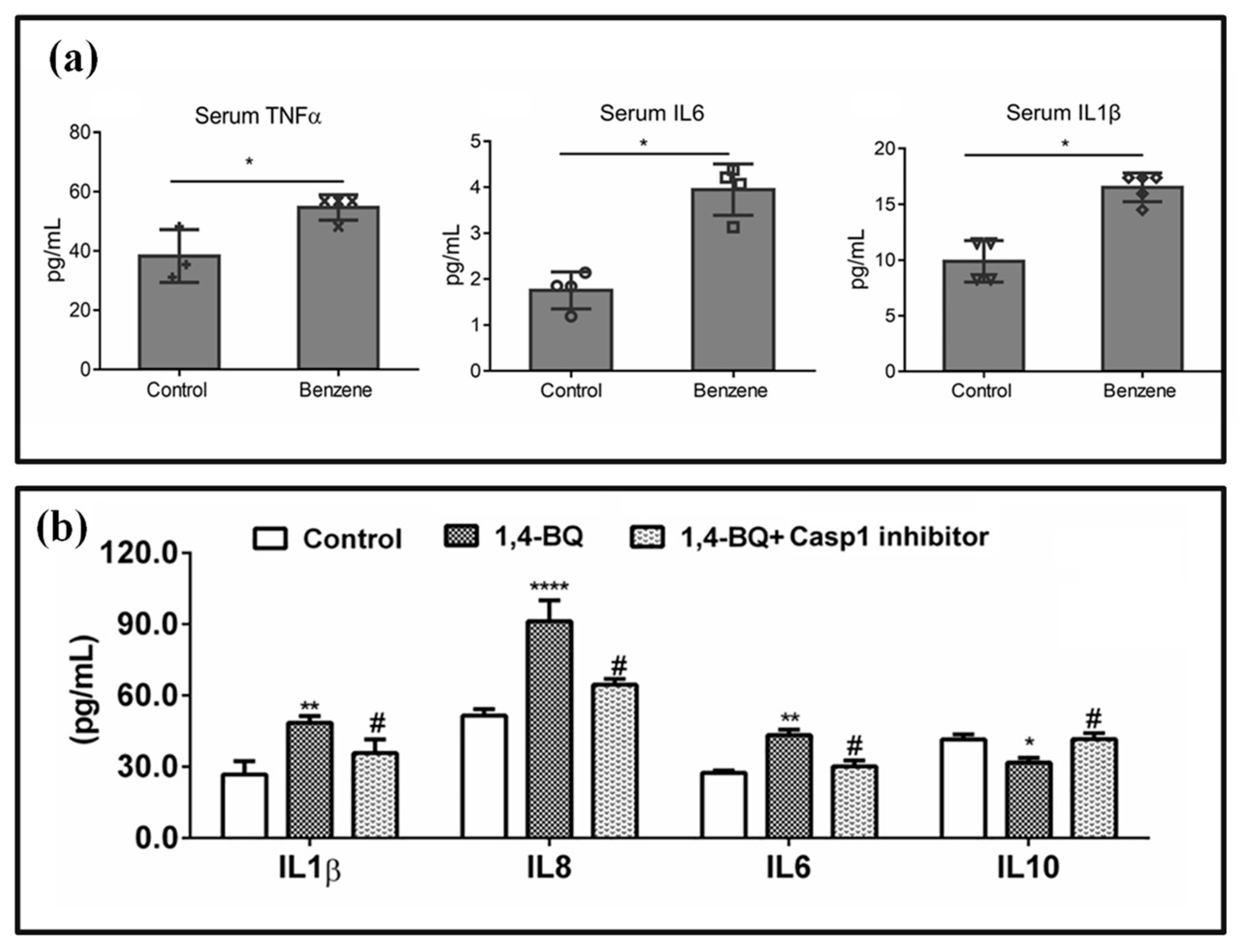
Ferroptosis is a novel form of iron-dependent cell death. Given that iron homeostasis is a key factor in blood development, studying the impact of ferroptosis on benzene-induced hematotoxicity is of great significance. Ren et al. [96] reported for the first time that the competitive endogenous RNA network Lnc-TC/miR-142-5p/CUL4B signaling axis promotes cell ferroptosis and participates in benzene hematotoxicity. Zhang et al. found that the ferroptosis inhibitor Lip-1 significantly inhibited oxidative stress, ferroptosis, and inflammation in NCM460 cells induced by 1,4-BQ [97]. Zhang et al. [98] reported that mice exposed to 50ppm benzene for 8 weeks showed a decrease in serum Fe2+ levels, inducing ferritin and inflammatory factor TNF-α, IL-1 β, elevated IL-6 levels, and uneven distribution of iron in the spleen and bone marrow, accompanied by inflammatory reactions and ferroptosis. The experimental results obtained from the enzyme-linked immunosorbent assay (ELISA) assay are shown in Figure 3a. Compared to the control group, benzene exposure significantly increased the concentrations of these three cytokines (p < 0.05), indicating that benzene triggers a systemic pro-inflammatory state in vivo. Further in vitro studies found that 1,4-BQ induced a significant increase in cell ROS expression and activated ferroptosis, indicating that the increased expression of regulatory factors IRP1, DHODH, and fatty acid metabolism enzyme ALOX12 is a key participant in regulating benzene mediated iron metabolism imbalance and ferroptosis, and revealing that the IRP1-DHODH-ALOX12 axis is involved in benzene-induced ferroptosis by sensing iron homeostasis disorder and inflammation. Sun et al. also found that benzene poisoning induced ferroptosis in mice, and intervention studies using ferroptosis inhibitors found that the Xc-/GPX4 axis and iron metabolism disorders, as well as activation of the NRF2 pathway, play an important role in benzene-induced ferroptosis [99]. It is anticipated that more biomarkers influencing the ferroptosis pathway will be discovered in the future, offering new foundations for treatment strategies for benzene poisoning.
Pyroptosis is a newly discovered form of inflammatory necrosis of cells, accompanied by the formation of cell membrane pores mediated by GSDM family proteins, the release of pro-inflammatory factors, and cellular morphology characterized by cell swelling, plasma membrane dissolution, chromatin fragmentation, and cell rupture [62,100]. Chronic inflammation can accelerate the progression of normal cells to malignant cells. Studies have suggested that chronic inflammation may be one of the important toxic mechanisms behind benzene-induced peripheral blood leukocyte reduction [101]. This provides a new approach to explore the toxic mechanism of benzene from the perspective of pyroptosis inflammation. Guo et al. found through population and in vitro studies that benzene metabolites can directly regulate the Aim2/Casp1 signaling pathway through TET2. As shown in Figure 3b, in cells exposed to 1,4-Benzoquinone, pro-inflammatory cytokines IL-1β (p < 0.01), IL-8 (**** p < 0.0001), IL-6 (p < 0.01), and the anti-inflammatory cytokine IL-10 (p < 0.05) were significantly elevated. Compared to the experimental group, the levels of cytokines in the control group treated with the Casp1 inhibitor were significantly reduced. This indicates that the cytokine production driven by 1,4-BQ is dependent on Casp1-mediated pyroptosis. These findings highlight the role of pyroptotic signaling in benzene-induced inflammatory dysregulation, showing that the Casp1 inhibitor effectively suppressed the expression of IL-1β, IL-8, IL-6, and IL-10 induced by 1,4-BQ [102]. Yang et al. have shown that HQ can activate aromatic hydrocarbon receptors (AhR) and mediate the toxic effects of HQ. By using HQ to infect JHP cells, in vitro studies have found that HQ can induce cell pyroptosis and cause inflammatory reactions. HQ also induces endoplasmic reticulum stress (ERS) by releasing ROS. AhR inhibitors intervene in advance to reverse HQ induced oxidative stress, ERS, and cell pyroptosis, indicating that AhR-mediated, HQ-induced ROS, ERS, and inflammatory reactions may play an important role in the toxic effects of benzene [62]. It is important to note that research on the mechanism by which benzene regulates cell pyroptosis toxicity is still in its early stages, and the complexity of pyroptosis-related molecular functions still poses great challenges in its application, requiring further exploration.
Figure 3. Benzene-induced PCD. (a) ELISA detected the levels of TNF-α, IL6, and IL1β in the plasma of mice. * p < 0.05 compared to the control group [98]. (b) Casp1 inhibitor inhibits 1,4-BQ-induced IL1β, IL8, IL6, and IL10 expression. * p < 0.05, ** p < 0.01, **** p < 0.0001, and # p < 0.05 compared to the 1,4-BQ group [102].
Chemosensors 13 00312 g003

4. Detection Methods for Benzene Poisoning

4.1. Biomarker Selection

Based on the above findings, we conclude that benzene is a significant environmental and occupational pollutant. Prolonged exposure to benzene can induce various cellular processes, including apoptosis, autophagy, and ferroptosis, thereby markedly increasing the risk of cancer. The International Agency for Research on Cancer (IARC) has classified benzene as a human carcinogen [103,104]. Benzene is highly volatile and is commonly found in environments such as semiconductor processing plants, steel processing facilities, and gas stations, where fossil fuels containing benzene are used extensively. Workers in these settings are often subjected to prolonged exposure to volatile benzene, which can lead to a myriad of health complications, including reproductive system disorders, various forms of cancer, and lymphoma [105,106]. In addition to inhalation, benzene can also penetrate the human body through the gastrointestinal tract and dermal exposure. As previously highlighted, benzene lacks a threshold effect; this indicates that even concentrations below the established minimum limit can lead to detrimental effects on the immune and reproductive systems, frequently accompanied by hematotoxicity and genotoxicity [107]. Therefore, continuous monitoring of individuals who are exposed to benzene over prolonged periods is of paramount importance to safeguard their health and mitigate potential risks [63,108]. Numerous studies have investigated and confirmed the mechanisms underlying benzene’s hematotoxicity; however, there remains a notable deficiency in the identification of biomarkers for early monitoring and warning. The OEL represents the maximum level of exposure that workers can safely experience over extended periods without incurring harmful health effects for the majority of individuals. This limit is commonly employed to assess and manage overall human exposure to benzene, serving as a critical benchmark for occupational health and safety practices. In 1946, to prevent acute benzene poisoning, ACGIH recommended setting the OEL for benzene at 100 ppm. This limit was subsequently revised in 1947 to 50 ppm, based on an eight-hour time-weighted average. As research into chronic benzene poisoning advanced, the recommended OEL continued to be progressively lowered. By 2014, the Health Council proposed an OEL of 0.2 ppm, which corresponds to a health-based recommended OEL (HBROEL) of 0.064 mg/m3 for an eight-hour time-weighted average. Environments with benzene concentrations below this threshold can be considered safe for human health [109]. Based on the HBROEL, environments can be categorized into several concentration levels: very low concentration (less than 0.032 mg/m3), low concentration (0.032–0.32 mg/m3), medium concentration (0.32–1.6 mg/m3), high concentration (1.6–3.2 mg/m3), and very high concentration (greater than 3.2 mg/m3) [39]. These classifications are essential for risk assessment and management in occupational settings where benzene exposure may occur.
Benzene that enters the human body undergoes metabolism primarily through Phase I and Phase II biochemical reactions. Phase I reactions entail the oxidation of benzene, which has been absorbed into the bloodstream, by cytochrome P450 enzymes (CYP450) in the liver. This metabolic process converts benzene into toxic intermediates, including phenol, hydroquinone, and benzoquinone. Compared with benzene, these metabolites exhibit increased polarity and enhanced water solubility, enabling more efficient excretion from the body [110]. Phase II reactions primarily involve the conjugation of intermediates generated in Phase I with endogenous substances, such as glucuronic acid and GSH. This process results in the formation of more polar, non-toxic, water-soluble metabolites. Notable examples of urinary benzene metabolites include t,t-MA and S-PMA [111]. These metabolites are predominantly filtered by the kidneys and excreted from the body via urine. However, it is important to note that a small quantity of these metabolites may still accumulate in the human body over time. Therefore, the selection of specific biomarkers is essential for accurately assessing the presence of benzene and determining exposure levels. Optimal biomarkers should exhibit high sensitivity and specificity, combined with appropriate detection methods, to ensure precise and reliable monitoring of benzene exposure. Effective biomarker selection is crucial not only for evaluating individual exposure but also for informing public health strategies aimed at reducing the risks associated with benzene toxicity.
A variety of biomarkers have been employed to assess benzene exposure, including benzene levels in blood, exhaled air, and urine [112,113,114], urea phenol [115], urinary benzene metabolites such as t,t-M, SPMA, 8-OHdG [116], HA, and MHA [117]. In addition, excessive benzene levels in the human body can disrupt normal immune and inflammatory processes, leading to elevated inflammatory markers such as C-reactive protein (CRP), interleukin-6 (IL-6), interleukin-1β (IL-1β), and oxidative stress indicators like abnormal SOD [118]. The definition of an optimal biomarker is one that demonstrates the highest sensitivity and specificity concerning exposure, health outcomes, or individual susceptibility. It is important to recognize that the primary metabolites resulting from benzene metabolism can vary depending on the concentration of exposure, which, in turn, influences the identification of optimal biomarkers. The subsequent section will provide a comprehensive overview of the selection of biomarkers and the measurement methodologies employed at various benzene exposure levels, highlighting the nuances that arise in different exposure contexts.
An optimal biomarker is defined as one that exhibits the highest sensitivity and specificity in relation to exposure, health outcomes, or individual susceptibility. It is crucial to understand that the primary metabolites produced during benzene metabolism can vary significantly based on the concentration of benzene exposure. Consequently, this variability leads to the identification of different optimal biomarkers for diverse exposure scenarios. In the following section, we will present a detailed examination of the selection of biomarkers and the measurement methodologies utilized across varying levels of benzene exposure. This discussion will encompass the rationale behind biomarker choice, the analytical techniques employed, and how these factors contribute to the accurate assessment of benzene exposure and its associated health risks. By exploring these aspects, we aim to enhance the understanding of biomarker utility in monitoring and managing benzene-related health effects.

4.2. Detection Methods for Benzene in Blood, Exhaled Air, and Urine

For patients with benzene poisoning, the most direct detection method involves measuring the concentration of benzene in blood, urine, or exhaled air. This approach is particularly effective at low benzene exposure levels (ranging from 0.032 to 0.32 mg/m3), as the detection results tend to be more accurate when compared to other biomarkers. However, the concentration of benzene in bodily fluids is generally found at the microgram or nanogram level, requiring the use of highly sensitive detection methods for measurement. Moreover, benzene’s volatile nature and short half-life present additional challenges. This volatility requires careful consideration during sample collection and handling to ensure the integrity of benzene in the samples is maintained [119].
Research indicates that measuring benzene levels in blood, exhaled air, and urine is an effective method for biological monitoring, particularly at concentrations below 0.032 mg/m3. It is crucial to monitor low-level benzene exposure, including short-term occupational exposure lasting only a few minutes, as this can pose significant health risks [120,121]. Due to benzene’s volatile and easily oxidized nature, directly detecting its concentration in bodily fluids requires the establishment of a dynamic headspace environment and rapid sample processing to ensure the accuracy of detection results. Angerer et al. proposed the use of dynamic headspace chromatography (DHGC) combined with a flame ionization detector (FID) for the detection of benzene content in blood [122]. In this method, a blood or urine sample is placed in a sealed container and heated, allowing for the extraction of gas samples through a gas sampler. These gas samples are then introduced into a gas chromatography (GC) column for separation. The benzene content within the separated compounds is subsequently detected using an FID, which boasts a detection limit of 80 ng/L. While this detection method demonstrates commendable sensitivity and accuracy, it is not without limitations. The procedure is complex, requires considerable time for detection, and necessitates specific instruments and controlled reaction conditions. These factors can pose challenges in clinical and research settings, where rapid and straightforward detection methods are often preferred.
Therefore, while DHGC-FID is a valuable tool for benzene detection, there is a need for continued exploration of methods that balance sensitivity, accuracy, and practicality in real-world applications.
Mirzaei et al. proposed the use of headspace solid-phase microextraction-gas chromatography-mass spectrometry (HS-SPME-GC-MS) to analyze the content of benzene in urine [123]. In this methodology, after the sample undergoes headspace treatment and transitions to the gas phase, solid-phase microextraction (SPME) is utilized to adsorb benzene molecules. These adsorbed molecules are then introduced into the gas chromatography-mass spectrometry (GC-MS) system for detection. This technique effectively enriches the benzene concentration from the sample while minimizing interference from the sample matrix, which can otherwise compromise test results. Despite its high accuracy, the HS-SPME-GC-MS method presents several challenges, including the complexity of operations and the time-consuming nature of the detection process. Furthermore, traditional methods often lack the capability for on-site sampling and rapid result delivery, which can be critical in clinical and environmental settings. To overcome these limitations, Geng et al. developed a portable headspace injector (portable HS) and portable gas chromatography-mass spectrometry (portable GC-MS). These innovations facilitate the rapid analysis of BTEX compounds (benzene, toluene, ethylbenzene, and xylene) in blood samples. The introduction of this portable device significantly enhances the efficiency of on-site sample collection and analysis, thereby improving the practicalities of toxicological monitoring and environmental assessments [124]. This advancement not only streamlines the detection process but also enables timely decision-making in response to potential benzene exposure, ultimately contributing to better public health outcomes.
Breath analysis serves as a biomonitoring method that presents numerous advantages for assessing human exposure to various volatile organic compounds, including benzene. Notably, while the presence of benzene in urine can interfere with its detection, benzene concentrations in exhaled breath demonstrate higher specificity, particularly at low exposure levels. ACGIH has established biological exposure indices (BEIs) for benzene in exhaled air, setting a limit of 0.08 ppm for mixed air (total exhaled air) and 0.12 ppm for alveolar air (end-exhaled air) [125]. For the analysis of benzene in exhaled breath, samples can be collected using specialized exhaled air collection tubes and subsequently analyzed using gas chromatography-tandem mass spectrometry (GC-MS/MS). However, the precision and accuracy of early detection methods face limitations [126]. To enhance detection accuracy, Plebani et al. [127] proposed a double-step sample collection method using Tedlar bags during the sampling phase to remove moisture from the gas. Additionally, a cryogenic trap is incorporated in the analysis phase to concentrate the air sample prior to its injection into the GC-MS system. SPME has proven to be an effective technique for sampling volatile compounds in the air. It allows for the integration of sampling and pre-concentration operations, thereby simplifying the experimental process. Menezes et al. [128] proposed employing SPME for the direct sampling of exhaled air. This approach involves exposing the SPME fiber directly to the air matrix for extraction while generating benzene standards through permeation. This method offers several advantages, including straightforward operation, reduced system adsorption of the analyte, and the capability to include multiple concentrations in a single dilution stage. Furthermore, Diba Ayache et al. [129] designed a benzene detection sensor based on quartz-enhanced photoacoustic spectroscopy. By using the infrared emission of a 14.85 μm quantum cascade laser (QCL), the content of benzene and toluene in air samples can be detected for a long time. The sensor is suitable for measuring and monitoring benzene concentrations in environments such as factories and gas stations. In summary, at low benzene exposure levels (0.032–0.32 mg/m3), direct measurement of benzene in blood, exhaled air, and urine remains one of the most effective methods for exposure assessment [37].

4.3. Trans, Trans Muconic Acid (t,t-MA)

After inhalation, benzene is primarily metabolized in the liver, where it is first oxidized to phenolic and quinonoid intermediates. This metabolic pathway involves further oxidation and carboxylation reactions. During this complex metabolic process, benzene is transformed into a variety of open-ring compounds and hydroxylated products, including t,t-MA, phenol, catechol, and S-PMA. Importantly, t,t-MA is recognized as the primary urinary metabolite of benzene. Studies have demonstrated a robust correlation between urinary t,t-MA levels and benzene concentrations in both air and blood, particularly at low exposure levels. This strong relationship renders t,t-MA a commonly utilized biological indicator for assessing benzene exposure. Its reliability in reflecting benzene levels in the environment and the body underscores its significance in biomonitoring efforts aimed at detecting and managing benzene-related health risks [130].
Kim et al. [131] conducted an experiment to investigate the relationship between urinary markers and benzene exposure concentrations. They found that when benzene exposure exceeded 0.65 mg/m3, the concentrations of t,t-MA and S-PMA in urine samples continued to increase. Cui et al. [132] conducted experiments using ultra-high-performance liquid chromatography-tandem mass spectrometry (UPLC-MS/MS) to determine the concentrations of t,t-MA and S-PMA in urine. Figure 4a shows the relationship between the naturally logarithm-transformed benzene exposure levels (mg/m3) and t,t-MA values (μg/g creatinine). The results indicated a strong positive linear correlation between the concentration of benzene exposure in the air and the concentration of t,t-MA in urine. They calculated the environmental benzene intake (DI) and back-calculated airborne benzene levels (BCABL) using a pharmacokinetic model. The results indicated that higher logarithmic values of t,t-MA in urine were generally associated with higher logarithmic values of benzene concentration in the air within the range of 1.10–86.91 mg/m3, supporting the rationale for t,t-MA as a biomarker of benzene exposure. t,t-MA is a specific metabolite of benzene in the human body, characterized by a longer half-life, higher sensitivity, and greater specificity. It provides a more comprehensive evaluation of benzene metabolism and the risks associated with long-term exposure.
Dispersive liquid–liquid microextraction (DLLME) and SPME are currently the two main methods used for detecting t,t-MA in urine. These methods offer the advantages of reduced organic solvent usage and a high pre-enrichment coefficient. Dehghani et al. [133] proposed the use of hydrophobic ionic liquids (ILs) as a substitute for traditional hazardous organic solvents. They developed an ionic DLLME method for the quantification of t,t-MA, which was integrated with high-performance liquid chromatography (HPLC) to enable the rapid detection of t,t-MA concentrations in urine samples. Figure 4b is a schematic diagram of the dispersive liquid–liquid microextraction–solidification of floating organic droplet (DLLME-SFOD) procedure. In this method, hydrochloric acid and sodium hydroxide solutions are used to adjust the pH value. 1-Undecanol is employed as the extraction solvent, which is mixed with methanol and then added to the sample solution to form a cloudy suspension. After centrifugation, the mixture is soaked in cold water until the organic solvent solidifies, followed by analysis using high-performance liquid chromatography with ultraviolet detector (HPLC-UV). This method enables the efficient enrichment of trace amounts of t,t-MA and simplifies phase separation using ice bath solidification, offering advantages of rapid operation and reduced solvent usage. It provides efficient and green technical support for the occupational assessment of benzene exposure. Pacenti et al. proposed measuring the t,t-MA concentration in urine using SPME coupled with gas chromatography-ion trap tandem mass spectrometry (MS) [134]. However, both methods have certain limitations. SPME requires solid adsorbents to adsorb the target substances, resulting in poor repeatability. On the other hand, DLLME is straightforward to operate and does not require additional characterization, but the solvents used as extractants and dispersants, such as chloroform and carbon tetrachloride, are toxic [135].
To enhance detection capabilities, further optimization technologies have been proposed. HPLC with a reverse-phase column, along with ultraviolet-visible spectrophotometry (UV-VIS) or MS detection, has become widely adopted. Gomes et al. developed a method for determining the concentration of t,t-MA in urine using HPLC-UV with an ion exclusion column. This method effectively reduces the interference of urine matrix components on t,t-MA detection [130]. Mansour proposed combining solidification of floating organic droplet (SFOD) with DLLME; this approach simplifies the operation of separating the target analytes and reduces the toxicity of organic solvents during the detection process by using an ice bath [136]. André et al. [137] synthesized a novel molecular imprinting polymer (MIP) for the selective extraction of t,t-MA from urine samples, employing molecular imprinting solid-phase extraction (MISPE) instead of conventional anion exchange extraction. They subsequently utilized HPLC with UV detection to separate and quantitatively analyze t,t-MA, achieving improved selectivity and stability. Due to the toxicity of the solvent used in DLLME and its density being greater than that of water—both of which can affect test results—Dehghani et al. proposed the use of DLLME-SFOD to extract t,t-MA from urine samples during the solidification process. They proposed a method based on the SFOD in conjunction with HPLC-UV for quantitative analysis. This method demonstrates excellent consistency and high recovery rates, and it allows for the simultaneous optimization of multiple variables [138]. Furthermore, Dominguez et al. designed a nanosensor based on gated mesoporous silica nanoparticles. Figure 4c illustrates the fluorescence “OFF-ON” sensing mechanism of nanoprobes for detecting t,t-MA. In the OFF state, the nanoprobe shows quenched fluorescence due to intermolecular interactions (such as π-π stacking) among the fluorophores. When t,t-MA is added, its dicarboxyl groups and conjugated double bonds bind to the nanoprobe via hydrogen bonding and π-π stacking, respectively, triggering the conformational rearrangement of the nanoprobe. This disrupts the quenching effect and restores fluorescent emission (ON state). By detecting the changes in fluorescence intensity, quantitative analysis of trace t,t-MA can be achieved, resulting in high sensitivity and rapid response characteristics [139].
Figure 4. The relationship between benzene exposure levels and the values of t,t-MA and detection methods for t,t-MA. (a) The relationship between the natural log-transformed benzene exposure levels (mg/m3) and t,t-MA values (μg/g creatinine). The results indicate that the levels of t,t-MA in urine increase with higher levels of benzene exposure [132]. (b) Extraction procedures of DLLME-SFOD and its application in the detection of t,t-MA, [133]. (c) The fluorescence “OFF-ON” sensing mechanism of nanoprobes for detecting t,t-MA [139].
Chemosensors 13 00312 g004

4.4. S-Phenylmercapturic Acid (S-PMA)
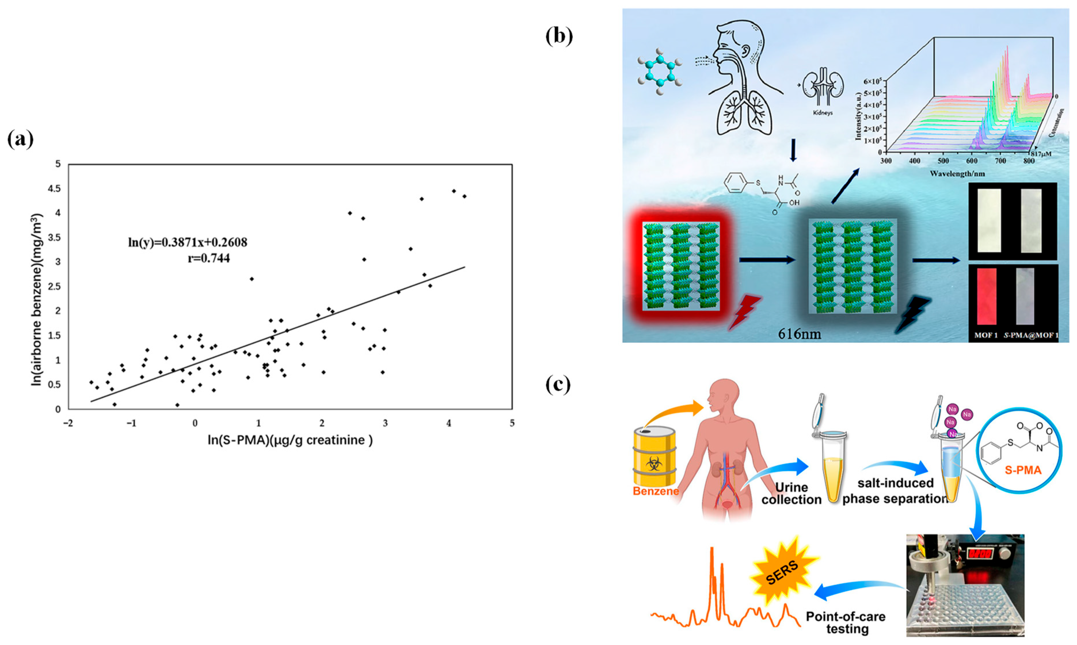
Related studies have shown that when benzene exposure exceeds 0.65 mg/m3, the concentrations of S-PMA and t,t-MA in urine continue to rise [140]. Compared to t,t-MA, S-PAM has a longer half-life (approximately 10 h) and better selectivity, and its concentration in urine is not affected by food additives such as potassium sorbate [141]. Some related studies have indicated that t,t-MA may not be a reliable biomarker for detecting low levels of benzene (<0.5 ppm) [47]. S-PMA is a mercapturic acid precursor formed from the intermediate metabolite of benzene, specifically epoxide benzene, in conjunction with GSH under the catalysis of GST. It is generated through a dehydration condensation reaction under acidic conditions and is excreted in urine. S-PMA is a metabolite of benzene in the human body characterized by strong specificity and high sensitivity. When benzene concentration in the air ranges from 0.035 to 73.85 mg·m−3, S-PMA demonstrates a good linear relationship with it, making it an ideal biomarker for occupational exposure to low concentrations of benzene [142]. Figure 5a shows the relationship between the naturally logarithm-transformed benzene exposure levels (mg/m3) and S-PMA values (μg/g creatinine) [132]. Pre-SPMA is an intermediate product in the metabolism of benzene to S-PMA in the human body. Katharina found that as the pH of urine decreases, a greater amount of pre-SPMA is converted to S-PMA, with complete conversion achieved when urine is treated with 37% hydrochloric acid, reaching a pH of 1.1. The pretreatment for adjusting the pH of urine significantly influences the test results [143].
S-PMA in urine can be detected using HPLC, liquid chromatography-mass spectrometry (LC/MS), gas chromatography (GC), and ELISA. Due to the presence of various biological matrices in urine and the low concentration of S-PMA, sample preparation is necessary before testing to minimize interference and enhance detection sensitivity. However, these detection methods are costly and involve complex extraction processes, posing challenges for the accurate detection of S-PMA. To enhance the sensitivity and specificity of the test and to eliminate interference from proteins, metabolites, and salts in urine, pre-treatment of the samples is often required. Corresponding new technologies have been proposed to address this issue. GC-MS technology is capable of separating target metabolites from complex matrices, making it suitable for the separation, identification, and quantitative detection of S-PMA. Shan et al. [144] synthesized a magnetic nanocomposite material based on activated carbon/diatomite-based materials (AC/DBMNs) and used it as an adsorbent to magnetically extract S-PMA from urine samples through magnetic solid-phase extraction (MSPE) technology. The extracted samples were then processed using HPLC, which eliminated the need for additional centrifugation or filtration steps. UPLC-MS/MS was employed to separate S-PMA in urine, leveraging UPLC’s excellent separation capabilities. The DI of benzene was calculated using a straightforward pharmacokinetic model based on the measured S-PMA levels, while the BCABL was subsequently calculated using the DI [132]. The LC-MS-MS analysis method dilutes and homogenizes the diluted solution and then places it into a chromatographic column for chromatographic separation to quantify S-PMA in urine samples [145]. To achieve rapid detection of S-PMA, Wei et al. proposed combining salt-induced phase separation (SIPS) with surface-enhanced Raman spectroscopy (SERS).
Existing detection methods have limitations such as complex operations, long processing times, and high detection costs. In recent years, fluorescent sensors for the rapid and convenient detection of S-PMA have been developed. Li et al. designed and synthesized a three-dimensional luminescent heterometal-organic framework (HMOF) with two types of pores by selecting d/f heterometal salts and two ligands with abundant coordination sites [146]. Figure 5b illustrates the fluorescence sensing mechanism of the heterometal-organic framework Zn-Eu-MOF for S-PMA. After benzene enters the human body through the respiratory tract and is metabolized into S-PMA, it is excreted through the kidneys. The porous structure of Zn-Eu-MOF can capture S-PMA, leading to concentration-dependent changes in the characteristic fluorescence at 616 nm by altering the luminescent microenvironment of Eu3+. Moreover, the higher the concentration of S-PMA, the more significant the signal change. Under ultraviolet light, the inherent red fluorescence of the MOF is significantly diminished upon binding with S-PMA, allowing for the identification of S-PMA binding through visual changes in luminescent color. Therefore, through spectral quantitative analysis and visual qualitative detection, highly selective and sensitive detection of S-PMA can be achieved. As illustrated in Figure 5c, urine samples undergo SIPS to enrich S-PMA and eliminate matrix interferences. Subsequently, SERS is employed to detect the concentrated S-PMA. The nanoparticle-mediated surface enhancement effect significantly amplifies the Raman characteristic signals of S-PMA. When integrated with miniaturized detection devices, this method facilitates point-of-care testing (POCT), enabling rapid and on-site diagnostic assessments for occupational health screening related to benzene exposure, as well as for environmental monitoring applications [147]. Additionally, the incorporation of chromatographic technologies such as high-performance liquid chromatography with HPLC-UV, high-performance liquid chromatography-tandem mass spectrometry (HPLC-MS/MS), and GC-MS has further enhanced the detection accuracy of S-PMA in urine. Table 1 lists the linear ranges, LoD, and sample types for t,t-MA and S-PMA under different detection methods; these data provide a reference for related analyses.
Figure 5. The relationship between benzene exposure levels and the values of S-PMA and detection methods for S-PMA. (a) The relationship between the natural log-transformed benzene exposure levels (mg/m3) and S-PMA values (μg/g creatinine) [132]. (b) A porous luminescent heterometal-organic framework, Zn-Eu-MOF, can achieve highly selective and sensitive detection of S-PMA through spectral quantitative analysis and visual qualitative detection [146]. (c) The combination of SIPS for enrichment and SERS enables highly sensitive detection of trace S-PMA. Together with the point-of-care testing (POCT) platform, this approach facilitates rapid on-site analysis, providing a technical solution for real-time monitoring of benzene exposure [147].
Chemosensors 13 00312 g005

4.5. Hippuric Acid and Methylhippuric Acid (HA and MHA)

In recent years, benzene homologues such as toluene, xylene, and styrene have been commonly used in building materials. The toxicity of these substances is comparable to that of benzene, and long-term exposure can lead to damage to the nervous and respiratory systems. For instance, toluene can directly inhibit nervous system function and is metabolized into benzoic acid in the human body, which may harm the liver and kidneys. Xylene can bind to hemoglobin, reducing oxygen transport capacity. Additionally, benzene homologues can generate corresponding biomarkers in the human body. For example, toluene is oxidized and conjugated with glycine to form hippuric acid (HA), which is excreted in urine, while xylene is oxidized and conjugated with glycine to form methylhippuric acid (MHA), also excreted in urine [148]. MHA has three isomers: 2-methylhippuric acid (2-MHA), 3-methylhippuric acid (3-MHA), and 4-methylhippuric acid (4-MHA), which differ based on the position of the methyl group. MHA and HA are metabolites of benzene in the human body. Consequently, the levels of benzene homologues in the human body can be assessed by measuring the concentrations of HA and MHA in urine. For individuals exposed to high concentrations of benzene over extended periods, the urinary levels of HA and MHA are significantly elevated compared to normal, making them reliable exposure biomarkers [117]. HPLC is typically utilized to analyze HA and MHA in urine samples. Due to the low concentrations of HA and MHA in urine, Tzanetou et al. developed two new methods combining GC-MS/MS and liquid chromatography-tandem mass spectrometry (LC-MS/MS) with headspace solid-phase microextraction (HS-SPME) for non-targeted detection of chemicals [149]. Two new methods combining GC-MS/MS and LC-MS/MS with HS-SPME were developed for the non-targeted detection of chemicals. Kurd et al. [150] designed a method based on a covalent organic framework (COF) for microextracting urine using the microextraction by packed sorbent (MEPS) technique, followed by sample preparation using an HPLC-UV detector system. Additionally, a novel magnetized imine-linked covalent organic framework (Fe3O4@TFPA-Bd) was synthesized, featuring Fe3O4 nanoparticles as the magnetic core, with benzidine (Bd) and Tris (4-formyl phenyl) amine (TFPA) serving as structural building blocks. Fe3O4@TFPA-Bd was developed as a microextraction adsorbent to isolate HA, MHA, tt-MA, and other target substances from urine samples [151]. Covalent organic framework-microextraction by packed sorbent (COF-MEPS) technology can efficiently separate multiple biomarkers of benzene and its compounds from urine samples using a single adsorbent, offering a rapid, straightforward, and environmentally friendly solution. Similarly, Han et al. developed a chitosan-modified magnetic Schiff base nanocomposite sphere (Fe3O4@SNW@Chitosan) that exhibits excellent hydrophilicity and superparamagnetic properties [152]. This material demonstrates favorable morphology for sample pre-concentration and effectively enriches and detects HA and 4-MHA in urine samples through MSPE combined with HPLC.
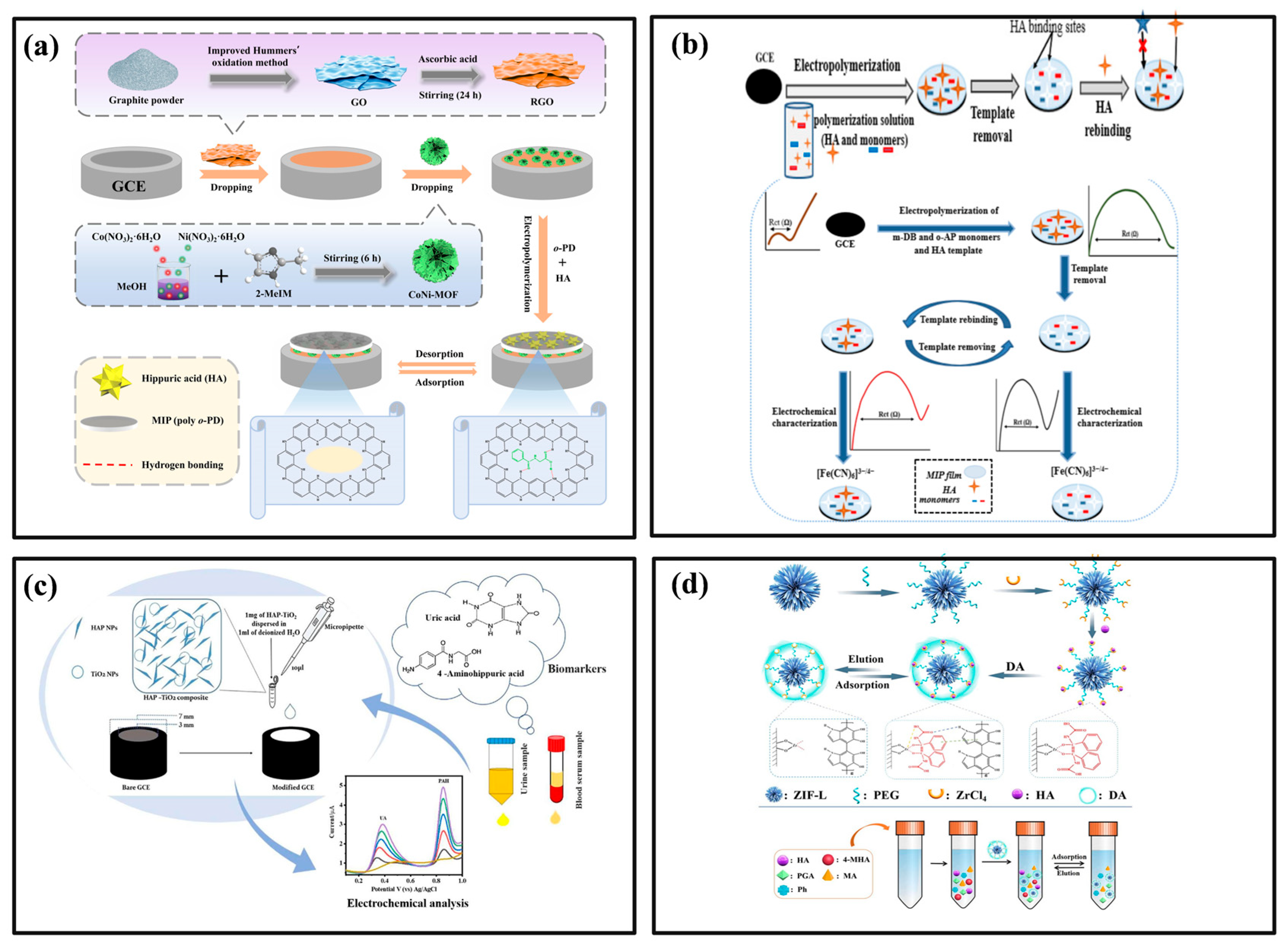
HA and MHA exhibit certain electrochemical activity, and electrochemical methods offer advantages such as low cost, high sensitivity, and rapid response times. In recent years, novel electrochemical methods for detecting HA and MHA have been proposed. As illustrated in Figure 6a, Gao et al. developed a molecularly imprinted electrochemical sensor comprising a molecularly imprinted polymer, reduced graphene oxide, and a cobalt–nickel metal–organic framework modified glassy carbon electrode (MIP/RGO/CoNi-MOF/GCE) [153]. This sensor enhances the electron transfer rate and conductivity of the electrode material through the interaction between RGO and CoNi-MOF, enabling sensitive detection of HA in urine samples. Karazan et al. proposed the use of electrochemical polymerization technology to detect HA. They constructed a molecularly imprinted polymer (MIP) film on the surface of a glass carbon electrode (GCE) using m-dihydroxybenzene (m-DB) and o-aminophenol (o-AP) as monomers for electropolymerization. The principle of the reaction and the preparation process are shown in Figure 6b [154]. This approach effectively enhances the selectivity and linear range of the sensor. The developed sensor demonstrates high selectivity, low limit of detection (LOD), and a wide linear range for detecting HA in samples, while also exhibiting good recovery rates. Anitta et al. developed a novel electrochemical sensor based on a hydroxyapatite-titanium dioxide (HAP-TiO2) nanocomposite [155]. As shown in Figure 6c, the GCE modified with this nanocomposite exhibits excellent electrocatalytic activity toward the oxidation of uric acid (UA) and para-aminobenzoic acid (PAH). This sensor enables rapid and accurate quantification of both analytes in biological samples, providing a promising tool for non-invasive assessment of liver and kidney functions. Zheng et al. developed an MIP based on a zirconium ion-modified zeolitic imidazolate framework (ZIF) immobilized with polyethylene glycol (PEG), referred to as ZIF@PEG@Zr@MIPs (the preparation process is shown in Figure 6d) [156]. This polymer serves as an adsorbent for SPE. By anchoring Zr4+ ions on the surface of ZIF, they created specific recognition sites for dopamine (DA) imprinting, which enhances the specificity of the sensor. The enhanced specific adsorption of HA and 4-MHA is achieved through π-π stacking interactions and hydrogen bonding between the target molecules and the imprinted sites, resulting in improved detection performance. Table 2 summarizes the detection methods for HA, MHA, and PAH mentioned above, along with their corresponding linear ranges and LoD, providing useful reference information for related analyses.

4.6. Immunological Markers

Excessive benzene levels in the human body can harm the blood and immune systems, leading to associated immune responses and oxidative stress. Early detection of benzene poisoning can be accomplished by identifying related biomarkers [55]. Immunoglobulin (Ig) refers to a globulin with similar activity and chemical structure to antibodies. It is a type of immune-active molecule that can specifically bind to antigens in the human body [158]. Benzene toxicity can trigger an immune response in the human body, leading to changes in the concentration of related immunoglobulins. Statistical data indicate that workers exposed to benzene for prolonged periods have lower levels of IgA and IgG in their serum compared to individuals without such exposure, while their IgE levels increase [157,159]. The relevant immunoglobulin content in human serum can be detected using ELISA or electrophoresis. Endothelial cells, which line blood vessels, play a crucial role in maintaining vascular homeostasis, regulating blood flow, and participating in immune responses. Upon cell activation and apoptosis, endothelial cells secrete markers such as CRP [160], IL-6 [161], IL-1β [162], tumor necrosis factor-α [163], and so on.
Research has established an association between outdoor benzene pollution and CRP levels in human serum. CRP levels rise with increasing environmental benzene concentrations, and in vitro studies indicate that benzene metabolites promote endothelial cell apoptosis and the formation of endothelial microparticles [164,165]. ELISA is considered the gold standard for detecting CRP. This method converts the analyte into a colored product through a color reaction, allowing for quantitative measurement. However, it has several disadvantages, including a long processing time, high equipment costs, and complex procedures [166]. New biosensors, including electrochemical, fluorescent, and optical biosensors, can significantly shorten detection time and reduce costs. For instance, Sheen et al. [167] designed an electrochemical impedance spectroscopy (EIS) biosensor with electrodes arranged in a circular array. This design simultaneously induces electrothermal convection and negative dielectrophoresis (n-DEP), facilitating the uniform dispersion of proteins and enhancing sensitivity and detection limits. Gao et al. [168] developed a label-free electrochemical sensor by immobilizing an electrochemical aptamer on a composite material made of gold nanoparticles (GNPs) and carboxylated graphene oxide (AuNPs/GO-COOH). The sensor utilizes carboxylated graphene oxide to facilitate the anchoring of inducer molecules, enhancing detection accuracy. By detecting shifts in the electrochemical response caused by changes in aptamer conformation, it enables rapid and accurate detection of CRP levels. Lee et al. [169] designed a photothermal biosensor (PTB) for detecting CRP concentrations in human saliva; the schematic diagram of the manufacturing and surface modification process of the sensor is shown in Figure 7a. The sensor consists of three parts: a platinum resistance temperature detector (Pt-RTD), a silicon dioxide layer, and a gold layer. The surface is sequentially modified with Cys3-conjugated protein G, anti-CRP antibodies, and bovine serum albumin (BSA) to construct a specific recognition interface. The sample containing CRP is then placed on the surface and incubated for 30 min. Finally, GNP conjugated with anti-CRP antibodies is added, and the sensing area is irradiated with a laser at a wavelength of 532 nm. The temperature change induced by the photothermal effect from the laser irradiation enables the quantitative detection of CRP.

4.7. Oxidative Stress Markers

Related studies have demonstrated that benzene poisoning in humans can lead to oxidative stress indicators, such as ROS [173], Heme Oxygenase-1 (HO-1) [174], 8-OHdG [175], and SOD [176]. ROS is primarily detected using fluorescent probe methods and chemiluminescence (CL) techniques. Peter et al. developed an improved dichlorodihydrofluorescein diacetate (DCFDA) fluorescent probe, which can be used to detect ROS in cell lines and achieve quantitative analysis of ROS levels [177]. ECL refers to the process in which electrochemical reactions occur at the electrode surface, leading to high-energy electron transfer reactions that generate excited-state luminophores. The emitted light signal can be directly detected using photodetectors such as photodiodes or CCD cameras [178]. In the biological analysis of ROS, a commonly used ECL system involves the luminescent reaction between luminol and hydrogen peroxide (H2O2). In an alkaline solution, luminol loses a proton to become an anion, which is subsequently oxidized to form a diazine quinone, leading to the generation of an excited-state 3-amino-phthalate at the electrode surface. By detecting the light signals emitted during the decay of 3-amino-phthalate to its ground state, ROS detection can be achieved [179]. In recent years, 3D cell culture and organ-on-a-chip models have frequently been used for the detection of ROS. For example, Soragni et al. developed a 3D chip model composed of human umbilical vein endothelial cells (HUVECs), which utilizes three dyes to stain live cells for ROS, dead cells, and DNA [170]. Figure 7b illustrates the chemical reaction principle of the 3D chip model for the quantitative detection of intracellular ROS and cell viability (Part A) and the strategy for confocal image analysis (Part B). Part A reveals the redox balance mechanism in the human body and the correlation between excessive ROS and oxidative stress. Part B describes the experimental workflow, where the analysis of fluorescent signals allows for the resolution of cell viability and ROS levels, enabling a multidimensional quantitative characterization of cell status within the 3D chip model. This approach highlights the potential of advanced cell culture techniques in studying cellular responses to oxidative stress while opening new avenues for drug testing and disease modeling. HO-1 is primarily detected using ELISA. Rücker et al. developed an ELISA-based assay for HO-1 enzyme activity. By analyzing protein blotting, the concentration of HO-1 was converted into optical density for quantitative analysis, enabling the identification and characterization of molecules potentially related to the treatment of inflammatory and autoimmune diseases [180]. HO-1 is primarily detected using ELISA. Rücker et al. developed an ELISA-based assay for HO-1 enzyme activity. By analyzing protein blotting, the concentration of HO-1 was converted into optical density for quantitative analysis, allowing for the identification and characterization of molecules potentially related to the treatment of inflammatory and autoimmune diseases [181]. 8-OHdG is a key biomarker commonly used to assess oxidative stress levels in the body. It is often detected using HPLC or electrochemical biosensors. For example, a carbon-based electrochemical sensor developed by Martins et al. demonstrates excellent electrochemical performance for the oxidation of 8-OHdG. In this sensor, the 8-OHdG molecules are oxidized on the conductive carbon paper substrate, and the content of 8-OHdG can be detected using differential pulse voltammetry (DPV). The sensor demonstrates good stability and sensitivity, making it suitable for POCT applications [182]. Wang et al. synthesized a novel peroxidase mimetic probe and employed a COF along with a multi-walled carbon nanotube carboxylic acid (MWCNT-COOH) to create an electrode sensing platform. They constructed an electrochemical aptamer sensor capable of quantitatively detecting 8-OHdG with high specificity and sensitivity [183]. To meet the detection needs of POCT and reduce detection costs, Ammanath et al. designed a disposable paper-based analytical device for the visual detection of 8-OHdG, as shown in Figure 7c. This device consists of a polyvinylidene fluoride (PVDF) membrane, filter paper, and a laminated plate. Cationic polythiophene (CPT) is immobilized on the PVDF membrane and interacts with the 8-OHdG aptamer molecules. In the absence of 8-OHdG, the aptamer binds to polythiophene, causing fluorescence quenching and resulting in a color change of the solution from orange to purple (the “OFF” state). When 8-OHdG is present in the urine, the aptamer binds to the target and folds into a G-quadruplex structure, thereby releasing its effect on polythiophene, restoring fluorescence and color, and causing the solution to gradually change from purple back to orange (the “ON” state). This color change allows for the rapid visual detection of 8-OHdG, providing a simple technical solution for the immediate diagnosis of oxidative stress [171]. Common SOD detection methods include immunoassay, electrochemiluminescence, fluorescence spectroscopy, and solid NMR spectroscopy. Abdussalam et al. [184] developed a CL sensor that operates based on the reaction between 1,4-Dithiothreitol (DTT) and lucigenin. In this system, the presence of SOD leads to the quenching of the CL signal. Notably, this sensor does not require the synthesis of a luminescent probe and exhibits a detection limit as low as 2.2 ng/mL. In recent years, detection methods for directly observing SOD levels with the naked eye have received significant attention for their potential applications in POCT. Yuan et al. [172] proposed a straightforward platinum-doped g-C3N4 photocatalytic biosensor that utilizes amaranth (AM) as an indicator. As shown in Figure 7d, when illuminated at specific wavelengths, AM can be oxidized by the superoxide radicals (·O2−) generated from g-C3N4. SOD specifically catalyzes the conversion of ·O2− radicals into oxygen and hydrogen peroxide, thereby reducing the degradation of AM and resulting in a red-colored solution. The accompanying figure illustrates the effectiveness of different wavelengths of light in driving the photo-oxidation process. It can be observed that the violet light with a wavelength range of 380 to 390 nm exhibits good photo-oxidation efficiency; thus, a violet LED lamp is chosen as the light source for illumination.

5. Conclusions and Recommendations

In summary, benzene remains a key hazard factor in the current occupational disease field, and benzene poisoning remains a major challenge in chemical poisoning prevention and control, posing a huge threat to the lives and health of workers as well as socio-economic development. At present, population studies and animal experiments have found that exposure to benzene can cause damage to multiple systems and organs in the body. Acute poisoning is mainly characterized by damage to the nervous system, while chronic poisoning is mainly characterized by damage to the hematopoietic system. Long-term low-dose exposure can also have effects on the digestive system, urinary system, and reproductive system. However, current research on the toxicity mechanism of benzene mainly focuses on hematopoietic toxicity, and the toxicity mechanism of benzene on the nervous system, reproductive system, respiratory system, etc., still needs further exploration. The incidence rate of leukemia in people with a long history of benzene exposure is far higher than that in normal people. Given that benzene has no threshold effect, exposure to low concentrations of benzene can still lead to peripheral blood abnormalities, occupational contraindications, and suspected benzene poisoning. Given that benzene has no threshold effect, exposure to low concentrations of benzene can still lead to peripheral blood abnormalities, occupational contraindications, and suspected benzene poisoning. Exploring highly sensitive, specific, simple, and feasible biomarkers for exposure assessment, biological monitoring, and early prevention and treatment of benzene-exposed populations has important guiding significance and application value.
The mechanism of benzene toxicity involves multiple pathways such as oxidative stress, immunotoxicity, cell death, genetic damage, and epigenetics. Related biomarkers include blood cell count, nucleic acids, proteins, etc. The main effect biomarkers detected after exposure to benzene are blood routine counts. Urine trans muconic acid and urine S-phenylmercaptouric acid are relatively reliable biological exposure markers after benzene metabolism, and urine samples are convenient, and detection methods are mature. In addition, the effect markers after benzene exposure include inflammatory markers IgA, IgG, IgM, IL-2, IL-4, IL-6, IL-17a, oxidative stress markers MDA, 8-OHdG, GST, SOD, genetic damage markers chromosome aberration, micronucleus rate, p14ARF, p15INK4b, LINE-1, ACSL1, PCD related markers miR-133a, lncRNA-VNN3, LncRNA-OBFC2A, Caspase-9, Caspase-3, let-7e-5p, TET2, etc. However, existing literature reports have not fully analyzed the sensitivity, specificity, and ROC curve of biomarkers in accordance with the requirements of evidence-based medicine, and further verification is needed. In addition, the roles of various signaling pathways in diseases often overlap, and different signaling pathways may be interrelated and interfere with each other. Therefore, exploring the common nodes of different pathways and studying multi-target comprehensive intervention methods are also of great significance for the prevention and treatment of benzene poisoning.

Funding

This research was funded by the National Key Research and Development Program of China (Grant No. 2022YFF1202700).

Conflicts of Interest

The authors declare no conflict of interest.

Abbreviations

·O2−superoxide radicals
1,4-BQ1,4-Benzoquinone
2-MHA2-methylhippuric acid
3-MHA3-methylhippuric acid
4-MHA4-methylhippuric acid
8-OHdG8-hydroxydeoxyguanine nucleoside
AAascorbic acid
AC/DBMNsactivated carbon/diatomite-based materials
ACGIHAmerican Conference of Governmental Industrial Hygienists
ACSL1acyl coA synthetase long-chain family member 1
AHRaryl hydrocarbon receptor
AMamaranth
BCABLback-calculated airborne benzene levels
Bdbenzidine
BEIsbiological exposure indices
BSAbovine serum albumin
CLchemiluminescence
COFcovalent organic framework
COF-MEPScovalent organic framework-microextraction by packed sorbent
CPTcationic polythiophene
CRPC-reactive protein
CYP450cytochrome P450 enzymes
DAdopamine
DHGCdynamic headspace chromatography
DHGC-FIDdirect headspace gas chromatography with flame ionization detection
DIbenzene intake
DLLMEdispersive liquid–liquid microextraction
DLLME-SFODdispersive liquid–liquid microextraction–solidification of floating organic droplet
DLLME-SPEdispersive liquid–liquid microextraction solid-phase extraction
DNMTsDNA methyltransferases
DPVdifferential pulse voltammetry
DTT1,4-Dithiothreitol
EAellagic acid
EISelectrochemical impedance spectroscopy
ELISAenzyme-linked immunosorbent assay
FIDflame ionization detector
GCgas chromatography
GC-MSgas chromatography-mass spectrometry
GC-MS/MSgas chromatography-tandem mass spectrometry
GNPgold nanoparticles
GPxglutathione peroxidase
GSHglutathione
GSTglutathione S-transferase
HAhippuric acid
HAP-TiO2hydroxyapatite-titanium dioxide
HBROELhealth-based recommended OEL
hMeDIPhydroxymethylated DNA immunoprecipitation
HMOFheterometal-organic framework
HPLChigh-performance liquid chromatography
HPLC-MS/MShigh-performance liquid chromatography-tandem mass spectrometry
HPLC-UVhigh-performance liquid chromatography with ultraviolet detector
HQhydroquinone
HS-SPMEheadspace solid-phase microextraction
HS-SPME-GC-MSheadspace solid-phase microextraction-gas chromatography-mass spectrometry
IL-1βinterleukin-1β
IL-6interleukin-6
ILsionic liquids
LC/MSliquid chromatography-mass spectrometry
LC-MS/MSliquid chromatography-tandem mass spectrometry
LODlow limit of detection
MEPSmicroextraction by packed sorbent
MHAmethylhippuric acid
MIPmolecular imprinting polymer
MISPEmolecular imprinting solid-phase extraction
MRImagnetic resonance imaging
MSmass spectrometry
MSPmethylation-specific PCR
MSPEmagnetic solid-phase extraction
MWCNT-COOHmulti-walled carbon nanotubes carboxylic acid
n-DEPnegative dielectrophoresis
Nrf2Nuclear factor erythroid 2-related factor 2
OELoccupational exposure limit
PCDprogrammed cell death
PAHpara-aminobenzoic acid
PEGpolyethylene glycol
POCTpoint-of-care testing
pre-SPMApre-S-phenylmercapturic acid
PTBphotothermal biosensor
Pt-RTDplatinum resistance temperature detector
QCLquantum cascade laser
qPCRreal-time quantitative PCR
RBCsred blood cells
ROSreactive oxygen species
SERSsurface-enhanced Raman spectroscopy
SFODsolidification of floating organic droplet
SIPSsalt-induced phase separation
SODsuperoxide dismutase
S-PMAS-Phenylmercapturic acid
SPMEsolid-phase microextraction
t,t-MAt,t-muconic acid
TFPAtris (4-formyl phenyl) amine
UAuric acid
UPLC-MS/MSultra-high-performance liquid chromatography-tandem mass spectrometry
UV-VISultraviolet-visible spectrophotometry
WBCswhite blood cells
ZIFzeolitic imidazolate framework

References

  1. Cao, Y.; Wang, T.; Xi, J.; Tian, W.; Liu, W.; Sun, Y.; Liu, W.; You, X.; Li, A.; Zhang, G.; et al. Benchmark dose estimation for benzene-exposed workers in China: Based on quantitative and multi-endpoint genotoxicity assessments. Environ. Pollut. 2023, 330, 121765. [Google Scholar] [CrossRef] [PubMed]
  2. Kim, D.-Y.; Kim, H.-S.; Lim, D.-S.; Kim, K.-Y. Benzene exposure assessment of printing workers treating petroleum-based cleaner in South Korea. Ind. Health 2023, 61, 283–290. [Google Scholar] [CrossRef]
  3. Wan, W.; Peters, S.; Portengen, L.; Olsson, A.; Schüz, J.; Ahrens, W.; Schejbalova, M.; Boffetta, P.; Behrens, T.; Brüning, T. Occupational benzene exposure and lung cancer risk: A pooled analysis of 14 case-control studies. Am. J. Respir. Crit. Care Med. 2024, 209, 185–196. [Google Scholar] [CrossRef] [PubMed]
  4. Talibov, M.; Sormunen, J.; Hansen, J.; Kjaerheim, K.; Martinsen, J.-I.; Sparen, P.; Tryggvadottir, L.; Weiderpass, E.; Pukkala, E. Benzene exposure at workplace and risk of colorectal cancer in four Nordic countries. Cancer Epidemiol. 2018, 55, 156–161. [Google Scholar] [CrossRef] [PubMed]
  5. Spycher, B.D.; Lupatsch, J.E.; Huss, A.; Rischewski, J.; Schindera, C.; Spoerri, A.; Vermeulen, R.; Kuehni, C.E.; The Swiss Paediatric Oncology Group; The Swiss National Cohort Study Group. Parental occupational exposure to benzene and the risk of childhood cancer: A census-based cohort study. Environ. Int. 2017, 108, 84–91. [Google Scholar] [CrossRef]
  6. Werder, E.J.; Engel, L.S.; Blair, A.; Kwok, R.K.; McGrath, J.A.; Sandler, D.P. Blood BTEX levels and neurologic symptoms in Gulf states residents. Environ. Res. 2019, 175, 100–107. [Google Scholar] [CrossRef]
  7. Thetkathuek, A.; Polyong, C.P.; Jaidee, W. Benzene health risk assessment for neurological disorders of gas station employees in Rayong Province, Thailand. Rocz. Państwowego Zakładu Hig. 2023, 74, 231–241. [Google Scholar]
  8. Hu, J.; Yu, E.; Liao, Z. Changes in cognitive function and related brain regions in chronic benzene poisoning: A case report. Ann. Transl. Med. 2021, 9, 81. [Google Scholar] [CrossRef]
  9. Wang, D.; Nie, L.; Shao, X.; Yu, H. Exposure profile of volatile organic compounds receptor associated with paints consumption. Sci. Total Environ. 2017, 603, 57–65. [Google Scholar] [CrossRef] [PubMed]
  10. Crossin, R.; Qama, A.; Andrews, Z.B.; Lawrence, A.J.; Duncan, J.R. The effect of adolescent inhalant abuse on energy balance and growth. Pharmacol. Res. Perspect. 2019, 7, e00498. [Google Scholar] [CrossRef]
  11. Wang, D.; Lin, D.; Yang, X.; Wu, D.; Li, P.; Zhang, Z.; Zhang, W.; Guo, Y.; Fu, S.; Zhang, N. Alterations in leukocyte telomere length and mitochondrial DNA copy number in benzene poisoning patients. Mol. Biol. Rep. 2024, 51, 309. [Google Scholar] [CrossRef] [PubMed]
  12. Ren, J.C.; Wang, T.; Wu, H.; Zhang, G.H.; Sun, D.; Guo, K.; Li, H.; Zhang, F.; Wu, W.; Xia, Z.; et al. Promoter hypermethylation in CSF3R induces peripheral neutrophil reduction in benzene-exposure poisoning. Environ. Mol. Mutagen. 2020, 61, 786–796. [Google Scholar] [CrossRef]
  13. Jin, Y.S.; Yi, Z.C.; Zhang, Y.J.; Long, R.; Yu, C.H. Proteomics Study of Benzene Metabolite Hydroquinone Induced Hematotoxicity in K562 Cells. Biomed. Environ. Sci. 2024, 37, 341–353. [Google Scholar] [PubMed]
  14. Janitz, A.E.; Campbell, J.E.; Magzamen, S.; Pate, A.; Stoner, J.A.; Peck, J.D. Benzene and childhood acute leukemia in Oklahoma. Environ. Res. 2017, 158, 167–173. [Google Scholar] [CrossRef]
  15. Liao, Q.; Zhang, Y.; Ma, R.; Zhang, Z.; Ji, P.; Xiao, M.; Du, R.; Liu, X.; Cui, Y.; Xing, X. Risk assessment and dose-effect of co-exposure to benzene, toluene, ethylbenzene, xylene, and styrene (BTEXS) on pulmonary function: A cross-sectional study. Environ. Pollut. 2022, 310, 119894. [Google Scholar] [CrossRef]
  16. Weaver, C.; Liu, S.-P.; Lu, J.-F.; Lin, B.-S. The effects of benzene exposure on apoptosis in epithelial lung cells: Localization by terminal deoxynucleotidyl transferase-mediated dUTP-biotin nick end labeling (TUNEL) and the immunocytochemical localization of apoptosis-related gene products. Cell Biol. Toxicol. 2007, 23, 201–220. [Google Scholar] [CrossRef]
  17. Warden, H.; Richardson, H.; Richardson, L.; Siemiatycki, J.; Ho, V. Associations between occupational exposure to benzene, toluene and xylene and risk of lung cancer in Montréal. Occup. Environ. Med. 2018, 75, 696–702. [Google Scholar] [CrossRef]
  18. Meo, S.A.; Alrashed, A.H.; Almana, A.A.; Altheiban, Y.I.; Aldosari, M.S.; Almudarra, N.F.; Alwabel, S.A. Lung function and fractional exhaled nitric oxide among petroleum refinery workers. J. Occup. Med. Toxicol. 2015, 10, 37. [Google Scholar] [CrossRef]
  19. Wichmann, F.A.; Müller, A.; Busi, L.E.; Cianni, N.; Massolo, L.; Schlink, U.; Porta, A.; Sly, P.D. Increased asthma and respiratory symptoms in children exposed to petrochemical pollution. J. Allergy Clin. Immunol. 2009, 123, 632–638. [Google Scholar] [CrossRef]
  20. Sauer, E.; Gauer, B.; Nascimento, S.; Nardi, J.; Göethel, G.; Costa, B.; Correia, D.; Matte, U.; Charão, M.; Arbo, M. The role of B7 costimulation in benzene immunotoxicity and its potential association with cancer risk. Environ. Res. 2018, 166, 91–99. [Google Scholar] [CrossRef] [PubMed]
  21. Hanke, J.; Dutkiewicz, T.; Piotrowski, J. The absorption of benzene through human skin. Int. J. Occup. Environ. Health 2000, 6, 104–111. [Google Scholar] [CrossRef] [PubMed]
  22. Frasch, H.F.; Barbero, A.M. In vitro human skin permeation of benzene in gasoline: Effects of concentration, multiple dosing and skin preparation. J. Expo. Sci. Environ. Epidemiol. 2018, 28, 193–201. [Google Scholar] [CrossRef] [PubMed]
  23. Han, L.; Wang, J.; Zhang, L.; Jing, J.; Zhang, W.; Liu, Z.; Gao, A. The role of N6-methyladenosine modification in benzene-induced testicular damage and the protective effect of melatonin. Chemosphere 2023, 319, 138035. [Google Scholar] [CrossRef]
  24. Mottola, F.; Iovine, C.; Carannante, M.; Santonastaso, M.; Rocco, L. In vitro combination of ascorbic and ellagic acids in sperm oxidative damage inhibition. Int. J. Mol. Sci. 2022, 23, 14751. [Google Scholar] [CrossRef]
  25. Marchetti, F.; Eskenazi, B.; Weldon, R.H.; Li, G.; Zhang, L.; Rappaport, S.M.; Schmid, T.E.; Xing, C.; Kurtovich, E.; Wyrobek, A.J. Occupational exposure to benzene and chromosomal structural aberrations in the sperm of Chinese men. Environ. Health Perspect. 2012, 120, 229–234. [Google Scholar] [CrossRef]
  26. Anigilaje, E.A.; Nasir, Z.A.; Walton, C. Exposure to benzene, toluene, ethylbenzene, and xylene (BTEX) at Nigeria’s petrol stations: A review of current status, challenges and future directions. Front. Public Health 2024, 12, 1295758. [Google Scholar] [CrossRef]
  27. Reutman, S.R.; LeMasters, G.K.; Knecht, E.A.; Shukla, R.; Lockey, J.E.; Burroughs, G.E.; Kesner, J.S. Evidence of reproductive endocrine effects in women with occupational fuel and solvent exposures. Environ. Health Perspect. 2002, 110, 805–811. [Google Scholar] [CrossRef]
  28. Lupo, P.J.; Symanski, E.; Waller, D.K.; Chan, W.; Langlois, P.H.; Canfield, M.A.; Mitchell, L.E. Maternal exposure to ambient levels of benzene and neural tube defects among offspring: Texas, 1999–2004. Environ. Health Perspect. 2011, 119, 397–402. [Google Scholar] [CrossRef]
  29. Yusoff, N.A.; Abd Hamid, Z.; Budin, S.B.; Taib, I.S. Linking benzene, in utero carcinogenicity and fetal hematopoietic stem cell niches: A mechanistic review. Int. J. Mol. Sci. 2023, 24, 6335. [Google Scholar] [CrossRef]
  30. Binsaleh, N.; Eltayeb, R.; Bashir, E.; Idris, H.; Althobiti, M.; Ahmed, H.; Khan, M.; Qanash, H. Insight into hematological parameters of petrol station workers. Eur. Rev. Med. Pharmacol. Sci. 2024, 28, 3135–3143. [Google Scholar] [PubMed]
  31. Sharma, S.; Rana, S.V.S. Melatonin improves liver function in benzene-treated rats. Arh. Za Hig. Rada I Toksikol. 2013, 64, 219–226. [Google Scholar] [CrossRef]
  32. Pérez-Herrera, N.; de León-Martínez, L.D.; Flores-Ramírez, R.; Barbier, O.; Ortega-Romero, M.; May-Euán, F.; Saldaña-Villanueva, K.; Perera-Rios, J.; Pérez-Vázquez, F.J. Evaluation of benzene exposure and early biomarkers of kidney damage in children exposed to solvents due to precarious work in Ticul, Yucatán, México. Ann. Glob. Health 2019, 85, 94. [Google Scholar] [CrossRef]
  33. Abd El-Shakour, A.; El-Ebiarie, A.S.; Ibrahim, Y.H.; Moneim, A.E.A.; El-Mekawy, A.M. Effect of benzene on oxidative stress and the functions of liver and kidney in rats. J. Environ. Occup. Health 2015, 4, 34–39. [Google Scholar]
  34. Boogaard, P.J. Human biomonitoring of low-level benzene exposures. Crit. Rev. Toxicol. 2022, 52, 799–810. [Google Scholar] [CrossRef]
  35. Zhang, H.; Li, H.; Peng, Z.J.; Cao, J.; Bao, J.M.; Li, L.; Wang, X.Z.; Ji, Y.Y.; Chen, Z.J. Meta-analysis of the effect of low-level occupational benzene exposure on human peripheral blood leukocyte counts in China. J. Environ. Sci. 2022, 114, 204–210. [Google Scholar] [CrossRef]
  36. Li, H.; Sun, Q.; Li, F.; Wang, B.; Zhu, B. Metabolomics of benzene exposure and development of biomarkers for exposure hazard assessment. Metabolites 2024, 14, 377. [Google Scholar] [CrossRef]
  37. Kivistö, H.; Pekari, K.; Peltonen, K.; Svinhufvud, J.; Veidebaum, T.; Sorsa, M.; Aitio, A. Biological monitoring of exposure to benzene in the production of benzene and in a cokery. Sci. Total Environ. 1997, 199, 49–63. [Google Scholar] [CrossRef]
  38. Thetkathuek, A.; Polyong, C.P.; Jaidee, W.; Sirivarasai, J. Comparison of urinary biomarkers concentrations in exposed and non-exposed petrol station workers in the Eastern Economic Corridor (EEC), Thailand. Rocz. Państwowego Zakładu Hig. 2022, 73, 109–119. [Google Scholar] [CrossRef] [PubMed]
  39. Rahimpoor, R.; Jalilian, H.; Mohammadi, H.; Rahmani, A. Biological exposure indices of occupational exposure to benzene: A systematic review. Heliyon 2023, 9, e21576. [Google Scholar] [CrossRef] [PubMed]
  40. Guengerich, F.P. Cytochrome P450 2E1 and its roles in disease. Chem.-Biol. Interact. 2020, 322, 109056. [Google Scholar] [CrossRef] [PubMed]
  41. Min, J.; Qu, X.-L.; Yan, B. Covalent-coordination tandem functionalization of a metal–organic framework (UiO-66) as a hybrid probe for luminescence detection of trans, trans-muconic acid as a biomarker of benzene and Fe3+. Analyst 2021, 146, 3052–3061. [Google Scholar] [CrossRef] [PubMed]
  42. Scherer, G.; Renner, T.; Meger, M. Analysis and evaluation of trans, trans-muconic acid as a biomarker for benzene exposure. J. Chromatogr. B Biomed. Sci. Appl. 1998, 717, 179–199. [Google Scholar] [CrossRef] [PubMed]
  43. Cocco, P.; Tocco, M.G.; Ibba, A.; Scano, L.; Ennas, M.G.; Flore, C.; Randaccio, F.S. trans, trans-Muconic acid excretion in relation to environmental exposure to benzene. Int. Arch. Occup. Environ. Health 2003, 76, 456–460. [Google Scholar] [CrossRef]
  44. Weaver, V.M.; Buckley, T.; Groopman, J.D. Lack of specificity of trans, trans-muconic acid as a benzene biomarker after ingestion of sorbic acid-preserved foods. Cancer Epidemiol. Biomark. Prev. 2000, 9, 749–755. [Google Scholar]
  45. Tevis, D.S.; Willmore, A.; Bhandari, D.; Bowman, B.; Biren, C.; Kenwood, B.M.; Jacob, P.; Liu, J.; Bello, K.; Hecht, S.S. Large differences in urinary benzene metabolite S-phenylmercapturic acid quantitation: A comparison of five LC–MS-MS methods. J. Anal. Toxicol. 2021, 45, 657–665. [Google Scholar] [CrossRef]
  46. Dougherty, D.; Garte, S.; Barchowsky, A.; Zmuda, J.; Taioli, E. NQO1, MPO, CYP2E1, GSTT1 and GSTM1 polymorphisms and biological effects of benzene exposure—A literature review. Toxicol. Lett. 2008, 182, 7–17. [Google Scholar] [CrossRef]
  47. Jalai, A.; Ramezani, Z.; Ebrahim, K. Urinary trans, trans-muconic acid is not a reliable biomarker for low-level environmental and occupational benzene exposures. Saf. Health Work 2017, 8, 220–225. [Google Scholar] [CrossRef]
  48. Wang, T.; Cao, Y.; Xia, Z.; Christiani, D.C.; Au, W.W. Review on novel toxicological effects and personalized health hazard in workers exposed to low doses of benzene. Arch. Toxicol. 2024, 98, 365–374. [Google Scholar] [CrossRef]
  49. Kebamo, T.E.; Yemane, T.; Arkew, M.; Walano, G.A.; Tantu, A.; Abose, A.; Haile, K.; Bawore, S.G.; Kiya, G.T. Hematological Parameters of Gasoline Station Workers at Hosanna Town, Southwest Ethiopia: A Comparative Cross-Sectional Study. J. Blood Med. 2024, 15, 21–28. [Google Scholar] [CrossRef] [PubMed]
  50. Elkama, A.; Şentürk, K.; Karahalil, B. Assessment of genotoxicity biomarkers in gasoline station attendants due to occupational exposure. Toxicol. Ind. Health 2024, 40, 337–351. [Google Scholar] [CrossRef]
  51. Giardini, I.; da Poça, K.S.; da Silva, P.V.B.; Andrade Silva, V.J.C.; Cintra, D.S.; Friedrich, K.; Geraldino, B.R.; Otero, U.B.; Sarpa, M. Hematological Changes in Gas Station Workers. Int. J. Environ. Res. Public Health 2023, 20, 5896. [Google Scholar] [CrossRef] [PubMed]
  52. Ahmadi, Z.; Moradabadi, A.; Abdollahdokht, D.; Mehrabani, M.; Nematollahi, M.H. Association of environmental exposure with hematological and oxidative stress alteration in gasoline station attendants. Environ. Sci. Pollut. Res. 2019, 26, 20411–20417. [Google Scholar] [CrossRef] [PubMed]
  53. Xu, K.; Huang, J.; Pu, Y.; Liang, G.; Yin, L.; Zhang, J.; Sun, R.; Pu, Y. Characterization of lymphocyte subsets and intestinal short-chain fatty acids in benzene-induced immunosuppressive mice. Environ. Sci. Pollut. Res. 2023, 30, 60907–60919. [Google Scholar] [CrossRef]
  54. Kirkeleit, J.; Ulvestad, E.; Riise, T.; Bråtveit, M.; Moen, B.E. Acute suppression of serum IgM and IgA in tank workers exposed to benzene. Scand. J. Immunol. 2006, 64, 690–698. [Google Scholar] [CrossRef]
  55. Li, P.; Wu, Y.; Zhang, Z.; Lin, D.; Wang, D.; Huang, X.; Zhang, Y. Proteomics analysis identified serum biomarkers for occupational benzene exposure and chronic benzene poisoning. Medicine 2019, 98, e16117. [Google Scholar] [CrossRef] [PubMed]
  56. Egeghy, P.P.; Tornero-Velez, R.; Rappaport, S.M. Environmental and biological monitoring of benzene during self-service automobile refueling. Environ. Health Perspect. 2000, 108, 1195–1202. [Google Scholar] [CrossRef]
  57. Riedel, K.; Ruppert, T.; Conze, C.; Scherer, G.; Adlkofer, F. Determination of benzene and alkylated benzenes in ambient and exhaled air by microwave desorption coupled with gas chromatography-mass spectrometry. J. Chromatogr. A 1996, 719, 383–389. [Google Scholar] [CrossRef]
  58. Demirci-Cekic, S.; Özkan, G.; Avan, A.N.; Uzunboy, S.; Çapanoğlu, E.; Apak, R. Biomarkers of oxidative stress and antioxidant defense. J. Pharm. Biomed. Anal. 2022, 209, 114477. [Google Scholar] [CrossRef]
  59. Wang, J.; Chen, Y.; Guo, X.; Zhang, W.; Ren, J.; Gao, A. LncRNA OBFC2A modulated benzene metabolites-induced autophagy and apoptosis by interacting with LAMP2. Food Chem. Toxicol. 2023, 178, 113889. [Google Scholar] [CrossRef]
  60. Han, L.; Zhang, W.; Wang, J.; Jing, J.; Zhang, L.; Liu, Z.; Gao, A. Shikonin targets to m6A-modified oxidative damage pathway to alleviate benzene-induced testicular injury. Food Chem. Toxicol. 2022, 170, 113496. [Google Scholar] [CrossRef] [PubMed]
  61. Rizk, A.A.; Abd El-Wahab, E.W.; El-Marakby, F.A.; El-Gazzar, R.M. Assessment of oxidative stress among refueling workers in an Egyptian setting. Environ. Sci. Pollut. Res. 2020, 27, 18099–18108. [Google Scholar] [CrossRef] [PubMed]
  62. Yang, X.; Dong, S.; Li, C.; Li, M.; Xing, C.; He, J.; Peng, C.; Shao, H.; Jia, Q. Hydroquinone triggers pyroptosis and endoplasmic reticulum stress via AhR-regulated oxidative stress in human lymphocytes. Toxicol. Lett. 2023, 376, 39–50. [Google Scholar] [CrossRef]
  63. Costa-Amaral, I.C.; Carvalho, L.V.; Santos, M.V.C.; Valente, D.; Pereira, A.C.; Figueiredo, V.O.; Souza, J.M.d.; Castro, V.S.; Trancoso, M.d.F.; Fonseca, A.S.A.; et al. Environmental assessment and evaluation of oxidative stress and genotoxicity biomarkers related to chronic occupational exposure to benzene. Int. J. Environ. Res. Public Health 2019, 16, 2240. [Google Scholar] [CrossRef] [PubMed]
  64. Salimi, A.; Khodaparast, F.; Bohlooli, S.; Hashemidanesh, N.; Baghal, E.; Rezagholizadeh, L. Linalool reverses benzene-induced cytotoxicity, oxidative stress and lysosomal/mitochondrial damages in human lymphocytes. Drug Chem. Toxicol. 2022, 45, 2454–2462. [Google Scholar] [CrossRef] [PubMed]
  65. Kubo, N.; Morita, M.; Nakashima, Y.; Kitao, H.; Egashira, A.; Saeki, H.; Oki, E.; Kakeji, Y.; Oda, Y.; Maehara, Y. Oxidative DNA damage in human esophageal cancer: Clinicopathological analysis of 8-hydroxydeoxyguanosine and its repair enzyme. Dis. Esophagus 2014, 27, 285–293. [Google Scholar] [CrossRef]
  66. Fenga, C.; Gangemi, S.; Teodoro, M.; Rapisarda, V.; Golokhvast, K.; Docea, A.O.; Tsatsakis, A.M.; Costa, C. 8-Hydroxydeoxyguanosine as a biomarker of oxidative DNA damage in workers exposed to low-dose benzene. Toxicol. Rep. 2017, 4, 291–295. [Google Scholar] [CrossRef]
  67. Mancini, M.; Mandruzzato, M.; Garzia, A.C.; Sahnane, N.; Magnani, E.; Macchi, F.; Oulad-Abdelghani, M.; Oudet, P.; Bollati, V.; Fustinoni, S.; et al. In vitro hydroquinone–induced instauration of histone bivalent mark on human retroelements (LINE-1) in HL60 cells. Toxicol. Vitr. 2017, 40, 1–10. [Google Scholar] [CrossRef]
  68. Seow, W.J.; Pesatori, A.C.; Dimont, E.; Farmer, P.B.; Albetti, B.; Ettinger, A.S.; Bollati, V.; Bolognesi, C.; Roggieri, P.; Panev, T.I. Urinary benzene biomarkers and DNA methylation in Bulgarian petrochemical workers: Study findings and comparison of linear and beta regression models. PLoS ONE 2012, 7, e50471. [Google Scholar] [CrossRef]
  69. Liu, Z.; Guo, X.; Zhang, W.; Wang, J.; Zhang, L.; Jing, J.; Han, L.; Gao, A. Oxidative stress-affected ACSL1 hydroxymethylation triggered benzene hematopoietic toxicity by inflammation and senescence. Food Chem. Toxicol. 2023, 180, 114030. [Google Scholar] [CrossRef]
  70. Scholten, B.; Vlaanderen, J.; Stierum, R.; Portengen, L.; Rothman, N.; Lan, Q.; Pronk, A.; Vermeulen, R. A quantitative meta-analysis of the relation between occupational benzene exposure and biomarkers of cytogenetic damage. Environ. Health Perspect. 2020, 128, 087004. [Google Scholar] [CrossRef]
  71. Maciel, L.A.; Feitosa, S.B.; Trolly, T.S.; Sousa, A.L. Genotoxic effects of occupational exposure among gas station attendants in Santarem, Para, Brazil. Rev. Bras. Med. Trab. 2019, 17, 247. [Google Scholar] [CrossRef]
  72. Singaraju, M.; Singaraju, S.; Parwani, R.N.; Wanjari, S.P. Cytogenetic biomonitoring in petrol station attendants: A micronucleus study. J. Cytol. 2012, 29, 1–5. [Google Scholar] [CrossRef] [PubMed]
  73. Lovreglio, P.; Doria, D.; Fracasso, M.E.; Barbieri, A.; Sabatini, L.; Drago, I.; Violante, F.S.; Soleo, L. DNA damage and repair capacity in workers exposed to low concentrations of benzene. Environ. Mol. Mutagen. 2016, 57, 151–158. [Google Scholar] [CrossRef] [PubMed]
  74. Pogribny, I.P.; Rusyn, I. Environmental toxicants, epigenetics, and cancer. Adv. Exp. Med. Biol. 2012, 745, 215–232. [Google Scholar]
  75. Spatari, G.; Allegra, A.; Carrieri, M.; Pioggia, G.; Gangemi, S. Epigenetic effects of benzene in hematologic neoplasms: The altered gene expression. Cancers 2021, 13, 2392. [Google Scholar] [CrossRef]
  76. Lubbert, M.; Oster, W.; Ludwig, W.; Ganser, A.; Mertelsmann, R.; Herrmann, F. A switch toward demethylation is associated with the expression of myeloperoxidase in acute myeloblastic and promyelocytic leukemias. Blood 1992, 80, 2066–2073. [Google Scholar] [CrossRef]
  77. Li, W.; Zou, C. NXNL2 Promotes Colon Cancer Proliferation and Metastasis by Regulating AKT Pathway. Appl. Biochem. Biotechnol. 2023, 195, 7685–7696. [Google Scholar] [CrossRef]
  78. Rivandi, M.; Khorrami, M.S.; Fiuji, H.; Shahidsales, S.; Hasanzadeh, M.; Jazayeri, M.H.; Hassanian, S.M.; Ferns, G.A.; Saghafi, N.; Avan, A. The 9p21 locus: A potential therapeutic target and prognostic marker in breast cancer. J. Cell. Physiol. 2018, 233, 5170–5179. [Google Scholar] [CrossRef]
  79. Park, S.S.; Lee, Y.-K.; Park, S.H.; Lim, S.B.; Choi, Y.W.; Shin, J.S.; Kim, Y.H.; Kim, J.-H.; Park, T.J. p15INK4B is an alternative marker of senescent tumor cells in colorectal cancer. Heliyon 2023, 9, e13170. [Google Scholar] [CrossRef]
  80. Jamebozorgi, I.; Majidizadeh, T.; Pouryaghoub, G.; Mahjoubi, F. Aberrant DNA methylation of two tumor suppressor genes, p14ARF and p15INK4b, after chronic occupational exposure to low level of benzene. Int. J. Occup. Environ. Med. 2018, 9, 145. [Google Scholar] [CrossRef]
  81. Xing, C.; Wang, Q.-f.; Li, B.; Tian, H.; Ni, Y.; Yin, S.; Li, G. Methylation and expression analysis of tumor suppressor genes p15 and p16 in benzene poisoning. Chem. Interact. 2010, 184, 306–309. [Google Scholar] [CrossRef]
  82. Fustinoni, S.; Rossella, F.; Polledri, E.; Bollati, V.; Campo, L.; Byun, H.-M.; Agnello, L.; Consonni, D.; Pesatori, A.C.; Baccarelli, A.P. Global DNA methylation and low-level exposure to benzene. Med. Lav. 2012, 103, 84–95. [Google Scholar]
  83. Nishikawa, T.; Izumo, K.; Miyahara, E.; Horiuchi, M.; Okamoto, Y.; Kawano, Y.; Takeuchi, T. Benzene induces cytotoxicity without metabolic activation. J. Occup. Health 2011, 53, 84–92. [Google Scholar] [CrossRef]
  84. Hu, J.; Ma, H.; Zhang, W.; Yu, Z.; Sheng, G.; Fu, J. Effects of benzene and its metabolites on global DNA methylation in human normal hepatic L02 cells. Environ. Toxicol. 2014, 29, 108–116. [Google Scholar] [CrossRef]
  85. Xuan, M.; Wu, Y.; Wang, H.; Ye, Z.; Wu, H.; Chen, Y.; Yang, H.; Tang, H. Effect of mir-92a-3p on hydroquinone induced changes in human lymphoblastoid cell cycle and apoptosis. Environ. Toxicol. 2023, 38, 1420–1430. [Google Scholar] [CrossRef]
  86. Yang, H.; Chen, Y.; Zeng, M.; Wu, H.; Zou, X.; Fang, T.; Zhai, L.; Liang, H.; Luo, H.; Tian, G.; et al. Long non-coding RNA LINC01480 is activated by Foxo3a and promotes hydroquinone-induced TK6 cell apoptosis by inhibiting the PI3K/AKT pathway. Ecotoxicol. Environ. Saf. 2023, 255, 114786. [Google Scholar] [CrossRef] [PubMed]
  87. Lee, J.-S.; Yang, E.J.; Kim, I.S. Hydroquinone-induced apoptosis of human lymphocytes through caspase 9/3 pathway. Mol. Biol. Rep. 2012, 39, 6737–6743. [Google Scholar] [CrossRef] [PubMed]
  88. Chen, Y.; Sun, P.; Bai, W.; Gao, A. MiR-133a regarded as a potential biomarker for benzene toxicity through targeting Caspase-9 to inhibit apoptosis induced by benzene metabolite (1, 4-Benzoquinone). Sci. Total Environ. 2016, 571, 883–891. [Google Scholar] [CrossRef]
  89. Chen, Y.; Zhang, W.; Guo, X.; Ren, J.; Gao, A. lncRNAVNN3 mediated benzene-induced hematotoxicity through promoting autophagy and apoptosis. Ecotoxicol. Environ. Saf. 2019, 185, 109672. [Google Scholar] [CrossRef]
  90. Wang, B.; Xu, S.; Wang, T.; Xu, K.; Yin, L.; Li, X.; Sun, R.; Pu, Y.; Zhang, J. LincRNA-p21 promotes p21-mediated cell cycle arrest in benzene-induced hematotoxicity by sponging miRNA-17-5p. Environ. Pollut. 2022, 296, 118706. [Google Scholar] [CrossRef] [PubMed]
  91. Wang, B.; Xu, S.; Sun, Q.; Li, X.; Wang, T.; Xu, K.; Yin, L.; Sun, R.; Pu, Y.; Zhang, J.; et al. Let-7e-5p, a promising novel biomarker for benzene toxicity, is involved in benzene-induced hematopoietic toxicity through targeting caspase-3 and p21. Ecotoxicol. Environ. Saf. 2022, 246, 114142. [Google Scholar] [CrossRef]
  92. Xu, L.; Liu, J.; Chen, Y.; Yun, L.; Chen, S.; Zhou, K.; Lai, B.; Song, L.; Yang, H.; Liang, H.; et al. Inhibition of autophagy enhances Hydroquinone-induced TK6 cell death. Toxicol. Vitr. 2017, 41, 123–132. [Google Scholar] [CrossRef]
  93. Qian, S.; Han, Y.; Shi, Y.; Xu, W.; Zhu, Y.; Jiang, S.; Chen, Y.; Yu, Z.; Zhang, S.; Yang, Y.; et al. Benzene induces haematotoxicity by promoting deacetylation and autophagy. J. Cell. Mol. Med. 2019, 23, 1022–1033. [Google Scholar] [CrossRef] [PubMed]
  94. Bizargity, P.; Schröppel, B. Autophagy: Basic principles and relevance to transplant immunity. Am. J. Transplant. 2014, 14, 1731–1739. [Google Scholar] [CrossRef] [PubMed]
  95. Harrath, A.H.; Alrezaki, A.; Jalouli, M.; Al-Dawood, N.; Dahmash, W.; Mansour, L.; Sirotkin, A.; Alwasel, S. Benzene exposure causes structural and functional damage in rat ovaries: Occurrence of apoptosis and autophagy. Environ. Sci. Pollut. Res. 2022, 29, 76275–76285. [Google Scholar] [CrossRef]
  96. Ren, J.; Wang, J.; Guo, X.; Zhang, W.; Chen, Y.; Gao, A. Lnc-TC/miR-142-5p/CUL4B signaling axis promoted cell ferroptosis to participate in benzene hematotoxicity. Life Sci. 2022, 310, 121111. [Google Scholar] [CrossRef] [PubMed]
  97. Zhang, L.; Kang, H.; Zhang, W.; Wang, J.; Liu, Z.; Jing, J.; Han, L.; Gao, A. Probiotics ameliorate benzene-induced systemic inflammation and hematopoietic toxicity by inhibiting Bacteroidaceae-mediated ferroptosis. Sci. Total Environ. 2023, 899, 165678. [Google Scholar] [CrossRef] [PubMed]
  98. Zhang, W.; Wang, J.; Liu, Z.; Zhang, L.; Jing, J.; Han, L.; Gao, A. Iron-dependent ferroptosis participated in benzene-induced anemia of inflammation through IRP1-DHODH-ALOX12 axis. Free Radic. Biol. Med. 2022, 193, 122–133. [Google Scholar] [CrossRef]
  99. Sun, R.; Liu, M.; Xu, K.; Pu, Y.; Huang, J.; Liu, J.; Zhang, J.; Yin, L.; Pu, Y. Ferroptosis is involved in the benzene-induced hematotoxicity in mice via iron metabolism, oxidative stress and NRF2 signaling pathway. Chem. Interact. 2022, 362, 110004. [Google Scholar] [CrossRef]
  100. Coll, R.C.; Schroder, K.; Pelegrín, P. NLRP3 and pyroptosis blockers for treating inflammatory diseases. Trends Pharmacol. Sci. 2022, 43, 653–668. [Google Scholar] [CrossRef]
  101. Wang, J.; Guo, X.; Chen, Y.; Zhang, W.; Ren, J.; Gao, A. Association between benzene exposure, serum levels of cytokines and hematological measures in Chinese workers: A cross-sectional study. Ecotoxicol. Environ. Saf. 2021, 207, 111562. [Google Scholar] [CrossRef] [PubMed]
  102. Guo, X.; Zhong, W.; Chen, Y.; Zhang, W.; Ren, J.; Gao, A. Benzene metabolites trigger pyroptosis and contribute to haematotoxicity via TET2 directly regulating the Aim2/Casp1 pathway. EBioMedicine 2019, 47, 578–589. [Google Scholar] [CrossRef] [PubMed]
  103. Loprieno, N. International Agency for Research on Cancer (IARC) monographs on the evaluation of carcinogenic risk of chemicals to man: “Relevance of data on mutagenicity”. Mutat. Res. Mutagen. Relat. Subj. 1975, 31, 201. [Google Scholar] [CrossRef]
  104. World Health Organization (WHO). International Agency for Research on Cancer; World Health Organization (WHO): Geneva, Switzerland, 2019. [Google Scholar]
  105. Nichols, L.; Sorahan, T. Cancer incidence and cancer mortality in a cohort of UK semiconductor workers, 1970–2002. Occup. Med. 2005, 55, 625–630. [Google Scholar] [CrossRef]
  106. Nethery, R.C.; Vega, S.; Frazier, A.L.; Laden, F. Mobile Source Benzene Regulations and Risk of Childhood and Young Adult Hematologic Cancers in Alaska: A Quasi-experimental Study. Epidemiology 2023, 34, 385–388. [Google Scholar] [CrossRef] [PubMed]
  107. Cox, L.A.; Schnatter, A.R.; Boogaard, P.J.; Banton, M.; Ketelslegers, H.B. Non-parametric estimation of low-concentration benzene metabolism. Chem. Interact. 2017, 278, 242–255. [Google Scholar] [CrossRef]
  108. Dugheri, S.; Mucci, N.; Cappelli, G.; Bonari, A.; Campagna, M.; Arcangeli, G.; Bartolucci, G. New fully automated gas chromatographic analysis of urinary S-phenylmercapturic acid in isotopic dilution using negative chemical ionization with isobutane as reagent gas. J. Mass Spectrom. 2019, 55, e4481. [Google Scholar] [CrossRef]
  109. Health Council of the Netherlands. Benzene—Health-Based Recommended Occupational Exposure Limit; Health Council of the Netherlands: The Hague, The Netherlands, 2014. [Google Scholar]
  110. Apelblat, A.; Manzurola, E.; Balal, N.A. The solubilities of benzene polycarboxylic acids in water. J. Chem. Thermodyn. 2006, 38, 565–571. [Google Scholar] [CrossRef]
  111. Bleasdale, C.; Kennedy, G.; MacGregor, J.O.; Nieschalk, J.; Pearce, K.; Watson, W.P.; Golding, B.T. Chemistry of muconaldehydes of possible relevance to the toxicology of benzene. Environ. Health Perspect. 1996, 104, 1201. [Google Scholar] [PubMed]
  112. Lovreglio, P.; Stufano, A.; Andreoli, R.; Tomasi, C.; Cagnazzi, P.; Barbieri, A.; Soleo, L.; De Palma, G. Urinary biomarkers of nucleic acid oxidation and methylation in workers exposed to low concentrations of benzene. Toxicol. Lett. 2020, 331, 235–241. [Google Scholar] [CrossRef]
  113. Ye, L.; Jiang, X.; Chen, L.; Chen, S.; Li, H.; Du, R.; You, W.; Peng, J.; Guo, P.; Zhang, R.; et al. Moderate body lipid accumulation in mice attenuated benzene-induced hematotoxicity via acceleration of benzene metabolism and clearance. Environ. Int. 2023, 178, 108113. [Google Scholar] [CrossRef]
  114. Soleimani, E. Benzene, toluene, ethylbenzene, and xylene: Current analytical techniques and approaches for biological monitoring. Rev. Anal. Chem. 2020, 39, 168–187. [Google Scholar] [CrossRef]
  115. Sun, M.; Li, H.; Zhou, X.L.; Wang, X.H.; Nie, H.X.; Li, X.; Zhang, H.M. Association of Urinary Phenol Concentration and Blood Biochemical Indices in Coke Oven Workers. Chin. J. Ind. Hyg. Occup. Dis. 2020, 38, 440–443. [Google Scholar]
  116. Sisto, R.; Cavallo, D.; Ursini, C.L.; Fresegna, A.M.; Ciervo, A.; Maiello, R.; Paci, E.; Pigini, D.; Gherardi, M.; Gordaini, A.; et al. Direct and oxidative DNA damage in a group of painters exposed to VOCs: Dose—Response relationship. Front. Public Health 2020, 8, 445. [Google Scholar] [CrossRef]
  117. Geraldino, B.R.; Nunes, R.F.N.; Gomes, J.B.; da Poça, K.S.; Giardini, I.; Silva, P.V.B.; Souza, H.P.; Otero, U.B.; Sarpa, M.; Silva, D.A.S. Evaluation of exposure to toluene and xylene in gasoline station workers. Adv. Prev. Med. 2021, 2021, 5553633. [Google Scholar] [CrossRef] [PubMed]
  118. Wen, H.; Yuan, L.; Wei, C.; Zhao, Y.; Qian, Y.; Ma, P.; Ding, S.; Yang, X.; Wang, X. Effects of combined exposure to formaldehyde and benzene on immune cells in the blood and spleen in Balb/c mice. Environ. Toxicol. Pharmacol. 2016, 45, 265–273. [Google Scholar] [CrossRef] [PubMed]
  119. Lei, N.; Li, W.; Zhao, D.; Li, W.; Liu, X.; Liu, L.; Yin, J.; Muddassir, M.; Wen, R.; Fan, L. A bifunctional luminescence sensor for biomarkers detection in serum and urine based on chemorobust Nickel(II) metal-organic framework. Spectrochim. Acta Part A Mol. Biomol. Spectrosc. 2023, 306, 123585. [Google Scholar] [CrossRef]
  120. Weisel, C.P. Benzene exposure: An overview of monitoring methods and their findings. Chem. Interact. 2010, 184, 58–66. [Google Scholar] [CrossRef]
  121. Kim, S.; Vermeulen, R.; Waidyanatha, S.; Johnson, B.A.; Lan, Q.; Smith, M.T.; Zhang, L.; Li, G.; Shen, M.; Yin, S.; et al. Modeling human metabolism of benzene following occupational and environmental exposures. Cancer Epidemiol. Biomark. Prev. 2006, 15, 2246–2252. [Google Scholar] [CrossRef]
  122. Angerer, J.; Scherer, G.; Schaller, K.H.; Müller, J. The determination of benzene in human blood as an indicator of environmental exposure to volatile aromatic compounds. Fresenius’ J. Anal. Chem. 1991, 339, 740–742. [Google Scholar] [CrossRef]
  123. Mirzaei, N.; Naddafi, K.; Raminnabizadeh; Yaghmaeian, K.; Assanvand, M.S.; Maroufizadeh, S.; Hoseini, M.; Adabi, S.; Yunesian, M. Urinary benzene as a biomarker of environmental exposure to benzene in males in the general population. Acta Medica Mediterr. 2016, 32, 1471–1475. [Google Scholar]
  124. Geng, Y.; Zhang, S.; Lin, J.; Zhu, X.; Gao, W.; Li, J.; Wang, C.; Wu, Y.; Han, R.; Tang, K.; et al. Determination of Ethanol and Aromatics in Blood by Headspace Portable Gas Chromatography-Mass Spectrometry (HS-PGC-MS). Anal. Lett. 2024, 58, 724–735. [Google Scholar] [CrossRef]
  125. Petrick, M.E.; Royster, L.H.; Royster, J.D.; Reist, P. Comparison of daily noise exposures in one workplace based on noise criteria recommended by ACGIH and OSHA. Am. Ind. Hyg. Assoc. J. 1996, 57, 924–928. [Google Scholar] [CrossRef]
  126. Amorim, L.C.; Carneiro, J.P.; Cardeal, Z.L. An optimized method for determination of benzene in exhaled air by gas chromatography–mass spectrometry using solid phase microextraction as a sampling technique. J. Chromatogr. B 2008, 865, 141–146. [Google Scholar] [CrossRef]
  127. Plebani, C.; Tranfo, G.; Salerno, A.; Panebianco, A.; Marcelloni, A.M. An optimized sampling and GC–MS analysis method for benzene in exhaled breath, as a biomarker for occupational exposure. Talanta 1999, 50, 409–412. [Google Scholar] [CrossRef] [PubMed]
  128. Menezes, H.C.; Amorim, L.C.A.; Cardeal, Z.L. Sampling of benzene in environmental and exhaled air by solid-phase microextraction and analysis by gas chromatography–mass spectrometry. Anal. Bioanal. Chem. 2009, 395, 2583–2589. [Google Scholar] [CrossRef]
  129. Ayache, D.; Trzpil, W.; Rousseau, R.; Kinjalk, K.; Teissier, R.; Baranov, A.N.; Bahriz, M.; Vicet, A. Benzene sensing by quartz enhanced photoacoustic spectroscopy at 14.85 µm. Opt. Express 2022, 30, 5531–5539. [Google Scholar] [CrossRef] [PubMed]
  130. Gomes, R.d.P.; Sanson, A.L.; Lobo, F.A.; Afonso, R.J.d.C.F.; Coutrim, M.X. Method for the determination of benzene metabolite t, t-muconic acid in urine by HPLC-UV with an Ion exclusion column. Separations 2016, 3, 14. [Google Scholar] [CrossRef]
  131. Kim, S.; Vermeulen, R.; Waidyanatha, S.; Johnson, B.A.; Lan, Q.; Rothman, N.; Smith, M.T.; Zhang, L.; Li, G.; Shen, M.; et al. Using urinary biomarkers to elucidate dose-related patterns of human benzene metabolism. Carcinog. 2005, 27, 772–781. [Google Scholar] [CrossRef] [PubMed]
  132. Cui, S.; Pang, B.; Yan, H.; Wu, B.; Li, M.; Xing, C.; Li, J. Using urinary biomarkers to estimate the benzene exposure levels in individuals exposed to benzene. Toxics 2022, 10, 636. [Google Scholar] [CrossRef]
  133. Abbaszadeh, S.; Yousefinejad, S.; Jafari, S.; Soleimani, E. In-syringe ionic liquid-dispersive liquid–liquid microextraction coupled with HPLC for the determination of trans, trans-muconic acid in human urine sample. J. Sep. Sci. 2021, 44, 3126–3136. [Google Scholar] [CrossRef]
  134. Pacenti, M.; Dugheri, S.; Villanelli, F.; Bartolucci, G.; Calamai, L.; Boccalon, P.; Arcangeli, G.; Vecchione, F.; Alessi, P.; Kikic, I.; et al. Determination of organic acids in urine by solid-phase microextraction and gas chromatography–ion trap tandem mass spectrometry previous ‘in sample’ derivatization with trimethyloxonium tetrafluoroborate. Biomed. Chromatogr. 2008, 22, 1155–1163. [Google Scholar] [CrossRef]
  135. Omidi, F.; Khadem, M.; Dehghani, F.; Seyedsomeah, M.; Shahtaheri, S.J. Ultrasound-assisted dispersive micro-solid-phase extraction based on N-doped mesoporous carbon and high-performance liquid chromatographic determination of 1-hydroxypyrene in urine samples. J. Sep. Sci. 2020, 43, 2602–2609. [Google Scholar] [CrossRef]
  136. Mansour, F.R.; Danielson, N.D. Solidification of floating organic droplet in dispersive liquid-liquid microextraction as a green analytical tool. Talanta 2017, 170, 22–35. [Google Scholar] [CrossRef]
  137. Vieira, A.C.; Zampieri, R.A.; de Siqueira, M.E.P.B.; Martins, I.; Figueiredo, E.C. Molecularly imprinted solid-phase extraction and high-performance liquid chromatography with ultraviolet detection for the determination of urinary trans, trans-muconic acid: A comparison with ionic exchange extraction. Analyst 2012, 137, 2462–2469. [Google Scholar] [CrossRef] [PubMed]
  138. Dehghani, F.; Omidi, F.; Heravizadeh, O.; Yousefinejad, S. Solidified floating organic droplet microextraction coupled with HPLC for rapid determination of trans, trans muconic acid in benzene biomonitoring. Sci. Rep. 2021, 11, 15751. [Google Scholar] [CrossRef] [PubMed]
  139. Domínguez, M.; Blandez, J.F.; Lozano-Torres, B.; de la Torre, C.; Licchelli, M.; Mangano, C.; Amendola, V.; Sancenón, F.; Martínez-Máñez, R. A Nanoprobe Based on Gated Mesoporous Silica Nanoparticles for The Selective and Sensitive Detection of Benzene Metabolite t,t-Muconic Acid in Urine. Chem. Eur. J. 2021, 27, 1306–1310. [Google Scholar] [CrossRef] [PubMed]
  140. Rappaport, S.M.; Waidyanatha, S.; Yeowell-O’Connell, K.; Rothman, N.; Smith, M.T.; Zhang, L.; Qu, Q.; Shore, R.; Li, G.; Yin, S. Protein adducts as biomarkers of human benzene metabolism. Chem.-Biol. Interact. 2005, 153, 103–109. [Google Scholar] [CrossRef]
  141. Gomes, A.P.; Barbosa, E.; Santos, A.L.A.d.; Lizot, L.F.; Sauer, E.; Garcia, S.C.; Linden, R.; Antunes, M.V.; Charao, M.F. A simple and sensitive LC-MS/MS method for the determination of S-phenylmercapturic acid in human urine. Química Nova 2021, 44, 334–340. [Google Scholar] [CrossRef]
  142. Mendes, M.P.R.; Silveira, J.N.; Andre, L.C. An efficient analytical method for determination of S-phenylmercapturic acid in urine by HPLC fluorimetric detector to assessing benzene exposure. J. Chromatogr. B 2017, 1063, 136–140. [Google Scholar] [CrossRef]
  143. Sterz, K.; Köhler, D.; Schettgen, T.; Scherer, G. Enrichment and properties of urinary pre-S-phenylmercapturic acid (pre-SPMA). J. Chromatogr. B 2010, 878, 2502–2505. [Google Scholar] [CrossRef]
  144. Shan, X.; Tan, S.; Shi, Y.; Shao, J.; Su, K.; Zhang, L.; Feng, H.; Ye, H. Activated carbon/diatomite-based magnetic nanocomposites for magnetic solid-phase extraction of S-phenylmercapturic acid from human urine. Biomed. Chromatogr. 2020, 34, e4834. [Google Scholar] [CrossRef]
  145. Bowman, B.A.; Lewis, E.V.; Goldy, D.W.; Kim, J.Y.; Elio, D.M.; Blount, B.C.; Bhandari, D. Assessment of urinary 6-hydroxy-2, 4-cyclohexadienyl mercapturic acid as a novel biomarker of benzene exposure. J. Anal. Toxicol. 2023, 47, 597–605. [Google Scholar] [CrossRef]
  146. Li, W.T.; Li, D.C.; Yang, Y.F.; Su, S.; Qie, S.W.; Jia, Y.J.; Hu, M. Identification of S-phenylmercapturic acid using heterometallic Zn-Eu MOF as a fluorescence sensor. J. Mol. Struct. 2025, 1321, 139974. [Google Scholar] [CrossRef]
  147. Wei, H.Y.; Zou, W.; Feng, R.; Liu, L.Y.; Zhang, M.P.; Meng, X.; Chen, W.W.; Jia, Q.; Wang, C.J. Point-of-Care Testing of Benzene Metabolite S-Phenylmercapturic Acid Using Salt-Induced Phase Separation Combined with Nanoparticle-Based Surface-Enhanced Raman Spectroscopy. ACS Appl. Nano Mater. 2024, 7, 16237–16244. [Google Scholar] [CrossRef]
  148. Qiao, H.; Liu, X.D.; Meng, X.J.; Li, J.; Niu, D.S.; Ding, X.W.; Nie, J. Determination of seven urinary metabolites of benzene, toluene and xylene by ultra-high performance liquid chromatography-triple quadrupole mass spectrometry. Chin. J. Ind. Hyg. Occup. Dis. 2019, 37, 303–307. [Google Scholar]
  149. Tzanetou, E.N.; Manea-Karga, E.; Baira, E.; Boutsikou, T.; Iliodromiti, Z.; Iacovidou, N.; Machera, K.; Kasiotis, K.M. Gas and Liquid Chromatography Mass Spectrometry as a Tool for Elucidating Volatile Organic Compounds (VOCs) and Metabolites in Maternal Milk: A Perspective on Infants’ Health Risk Assessment. Chemosensors 2024, 12, 30. [Google Scholar] [CrossRef]
  150. Kurd, N.; Bahrami, A.; Afkhami, A.; Shahna, F.G.; Assari, M.J.; Farhadian, M. Application of Fe3O4@ TbBd nanobeads in microextraction by packed sorbent (MEPS) for determination of BTEXs biomarkers by HPLC–UV in urine samples. J. Chromatogr. B 2022, 1197, 123197. [Google Scholar] [CrossRef] [PubMed]
  151. Kurd, N.; Bahrami, A.; Afkhami, A.; Shahna, F.G.; Assari, M.J.; Farhadian, M. A Novel Magnetized Imine-linked Covalent Organic Framework Sorbent (Fe3O4@ TFPA-Bd) for Microextraction of BTEX Biomarkers in Urinary Samples. J. Health Saf. Work 2023, 13, 474–479. [Google Scholar]
  152. Han, J.; Wang, H.; Li, Z.; Wang, Z. Preparation of chitosan-modified magnetic Schiff base network composite nanospheres for effective enrichment and detection of hippuric acid and 4-methyl hippuric acid. J. Chromatogr. A 2021, 1652, 462373. [Google Scholar] [CrossRef]
  153. Gao, H.; Chai, J.; Jin, C.; Tian, M. Molecularly imprinted electrochemical sensor based on CoNi-MOF/RGO nanocomposites for sensitive detection of the hippuric acid. Anal. Chim. Acta 2024, 1296, 342307. [Google Scholar] [CrossRef]
  154. Karazan, Z.M.; Roushani, M. A new method for electrochemical determination of Hippuric acid based on molecularly imprinted copolymer. Talanta 2022, 246, 123491. [Google Scholar] [CrossRef]
  155. Anitta, S.; Sekar, C. HAP-TiO2 nanocomposites based electrochemical sensor for selective and simultaneous detection of para-aminohippuric acid and uric acid. Microchem. J. 2022, 181, 107704. [Google Scholar] [CrossRef]
  156. Zheng, J.; Gao, H.; Jin, C.; Tian, M. Selective sorption of hippuric acid and 4-methylhippuric acid by polyethylene glycol modified ZIF based molecular imprinted polymer. Microchem. J. 2024, 199, 110026. [Google Scholar] [CrossRef]
  157. Ibrahim, K.S.; Amer, N.M.; El-dossuky, E.A.; Emara, A.M.; Abd El-Fattah, A.E.-S.M.; Shahy, E.M. Hepatic Dysfunction and Immune Suppression among Egyptian Workers Occupationally Exposed to Benzene. Int. Public Health Forum 2014, 1, 1. [Google Scholar]
  158. Vermeulen, R.; Portengen, L.; Li, G.; Gilbert, E.S.; Dores, G.M.; Ji, B.-T.; Hayes, R.; Yin, S.; Rothman, N.; Linet, M.S. Benzene exposure and risk of benzene poisoning in Chinese workers. Occup. Environ. Med. 2022, 79, 610–617. [Google Scholar] [CrossRef]
  159. Zhang, Y.; Liu, W.; Zeng, W.; Lin, Q.; Liu, Y. A study on the effects of exposure to benzene on the activity of immunoglobulin E. Zhonghua Lao Dong Wei Sheng Zhi Ye Bing Za Zhi = Zhonghua Laodong Weisheng Zhiyebing Zazhi = Chin. J. Ind. Hyg. Occup. Dis. 2017, 35, 380–382. [Google Scholar]
  160. Sajid Jabbar, A.; Ali, E.T. Impact of Petroleum Exposure on Some Hematological Indices, Interleukin-6, and Inflammatory Markers of Workers at Petroleum Stations in Basra City. J. Environ. Public Health 2020, 2020, 7693891. [Google Scholar] [CrossRef]
  161. Hashemi, F.; Hamidinejad, F.S.; Hoepner, L.; Rafiee, A.; Abbasi, A.; Hoseini, M. BTEX exposure of pregnant women and associations with pro-inflammatory cytokines (IL-6 and TNF-α). Air Qual. Atmos. Health 2021, 15, 707–719. [Google Scholar] [CrossRef]
  162. Werder, E.J.; Beier, J.I.; Sandler, D.P.; Falkner, K.C.; Gripshover, T.; Wahlang, B.; Engel, L.S.; Cave, M.C. Blood BTEXS and heavy metal levels are associated with liver injury and systemic inflammation in Gulf states residents. Food Chem. Toxicol. 2020, 139, 111242. [Google Scholar] [CrossRef] [PubMed]
  163. Dignat-George, F.; Boulanger, C.M. The many faces of endothelial microparticles. Arterioscler. Thromb. Vasc. Biol. 2011, 31, 27–33. [Google Scholar] [CrossRef]
  164. Sangaramoorthy, M.; Yang, J.; Tseng, C.; Wu, J.; Ritz, B.; Larson, T.V.; Fruin, S.; Stram, D.O.; Park, S.-S.L.; Franke, A.A. Particulate matter, traffic-related air pollutants, and circulating C-reactive protein levels: The Multiethnic Cohort Study. Environ. Pollut. 2023, 332, 121962. [Google Scholar] [CrossRef]
  165. Li, Q.; Ke, N.; Sundaram, R.; Wong-Staal, F. NR4A1, 2, 3 an orphan nuclear hormone receptor family involved in cell apoptosis and carcinogenesis. Histol. Histopathol. 2006, 21, 533–540. [Google Scholar] [PubMed]
  166. Songjaroen, T.; Feeny, R.M.; Mensack, M.M.; Laiwattanapaisal, W.; Henry, C.S. Label-free detection of C-reactive protein using an electrochemical DNA immunoassay. Sens. Bio-Sens. Res. 2016, 8, 14–19. [Google Scholar] [CrossRef]
  167. Sheen, H.-J.; Panigrahi, B.; Kuo, T.-R.; Hsu, W.-C.; Chung, P.-S.; Xie, Q.-Z.; Lin, C.-Y.; Chang, Y.-S.; Lin, C.-T.; Fan, Y.-J. Electrochemical biosensor with electrokinetics-assisted molecular trapping for enhancing C-reactive protein detection. Biosens. Bioelectron. 2022, 210, 114338. [Google Scholar] [CrossRef] [PubMed]
  168. Gao, H.; Bai, Y.; He, B.; Tan, C.S. A Simple Label-Free Aptamer-Based Electrochemical Biosensor for the Sensitive Detection of C-Reactive Proteins. Biosensors 2022, 12, 1180. [Google Scholar] [CrossRef]
  169. Lee, S.H.; Choi, S.; Kwon, K.; Bae, N.-H.; Kwak, B.S.; Cho, W.C.; Lee, S.J.; Jung, H.-I. A photothermal biosensor for detection of C-reactive protein in human saliva. Sens. Actuators B Chem. 2017, 246, 471–476. [Google Scholar] [CrossRef]
  170. Soragni, C.; Rabussier, G.; Lanz, H.L.; Bircsak, K.M.; de Windt, L.J.; Trietsch, S.J.; Murdoch, C.E.; Ng, C.P. A versatile multiplexed assay to quantify intracellular ROS and cell viability in 3D on-a-chip models. Redox Biol. 2022, 57, 102488. [Google Scholar] [CrossRef]
  171. Ammanath, G.; Yildiz, U.H.; Palaniappan, A.; Liedberg, B. Luminescent device for the detection of oxidative stress biomarkers in artificial urine. ACS Appl. Mater. Interfaces 2018, 10, 7730–7736. [Google Scholar] [CrossRef]
  172. Yuan, Q.; Li, L.; Tang, Y.; Zhang, X. A facile Pt-doped g-C3N4 photocatalytic biosensor for visual detection of superoxide dismutase in serum samples. Sens. Actuators B Chem. 2020, 318, 128238. [Google Scholar] [CrossRef]
  173. Gut, I.; Nedelcheva, V.; Soucek, P.; Stopka, P.; Tichavska, B. Cytochromes P450 in benzene metabolism and involvement of their metabolites and reactive oxygen species in toxicity. Environ. Health Perspect. 1996, 104, 1211–1218. [Google Scholar] [PubMed]
  174. Sarma, S.N.; Kim, Y.-J.; Song, M.; Ryu, J.-C. Induction of apoptosis in human leukemia cells through the production of reactive oxygen species and activation of HMOX1 and Noxa by benzene, toluene, and o-xylene. Toxicology 2011, 280, 109–117. [Google Scholar] [CrossRef] [PubMed]
  175. Lagorio, S.; Tagesson, C.; Forastiere, F.; Iavarone, I.; Axelson, O.; Carere, A. Exposure to benzene and urinary concentrations of 8-hydroxydeoxyguanosine, a biological marker of oxidative damage to DNA. Occup. Environ. Med. 1994, 51, 739–743. [Google Scholar] [CrossRef]
  176. Amin, M.M.; Rafiei, N.; Poursafa, P.; Ebrahimpour, K.; Mozafarian, N.; Shoshtari-Yeganeh, B.; Hashemi, M.; Kelishadi, R. Association of benzene exposure with insulin resistance, SOD, and MDA as markers of oxidative stress in children and adolescents. Environ. Sci. Pollut. Res. 2018, 25, 34046–34052. [Google Scholar] [CrossRef]
  177. Peter, A.; Jose, J.; Bhat, S.G.; Abhitha, K. A modified fluorescent probe protocol for evaluating the reactive oxygen species generation by metal and metal oxide nanoparticles in Gram-positive and Gram-negative organisms. Results Eng. 2024, 24, 102925. [Google Scholar] [CrossRef]
  178. Zhang, J.; Arbault, S.; Sojic, N.; Jiang, D. Electrochemiluminescence imaging for bioanalysis. Annu. Rev. Anal. Chem. 2019, 12, 275–295. [Google Scholar] [CrossRef]
  179. Yan, F.; Zang, Y.; Sun, J.; Sun, Z.; Zhang, H. Sensing mechanism of reactive oxygen species optical detection. TrAC Trends Anal. Chem. 2020, 131, 116009. [Google Scholar] [CrossRef]
  180. Lü, R. Reaction-based small-molecule fluorescent probes for dynamic detection of ROS and transient redox changes in living cells and small animals. J. Mol. Cell. Cardiol. 2017, 110, 96–108. [Google Scholar] [CrossRef]
  181. Rücker, H.; Amslinger, S. Identification of heme oxygenase-1 stimulators by a convenient ELISA-based bilirubin quantification assay. Free Radic. Biol. Med. 2015, 78, 135–146. [Google Scholar] [CrossRef] [PubMed]
  182. Martins, G.V.; Tavares, A.P.; Fortunato, E.; Sales, M.G.F. based sensing device for electrochemical detection of oxidative stress biomarker 8-hydroxy-2′-deoxyguanosine (8-OHdG) in point-of-care. Sci. Rep. 2017, 7, 14558. [Google Scholar] [CrossRef] [PubMed]
  183. Wang, J.; Zhou, Q.; Li, Z.; Ming, P.; Sun, D.; Zhai, H. An ultrasensitive aptasensor for 8-hydroxy-2′-deoxyguanosine of human urine detection based on COFTAPB-DMTP@ MWCNT-COOH nanocomposites and CeFeOx-C@ Au@ Apt nanoprobe. Microchem. J. 2024, 203, 110907. [Google Scholar] [CrossRef]
  184. Abdussalam, A.; Chen, Y.; Yuan, F.; Ma, X.; Lou, B.; Xu, G. Dithiothreitol–Lucigenin Chemiluminescent System for Ultrasensitive Dithiothreitol and Superoxide Dismutase Detection. Anal. Chem. 2022, 94, 11023–11029. [Google Scholar] [CrossRef] [PubMed]
Figure 1. Benzene-induced oxidative stress and gene damage. (a) The mechanism by which hydroquinone (HQ) induces inflammatory processes and leads to cell pyroptosis [62]. (b) By analyzing the changes in chromatin marks after treatment, a gradual effect was observed from week 1 to week 4. It was found that modifications essentially disappeared after short-term exposure, while H3K4me3 levels continued to rise after prolonged exposure [67]. (c) Association of SPMA with DNA methylation in Alu, LINE-1, MAGE, and p15 methylation. p-values shown correspond to main associations of SPMA [68]. (d) Effect of hydroxymethylation process on the expression of the ACSL1 gene. The effects of 1,4-BQ and the hydroxymethylation inhibitor DMOG on the hydroxymethylation level of the ACSL1 gene promoter region and the expression level of ACSL1. Compared with the control group, * p < 0.05; compared with the 1,4-BQ exposure group, # p < 0.05, n = 3 [69].
Chemosensors 13 00312 g001
Figure 2. Benzene-induced PCD. (a) Relative cell viability of TK6 cells exposed to 20 μM HQ for 12, 24, 48, and 72 h. ** p < 0.01 and *** p < 0.001 compared to the control group [86]. (b) After treatment with 1,4-BQ, the expression of lncRNAVNN3 was measured by qRT-PCR, and it was found that the knockdown of lncRNAVNN3 attenuated the expression of autophagy-associated and apoptosis-associated proteins induced by 1,4-BQ. The expression of lncRNA, autophagy-associated proteins, and apoptosis-associated proteins was measured by qRT-PCR after treatment with lncRNA-NC, lncRNA-VNN3i, and 1,4-BQ. Compared to the control group, * p < 0.05, ** p < 0.01, *** p < 0.001 compared to the control group and # p < 0.05 compared to the lncRNA-NC + 1,4-BQ group [89].
Chemosensors 13 00312 g002
Figure 6. Schematic diagram of the detection methods for HA, MHA, and PAH. (a) Schematic illustration of the MIP/RGO/CoNi-MOF/GCE sensor fabrication for detection of HA [153]. (b) Schematic illustration of detecting HA using MIP-PoAP/PmDB-GCE films and the molecular imprinting method [154]. (c) Schematic illustration of an electrochemical sensor based on HAP-TiO2 nanocomposites for the simultaneous detection of UA and PAH in urine, there is a positive correlation between the concentration of the target analytes and the electrochemical signal peak values [155]. (d) Schematic illustration of the preparation process of the ZIF@PEG@Zr@MIPs adsorbent and its specific adsorption of HA and MHA in samples [156].
Chemosensors 13 00312 g006
Figure 7. (a) Schematic illustration of the fabrication process and surface modification of the PTB for detecting CRP in human saliva [169]. (b) Chemical reaction principles and image-based assay of a 3D chip model for quantitative measurement of intracellular ROS and cell viability [170]. (c) Schematic diagram of a luminescent paper-based device for visual detection of 8-OHdG in urine, enabling immediate diagnosis of oxidative stress [171]. (d) Photocatalytic sensor for visual detection of SOD concentration in serum samples, demonstrating the driving effect of different wavelength LED light sources on the photo-oxidation process [172].
Chemosensors 13 00312 g007
Table 1. Detection methods, linear ranges, and LoD for t,t-MA and S-PMA.
MaterialsDetection MethodLinear RangeLoDSample TypesReference
t,t-MAHPLC-UV5−500 µg/L0.11 µg/LHuman urine[132]
UPLC-MS/MS3.3−1000 μg/L3.3 μg/LHuman urine[134]
DLLME/HPLC-UV0.029−10 μg/mL0.011 μg/mLHuman urine[135]
MIP/HPLC-UV0.3−10 mg/L0.3 mg/LHuman urine[139]
SFOC/HPLC-UV0.02−5 μg/mL0.006 μg/mLHuman urine[140]
Nanoprobe Based on Gated Mesoporous Silica Nanoparticles0.025–0.225 mM0.017 mMHEPES suspension/Human urine[141]
S-PMAUPLC-MS/MS0.17−50 μg/L0.17 μg/LHuman urine[134]
LC-MS/MS0.5−500 ng/mL0.5 ng/mLHuman urine[143]
SPE/HPLC10−100 μg/L0.22 μg/LHuman urine[144]
AC/DBMNs/HPLC-UV-vis0.03−1.0 mg/L0.01 mg/LStandard sample/Human urine[146]
luminescent HMOF3.70−180 μM0.03 μMHuman urine[148]
SIPS-SERS0−5 ppm1.06 ppbStandard sample/Human urine[149]
Table 2. Detection methods, linear ranges, and LoD for HA, MHA, and PAH.
MaterialsDetection MethodLinear RangeLoDSample TypesReference
HACOFs-MEPS/HPLC-UV0.1–50 µg/mL0.05 µg/mLHuman urine[152]
Fe3O4@TFPA-Bd-MEPS/HPLC0.16–25 µg/mL0.05 µg/mLHuman urine[153]
Fe3O4@SNW@Chitosan-MSPE/HPLC1–1000 μg/L0.3 μg/LHuman urine[154]
MIP/RGO/CoNi-MOF/GCE2–800 nM0.97 nMHuman urine[155]
MIP/GCE0.05–500 nM0.012 nMHuman urine and blood serum[156]
MHAZIF@PEG@Zr@MIPs-SPE/HPLC-UV0.03–100 mg/L0.015 mg/LHuman urine[157]
COFs-MEPS/HPLC-UV0.1–25 µg/mL0.05 µg/mLHuman urine[152]
Fe3O4@TFPA-Bd-MEPS/HPLC0.16–25 µg/mL0.05 µg/mLHuman urine[153]
Fe3O4@SNW@Chitosan-MSPE/HPLC1–1000 μg/L0.2 μg/LHuman urine[154]
ZIF@PEG@Zr@MIPs-SPE/HPLC-UV0.02–100 mg/L0.011 mg/LHuman urine[157]
PAHHAP-TiO2/GCE50 nM–5.76 mM37 nMHuman urine[158]
Disclaimer/Publisher’s Note: The statements, opinions and data contained in all publications are solely those of the individual author(s) and contributor(s) and not of MDPI and/or the editor(s). MDPI and/or the editor(s) disclaim responsibility for any injury to people or property resulting from any ideas, methods, instructions or products referred to in the content.