Article

Breathprint-Based Endotyping of COPD and Bronchiectasis COPD Overlap Using Electronic Nose Technology: A Prospective Observational Study

by
Vitaliano Nicola Quaranta
,
Maria Francesca Grimaldi
,
Silvano Dragonieri
*,
Alessio Marinelli
,
Andrea Portacci
,
Maria Rosaria Vulpi
and
Giovanna Elisiana Carpagnano
Department of Respiratory Diseases, University of Bari Aldo Moro, 70121 Bari, Italy
*
Author to whom correspondence should be addressed.
Chemosensors 2025, 13(8), 311; https://doi.org/10.3390/chemosensors13080311 (registering DOI)
Submission received: 9 July 2025 / Revised: 12 August 2025 / Accepted: 14 August 2025 / Published: 16 August 2025
(This article belongs to the Special Issue Detection of Volatile Organic Compounds in Complex Mixtures)

Abstract

Chronic obstructive pulmonary disease (COPD) is a heterogeneous syndrome with multiple clinical and inflammatory phenotypes. The coexistence of bronchiectasis, known as bronchiectasis–COPD overlap (BCO), identifies a subgroup with increased morbidity and mortality. Non-invasive breath analysis using electronic noses (e-noses) has shown promise in identifying disease-specific volatile organic compound (VOC) patterns (“breathprints”). Our aim was to evaluate the ability of an e-nose to differentiate between COPD and BCO patients, and to assess its utility in detecting inflammatory endotypes (neutrophilic vs. eosinophilic). In a monocentric, prospective, real-life study, 98 patients were enrolled over nine months. Forty-two patients had radiologically confirmed BCO, while fifty-six had COPD without bronchiectasis. Exhaled breath samples were analyzed using the Cyranose 320 e-nose. Principal component analysis (PCA) and discriminant analysis were used to identify group-specific breathprints and inflammatory profiles. PCA revealed significant breathprint differences between BCO and COPD (p = 0.021). Discriminant analysis yielded an overall accuracy of 69.6% (AUC 0.768, p = 0.037). The highest classification performance (76.8%) was achieved when distinguishing eosinophilic COPD from neutrophilic BCO. These findings suggest distinct inflammatory profiles that may be captured non-invasively. E-nose technology holds potential for the non-invasive endotyping of COPD, especially in identifying neutrophilic BCO as a unique inflammatory entity. Breathomics may support early, personalized treatment strategies.

1. Introduction

Chronic obstructive pulmonary disease (COPD) is a leading cause of morbidity and mortality worldwide, affecting more than 300 million people and accounting for approximately 3.2 million deaths annually, making it the third leading cause of death globally [1,2,3]. COPD is defined by persistent respiratory symptoms and airflow limitation due to airway and/or alveolar abnormalities, often caused by significant exposure to noxious particles or gases, primarily from cigarette smoking and environmental pollutants [1].
One of the most critical challenges in the management of COPD is its clinical and biological heterogeneity. While airflow limitation is the hallmark of the disease, COPD includes a spectrum of overlapping phenotypes, including chronic bronchitis, emphysema, frequent exacerbator, and eosinophilic COPD. These phenotypes are associated with different inflammatory pathways, clinical trajectories, and responses to pharmacological treatment [4]. As such, the shift from phenotype to endotype-based classification—where disease subtypes are defined by distinct pathobiological mechanisms—has become increasingly relevant in the era of precision medicine.
Among these phenotypic subgroups, a distinct entity has gained attention in recent years: the overlap between COPD and bronchiectasis, commonly referred to as bronchiectasis–COPD overlap (BCO) [5]. Bronchiectasis is a chronic airway disease characterized by permanent dilation of the bronchi, chronic cough, purulent sputum production, and recurrent infections. The co-presence of bronchiectasis in patients with COPD identifies a subset with worse clinical outcomes, including increased bacterial colonization (particularly by Pseudomonas aeruginosa), more frequent exacerbations, accelerated lung function decline, reduced quality of life, and higher mortality [6,7,8,9].
High-resolution computed tomography (HRCT) has been the cornerstone of bronchiectasis diagnosis. However, this approach presents logistical, financial, and radiation-related limitations, especially in the context of routine screening or longitudinal monitoring. Furthermore, the presence of bronchiectasis is often underdiagnosed due to its subclinical presentation in some COPD patients [8]. There is a growing need for non-invasive, rapid, and reliable diagnostic tools that can assist in identifying COPD endotypes, particularly those with a higher inflammatory and infectious burden such as BCO.
Breathomics, the comprehensive analysis of volatile organic compounds (VOCs) in exhaled air, offers a novel and non-invasive window into the host’s metabolic and inflammatory status. VOCs originate from endogenous metabolic processes as well as exogenous sources (such as bacterial metabolism, oxidative stress, and environmental exposure), and their profiles—termed “breathprints”—may reflect disease-specific biochemical alterations [10]. Among breathomics technologies, electronic noses (e-noses) represent a promising class of portable devices that mimic the human olfactory system using arrays of chemical sensors capable of detecting complex VOC mixtures [11].
The application of e-nose technology in respiratory medicine has gained momentum over the past decade. Several studies have demonstrated its utility in distinguishing between healthy individuals and those with respiratory diseases such as asthma, COPD, lung cancer, and interstitial lung diseases [12,13]. In COPD, breathprints have been shown to correlate with exacerbation risk, airway inflammation, and microbial colonization patterns [14,15,16]. However, little is known about the specific VOC signatures of BCO patients and whether e-noses can capture the complex inflammatory milieu that characterizes this overlap syndrome.
In particular, neutrophilic inflammation plays a central role in BCO pathogenesis. Unlike eosinophilic COPD, which often responds to inhaled corticosteroids or biologics targeting type 2 inflammation, neutrophilic airway disease is often more resistant to conventional therapies and may reflect a background of chronic bacterial infection and proteobacterial dysbiosis [17,18,19,20,21]. Identifying neutrophilic versus eosinophilic patterns using non-invasive tools could support more personalized treatment decisions, such as the use of macrolides, anti-infective agents, or neutrophil-modulating drugs.
This study aimed to evaluate the effectiveness of a polymer-sensor-based e-nose (Cyranose 320) in discriminating between COPD patients with and without bronchiectasis, and to explore its ability to identify inflammatory endotypes, particularly neutrophilic versus eosinophilic BCO. We hypothesize that BCO patients, especially those with neutrophilic inflammation, exhibit distinct breathprints due to altered VOC profiles arising from chronic inflammation, bacterial colonization, and tissue damage. If successful, this approach could pave the way for the integration of breathomics into routine clinical practice, offering a non-invasive tool for COPD phenotyping and therapeutic stratification.

2. Materials and Methods

2.1. Study Design and Objectives

This prospective, observational, monocentric study was conducted in a real-life clinical setting at the Pneumology Unit of the University of Bari, Italy, from April to December 2024. The primary objective was to evaluate the ability of an electronic nose (e-nose) device to discriminate between COPD patients with and without radiologically confirmed bronchiectasis. A secondary objective was to assess whether the e-nose could differentiate between inflammatory endotypes—specifically neutrophilic and eosinophilic patterns—based on exhaled volatile organic compounds (VOCs). The study adhered to the principles of the Declaration of Helsinki and was approved by the local institutional ethics board (protocol number 46403/15). All participants provided written informed consent prior to enrolment.

2.2. Study Population

Patients were consecutively recruited from outpatient clinics dedicated to chronic airway diseases. Eligible participants were adults aged 40 years or older with a confirmed diagnosis of COPD, established according to the GOLD 2024 criteria [1]. Specifically, patients had to exhibit a post-bronchodilator ratio of forced expiratory volume in one second (FEV1) to forced vital capacity (FVC) of less than 0.70, indicating persistent and non-reversible airflow obstruction.
Two distinct subgroups were defined within the study population. The first group consisted of patients with COPD who had no evidence of bronchiectasis on high-resolution computed tomography (HRCT). The second group included patients who met COPD criteria and had radiologically confirmed bronchiectasis, thereby fulfilling the definition of bronchiectasis–COPD overlap (BCO). HRCT was interpreted by an experienced thoracic radiologist, and the presence of bronchiectasis was defined by accepted morphological criteria, including lack of tapering of the bronchi, visualization of the bronchi in the peripheral lung fields, and internal diameter of the bronchus exceeding that of the adjacent artery.
Patients were excluded if they had experienced a recent respiratory tract infection or exacerbation in the preceding four weeks, had used antibiotics or systemic corticosteroids in the prior 30 days, or were unable to perform breath collection due to cognitive or physical impairment. Patients with other significant respiratory comorbidities (e.g., cystic fibrosis, interstitial lung disease, lung cancer) were also excluded to minimize the confounding effects on VOC profiles. Individuals with a known diagnosis of diabetes mellitus were excluded from the study to minimize metabolic interference with exhaled VOC profiles.

2.3. Clinical and Functional Assessment

At baseline, a comprehensive clinical evaluation was conducted. This included detailed medical history, smoking status, body mass index (BMI), symptom burden using the modified Medical Research Council (mMRC) dyspnea scale and COPD Assessment Test (CAT), and exacerbation history over the previous year. Lung function was assessed using standardized spirometry according to ATS/ERS guidelines. The best of three reproducible post-bronchodilator measurements was recorded.
Routine laboratory tests were performed, including complete blood counts with a differential, C-reactive protein, and arterial blood gas analysis. Eosinophilic inflammation was defined as blood eosinophil count ≥ 300 cells/μL or sputum eosinophils > 3%. Neutrophilic inflammation was defined by sputum neutrophils > 60%, when available, or clinical surrogates such as purulent expectoration, frequent infective exacerbations, and bacterial colonization documented by sputum culture.

2.4. Breath Sample Collection and Pre-Analytical Conditions

To ensure the reproducibility and reliability of VOC measurements, all patients underwent a standardized pre-sampling protocol. Patients were instructed to avoid eating, drinking (except water), and smoking for at least two hours before the procedure. Upon arrival, each participant rested in a controlled environment for at least 10 min to minimize external influences on VOC production.
Breath samples were collected using a sterile Tedlar® bag (3 L volume) via a disposable, single-use mouthpiece connected to a two-way non-rebreathing valve. Patients were instructed to perform tidal breathing through the mouthpiece for 60 s. Environmental air samples were collected simultaneously to assess and control for ambient VOC contamination. All samples were labeled and analyzed immediately after collection to avoid degradation or loss of volatile compounds.
The breath sampling room was maintained under controlled environmental conditions: temperature was stabilized between 22 °C and 24 °C, and relative humidity was maintained between 40% and 60%. The room was free of chemical contaminants, and airflow was regulated to ensure adequate ventilation while avoiding VOC dilution or accumulation.

2.5. Electronic Nose Device and Signal Acquisition

VOC profiles were analyzed using the Cyranose® 320 (Sensigent, Pasadena, CA, USA), a portable electronic nose device that integrates a nanocomposite polymer sensor array consisting of 32 sensors. Each sensor responds to VOCs by altering its electrical resistance in a unique and semi-specific manner. The collective response generates a multidimensional signal vector that forms a “breathprint” for each individual sample (Figure 1).
The 32 sensors of the Cyranose® 320 have semi-selective responses to broad chemical classes, including alkanes, aromatics, aldehydes, ketones, alcohols, and sulfur-containing compounds (Table 1). The array does not directly quantify individual VOCs but produces composite patterns capable of discriminating among hundreds of potential VOC mixtures. The breadth of detectable profiles depends on the diversity of VOCs present and the statistical methods applied to the multidimensional sensor data.
The Tedlar® bag was connected directly to the device’s intake port. Each sample underwent a 60-s exposure to the sensor array. The resulting changes in resistance were recorded and normalized using the fractional difference in resistance formula: ΔR/R, where R is baseline sensor resistance under ambient air, and ΔR is the difference of resistance during exposure to the breath sample.
Two consecutive breath samples were collected and analyzed for each patient to ensure intra-individual reproducibility. Additionally, ambient air readings were subtracted from each breath sample to calculate the alveolar gradient and minimize background interference.
Environmental blanks were acquired for each subject and subtracted from breath responses after ΔR/R normalization. For device stability, a standard purge/zeroing cycle and warm-up were performed before each run, with inter-run flushing to avoid carry-over. These procedures, together with background subtraction and multivariate normalization, mitigate baseline drift and environmental variability.

2.6. Data Processing and Statistical Analysis

Raw sensor data were first subjected to baseline correction and normalization. Principal component analysis (PCA) was employed to reduce the dimensionality of the dataset while retaining the maximum variance. PCA allowed for the visualization of group clustering and identification of key discriminant components.
Subsequently, linear discriminant analysis (LDA) with leave-one-out cross-validation was performed to assess the classification accuracy of the e-nose in differentiating between COPD and BCO patients, as well as among inflammatory endotypes. Receiver operating characteristic (ROC) curves were constructed, and the area under the curve (AUC) was calculated to quantify the discriminatory power of the models.
Baseline clinical characteristics were compared between groups to assess potential confounders. Continuous variables (e.g., age, FEV1, FVC, BMI, CAT, mMRC) were expressed as the mean ± standard deviation and compared using Student’s t-test for independent samples after verifying approximate normal distribution. Categorical variables (sex distribution, proportion of patients with eosinophils ≥ 300 cells/μL) were reported as counts and percentages, and compared using the Chi-square test. A p-value < 0.05 was considered statistically significant.
All statistical analyses were conducted using SPSS version 26.0 (IBM Corp., Armonk, NY, USA).

3. Results

3.1. Study Population Characteristics

A total of 98 patients with confirmed COPD were enrolled, including 56 patients (57.1%) with COPD without bronchiectasis and 42 patients (42.9%) with bronchiectasis–COPD overlap (BCO). The mean age was similar between the groups (69.2 ± 8.4 years for COPD vs. 70.5 ± 7.9 years for BCO; p = 0.44). Both groups showed a predominance of male patients, with 38 males and 18 females in the COPD group, and 30 males and 12 females in the BCO group (p = 0.92, Table 2).
Patients with BCO exhibited significantly more severe airflow limitation, with mean FEV1 % predicted at 52.3 ± 16.5% compared to 67.3 ± 22.3% in the COPD group (p < 0.01). Similarly, FVC % predicted was lower in BCO patients (66.7 ± 17.8%) versus those with COPD only (83.2 ± 25.8%, p < 0.01, Table 1).
There were no significant differences in BMI (26.8 ± 4.1 kg/m2 for COPD vs. 25.9 ± 4.3 kg/m2 for BCO; p = 0.28), CAT scores (18.5 ± 6.2 vs. 19.4 ± 7.1; p = 0.51), or mMRC scores (2.1 ± 0.8 vs. 2.3 ± 0.9; p = 0.26, Table 1).
Regarding inflammatory profiles, the proportion of patients with blood eosinophil counts ≥ 300 cells/μL was comparable between the groups (35.7% in COPD vs. 33.3% in BCO; p = 0.80, Table 2).

3.2. Principal Component Analysis

Principal component analysis (PCA) was conducted on the raw e-nose data derived from the 32-sensor array. This analysis aimed to reduce data complexity and identify patterns or clusters that could separate the patient subgroups based on their VOC profiles.
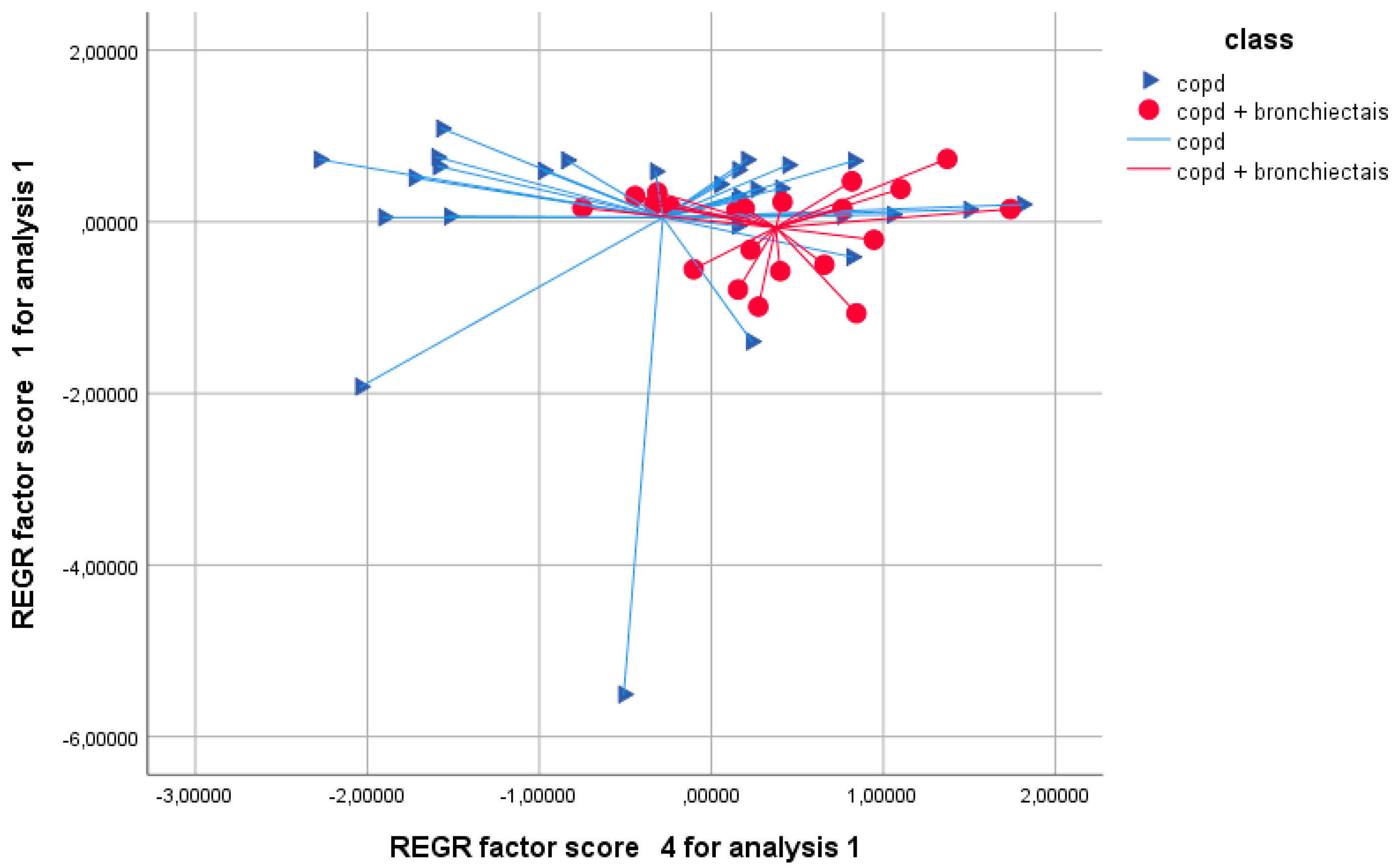
PCA identified four principal components that, together, accounted for 99.22% of the total variance in the dataset. The greatest discriminatory power between the two patient groups was found in the fourth principal component (PC4), which revealed a statistically significant difference in breathprint signatures as follows: mean PC4 score for the BCO group: 0.3748 ± 0.6337; mean PC4 score for the COPD group: –0.2811 ± 1.1354, p = 0.021 (Table 3)
These results suggest that the e-nose was able to detect reproducible differences in the exhaled VOC patterns of patients with bronchiectasis compared to those without. Although PC4 does not explain the majority of the variance, its statistical significance indicates that subtle yet relevant differences in VOC composition may underlie the pathophysiological divergence between the groups.
The PCA scatter plot demonstrated a partial but distinct separation of the two populations along the PC4 axis, supporting the presence of different metabolic or inflammatory signatures captured by the breathprints.

3.3. Discriminant Analysis and Model Performance

To assess the classification performance of the e-nose in distinguishing between BCO and non-BCO COPD patients, linear discriminant analysis (LDA) was applied, followed by cross-validation using the leave-one-out method.
The overall cross-validated classification accuracy was 69.6%, indicating a statistically significant ability of the model to distinguish the two patient groups based on breathprint data alone (Figure 2). The area under the ROC curve (AUC) for this model was 0.768 (95% CI: 0.618–0.919), with a p-value of 0.037, further supporting the discriminative capability of the VOC-based approach.
While this level of accuracy may not be sufficient for stand-alone diagnostic purposes, it is encouraging given the complexity of VOC signatures and the known overlap in clinical features between COPD and BCO. These findings suggest that breathprints obtained via e-nose could serve as complementary biomarkers in COPD endotyping and risk stratification.

3.4. Subgroup Analysis by Inflammatory Endotype

To explore whether the e-nose could detect the differences in breathprints associated with inflammatory endotypes, we performed subgroup analysis focusing on three distinct comparisons: (1) eosinophilic COPD vs. neutrophilic BCO; (2) neutrophilic COPD vs. neutrophilic BCO; and (3) eosinophilic COPD vs. neutrophilic COPD.
The greatest classification performance was observed in differentiating eosinophilic COPD from neutrophilic BCO patients. In this comparison, the cross-validated classification accuracy reached 76.8%, indicating a strong signal for differentiation. This was followed by a 74.7% accuracy rate in distinguishing neutrophilic COPD from neutrophilic BCO (Table 4 and Table 5, Figure 3).
These findings underscore the e-nose’s potential in identifying not only structural overlap syndromes like BCO, but also in differentiating inflammatory endotypes based on the VOC signature in exhaled breath. Given that neutrophilic inflammation is often associated with chronic infection, oxidative stress, and bacterial colonization, it is plausible that the altered VOC profile detected by the e-nose reflects these underlying processes.
No significant classification was observed between eosinophilic and neutrophilic COPD in the absence of bronchiectasis, suggesting that structural changes in the airways (i.e., bronchiectasis) may exert a more dominant effect on the VOC profile than eosinophilic versus neutrophilic inflammation alone.

4. Discussion

This prospective real-life study demonstrates that e-nose technology can discriminate between patients with COPD and those with bronchiectasis–COPD overlap (BCO) based on exhaled volatile organic compound (VOC) profiles. Breath analysis via e-nose offers a unique, non-invasive means of assessing the airway’s molecular milieu, capturing real-time snapshots of the volatile metabolome, the so called “breathprint”, generated by endogenous processes such as inflammation, oxidative stress, tissue remodeling, and microbiome metabolism.
Our findings align with the growing literature supporting breathomics in respiratory medicine. In our cohort, the e-nose distinguished breathprints between BCO and COPD patients, with PC4 showing significantly higher scores in BCO. The overall classification accuracy was adequate and ROC analysis confirmed that separation was unlikely due to chance. Importantly, discriminatory performance improved when evaluating specific inflammatory subgroups, reaching up to 76.8% accuracy in differentiating eosinophilic COPD from neutrophilic BCO.
These results suggest that VOC signatures reflect underlying biological processes, including neutrophilic airway inflammation that predominates in bronchiectasis. This capacity to non-invasively profile inflammatory phenotypes could guide personalized treatment strategies and rapid bedside stratification.
BCO patients in our study demonstrated more severe lung function impairment and a breathprint indicative of distinct metabolic and inflammatory pathways. Previous studies reported chronic neutrophilic inflammation in BCO, often driven by persistent bacterial colonization (e.g., Pseudomonas aeruginosa, Haemophilus influenzae). Such chronic infections sustain elevated cytokines (IL-8, GM-CSF, TNF-α) and proteolytic enzymes like neutrophil elastase, leading to epithelial damage and altered local metabolome, i.e., features captured within VOC profiles.
Huang et al. [4] identified five unique endotypes within BCO, including a neutrophilic–proteobacterial cluster associated with worse prognosis. E-nose technology may detect these patterns, acting as surrogate markers of such biologically defined clusters and facilitating precision medicine approaches.
Although neutrophilic inflammation predominates in bronchiectasis, a relevant subset of patients presents eosinophilic airway inflammation responsive to corticosteroids or biologics. However, blood eosinophil counts show moderate reliability over time, with up to 50% of patients changing eosinophilic status during multi-year follow up [22,23]. Breathomics may complement blood biomarkers, as VOC profiles reflecting type 2 inflammation have been identified in asthma and could support the detection and monitoring of eosinophilic endotypes in bronchiectasis [24].
Currently, bronchiectasis and BCO management follows a “one-size-fits-all” approach despite pathophysiological differences [25,26]. E-nose technology could serve as a rapid triage tool to identify patients likely to benefit from specific therapies, such as inhaled corticosteroids or biologics in eosinophilic inflammation, or macrolides, mucolytics, and antibiotics in neutrophilic BCO. Moreover, it may aid the pharmacodynamic monitoring of anti-neutrophilic therapies (e.g., brensocatib) [27,28,29], detecting breathprint changes correlating with treatment response or early relapse.
The portability and ease-of-use of the e-nose make it suitable for outpatient clinics or even home-based monitoring, potentially guiding antibiotic stewardship and exacerbation prediction [30].
Breathomics complements systemic or airway biomarker research. While serum markers such as adiponectin and cytokine profiles have been studied in BCO, they may not always reflect airway inflammation and can be confounded by comorbidities [31]. In contrast, breathomics provides a localized, dynamic reflection of airway inflammation and microbial activity. Unlike high-throughput omics requiring reagents and laboratory processing, e-nose technology is inexpensive and yields immediate results.
Although the Cyranose® 320 platform does not identify individual VOCs, previous GC-MS-based studies on COPD and bronchiectasis have reported alterations in several chemical classes, including alkanes, aldehydes, ketones, alcohols, aromatic hydrocarbons, and sulfur-containing compounds [32,33]. In bronchiectasis with neutrophilic inflammation and chronic bacterial colonization, bacterial metabolites such as short-chain fatty acids and nitrogen-containing compounds have also been described. These findings suggest that the breathprints detected in our study may reflect a combination of oxidative stress markers, metabolic byproducts, and microbial metabolites characteristic of COPD and BCO pathophysiology.
Gastrointestinal gases (e.g., hydrogen, methane, sulfur compounds) can be present in exhaled breath and theoretically influence VOC patterns. In this study, standardized pre-sampling fasting, avoidance of recent smoking, and alveolar breath collection were employed to minimize their impact. While Cyranose® 320 does not differentiate GI-derived VOCs from airway-derived VOCs, it is likely that the discriminant patterns observed in COPD and BCO primarily reflect airway inflammation, oxidative stress, and microbial activity. Future studies could integrate GI gas quantification or employ sampling methods optimized for alveolar fraction enrichment to further reduce potential confounding.
Moreover, the accuracy of e-nose measurements is highly dependent on standardized sampling and pre-analytical handling, as VOC profiles can be influenced by recent diet, smoking, environmental exposures, and even circadian variations. To minimize these effects, our protocol included pre-sampling fasting, acclimatization in a controlled environment, alveolar breath enrichment, and immediate analysis. Ambient air blanks were subtracted to reduce background interference. This approach aligns with systematic methodological recommendations, such as those by Di Gilio et al. [34], who emphasize that differences in sampling volume, fraction, and handling can significantly affect VOC measurement reproducibility. While these measures reduce variability, residual time-of-day effects and other pre-analytical factors cannot be fully excluded. Future studies should systematically investigate the impact of sampling time and standardized pre-treatment on breathprint reproducibility.
Despite promising results, this study has limitations. The sample size was modest, warranting caution in interpretation. Although our sample size was relatively limited, it was chosen to ensure a standard error of ≤10% for group-level estimates, as in previous breathomics studies. The observed discriminant performance supports the feasibility of e-nose technology for breathprint-based endotyping. Nevertheless, larger multicenter studies are warranted to validate these findings and improve generalizability. Moreover, a key limitation of the Cyranose® 320 platform is the lack of compound-level resolution. The device identifies breathprint patterns but does not specify individual VOCs contributing to group separation. Future studies should integrate gas chromatography–mass spectrometry (GC-MS) or proton transfer reaction–mass spectrometry (PTR-MS) to identify the specific VOCs responsible for the observed breathprint differences. Furthermore, the influence of diet, circadian variation, and medications on VOC profiles cannot be fully excluded, and not all patients underwent sputum analysis.
Given the promising preliminary results of this study, several avenues for future research are warranted to fully realize the potential of e-nose technology in the management of bronchiectasis and COPD.
Firstly, the validation of VOC signatures against established biological markers is crucial. Correlating breathprint data with sputum cytokine levels, proteomic profiles, and microbiome composition would enhance the biochemical interpretability of the patterns detected by the e-nose. This integration could help identify the specific metabolic or inflammatory pathways contributing to the discriminant breathprints observed in different endotypes.
Secondly, longitudinal studies are needed to assess how breathprints evolve over time, particularly in response to treatment or during exacerbations. Such studies would provide insights into the temporal dynamics of VOC profiles, enabling clinicians to monitor disease progression more effectively and adjust therapeutic strategies in a timely manner. For example, detecting early breathprint changes indicative of an impending exacerbation or treatment failure could facilitate preemptive interventions, potentially improving clinical outcomes.
Thirdly, integrating e-nose data with artificial intelligence (AI) algorithms represents a promising strategy to enhance diagnostic accuracy and clinical utility. Machine learning models could be trained to recognize complex VOC patterns, automate endotype classification, and even predict treatment responses based on breathprint changes. This approach would support the development of rapid, reproducible, and clinician-friendly diagnostic tools within precision medicine frameworks [35].
Furthermore, it is important to assess the cost-effectiveness of implementing e-nose technology in routine clinical practice. While breath analysis offers several advantages in terms of non-invasiveness and rapidity, health economic evaluations are necessary to determine whether its integration translates into measurable benefits in clinical outcomes, healthcare resource utilization, and patient quality of life.
Although purge/zeroing, background subtraction and normalization reduce short-term variability, long-term sensor drift is inherent to polymer-sensor e-noses. Our single-center study minimized this via standardized acquisition and cross-validation; however, future multicenter/longitudinal studies should incorporate external references and drift-correction strategies to ensure inter-day and inter-site transferability.
Finally, future studies should consider multicenter designs with larger and more diverse patient cohorts to confirm the generalizability and reproducibility of findings across different settings. Collaborative research incorporating e-nose technology alongside omics platforms such as microbiomics, proteomics, and transcriptomics could provide a multidimensional view of airway disease biology, ultimately paving the way toward composite biomarker panels for diagnosis, prognosis, and treatment monitoring.
Looking ahead, several research avenues are worth pursuing. Longitudinal studies should investigate how breathprints evolve over time and respond to treatment, potentially enabling the VOC-based monitoring of disease activity and early detection of exacerbations. Combining e-nose technology with other omics platforms (microbiome, transcriptome, proteome) may uncover composite biomarkers for precision endotyping. Additionally, machine learning algorithms can be trained to enhance diagnostic and prognostic accuracy from breath data. Future validation in large, multicenter cohorts is essential to support clinical adoption. Finally, e-nose applications may extend to the pharmacodynamic monitoring of targeted therapies, such as biologics or anti-neutrophilic drugs, helping guide treatment decisions in real time.

5. Conclusions

This prospective real-life study demonstrates that e-nose technology can non-invasively discriminate between COPD and BCO based on exhaled VOC profiles. Significant differences were observed in breathprints, particularly in inflammatory endotype comparisons, with the highest classification accuracy when distinguishing eosinophilic COPD from neutrophilic BCO. These results support the feasibility of breathomics for patient stratification, and highlight its potential role in guiding personalized treatment strategies. Further multicenter and longitudinal studies integrating compound-level VOC identification are warranted to validate these findings and enhance clinical translation.

Author Contributions

Conceptualization, V.N.Q. and S.D.; methodology, S.D.; software, A.M.; validation, A.P., A.M. and S.D.; formal analysis, V.N.Q.; investigation, M.F.G.; resources, A.P.; data curation, V.N.Q.; writing—original draft preparation, S.D.; writing—review and editing, A.M.; visualization, A.P.; supervision, G.E.C.; project administration, M.R.V.; funding acquisition, M.R.V. All authors have read and agreed to the published version of the manuscript.

Funding

This research received no external funding.

Institutional Review Board Statement

The study was conducted in accordance with the Declaration of Helsinki, and approved by the Institutional Review Board of Policlinico di Bari (protocol number 46403/15), date of approval 27 May 2015.

Informed Consent Statement

Informed consent was obtained from all the subjects involved in the study.

Data Availability Statement

The dataset is available upon reasonable request.

Conflicts of Interest

The authors declare no conflicts of interest.

References

  1. Global Strategy for the Diagnosis, Management, and Prevention of Chronic Obstructive Pulmonary Disease. Global Initiative for Chronic Obstructive Lung Disease. Available online: https://goldcopd.org/2024-gold-report/ (accessed on 22 October 2024).
  2. Wang, Z.; Lin, J.; Liang, L.; Huang, F.; Yao, X.; Peng, K.; Gao, Y.; Zheng, J. Global, regional, and national burden of chronic obstructive pulmonary disease and its attributable risk factors from 1990 to 2021: An analysis for the Global Burden of Disease Study 2021. Respir. Res. 2025, 26, 2. [Google Scholar] [CrossRef]
  3. Boers, E.; Barrett, M.; Su, J.G.; Benjafield, A.V.; Sinha, S.; Kaye, L.; Zar, H.J.; Vuong, V.; Tellez, D.; Gondalia, R.; et al. Global Burden of Chronic Obstructive Pulmonary Disease Through 2050. JAMA Netw. Open. 2023, 6, 2346598. [Google Scholar] [CrossRef]
  4. Huang, J.T.J.; Cant, E.; Keir, H.R.; Barton, A.K.; Kuzmanova, E.; Shuttleworth, M.; Pollock, J.; Finch, S.; Polverino, E.; Mathieu, B.; et al. Endotyping Chronic Obstructive Pulmonary Disease, Bronchiectasis, and the “Chronic Obstructive Pulmonary Disease-Bronchiectasis Association”. Am. J. Respir. Crit. Care Med. 2022, 206, 417–426. [Google Scholar] [CrossRef] [PubMed]
  5. Alam, M.A.; Mangapuram, P.; Fredrick, F.C.; Singh, B.; Singla, A.; Kumar, A.; Jain, R. Bronchiectasis-COPD Overlap Syndrome: A Comprehensive Review of its Pathophysiology and Potential Cardiovascular Implications. Ther. Adv. Pulm. Crit. Care Med. 2024, 19, 29768675241300808. [Google Scholar] [CrossRef]
  6. Aliberti, S.; Sotgiu, G.; Lapi, F.; Gramegna, A.; Cricelli, C.; Blasi, F. Prevalence and incidence of bronchiectasis in Italy. BMC Pulm. Med. 2020, 20, 15. [Google Scholar] [CrossRef] [PubMed]
  7. Martinez-Garcia, M.A.; Miravitlles, M. Bronchiectasis in COPD patients: More than a comorbidity? Int. J. Chronic Obstr. Pulm. Dis. 2017, 12, 1401–1411. [Google Scholar] [CrossRef]
  8. Polverino, E.; Dimakou, K.; Hurst, J.; Martinez-Garcia, M.A.; Miravitlles, M.; Paggiaro, P.; Shteinberg, M.; Aliberti, S.; Chalmers, J.D. The overlap between bronchiectasis and chronic airway diseases: State of the art and future directions. Eur. Respir. J. 2018, 52, 1800328. [Google Scholar] [CrossRef]
  9. Raboso, B.; Pou, C.; Abril, R.; Erro, M.; Sánchez, C.; Manzano, C.; Zamarrón, E.; Suarez-Cuartin, G.; González, J. Bronchiectasis. Open Respir. Arch. 2024, 6, 100339. [Google Scholar] [CrossRef] [PubMed]
  10. Ibrahim, W.; Carr, L.; Cordell, R.; Wilde, M.J.; Salman, D.; Monks, P.S.; Thomas, P.; Brightling, C.E.; Siddiqui, S.; Greening, N.J. Breathomics for the clinician: The use of volatile organic compounds in respiratory diseases. Thorax 2021, 76, 514–521. [Google Scholar] [CrossRef]
  11. Dragonieri, S.; Pennazza, G.; Carratu, P.; Resta, O. Electronic Nose Technology in Respiratory Diseases. Lung 2017, 195, 157–165. [Google Scholar] [CrossRef]
  12. Ibrahim, B.; Basanta, M.; Cadden, P.; Singh, D.; Douce, D.; Woodcock, A.; Fowler, S.J. Non-invasive phenotyping using exhaled volatile organic compounds in asthma. Thorax 2011, 66, 804–809. [Google Scholar] [CrossRef]
  13. Ibrahim, W.; Natarajan, S.; Wilde, M.; Cordell, R.; Monks, P.S.; Greening, N.; Brightling, C.E.; Evans, R.; Siddiqui, S. A systematic review of the diagnostic accuracy of volatile organic compounds in airway diseases and their relation to markers of type-2 inflammation. ERJ Open Res. 2021, 7, 00030-2021. [Google Scholar] [CrossRef] [PubMed]
  14. Ahmed, W.M.; Lawal, O.; Nijsen, T.M.; Goodacre, R.; Fowler, S.J. Exhaled Volatile Organic Compounds of Infection: A Systematic Review. ACS Infect. Dis. 2017, 3, 695–710. [Google Scholar] [CrossRef] [PubMed]
  15. Filipiak, W.; Ager, C.; Troppmair, J. Predicting the future from the past: Volatile markers for respiratory infections. Eur. Respir. J. 2017, 49, 1700264. [Google Scholar] [CrossRef]
  16. Kwon, J.W.; Park, H.W.; Kim, W.J.; Kim, M.G.; Lee, S.J. Exposure to volatile organic compounds and airway inflammation. Environ. Health 2018, 17, 65. [Google Scholar] [CrossRef]
  17. Angrill, J.; Agustí, C.; De Celis, R.; Filella, X.; Rañó, A.; Elena, M.; De La Bellacasa, J.P.; Xaubet, A.; Torres, A. Bronchial inflammation and colonization in patients with clinically stable bronchiectasis. Am. J. Respir. Crit. Care Med. 2001, 164, 1628–1632. [Google Scholar] [CrossRef]
  18. Chalmers, J.D.; Aliberti, S.; Filonenko, A.; Shteinberg, M.; Goeminne, P.C.; Hill, A.T.; Fardon, T.C.; Obradovic, D.; Gerlinger, C.; Giovanni, S.; et al. Characterization of the “Frequent Exacerbator Phenotype” in Bronchiectasis. Am. J. Respir. Crit. Care Med. 2018, 197, 1410–1420. [Google Scholar] [CrossRef]
  19. Chalmers, J.D.; Metersky, M.; Aliberti, S.; Morgan, L.; Fucile, S.; Lauterio, M.; McDonald, P.P. Neutrophilic inflammation in bronchiectasis. Eur. Respir. Rev. 2025, 34, 240179. [Google Scholar] [CrossRef]
  20. Chen, W.; Ran, S.; Li, C.; Li, Z.; Wei, N.; Li, J.; Li, N. Elevated Eosinophil Counts in Acute Exacerbations of Bronchiectasis: Unveiling a Distinct Clinical Phenotype. Lung 2024, 202, 53–61. [Google Scholar] [CrossRef]
  21. Oscullo, G.; Gómez-Olivas, J.D.; Ingles, M.; Mompean, S.; Martinez-Perez, R.; Suarez-Cuartin, G.; Rosa-Carrillo, D.; Martinez-Garcia, M.A. Bronchiectasis-COPD Overlap Syndrome: Role of Peripheral Eosinophil Count and Inhaled Corticosteroid Treatment. J. Clin. Med. 2023, 12, 6417. [Google Scholar] [CrossRef] [PubMed]
  22. Martínez-García, M.A.; Olveira, C.; Girón, R.; García-Clemente, M.; Máiz, L.; Sibila, O.; Golpe, R.; Rodríguez-Hermosa, J.L.; Barreiro, E.; Raúl, M.; et al. Reliability of blood eosinophil count in steady-state bronchiectasis. Pulmonology 2025, 31, 2416836. [Google Scholar] [CrossRef]
  23. Shoemark, A.; Shteinberg, M.; De Soyza, A.; Haworth, C.S.; Richardson, H.; Gao, Y.; Perea, L.; Dicker, A.J.; Goeminne, P.C.; Cant, E.; et al. Characterization of Eosinophilic Bronchiectasis: A European Multicohort Study. Am. J. Respir. Crit. Care Med. 2022, 205, 894–902. [Google Scholar] [CrossRef]
  24. Alahmadi, F.H.; Wilkinson, M.; Keevil, B.; Niven, R.; Fowler, S.J. Short- and medium-term effect of inhaled corticosteroids on exhaled breath biomarkers in severe asthma. J. Breath. Res. 2022, 16, 047101. [Google Scholar] [CrossRef]
  25. Chalmers, J.D.; Elborn, S.; Greene, C.M. Basic, translational and clinical aspects of bronchiectasis in adults. Eur. Respir. Rev. 2023, 32, 230015. [Google Scholar] [CrossRef]
  26. Polverino, E.; Goeminne, P.C.; McDonnell, M.J.; Aliberti, S.; Marshall, S.E.; Loebinger, M.R.; Murris, M.; Cantón, R.; Torres, A.; Katerina, D.; et al. European Respiratory Society guidelines for the management of adult bronchiectasis. Eur. Respir. J. 2017, 50, 1700629. [Google Scholar] [CrossRef]
  27. Chalmers, J.D.; Shteinberg, M.; Mall, M.A.; O’Donnell, A.E.; Watz, H.; Gupta, A.; Frahm, E.; Eleftheraki, A.; Rauch, J.; Sanjay, H.C.; et al. Cathepsin C (dipeptidyl peptidase 1) inhibition in adults with bronchiectasis: AIRLEAF, a phase II randomised, double-blind, placebo-controlled, dose-finding study. Eur. Respir. J. 2025, 65, 2401551. [Google Scholar] [CrossRef]
  28. Tanabe, N.; Matsumoto, H.; Kogo, M.; Morimoto, C.; Nomura, N.; Hayashi, Y.; Sakamoto, R.; Oguma, T.; Nagasaki, T.; Sunadome, H.; et al. Exploring the roles of airway dipeptidyl peptidase 1 in obstructive airway disease. ERJ Open Res. 2025, 11, 00841–02024. [Google Scholar] [CrossRef] [PubMed]
  29. Chalmers, J.D.; Burgel, P.R.; Daley, C.L.; De Soyza, A.; Haworth, C.S.; Mauger, D.; Mange, K.; Teper, A.; Fernandez, C.; Conroy, D.; et al. Brensocatib in non-cystic fibrosis bronchiectasis: ASPEN protocol and baseline characteristics. ERJ Open Res. 2024, 10, 00151–0202. [Google Scholar] [CrossRef] [PubMed]
  30. Martins, M.; Keir, H.R.; Chalmers, J.D. Endotypes in bronchiectasis: Moving towards precision medicine. A narrative review. Pulmonology 2023, 29, 505–517. [Google Scholar] [CrossRef]
  31. Nigro, E.; Mosella, M.; Daniele, A.; Mallardo, M.; Accardo, M.; Bianco, A.; Perrotta, F.; Scialò, F. Adiponectin Increase in Patients Affected by Chronic Obstructive Pulmonary Disease with Overlap of Bronchiectasis. Life 2023, 13, 444. [Google Scholar] [CrossRef] [PubMed]
  32. Gu, S.Y.; Lu, H.W.; Bai, J.W.; Yang, J.W.; Mao, B.; Yu, L.; Xu, J.F. The role of volatile organic compounds for assessing characteristics and severity of non-cystic fibrosis bronchiectasis: An observational study. Front. Med. 2024, 11, 1345165. [Google Scholar] [CrossRef]
  33. Kuo, P.H.; Jhong, Y.C.; Kuo, T.C.; Hsu, Y.T.; Kuo, C.H.; Tseng, Y.J. A Clinical Breathomics Dataset. Sci. Data 2024, 11, 203. [Google Scholar] [CrossRef] [PubMed]
  34. Di Gilio, A.; Palmisani, J.; Ventrella, G.; Facchini, L.; Catino, A.; Varesano, N.; Pizzutilo, P.; Galetta, D.; Borelli, M.; Barbieri, P.; et al. Breath Analysis: Comparison among Methodological Approaches for Breath Sampling. Molecules 2020, 25, 5823. [Google Scholar] [CrossRef] [PubMed]
  35. Dai, J.; Xie, G.; Huo, X.; Li, J.; Deng, S.; Su, Y. Implantable and Biodegradable Smart Textiles for Continuous Limb and Gastrointestinal Motility Monitoring. Small 2025, 21, 2407773. [Google Scholar] [CrossRef] [PubMed]
Figure 1. Schematic workflow of the Cyranose 320 e-nose.
Chemosensors 13 00311 g001
Figure 2. Principal component analysis (PCA) scatter plot showing separation between COPD (blue triangles) and bronchiectasis–COPD overlap (BCO; red circles) patients based on exhaled VOC profiles. The plot displays REGR factor scores for principal component 4 (x-axis) and principal component 1 (y-axis), demonstrating a partial but distinct clustering of BCO patients compared to COPD-only patients, supporting the presence of different metabolic or inflammatory breathprint signatures (cross-validated classification accuracy = 69.6%).
Chemosensors 13 00311 g002
Figure 3. Principal component analysis (PCA) scatter plots comparing COPD and bronchiectasis–COPD overlap (BCO) patients. (A) PCA plot showing distribution of patients with COPD (blue triangles) versus BCO (red squares) based on their exhaled volatile organic compound (VOC) breathprints. (B) PCA plot comparing eosinophilic COPD (green triangles) and neutrophilic BCO (red squares). In both panels, PC4 is plotted against PC1, demonstrating partial but distinct clustering between groups, suggesting different metabolic and inflammatory VOC signatures captured by the electronic nose.
Chemosensors 13 00311 g003
Table 1. Clinical characteristics of the study population. Values are shown as the mean ± standard deviation (SD), unless differently specified.
CharacteristicCOPD (n = 56)BCO (n = 42)p-Value
Number of patients5642
Sex (M/F)38\1830\120.92
Age (years)69.2 ± 8.470.5 ± 7.90.44
FEV1 (% predicted)67.3 ± 22.352.3 ± 16.5<0.01
FVC (% predicted)83.2 ± 25.866.7 ± 17.8<0.01
BMI (kg/m2)26.8 ± 4.125.9 ± 4.30.28
CAT score18.5 ± 6.219.4 ± 7.10.51
mMRC score2.1 ± 0.82.3 ± 0.90.26
Eosinophils ≥ 300 cells/μL (n, %)20 (35.7%)14 (33.3%)0.80
Table 2. Description of the 32-sensor array in the Cyranose® 320.
Sensor No.Polymer Coating MaterialGeneral VOC Sensitivity Profile
S1Poly(4-vinylphenol)Polar alcohols, phenols
S2Poly(ethylene-co-vinyl alcohol)Small alcohols, ketones
S3Poly(vinylpyridine)Amines, basic VOCs
S4Poly(styrene)Aromatic hydrocarbons
S5Poly(vinyl acetate)Esters, ketones
S6Poly(butadiene)Non-polar hydrocarbons
S7Poly(vinylidene chloride-co-acrylonitrile)Halogenated compounds
S8Poly(methyl methacrylate)Ketones, esters
S9Poly(ethylene oxide)Alcohols, ethers
S10Poly(isobutylene)Non-polar VOCs
S11Poly(ethylene-co-propylene)Alkanes, alkenes
S12Poly(caprolactone)Esters, aldehydes
S13Poly(acrylic acid)Polar VOCs
S14Poly(vinyl alcohol)Alcohols, aldehydes
S15Poly(2-vinylpyridine)Amines
S16Poly(ethylene-co-vinyl acetate)Ketones, esters
S17Poly(tetrafluoroethylene)Fluorinated VOCs
S18Poly(propylene glycol)Alcohols, glycols
S19Poly(4-methyl-1-pentene)Alkanes, non-polar VOCs
S20Poly(ethylene terephthalate)Aromatics, esters
S21Poly(vinyl chloride)Chlorinated hydrocarbons
S22Poly(phenylene oxide)Aromatics, phenols
S23Poly(acrylonitrile)Nitriles, polar VOCs
S24Poly(lactic acid)Aldehydes, ketones
S25Poly(ethylene glycol)Alcohols, glycols
S26Poly(butyl methacrylate)Esters, ketones
S27Poly(oxymethylene)Aldehydes
S28Poly(caprylic acid)Fatty acids
S29Poly(urethane)Ketones, aldehydes
S30Poly(dimethylsiloxane)Non-polar VOCs
S31Poly(vinyl methyl ether)Ethers, aldehydes
S32Poly(ethylene naphthalate)Aromatics, hydrocarbons
Table 3. PCA factor scores for COPD and BCO patients. Mean ± SD values are shown for each principal component (PC1–PC4). PC4 demonstrated a statistically significant difference between groups (p = 0.021), indicating distinct VOC breathprint patterns in BCO compared to COPD. No significant differences were observed for PC1, PC2, or PC3.
Principal ComponentCOPD (Mean ± SD) BCO (Mean ± SD) p-Value
PC1 0.0501 ± 1.2590 −0.0668 ± 0.5019 0.690
PC2 −0.0493 ± 1.0085 0.0657 ± 1.0095 0.695
PC3 −0.0545 ± 1.2115 0.0726 ± 0.6394 0.664
PC4 −0.2811 ± 1.1354 0.3748 ± 0.6337 0.021
Table 4. Principal component analysis (PCA) factor scores by inflammatory phenotype and group. Mean ± SD values are shown for each principal component (PC1–PC4) in COPD and BCO patients, stratified by blood eosinophil counts: eosinophilic: ≥300 cells/μL; neutrophilic: <300 cells/μL. PC4 showed a statistically significant difference (p = 0.014) across groups, suggesting distinct VOC breathprint signatures particularly in neutrophilic BCO patients. No significant differences were observed for PC1, PC2, or PC3.
PCCOPD Eos ≥ 300 Cells/μLBCO Eos ≥ 300 Cells/μLCOPD Eos < 300 Cells/μLBCO Eos < 300 Cells/μLp-Value
PC10.13 ± 0.73−0.02 ± 1.480.06 ± 0.41−0.09 ± 0.550.889
PC2−0.29 ± 0.780.11 ± 1.12−0.02 ± 0.990.08 ± 0.970.488
PC3−0.14 ± 0.70−0.13 ± 1.310.32 ± 0.680.12 ± 0.840.430
PC4−0.19 ± 1.19−0.23 ± 1.09−0.09 ± 0.790.49 ± 0.600.014
Table 5. Cross-validated classification accuracy for inflammatory subgroup comparisons.
ComparisonCross-Validated Classification Accuracy (%)
COPD eosinophilic vs. BCO eosinophilic33.4
COPD eosinophilic vs. COPD neutrophilic41.2
COPD eosinophilic vs. BCO neutrophilic76.8
BCO eosinophilic vs. COPD neutrophilic44.0
BCO vs. BCO neutrophilic38.6
COPD neutrophilic vs. BCO neutrophilic74.7
Disclaimer/Publisher’s Note: The statements, opinions and data contained in all publications are solely those of the individual author(s) and contributor(s) and not of MDPI and/or the editor(s). MDPI and/or the editor(s) disclaim responsibility for any injury to people or property resulting from any ideas, methods, instructions or products referred to in the content.