Article

A Carbazole-Based Aggregation-Induced Emission “Turn-On” Sensor for Mercury Ions in Aqueous Solution

by
Remya Radha
1,
Mohammed S. Valliyengal
1 and
Mohammad H. Al-Sayah
1,2,3,*
1
Department of Biology, Chemistry, and Environmental Sciences, American University of Sharjah, Sharjah P.O. Box 26666, United Arab Emirates
2
Materials Science and Engineering Program, College of Arts and Sciences, American University of Sharjah, Sharjah P.O. Box 26666, United Arab Emirates
3
Materials Research Center, College of Arts and Sciences, American University of Sharjah, Sharjah P.O. Box 26666, United Arab Emirates
*
Author to whom correspondence should be addressed.
Chemosensors 2025, 13(8), 276; https://doi.org/10.3390/chemosensors13080276
Submission received: 7 June 2025 / Revised: 9 July 2025 / Accepted: 22 July 2025 / Published: 25 July 2025

Abstract

The development of rapid detection methods to identify mercury ions in aqueous solutions is crucial for effectively monitoring environmental contamination. Fluorescent chemical sensors offer a fast and reliable approach to detect and analyze these metal ions. In this study, a sensor utilizing aggregation-induced emission (AIE) is introduced as a ’turn-on’ fluorescent sensor specifically designed for mercury ions in aqueous solutions. The sensor, based on carbazole, forms aggregates in aqueous solutions, resulting in a significant 800% enhancement of its fluorescence signal. When elemental iodine is added to the solution, the fluorescence of the aggregates is quenched by 90%. However, upon subsequent addition of mercury ions, the fluorescence is regenerated, and the intensity of the emission signal is directly proportional to the concentration of the ions across a wide concentration range. The carbazole-iodine complex acts as a fluorescent probe, enabling the detection of mercury ions in aqueous solutions.

Graphical Abstract

1. Introduction

Over the past few decades, fluorescent sensors, particularly those with aggregation-induced emission (AIE) properties, have become highly sought-after features in advanced functional materials [1,2,3,4]. They offer high sensitivity and versatility in detecting ions, biomolecules, and cellular components [1,5], making them have broad applications in biosciences, electronics, and energy organic light-emitting diodes (OLEDs), organic nano-dots for bio-imaging, and fluorescent sensors [6,7,8,9,10,11,12,13]. Unlike conventional fluorescent dyes, which typically suffer from aggregation-caused quenching (ACQ) due to pi–pi stack interactions during aggregation, AIE fluorophores exhibit an intriguing property of increased emission intensity upon aggregation [14,15,16]. This enhancement is attributed to the constrained intramolecular motions that result in twisted molecular structures, preventing intermolecular pi–pi interactions and intermolecular pi–pi conjugation [17]. Consequently, these interactions decrease the rate of non-radiative decay and significantly enhance the emission of AIE fluorophores [18].
AIE fluorophores have been used for the development of fluorescent chemical sensors [19]. One extensively studied fluorophore is tetraphenylethylene (TTPE), which has found numerous applications, including its use as a fluorescent sensor for metals and nitroaromatics [20,21]. Recently, Zhang and colleagues reported a TTPE-based ’turn-off’ sensor for iodide anions, wherein the sensor–iodide complex subsequently serves as a ’turn-on’ sensor for Hg2+ metal ions [22]. In this system, the AIE of the sensor is quenched upon the addition of iodide anions. However, upon binding with subsequently added Hg2+ cations, the emission of the sensor is regenerated. Additionally, a fluorescent sensor based on 4-diethylamino (benzylideneamino)-3′,6′-dihydroxyspiro[isoindoline-1,9-xanthen]-3-one was developed for the selective detection of 4-nitroaniline (4-NA) via a photoinduced electron transfer mechanism. The sensor was efficiently used for the fabrication of on-site detection through handheld kits for 4-NA and for the latent fingerprints on various surfaces [23].
We have recently reported [6] the AIE behavior of a carbazole-barbiturate (CB) chromophore upon aggregation in aqueous solutions (fw = 99%). CB consists of a carbazole fluorophore and two barbiturate moieties, forming a highly conjugated structure with a large aromatic surface and a pi-rich system (Figure 1) This structure is the driving force for aggregation in aqueous solution. Yet, the AIE behavior of CB is attributed to the restricted rotation around the bond connecting the barbiturate with the carbazole in the aggregates, which prevents conjugation between the two units and suppresses the non-radiative relaxation channels, similar to the system that was reported by Yin et al. earlier [24].
Herein, we report on the investigations on CB as an AIE sensor for the detection of Hg2+ ions in aqueous solution. We hypothesized that the aggregates of CB can form iodine complexes (CB-I) due to the low solubility of elemental iodine in water and the interaction of iodine with the pi-system of CB. Subsequently, this complexation will significantly affect the photoelectronic properties of CB and its absorbance and quenching of CB emission. Yet, the presence of Hg2+ ions in the solution leads to the breakup of the CB-I complex and regeneration of the CB emission. This change in the emission intensity is then correlated to the concentration of Hg2+ ions in the solution, providing a quick detection probe for these ions. The development of such rapid and cost-effective detection methods to monitor the presence of mercury ions in aqueous solutions is of high importance due to the significant global concern regarding mercury pollution and its high toxicity [22]. The bioaccumulation of mercury ions by microorganisms can result in their conversion into hazardous organic mercury compounds, which subsequently enter the food chain and accumulate in animals and humans [25,26,27]. Such accumulation of mercury heavy metal ion causes severe health problems, including kidney failure, damage to the central nervous system, vision and hearing loss, tubular necrosis, and proteinuria [28,29]. Moreover, it acts as a neurotoxin and causes immune system dysfunction and even death of individuals [30,31].
The results in this paper show that the emission intensity of the CB is enhanced upon aggregation in aqueous solutions. Upon the introduction of elemental iodine (I2) into the CB solution, the CB–iodine complex (CB-I) is formed, leading to the quenching of the fluorescence from the aggregated sensor. However, upon the subsequent addition of Hg2+ ions, the emission of CB is regenerated, displaying a linear increase in emission intensity corresponding to the concentration of Hg2+ ions in solution. Hence, the CB-I system can be utilized as a fluorescent sensor for the detection and quantification of Hg2+ ions in aqueous solution.

2. Experimental Section

2.1. Materials and Instrumentation

All metal anions, iodine, and other chemicals used in the study were of the highest analytical grade and purchased from Sigma Aldrich (Saint Louis, MO, USA). UV-visible (UV-vis) absorption spectra were collected with a UV-1800 UV-Vis spectrophotometer (Shimadzu, Kyoto, Japan). Fluorescence emission and spectrum analysis were recorded on a FLSP920 Series of Fluorescence spectrometer (Edinburgh Instruments Ltd., Livingston, UK).Dynamic light scattering experiments and particle size distribution data were collected with the help of a Dyanopro Nanostar laser photometer (WYATT, Santa Barbara, CA, USA). Fourier Transform Infrared Spectrophotometry analysis on samples was conducted using a PerkinElmer FT-IR Spectrometer (Shelton, CT, USA).

2.2. Preparation of CB Aggregates

The chromophore was synthesized following the method previously reported [7], with its structure and purity verified as detailed therein. A stock solution of CB sensor was prepared in Dimethyl sulfoxide (DMSO) with a concentration of 1 mM. Aliquots of the stock solution (20 µL) were transferred to glass tubes containing appropriate volume fractions of DMSO and water to obtain 10 µM sensor solutions with different water contents (0–99 vol%) (total volume of mixture was 2 mL). The solutions were mixed vigorously, and the fluorescence emission from each solution was recorded immediately. The fluorescence measurements were taken at a scanning excitation wavelength of 465 nm and with an emission wavelength range of 485–800 nm.

2.3. Particle Size Distribution Analysis on Nanoaggregates

Particle size analysis on various aggregates of both CB and CB-I in water–DMSO solutions (fw = 30%, 70% and 99%) was obtained by dynamic light scattering experiments using the Dyanopro Nanostar Laser Photometer. CB and CB-I aggregates were prepared such that the concentrations of CB and iodine were maintained as 10 µM and 400 µM, respectively, in a total volume of 500 µL. The resultant aggregates, without any dilutions, were directly taken for light scattering experiments.

2.4. Fourier Transform Infrared Spectrophotometry (FTIR)

The interaction of iodine with the CB molecules was analyzed using FTIR spectroscopy. Samples of CB (0.5 mg) were dissolved in chloroform (1 mL), and iodine was added at 20, 60, and 100 molar equivalents relative to CB. For each sample (including the blank without iodine), approximately 20 µL was drop-cast onto a clean KBr disk and allowed to dry under ambient conditions to form a thin film, and FTIR spectra were recorded for each.

2.5. Fluorescence Detection of Iodine and Hg2+

The quenching property of iodine on CB was checked with varying levels of iodine. Aqueous solution of aggregated CB (10 µM in 1% DMSO, 2 mL) and iodine stock solution (prepared in DMSO), maintaining 10, 20, 40, 80, 160, 240, 320, and 400 µM of I2 in the solution, were taken for the analysis. The solutions were mixed properly and left at room temperature for 5 min. The UV absorption (250–600 nm) and emission spectrum of the CB-I solutions (0–400 µM of iodine) were recorded with an excitation wavelength at 465 nm and emission range of 485–800 nm.
To monitor the effect of Hg2+ on CB-I, aliquots of Hg2+ solution (stock of 1 mM prepared in a solution of CB-I) were then titrated into CB-I solution and mixed for 1 min. The emission spectrum was recorded after each addition of the aliquots (0–200 µM). The fluorescence measurements were recorded at an excitation wavelength of 465 nm. The limit of detection (LOD) was calculated as LOD = 3.3 σres/S, where σres is the residual standard deviation of the relative change in fluorescence (f-f0/f0) and S is the slope of the regression line [32].

2.6. Study on Fluorescence Response of CB Towards Different Cationic Metal Ions

The fluorescence response of CB-I towards a series of cationic metal compounds was monitored by recording emission spectra of CB-I (10 µM in 1% DMSO) in the presence of 10 molar equivalents of each metal ion (100 µM). The metal compounds used were (corresponding metal cations given in brackets) silver nitrate (Ag+), potassium chloride (K+), sodium nitrate (Na+), lead nitrate (Pb2+), iron chloride(III)-hexahydrate (Fe3+), copper sulfate-5-hydrate (Cu2+), calcium chloride (Ca2+), nickel (II) nitrate hexahydrate (Ni2+), zinc nitrate hexahydrate (Zn2+), aluminium chloride (Al3+), and mercury-II-nitrate (Hg2+). The fluorescence measurements were recorded at an excitation wavelength of 465 nm and emission wavelength range of 485–800 nm.

3. Results and Discussion

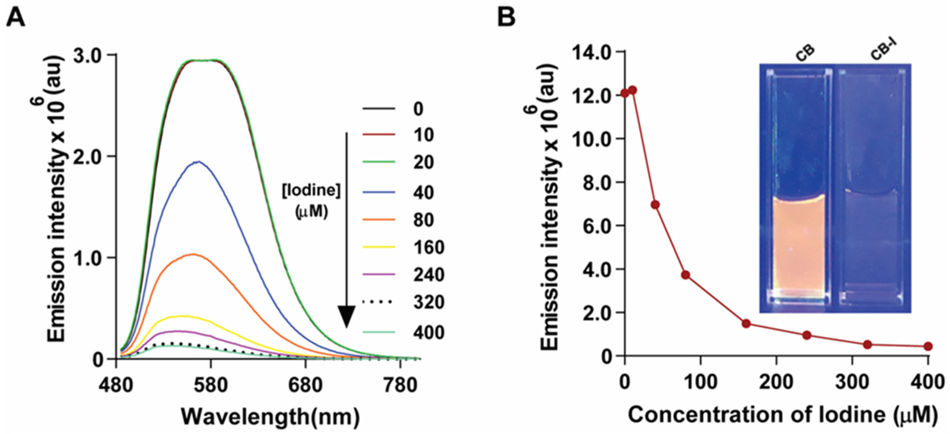
3.1. Effect of Elemental Iodine on CB

The synthesis and AIE behavior of carbazole-barbiturate (CB) chromophore upon aggregation in aqueous solvents in aqueous solutions (fw = 99%) was recently reported in a previous paper [7]. Dynamic light scattering (DLS) measurement of the CB solution at fw = 99% showed the presence of aggregates with a mean radius size of ~77.6 ± 5.09 nm (Figure 2A) and a wide polydispersity of 21.7 ± 1.26%. Previous reports have shown that compounds and polymers with extended π systems (e.g., carbazole-based frameworks) form iodine complexation via charge-transfer or van der Waals interactions [33]. Thus, aqueous solutions of CB with varying concentrations of iodine were prepared from stocks of CB aqueous solutions and DMSO solutions of elemental iodine. The obtained mixtures maintained a constant concentration of CB at 10 µM and fw = 99% with varying concentrations of iodine (10, 20, 40, 80, 160, 240, 320, 400 µM).
DLS analysis on CB solutions with iodine (400 µM) showed the presence of aggregates with a mean radius size of ~56.1 ± 2.73 nm (Figure 2B) and a wide polydispersity of 23.1 ± 1.4%. The addition of iodine to the aqueous solution of CB showed significant changes in the spectroscopic profiles of CB (Figure 3). However, the UV spectra of CB upon addition of iodine showed a red shift of the CB peak at 275 nm, which is attributed predominantly to π–π* transitions localized on the conjugated carbazole backbone, suggesting a strong interaction of iodine with the conjugated system of CB aggregates. In addition, the enhancement in the intensity of the peaks at 320 nm and 450 nm indicates a charge-transfer (CT) complex formation between CB and iodine [34,35]. This interaction is enabled by the low solubility of iodine in water and its high affinity for pi-rich systems.
FTIR spectroscopy (Figure 4) corroborates this mechanism as progressive attenuation of the carbonyl stretch (~1755 cm−1), and the aromatic C=C/C–N band (~1525 cm−1) indicates electron density withdrawal from both barbiturate and carbazole units into iodine. Conversely, the alkyl-associated bending mode (~1384 cm−1) becomes more IR-active, reflecting increased polarizability in the N-alkyl chain as a result of CT interaction [36,37,38]. Taken together, these spectral changes confirm a non-covalent, reversible CT mechanism, rather than chemical modification.
Moreover, minor oxidation of the organic compounds could lead to the formation of iodide ions upon reduction of elemental iodine. The presence of minute amounts of the iodide ions initiates the formation of polyiodides (I3, I5, etc.), which in turn form complexes with pi-rich CB aggregates (CB-I) [39,40,41]. This strong interaction of iodine with CB in CB-I is also reflected in the changes in its emission spectra. CB exhibits a broad emission with a full width at half maximum (FWHM) exceeding 70 nm, a characteristic of AIE systems. This is usually attributed to the charge-transfer character in such donor–acceptor systems, intersystem crossing, and varying aggregation states [16,42].
Figure 5A shows the fluorescence spectrum of CB and the changes upon the addition of iodine solution to the sensor’s solution. Significant quenching of fluorescence was observed as the iodine concentration increased, reaching less than 10% of the original emission upon addition of 40 molar equivalents of iodine (Figure 5B). The quenching of emission was also visually observed under UV light by the naked eye (Figure 5B: inset). Such a quenching behavior by polyiodides of carbazole-based AIE aggregates was previously reported by Zhang and coworkers, and it was attributed to the formation of charge-transfer complexes [22]. These features allow CB to function as a “turn-off” fluorescent sensor for elemental iodine in water. Iodine is an essential micronutrient that plays a crucial role in the production of thyroid hormones, brain development, and various biological functions [43].

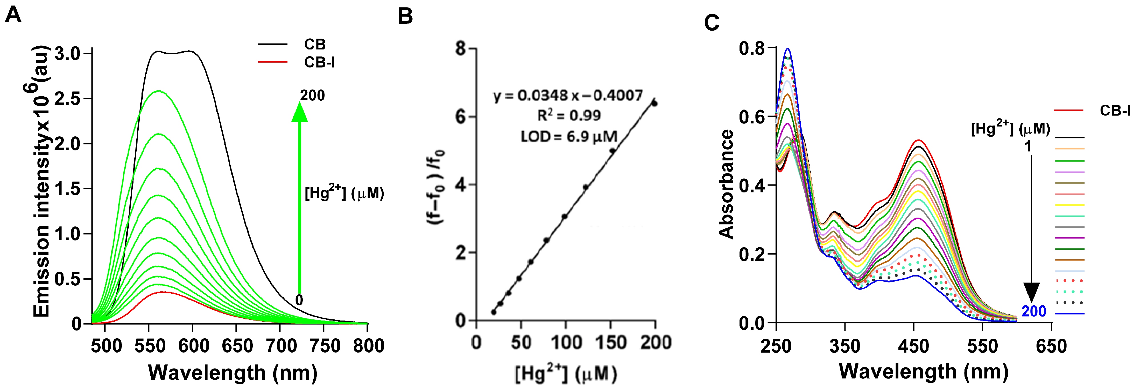
3.2. Effect of Hg2+ on Emission of CB-I

The formation of polyiodide in aqueous solutions can be inhibited by the presence of metal ions such as Hg2+ that bind preferably to the iodide anion [41]. It is well established that the mercury ions can react with polyiodides to form either soluble metal–iodine complexes or insoluble salts [44,45]. Thus, the presence of mercury ions will prevent the interaction of iodine complexation with the organic compounds. Therefore, we proposed that the presence of Hg2+ ions in a solution of CB-I will inhibit the formation of polyiodides that bind to CB, thereby quenching its fluorescence. As a result, the fluorescence of CB will be restored, allowing CB-I to function as a “turn on” fluorescent sensor for Hg2+ ions.
In order to test this hypothesis, we monitored the change in the fluorescence of CB-I solution (an aqueous solution (fw = 99%) of CB at 10 µM and iodine at 400 µM) upon titrating with Hg(NO3)2 solution. As the concentration of Hg2+ (0–200 µM) in CB-I aggregated solution increased, a significant enhancement of the fluorescence of CB-I solution was observed (Figure 6A). The relative change in fluorescence was linearly correlated (Figure 6B) with the increase in the concentration of Hg2+ reaction, reaching ~7 times that of CB-I at [Hg2+] at 200 µM. The fluorescence enhancement was even visually observed, as can be seen from the photographs taken of CB-I solution under UV irradiation (Figure 7A) before and after the addition of Hg2+ ions. The turn-on of emission in the presence of Hg2+ can be attributed to the formation of HgI2 and/or HgI42−, which prevents the formation of CB-I and, hence, eliminates the quenching effect of iodine [41], and possibly to the removal or reorganization of hydration-shell water during complex formation, as hydration dynamics are known to influence emission properties in related systems [46]. This is further supported by the observed changes in the UV-absorption (Figure 6C) spectra of CB-I upon the addition of Hg2+. The changes show a gradual decrease in absorption intensity at 450 nm and an increase in the peak intensity at ~275 nm; hence, retaining the original spectra of CB.

3.3. Effect of Metal Cations on the Fluorescent Detection of Hg2+ Ions

To assess the selectivity of CB-I for Hg2+ ions, we tested the effect of other metals on the fluorescence of CB-I. Thus, the fluorescence spectrum on CB-I solution (10 µM) was recorded in the presence of 10 molar equivalents for each of the following metal ions: Ag+, K+, Na+, Pb2+, Fe3+, Cu2+, Ca2+, Ni2+, Zn2+, Al3+, and Hg2+ (100 µM). As depicted in (Figure 7B), the addition of Hg2+ ions highly enhanced (~5-fold) the fluorescence emission intensity of CB-I by reducing its quenching effect, while the presence of Ag+ and K+ slightly lit up the fluorescence but to a much lower extent than Hg2+ ions. Yet, the presence of other metals had an insignificant effect on the fluorescence intensity of CB-I. The difference in the fluorescence intensity was even visible to the naked eye when comparing the photograph taken under UV irradiation for each metal–CB-I solution (Figure 7A). These results imply that CB-I has a higher sensitivity and selectivity towards Hg2+ ions than the other tested metal ions, especially doubly charged metal ions.
We further investigated the interference of each one of these metal ions on the detection of Hg2+ ions by monitoring the change in the fluorescence of CB-I solution (10 µM) upon addition of 10 molar equivalents of Hg2+ (100 µM) in the presence of a similar concentration (100 µM) of each one of the other metal ions. The results depicted in Figure 7C show that singly charged ions had a positive enhancing effect on that of Hg2+, while doubly charged ions had a negative interference effect on the detection of Hg2+.

4. Conclusions

This paper introduced a carbazole-based compound that forms aggregates in aqueous solutions, exhibiting a large enhancement of fluorescence. The emission of this AIE fluorogen (CB) in aqueous solutions (fw = 99%) was quenched by the presence of 40 molar equivalents of elemental iodine to less than 10%. The fluorescence of the CB-I aggregates was recovered upon the addition of Hg2+ ions into the aqueous medium. The enhancement in the emission of CB-I was linearly correlated with the concentration of Hg2+ ions in solution, indicating that CB-I can be used as an optical probe for detecting and measuring the concentration of Hg2+ ions with LOD = 6.9 µM. The study also showed that other metal ions in solution (K+, Na+, Pb2+, Fe2+, Cu2+, Ca2+, Ni2+, Zn2+, and Al3+) had an insignificant effect on the fluorescence enhancement of CB-I with the exception of Ag+ ions, which exhibited some enhancement but to a much lower extent than that of Hg2+ ions. The presence of any of these ions at high concentrations in solutions, along with Hg2+ ions, however, could slightly interfere with the emission enhancement of CB-I by Hg2+ ions. One limitation of this study is that the selectivity assessment is based on stability constants at room temperature, without accounting for thermodynamic parameters that vary with temperature.

Author Contributions

R.R.: Experiment conduction, draft layout, data collection, analysis of data, writing original draft, manuscript editing; M.S.V.: CB and CB-I characterization experiments, contributed to writing; M.H.A.-S.: Conceptualization, Synthesis of CB, experiments design, guidance, funding acquisition, editing draft. All authors have read and agreed to the published version of the manuscript.

Funding

The study was funded by the American University of Sharjah, UAE (grants: FRG16-R-01 and FRG24-C-S08). This paper represents the opinions of the authors and does not mean to represent the position or opinions of the American University of Sharjah.

Institutional Review Board Statement

Not applicable.

Informed Consent Statement

Not applicable.

Data Availability Statement

The data supporting this study will be provided to readers upon reasonable request.

Acknowledgments

The authors acknowledge the technical support of the BCE Department, the Office of Research, at the American University of Sharjah. The authors acknowledge Ghaleb A. Husseini and Vinod Paul, Department of Chemical and Biological Engineering, for the support given for the DLS facilities.

Conflicts of Interest

The authors declare that there are no known conflicts of interest associated with the study.

References

  1. Keremane, K.S.; Acharya, M.G.; Naik, P.; Malakar, C.C.; Wang, K.; Poudel, B. Recent Advances in Aggregation-Induced Emission (AIE) Fluorescent Sensors for Biomolecule Detection. Chemosensors 2025, 13, 174. [Google Scholar] [CrossRef]
  2. Duo, Y.; Xiang, Z.; Gao, G.; Luo, G.; Tang, B.Z. Biomedical Application of Aggregation-Induced Emission Luminogen-Based Fluorescent Sensors. TrAC Trends Anal. Chem. 2023, 167, 117252. [Google Scholar] [CrossRef]
  3. He, Q.; Wang, M.; Zhao, L.; Xu, B.; Tian, W.; Zhang, L. Near-infrared Aggregation-induced Emission Materials: Bibliometric Analysis and Their Application in Biomedical Field. Aggregate 2024, 5, e505. [Google Scholar] [CrossRef]
  4. Esmaelpourfarkhani, M.; Ramezani, M.; Alibolandi, M.; Abnous, K.; Taghdisi, S.M. Aggregation-Induced Emission-Based Aptasensors for the Detection of Various Targets: Recent Progress. Talanta 2025, 292, 127995. [Google Scholar] [CrossRef] [PubMed]
  5. Ren, K.; Zhang, B.; Guo, J.; Cao, H.; Cheng, J.; Guo, J.; Li, D. Aggregation-Induced Emission(AIE)for next-Generation Biosensing and Imaging: A Review. Biosens. Bioelectron. 2025, 271, 117067. [Google Scholar] [CrossRef] [PubMed]
  6. He, Z.; Ke, C.; Tang, B.Z. Journey of Aggregation-Induced Emission Research. ACS Omega 2018, 3, 3267–3277. [Google Scholar] [CrossRef]
  7. Radha, R.; Vitor, R.F.; Al-Sayah, M.H. A Fluorescence-Based Chemical Sensor for Detection of Melamine in Aqueous Solutions. Chemosensors 2021, 10, 13. [Google Scholar] [CrossRef]
  8. Radha, R.; Al-Sayah, M.H. Development of Liposome-Based Immunoassay for the Detection of Cardiac Troponin I. Molecules 2021, 26, 6988. [Google Scholar] [CrossRef]
  9. Radha, R.; Shahzadi, S.K.; Al-Sayah, M.H. Fluorescent Immunoassays for Detection and Quantification of Cardiac Troponin I: A Short Review. Molecules 2021, 26, 4812. [Google Scholar] [CrossRef]
  10. Zha, B.; Li, H.; Ren, S.; Wu, J.-R.; Wang, H. Aggregation-Induced Emission (AIE) Probes in Fluorescent Sensing: Progress and Applications for Pesticide Detection. Appl. Sci. 2024, 14, 8947. [Google Scholar] [CrossRef]
  11. Yang, L.; Xiong, L.-H.; He, X. Applications of Aggregation-Induced Emission Materials in Immunology: From Diagnostics to Immunotherapy. Chem. Biomed. Imaging 2025, cbmi.5c00016. [Google Scholar] [CrossRef]
  12. Asthana, S.; Mouli, M.S.S.V.; Tamrakar, A.; Wani, M.A.; Mishra, A.K.; Pandey, R.; Pandey, M.D. Recent Advances in AIEgen-Based Chemosensors for Small Molecule Detection, with a Focus on Ion Sensing. Anal. Methods 2024, 16, 4431–4484. [Google Scholar] [CrossRef]
  13. Zeng, Y.; Huang, D.; Li, B.; Wang, J.; Hu, R. AIE Polymers for Biosensing Applications. Polym. Chem. 2025, 16, 1897–1928. [Google Scholar] [CrossRef]
  14. Zhang, Y.; Huang, Y.; Miao, R.; Chen, H. Inorganic-Based Aggregation-Induced Luminescent Materials: Recent Advances and Perspectives. Small Struct. 2023, 4, 2300157. [Google Scholar] [CrossRef]
  15. Huang, Y.; Xing, J.; Gong, Q.; Chen, L.-C.; Liu, G.; Yao, C.; Wang, Z.; Zhang, H.-L.; Chen, Z.; Zhang, Q. Reducing Aggregation Caused Quenching Effect through Co-Assembly of PAH Chromophores and Molecular Barriers. Nat. Commun. 2019, 10, 169. [Google Scholar] [CrossRef] [PubMed]
  16. Zhu, C.; Kwok, R.T.K.; Lam, J.W.Y.; Tang, B.Z. Aggregation-Induced Emission: A Trailblazing Journey to the Field of Biomedicine. ACS Appl. Bio Mater. 2018, 1, 1768–1786. [Google Scholar] [CrossRef]
  17. Diana, R.; Panunzi, B. Zinc (II) and AIEgens: The “Clip Approach” for a Novel Fluorophore Family. A Review. Molecules 2021, 26, 4176. [Google Scholar] [CrossRef]
  18. Diana, R.; Caruso, U.; Gentile, F.S.; Di Costanzo, L.; Panunzi, B. A Novel L-Shaped Fluorescent Probe for AIE Sensing of Zinc (II) Ion by a DR/NIR Response. Molecules 2021, 26, 7347. [Google Scholar] [CrossRef]
  19. Bhardwaj, V.; Nurchi, V.M.; Sahoo, S.K. Mercury Toxicity and Detection Using Chromo-Fluorogenic Chemosensors. Pharmaceuticals 2021, 14, 123. [Google Scholar] [CrossRef]
  20. Jagadhane, K.S.; Bhosale, S.R.; Gunjal, D.B.; Nille, O.S.; Kolekar, G.B.; Kolekar, S.S.; Dongale, T.D.; Anbhule, P.V. Tetraphenylethene-Based Fluorescent Chemosensor with Mechanochromic and Aggregation-Induced Emission (AIE) Properties for the Selective and Sensitive Detection of Hg2+ and Ag+ Ions in Aqueous Media: Application to Environmental Analysis. ACS Omega 2022, 7, 34888–34900. [Google Scholar] [CrossRef]
  21. Zhang, Y.; Xiong, T.; Möslein, A.F.; Mollick, S.; Kachwal, V.; Babal, A.S.; Amin, N.; Tan, J.-C. Nanoconfinement of Tetraphenylethylene in Zeolitic Metal-Organic Framework for Turn-on Mechanofluorochromic Stress Sensing. Appl. Mater. Today 2022, 27, 101434. [Google Scholar] [CrossRef]
  22. Zhang, R.X.; Li, P.F.; Zhang, W.J.; Li, N.; Zhao, N. A Highly Sensitive Fluorescent Sensor with Aggregation-Induced Emission Characteristics for the Detection of Iodide and Mercury Ions in Aqueous Solution. J. Mater. Chem. C 2016, 4, 10479–10485. [Google Scholar] [CrossRef]
  23. Muqadas; Shabbir, A.; Khan, M.; Assiri, M.A.; Khan, A.M.; Shahzad, S.A. Synthesis and Construction of AIE Based Fluorescent Sensor for Highly Selective Solid Phase Sensing of 4-Nitroaniline: Solvatochromism, Acidochromism, Mechanochromism and Applications in Latent Fingerprints and Invisible Ink. Microchem. J. 2025, 214, 114129. [Google Scholar] [CrossRef]
  24. Yin, G.; Ma, Y.; Xiong, Y.; Cao, X.; Li, Y.; Chen, L. Enhanced AIE and Different Stimuli-Responses in Red Fluorescent (1,3-Dimethyl)Barbituric Acid-Functionalized Anthracenes. J. Mater. Chem. C 2016, 4, 751–757. [Google Scholar] [CrossRef]
  25. Wu, Y.-S.; Osman, A.I.; Hosny, M.; Elgarahy, A.M.; Eltaweil, A.S.; Rooney, D.W.; Chen, Z.; Rahim, N.S.; Sekar, M.; Gopinath, S.C.B.; et al. The Toxicity of Mercury and Its Chemical Compounds: Molecular Mechanisms and Environmental and Human Health Implications: A Comprehensive Review. ACS Omega 2024, 9, 5100–5126. [Google Scholar] [CrossRef] [PubMed]
  26. Kumar, V.; Umesh, M.; Shanmugam, M.K.; Chakraborty, P.; Duhan, L.; Gummadi, S.N.; Pasrija, R.; Jayaraj, I.; Dasarahally Huligowda, L.K. A Retrospection on Mercury Contamination, Bioaccumulation, and Toxicity in Diverse Environments: Current Insights and Future Prospects. Sustainability 2023, 15, 13292. [Google Scholar] [CrossRef]
  27. Azmi, H.; Melles, S.J.; Laursen, A.E. Does Selenium Offset Methylmercury Toxicity across Trophic Levels in a Primary Producer, Auxenochlorella Pyrenoidosa, and a Detritovore, Aeolosoma Variegatum? FACETS 2025, 10, 1–13. [Google Scholar] [CrossRef]
  28. Charkiewicz, A.E.; Omeljaniuk, W.J.; Garley, M.; Nikliński, J. Mercury Exposure and Health Effects: What Do We Really Know? Int. J. Mol. Sci. 2025, 26, 2326. [Google Scholar] [CrossRef]
  29. Balali-Mood, M.; Naseri, K.; Tahergorabi, Z.; Khazdair, M.R.; Sadeghi, M. Toxic Mechanisms of Five Heavy Metals: Mercury, Lead, Chromium, Cadmium, and Arsenic. Front. Pharmacol. 2021, 12, 643972. [Google Scholar] [CrossRef]
  30. Silbergeld, E.; Silva, I.; Nyland, J. Mercury and Autoimmunity: Implications for Occupational and Environmental Health. Toxicol. Appl. Pharmacol. 2005, 207, 282–292. [Google Scholar] [CrossRef]
  31. Kang, B.; Wang, J.; Guo, S.; Yang, L. Mercury-Induced Toxicity: Mechanisms, Molecular Pathways, and Gene Regulation. Sci. Total Environ. 2024, 943, 173577. [Google Scholar] [CrossRef]
  32. Sanchez, J.M. Estimating Detection Limits in Chromatography from Calibration Data: Ordinary Least Squares Regression vs. Weighted Least Squares. Separations 2018, 5, 49. [Google Scholar] [CrossRef]
  33. Chen, T.; Zhang, W.; Li, B.; Huang, W.; Lin, C.; Wu, Y.; Chen, S.; Ma, H. Adsorptive Separation of Aromatic Compounds from Alkanes by π–π Interactions in a Carbazole-Based Conjugated Microporous Polymer. ACS Appl. Mater. Interfaces 2020, 12, 56385–56392. [Google Scholar] [CrossRef]
  34. Refat, M.S.; El-Didamony, A.M.; Grabchev, I. UV–Vis, IR Spectra and Thermal Studies of Charge Transfer Complex Formed between Poly(Amidoamine) Dendrimers and Iodine. Spectrochim. Acta. A. Mol. Biomol. Spectrosc. 2007, 67, 58–65. [Google Scholar] [CrossRef] [PubMed]
  35. Pullen, S.; Walker, L.A.; Sension, R.J. Femtosecond Studies of the Iodine–Mesitylene Charge-Transfer Complex. J. Chem. Phys. 1995, 103, 7877–7886. [Google Scholar] [CrossRef]
  36. Wang, X.; Yu, C.; Guo, H.; Cheng, Y.; Li, Y.; Zheng, D.; Feng, S.; Lin, Y. Robust Fluorescent Detection of Iodine Vapor by a Film Sensor Based on a Polymer of Intrinsic Microporosity. Chem. Eng. J. 2022, 438, 135641. [Google Scholar] [CrossRef]
  37. Li, Q.; Guo, W.; Wang, Z.; Tan, L.; Shang, L. Tailoring Electron-Rich Fluorescent Supramolecular Organic Frameworks for Efficient Capture and Visual Monitoring of Iodine. Adv. Funct. Mater. 2025, 35, 2413694. [Google Scholar] [CrossRef]
  38. Ciureanu, M. Infrared Studies of Iodine Complexes with Several Aromatic Amines. Adv. Mol. Relax. Interact. Process. 1979, 15, 25–35. [Google Scholar] [CrossRef]
  39. Calabrese, V.T.; Khan, A. Polyiodine and Polyiodide Species in an Aqueous Solution of Iodine + KI: Theoretical and Experimental Studies. J. Phys. Chem. A 2000, 104, 1287–1292. [Google Scholar] [CrossRef]
  40. Mhatre, A.M.; Chappa, S.; Ojha, S.; Pandey, A.K. Functionalized Glass Fiber Membrane for Extraction of Iodine Species. Sep. Sci. Technol. 2019, 54, 1469–1477. [Google Scholar] [CrossRef]
  41. Nashukha, H.L.; Sitanurak, J.; Sulistyarti, H.; Nacapricha, D.; Uraisin, K. Simple and Equipment-Free Paper-Based Device for Determination of Mercury in Contaminated Soil. Molecules 2021, 26, 2004. [Google Scholar] [CrossRef]
  42. Liu, W.; Zhang, S.; Jiang, T.; Li, S.; Xie, Z.; Zhao, Y.; Zhang, J.; Redshaw, C.; Feng, X.; Lam, J.W.Y.; et al. Pyrene-Based Blue Aggregation-Induced Emission Luminogens: The Synergistic Effect of Through-Space Conjugation for High Exciton Utilization Efficiency and Narrow-Band and Blue OLEDs. Adv. Opt. Mater. 2025, 13, 2402567. [Google Scholar] [CrossRef]
  43. Cho, E.; Perebikovsky, A.; Benice, O.; Holmberg, S.; Madou, M.; Ghazinejad, M. Rapid Iodine Sensing on Mechanically Treated Carbon Nanofibers. Sensors 2018, 18, 1486. [Google Scholar] [CrossRef] [PubMed]
  44. Qu, W.; Liu, J.; Shen, F.; Wei, P.; Lei, Y. Mechanism of Mercury-Iodine Species Binding on Carbonaceous Surface: Insight from Density Functional Theory Study. Chem. Eng. J. 2016, 306, 704–708. [Google Scholar] [CrossRef]
  45. Bayindir, S.; Hussein, A.S.; Lafzi, F.; Toprak, M. The Switchable Fluorescent Detection of Mercury, Copper, and Iodine in Real Water Samples Using Bola-Amphiphilic-Tetraphenylethene. J. Mol. Liq. 2023, 382, 121939. [Google Scholar] [CrossRef]
  46. Zheng, Y.; Li, Q.; Wang, C.; Su, M. Enhanced Turn-On Fluorescence Detection of Aqueous Lead Ions with Size-Shrinkable Hydrogels. ACS Omega 2021, 6, 11897–11901. [Google Scholar] [CrossRef]
Figure 1. The chemical structure of the AIE fluorophore CB.
Chemosensors 13 00276 g001
Figure 2. Dynamic light scattering measurements of sensor aggregates in DMSO/water mixture with fw = 99%. Size distribution pattern of CB aggregates in absence (A) and the presence (B) of iodine (400 µM).
Chemosensors 13 00276 g002
Figure 3. UV-vis absorption spectra of CB in presence of iodine (0–400 µM).
Chemosensors 13 00276 g003
Figure 4. FTIR of CB and CB-I drop-cast films at different concentrations of iodine; the highlighted signals reflect the nature of interactions between CB and iodine.
Chemosensors 13 00276 g004
Figure 5. Fluorescence quenching effect of iodine on sensor CB. (A) Emission spectra of CB (10 µM) in aqueous solution (with 1% DMSO) in presence of iodine (0–400 µM). (B) Plot of total fluorescence emission calculated for Figure 5B data versus the concentration of I (0–400 µM), excitation wavelength: 465 nm. Inset: photographs of CB (10 µM in 1% DMSO) in the absence and presence of iodine (400 µM) visualized under UV irradiation.
Chemosensors 13 00276 g005
Figure 6. Emission and absorbance properties of CB-I in presence of Hg2+. (A) Emission spectra of CB-I in aqueous solution with 1%DMSO in the presence of Hg2+ (0–200 µM), excitation wavelength of 465 nm. (B) Linear fit for (ff0/f0) versus concentration of Hg2+, where f0 is the fluorescence emission of CB-I in absence of Hg2+ and f is fluorescence emission of CB-I in presence of varying levels of Hg2+ (20–200 µM). (C) UV-vis absorption spectra of CB-I in aqueous solution with 1%DMSO in the presence of Hg2+ (0–200 µM).
Chemosensors 13 00276 g006
Figure 7. Fluorescence assay on CB-I in presence of metal cations. (A) Photographs of CB (10 µM in 1% DMSO) in the presence of different metal ions (100 µM) under UV irradiation. (B) Emission spectra of iodine-quenched aqueous solution of CB (10 µM in 1% DMSO) in the presence of different metal ions (100 µM). (C) Bar plot showing the comparison between the fluorescence effect of CB-I in the presence of different metal ions and the increase in fluorescence upon the addition of Hg2+.
Chemosensors 13 00276 g007
Disclaimer/Publisher’s Note: The statements, opinions and data contained in all publications are solely those of the individual author(s) and contributor(s) and not of MDPI and/or the editor(s). MDPI and/or the editor(s) disclaim responsibility for any injury to people or property resulting from any ideas, methods, instructions or products referred to in the content.

