Article

Design and Synthesis of Arylboronic Acid Chemosensors for the Fluorescent-Thin Layer Chromatography (f-TLC) Detection of Mycolactone

by
Gideon Atinga Akolgo
1,
Benjamin M. Partridge
2,
Timothy D. Craggs
2,
Kingsley Bampoe Asiedu
3 and
Richard Kwamla Amewu
1,*
1
Department of Chemistry, University of Ghana, Legon-Accra P.O. Box LG56, Ghana
2
School of Physical and Mathematical Sciences, Dainton Building, University of Sheffield, Sheffield S3 7HF, UK
3
Department of Neglected Tropical Diseases, World Health Organization, 1211 Geneva, Switzerland
*
Author to whom correspondence should be addressed.
Chemosensors 2025, 13(7), 244; https://doi.org/10.3390/chemosensors13070244
Submission received: 27 February 2025 / Revised: 27 March 2025 / Accepted: 7 April 2025 / Published: 9 July 2025

Abstract

Fluorescent chemosensors are increasingly becoming relevant in recognition chemistry due to their sensitivity, selectivity, fast response time, real-time detection capability, and low cost. Boronic acids have been reported for the recognition of mycolactone, the cytotoxin responsible for tissue damage in Buruli ulcer disease. A library of fluorescent arylboronic acid chemosensors with various signaling moieties with certain beneficial photophysical characteristics (i.e., aminoacridine, aminoquinoline, azo, BODIPY, coumarin, fluorescein, and rhodamine variants) and a recognition moiety (i.e., boronic acid unit) were rationally designed and synthesised using combinatorial approaches, purified, and fully characterised using a set of complementary spectrometric and spectroscopic techniques such as NMR, LC-MS, FT-IR, and X-ray crystallography. In addition, a complete set of basic photophysical quantities such as absorption maxima (λabsmax), emission maxima (λemmax), Stokes shift (∆λ), molar extinction coefficient (ε), fluorescence quantum yield (ΦF), and brightness were determined using UV-vis absorption and fluorescence emission spectroscopy techniques. The synthesised arylboronic acid chemosensors were investigated as chemosensors for mycolactone detection using the fluorescent-thin layer chromatography (f-TLC) method. Compound 7 (with a coumarin core) emerged the best (λabsmax = 456 nm, λemmax = 590 nm, ∆λ = 134 nm, ε = 52816 M−1cm−1, ΦF = 0.78, and brightness = 41,197 M−1cm−1).

1. Introduction

Over the years, researchers, inspired by certain biological processes in nature, looked for new concepts that allow for the selective molecular recognition of target molecules that play important roles in complex chemical processes in various disciplines such as medicine, biology, agriculture, biomedicine, environment, and social sciences [1]. Molecular recognition is achieved by the utility of chemosensors. Chemosensors are molecules of abiotic origin that signal the presence of matter or energy. A fluorescent chemosensor, therefore, is a compound that interacts with an analyte in such a way as to produce a detectable fluorescent signal [2]. Fluorescent chemosensors have been of particular interest in supramolecular chemistry because of their ability to recognise guest species as well as their high selectivity, sensitivity, and simplicity [3,4]. Generally, a fluorescent chemosensor is composed of a recognition moiety that interacts with an analyte of interest and relays the recognition event through a signalling unit. Thus, the engineering of an appropriate fluorescent chemosensor with the right recognition and signalling moieties can result in favourable interactions with specific analytes.
A boronic acid motif can be explored as a recognition unit in the design of chemosensors to exploit the 1,3-diol moieties on the structure of the analyte. This is because boronic acids and their derivatives have been known for decades to form five- or six-membered cyclic boronate esters with both cis-1,2- and 1,3-diols that are present on biologically relevant molecules through covalent bonding [5,6,7,8,9,10]. The phenomenon of the complexation of boronic acids with polyols dates to over 70 years but remains an area of interest. The first reported use of boronic acids as recognition units for cis-1,2 or 1,3-diol-containing compounds was published by Lorand and Edwards in 1959 [11]. They demonstrated the complexation equilibria of aqueous benzeneboronate ion with several polyols and compared the association constants to that of borate. Following the early seminal work of Lorand and Edwards, boronic acids have greatly evolved into an area of research for molecular recognition, thus gaining significant interest in the design of many fluorescent chemosensors. This is because of their selective binding behaviour for a considerable number of biological molecules possessing polyhydroxy motifs, their high stability, environmental friendliness, general low toxicity, and their relatively inexpensive nature. The first fluorescent boronic-acid-based sensor was developed later in 1992 by Yoon and Czarnik for saccharide sensing [12]. Since then, boronic acids have attracted considerable interest in molecular recognition, in which the boronic acid functionality has increasingly become a very important recognition moiety in the design and synthesis of molecular recognition chemosensors, taking advantage of this unique intrinsic affinity for cis-1,2 and/or 1,3-diols. The selective binding behaviour of the boronic acid unit has been explored for the detection of diol-containing analytes based on the “lock-and-key” concept of enzyme-substrate recognition [1]. The unique properties of boronic acid functionality have positioned it as an invaluable receptor and have shown great promise for the specific recognition and detection of various species in real-world situations [13]. They have become popular as sensors for various biological polyol molecules including monosaccharides and in particular glucose [14], catecholamines [15], and dopamine. Others include various polysaccharides, glycoproteins, and glycated haemoglobin [16,17,18,19,20,21], ionic compounds [22,23,24], anions such as cyanides and fluoride [9,25,26,27], water traces in solvents [28,29], and hydrogen peroxide (H2O2) [30,31]. Furthermore, boronic acids have also been employed as biochemical tools for a variety of applications, including but not limited to enzyme inhibitors [32], cell delivery methods [33], whole-body diagnostic imaging [34], and tumour cell imaging [35,36].
The pathogenesis of Mycobacterium ulcerans disease is widely dependent on the activity of a unique toxin called mycolactone [37]. Mycolactone 1, plays a significant role in the virulence, pathogenicity, and cytotoxicity of Buruli ulcer (BU) disease. It has immunosuppressive properties and is responsible for the tissue damage seen in BU [38,39]. Structurally, mycolactone A/B is a polyketide macrolide toxin composed of a 12-membered macrocyclic lactone core (C1–C11) with two laterally attached side chains; a C-linked northern side chain (C12–C20) with two hydroxyl groups in 1,3-diol positions at C17 and C19; and an esterified C5-O-linked highly polyunsaturated acyl southern side chain (C1’–C16’) possessing a conjugated pentaenoic acid ester chromophore and three hydroxyl groups, two of which are in 1,3-diol positions at C13’ and C15’ [40,41] (Figure 1).
The secretion of mycolactone is a distinguishing feature of M. ulcerans amongst other human mycobacteria. According to Hong et al., mycolactone A/B appears to be biosynthetically limited to M. ulcerans, uniformly distributed inside the infected tissue, and demonstrated to diffuse beyond the sites of primary infection. For these reasons, mycolactone-specific detection is crucial for the diagnosis of BU [43]. Histopathological studies have shown that in tissues, mycolactone is widely distributed compared to the causative organism [44]. Consequently, the detection of its presence in biological samples is necessary for the diagnosis of the disease. Based on these findings, Kishi and colleagues developed a sensitive and practical flourescent-thin layer chromatographine (f-TLC) technique for detecting the toxin. The diagnostic method utilises the derivatisation of diol motifs of mycolactone A/B using a 2-naphthylboronic acid that forms six-membered cyclic boronate esters, which can be detected on a TLC plate as an adduct (Figure 2). Accordingly, the 2-naphthylboronic acid and mycolactone work as an acceptor–donor FRET pair. The boronic acid unit reacts selectively with 1,3-diol(s) moieties of mycolactone to form boronate esters, significantly enhancing fluorescent intensity. The proposed general mechanism involves the FRET process, where a donor, in this case the pentaenoate chromophore of mycolactone, is excited at a defined wavelength (365 nm), and the energy is transferred, via non-radiative dipole–dipole interactions, to a properly selected acceptor (the 2-naphthylboronic acid), which emits through radiative relaxation. The free 2-naphthylboronic acid with the lowest (np*) excited singlet state (S1) has the lower absorptivity and so it is weakly fluorescent. On the other hand, the hybridisation of boron atoms plays a secondary role in affecting fluorescence intensity. Therefore, binding with mycolactone at the right temperature results in the formation of the cyclic boronate, in which the hybridisation of boron atom changes from sp2 to sp3, has the lowest (pp*)-excited singlet state, and results in a large enhancement of fluorescence emission (Figure 2).
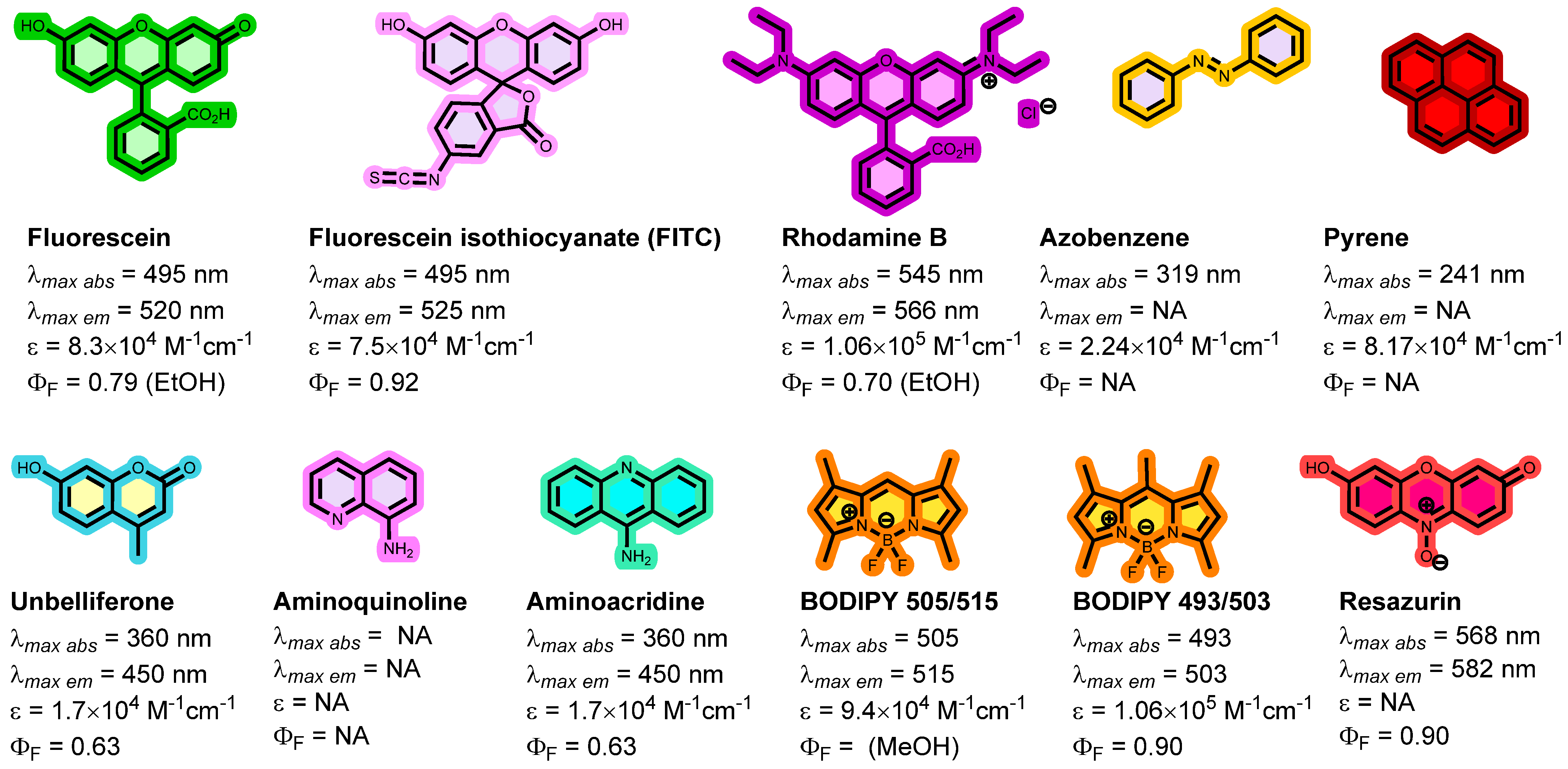
This idea led to research into alternative commercially available boronic acids, and it was discovered that 9,9-Diphenyl-9H-fluoren-4-yl)boronic acid (BA18) could be a viable substitute for 2-naphthylboronic acid (BA) [45]. The f-TLC method holds great promise because it is relatively cheap, simple to use, and rapid. [42,46,47]. Although the method has been successfully evaluated using clinical samples [48,49], it is hampered by the fact that it is non-quantitative. There are also concerns about interferences from other co-extracted and co-eluted lipids which make reading the results of the method subjective [48]. There is a need to improve upon the low detection and quantification rates as well as discriminate the mycolactone from other autofluorescent co-extracted human tissues. This study aimed to address these principal challenges associated with the f-TLC technique for the diagnosis of Buruli ulcers. To the best of our knowledge, no chemosensor has been designed for the molecular recognition of mycolactone. Considering the broad application of boron-based fluorescence sensors, our goal was to incorporate the boronic acid motif into various fluorescent dyes as chemosensors for mycolactone detection. Considering the unique structure of mycolactone, we hypothesised that boronate ester formation between the diol moieties of mycolactone and a boronic acid binding site attached to a fluorescent molecule would allow for signal transduction that could be exploited for mycolactone detection. Therefore, we employed various fluorescent dyes (fluorophores) such as 9-aminoacridine, 8-aminoquinoline, azo, BODIPYs, coumarins, fluorescein, and rhodamine dyes, which are all amenable to boronic acid tethering to design, synthesise, and characterise various fluorescent arylboronic acid dye chemosensors. These are already well-known fluorophores with excellent photophysical properties including large excitation and emission maxima, extinction coefficient, high fluorescence quantum yield (Φ), large Stokes shift, high brightness, adequate solubility, good thermal and photostability, and sufficient chemical stability [50,51,52] (Figure 3). Secondly, these dyes are commercially available and can be coupled to diverse functional groups.

2. Materials and Methods

2.1. Reagents and Instruments

Standard laboratory apparatus and glassware were used for all reactions. Unless otherwise specified, all solvents and reagents were used as supplied by commercial vendors without further purification. All air-sensitive reactions were conducted in oven-dried apparatus under nitrogen atmosphere. Aluminium-backed Merck silica gel 60 F254 plates pre-coated with silica were used for thin-layer chromatography (TLC). Plates were visualised using either ultraviolet light of 254 nm or 365 nm or by dipping the plates into solutions of vanillin, ninhydrin, or potassium permanganate followed by heating. All column chromatography was performed under ‘flash’ conditions using silica gel mesh 40–63. Fourier transform infrared (FT-IR) spectra were recorded on a Perkin Elmer 100 FT-IR instrument on the neat compounds. The spectrum was processed in the software and displayed. The relevant and characteristic absorptions were reported in wavenumbers (cm−1), with the intensities of the bands recorded as broad (b), strong (s), medium (m), and weak (w). NMR spectroscopy was carried out using Brüker Avance III 400 and 500 MHz spectrometers. 1H NMR spectra, 13C NMR spectra, and 11B NMR were obtained at the indicated 101, 126, 128, 377, 400, and 500 MHz as dilute solutions in various deuterated solvents at room temperature. All spectra were recorded in deuterated dimethyl sulfoxide (DMSO-d6) or deuterated chloroform (CDCl3) obtained from Sigma Aldrich. Chemical shifts (δ) are reported in parts per million (ppm) relative to the following residual solvent peaks (δH: CHCl3 = 7.26 ppm, DMSO-d6 = 2.50 ppm) or the solvent itself (δC: CDCl3 = 77.0 ppm, DMSO-d6 = 39.52 ppm). For 11B NMR, BF3·OEt2 (δ = 0 ppm) was used as an external standard. Coupling constants (J) were measured in hertz (Hz). Multiplicities are reported as singlet (s), broad singlet (br s), doublet (d), doublet triplet (dt), triplet doublet (td), doublet doublet of doublets of doublets (ddd), quartet (q), broad quartet (br q), or multiplet (m). High-resolution mass spectra were recorded using a 1260 Infinity LC (Agilent Technologies), coupled to a Quadrupole–Time of Flight tandem mass spectrometer 6530 Infinity Q-ToF detector (Agilent Technologies) by a Jet Stream ESI interface (Agilent Technologies). High-resolution MS spectra were acquired in the positive mode in the 100–2400 m/z range. Single crystal X-ray Diffraction (SCXRD) data were collected on a Bruker D8 VENTURE diffractometer (Bruker AXS, Karlsruhe, Germany), equipped with a PHOTON 100 CMOS detector with graphite-monochromatised Cu-Kα radiation (λ = 1.54178 Å) or a Bruker X8 Apex-II diffractometer, equipped with an Apex-II CCD area detector diffractometer (Bruker, Karlsruhe, Germany) with graphite-monochromatised Mo-Kα radiation (λ = 0.71073 Å). A suitable crystal was mounted in Fomblin oil on a MiTeGen MicroLoop and cooled in a stream of cold N2 to 100 K. Data were corrected for absorption using empirical methods (SADABS) [53,54,55] based upon symmetry-equivalent reflections combined with measurements at different azimuthal angles. All crystal structures were solved and refined against F2 values using ShelXT [56] for the solution and ShelXL [57] for refinement, accessed via the Olex2 program [58]. All non-hydrogen atoms were refined with anisotropic displacement parameters. All hydrogen atoms were added at calculated positions and refined with a riding model and isotropic displacement parameters fixed in magnitude relative to the attached carbon atoms. Details regarding the structures and a summary table of crystallographic and data collection parameters are provided in Electronic Supplementary Information (Table S1).

2.2. Synthesis and Characterisation

Synthesis of 4-(4,4,5,5-tetramethyl-1,3,2-dioxaborolan-2-yl)benzaldehyde 2: Using a variation of the procedure of Miyaura et al. [59], a mixture of 4-bromobenzaldehyde (1.0 g, 5.4 mmol, 1.0 equiv.), B2pin2 (1.65 g, 6.5 mmol, 1.2 equiv.), KOAc (1.59 g, 16.2 mmol, 3.0 equiv.), and PdCl2(dppf) (119 mg, 0.162 mmol) in dry 1,4-dioxane (20 mL) in an oven-dried two-necked round-bottom flask was degassed for 5–10 min using N2 gas and stirred at 80 °C for 16 h (overnight). Upon completion (as monitored by TLC), the reaction mixture was allowed to cool, filtered through a plug of celite, washed with EtOAc (200 mL), extracted twice with water (2 × 50 mL), and then with brine. The organic layer was dried with anhydrous MgSO4, filtered, and concentrated in vacuo. The crude product was purified on silica gel using DCM (100%) to obtain product 2 as a white solid (872 mg, 70% yield). 1H NMR (400 MHz, CDCl3) δH 10.02 (s, 1H, H1), 7.94 (d, J = 8.2 Hz, 2H, H5), 7.83 (d, J = 8.2 Hz, 2H, H4), 1.33 (s, 12H, H8). 13C NMR (101 MHz, CDCl3) δC 192.9 (C2), 138.4 (C3), 135.6 (C4), 129.0 (C5), 84.7 (C7), 25.2 (C8) (C6 bonded to B not observed due to broadening). LC-MS: retention time: 9.4 min; ESI-QTOF HRMS (m/z): for [C13H18BO3]+ [M + H]+: exact mass calcd.: 233.1349; found: 233.1358; for [C13H17BO3Na]+ [M + Na]+: exact mass calcd.: 255.1168; found: 255.1172.
1-(4-(4,4,5,5-tetramethyl-1,3,2-dioxaborolan-2-yl)phenyl)ethan-1-one 3: This title compound was prepared as described above for 2 using 4-bromoacetophenone (5.0 g, 25.1 mmol, 1.0 equiv.), and B2pin2 (7.65 g, 30.1 mmol, 1.2 equiv.), KOAc (7.39 g, 75.3 mmol, 3.0 equiv.), PdCl2(dppf) (551 mg, 0.75 mmol) in 1,4-dioxane (20 mL). The crude product was purified on silica gel using hexane/ethyl acetate = 50:1 (V:V) to obtain product 3 as a white solid (6.15 g, 96% yield). 1H NMR (500 MHz, CDCl3) δH 7.90 (d, J = 6.3 Hz, 2H, H4), 7.87 (d, J = 6.3 Hz, 2H, H5), 2.58 (s, 3H, H1), 1.33 (s, 12H, H8). 13C NMR (126 MHz, CDCl3) δC 198.7 (C2), 139.3 (C3), 135.2 (C5), 127.6 (C4), 84.5 (C7), 27.0 (C1), 25.2 (C8) (C6 bonded to B not observed due to broadening).

2.3. Categories of Fluorescent Arylboronic Acid Chemosensor Dyes

2.3.1. Coumarin Dyes

Synthesis of 7-(diethylamino)-2H-chromen-2-one 4: A mixture of 4-(diethylamino) salicylaldehyde (6.0 g, 28.69 mmol) and diethyl malonate (10.84 g, 67.75 mmol) in 90 mL anhydrous ethanol was treated with 3 mL of piperidine and then refluxed for 13 h. After removing the solvent, 120 mL of mixed solution (concentrated HCl:glacial acetic acid = 1:1 (V:V)) was added to the crude product and refluxed for another 7 h. After cooling the reaction solution to room temperature, it was transferred into 300 mL of water and the pH was adjusted to 5 using NaOH. The solid was filtered and purified over silica gel (hexane: ethyl acetate = 9:1 (V:V) to give a beige solid 4 (yield: 85%). 1H NMR (500 MHz, CDCl3) δH 7.53 (d, J = 9.3 Hz, 1H, H9), 7.23 (d, J = 8.7 Hz, 1H, H5), 6.56 (dd, J = 8.8, 2.5 Hz, 1H, H4), 6.46 (d, J = 2.5 Hz, 1H, H6), 6.01 (d, J = 9.3 Hz, 1H, H10), 3.40 (q, J = 7.2 Hz, 4H, H2), 1.20 (t, J = 7.2 Hz, 6H, H1). 13C NMR (126 MHz, CDCl3) δC 162.4 (C11), 156.7 (C3), 150.7 (C7), 143.8 (C9), 128.8 (C5), 109.0 (C10), 108.7 (C8), 108.3 (C4), 97.4 (C6), 44.8 (C2), 12.4 (C1).
Synthesis of 7-(diethylamino)-2-oxo-2H-chromene-3-carbaldehyde 5: Under N2, anhydrous DMF (4.20 g, 57.5 mmol, 4.4 mL, 12.5 equiv.) was dropped into phosphoryl chloride (POCl3) (1.76 g, 11.5 mmol, 1.1 mL, 2.5 equiv.) with stirring for 6 h in an ice bath. The solution of 7-(diethylamino)-2H-chromen-2-one 4 (1.00 g, 4.6 mmol, 1.0 equiv.) in anhydrous 1,2-dichloroethane (50 mL) was added to the above solution, and the mixture was stirred at 60 °C for 12 h. After completing the process, the mixture was poured into ice water and neutralised with NaOH solution (20%) to pH 7. The formed precipitate was filtered off and washed three times with water. The residue was chromatographed on silica, eluting with ethyl acetate/hexane (1:1, V:V) to form 5 as an orange-red solid product (yield: 79%). 1H NMR (500 MHz, CDCl3) δH 10.10 (s, 1H, H13), 8.23 (s, 1H, H9), 7.39 (d, J = 9.0 Hz, 1H, H5), 6.63 (dd, J = 9.0, 2.5 Hz, 1H, H4), 6.47 (d, J = 2.5 Hz, 1H, H6), 3.46 (q, J = 7.2 Hz, 4H, H2), 1.24 (t, J = 7.1 Hz, 6H, H1). 13C NMR (126 MHz, CDCl3) δC 188.0 (C12), 162.0 (C11), 159.0 (C3), 153.6 (C7), 145.5 (C9), 132.6 (C5), 114.5 (C10), 110.3 (C8), 108.4 (C4), 97.3 (C6), 45.4 (C2), 12.6 (C1). FTIR (cm−1): υ (C–H st) 2879, υ (C=O st, carboxyl) 1709, υ (C=C st) 1609.
Synthesis of (E)-7-(diethylamino)-3-(3-oxo-3-(4-(4,4,5,5-tetramethyl-1,3,2-dioxaborolan-2-yl)phenyl)prop-1-en-1-yl)-2H-chromen-2-one 6: compound 5 (0.50 g, 2.0 mmol, 1.0 equiv.) and 1-(4-(4,4,5,5-tetramethyl-1,3,2-dioxaborolan-2-yl)phenyl)ethan-1-one 3 (0.98 g, 4.0 mmol, 2.0 equiv.) were added to 20 mL of the mixed solvent (CH2Cl2/anhydrous CH3CH2OH = 1:1 (V:V)), then 10 drops of pyrrolidine was dropped into the above solution. The mixture was stirred at room temperature for 4 d, and the solvent was removed under reduced pressure. The residue was added to 40 mL of hexane/ethyl acetate (V:V=1:1) to yield red precipitations. The precipitates were collected on a filter funnel to give compound 6 as a bright red solid (yield: 51%). 1H NMR (500 MHz, DMSO-d6) δH 8.48 (s, 1H, H9), 8.03 (d, J = 3.2 Hz, 1H, H18), 8.00 (d, J = 4.4 Hz, 2H, H17), 7.85 (d, J = 7.7 Hz, 2H, H16), 7.67 (d, J = 15.4 Hz, 1H, H12), 7.50 (d, J = 8.9 Hz, 1H, H5), 6.80 (dd, J = 9.0, 2.4 Hz, 1H, H4), 6.60 (d, J = 2.3 Hz, 1H, H6), 3.48 (q, J = 7.1 Hz, 4H, H2), 1.32 (s, 12H, H20), 1.15 (t, J = 7.0 Hz, 6H, H1). 13C NMR (126 MHz, DMSO-d6) δC 189.1 (C14), 160.0 (C11), 156.5 (C3), 152.0 (C7), 146.0 (C12), 140.0 (C9), 139.9 (C15), 134.8 (C17), 130.7 (C5), 127.4 (C16), 120.7 (C10), 113.1 (C13), 110.0 (C8), 108.4 (C4), 96.3 (C6), 84.1 (C19), 44.3 (C2), 24.7 (C20), 12.4 (C1) (C18 bonded to B not observed due to broadening).
(E)-(4-(3-(7-(diethylamino)-2-oxo-2H-chromen-3-yl)acryloyl)phenyl)boronic acid 7: Deprotection of pinacol boronate esters was performed according to the procedure by Akgun [60]. (E)-7-(diethylamino)-3-(3-oxo-3-(4-(4,4,5,5-tetramethyl-1,3,2-dioxaborolan-2-yl)phenyl)prop-1-en-1-yl)-2H-chromen-2-one 6 (150 mg, 0.36 mmol, 1.0 equiv.) was dissolved in THF: water (4:1 mL) (4:1 V:V). Then sodium periodate (231 mg, 1.08 mmol, 3.0 equiv.) was added to the solution and stirred at room temperature for 30 min under an ambient atmosphere. Finally, the reaction mixture was stirred for 24 h at room temperature after adding HCl (0.2 mL, 1 N). The mixture was then concentrated in vacuo. After that, it was dissolved in EtOAc (30 mL) and washed with water (8 mL) and brine (8 mL). The organic layer was dried using MgSO4, filtered, and concentrated in vacuo to obtain pure (E)-(4-(3-(7-(diethylamino)-2-oxo-2H-chromen-3-yl)acryloyl)phenyl)boronic acid 7 (yield: 90%). 1H NMR (400 MHz, DMSO-d6) δH 8.47 (s, 1H, H9), 8.29 (s, 2H, H19), 8.06 (d, J = 19.8 Hz, 1H, H5), 8.00–7.95 (m, 4H, H16, H17), 7.66 (d, J = 15.4 Hz, 1H, H12), 7.48 (d, J = 9.0 Hz, 1H, H13), 6.78 (dd, J = 9.0, 2.4 Hz, 1H, H4), 6.59 (d, J = 2.4 Hz, 1H, H6), 3.47 (q, J = 7.0 Hz, 4H, H2), 1.14 (t, J = 7.0 Hz, 6H, H1). 13C NMR (101 MHz, DMSO-d6) δC 189.2 (C14), 160.0 (C11), 156.5 (C3, 151.9 (C7), 145.5 (C12), 139.4 (C9), 139.0 (C15), 134.4 (C17), 130.7 (C5), 127.0 (C16), 120.9 (C10), 113.2 (C13), 109.9 (C8), 108.4 (C4), 96.3 (C6), 44.3 (C2), 12.4 (C1) (C18 bonded to B not observed due to broadening). 11B NMR (128 MHz, DMSO-d6) δB 28.4. LC-MS: retention time: 8.8 min; ESI-QTOF HRMS (m/z): for [C22H23BNO5]+ [M + H]+: exact mass calcd.: 392.1669; found: 392.1680. FTIR (cm−1): υ (O–H st) 3378, υ (C–H st) 2974, υ (C=O st, carboxyl) 1719, υ (C=C st) 1560.
(4-(((4-methyl-2-oxo-2H-chromen-7-yl)oxy)methyl)phenyl)boronic acid 22: 4-Bromomethylphenylboronic acid (297 mg, 1.0 mmol) and 4-methylumbelliferone (4-MU) (194 mg, 1.1 mmol) were dissolved in 5 mL dry dimethylformamide (DMF). Cesium chloride (360 mg, 1.1 mmol) was then added and stirred for 1.5 h at 70 °C. After cooling the mixture to room temperature, it was extracted using CH2Cl2 and deionised water. The organic layer was collected, dried over anhydrous magnesium sulphate, and concentrated. The crude material was purified using a silica column (hexane: ethyl acetate = 5:3) to afford 22 as a white solid (298 mg, 76%). 1H NMR (400 MHz, DMSO-d6) δH 8.06 (s, 2H, H16), 7.81 (d, J = 7.8 Hz, 2H, H14), 7.67 (d, J = 8.6 Hz, 1H, H9), 7.42 (d, J = 8.0 Hz, 2H, H13), 7.06–7.01 (m, 2H, H6, H7), 6.20 (d, J = 1.4 Hz, 1H, H2), 5.23 (s, 2H, H11), 2.38 (d, J = 1.3 Hz, 3H, H4). 13C NMR (101 MHz, DMSO-d6) δC 161.3 (C8), 160.1 (C1), 154.7 (C10), 153.4 (C3), 138.0 (C12), 134.3 (C14), 126.7 (C13), 126.5 (C6), 113.3 (C5), 112.7 (C2), 111.2 (C7), 101.7 (C9), 69.8 (C11), 18.1 (C4) (C15 bonded to B not observed due to broadening). 11B NMR (128 MHz, DMSO-d6) δB 28.9. LC-MS: retention time: 7.5 min; ESI-QTOF HRMS (m/z): for [C17H16BO5]+ [M + H]+: exact mass calcd.: 311.1091; found: 311.1096. FTIR (cm−1): υ(N–H st) 3328, υ (O–H st) 3224, υ (C–H st) 2938, υ (C=O st, carboxyl) 1691, υ (C=C st) 1604.

2.3.2. 9-Aminoacridine Dyes

Synthesis of N-(acridin-9-yl)-2-chloroacetamide 23: Chloroacetyl chloride (1.15 g, 10.2 mmol, 0.8 mL, 2.0 equiv.) in THF (20 mL) was added dropwise while stirring to a mixture of 9-aminoacridine (1.00 g, 4.0 mmol, 1.0 equiv.) and TEA (1.03 g, 10.2 mmol, 1.4 mL, 2.0 equiv.) in THF at 0–5 °C. The reaction mixture was then stirred for 4 h. The solvent was evaporated under reduced pressure. The residue was washed with water to remove TEA.HCl before being dried and recrystallised from ethanol to obtain 23 (yield: 59%). 1H NMR (400 MHz, DMSO-d6) δH 12.19 (s, 1H, H9), 8.48 (d, 4H, H3), 8.23 (dd, J = 9.0, 6.5 Hz, 2H, H1), 7.87 (dd, 2H, H2), 4.84 (s, 2H, H11). 13C NMR (101 MHz, DMSO-d6) δC 166.6 (C10), 141.5 (C6, C8), 135.8 (C5, C7), 126.9 (C3), 125.9 (C1), 121.4 (C2), 43.2 (C11). LC-MS: retention time: 4.7 min; ESI-QTOF HRMS (m/z): for [C15H12ClN2O]+ [M + H]+: exact mass calcd.: 271.0638; found: 271.0750.
(4-(((2-(acridin-9-ylamino)-2-oxoethyl)amino)methyl)phenyl)boronic acid 24: N-(acridin-9-yl)-2-chloroacetamide 23 (536 mg, 1.98 mmol, 1.1 equiv.), (4-(aminomethyl)phenyl)boronic acid (270 mg, 1.80 mmol, 1.0 equiv.), and triethylamine (TEA, 0.5 mL, 3.60 mmol, 2.0 equiv.) were dissolved in acetonitrile (MeCN, 30 mL), stirred, and refluxed for 1 d. The oily residue was dissolved in methylene chloride and washed five times with 10 mL of water. The organic layer was dried over anhydrous Na2SO4. The solvent was removed under reduced pressure to obtain the crude product, which was purified by silica gel column chromatography (Hexanes: EtOAc-9:1) (yield: 22%). 1H NMR (400 MHz, DMSO-d6) δH 9.56 (s, 1H, H8), 9.24 (s, 1H, H11), 7.91 (s, 2H, H17), 7.58 (d, J = 7.9 Hz, 2H, H14), 7.49 (dd, J = 7.9, 1.5 Hz, 2H, H2), 7.28 (ddd, J = 8.3, 5.8, 1.5 Hz, 2H, H3), 7.02–6.95 (m, 4H, H1, H4), 6.90 (d, J = 7.7 Hz, 2H, H15), 3.23 (s, 2H, H12), 2.95 (s, 2H, H10). 13C NMR (101 MHz, DMSO-d6) δC 171.4 (C9), 140.3 (C5), 138.6 (C7), 134.0 (C13), 128.7 (C15), 128.0 (C3), 126.6 (C14), 119.5 (C1), 119.1 (C2), 114.9 (C6), 114.1 (C4), 53.3 (C12), 52.1 (C10) (C16 bonded to B not observed due to broadening). 11B NMR (128 MHz, DMSO-d6) δB 29.18. LC-MS: retention time: 0.5 min; ESI-QTOF HRMS (m/z): for [C22H21BN3O3]+ [M + H]+: exact mass calcd.: 386.1676; found: 386.1683. FTIR (cm−1): υ (N–H st) 3328, υ (C-H st, carboxyl) 2976, υ (C=O st, amide) 1673, υ (C=C st and C=N st) 1613–1477.

2.3.3. 8-Aminoquinoline Dyes

Synthesis of 2-(tert-butoxycarbonylamino)acetic acid 25: Based on the procedure adopted from Ear et al. [61], Glycine (10.0 g, 133.2 mmol, 1.0 equiv.) was dissolved in a dioxane–water (1:1) (200 mL) mixture. Triethylamine (18.5 mL, 133.2 mmol, 1.0 equiv.) and di-tert-butyl dicarbonate (Boc2O, 31.98 g, 146.5 mmol, 1.1 equiv.) were added and the mixture was stirred for 2 h at room temperature. After evaporation of the volatiles, they were redissolved in water/diethyl ether (150 mL/250 mL) for extraction. The aqueous phase was recovered, and the pH was adjusted to 2 with a 2 M solution of HCl. The mixture was extracted with ethyl acetate (2 × 150 mL), dried over MgSO4, and concentrated in vacuo, to yield white crystalline product 25, yield (15.08 g, 65% yield). The data were consistent with the literature [61]. 1H NMR (400 MHz, DMSO-d6) δH 7.05 (t, J = 6.2 Hz, 1H, H4), 3.57 (d, J = 6.2 Hz, 2H, H5), 1.38 (s, 9H, H1). 13C NMR (101 MHz, DMSO-d6) δC 171.8 (C6), 155.9 (C3), 78.1 (C2), 41.8 (C5), 28.2 (C1). LC-MS: retention time: 4.7 min; ESI-QTOF HRMS (m/z): for [C14H17BNO2]+ [M + H]+: exact mass calcd.: 198.0742; found: 198.0756.
Synthesis of tert-butyl (2-oxo-2-(quinolin-8-ylamino)ethyl)carbamate 26: A mixture of 8-aminoquinoline (2.56 g, 17.7 mmol, 1.0 equiv.), DMAP (108 mg, 5 mol%), and Boc-Gly-OH, 25 (1.22 g, 6.94 mmol, 2.0 equiv.) in dichloromethane (100 mL) was stirred in an ice bath (~0 °C) for 30 min, followed by an addition of EDC·HCl (1.33 g, 6.94 mmol, 2.0 equiv.). The reaction mixture was stirred at 0 °C for 2 h and stirred overnight at room temperature. The crude was extracted with NH4Cl (aq.) (100 mL). The organic layer was dried over MgSO4, filtered, and concentrated under a vacuum. The crude product was purified through silica gel column chromatography using hexane/ethyl acetate (3:2) as an eluent to afford tert-butyl (2-oxo-2-(quinolin-8-ylamino)ethyl)carbamate 26 as a white solid (4.95 g, 93% yield). The data were consistent with the literature [62]. 1H NMR (500 MHz, CDCl3) δH 10.26 (s, 1H, H10), 8.74 (d, J = 4.2 Hz, 1H, H1), 8.70 (dd, J = 6.3, 2.7 Hz, 1H, H3), 8.10 (d, J = 8.3 Hz, 1H, H7), 7.48 (d, J = 8.4 Hz, 1H, H5), 7.46 (s, 1H, H2), 7.40 (dd, J = 8.3, 4.2 Hz, 1H, H6), 5.51 (s, 1H, H13), 4.11 (d, J = 5.9 Hz, 2H, H12), 1.50 (s, 9H, H16). 13C NMR (126 MHz, CDCl3) δC 168.1 (C11), 156.1 (C14), 148.3 (C1), 138.4 (C9), 136.4 (C8), 133.9 (C3), 128.0 (C4), 127.3 (C6), 122.0 (C2), 121.7 (C5), 116.8 (C7), 80.3 (C15), 45.4 (C12), 28.4 (C16). LC-MS: retention time: 8.0 min; ESI-QTOF HRMS (m/z): for [C16H20N3O3]+ [M + H]+: exact mass calcd.: 302.1505; found: 302.1523.
Boc deprotection of tert-butyl (2-oxo-2-(quinolin-8-ylamino)ethyl)carbamate 27: Based on the procedure by Ear et al., tert-butyl (2-oxo-2-(quinolin-8-ylamino)ethyl)carbamate 26 (3.00 g, 9.96 mmol, 1.0 equiv.) was dissolved in CH2CI2 (150 mL) and HCI-dioxane (4 M solution, 20 mL) was added and the mixture was stirred at rt overnight. The reaction was monitored by TLC. After completion of the reaction, the solvents were removed in vacuo to obtain the hydrochloride salt of the amine. The residue was dissolved in diethyl ether and extracted with NaOH (2 M) and Et2O. The organic layer was dried over MgSO4 and the solvent was evaporated under reduced pressure to obtain the product 2-amino-N-(quinolin-8-yl)acetamide 27 (1.68 g, 84% yield). 1H NMR (500 MHz, CDCl3) δH 11.25 (s, 1H, H10), 8.85 (d, 1H, H1), 8.82 (dd, 1H, H3), 8.14 (d, 1H, H7), 7.56–7.47 (m, 2H, H2, H5), 7.43 (dd, J = 8.3, 4.2 Hz, 1H, H6), 3.64 (s, 2H, H13), 1.81 (s, 2H, H12). 13C NMR (126 MHz, CDCl3) δC 171.9 (C11), 148.9 (C1), 139.3 (C9), 136.6 (C8), 134.6 (C3), 128.4 (C4), 127.7 (C6), 122.1 (C2), 121.9 (C5), 116.9 (C7), 46.5 (C12). LC-MS: retention time: 1.1 min; ESI-QTOF HRMS (m/z): for [C11H12N3O]+ [M + H]+: exact mass calcd.: 202.0980; found: 202.0997; for [C11H11N3ONa]+ [M + Na]+: exact mass calcd.: 224.0800; found: 224.0804.
Synthesis of N-(quinolin-8-yl)-2-((4-(4,4,5,5-tetramethyl-1,3,2-dioxaborolan-2-yl)benzyl)amino)acetamide 28: 2-amino-N-(quinolin-8-yl)acetamide 27 (0.82 g, 4.07 mmol, 1.2 equiv.) and 4-(4,4,5,5-tetramethyl-1,3,2-dioxaborolan-2-yl)benzaldehyde 2 (0.79 g, 3.39 mmol, 1.0 equiv.) were dissolved in 1, 2-dichloroethane (50 mL). The reaction mixture was then charged with sodium triacetoxyborohydride (1.08 g, 1.5 equiv.). The reaction was then stirred under nitrogen for 1 h. After an hour, additional sodium triacetoxyborohydride (0.75 g, 1.0 equiv.) was added to the reaction and the mixture was stirred for a further 1 h. The reaction mixture was then quenched with saturated NaHCO3 solution (100 mL). The aqueous phase was extracted with CH2Cl2 (2 × 100 mL), and the combined organics dried over anhydrous MgSO4 and concentrated to dryness in vacuo to afford N-(quinolin-8-yl)-2-((4-(4,4,5,5-tetramethyl-1,3,2-dioxaborolan-2-yl)benzyl)amino)acetamide 28 as a white solid (0.75 g, 53% yield). 1H NMR (400 MHz, CDCl3) δH 11.61 (s, 1H, H10), 8.92 (dd, J = 4.2, 1.7 Hz, 1H, H1), 8.82 (dd, J = 7.0, 2.0 Hz, 1H, H3), 8.17 (d, J = 8.2 Hz, 1H, H7), 7.83 (d, J = 7.4 Hz, 2H, H17), 7.62 (d, J = 7.8 Hz, 2H, H16), 7.59–7.49 (m, 2H, H2, H5), 7.48 (dd, J = 8.3, 4.3 Hz, 1H, H6), 3.94 (s, 2H, H14), 3.61 (s, 2H, H12), 1.36 (s, 12H, H20). 13C NMR (101 MHz, CDCl3) δC 170.4 (C11), 148.6 (C1), 142.8 (C9), 139.1 (C15), 136.3 (C8), 135.2 (C17), 134.5 (C3), 128.2 (C4), 128.0 (C16), 127.5 (C6), 121.8 (C2), 121.7 (C5), 116.6 (C7), 83.9 (C19), 54.4 (C14), 53.4 (C12), 25.0 (C20) (C18 bonded to B not observed due to broadening). 11B NMR (128 MHz, CDCl3) δB 31.6. LC-MS: retention time: 6.9 min; ESI-QTOF HRMS (m/z): for [C24H29BN3O3]+ [M + H]+: exact mass calcd.: 418.2302; found: 418.2320; for [C24H28BN3O3Na]+ [M + Na]+: exact mass calcd.: 440.2121; found: 440.2123.
(4-(((2-oxo-2-(quinolin-8-ylamino)ethyl)amino)methyl)phenyl)boronic acid 29: Deprotection of pinacol boronate esters was performed according to the procedure by Akgun [60]. The title compound was prepared as described above for 7. N-(quinolin-8-yl)-2-((4-(4,4,5,5-tetramethyl-1,3,2-dioxaborolan-2-yl)benzyl)amino)acetamide 28 (150 mg, 0.36 mmol, 1.0 equiv.) was dissolved in THF: water (4:1 mL) (4:1 V:V). Then, sodium periodate (231 mg, 1.08 mmol, 3.0 equiv.) was added to the solution and stirred at room temperature for 30 min under an ambient atmosphere. Lastly, the reaction mixture was stirred for 24 h at room temperature after adding HCl (0.2 mL, 1 N). The mixture was then concentrated in vacuo. After that, it was dissolved in EtOAc (30 mL) and washed with water (8 mL), and brine (8 mL). The organic layer was dried using MgSO4, filtered, and concentrated in vacuo to obtain pure (4-(((2-oxo-2-(quinolin-8-ylamino)ethyl)amino)methyl)phenyl)boronic acid 29 as a white solid (yield: 76%). 1H NMR (400 MHz, DMSO-d6) δH 11.62 (s, 1H, H10), 8.98 (dd, J = 4.2, 1.7 Hz, 1H, H1), 8.71 (dd, J = 7.6, 1.4 Hz, 1H, H3), 8.43 (dd, J = 8.3, 1.7 Hz, 1H, H7), 8.00 (s, 2H, H19), 7.78 (d, J = 8.0 Hz, 2H, H17), 7.69 – 7.68 (m, 1H, H13), 7.66 (dd, J = 2.7, 1.4 Hz, 1H, H6), 7.62 – 7.57 (m, 2H, H2, H6), 7.53 (d, J = 7.9 Hz, 2H, H16), 3.82 (s, 2H, H14), 3.40 (s, 2H, H12). 13C NMR (101 MHz, DMSO-d6) δC 170.5 (C11), 149.0 (C1), 138.1 (C9), 136.6 (C15), 134.2 (C8), 134.1 (C17), 133.9 (C3), 127.9 (C4), 127.4 (C16), 127.1 (C6), 122.3 (C2), 121.7 (C5), 115.4 (C7), 53.2 (C14), 52.6 (C12). 11B NMR (128 MHz, DMSO-d6) δB 29.09. LC-MS: retention time: 5.2 min; ESI-QTOF HRMS (m/z): for [C18H19BN3O3]+ [M + H]+: exact mass calcd.: 336.1519; found: 336.1524. FTIR (cm−1): υ (O–H st) 3265, υ (C–H st) 2829, υ (C=O st, amide) 1669, υ (C=C st and C=N st) 1604–1524.

2.3.4. Fluorescein Dyes

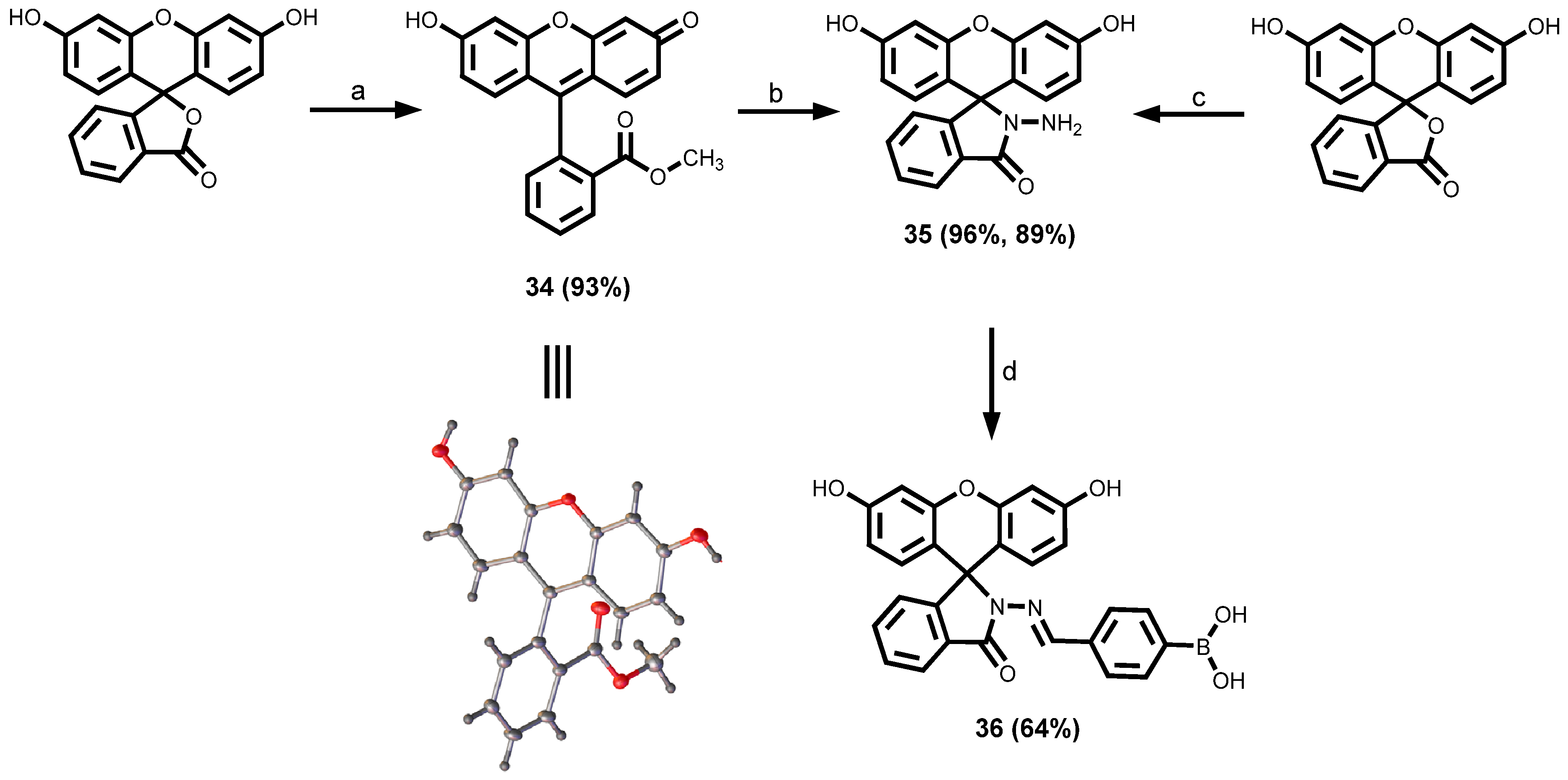
Synthesis of fluorescein methyl ester 34: To fluorescein (1.0 g, 3.1 mmol) methanol solution (10 mL) in a 50 mL round-bottom flask was added concentrated sulfuric acid (98%) (1 mL). The solution was refluxed and stirred for 4 h. After cooling, excess methanol was removed under reduced pressure and excess water was added to the residue. The red solid formed was washed with water several times and filtered in a vacuum until almost free from fluorescence. After drying in a vacuum, the crude product was redissolved and columned using CH2Cl2: methanol (10:1) to obtain red solid fluorescein methyl ester 34 (93% yield). 1H NMR (400 MHz, DMSO-d6) δH 8.31 (dd, J = 7.8, 1.3 Hz, 1H, H18), 7.96 (td, J = 7.5, 1.4 Hz, 1H, H16), 7.88 (td, J = 7.7, 1.4 Hz, 1H, H17), 7.55 (dd, J = 7.5, 1.4 Hz, 1H, H15), 7.31 (d, J = 9.2 Hz, 2H, H6, H10), 7.24 (d, J = 2.2 Hz, 2H, H2, H12), 7.10 (dd, J = 9.2, 2.2 Hz, 2H, H3, H13), 3.57 (s, 3H, H21). 13C NMR (101 MHz, DMSO-d6) δC 171.5, 165.0, 158.1, 133.3, 133.1, 132.3, 130.9, 130.3, 129.1, 120.7, 116.0, 102.5, 52.6. LC-MS: retention time: 6.8 min; ESI-QTOF HRMS (m/z): for [C21H15O5]+ [M + H]+: exact mass calcd.: 347.0920; found: 347.0934.
Synthesis of fluorescein hydrazide 35: Fluorescein methyl ester 34 (0.40 g) and hydrazine hydrate (0.24 g, 4.8 mmol) were added to methanol (5 mL), refluxed, and stirred for 6 h. After collecting by filtration, the light brown precipitate was washed with a small amount of methanol and water and dried in a vacuum to obtain the crude product. The crude material was pre-adsorbed onto silica and purified by column chromatography using CH2Cl2: MeOH (10:1) to obtain pure straw-yellow fluorescein hydrazide 35 (96% yield). 1H NMR (400 MHz, DMSO-d6) δH 9.80 (s, 2H, H22), 7.77 (ddd, J = 5.2, 2.4, 0.8 Hz, 1H, H18), 7.50–7.47 (m, 2H, H16, H17), 7.00–6.98 (m, 1H, H15), 6.59 (d, J = 2.3 Hz, 2H, H6, H10), 6.45 (dd, J = 8.6, 2.4 Hz, 2H, H2, H12), 6.40 (d, J = 8.6 Hz, 2H, H3, H13), 4.38 (s, 2H, H21). 13C NMR (101 MHz, DMSO-d6) δC 165.5, 158.2, 152.4, 151.5, 132.6, 129.3, 128.4, 128.0, 123.4, 122.4, 112.0, 110.0, 102.4, 64.6. LC-MS: retention time: 6.5 min; ESI-QTOF HRMS (m/z): for [C20H15N2O4]+ [M + H]+: exact mass calcd.: 347.1032; found: 347.1028.
(E)-(4-(((3’,6’-dihydroxy-3-oxospiro[isoindoline-1,9’-xanthen]-2-yl)imino)methyl)phenyl)boronic acid 36: 4-formylphenylboronic acid (0.391 mL, 1.77 mmol) was dissolved in 20 mL of a mixture of ethanol/toluene (90:10) and then 35 (0.200 g, 0.578 mmol) was added. A Dean-Stark trap was fixed to the reaction vessel and filled with 10 mL of the same solvent mixture for the azeotropic removal of water. The reaction was then allowed to stir for 16 h at 100 °C, at which time TLC showed that the starting material had fully reacted. The solvent was removed under vacuum, and the product was recrystallised to obtain the product 36 (64% yield). 1H NMR (400 MHz, DMSO-d6) δH 9.92 (s, 2H, H27), 8.96 (s, 1H, H21), 8.10 (s, 2H, H26), 7.92 (d, J = 7.3 Hz, 1H, H18), 7.75 (d, J = 7.9 Hz, 2H, H24), 7.68 – 7.55 (m, 2H, H16, H17), 7.38 (d, J = 8.0 Hz, 2H, H23), 7.14 (d, J = 7.4 Hz, 1H, H15), 6.67 (d, J = 2.2 Hz, 2H, H6, H10), 6.51 (d, J = 8.6 Hz, 2H, H2, H12), 6.46 (dd, J = 8.6, 2.3 Hz, 2H, H). 13C NMR (101 MHz, DMSO-d6) δC 163.7, 158.6, 152.2, 150.5, 148.9, 135.8, 134.5, 134.1, 129.1, 128.9, 128.0, 125.7, 123.8, 123.2, 112.4, 110.1, 102.5, 65.3. 11B NMR (128 MHz, DMSO-d6) δB 28.1. LC-MS: retention time: 7.4 min; ESI-QTOF HRMS (m/z): for [C27H20BN2O6]+ [M + H]+: exact mass calcd.: 479.1414; found: 479.1412. FTIR (cm−1): υ (O–H st) 3287, υ (C=O st, amide) 1668, υ (C=C st and C=N st) 1600–1318.
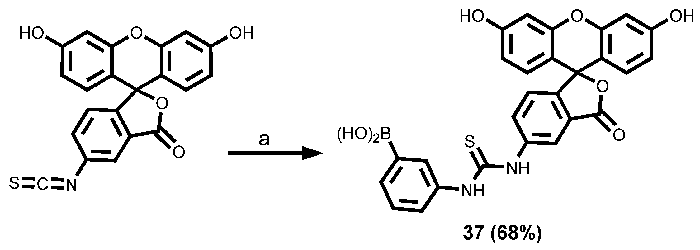
3-(3-(3’,6’-Dihydroxy-3-oxo-3H-spiro[isobenzofuran-1,9’-xanthene]-5-yl)thioureido)phenylboronic acid 37: 3-Aminobenzeneboronic acid (0.35 g, 2.57 mmol) was added to a solution of fluorescein isothiocyanate isomer I (1.00 g, 2.57 mmol) in DMF (5 mL). The reaction mixture was stirred at room temperature for 12 h and then poured into methanol (10 mL). The solvents were removed in vacuo; the residue was then dissolved in the minimum amount of fresh methanol. Chloroform was added and product 37 was obtained as a bright orange precipitate (920 mg, 68% yield). LC-MS: retention time: 7.0 min; ESI-QTOF HRMS (m/z): for [C27H20BN2O7S]+ [M + H]+: exact mass calcd.: 527.1084; found: 527.1123.

2.3.5. Rhodamine Dyes

Synthesis of rhodamine B hydrazide 38: To 0.4 g of rhodamine B (I) dissolved in 15 mL of methanol, an excessive hydrazine hydrate (0.5 mL) was added and then the reaction solution was refluxed till the pink colour disappeared. After that, the cooled reaction solution was poured into distilled water and extracted with ethyl acetate (6 × 25 mL). The combined extracts were dried with sodium sulphate anhydrous and filtered, and the solvent was evaporated to obtain rhodamine B hydrazide 38 in 68% yield. 1H NMR (400 MHz, DMSO-d6) δH 7.79–7.73 (m, 1H, H12), 7.50–7.43 (m, 2H, H10, H11), 7.01–6.95 (m, 1H, H9), 6.37 (t, J = 1.4 Hz, 2H, H2), 6.33 (d, J = 1.4 Hz, 4H, H3, H6), 4.26 (s, 2H, H15), 3.31 (q, 8H, H16), 1.08 (t, J = 7.0 Hz, 12H, H17). 13C NMR (101 MHz, DMSO-d6) δC 165.3 (C14), 153.0 (C5), 151.9 (C1), 148.1 (C8), 132.4 (C10), 129.6 (C13), 128.1 (C11), 127.7 (C3), 123.5 (C9), 122.1 (C12), 107.8 (C4), 105.5 (C2), 97.4 (C6), 64.8 (C7), 43.7 (C16), 12.5 (C17). LC-MS: retention time: 8.6 min; ESI-QTOF HRMS (m/z): for [C28H33N4O2]+ [M + H]+: exact mass calcd.: 457.2604; found: 457.2596.
(E)-(4-(((2-(3’,6’-bis(diethylamino)-3-oxospiro[isoindoline-1,9’-xanthen]-2-yl)ethyl)imino)methyl)phenyl)boronic acid 39: 4-formylphenylboronic acid (0.391 mL, 1.77 mmol) was dissolved in 20 mL of a mixture of ethanol/toluene (90:10) and then 38 (0.200 g, 0.578 mmol) was added. A Dean–Stark trap was fixed to the reaction vessel and filled with 10 mL of the same solvent mixture for the azeotropic removal of water. The reaction was then allowed to stir for 16 h at 100 °C, at which time TLC showed that the starting material 38 had fully reacted. The solvent was removed under vacuum, and the product was recrystallised to obtain product 39 (73% yield). 1H NMR (400 MHz, CDCl3) δH 8.07 (d, J = 7.8 Hz, 1H, H12), 8.00 (d, J = 6.4 Hz, 1H, H10), 7.74 (d, J = 7.8 Hz, 1H, H11), 7.65 – 7.62 (m, 3H, H9, H20), 7.56 – 7.53 (d, J = 7.1 Hz, 2H, H18), 7.47 (d, J = 7.1 Hz, 2H, H17), 7.14 – 7.09 (m, 1H, H15), 6.52 (dd, J = 8.9, 6.5 Hz, 2H, H2), 6.46 (dd, J = 14.9, 2.6 Hz, 2H, H3), 6.24 (ddd, J = 9.1, 7.2, 2.6 Hz, 2H, H6), 3.31 (q, J = 9.0, 8.1 Hz, 8H, H16), 1.15 (t, J = 6.6 Hz, 12H, H17). 13C NMR (101 MHz, CDCl3) δC 165.3, 153.4, 153.2, 152.0, 149.1, 149.1, 147.1, 137.5, 135.63, 134.0, 133.6, 133.4, 128.4, 128.1, 127.0, 124.0, 123.6, 108.2, 106.0, 98.0, 58.6, 44.5, 12.7. 11B NMR (128 MHz, CDCl3) δB 29.9. LC-MS: retention time: 10.2 min; ESI-QTOF HRMS (m/z): for [C35H38BN4O4]+ [M + H]+: exact mass calcd.: 589.2986; found: 589.2994. FTIR (cm−1): υ (O–H st) 3310, υ (C–H st) 2893, υ (C=O st, amide) 1696, υ (C=C st and C=N st) 1614–1306.

2.3.6. BODIPY Dyes

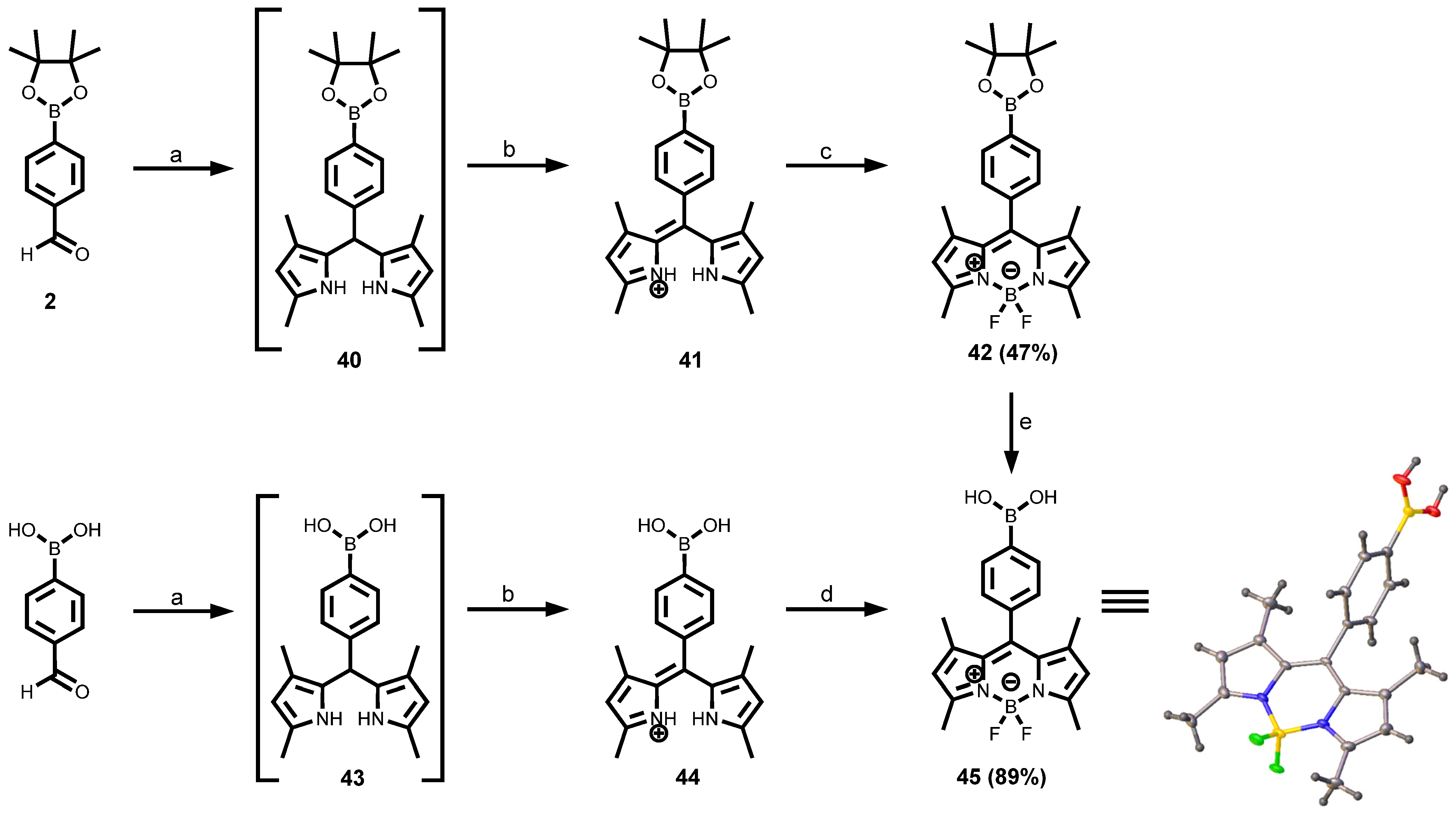
Synthesis of protected BODIPY dye 42: A mixture of a protected 4-formylphenylboronic acid 2 (1.50 g, 6.5 mmol, 1.0 equiv.) and 2,4-dimethyl-1H-pyrrole (1.24 g, 13.0 mmol, 2.0 equiv.) was dissolved in anhydrous DCM (40 mL) under nitrogen. TFA (0.4 mL) was added to the mixture and stirred at room temperature for 3 h. The consumption of the aldehyde was monitored by TLC. A solution of p-chloranil in DCM (1.92 g, 7.8 mmol, 1.2 equiv.) was added at 0 °C and stirred for 30 min. TEA (3.95 g, 5.4 mL, 39.0 mmol, 6.0 equiv.) was added at 0 °C and stirred for a further 30 min. Following dropwise addition of BF3·OEt2 (9.23 g, 8.0 mL, 65.0 mmol, 10.0 equiv.) to the reaction mixture, it was stirred for 12 h at room temperature. The crude product was obtained by evaporating the mixture under reduced pressure. The crude product was purified by silica gel column using hexane/ethyl acetate (1:0 → 9:1) to obtain the desired product 42 in 47% yield. 1H NMR (500 MHz, CDCl3) δH 7.90 (d, J = 7.5 Hz, 2H, H10), 7.29 (d, J = 7.5 Hz, 2H, H9), 5.97 (s, 2H, H3), 2.55 (s, 6H, H6), 1.38 (s, 12H, H13), 1.36 (s, 6H, H7). 13C NMR (126 MHz, CDCl3) δC 155.6 (C4), 143.4 (C1), 141.8 (C2), 138.0 (C8), 135.5 (C10), 131.4 (C5), 127.5 (C9), 121.3 (C3), 84.3 (C12), 29.8 (C7), 25.1 (C13), 14.7 (C6) (C11 bonded to B not observed due to broadening). LC-MS: retention time: 12.4 min; ESI-QTOF HRMS (m/z): for [C25H31B2F2N2O2]+ [M + H]+: exact mass calcd.: 451.2540; found: 451.2555.
Deprotection of protected BODIPY dye 42 to obtain 45: Deprotection of pinacol boronate esters was performed according to the procedure by Akgun [60]. BODIPY dye 42 (150 mg, 0.36 mmol, 1.0 equiv.) was dissolved in THF: water (4:1 mL) (4:1 V:V). Then sodium periodate (231 mg, 1.08 mmol, 3.0 equiv.) was added to the solution and stirred at room temperature for 30 min under an ambient atmosphere. Lastly, HCl (0.2 mL, 1 N) was added to the reaction mixture, which was stirred for 24 h at room temperature. The reaction mixture was concentrated in vacuo. Then it was dissolved in EtOAc (30 mL) and washed with water (1 × 8 mL), and brine (1 × 8 mL). The organic fraction was dried (MgSO4), and the solids were removed by gravity filtration. The crude product was preabsorbed onto 5 mL of silica and dried under vacuum. Flash chromatography (1:3 hexanes/EtOAc) provided a pure deprotected BODIPY dye 45 (89% yield) as a reddish-orange powder after solvent removal. 1H NMR (400 MHz, DMSO-d6) δH 8.25 (s, 2H, H12), 7.96 (d, J = 7.9 Hz, 2H, H10), 7.33 (d, J = 7.9 Hz, 2H, H9), 6.17 (s, 2H, H3), 2.44 (s, 6H, H6), 1.32 (s, 6H, H7). 13C NMR (101 MHz, DMSO-d6) δC 154.8, 150.9, 142.8, 142.2, 135.7, 134.9, 130.6, 129.2, 126.7, 121.4, 113.8, 101.6, 14.3, 14.0. LC-MS: retention time: 9.0 min; ESI-QTOF HRMS (m/z): for [C19H21B2F2N2O2]+ [M + H]+: exact mass calcd.: 369.1757; found: 369.1763. FTIR (cm−1): υ (O–H st) 3201, υ(C–H st) 2916, υ(C=C st and C=N st) 1545–1454.

2.3.7. Azo Dyes

Synthesis of (2-((m-tolylamino)methyl)phenyl)boronic acid 46: 2-formylbenzeneboronic acid (3.00 g, 20.1 mmol, 1.0 equiv.) and m-toluidine (2.7 mL, 24.1 mmol, 1.1 equiv.) were dissolved in 1, 2-dichloroethane (50 mL). The reaction mixture was then charged with sodium triacetoxyborohydride (6.39 g, 1.5 equiv.). The reaction was then stirred under nitrogen for 1 h. After an hour, additional sodium triacetoxyborohydride (4.26 g, 1.0 equiv.) was added to the reaction and stirred further for another 1 h. The reaction mixture was then quenched with saturated NaHCO3 solution (100 mL). The aqueous phase was extracted with CH2Cl2 (2 × 100 mL), and the combined organics dried over anhydrous MgSO4 and concentrated to dryness in vacuo to afford (2-((m-tolylamino)methyl)phenyl)boronic acid 46 as a cream-coloured solid (3.64 g, 75% yield). The material was used without further purification. 1H NMR (400 MHz, CDCl3) δH 7.74 (dd, J = 7.6, 1.5 Hz, 1H, H5), 7.45 (m, 2H, H2, H12), 7.38–7.32 (m, 3H, H6, H16), 7.02–6.97 (m, 2H, H1, H11), 6.66 (m, 2H, H10, H14), 6.52 (s, 1H, H8), 4.36 (s, 2H, H7), 2.14 (s, 3H, H15). LC-MS: retention time: 6.4 min; ESI-QTOF HRMS (m/z): for [C14H17BNO2]+ [M + H]+: exact mass calcd.: 242.1352; found: 242.1354.
(E)-(2-(((3-methyl-4-((4-nitrophenyl)diazenyl)phenyl)amino)methyl)phenyl)boronic acid 47a: 4-nitroaniline (504 mg, 3.64 mmol, 1.1 equiv.) was mixed in water (2 mL), methanol (2 mL), and hydrochloric acid (2 mL, 5.0 M) and then cooled to 0–5 °C on an ice-bath. A chilled solution of sodium nitrite (186 mg, 2.69 mmol, 1.3 equiv.) was added dropwise. Excess nitrite was destroyed by the addition of sulfamic acid after stirring for 5 min. (2-((m-tolylamino)methyl)phenyl)boronic acid (500 mg, 2.07 mmol, 1.0 equiv.) was dissolved in methanol (3 mL) and dilute hydrochloric acid (2 mL, 1 M), then added dropwise to the reaction mixture, which quickly turned red. Sodium acetate was added to raise the pH of the solution to 4 and this was then left to stir at 0–5 °C for 3 h. Sodium hydroxide (2 M) was slowly added to raise the pH to 7. The resulting precipitate was collected by suction filtration and dried in a desiccator overnight to afford product 47a as a dark red solid (44% yield). 1H NMR (400 MHz, DMSO-d6) δH 9.86 (s, 1H), 8.39 (d, J = 8.9 Hz, 2H), 8.33 (d, J = 8.9 Hz, 1H), 8.18 (s, 1H), 8.01 (d, J = 8.9 Hz, 2H), 7.92–7.85 (m, 2H), 7.75 (d, J = 9.1 Hz, 1H), 7.58–7.53 (m, 1H), 7.47 (s, 2H, 2H), 7.37–7.30 (m, 1H), 4.63 (s, 2H), 2.73 (s, 3H, 3H). 13C NMR (101 MHz, DMSO-d6) δC 156.8, 156.1, 153.6, 150.5, 148.3, 147.5, 146.5, 144.1, 142.6, 141.6, 141.5, 133.7, 130.4, 130.3, 129.0, 125.0, 123.0, 122.5, 122.4, 118.7, 116.3, 116.1, 52.7, 17.7. 11B NMR (128 MHz, DMSO-d6) δB 32.0. LC-MS: retention time: 9.9 min; ESI-QTOF HRMS (m/z): for [C20H20BN4O4]+ [M + H]+: exact mass calcd.: 391.1578; found: 391.1587.
(E)-4-((4-((2-boronobenzyl)amino)-2-methylphenyl)diazenyl)benzoic acid 47b: The above procedure was repeated using 4-aminobenzoic acid (0.11 g, 0.83 mmol) instead of 4-nitroaniline. All other reagents were used in the same mole ratios to afford 47b as an orange-red solid (62% yield). 1H NMR (400 MHz, DMSO-d6) δH 13.09 (s, 1H), 9.79 (s, 1H), 8.15–8.08 (m, 2H), 8.05 (dd, J = 8.6, 1.9 Hz, 1H), 7.90 (dd, J = 8.0, 6.3 Hz, 3H), 7.79 (dd, J = 8.5, 1.7 Hz, 1H), 7.72 (d, J = 9.1 Hz, 1H), 7.68 (dd, J = 9.1, 2.3 Hz, 1H), 7.56 (d, J = 2.4 Hz, 1H), 7.48 (s, 2H), 7.35 (dd, J = 7.8, 4.3 Hz, 1H), 4.64 (s, 2H), 2.73 (s, 3H). 13C NMR (101 MHz, DMSO-d6) δC 166.9, 155.1, 149.8, 148.4, 144.0, 140.6, 131.6, 130.6, 130.5, 130.4, 130.2, 122.5, 122.2, 118.8, 116.1, 116.0, 52.7, 48.6, 30.4, 17.7. 11B NMR (128 MHz, DMSO-d6) δB 30.4. LC-MS: retention time: 8.2 min; ESI-QTOF HRMS (m/z): for [C21H21BN3O4]+ [M + H]+: exact mass calcd.: 390.1625; found: 390.1634. FTIR (cm−1): υ(O–H st) 3442, υ(C–H st) 2925, υ(C=O st, carboxyl) 1677, υ(C=C st and C=N st) 1677–1600.
(E)-3-((4-((2-boronobenzyl)amino)-2-methylphenyl)diazenyl)benzoic acid 47c: The previous experiment was repeated using 3-aminobenzoic acid (499 mg, 3.64 mmol) instead of p-anisidine. All other reagents were used in the same mole ratios to afford 47c as a red solid (25% yield). 1H NMR (400 MHz, DMSO-d6) δH 13.20 (s, 1H), 9.77 (s, 1H), 8.34 (t, J = 1.8 Hz, 1H), 8.25–8.16 (m, 1H), 8.10–8.03 (m, 3H), 7.89 (d, J = 7.3 Hz, 1H), 7.72 (d, J = 9.1 Hz, 2H), 7.70–7.66 (m, 2H), 7.56 (d, J = 2.4 Hz, 1H), 7.48 (s, 2H), 4.64 (s, 2H), 2.73 (s, 3H). LC-MS: retention time: 8.5 min; ESI-QTOF HRMS (m/z): for [C21H21BN3O4]+ [M + H]+: exact mass calcd.: 390.1625; found: 390.1630.
(E)-(2-(((4-((4-methoxyphenyl)diazenyl)-3-methylphenyl)amino)methyl)phenyl)boronic acid 47d: The previous experiment was repeated using p-anisidine (0.61 g, 4.98 mmol, 6 equiv.) instead of 4-aminobenzoic acid. Purification was performed by washing the product (in dichloromethane) with 10% sodium hydrogen carbonate solution (w/w) to remove residual acetic acid, to yield 47d as an orange-brown solid (56% yield). 1H NMR (400 MHz, DMSO-d6) δH 9.67 (s, 1H), 7.89 (d, J = 7.3 Hz, 1H), 7.84 (d, J = 8.9 Hz, 2H), 7.74–7.71 (m, 1H), 7.65–7.62 (m, 2H), 7.53 (d, J = 2.0 Hz, 1H), 7.48–7.45 (m, 2H), 7.34 (ddt, J = 9.2, 6.6, 3.2 Hz, 1H), 7.10 (d, J = 9.0 Hz, 2H), 7.08–7.02 (m, 1H), 4.61 (s, 2H), 3.85 (s, 3H), 2.69 (s, 3H). 13C NMR (101 MHz, DMSO-d6) δC 161.1, 148.4, 148.3, 146.9, 143.8, 138.9, 130.3, 130.0, 126.6, 126.4, 124.0, 123.4, 123.4, 122.5, 118.8, 115.9, 115.7, 114.5, 114.3, 55.5, 52.7, 17.7. 11B NMR (128 MHz, DMSO-d6) δB 29.9. LC-MS: retention time: 9.8 min; ESI-QTOF HRMS (m/z): for [C21H23BN3O3]+ [M + H]+: exact mass calcd.: 376.1832; found: 376.1836.

2.3.8. Sudan I Dyes Boronic Acid

Synthesis of Sudan I dyes boronic acid 48: To a heated solution (50 °C) of Sudan I (347 mg, 1.39 mmol, 1.2 equiv.) in acetone (20 mL), potassium carbonate, K2CO3 (128 mg, 0.93 mmol, 0.80 equiv.), and 4-(bromomethyl)phenylboronic acid (250 mg, 1.16 mmol, 1.0 equiv.) were added. After 4 h of stirring and heating, additional potassium carbonate, K2CO3 (128 mg, 0.93 mmol, 0.80 equiv.) was added, and the suspension was stirred overnight. Finally, the reaction mixture was diluted with EtOAc and the organic layer was washed with 1 M hydrochloric acid (2 × 10 mL) and brine (20 mL), dried over Na2SO4, and concentrated in vacuo. The resulting solid was further purified using silica gel chromatography affording a pure Sudan I dye boronic acid 48 product. LC-MS: retention time: 9.7 min; ESI-QTOF HRMS (m/z): for [C23H19BN2O3]+ [M + H]+: exact mass calcd.: 383.2260; found: 383.1572.

2.4. Measurements of Photophysical Properties of Synthesised Compounds

Photophysical quantities such as absorption maxima (λabsmax), emission maxima (λemmax), Stokes shift (∆λ), molar extinction coefficient (ε), fluorescence quantum yield (ΦF), and brightness were determined from absorption and emission data. Photophysical measurements were performed at ambient temperature (25 °C). UV-vis absorption spectra were recorded on a Shimadzu UV-1800 spectrophotometer between 200 and 800 nm using quartz cuvettes of 1 cm optical path length. The molar extinction coefficients (ε) of the different arylboronic acid dyes were determined at the respective maximum absorption wavelength using the Beer–Lambert law:
A = εcl
where A is absorbance; ε is the extinction coefficient for the dye; c is the sample concentration; l is the pathlength of the sample cuvette. Fluorescence spectra of the synthesised compounds were acquired using a Shimadzu RF-6000 spectrofluorometer (Shimadzu, Kyoto, Japan). Emission spectra were recorded at room temperature with excitation wavelengths set at the λabsmax of the respective dyes and emission range 250–800 nm. The excitation and emission slit widths were set at 5 nm. Fluorescence quantum yields were determined by the relative comparison procedure [63,64], using either rhodamine B (ΦF = 0.70 in ethanol) [65] or quinine sulphate (ΦF = 0.51 in 0.05 M H2SO4) [4,66]. Five different solutions with increasing concentrations of each of the synthesised compounds and the reference standards (rhodamine B or quinine sulphate) were prepared in different solutions and the absorbance of each solution was determined to be between 0.01–0.1 in 1 cm optical path length cuvette using a Shimadzu UV-1800 Spectrophotometer. Fluorescence spectra and the integrated fluorescence intensities for varying concentrations of solutions were measured using a Shimadzu RF-6000 spectrofluorometer. For each solution, graphs of the integrated fluorescence intensities as a function of the solution absorption were plotted in each case for both the synthesised dye and the reference standard (either rhodamine B or quinine sulphate). The resulting data points were fitted with linear plots and the slopes were calculated. The quantum yield (ΦF) is calculated using the slope of the line determined from the plot of the absorbance against the integrated fluorescence intensities. In this case, the quantum yield of the unknown sample can be calculated using the following equation:
Φ F , S = Φ F , R m S m R η S 2 η R 2
where m is the slope of the line obtained from the plot of the integrated fluorescence intensity vs. absorbance, η is the refractive index of solvent, and the subscripts R and S refer to the reference and unknown sample, respectively; ΦS and ΦR are the fluorescence quantum yield of the sample and that of the standard, respectively. The quantum yields were corrected for the differing refractive index of the solvent used for the sample and reference. Refractive indices (25 °C) were taken to be 1.3326, 1.3593, and 1.4793 for water, ethanol, and DMSO, respectively [67]. In instances where the same solvent was used for both the reference and unknown sample as a solvent, η S 2 / η R 2 will be 1, so the fluorescence quantum yield of the unknown (ΦS) was obtained directly from the quotient of the two slopes.

3. Results and Discussion

3.1. Synthesis of Various Arylboronic Acid Chemosensor Fluorescent Dyes

3.1.1. Synthesis of the Building Blocks Through Miyaura Borylation

The syntheses of the various boronic ester building blocks were accomplished via a palladium-catalysed Miyaura borylation of aryl halides with a diboron reagent such as bis(pinacolato)diboron (B2pin2). Potassium acetate (KOAc) was employed as the base, and the reaction was carried out in 1,4-dioxane at 80 °C for 16 h [59,68,69]. The various pinacol-protected boronate esters were readily purified by silica gel chromatography to obtain analytically pure and white crystalline pinacol boronate ester solids with yields ranging from 70% to 96% (Scheme 1). They were characterised by 1H and 13C NMR, FTIR, and HRMS, and the data are provided in the Materials and Methods section with the corresponding spectra available in the Electronic Supplementary Information (Figures S1–S5). Interestingly, these boronate ester solids are indefinitely bench-stable under air compared to their corresponding boronic acids.

3.1.2. Coumarin-Tagged Boronic Acids

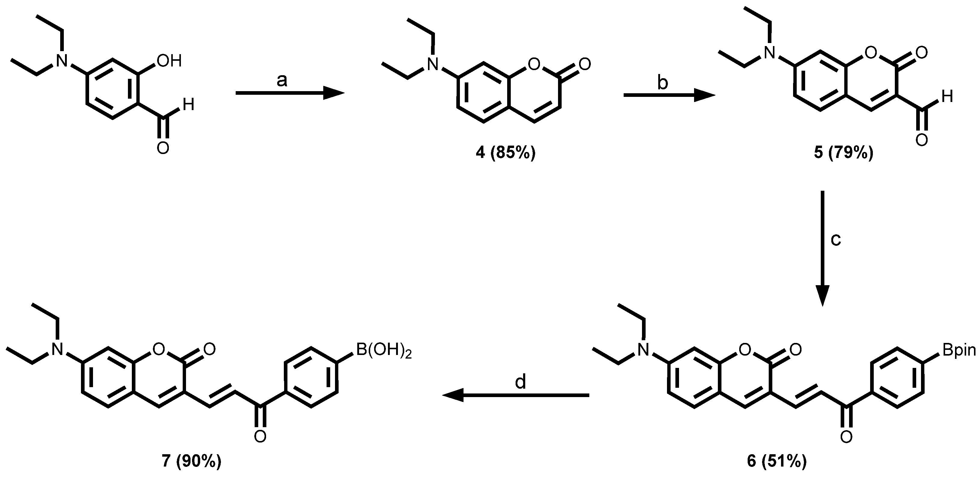
Coumarin and its derivatives are widely used as fluorescent probes because of their excellent chromogenic and fluorogenic properties such as high fluorescence quantum yields, large Stokes shifts, excellent photostability, and thermal stability [70,71,72,73,74,75,76]. Based on this knowledge, two coumarin-tagged boronic acids were designed and synthesised (Scheme 2 and Scheme 3). The synthesis of the target compound 7 was achieved in four chemical steps as shown in Scheme 2. Characterisation was carried out by 1H and 13C NMR, FTIR, and HRMS, and the data are provided in the Materials and Methods section and the corresponding spectra are available in the Electronic Supplementary Information (Figures S6–S18). The first step of the synthesis was via a Knoevenagel condensation reaction of 4-(diethylamino)-2-hydroxybenzaldehyde with diethyl malonate in the presence of piperidine, cyclised, and then decarboxylated to afford 7-(diethylamino)-2H-chromen-2-one 4 in good yield (85%). Subsequently, compound 4 was subjected to Vilsmeier–Haack formylation in the presence of 1,2-dichloroethane (DCE) to produce 7-(diethylamino)coumarin-3-carbaldehyde 5 in 79% [76].
The formylation was deemed successful by the confirmation of the distinctive and unmistakable singlet at δH = 10.10 ppm observed in the 1H NMR spectrum, which integrated for a single proton. This is characteristic of aldehydic protons [77].
Next, a base-catalysed Claisen–Schmidt condensation of 7-(diethylamino)coumarin-3-carbaldehyde 5 with 1-(4-(4,4,5,5-tetramethyl-1,3,2-dioxaborolan-2-yl)phenyl)ethan-1-one 3 building block using a catalytic amount of pyrrolidine in CH2Cl2/EtOH afforded the chalcone molecule 6 (yield = 51%) with extended conjugations [78]. Afterwards, the chalcone 6 which had the pinacol-protecting group was subjected to an oxidative hydrolysis deprotection protocol with aqueous sodium periodate (NaIO4). This led to the smooth formation of the corresponding boronic acid 7 with excellent yield (up to 90%) [8,79]. 1H NMR analysis of the product showed that the desired final compound was prepared following the disappearance of the Bpin peak at δH = 1.32 ppm (see Supplementary Materials Figure S14).
Another coumarin fluorescent dye arylboronic acid was synthesised from 7-hydroxycoumarin (umbelliferone). Unbelliferone is a known fluorophore with a high quantum yield [80,81]. The coumarin-tagged boronic acid derivative in Scheme 3 was synthesised according to the procedure reported by Palanisamy et al. via a one-step alkylation reaction of 4-methylumbelliferone with 4-bromomethylphenylboronic acid in the presence of anhydrous potassium carbonate in dry DMF at 70 °C [82] to afford 22 in 76% yield after purification. The product was fully characterised by 1H and 13C NMR, and HRMS data are provided in the Materials and Methods section and the corresponding spectra are available in the Electronic Supplementary Information (Figures S19–S23).

3.1.3. 9-Aminoacridine-Tagged Boronic Acid Dyes

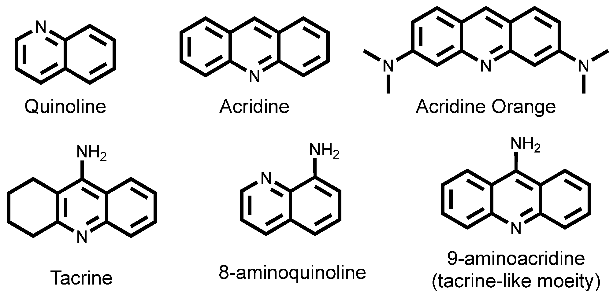
In another development, 9-aminoacridine-tagged boronic acid dyes were prepared. 9-Aminoacridine is a quinoline derivative that contains a tacrine-like moiety (Figure 4). Acridine and its derivatives are important quinoline structural derivatives that are generally planar tricyclic aromatic molecules which fluoresce at shorter wavelength regions [83,84]. Acridine-based fluorescence chemosensors have recently been synthesised and employed by Wang et al. for the selective detection of Fe3+ and Ni2+ ions [85].
Harnessing the advantages of the acridine moiety, a new arylboronic acid fluorescent chemosensor was prepared by coupling an aminoacridine fluorophore to a (4-(aminomethyl)phenyl)boronic acid via a chloroacetyl chloride linker in three steps (Scheme 4). N-(acridin-9-yl)-2-chloroacetamide 23 was prepared by reacting a commercially available 9-aminoacridine with chloroacetyl chloride according to chloroacetylation reaction procedures described in the literature [86,87,88]. This step was carried out in the presence of a catalytic amount of triethylamine (Et3N). Chloroacetyl chloride plays a key role in synthetic and biological chemistry as a bifunctional linker for alcohols and amines [89]. Thus, it was employed to prepare the N-(acridin-9-yl)-2-chloroacetamide 23 as a suitable building block for the next step. The reaction was run overnight under room temperature, and the isolated yield was 59%. (4-(Aminomethyl)phenyl)boronic acid was then alkylated with the N-(9-acridinyl)-2-chloroacetamide using triethylamine as a catalyst under reflux in acetonitrile (CH3CN) for 24 h to provide the target compound (4-(((2-(acridin-9-ylamino)-2-oxoethyl)amino)methyl)phenyl)boronic acid 24 in 22% yield.

3.1.4. 8-Aminoquinoline-Tagged Boronic Acid Dyes

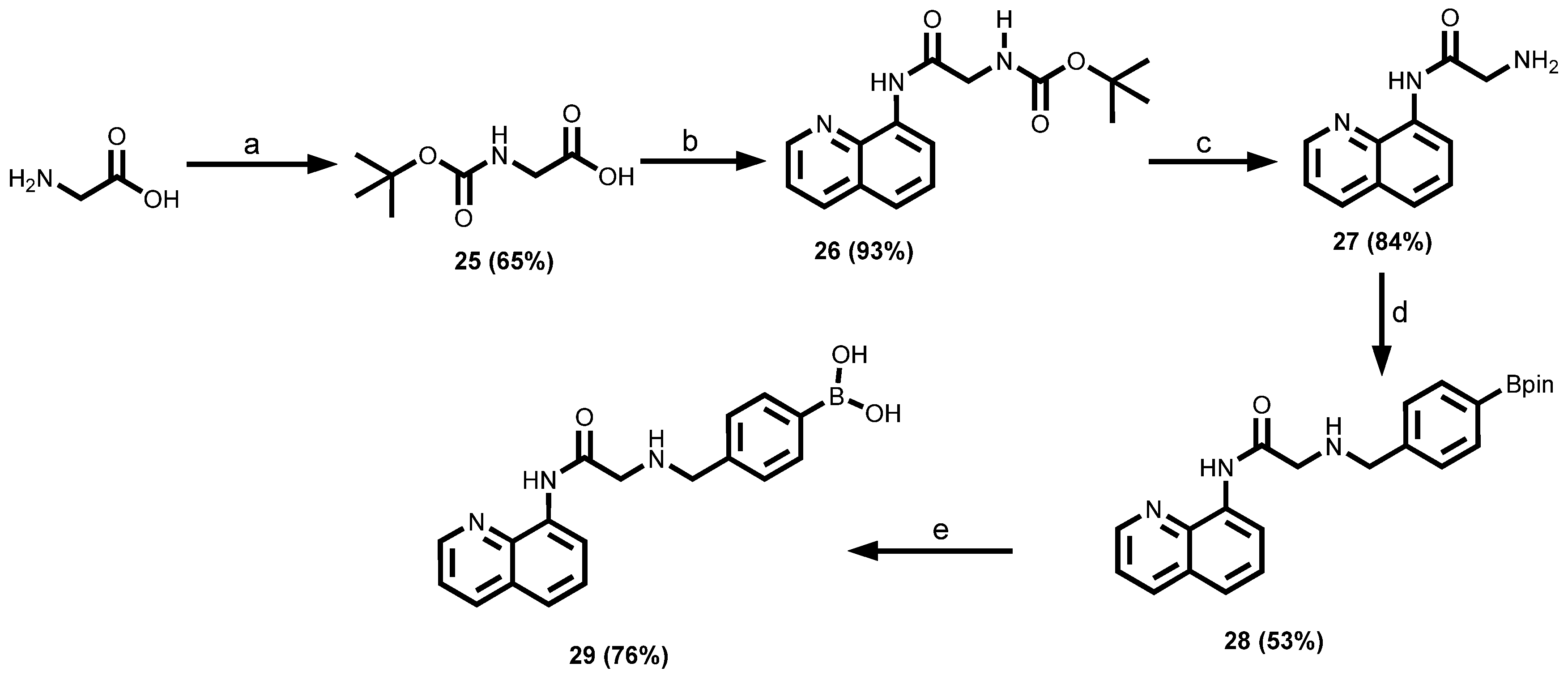
8-aminoquinoline contains a quinoline moiety which is a well-known fluorophore unit [90]. Quinolines are particularly desirable because of their good coordination properties and their ability to form hydrogen bonds as a result of the presence of nitrogen in the ring as well as their small molecular size [90,91]. Furthermore, they are well known for their good metal affinity and hence are becoming leading candidates in the design of fluorescent probes for metals such as zinc and their applications in more complex biological research [90]. More importantly, the chemistry of quinoline is also well established and a plethora of synthetic methods are available to functionalise the core moiety [90]. With these advantages, attempts were made to synthesise an aminoquinoline-tagged boronic acid 29. The synthesis was achieved in five steps as depicted in Scheme 5. The first step involves Boc-protection of glycine by reacting with di-tert-butyl dicarbonate (Boc2O) under basic conditions to afford Boc-Gly-OH under 2 h. This was followed by amide coupling between the carboxylic acid functionality of the N-Boc-protected glycine 25 and 8-aminoquinoline in CH2Cl2 in the presence of EDC.HCl/DMAP as the activation/amide coupling agent.
The next step was to obtain the free amine through deprotection of the N-Boc group by using the traditional approach based on TFA-induced cleavage as reported previously [92] or with HCl (6 M) in dioxane at room temperature [93]. Both methods gave the desired products in excellent yields. The resultant free base was characterised by NMR and used in the next step without further purification. After the successful deprotection of the N-Boc group to obtain the free amine 27, it was then coupled to the aldehyde functionality of the pinacol-protected 4-formylphenylboronic acid 2 building block that was previously synthesised under reductive amination conditions to give the aminomethyl derivative 28. Sodium triacetoxyborohydride (NaBH(OAc)3) was chosen as the reducing agent over others such as sodium cyanoborohydride (NaBH3CN) and the less bulky hydride reagent NaBH4 because of its high efficiency in reductive amination reactions with unreactive amines, leading to faster, better yields, and with fewer side products. Secondly, NaBH(OAc)3 has been reported to be more selective for direct reductive aminations of ketones and aldehydes relative to the sodium cyanoborohydride alternative [94]. The other advantage of triacetoxyborohydride is the avoidance of the production of toxic by-products such as NaCN and HCN that are usually associated with the use of NaBH3CN [27]. 1,2-dichloroethane (DCE) was preferred as a reaction solvent even though other similar amination reactions have been carried out in other solvents such as tetrahydrofuran (THF) and acetonitrile (CH3CN). The final step was the deprotection of the boronate ester 28 using the optimised procedure of NaIO4 to afford (4-(((2-oxo-2-(quinolin-8-ylamino)ethyl)amino)methyl)phenyl)boronic acid 29 in 76% yield.

3.1.5. Fluorescein-Tagged Boronic Acid Dyes

The synthesis of fluorescein-tagged boronic acid 36 was performed following the procedure in Scheme 6. First, esterification of cyclic fluorescein was carried out in the presence of a catalytic amount of concentrated sulfuric acid in methanol to afford fluorescein methyl ester 34 in an excellent yield (93%). The fluorescein methyl ester intermediate 34 was first characterised by X-ray crystallography (the crystallographic data are shown in Table S1). Then the ester was refluxed for 6 h with an excess amount of hydrazine hydrate in methanol via hydrazinolysis to produce the desired fluorescein hydrazide 35 with a yield of 96% [95]. An alternative method for the preparation of fluorescein hydrazide 35 (in 89% yield) involved the direct hydrazinolysis of the cyclic fluorescein using excess hydrazine hydrate under reflux in methanol for 12 h. Finally, through a Schiff base reaction, the fluorescein hydrazide 35 was coupled to 4-formylphenylboronic acid to form the fluorescein-tagged arylboronic acid chemosensor 36 in 64% yield.
A second fluorescein boronic acid was synthesised via a one-step reaction of fluorescein isothiocyanate isomer I and 3-aminobenzeneboronic acid (Scheme 7). The fluorescein isothiocyanate contains an electrophilic carbon atom that readily reacted with nucleophile (amine), using dimethylformamide (DMF) as the solvent to form thiourea compound 37 in 68% yield [96,97].

3.1.6. Rhodamine-Tagged Boronic Acid Dye

Rhodamine dye fluorophores are part of the family of xanthenes along with fluorescein and eosin dyes [98]. The structures of xanthene chromophore and rhodamine dyes are shown in Figure 5. The carboxylic acid group of rhodamine dyes undergo intramolecular cyclisation. The ring-opened form is highly fluorescent, whereas the spirocyclic spirolactone form is essentially nonfluorescent [98,99].
Rhodamine dyes and their derivatives have also been used extensively, in particular as chemosensors either in vitro or in vivo in the detection of metals including Hg(II) [100,101,102], peroxynitrite [51], Cu(II) [103], Fe(III) [104], Cr(III) [104]. Rhodamine derivatives are also employed as molecular switches [105]. Rhodamine B was chosen as the fluorescent dye for use in this study due to the high fluorescence quantum efficiency, high molar extinction coefficients, and low cost. A rhodamine-tagged boronic acid 39 was readily synthesised from rhodamine B by a two-step reaction (Scheme 8), where rhodamine B was treated with excess hydrazine in methanol to give rhodamine B hydrazide 38 in 68% yield. The hydrazide was further converted to the designed product 39 in 73% yield through a Schiff base reaction using 4-formylphenylboronic acid.

3.1.7. BODIPY-Tagged Boronic Acid Dyes

4,4-difluoro-4-bora-3a-4a-diaza-s-indacene (simply known as BODIPY, difluoroboron dipyrromethene) and its derivatives are fluorescent organic dye molecules that were first discovered by Treibs and Kreuzer in 1968 [106] (Figure 6). Since then, several BODIPY and their derivatives have become the most popular dye molecules among the multitude of fluorescent dyes available with widespread applications in various areas, including tunable laser dyes, biological labelling [107,108,109], as fluorescent switches, fluorophores in sensors and labels, and light-harvesting systems in electroluminescent devices [110,111,112]. The interest in BODIPY chromophores can be attributed to their favourable physicochemical characteristics and desirable photophysical properties [113,114], such as high fluorescence quantum yields, relatively large molar absorption coefficients, narrow emission bandwidths, and tunable fluorescent properties, as well as relatively high thermal and photochemical stabilities [115,116,117,118]. These molecules generally have excitation/emission wavelength in the visible region with stable excited states. Other advantages include ease of modification of BODIPY chemically for the synthetic accessibility of various derivatives and good solubility in common solvents [119]. As a result, a significant number of BODIPY derivatives have since been synthesised and characterised [115,116]. Also, there is an abundance of literature that supports the synthetic versatility of the BODIPY chromophore which allows for far-reaching structural modifications, resulting in the alteration of the electronic, optical, and chemical properties of the dye [120].
Thus, a procedure for the introduction of an arylboronic acid unit onto a BODIPY core was developed to take advantage of the desirable photophysical properties outlined above. Two approaches were employed to synthesise the boron dipyrromethene (BODIPY)-tagged boronic acid dye 45 (Scheme 9). The first route involved the use of the pinacol-protected 4-formylphenylboronic acid 2, whereas in the second route, 4-formylphenylboronic acid was attempted directly without protecting it to reduce the number of steps, especially in instances where purification and hydrolysis of the boronate proved challenging. The synthesis began with an acid-catalysed condensation between two equivalents of 2,4-dimethylpyrrole and a suitable electrophile such as pinacol-protected 4-formylphenylboronic acid 2 to form a dipyrromethane intermediate 40, followed by the addition of a few drops of trifluoroacetic acid (TFA). The complete consumption of the aldehyde was monitored by TLC. The dipyrromethane intermediate 40 was unstable and was therefore not isolated but converted directly to a dipyrromethene 41 in an oxidation step carried out using 2,3,5,6-tetrachloro-p-benzoquinone (p-chloranil) as an oxidant [121]. The dipyrromethene 41 was subsequently subjected to a boron trifluoride etherate (BF3·Et2O) complexation reaction catalysed by triethylamine (Et3N) to afford the borondifluoride complex 42 in 47% yield [122,123]. The last step involved the deprotection of the boronate ester 42, in the case where the protected boronate aldehyde was used, to obtain the BODIPY dye 45 as a red crystalline solid (89% yield).
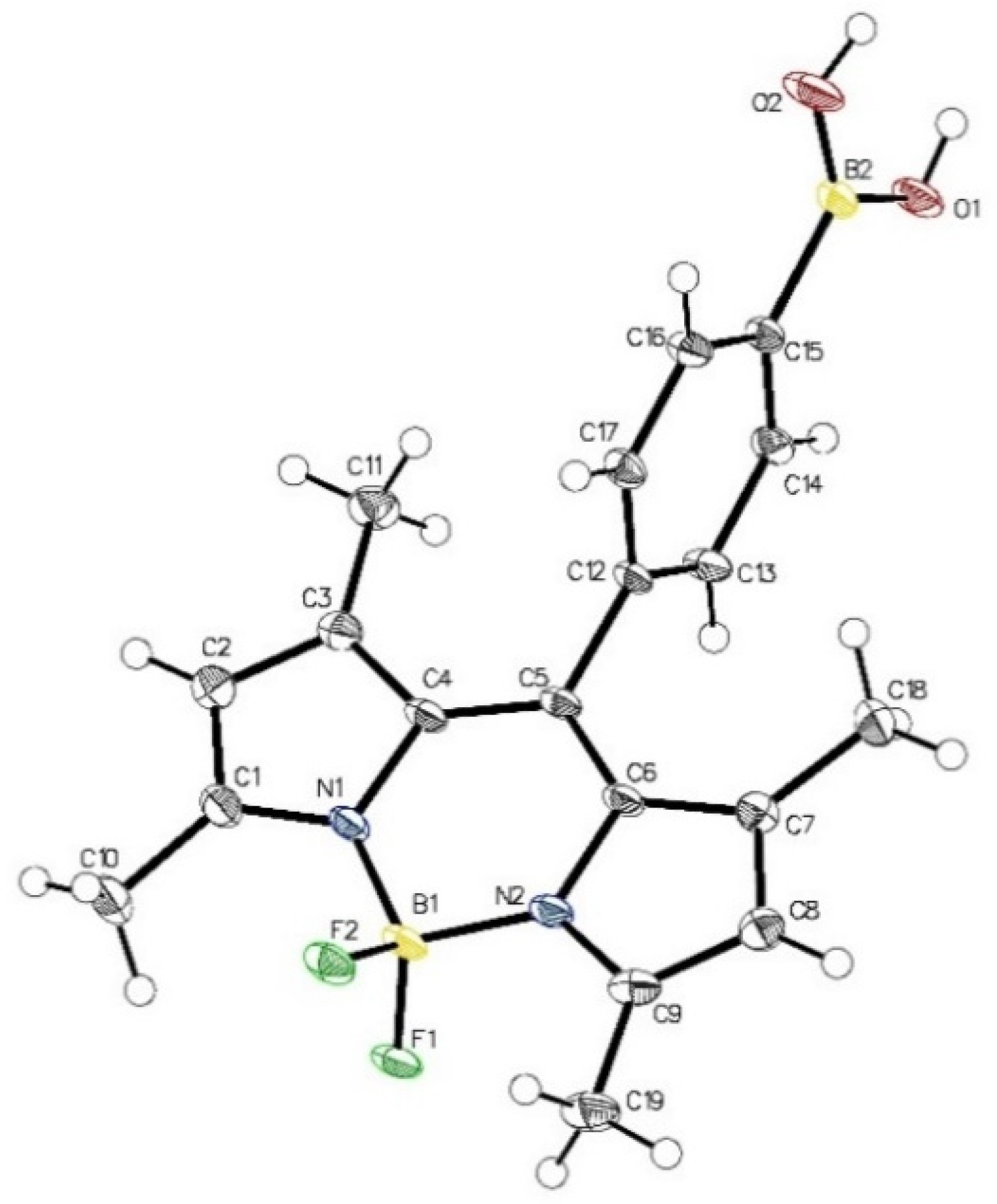
A single-crystal diffraction analysis was measured for crystals of 45, and the Oak Ridge thermal ellipsoid plot (ORTEP) diagram is displayed in Figure 7 and the crystallographic data are shown in Supplementary Materials (Table S1). The crystal structure of BODIPY 45 reveals that the molecule is virtually planar. This is corroborated by the following structure refinement data. The boron atom has a slightly distorted tetrahedral coordination with the two fluorine atoms being perpendicularly oriented with respect to the dipyrrin plane. The B1–N1 and B1–N2 bond lengths are 1.536(2) Å and 1.534(2) Å respectively. Likewise, the B1–F1 and B1–F2 bond lengths are 1.412(2) Å and 1.407(2) Å respectively. The bond distances of the two B–N and two B–F are virtually identical, which is an indication that 45 possess single B–N and B–F bonds. Also, the average N1–B1–N2 and F1–B1–F2 angles are 107.75(12) ° and 106.82(12)°, respectively. This also presupposes that there is an expected delocalisation of the positive charge.

3.1.8. Azo-Tagged Boronic Acid Dyes

Boronic acid azo dyes were used in the 1940s for investigations into the treatment of cancer by boron neutron capture therapy (BNCT) [124]. However, in 1991, a boronic acid azo dye synthesised from 4-aminophenylboronic acid was found to be sensitive to a selection of saccharides [125]. Subsequently, various conjugates of azo dyes have been developed as saccharide and sugar sensors [126,127,128]. A variety of azo-tagged boronic acid dyes in which subtle modifications are made to the electronic configuration of the azo chromophore by varying the substituent groups on the azo aromatic ring were synthesised as depicted in Scheme 10. The first step involved the synthesis of (2-((m-tolylamino)methyl)phenyl)boronic acid 46 by reductive amination, followed by diazotisation of various anilines containing electron-withdrawing groups such as p-NO2, p-COOH, p-OCH3, and m-COOH in the presence of an aqueous solution of sodium nitrite (NaNO2) in concentrated HCl medium at 0 °C to form the corresponding diazonium salts. The destruction of excess sodium nitrite after the diazotisation reaction step was required because nitrites are toxic and can be harmful to the environment. This was achieved by using a common nitrous acid scavenger such as sulfamic acid (H3NSO3) that reacts with nitrous acid (HNO2) to form stable and non-toxic compounds [129,130]. The diazonium salts that were formed in the previous step were not isolated but coupled in situ with (2-((m-tolylamino)methyl)phenyl)boronic acid 46 in a basic medium (NaOH) to give the respective target products, which were fully characterised by 1H NMR, 13C NMR, and HR-LCMS spectroscopic and spectrometric analysis. It is worth noting that the pH of the reaction mixture plays a significant role in the coupling reaction and therefore a careful balance must be struck between the equilibria governing the coupling component and the diazo component. The coupling reactions of the various aniline diazonium salts with the (2-((m-tolylamino)methyl)phenyl)boronic acid 46 occur predominantly para to the electron-donating anilinic nitrogen because of steric effects. This was directed by incorporating a methyl group meta to the anilinic nitrogen in the amine coupling agent, and it resulted in the exclusive para coupling products. This approach allowed for the synthesis of target compounds (47a–d) and avoided the potentially difficult separation of isomers [131].

3.1.9. Sudan I Boronic Acid Dye

Another azo boronic acid derivative was synthesised from Sudan I dye according to the one-step procedure in Scheme 11. Sudan I was reacted with 4-bromomethylphenylboronic acid in the presence of anhydrous potassium carbonate in dry acetone at 50 °C to afford 48. The product was characterised by HRMS.

3.2. Photophysical Properties of the Different Arylboronic Acid Chemosensor Dyes

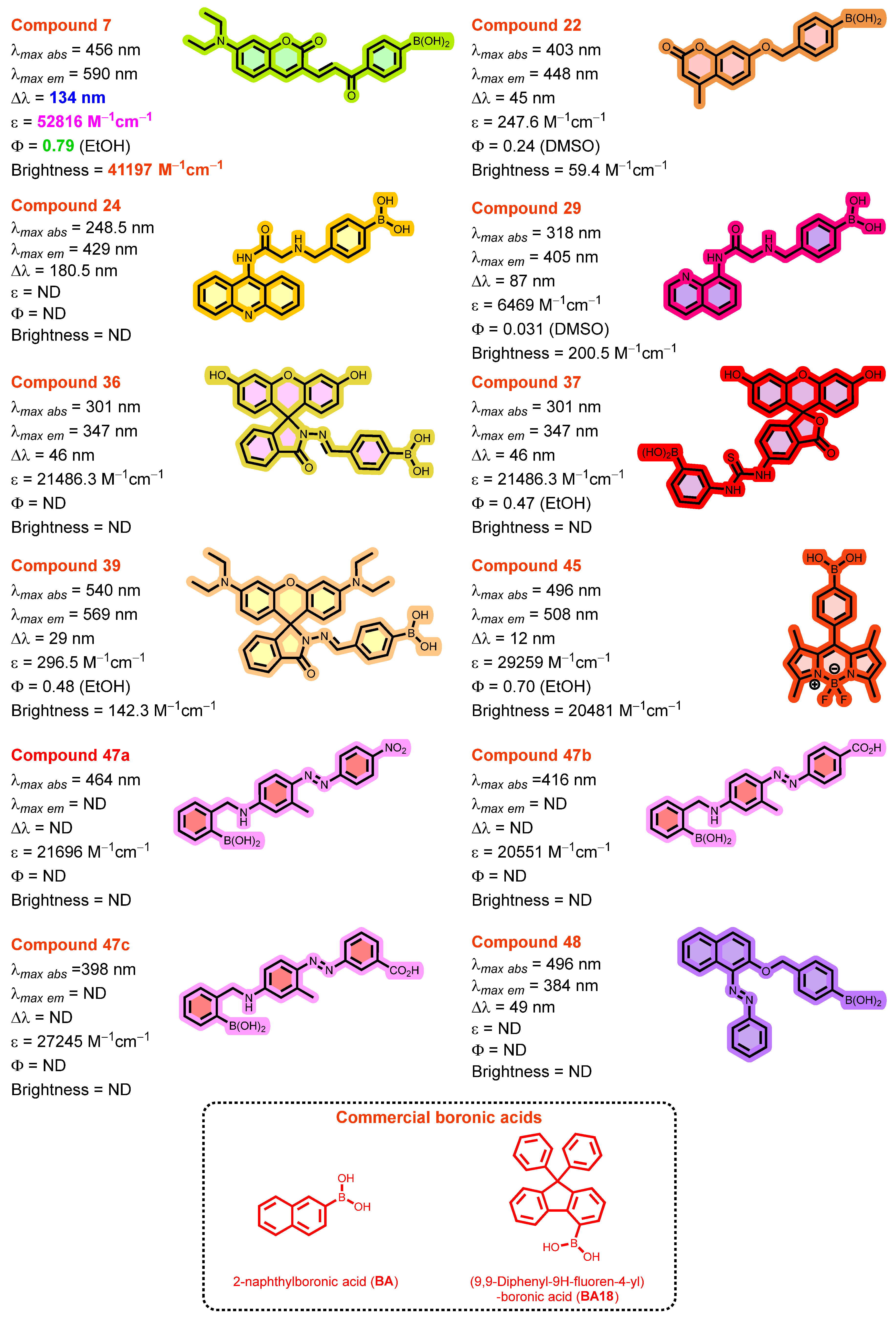
A complete set of basic photophysical quantities such as absorption maxima (λabsmax), emission maxima (λemmax), Stokes shift (∆λ), molar extinction coefficient (ε), fluorescence quantum yield (ΦF), and brightness of twelve (12) synthesised arylboronic acid chemosensors and two (2) commercially available boronic acids (2-naphthylboronic acid (BA) and 9,9-diphenyl-9H-fluoren-4-ylboronic acid (BA18)) were determined in different solvents including ethanol, dimethyl sulfoxide, and methanol through absorption and emission spectroscopy techniques. The data are summarised in Figure 8 and Table 1.
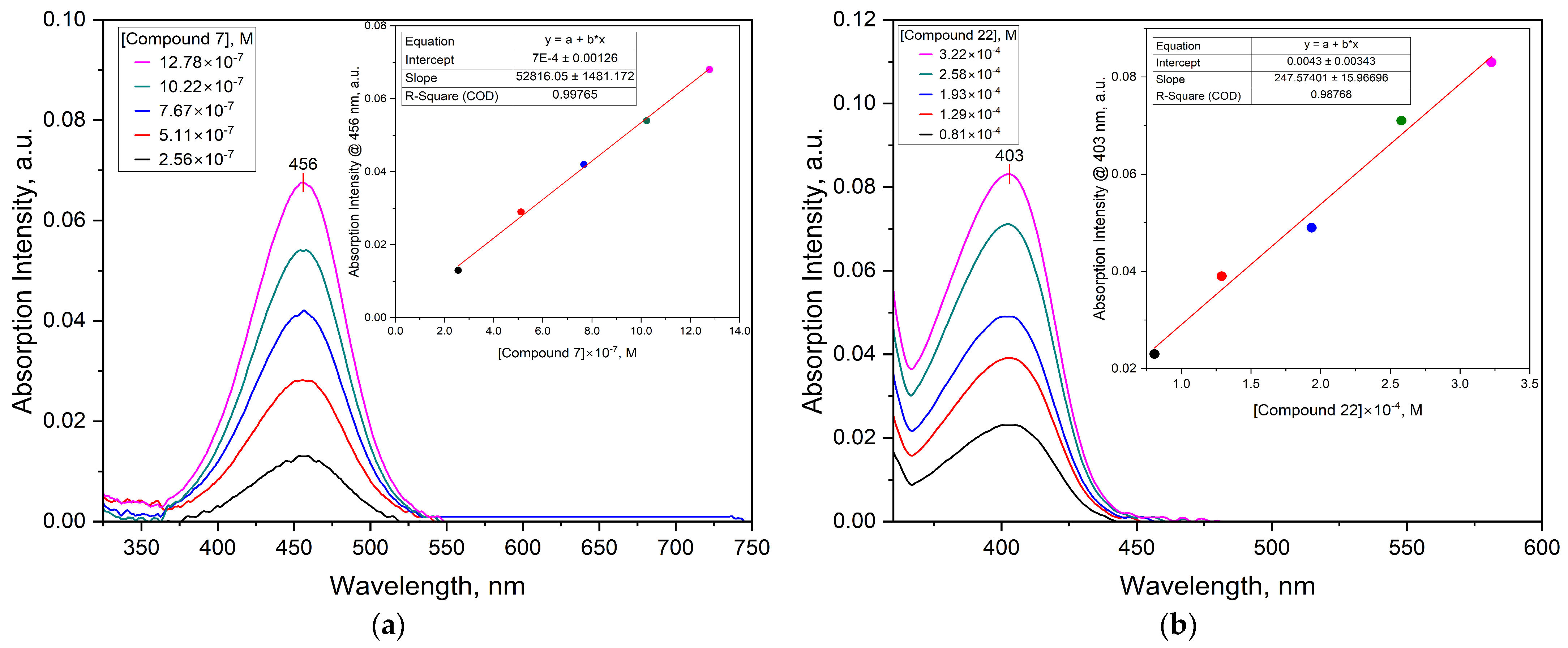
The UV-vis absorption spectra of the different synthesised compounds are depicted in Figure 9 (see also the Supplementary Materials). The absorption maxima wavelengths are recorded in Table 1. The UV–vis spectroscopy was also used to obtain the experimental extinction coefficient (ε). It was observed that the synthesised compounds were characterised by ε ranging from very low values for 22 (ε = 248 ±16 M−1cm−1 at 403 nm) to the largest for 7 (ε = 52,800 ± 1481 M−1cm−1 at 456 nm).
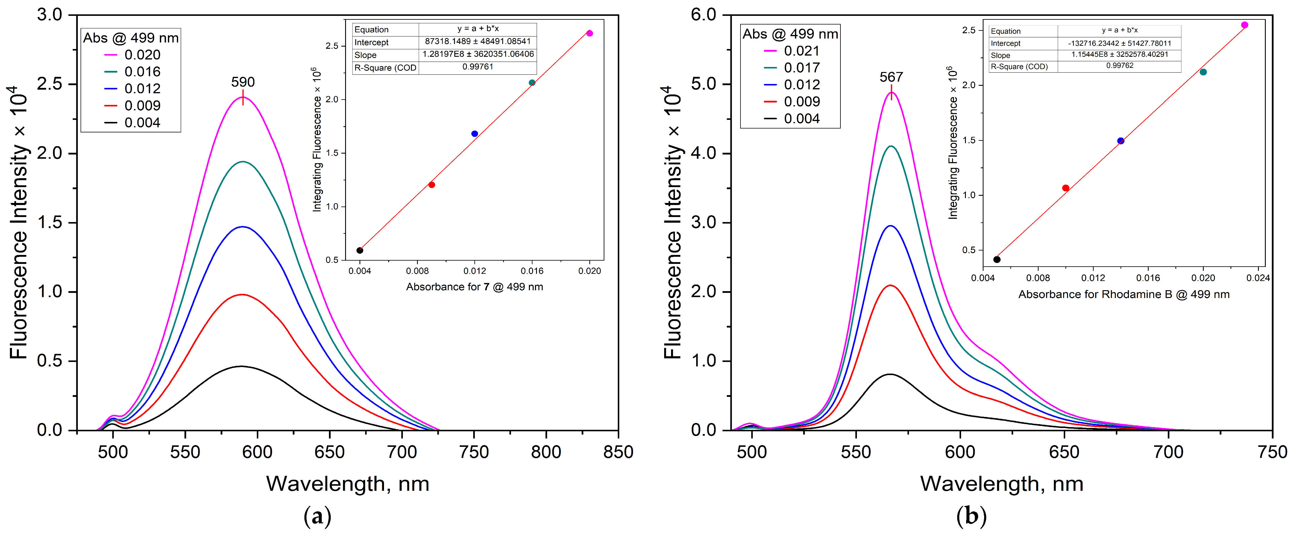
Additionally, the fluorescence emission curves were obtained by exciting the compounds at their respective absorption maxima. The normalised fluorescence emission spectra of the synthesised compounds are shown in Figure 10 and Figure S90. The maximum emission bands are as follows: 37em = 525 nm at λex = 480 nm), 7em = 590 nm at λex = 456 nm), 29em = 405 nm at λex = 318 nm), 36em = 347 nm at λex = 301 nm), 22em = 448 nm at λex = 403 nm), 39em = 569 nm at λex = 540 nm), and 45em = 508 nm at λex = 496 nm). The Stokes shift (∆λ)—defined as the difference between absorption maxima (λabsmax) and emission maxima (λemmax)—was calculated for the various compounds. Fluorescence spectra of the varying concentrations of unknown samples and references with the absorbance values and integrated intensities (area under curve) of both samples and corresponding references are shown in Table 2. The relative fluorescence quantum yield (ΦF) was determined relative to Rhodamine B in ethanol and Quinine sulphate in 0.1 M H2SO4 from fluorescence measurements. Quantum yield measurements require low absorbance (optical density), typically below 0.1 at the longest wavelength absorption maxima. Therefore, all solutions of all synthesised arylboronic dyes were prepared at low concentrations to limit the absorbance values to less than 0.1 in order to avoid any complications with dimer or aggregate formation [132], reduce or minimise possible non-linear effects arising from the inner filter (reabsorption effects) [64,132], and eliminate concentration-quenching effects, internal filter effects, and errors arising from uneven distribution of the excited species in the detected volume [65], which altogether may otherwise skew the resulting quantum yield.
Finally, the brightness of each of the synthesised dyes was calculated from the molar absorption coefficient at the excitation wavelength and the fluorescence quantum yield values. Brightness is defined as the product of ε and ΦF (B = ΦF × ε). The brightness values allow for more practically meaningful comparisons between different dyes and, as a result, can be used to determine the analytical sensitivity from the fluorophore side [52,133,134]. From the data obtained, the fluorescence emissions of most of the compounds occurred at longer wavelengths in the visible region of the electromagnetic spectrum as compared to the absorption spectra. This can be explained by the general principle that there are usually energy losses associated with absorption and emission spectra, thus shifting the fluorescence spectra to longer wavelengths [135]. The fluorescence observed can be attributed to n→π* or π→π* transition states [136]. The ∆λ values ranged from the least for 45 (∆λ = 12 nm) to sufficiently large Stokes shift values (∆λ > 80 nm) for 29, 7, and 24 with ∆λ values of 87, 134, and 180.5 nm respectively. The BODIPY analogue 45 showed an intense absorption band with a maximum at 496 nm assigned at spin-allowed π-π* transitions, high molar extinction coefficient (ε = 29,259 ± 450 M−1cm−1 at 496 nm), emission maximum at 508 nm with the lowest calculated ∆λ value of 12 nm. These properties are characteristic of the BODIPY chromophore subunits and consistent with reported data [115,137]. On the other hand, 7 with a high ∆λ value of 134 nm has the coumarin core moiety.
Some of the synthesised compounds such as 47a47c did not show fluorescence. Incidentally, all three compounds are azo dyes and this was not surprising because as a general rule, these categories of dyes do not fluoresce [138]. Here, the absorbed energy is dissipated by the medium or emitted as phosphorescence, which has a longer lifetime for the excited state [136]. However, all three synthesised azo dyes were characterised by high molar extinction coefficient values for example 47a (ε = 21,696 ± 1997 M−1cm−1 at 464 nm), 47b (ε = 20,551 ± 360 M−1cm−1 at 416 nm), and 47c (ε = 27,245 ± 467 M−1cm−1 at 398 nm). The quantum yields for these dyes were not determined due to a lack of significant fluorescence emissions at their respective absorption maxima values and also because very small quantum yield values have been acquired for other azo dyes [139,140]. Unlike the azo dyes 47a47c, this was not the case for 48, a Sudan I boronic acid derivative which is also an azo dye. Fluorescence was observed with 48, giving an emission maximum at 384 nm and a ∆λ value of 49 nm. As a general principle, small-molecule fluorophores require a high degree of aromaticity to have absorptions in the visible region of the spectrum [4,135]. The compound with the largest molar extinction coefficient (ε = 52,816 ± 1481 M−1cm−1 at 456 nm), a ∆λ value of 134 nm, ΦF = 0.78, and brightness value (41,196.6 M−1cm−1) was 7, a coumarin derivative. This is typical of coumarin derivatives, which are usually characterised by high molar extinction coefficients in the near-UV and visible range and high fluorescence emission, and, as a result, have found many uses as fluorescent chromophores for several applications [141,142].
Based on the excellent photophysical properties of 7, its fluorescent properties toward mycolactone were investigated. Sensitive detection of mycolactone was achieved through fluorescence spectroscopy as shown in Figure 11. For instance, free 7 (2.5 × 10⁻³ mg/mL) exhibited weaker fluorescence intensity at the emission maximum of 581 nm. However, the fluorescence intensity gradually increased as the concentration of mycolactone increased from 0 to 8.0 × 10⁻³ mg/mL (0–1.6 equiv.). The enhanced fluorescence of 7 was also visually observed on f-TLC plates when coupled with mycolactone and under a 365 nm UV lamp, as shown in Figure 12.
This can be explained by the coupling of the boronic acid moiety in the structure of 7 with the 1,3-diol moieties of mycolactone to form six-membered cyclic boronate esters, as has been established in the previous section on the proof-of-concept studies. The formation of the boronate coupled with the chromophore of the mycolactone resulted in the enhanced fluorescence. A calibration curve was obtained between the concentration of mycolactone and fluorescence intensity at 581 nm with good linearity (R2 = 0.9886) (inset, Figure 11). The limit of detection (LOD) and limit of quantification (LOQ) were estimated to be 0.85 ng/µL (signal-to-noise ratio (S/N) × 3) and 2.84 ng/µL (signal-to-noise ratio (S/N) × 10), respectively; where N is the standard deviation (SD) of the peak area (n = 8), taken as a measure of the noise, and B is the slope of the corresponding calibration curve. This reveals that the 7 could detect a nanomolar-level concentration of mycolactone.

3.3. Detection of Mycolactone by the f-TLC Method Using the Synthesised Dyes

Boronic acids form reversible cyclic boronate complexes with 1,2- or 1,3-diols and this property. Neutral boronic acids are electrophilic because of the vacant p-orbital on the boron, and thus are highly reactive toward nucleophiles, such as diols [143]. The interaction between a boronic acid and a diol is one of the most powerful functional group interactions [144]. This unique property makes boronic acids useful selective and sensitive detection reagents for diols in TLC. For instance, boronic acids have been employed in the detection of mycolactone on thin-layer chromatography (TLC) [44,46]. Here, the synthesised aryl boronic acids coupled to mycolactone and the resulting fluorescent boronate complexes were visualised under a benchtop UV lamp (365 nm). The detection of mycolactone spots on TLC plates was optimised by using different concentrations of the synthesised boronic acids. Figure 12 shows the images of the fluorescence response of 5 µL/spot of synthetic mycolactone after dipping in 0.1 mM and 0.01 mM acetone concentrations of various synthesised fluorescent aryl boronic acid chemosensors, together with the two boronic acids previously reported for use in the f-TLC method which were obtained commercially (BA and BA18) [44,45,48].
First, a remarkable enhancement of fluorescence intensity of mycolactone was generally observed after dipping in either 0.01 mM or 0.1 mM solutions of the various boronic acids, particularly after heating. In this study, glass-backed TLC plates are used for the purpose of heating. Heating the plates to approximately 100 °C for just 60 s was sufficient to ensure the complete formation of the cyclic boronate esters of the various boronic acid sensors with the 1,3-diols motifs of the mycolactone. The boronic acid motif that is incorporated into various dyes has an empty sp2 hybridised p-orbital on the boron centre with O−B−O bond angle of ~120°. It therefore readily binds covalently to 1,3-diols moieties of mycolactone and adopts sp3 hybridisation in the cyclic boronate ester product, with the boron adopting a tetrahedral configuration with O−B−O ~109.5°, like those in the literature [143]. This then triggers a change in the spectroscopic properties of the adduct because of significant perturbation of the π system. Secondly, the enhanced fluorescence after coupling was also attributed to the various signalling moieties—minoacridine, aminoquinoline, azo, BODIPY, coumarin, fluorescein, and rhodamine variants. They act as excellent fluorescent enhancers by stimulating the polarisation of the C–B bond due to extended π-conjugation systems. This induced the production of a rapid signal because of the highly polar C–B bond along with the corresponding boronate unit.
Generally, it was observed that the fluorescence band intensities of mycolactone adducts on TLC plates dipped in 0.1 mM acetone solutions of the synthesised chemosensors appeared more intense than those dipped in the same concentration of the commercially available BA and BA18 solutions, except for 37, 48, 47c, 39, and 45. This observation was similar when 0.01 mM solutions of the boronic acids were used. Unlike other spots, it was observed that the mycolactone adduct spots appeared black on different backgrounds in solutions of 37, 24, 39, and 45. Compound 37 produced the most intense black spot in the 0.01 mM solution while the 24 plate had the most intense black spot in the 0.1 mM solution.
Fluorescence is observable only if the absorbed energy is not fully dissipated during internal conversion. Typically, compounds that come into contact with the stationary phase lose some of their energy through transfer to the layer, which lowers the intensity of their fluorescence. When adsorbed onto silica gel, compounds that glow weakly in solution may either exhibit no fluorescence at all or fluorescence at wavelengths that are significantly longer than those observed in solution. An excited molecule in TLC quickly releases its vibrational energy as heat into the surrounding medium; in other words, the activated molecules transfer their excess photons to the TLC layer or to nearby molecules. Higher sample concentrations raise the likelihood of an excited and unexcited molecule colliding in a TLC layer [136]. Compounds 7, 29, 47a, and 47b had outstanding fluorescent bands on TLC, especially in 0.1 mM solutions. In the case of 37, 47c, 36, 39, and 45 the mycolactone adduct of TLC plates dipped in 0.1 mM solutions showed weak fluorescent band intensities in comparison to the band intensities of plates that were dipped in the 10-fold lower concentrations of the same boronic acids. This could partly be a result of fluorescence quenching leading to the reduced fluorescence bands on TLC [136]. Should this be the case, then, this concentration range might be unsuitable for evaluating these boronic acids because of the dramatic concentration quenching of fluorescence.
Compounds 36 and 37 are both fluorescein-based chemosensors. However, whereas 37 was fluorescent, 36 was weakly fluorescent. Generally, fluorescein dyes possess a unique spirolactam ring structure that exhibits a specific close-and-open feature with a ‘turn-on’ fluorescence response. However, when compound 36 was reacted with mycolactone, there was a fluorescence turn-on effect on TLC. This could be attributed the fact that the analyte, upon binding, was able induce the opening of the spirolactam ring which led to the enhancement in the fluorescence spot that was observed [145]. The fluorescence of compound 37 was strongly quenched when the adduct was formed on TLC in contrast to that of 36. Unlike compound 36, the fluorescence of compound 37 adduct was significantly quenched on TLC. It is thought that a photoinduced electron-transfer mechanism causes compound 37 adduct fluorescence to be quenched. Compound 37 is a derivative of fluorescein isothiocyanate isomer I (FITC), and FITC fluorescence frequently exhibits self-quenching. Self-quenching usually happens at high concentrations due to increased energy transfer between interacting dye molecules as their concentration increases. Thus, it is possible that the high concentrations may be the cause of the quenching seen in 37 [146,147].
Compounds 7 and 22 both have a coumarin core; however, 7 was fluorescent, whereas 22 was non-fluorescent. In the design of compound 7, the π-conjugation was extended at 3-position on the coumarin ring. The extended π-electron moiety leads to a red shift in the absorption and emission spectra. This means that the emitted light had a longer wavelength, and can also enhance fluorescence intensity [148]. Secondly, the enhanced fluorescence intensity observed in compound 7 could also be attributed to the presence of the electron-donating diethylamino moiety at the 7-position which influences the overall electronic structure of the molecule. This makes compound 7 a more efficient emitter of light, in which the emitted light had a longer wavelength [149]. On the other hand, compound 22 is a 7-hydroxycoumarin (unbelliferone) derivative and its fluorescence is entirely dependent on the excitation of the phenolic form only. Thus, it is non-fluorescent in its neutral form, but becomes strongly fluorescent as the phenolate anion in alkaline medium due to the blue emission of the phenolate anion [150]. Therefore, the non-fluorescence that was observed in 22 could possibly be explained by the derivation of the 7-hydroxy group to an ether linker to the methylphenylboronic acid recognition unit. In addition to the coumarin substitution, the fluorescence intensities in 7 and 22 could also be influenced by the solvent and solution pH [151,152]. The intensity of the fluorescence increases with increasing solution pH [152], and the absorbance of coumarin derivatives is red-shifted with increasing solvent polarity, while the emission of the coumarin moieties is broadened and shifted to higher wavelengths because of reinforced hydrogen bonding [148].
Solvent effect could also be attributed to the fluorescence intensity patterns that were observed in the aminoacridine adduct 24 and the 8-aminoquinoline adduct 29. Hence, we observed that, whereas fluorescence enhancement was seen in the case of the adduct of compound 29, that of 24 adduct was substantially quenched. As with the earlier sensors that contained the 8-aminoquinoline moiety, we proposed that the complexation between mycolactone and compounds 24 and 29 provided the “off-on” response via an internal charge transfer effect [153,154].
There was a turn-on effect in the fluorescence of the adducts of the azo derivatives 47ac and 48. The boronic-acid-appended azo dyes are by themselves coloured in nature because of their unique chromophoric properties and can produce colour change in the presence of diols due to the enhancement of the Lewis acidity of boron upon binding. One possible explanation for the colorimetric turn-on response of these derivatives upon binding with mycolactone would be the combination of chromophoric characteristics and the Lewis acidity of boron in the complex. The azo chromophores have the capacity to transition into a donor-π-acceptor compound when Lewis bases such as the diols moietie on mycolactone coordinate at the Lewis acidic boron centre of the dyes, and this is accompanied by an increase in intramolecular charge-transfer interactions leading to the turn-on effect [155,156].
Under 365 nm UV irradiation, BODIPY compound 45 exhibited intense fluorescence and produced an intense bright green colour. However, the mycolactone adduct that was formed on TLC was effectively quenched. The significant quenching can be attributed to photoinduced electron transfer between mycolactone, lying in close proximity to boronic acid moiety of the BODIPY dye. In the case of compound 45 where the BODIPY is appended to boronic acid moiety as a recognition unit, boron assumes sp2 hybridisation and hence is strongly fluorescent upon UV irradiation. However, the fluorescence is almost completely quenched in the adduct where boron, upon binding to the diol moieties of mycolactone, assumes sp3-hybridised status [157,158,159]. Similarly, the rhodamine-based derivative 39, which had a maximum absorption at 540 nm, was colourless in solution and turned pink when viewed with the naked eye. Under 365 light, it fluoresced orange at a maximum of 569 nm because of the spirolactam ring opening [99]. On TLC, however, quenching was noted following the binding of the diol moieties of mycolactone to the boronic acid unit of compound 39. This could be attributed to the inability of the complexation to facilitate the opening of the spirolactam unit of the rhodamine moiety of 39. This shows that the energy transfer mechanism did not take place, resulting in the quenching of the fluorescence. Additionally, it is commonly known that other environmental factors including solvent polarity, viscosity, and temperature can impact the fluorescence properties of rhodamine dyes [160,161]. For instance, the rate of collisions increases with temperature, resulting in a greater amount of collisional quenching. The high temperature (~100 °C) used in the adduct formation of compound 39 could possibly promote the non-radiative process of its diethylamino groups and results in a condition that resembles a non-emissive TICT (twisted intramolecular charge transfer), resulting in the quenching in fluorescence intensity as was observed [162,163].

4. Conclusions

This paper has demonstrated synthetic approaches for the synthesis of a library of fluorescent arylboronic acid chemosensors by exploiting a range of dyes (fluorophores) of interest with beneficial photophysical characteristics. All the synthesised molecules have a boronic acid motif (recognition moiety) linked to a fluorescent dye which serves as a signalling moiety for possible complexation with the 1,3-diol moieties of mycolactone. All the synthesised dyes have been completely characterised using a set of complementary spectrometric and spectroscopic techniques such as NMR, LC-MS, FT-IR, and in some cases, X-ray crystallography. Following the successful characterisation of the synthesised dyes, their photophysical properties were determined. Likewise, their performance on TLC was also investigated in comparison to BA and BA18. The findings of the study indicated that the synthesised boronic acids were able to detect mycolactone both in solution and on TLC plates selectively and sensitively. Dye 7 had a better detection limit than BA and BA18. However, the diagnostic performance of the synthesised dye 7 is yet to be evaluated using real clinical samples of suspected BU cases. From the results, it can be concluded that all the synthesised arylboronic acids are selective for the detection of mycolactone and 7 gave the best outcomes in terms of fluorescence on TLC plates. It returned λabsmax = 456 nm, λemmax = 590 nm, ∆λ = 134 nm, ε = 52,816 M−1cm−1, ΦF = 0.78, and brightness = 41,197 M−1cm−1.

Supplementary Materials

The following supporting information can be downloaded at: https://www.mdpi.com/article/10.3390/chemosensors13070244/s1, Figure S1–S87: 1H NMR, 13C NMR, 11B NMR, ESI-QTOF HRMS, and FTIR spectra of synthesized compounds; Figure S88: ORTEP representation of the molecular structure of BODIPY dye 45; Figure S89: Absorbance spectra of various dyes in different organic solvents; Figure S90: Fluorescence emission spectra of synthesized compounds; Table S1: Crystal data and refinement details for the X-ray structure determinations of 34 and 45.

Author Contributions

Conceptualisation, G.A.A., B.M.P., T.D.C. and R.K.A.; methodology, G.A.A., B.M.P., T.D.C. and R.K.A.; validation, G.A.A., B.M.P., T.D.C., K.B.A. and R.K.A.; formal analysis, G.A.A., B.M.P. and R.K.A.; investigation, G.A.A., B.M.P., T.D.C. and R.K.A.; resources, B.M.P., K.B.A. and R.K.A.; data curation, G.A.A., B.M.P. and R.K.A.; writing—original draft preparation, G.A.A.; writing—review and editing, G.A.A., B.M.P., T.D.C. and R.K.A.; visualisation, G.A.A.; supervision, B.M.P., T.D.C. and R.K.A.; project administration, R.K.A. All authors have read and agreed to the published version of the manuscript.

Funding

This research was not funded by any organisation. Mass spectrometric and X-ray crystallography analysis was performed at the University of Sheffield.

Institutional Review Board Statement

Not applicable.

Informed Consent Statement

Not applicable.

Data Availability Statement

Data are contained within the article and supplementary materials.

Acknowledgments

The authors are grateful to the late Kishi Yoshito for generously donating synthetic mycolactone A/B through the World Health Organization for the study and to the Erasmus+ mobility grant between University of Ghana and University of Sheffield and Ghana National Petroleum Company (GNPC) for funding Gideon Atinga Akolgo’s Doctoral research.

Conflicts of Interest

Author Kingsley Bampoe Asiedu was employed by the World Health Organization. The remaining authors declare that the research was conducted in the absence of any commercial or financial relationships that could be construed as a potential conflict of interest.

References

  1. Whyte, G.F.; Vilar, R.; Woscholski, R. Molecular recognition with boronic acids—Applications in chemical biology. J. Chem. Biol. 2013, 6, 161–174. [Google Scholar] [CrossRef] [PubMed]
  2. Czarnik, A.W. Chemosensors of Ion and Molecule Recognition; Springer Science & Business Media: Berlin, Germany, 2012; Volume 492. [Google Scholar]
  3. Wu, J.-S.; Liu, W.-M.; Zhuang, X.-Q.; Wang, F.; Wang, P.-F.; Tao, S.-L.; Zhang, X.-H.; Wu, S.-K.; Lee, S.-T. Fluorescence Turn On of Coumarin Derivatives by Metal Cations:  A New Signaling Mechanism Based on C=N Isomerization. Org. Lett. 2007, 9, 33–36. [Google Scholar] [CrossRef]
  4. Valeur, B.; Berberan-Santos, M.N. Molecular Fluorescence: Principles and Applications; John Wiley & Sons: Hoboken, NJ, USA, 2012. [Google Scholar]
  5. Tharmaraj, V.; Pitchumani, K. d-Glucose sensing by (E)-(4-((pyren-1-ylmethylene)amino)phenyl) boronic acid via a photoinduced electron transfer (PET) mechanism. RSC Adv. 2013, 3, 11566–11570. [Google Scholar] [CrossRef]
  6. Curtius, H.; Kaiser, G.; Müller, E.; Bosbach, D. Radionuclide release from research reactor spent fuel. J. Nucl. Mater. 2011, 416, 211–215. [Google Scholar] [CrossRef]
  7. Wu, X.; Chen, X.-X.; Jiang, Y.-B. Recent advances in boronic acid-based optical chemosensors. Analyst 2017, 142, 1403–1414. [Google Scholar] [CrossRef]
  8. Hall, D.G. Boronic Acids; John Wiley & Sons: Hoboken, NJ, USA, 2011. [Google Scholar] [CrossRef]
  9. Springsteen, G.; Wang, B. A detailed examination of boronic acid–diol complexation. Tetrahedron 2002, 58, 5291–5300. [Google Scholar] [CrossRef]
  10. Springsteen, G.; Wang, B. Alizarin Red S. as a general optical reporter for studying the binding of boronic acids with carbohydrates. Chem. Commun. 2001, 32, 1608–1609. [Google Scholar] [CrossRef]
  11. Lorand, J.P.; Edwards, J.O. Polyol complexes and structure of the benzeneboronate ion. J. Org. Chem. 1959, 24, 769–774. [Google Scholar] [CrossRef]
  12. Yoon, J.; Czarnik, A.W. Fluorescent chemosensors of carbohydrates. A means of chemically communicating the binding of polyols in water based on chelation-enhanced quenching. J. Am. Chem. Soc. 1992, 114, 5874–5875. [Google Scholar] [CrossRef]
  13. Guo, Z.; Shin, I.; Yoon, J. Recognition and sensing of various species using boronic acid derivatives. Chem. Commun. 2012, 48, 5956–5967. [Google Scholar] [CrossRef]
  14. Lacina, K.; Skládal, P.; James, T.D. Boronic acids for sensing and other applications—a mini-review of papers published in 2013. Chem. Cent. J. 2014, 8, 60. [Google Scholar] [CrossRef] [PubMed]
  15. Chaicham, A.; Sahasithiwat, S.; Tuntulani, T.; Tomapatanaget, B. Highly effective discrimination of catecholamine derivatives via FRET-on/off processes induced by the intermolecular assembly with two fluorescence sensors. Chem. Commun. 2013, 49, 9287–9289. [Google Scholar] [CrossRef]
  16. Kaur, G.; Fang, H.; Gao, X.; Li, H.; Wang, B. Substituent effect on anthracene-based bisboronic acid glucose sensors. Tetrahedron 2006, 62, 2583–2589. [Google Scholar] [CrossRef]
  17. Wu, W.; Mitra, N.; Yan, E.C.; Zhou, S. Multifunctional hybrid nanogel for integration of optical glucose sensing and self-regulated insulin release at physiological pH. ACS Nano 2010, 4, 4831–4839. [Google Scholar] [CrossRef]
  18. Wu, W.; Zhou, T.; Berliner, A.; Banerjee, P.; Zhou, S. Glucose-Mediated Assembly of Phenylboronic Acid Modified CdTe/ZnTe/ZnS Quantum Dots for Intracellular Glucose Probing. Angew. Chem. Int. Ed. 2010, 49, 6554–6558. [Google Scholar] [CrossRef]
  19. Huang, Y.-J.; Ouyang, W.-J.; Wu, X.; Li, Z.; Fossey, J.S.; James, T.D.; Jiang, Y.-B. Glucose sensing via aggregation and the use of “knock-out” binding to improve selectivity. J. Am. Chem. Soc. 2013, 135, 1700–1703. [Google Scholar] [CrossRef]
  20. Zhai, W.; Male, L.; Fossey, J.S. Glucose selective bis-boronic acid click-fluor. Chem. Commun. 2017, 53, 2218–2221. [Google Scholar] [CrossRef] [PubMed]
  21. Pohanka, M. Glycated Hemoglobin and Methods for Its Point of Care Testing. Biosensors 2021, 11, 70. [Google Scholar] [CrossRef]
  22. Zhu, X.; Zhou, X.; Xing, D. Ultrasensitive and selective detection of mercury (II) in aqueous solution by polymerase assisted fluorescence amplification. Biosens. Bioelectron. 2011, 26, 2666–2669. [Google Scholar] [CrossRef]
  23. Guan, R.; Chen, H.; Cao, F.; Cao, D.; Deng, Y. Two fluorescence turn-on chemosensors for cyanide anions based on pyridine cation and the boronic acid moiety. Inorg. Chem. Commun. 2013, 38, 112–114. [Google Scholar] [CrossRef]
  24. Lee, S.A.; You, G.R.; Choi, Y.W.; Jo, H.Y.; Kim, A.R.; Noh, I.; Kim, S.-J.; Kim, Y.; Kim, C. A new multifunctional Schiff base as a fluorescence sensor for Al3+ and a colorimetric sensor for CN in aqueous media: An application to bioimaging. Dalton Trans. 2014, 43, 6650–6659. [Google Scholar] [CrossRef] [PubMed]
  25. Pizer, R.; Tihal, C. Equilibria and reaction mechanism of the complexation of methylboronic acid with polyols. Inorg. Chem. 1992, 31, 3243–3247. [Google Scholar] [CrossRef]
  26. Pizer, R.D.; Tihal, C.A. Mechanism of boron acid/polyol complex formation. comments on the trigonal/tetrahedral interconversion on boron. Polyhedron 1996, 15, 3411–3416. [Google Scholar] [CrossRef]
  27. DeFrancesco, H.; Dudley, J.; Coca, A. Boron chemistry: An overview. In Boron Reagents in Synthesis; American Chemical Society: Washington, DC, USA, 2016; pp. 1–25. [Google Scholar]
  28. Ooyama, Y.; Furue, K.; Uenaka, K.; Ohshita, J. Development of highly-sensitive fluorescence PET (photo-induced electron transfer) sensor for water: Anthracene–boronic acid ester. RSC Adv. 2014, 4, 25330–25333. [Google Scholar] [CrossRef]
  29. Ooyama, Y.; Matsugasako, A.; Oka, K.; Nagano, T.; Sumomogi, M.; Komaguchi, K.; Imae, I.; Harima, Y. Fluorescence PET (photo-induced electron transfer) sensors for water based on anthracene–boronic acid ester. Chem. Commun. 2011, 47, 4448–4450. [Google Scholar] [CrossRef] [PubMed]
  30. Chung, C.; Srikun, D.; Lim, C.S.; Chang, C.J.; Cho, B.R. A two-photon fluorescent probe for ratiometric imaging of hydrogen peroxide in live tissue. Chem. Commun. 2011, 47, 9618–9620. [Google Scholar] [CrossRef]
  31. Nonaka, H.; An, Q.; Sugihara, F.; Doura, T.; Tsuchiya, A.; Yoshioka, Y.; Sando, S. Phenylboronic Acid-based 19F MRI Probe for the Detection and Imaging of Hydrogen Peroxide Utilizing Its Large Chemical-Shift Change. Anal. Sci. 2015, 31, 331–335. [Google Scholar] [CrossRef]
  32. Fu, H.; Fang, H.; Sun, J.; Wang, H.; Liu, A.; Sun, J.; Wu, Z. Boronic acid-based enzyme inhibitors: A review of recent progress. Curr. Med. Chem. 2014, 21, 3271–3280. [Google Scholar] [CrossRef]
  33. Cai, B.; Luo, Y.; Guo, Q.; Zhang, X.; Wu, Z. A glucose-sensitive block glycopolymer hydrogel based on dynamic boronic ester bonds for insulin delivery. Carbohydr. Res. 2017, 445, 32–39. [Google Scholar] [CrossRef]
  34. Fang, H.; Kaur, G.; Wang, B. Progress in boronic acid-based fluorescent glucose sensors. J. Fluoresc. 2004, 14, 481–489. [Google Scholar] [CrossRef]
  35. Chang, M.-H.; Chang, C.-N. Synthesis of three fluorescent boronic acid sensors for tumor marker Sialyl Lewis X in cancer diagnosis. Tetrahedron Lett. 2014, 55, 4437–4441. [Google Scholar] [CrossRef]
  36. Chu, Y.; Wang, D.; Wang, K.; Liu, Z.L.; Weston, B.; Wang, B. Fluorescent conjugate of sLex-selective bisboronic acid for imaging application. Bioorganic Med. Chem. Lett. 2013, 23, 6307–6309. [Google Scholar] [CrossRef] [PubMed]
  37. George, K.M.; Chatterjee, D.; Gunawardana, G.; Welty, D.; Hayman, J.; Lee, R.; Small, P.L. Mycolactone: A polyketide toxin from Mycobacterium ulcerans required for virulence. Science 1999, 283, 854–857. [Google Scholar] [CrossRef] [PubMed]
  38. Demangel, C.; Stinear, T.P.; Cole, S.T. Buruli ulcer: Reductive evolution enhances pathogenicity of Mycobacterium ulcerans. Nat. Rev. Microbiol. 2009, 7, 50–60. [Google Scholar] [CrossRef]
  39. George, K.M.; Pascopella, L.; Welty, D.M.; Small, P.L. A Mycobacterium ulcerans toxin, mycolactone, causes apoptosis in guinea pig ulcers and tissue culture cells. Infect. Immun. 2000, 68, 877–883. [Google Scholar] [CrossRef]
  40. Scherr, N.; Gersbach, P.; Dangy, J.P.; Bomio, C.; Li, J.; Altmann, K.H.; Pluschke, G. Structure-activity relationship studies on the macrolide exotoxin mycolactone of Mycobacterium ulcerans. PLoS Neglected Trop. Dis. 2013, 7, e2143. [Google Scholar] [CrossRef]
  41. Saint-Auret, S.; Chany, A.C.; Casarotto, V.; Tresse, C.; Parmentier, L.; Abdelkafi, H.; Blanchard, N. Total Syntheses of Mycolactone A/B and its Analogues for the Exploration of the Biology of Buruli Ulcer. Chimia 2017, 71, 836–840. [Google Scholar] [CrossRef] [PubMed]
  42. Spangenberg, T.; Kishi, Y. Highly sensitive, operationally simple, cost/time effective detection of the mycolactones from the human pathogen Mycobacterium ulcerans. Chem. Commun. 2010, 46, 1410–1412. [Google Scholar] [CrossRef]
  43. Hong, H.; Coutanceau, E.; Leclerc, M.; Caleechurn, L.; Leadlay, P.F.; Demangel, C. Mycolactone diffuses from Mycobacterium ulcerans–infected tissues and targets mononuclear cells in peripheral blood and lymphoid organs. PLoS Neglected Trop. Dis. 2008, 2, e325. [Google Scholar] [CrossRef]
  44. Sarfo, F.S.; Phillips, R.O.; Rangers, B.; Mahrous, E.A.; Lee, R.E.; Tarelli, E.; Asiedu, K.B.; Small, P.L.; Wansbrough-Jones, M.H. Detection of Mycolactone A/B in Mycobacterium ulcerans-Infected Human Tissue. PLoS Neglected Trop. Dis. 2010, 4, e577. [Google Scholar] [CrossRef]
  45. Akolgo, G.A.; Partridge, B.M.; Craggs, T.D.; Amewu, R.K. Alternative boronic acids in the detection of Mycolactone A/B using the thin layer chromatography (f-TLC) method for diagnosis of Buruli ulcer. BMC Infect. Dis. 2023, 23, 495. [Google Scholar] [CrossRef] [PubMed]
  46. Kishi, Y. Chemistry of mycolactones, the causative toxins of Buruli ulcer. Proc. Natl. Acad. Sci. USA 2011, 108, 6703–6708. [Google Scholar] [CrossRef] [PubMed]
  47. Converse, P.J.; Xing, Y.; Kim, K.H.; Tyagi, S.; Li, S.Y.; Almeida, D.V.; Nuermberger, E.L.; Grosset, J.H.; Kishi, Y. Accelerated detection of mycolactone production and response to antibiotic treatment in a mouse model of Mycobacterium ulcerans disease. PLoS Neglected Trop. Dis. 2014, 8, e2618. [Google Scholar] [CrossRef] [PubMed]
  48. Amewu, R.K.; Akolgo, G.A.; Asare, M.E.; Abdulai, Z.; Ablordey, A.S.; Asiedu, K. Evaluation of the fluorescent-thin layer chromatography (f-TLC) for the diagnosis of Buruli ulcer disease in Ghana. PLoS ONE 2022, 17, e0270235. [Google Scholar] [CrossRef]
  49. Wadagni, A.; Frimpong, M.; Phanzu, D.M. Simple, rapid Mycobacterium ulcerans disease diagnosis from clinical samples by fluorescence of mycolactone on thin layer chromatography. PLoS Neglected Trop. Dis. 2015, 9, e0004247. [Google Scholar] [CrossRef]
  50. Yuan, L.; Lin, W.; Yang, Y.; Chen, H. A unique class of near-infrared functional fluorescent dyes with carboxylic-acid-modulated fluorescence ON/OFF switching: Rational design, synthesis, optical properties, theoretical calculations, and applications for fluorescence imaging in living animals. J. Am. Chem. Soc. 2012, 134, 1200–1211. [Google Scholar] [CrossRef]
  51. Yang, X.-F.; Guo, X.-Q.; Zhao, Y.-B. Development of a novel rhodamine-type fluorescent probe to determine peroxynitrite. Talanta 2002, 57, 883–890. [Google Scholar] [CrossRef]
  52. Lavis, L.D.; Raines, R.T. Bright building blocks for chemical biology. ACS Chem. Biol. 2014, 9, 855–866. [Google Scholar] [CrossRef] [PubMed]
  53. Sheldrick, G.M. Program for Empirical Absorption Correction of Area Detector Data. SADABS; University of Göttingen: Göttingen, Germany, 1996. [Google Scholar]
  54. Krause, L.; Herbst-Irmer, R.; Sheldrick, G.M.; Stalke, D. Comparison of silver and molybdenum microfocus X-ray sources for single-crystal structure determination. J. Appl. Crystallogr. 2015, 48, 3–10. [Google Scholar] [CrossRef]
  55. Blessing, R.H. An empirical correction for absorption anisotropy. Acta Crystallogr. Sect. A Found. Crystallogr. 1995, 51, 33–38. [Google Scholar] [CrossRef]
  56. Sheldrick, G. SHELXT—Integrated space-group and crystal-structure determination. Acta Crystallogr. Sect. A 2015, 71, 3–8. [Google Scholar] [CrossRef]
  57. Sheldrick, G. Crystal structure refinement with SHELXL. Acta Crystallogr. Sect. C 2015, 71, 3–8. [Google Scholar] [CrossRef] [PubMed]
  58. Dolomanov, O.; Bourhis, L.; Gildea, R.; Howard, J.; Puschmann, H. Johnkoivulaite, Cs(Be2B)Mg2Si6O18, A new mineral of the beryl group from the gem deposits of Mogok, Myanmar. Am. Miner. 2021, 106, 1844–1851. [Google Scholar] [CrossRef]
  59. Ishiyama, T.; Murata, M.; Miyaura, N. Palladium(0)-Catalyzed Cross-Coupling Reaction of Alkoxydiboron with Haloarenes: A Direct Procedure for Arylboronic Esters. J. Org. Chem. 1995, 60, 7508–7510. [Google Scholar] [CrossRef]
  60. Akgun, B. Boronic Esters as Bioorthogonal Probes in Site-Selective Labeling of Proteins. Ph.D. Thesis, University of Alberta, Edmonton, AB, Canada, 2018. [Google Scholar]
  61. Ear, A.; Amand, S.; Blanchard, F.; Blond, A.; Dubost, L.; Buisson, D.; Nay, B. Direct biosynthetic cyclization of a distorted paracyclophane highlighted by double isotopic labelling of l-tyrosine. Org. Biomol. Chem. 2015, 13, 3662–3666. [Google Scholar] [CrossRef]
  62. Promchat, A.; Wongravee, K.; Sukwattanasinitt, M.; Praneenararat, T. Rapid Discovery and Structure-Property Relationships of Metal-Ion Fluorescent Sensors via Macroarray Synthesis. Sci. Rep. 2019, 9, 10390. [Google Scholar] [CrossRef]
  63. Lawson-Wood, K.; Upstone, S.; Evans, K. Determination of Relative Fluorescence Quantum Yields using the FL6500 Fluorescence Spectrometer. Fluoresc. Spectrosc 2018, 4, 1–5. [Google Scholar]
  64. Würth, C.; Grabolle, M.; Pauli, J.; Spieles, M.; Resch-Genger, U. Relative and absolute determination of fluorescence quantum yields of transparent samples. Nat. Protoc. 2013, 8, 1535–1550. [Google Scholar] [CrossRef]
  65. Brouwer, A.M. Standards for photoluminescence quantum yield measurements in solution (IUPAC Technical Report). Pure Appl. Chem. 2011, 83, 2213–2228. [Google Scholar] [CrossRef]
  66. Velapoldi, R.A.; Mielenz, K. A Fluorescence Standard Reference Material, Quinine Sulfate Dihydrate; US Department of Commerce: Washington, DC, USA; National Bureau of Standards: Gaithersburg, MD, USA, 1980. [Google Scholar]
  67. Herráez, J.V.; Belda, R. Refractive Indices, Densities and Excess Molar Volumes of Monoalcohols + Water. J. Solut. Chem. 2006, 35, 1315–1328. [Google Scholar] [CrossRef]
  68. Ishiyama, T.; Itoh, Y.; Kitano, T.; Miyaura, N. Synthesis of arylboronates via the palladium (0)-catalyzed cross-coupling reaction of tetra (alkoxo) diborons with aryl triflates. Tetrahedron Lett. 1997, 38, 3447–3450. [Google Scholar] [CrossRef]
  69. Ishiyama, T.; Ishida, K.; Miyaura, N. Synthesis of pinacol arylboronates via cross-coupling reaction of bis (pinacolato) diboron with chloroarenes catalyzed by palladium (0)–tricyclohexylphosphine complexes. Tetrahedron 2001, 57, 9813–9816. [Google Scholar] [CrossRef]
  70. Wu, C.; Wang, J.; Shen, J.; Bi, C.; Zhou, H. Coumarin-based Hg2+ fluorescent probe: Synthesis and turn-on fluorescence detection in neat aqueous solution. Sens. Actuators B Chem. 2017, 243, 678–683. [Google Scholar] [CrossRef]
  71. Pang, B.-J.; Li, Q.; Li, C.-R.; Yang, Z.-Y. A highly selective and sensitive coumarin derived fluorescent probe for detecting Hg2+ in 100% aqueous solutions. J. Lumin. 2019, 205, 446–450. [Google Scholar] [CrossRef]
  72. Ma, S.; Wang, K.-N.; Xing, M.; Feng, F.; Pan, Q.; Cao, D. A coumarin-boronic ester derivative as fluorescent chemosensor for detecting H2O2 in living cells. Inorg. Chem. Commun. 2021, 124, 108414. [Google Scholar] [CrossRef]
  73. Lin, C.; Zhang, M.; Yan, X.; Zhang, R.; He, X.; Yuan, Y. A Coumarin-boronic Based Fluorescent “ON-OFF” Probe for Hg2+ in Aqueous Solution. Z. Anorg. Und Allg. Chem. 2020, 646, 1892–1899. [Google Scholar] [CrossRef]
  74. Li, Q.; Hu, Y.; Hou, H.-N.; Yang, W.-N.; Hu, S.-L. A new coumarin-carbonothioate-based turn-on fluorescent chemodosimeter for selective detection of Hg2+. Inorganica Chim. Acta 2018, 471, 705–708. [Google Scholar] [CrossRef]
  75. Abdallah, M.; Hijazi, A.; Graff, B.; Fouassier, J.-P.; Rodeghiero, G.; Gualandi, A.; Dumur, F.; Cozzi, P.G.; Lalevée, J. Coumarin derivatives as versatile photoinitiators for 3D printing, polymerization in water and photocomposite synthesis. Polym. Chem. 2019, 10, 872–884. [Google Scholar] [CrossRef]
  76. Wang, Y.; Li, Y.; Yu, T.; Su, W.; Ma, H.; Zhao, Y.; Li, X.; Zhang, H. Functionalized coumarin derivatives containing aromatic-imidazole unit as organic luminescent materials. Dye. Pigment. 2020, 173, 107958. [Google Scholar] [CrossRef]
  77. Klinck, R.; Stothers, J. Nuclear Magnetic Resonance Studies: Part I. The Chemical Shift of the Formyl Proton in Aromatic Aldehydes. Can. J. Chem. 1962, 40, 1071–1081. [Google Scholar] [CrossRef]
  78. Bai, H.; Sun, Q.; Tian, H.; Qian, J.; Zhang, L.; Zhang, W. A Long-Wavelength Fluorescent Probe for Saccharides Based on Boronic-Acid Receptor. Chin. J. Chem. 2013, 31, 1095–1101. [Google Scholar] [CrossRef]
  79. Akgun, B.; Hall, D.G. Fast and Tight Boronate Formation for Click Bioorthogonal Conjugation. Angew. Chem. 2016, 128, 3977–3981. [Google Scholar] [CrossRef]
  80. Moriya, T. Excited-state reactions of coumarins. VII. The solvent-dependent fluorescence of 7-hydroxycoumarins. Bull. Chem. Soc. Jpn. 1988, 61, 1873–1886. [Google Scholar] [CrossRef]
  81. Aaron, J.-J.; Buna, M.; Parkanyi, C.; Antonious, M.S.; Tine, A.; Cisse, L. Quantitative treatment of the effect of solvent on the electronic absorption and fluorescence spectra of substituted coumarins: Evaluation of the first excited singlet-state dipole moments. J. Fluoresc. 1995, 5, 337–347. [Google Scholar] [CrossRef]
  82. Palanisamy, S.; Wu, P.-Y.; Wu, S.-C.; Chen, Y.-J.; Tzou, S.-C.; Wang, C.-H.; Chen, C.-Y.; Wang, Y.-M. In vitro and in vivo imaging of peroxynitrite by a ratiometric boronate-based fluorescent probe. Biosens. Bioelectron. 2017, 91, 849–856. [Google Scholar] [CrossRef] [PubMed]
  83. Wang, C.; Wang, P.; Liu, X.; Fu, J.; Xue, K.; Xu, K. Novel enantioselective fluorescent sensors for tartrate anion based on acridinezswsxa. Luminescence 2017, 32, 1313–1318. [Google Scholar] [CrossRef]
  84. Dai, Y.; Xu, K.; Li, Q.; Wang, C.; Liu, X.; Wang, P. Acridine-based complex as amino acid anion fluorescent sensor in aqueous solution. Spectrochim. Acta Part A Mol. Biomol. Spectrosc. 2016, 157, 1–5. [Google Scholar] [CrossRef] [PubMed]
  85. Wang, C.; Fu, J.; Yao, K.; Xue, K.; Xu, K.; Pang, X. Acridine-based fluorescence chemosensors for selective sensing of Fe3+ and Ni2+ ions. Spectrochim. Acta Part A Mol. Biomol. Spectrosc. 2018, 199, 403–411. [Google Scholar] [CrossRef]
  86. Turan-Zitouni, G.; Kaplancikli, Z.; Uçucu, Ü.; Özdemir, A.; Chevallet, P.; Tunali, Y. Synthesis of some 2-[(benzazole-2-yl) thio]-diphenylmethylacetamide derivatives and their antimicrobial activity. Phosphorus Sulfur Silicon 2004, 179, 2183–2188. [Google Scholar] [CrossRef]
  87. Kaplancikli, Z.A.; Turan-Zitouni, G.; Revial, G.; Guven, K. Synthesis and study of antibacterial and antifungal activities of novel 2-[[(benzoxazole/benzimidazole-2-yl) sulfanyl] acetylamino] thiazoles. Arch. Pharmacal Res. 2004, 27, 1081–1085. [Google Scholar] [CrossRef]
  88. Demir Özkay, Ü.; Özkay, Y.; Can, Ö.D. Synthesis and analgesic effects of 2-(2-carboxyphenylsulfanyl)-N-(4-substitutedphenyl) acetamide derivatives. Med. Chem. Res. 2011, 20, 152–157. [Google Scholar] [CrossRef]
  89. Olszewska, A.; Pohl, R.; Brázdová, M.; Fojta, M.; Hocek, M. Chloroacetamide-Linked Nucleotides and DNA for Cross-Linking with Peptides and Proteins. Bioconjugate Chem. 2016, 27, 2089–2094. [Google Scholar] [CrossRef] [PubMed]
  90. Czaplinska, B.; Spaczynska, E.; Musiol, R. Quinoline fluorescent probes for zinc–from diagnostic to therapeutic molecules in treating neurodegenerative diseases. Med. Chem. 2018, 14, 19–33. [Google Scholar] [CrossRef]
  91. Sousa, K.A.P.; Lima, F.M.R.; Monteiro, T.O.; Silva, S.M.; Goulart, M.O.F.; Damos, F.S.; Luz, R.d.C.S. Amperometric Photosensor Based on Acridine Orange/TiO2 for Chlorogenic Acid Determination in Food Samples. Food Anal. Methods 2018, 11, 2731–2741. [Google Scholar] [CrossRef]
  92. López-Soria, J.M.; Pérez, S.J.; Hernández, J.N.; Ramírez, M.A.; Martín, V.S.; Padrón, J.I. A practical, catalytic and selective deprotection of a Boc group in N,N′-diprotected amines using iron(iii)-catalysis. RSC Adv. 2015, 5, 6647–6651. [Google Scholar] [CrossRef]
  93. Han, G.; Tamaki, M.; Hruby, V. Fast, efficient and selective deprotection of the tert-butoxycarbonyl (Boc) group using HCL/dioxane (4 M). J. Pept. Res. Off. J. Am. Pept. Soc. 2001, 58, 338–341. [Google Scholar] [CrossRef] [PubMed]
  94. Abdel-Magid, A.F.; Carson, K.G.; Harris, B.D.; Maryanoff, C.A.; Shah, R.D. Reductive amination of aldehydes and ketones with sodium triacetoxyborohydride. studies on direct and indirect reductive amination procedures1. J. Org. Chem. 1996, 61, 3849–3862. [Google Scholar] [CrossRef]
  95. Rahman, A.F.M.M.; Park, S.-E.; Kadi, A.A.; Kwon, Y. Fluorescein Hydrazones as Novel Nonintercalative Topoisomerase Catalytic Inhibitors with Low DNA Toxicity. J. Med. Chem. 2014, 57, 9139–9151. [Google Scholar] [CrossRef]
  96. Kaschula, C.H.; Hunter, R. Synthesis and structure–activity relations in allylsulfide and isothiocyanate compounds from garlic and broccoli against in vitro cancer cell growth. Stud. Nat. Prod. Chem. 2016, 50, 1–43. [Google Scholar]
  97. Podhradský, D.; Drobnica, Ľ.; Kristian, P. Reactions of cysteine, its derivatives, glutathione, coenzyme A, and dihydrolipoic acid with isothiocyanates. Experientia 1979, 35, 154–155. [Google Scholar] [CrossRef]
  98. Beija, M.; Afonso, C.A.; Martinho, J.M. Synthesis and applications of Rhodamine derivatives as fluorescent probes. Chem. Soc. Rev. 2009, 38, 2410–2433. [Google Scholar] [CrossRef] [PubMed]
  99. Kim, H.N.; Lee, M.H.; Kim, H.J.; Kim, J.S.; Yoon, J. A new trend in rhodamine-based chemosensors: Application of spirolactam ring-opening to sensing ions. Chem. Soc. Rev. 2008, 37, 1465–1472. [Google Scholar] [CrossRef] [PubMed]
  100. Wu, D.; Huang, W.; Duan, C.; Lin, Z.; Meng, Q. Highly sensitive fluorescent probe for selective detection of Hg2+ in DMF aqueous media. Inorg. Chem. 2007, 46, 1538–1540. [Google Scholar] [CrossRef] [PubMed]
  101. Yang, Y.-K.; Yook, K.-J.; Tae, J. A Rhodamine-Based Fluorescent and Colorimetric Chemodosimeter for the Rapid Detection of Hg2+ Ions in Aqueous Media. J. Am. Chem. Soc. 2005, 127, 16760–16761. [Google Scholar] [CrossRef]
  102. Kim, S.K.; Swamy, K.; Chung, S.-Y.; Kim, H.N.; Kim, M.J.; Jeong, Y.; Yoon, J. New fluorescent and colorimetric chemosensors based on the rhodamine and boronic acid groups for the detection of Hg2+. Tetrahedron Lett. 2010, 51, 3286–3289. [Google Scholar] [CrossRef]
  103. Xiang, Y.; Tong, A.; Jin, P.; Ju, Y. New Fluorescent Rhodamine Hydrazone Chemosensor for Cu(II) with High Selectivity and Sensitivity. Org. Lett. 2006, 8, 2863–2866. [Google Scholar] [CrossRef]
  104. Mao, J.; Wang, L.; Dou, W.; Tang, X.; Yan, Y.; Liu, W. Tuning the selectivity of two chemosensors to Fe (III) and Cr (III). Org. Lett. 2007, 9, 4567–4570. [Google Scholar] [CrossRef]
  105. Bossi, M.; Belov, V.; Polyakova, S.; Hell, S.W. Reversible red fluorescent molecular switches. Angew. Chem. Int. Ed. 2006, 45, 7462–7465. [Google Scholar] [CrossRef]
  106. Treibs, A.; Kreuzer, F.H. Difluorboryl-komplexe von di-und tripyrrylmethenen. Justus Liebigs Ann. Chem. 1968, 718, 208–223. [Google Scholar] [CrossRef]
  107. Ojida, A.; Sakamoto, T.; Inoue, M.-A.; Fujishima, S.-H.; Lippens, G.; Hamachi, I. Fluorescent BODIPY-based Zn (II) complex as a molecular probe for selective detection of neurofibrillary tangles in the brains of Alzheimer’s disease patients. J. Am. Chem. Soc. 2009, 131, 6543–6548. [Google Scholar] [CrossRef]
  108. Smith, N.W.; Alonso, A.; Brown, C.M.; Dzyuba, S.V. Triazole-containing BODIPY dyes as novel fluorescent probes for soluble oligomers of amyloid Aβ1–42 peptide. Biochem. Biophys. Res. Commun. 2010, 391, 1455–1458. [Google Scholar] [CrossRef]
  109. Beatty, K.E.; Szychowski, J.; Fisk, J.D.; Tirrell, D.A. A BODIPY-cyclooctyne for protein imaging in live cells. ChemBioChem 2011, 12, 2137–2139. [Google Scholar] [CrossRef] [PubMed]
  110. Lin, H.-Y.; Huang, W.-C.; Chen, Y.-C.; Chou, H.-H.; Hsu, C.-Y.; Lin, J.T.; Lin, H.-W. BODIPY dyes with β-conjugation and their applications for high-efficiency inverted small molecule solar cells. Chem. Commun. 2012, 48, 8913–8915. [Google Scholar] [CrossRef] [PubMed]
  111. Altan Bozdemir, O.; Erbas-Cakmak, S.; Ekiz, O.O.; Dana, A.; Akkaya, E.U. Towards unimolecular luminescent solar concentrators: BODIPY-based dendritic energy-transfer cascade with panchromatic absorption and monochromatized emission. Angew. Chem. 2011, 123, 11099–11104. [Google Scholar] [CrossRef]
  112. Rousseau, T.; Cravino, A.; Ripaud, E.; Leriche, P.; Rihn, S.; De Nicola, A.; Ziessel, R.; Roncali, J. A tailored hybrid BODIPY–oligothiophene donor for molecular bulk heterojunction solar cells with improved performances. Chem. Commun. 2010, 46, 5082–5084. [Google Scholar] [CrossRef]
  113. Karolin, J.; Johansson, L.B.-A.; Strandberg, L.; Ny, T. Fluorescence and absorption spectroscopic properties of dipyrrometheneboron difluoride (BODIPY) derivatives in liquids, lipid membranes, and proteins. J. Am. Chem. Soc. 1994, 116, 7801–7806. [Google Scholar] [CrossRef]
  114. Kollmannsberger, M.; Gareis, T.; Heinl, S.; Daub, J.; Breu, J. Electrogenerated Chemiluminescence and Proton-Dependent Switching of Fluorescence: Functionalized Difluoroboradiaza-s-indacenes. Angew. Chem. Int. Ed. Engl. 1997, 36, 1333–1335. [Google Scholar] [CrossRef]
  115. Loudet, A.; Burgess, K. BODIPY dyes and their derivatives: Syntheses and spectroscopic properties. Chem. Rev. 2007, 107, 4891–4932. [Google Scholar] [CrossRef]
  116. Ulrich, G.; Ziessel, R.; Harriman, A. The chemistry of fluorescent bodipy dyes: Versatility unsurpassed. Angew. Chem. Int. Ed. 2008, 47, 1184–1201. [Google Scholar] [CrossRef]
  117. Boens, N.; Leen, V.; Dehaen, W. Fluorescent indicators based on BODIPY. Chem. Soc. Rev. 2012, 41, 1130–1172. [Google Scholar] [CrossRef]
  118. Biagiotti, G.; Purić, E.; Urbančič, I.; Krišelj, A.; Weiss, M.; Mravljak, J.; Gellini, C.; Lay, L.; Chiodo, F.; Anderluh, M. Combining cross-coupling reaction and Knoevenagel condensation in the synthesis of glyco-BODIPY probes for DC-SIGN super-resolution bioimaging. Bioorganic Chem. 2021, 109, 104730. [Google Scholar] [CrossRef] [PubMed]
  119. Mahato, P.; Saha, S.; Das, P.; Agarwalla, H.; Das, A. An overview of the recent developments on Hg2+ recognition. RSC Adv. 2014, 4, 36140–36174. [Google Scholar] [CrossRef]
  120. Bartelmess, J.; De Luca, E.; Signorelli, A.; Baldrighi, M.; Becce, M.; Brescia, R.; Nardone, V.; Parisini, E.; Echegoyen, L.; Pompa, P.P. Boron dipyrromethene (BODIPY) functionalized carbon nano-onions for high resolution cellular imaging. Nanoscale 2014, 6, 13761–13769. [Google Scholar] [CrossRef]
  121. Brückner, C.; Karunaratne, V.; Rettig, S.J.; Dolphin, D. Synthesis of meso-phenyl-4, 6-dipyrrins, preparation of their Cu (II), Ni (II), and Zn (II) chelates, and structural characterization of bis [meso-phenyl-4, 6-dipyrrinato] Ni (II). Can. J. Chem. 1996, 74, 2182–2193. [Google Scholar] [CrossRef]
  122. Shah, M.; Thangaraj, K.; Soong, M.L.; Wolford, L.T.; Boyer, J.H.; Politzer, I.R.; Pavlopoulos, T.G. Pyrromethene–BF2 complexes as laser dyes: 1. Heteroat. Chem. 1990, 1, 389–399. [Google Scholar] [CrossRef]
  123. Jameson, L.P.; Dzyuba, S.V. Expeditious, mechanochemical synthesis of BODIPY dyes. Beilstein J. Org. Chem. 2013, 9, 786–790. [Google Scholar] [CrossRef]
  124. Snyder, H.; Weaver, C. The preparation of some azo boronic acids. J. Am. Chem. Soc. 1948, 70, 232–234. [Google Scholar] [CrossRef]
  125. Smoum, R.; Srebnik, M. Boronated saccharides: Potential applications. Stud. Inorg. Chem. 2005, 22, 391. [Google Scholar]
  126. Egawa, Y.; Miki, R.; Seki, T. Colorimetric sugar sensing using boronic acid-substituted azobenzenes. Materials 2014, 7, 1201–1220. [Google Scholar] [CrossRef]
  127. Hansen, J.S.; Christensen, J.B.; Petersen, J.F.; Hoeg-Jensen, T.; Norrild, J.C. Arylboronic acids: A diabetic eye on glucose sensing. Sens. Actuators B Chem. 2012, 161, 45–79. [Google Scholar] [CrossRef]
  128. DiCesare, N.; Lakowicz, J.R. Spectral Properties of Fluorophores Combining the Boronic Acid Group with Electron Donor or Withdrawing Groups. Implication in the Development of Fluorescence Probes for Saccharides. J. Phys. Chem. A 2001, 105, 6834–6840. [Google Scholar] [CrossRef] [PubMed]
  129. Beda, N.; Nedospasov, A. A spectrophotometric assay for nitrate in an excess of nitrite. Nitric Oxide 2005, 13, 93–97. [Google Scholar] [CrossRef]
  130. Sinan, R.; Al-Uzri, W.A. Spectrophotometric Method for Determination of Sulfamethoxazole in Pharmaceutical Preparations by Diazotization-Coupling Reaction. Al-Nahrain J. Sci. 2011, 14, 9–16. [Google Scholar] [CrossRef]
  131. Gordon, P.F.; Gregory, P. Organic Chemistry in Colour; Springer Science & Business Media: Berlin, Germany, 2012. [Google Scholar]
  132. Dhami, S.; de Mello, A.; Rumbles, G.; Bishop, S.; Phillips, D.; Beeby, A. Phthalocyanine fluorescence at high concentration: Dimers or reabsorption effect? Photochem. Photobiol. 1995, 61, 341–346. [Google Scholar] [CrossRef]
  133. Resch-Genger, U.; Grabolle, M.; Cavaliere-Jaricot, S.; Nitschke, R.; Nann, T. Quantum dots versus organic dyes as fluorescent labels. Nat. Methods 2008, 5, 763–775. [Google Scholar] [CrossRef]
  134. Cavazos-Elizondo, D.; Aguirre-Soto, A. Photophysical Properties of Fluorescent Labels: A Meta-Analysis to Guide Probe Selection Amidst Challenges with Available Data. Anal. Sens. 2022, 2, e202200004. [Google Scholar]
  135. Lakowicz, J.R. Principles of Fluorescence Spectroscopy; Springer: Berlin/Heidelberg, Germany, 2006. [Google Scholar]
  136. Spangenberg, B.; Poole, C.F.; Weins, C. Quantitative Thin-Layer Chromatography: A Practical Survey; Springer Science & Business Media: Berlin, Germany, 2011. [Google Scholar]
  137. Giacomazzo, G.E.; Palladino, P.; Gellini, C.; Salerno, G.; Baldoneschi, V.; Feis, A.; Scarano, S.; Minunni, M.; Richichi, B. A straightforward synthesis of phenyl boronic acid (PBA) containing BODIPY dyes: New functional and modular fluorescent tools for the tethering of the glycan domain of antibodies. RSC Adv. 2019, 9, 30773–30777. [Google Scholar] [CrossRef] [PubMed]
  138. Isak, S.J.; Eyring, E.M.; Spikes, J.D.; Meekins, P.A. Direct blue dye solutions: Photo properties. J. Photochem. Photobiol. A Chem. 2000, 134, 77–85. [Google Scholar] [CrossRef]
  139. Dinçalp, H.; Toker, F.; Durucasu, İ.; Avcıbaşı, N.; Icli, S. New thiophene-based azo ligands containing azo methine group in the main chain for the determination of copper(II) ions. Dye. Pigment. 2007, 75, 11–24. [Google Scholar] [CrossRef]
  140. Joshi, H.; Kamounah, F.S.; Gooijer, C.; van der Zwan, G.; Antonov, L. Excited state intramolecular proton transfer in some tautomeric azo dyes and schiff bases containing an intramolecular hydrogen bond. J. Photochem. Photobiol. A Chem. 2002, 152, 183–191. [Google Scholar] [CrossRef]
  141. Rahal, M.; Mokbel, H.; Graff, B.; Toufaily, J.; Hamieh, T.; Dumur, F.; Lalevée, J. Mono vs. difunctional coumarin as photoinitiators in photocomposite synthesis and 3D printing. Catalysts 2020, 10, 1202. [Google Scholar] [CrossRef]
  142. Rahal, M.; Graff, B.; Toufaily, J.; Hamieh, T.; Noirbent, G.; Gigmes, D.; Dumur, F.; Lalevée, J. 3-Carboxylic Acid and Formyl-Derived Coumarins as Photoinitiators in Photo-Oxidation or Photo-Reduction Processes for Photopolymerization upon Visible Light: Photocomposite Synthesis and 3D Printing Applications. Molecules 2021, 26, 1753. [Google Scholar] [CrossRef] [PubMed]
  143. Marco-Dufort, B.; Iten, R.; Tibbitt, M.W. Linking molecular behavior to macroscopic properties in ideal dynamic covalent networks. J. Am. Chem. Soc. 2020, 142, 15371–15385. [Google Scholar] [CrossRef]
  144. Yan, J.; Fang, H.; Wang, B. Boronolectins and fluorescent boronolectins: An examination of the detailed chemistry issues important for the design. Med. Res. Rev. 2005, 25, 490–520. [Google Scholar] [CrossRef] [PubMed]
  145. Aruna; Verma, V.P.; Singh, A.P.; Shrivastava, R. Recent advancement in development of fluorescein based molecular probes for analytes sensing. J. Mol. Struct. 2024, 1295, 136549. [Google Scholar] [CrossRef]
  146. Lakowicz, J.R.; Malicka, J.; D’Auria, S.; Gryczynski, I. Release of the self-quenching of fluorescence near silver metallic surfaces. Anal. Biochem. 2003, 320, 13–20. [Google Scholar] [CrossRef]
  147. Imhof, A.; Megens, M.; Engelberts, J.J.; de Lang, D.T.N.; Sprik, R.; Vos, W.L. Spectroscopy of Fluorescein (FITC) Dyed Colloidal Silica Spheres. J. Phys. Chem. B 1999, 103, 1408–1415. [Google Scholar] [CrossRef]
  148. Cazin, I.; Rossegger, E.; de la Guedes Cruz, G.; Griesser, T.; Schlögl, S. Recent Advances in Functional Polymers Containing Coumarin Chromophores. Polymers 2021, 13, 56. [Google Scholar] [CrossRef]
  149. Al-Kindy, S.M.Z.; Al-Sharji, N.; Al-Harasi, A.F.; Suliman, F.O.; Al-Lawati, H.J.; Schulman, S. Synthesis and spectroscopic study of 2,7-diethylamino-2-oxo-2H-chromen-3-yl benzothiazole-6-sulfonyl chlorides and its derivatives. Arab. J. Chem. 2017, 10, S114–S120. [Google Scholar] [CrossRef]
  150. Christopoulos, T.K.; Diamandis, E.P. 14—Fluorescence Immunoassays. In Immunoassay; Diamandis, E.P., Christopoulos, T.K., Eds.; Academic Press: San Diego, CA, USA, 1996; pp. 309–335. [Google Scholar]
  151. Jones, G.; Jackson, W.R.; Choi, C.Y.; Bergmark, W.R. Solvent effects on emission yield and lifetime for coumarin laser dyes. Requirements for a rotatory decay mechanism. J. Phys. Chem. 1985, 89, 294–300. [Google Scholar] [CrossRef]
  152. Hoshiyama, M.; Kubo, K.; Igarashi, T.; Sakurai, T. Complexation and proton dissociation behavior of 7-hydroxy-4-methylcoumarin and related compounds in the presence of β-cyclodextrin. J. Photochem. Photobiol. A Chem. 2001, 138, 227–233. [Google Scholar] [CrossRef]
  153. Ma, Y.; Chen, H.; Wang, F.; Kambam, S.; Wang, Y.; Mao, C.; Chen, X. A highly sensitive and selective ratiometric fluorescent sensor for Zn2+ ion based on ICT and FRET. Dye. Pigment. 2014, 102, 301–307. [Google Scholar] [CrossRef]
  154. Du, J.; Fan, J.; Peng, X.; Li, H.; Sun, S. The quinoline derivative of ratiometric and sensitive fluorescent zinc probe based on deprotonation. Sens. Actuators B Chem. 2010, 144, 337–341. [Google Scholar] [CrossRef]
  155. Ward, C.J.; Patel, P.; James, T.D. Boronic acid appended azo dyes–colour sensors for saccharides. J. Chem. Soc. Perkin Trans. 1 2002, 4, 462–470. [Google Scholar] [CrossRef]
  156. Oehlke, A.; Auer, A.A.; Schreiter, K.; Friebe, N.; Spange, S. Highly Lewis Acidic Arylboronate Esters Capable of Colorimetric Turn-On Response. Chem. A Eur. J. 2015, 21, 17890–17896. [Google Scholar] [CrossRef]
  157. Hansen, J.S.; Petersen, J.F.; Hoeg-Jensen, T.; Christensen, J.B. Buffer and sugar concentration dependent fluorescence response of a BODIPY-based aryl monoboronic acid sensor. Tetrahedron Lett. 2012, 53, 5852–5855. [Google Scholar] [CrossRef]
  158. Corminboeuf, C.; Prlj, A.; Fabrizio, A. Rationalizing Fluorescence Quenching in meso-BODIPY Dyes. Phys. Chem. Chem. Phys. 2016, 18, 32668–32672. [Google Scholar] [CrossRef]
  159. Prlj, A.; Vannay, L.; Corminboeuf, C. Fluorescence Quenching in BODIPY Dyes: The Role of Intramolecular Interactions and Charge Transfer. Helv. Chim. Acta 2017, 100, e1700093. [Google Scholar] [CrossRef]
  160. Snare, M.; Treloar, F.; Ghiggino, K.; Thistlethwaite, P. The photophysics of rhodamine B. J. Photochem. 1982, 18, 335–346. [Google Scholar] [CrossRef]
  161. Casey, K.G.; Quitevis, E.L. Effect of solvent polarity on nonradiative processes in xanthene dyes: Rhodamine B in normal alcohols. J. Phys. Chem. 1988, 92, 6590–6594. [Google Scholar] [CrossRef]
  162. Vogel, M.; Rettig, W.; Sens, R.; Drexhage, K.H. Structural relaxation of rhodamine dyes with different N-substitution patterns: A study of fluorescence decay times and quantum yields. Chem. Phys. Lett. 1988, 147, 452–460. [Google Scholar] [CrossRef]
  163. Feng, Y.; Liu, W.; Mercadé-Prieto, R.; Chen, X.D. Dye-protein interactions between Rhodamine B and whey proteins that affect the photoproperties of the dye. J. Photochem. Photobiol. A Chem. 2021, 408, 113092. [Google Scholar] [CrossRef]
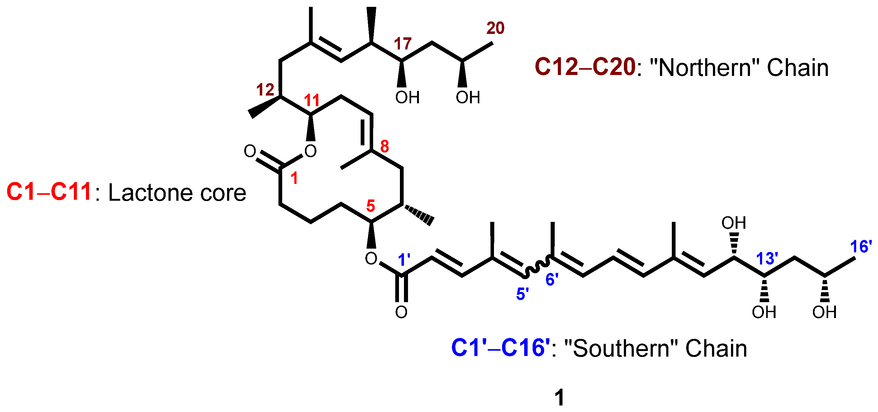
Figure 1. Structure of mycolactone A/B. Reproduced with permission from Akolgo et al., BMC Infectious Diseases, published by Springer Nature, 2023 [42].
Chemosensors 13 00244 g001
Figure 2. A schematic representation of the proposed mechanism of fluorescence of mycolactone and 2-naphthylboronic acid, as observed on TLC.
Chemosensors 13 00244 g002
Figure 3. Structures of common fluorescent dyes.
Chemosensors 13 00244 g003
Scheme 1. Palladium-catalysed Miyaura borylation of aryl halides utilising bis(pinacolato)diboron. Reagents and conditions: (a) bis(pinacolato)diboron, KOAc, Pd(dppf)Cl2, 1,4-dioxane, 80 °C, 16 h.
Chemosensors 13 00244 sch001
Scheme 2. Synthesis routes of coumarin-tagged boronic acid 7. Reagents and conditions: (a) diethyl malonate, piperidine, EtOH, reflux, 13 h (4, 85%); (b) DMF/POCl3, 1,2-dichloroethane, 0 °C → 60 °C, 12 h (5, 79%); (c) 3, CH2Cl2/anhydrous CH3CH2OH=1:1 (V:V), pyrrolidine, room temperature, 4 d (6, 51%); (d) NaIO4, THF/H2O (4:1), room temperature, 30 min (7, 90%).
Chemosensors 13 00244 sch002
Scheme 3. Synthetic route to arylboronic acid dye 22. Reagents and conditions: (a) K2CO3, DMF, 70 °C, reflux, 1.5 h (22, 76%).
Chemosensors 13 00244 sch003
Figure 4. Structures of quinoline and quinoline derivatives.
Chemosensors 13 00244 g004
Scheme 4. Synthetic route to (4-(((2-(acridin-9-ylamino)-2-oxoethyl)amino)methyl)phenyl)boronic acid 24. Reagents and conditions: (a) chloroacetyl chloride, Et3N, THF, 0 °C to room temperature, overnight (23, 59%); (b) (4-(aminomethyl)phenyl)boronic acid, Et3N, CH3CN, reflux, 24 h (24, 22%).
Chemosensors 13 00244 sch004
Scheme 5. Synthetic route to (4-(((2-oxo-2-(quinolin-8-ylamino)ethyl)amino)methyl)phenyl)boronic acid 18. Reagents and conditions: (a) (Boc)2O, Et3N, 1,4-dioxane/H2O (1:1), room temperature, 2 h (6, 65%); (b) 8-aminoquinoline, EDC.HCl, DMAP, CH2Cl2, room temperature, overnight (15, 93%); (c) HCl/dioxane (6 N), CH2Cl2, room temperature, overnight (16, 84%); (d) 2, NaBH(OAc)3, 1,2-dichloroethane, room temperature, 2 h (28, 53%); (e) NaIO4, THF/H2O (4:1), room temperature, 30 min (29, 76%).
Chemosensors 13 00244 sch005
Scheme 6. Synthetic route to (E)-(4-(((3’,6’-dihydroxy-3-oxospiro[isoindoline-1,9’-xanthen]-2-yl)imino)methyl)phenyl)boronic acid. Reagents and conditions: (a) MeOH/H2SO4, reflux, 6 h (34, 93%); (b) NH2NH2•H2O/MeOH, reflux, 6 h (35, 96%); (c) NH2NH2•H2O/MeOH, reflux, 12 h (35, 89%); (d) 4-formylphenylboronic acid, EtOH, reflux, 5 h (36, 64%).
Chemosensors 13 00244 sch006
Scheme 7. Synthesis of (3-(3-(3’,6’-dihydroxy-3-oxo-3H-spiro[isobenzofuran-1,9’-xanthen]-5-yl)thioureido)phenyl)boronic acid. Reagents and conditions: (a) 3-aminobenzeneboronic acid, DMF, room temperature, 12 h (37, 68%).
Chemosensors 13 00244 sch007
Figure 5. Molecular structure of Xanthene and Rhodamine B dye.
Chemosensors 13 00244 g005
Scheme 8. Synthesis of (E)-(4-(((3’,6’-bis(diethylamino)-3-oxospiro[isoindoline-1,9’-xanthen]-2-yl)imino)methyl)phenyl)boronic acid 39. Reagents and conditions: (a) NH2NH2•H2O/MeOH, reflux, 6 h (38, 68%); (b) 4-formylphenylboronic acid, EtOH, reflux, 5 h (39, 73%).
Chemosensors 13 00244 sch008
Figure 6. Representation of the BODIPY framework showing the IUPAC numbering system, delocalised structures, dipyrromethene, and dipyrromethene cores.
Chemosensors 13 00244 g006
Scheme 9. Synthesis of BODIPY-tagged boronic acid 45 via two routes. Reagents and conditions: (a) 2,4-dimethylpyrrole, CH2Cl2, 0 °C to room temperature, 3 h; (b) p-chloranil, CH2Cl2, 0 °C to room temperature, 30 min; (c) Et3N, BF3•OEt2, CH2Cl2, 0 °C to room temperature, 12 h (42, 47%); (d) Et3N, BF3•OEt2, CH2Cl2, 0 °C to room temperature, 12 h (45, 89%); (e) NaIO4, THF/H2O (4:1), room temperature, 30 min (45, 89%).
Chemosensors 13 00244 sch009
Figure 7. ORTEP representation of the compound 45 investigated by X-ray structure analysis; atomic labelling shown with 30% probability displacement ellipsoids.
Chemosensors 13 00244 g007
Scheme 10. Synthetic route to (E)-(2-(((3-methyl-4-(phenyldiazenyl)phenyl)amino)methyl)phenyl)boronic acid derivatives. Reagents and conditions: (a) NaBH(OAc)3, 1,2-dichloroethane, room temperature, 2 h (46, 75%); (b) NaNO2/conc HCl, 0 °C to 5 °C; (c) pH 4, 0 °C to 5 °C, 3 h.
Chemosensors 13 00244 sch010
Scheme 11. Preparation of Sudan I boronic acid dye 48. Reagents and conditions: (a) K2CO3, acetone, reflux, 50 °C, overnight.
Chemosensors 13 00244 sch011
Figure 8. Structures of synthesised fluorescent arylboronic acid dyes were included in this study. The characteristic chromophore cores of the structures are highlighted in different colours.
Chemosensors 13 00244 g008
Figure 9. Absorbance spectra of various dyes in different organic solvents; insets: respective linearity plots for the evaluation of molar extinction coefficients (ε) for the dyes at the absorption maxima: (a) compound 7; (b) compound 22.
Chemosensors 13 00244 g009
Figure 10. Fluorescence emission spectra of synthesised compounds (left) and reference compounds (Rhodamine B or Quinine sulphate (right)) with varying absorbance. Inset: calibration curves of integrated fluorescence intensity (area under curve) against absorbance for the following samples, respectively: (a) compound 7; (b) Rhodamine B at 499 nm.
Chemosensors 13 00244 g010
Figure 11. Fluorescence spectra of coumarin dye 7 (0.0025 mg/mL) upon gradual addition of serial concentrations of mycolactone (from bottom to top, 0–0.008 mg/mL) in ethanol. Inset: plot of the linear relationship between the fluorescence intensity and varying concentrations of mycolactone.
Chemosensors 13 00244 g011
Figure 12. Images of the fluorescence response of 5 µL/spot of synthetic mycolactone after dipping in 0.1- and 0.01-mM acetone concentrations of various synthesised fluorescent aryl boronic acid chemosensors compared to two commercial boronic acids under 365 nm UV light.
Chemosensors 13 00244 g012aChemosensors 13 00244 g012b
Table 1. Photophysical properties of synthesised fluorescent arylboronic acid dyes.
DyeMW
[gmol−1]
Solventλabsmax
[nm]
λemmax [nm]Stokes Shift (∆λ) [nm]ε
[M−1cm−1]
Quantum Yield (ΦF)Brightness
[M−1cm−1]
7391.2EtOH45659013452,816.10.7841,196.6
22310.1DMSO40344845247.60.2459.4
24385.2MeOH248.5429180.5ND ND
29335.2DMSO318405876468.80.031200.5
36478.3EtOH3013474621,486.3 ND
37526.3EtOH480525459368.70.474403.3
39588.5EtOH54056929296.50.48142.3
45368.0EtOH4965081229,259.10.7020,481.4
47a390.2EtOH464--21,695.7 ND
47b389.2EtOH416--20,550.8 ND
47c389.2EtOH398--27,245.4 ND
48382.2MeOH33538449ND ND
BA172.0MeOH27532853ND ND
BA18362.2MeOH27033363ND ND
Maximum absorption wavelength (λabsmax); maximum emission wavelength (λemmax); Stokes shift (∆λ): calculated as the difference between λabsmax and λemmax; molar extinction coefficient (ε); fluorescence quantum yield (ΦF): calculated by compared to fluorescence reference standards with known quantum yield values such as Rhodamine B (ΦF = 0.70 in EtOH), Quinine sulphate (ΦF = 0.51 in 0.1 M H2SO4); and brightness: this parameter was calculated using the following formula: brightness = extinction coefficient (ε) × fluorescence quantum yield (ΦF); ND = not determined.
Table 2. Absorbance and integrated fluorescence intensities (areas) for various samples and references.
Integrated Fluorescence Intensity
Absorbance @ 499 nm7Rhodamine B
0.0202619195.02549829.2
0.0162158588.52121340.3
0.0121682211.21495319.2
0.0091204913.31066673.4
0.004591676.7415287.3
Slope128197000.0115445000.0
Absorbance @ 358 nm22Quinine sulphate
0.0722384944.09169983.2
0.0582080661.17757302.9
0.0511560798.86858240.1
0.0381032764.15131493.2
0.028510273.43945263.9
Slope 43905200.0120830000.0
Absorbance @ 332 nm29Quinine sulphate
0.080406256.110254500.0
0.066331390.98717544.0
0.053269142.87633669.7
0.040195980.95868201.1
0.030134100.44435606.5
Slope5383876.6114494000.0
Absorbance @ 502.5 nm37Rhodamine B
0.0232014294.32863728.4
0.0201785124.72380927.6
0.0141300128.51745938.7
0.010942578.11216051.1
0.005455519.6489246.6
Slope861649000.0128111000.0
Absorbance @ 544.5 nm39Rhodamine B
0.0786696547.28871962.3
0.0646251111.17476185.8
0.0484931408.85593976.4
0.0323405442.53872951.0
0.0171858555.01555633.8
Slope 81524200.0118451000.0
Absorbance @ 502.5 nm45Rhodamine B
0.0233049627.72863728.4
0.0192613267.22380927.6
0.0141973366.81745938.7
0.0101360672.81216051.1
0.005731821.0489246.6
Slope 127606000.0128111000.0
Disclaimer/Publisher’s Note: The statements, opinions and data contained in all publications are solely those of the individual author(s) and contributor(s) and not of MDPI and/or the editor(s). MDPI and/or the editor(s) disclaim responsibility for any injury to people or property resulting from any ideas, methods, instructions or products referred to in the content.