Article

Ligand Differentiation Ability of Insect Odorant Receptors in Heterologously Expressed Cells as Potential Biosensor Elements

Research Center for Advanced Science and Technology (RCAST), The University of Tokyo, 4-6-1 Komaba, Meguro, Tokyo 153-8904, Japan
*
Author to whom correspondence should be addressed.
Chemosensors 2025, 13(8), 273; https://doi.org/10.3390/chemosensors13080273
Submission received: 13 June 2025 / Revised: 11 July 2025 / Accepted: 15 July 2025 / Published: 23 July 2025

Abstract

The extensive diversity of volatile organic compounds, along with their minor structural variations, presents significant challenges in the development of chemosensory-based biosensors. Previously, we generated sensor cells expressing insect odorant receptors (ORs) in Sf21 cells, demonstrating their potential as cell-based odorant sensor elements. However, it remains unclear whether the selectivity of cells expressing ORs in vitro for diverse compounds aligns with the receptor’s in vivo performance, aside from the response to target compounds. To address this, we assessed the ligand responses of sensor cells expressing ORs from Drosophila melanogaster using a high-throughput calcium imaging system. Our results demonstrate that in vitro receptor responses exhibit ligand selectivity comparable to in vivo conditions across different chemical categories. Broadly tuned OR-expressing sensor cells (Or13a, Or47a, and Or98a) displayed differential affinities, whereas the narrowly tuned Or56a-expressing sensor cells selectively responded to geosmin. Moreover, cell responses varied with subtle differences in chemical structure, including carbon chain length and functional group positioning. These findings provide valuable insights into insect OR–ligand interactions in vitro, demonstrating that receptor selectivity in sensor cells closely mirrors in vivo conditions. In addition to this consistency, our results highlight the subtle ligand differentiation capabilities of sensor cells enabling fluorescence-based visualization of receptor–ligand interactions.

1. Introduction

Volatile organic compounds are abundant in the environment; some function as odorants, conveying vital sensory information that influences animal behavior. Many odorants are strongly associated with food or environmental safety, and certain subtle odorant cues can serve as indicators of underlying diseases. However, minor variations in the chemical structures of these odorants elicit a wide range of olfactory responses, presenting challenges to the development of chemosensory-based biosensors.
Insects possess highly sensitive olfactory systems that play a crucial role in modulating behavior. The ability of insects to sense odorants is attributed to the large number of odorant receptors (ORs) and their corresponding ligand repertoire. Just a few molecules of pheromones released by female Bombyx mori can modulate male searching behavior [1]. Drosophila melanogaster, via ORs and the odorant receptor co-receptor (Orco) expressed on the membrane of olfactory receptor neurons (ORNs), find citrus fruits as oviposition sites, and via certain ORN-triggered innate avoidance circuits evade toxic microbes [2,3]. Because of their large-band odorant detection ability, human cancers can be detected by the insect olfactory system [4]. Additionally, the OR-Orco complex localized on the ORN membrane of insects can be used to detect illicit substances [5].
Unlike in mammals, which use seven-transmembrane helix (7TMH) G protein-coupled receptors as olfactory receptors [6], insect ORs are also 7TMH proteins but with an intracellular N-terminus and extracellular C-terminus [7]. Moreover, insect ORs require the presence of the co-receptor Orco [8], with downstream signal transduction beginning with OR-Orco complexes forming an odorant-gated non-selective cation channel permeable to Ca2+, Na+, and K+ ions [9,10]. The advancement of sequencing and bioinformatics tools has reduced costs and facilitated the exploration of the OR gene family in several insect species [11]. Analysis of the D. melanogaster genome has identified 60 genes encoding ORs, constituting the molecular foundation for odorant ligand detection [12,13,14]. Generally, one odorant can activate more than one OR. To comprehensively decode the chemosensory capability of D. melanogaster, researchers have developed a database to compare and combine extensive odor response profiles [15]. A comprehensive understanding of ligand–OR binding profiles offers potential for the development of OR-based sensing elements.
While research on ORNs and their corresponding ORs has generated glomeruli- or sensillum-based in vivo physiological data, expression of the OR-Orco complex in heterologous cells has aided the identification of single ORs, their corresponding ligands, and advanced in vitro biosensor applications. Initially, this examination was conducted in vertebrate cells, such as Xenopus oocytes [16], HEK293 cells [17], HeLa cells [10], and insect cell lines [18,19,20]. Long-term usability and stability are critical for biosensor development. We previously developed sensor cell lines using Sf21 cells from pupal ovarian tissues of the noctuid moth, Spodoptera frugiperda, to construct long-term chemical sensing elements [21]. However, the capacity for heterologously expressed insect ORs to replicate ligand selectivity observed in their native biological environments, as well as their ability to respond to diverse compound classes and subtle structural variations, remains largely unexplored. Based on this, we aimed to employ a high-throughput strategy to demonstrate the ligand selectivity of insect ORs under an in vitro expression system.
In this study, we aimed to characterize the ligand differentiation ability of heterologously expressed insect ORs by testing a range of chemicals, from distinct chemical groups to those with subtle variations in carbon chain length and functional group positioning. The selected ligands were based on documented in vivo OR–ligand responses, covering a spectrum from high to low affinity, which, in turn, elicits receptor responses of varying intensities in vivo. Using these ligands, we aimed to conduct in vitro studies to examine receptor activity, addressing the common limitation of in vitro experiments, which typically report only strong receptor responses while lacking data on weaker interactions. To assess receptor activity, we aimed to employ a high-throughput calcium imaging plate reader to measure ligand-induced fluorescence changes in sensor cells expressing D. melanogaster ORs, including Or13a, Or47a, Or98a, and Or56a. These ORs have been previously characterized in vivo, with Or13a, Or47a, and Or98a recognized as broadly tuned receptors, whereas Or56a is narrowly tuned and highly selective for geosmin. Investigating their responses in a heterologous expression system allows us to explore whether their ligand selectivity and tuning properties are preserved in vitro. Moreover, evaluating how these receptors respond to subtle structural differences among ligands in an in vitro setting can provide insights into their functional mechanisms and potential applications in high-throughput chemical screening. This approach is expected to contribute to identifying ligands for uncharacterized ORs and enhancing the applicability of OR-based platforms in mixture of odorant detection.

2. Materials and Methods

2.1. Odorant Preparation

The chemicals used in this study are listed in Supplementary Table S1. Odorants were dissolved in dimethyl sulfoxide (DMSO; 043-07216, Wako Pure Chemical Industries, Ltd., Osaka, Japan) to create 1 M stock solutions and stored at −30 °C until use. The assay buffer solution was prepared using modified Ringer’s solution containing 140 mM NaCl, 5.6 mM KCl, 4.5 mM CaCl2, 11.26 mM MgCl2, 11.32 mM MgSO4, 5 mM HEPES, and 9.4 mM D-glucose, adjusted to pH 7.2, with the addition of 0.1% (v/v) DMSO. Odorant sample solutions were diluted to different experimental concentrations using the assay buffer, maintaining a final DMSO concentration of 0.1% (v/v).

2.2. Construction of Odorant-Sensing Cells

Four odorant sensor cell lines were used in this study. Two cell lines expressing the D. melanogaster ORs Or13a and Or56a have been described in previous studies by our group [21,22,23]. Two novel cell lines expressing Or47a and Or98a were constructed following a similar protocol [21]. Sf21 cells derived from S. frugiperda were purchased from Life Technologies (Life Technologies, Carlsbad, CA, USA) and maintained in tissue culture flasks (Falcon Plastics Co., Los Angeles, CA, USA) as adherent cultures using Grace’s insect medium (Life Technologies, Carlsbad, CA, USA) at room temperature (27 °C). Sf21 cells were transfected with two vectors: an odorant receptor and a co-receptor dual-expression vector engineered using the pIB/V5-His vector (Invitrogen, Carlsbad, CA, USA). Additionally, a vector containing the gene for the calcium indicator fluorescent protein GCaMP6s [24] was engineered using the pIZ/V5-His vector (Invitrogen, Carlsbad, CA, USA). Four OR-Orco-expressing sensor cell lines, 8 × 10⁵ Sf21 cells seeded in 35-mm-diameter dishes (Iwaki, Chiba, Japan), were incubated for 24 h before transfection with TransIT-Insect (Takara Bio Inc., Shiga, Japan). After distributing the DNA complexes to cells, the culture dishes were incubated for 48 h. Subsequently, the cells were transferred into tissue culture flasks (surface area 25 cm2) containing 5 mL of insect medium supplemented with antibiotics, including 10 µg/mL Blasticidin S HCl (Gibco, A11139-03, Waltham, MA, USA), 100 µg/mL Zeocin (Invitrogen, R25001, Carlsbad, CA, USA), and 10 µg/mL of Gentamicin (Gibco, 15710-064, Waltham, MA, USA), each at their respective final concentrations and maintained for approximately one month [21,23]. After subculture, single-cell clones were manually selected using a limiting dilution method and cultured to establish stable odorant-sensing cell lines [23] after ligand testing based on highly responsive ligands listed in the OR responses database. Briefly, a functional ion channel formed by the OR-Orco complex is present on the cell membrane, and odorant binding triggers calcium influx (Figure 1). This influx is then detected using the calcium indicator protein GCaMP6s, resulting in fluorescent emission under excitation light.

2.3. Microplate Reader Calcium Imaging

All OR-Orco-expressing sensor cell lines and cell lines expressing only GCaMP6s were maintained at 27 °C in a non-humidified, CO2-free incubator and were passaged twice a week. To prepare the cell suspension solution before the experiment, the growth medium was replaced by centrifuging the cells at 500× g for 5 min at room temperature (25 °C). After removing the supernatant, the cells were resuspended in an assay buffer to adjust their concentration to 1 × 10⁶ cells/mL.
For fluorescence imaging, 80 µL of the cell suspension in assay buffer was plated in a 96-well assay plate (Corning catalog no. 3340, Corning, NY, USA). Then, 40 µL of odorant sample solution was added to each well from a 96-well sample plate containing 80 µL of assay buffer (Corning, catalog no. 3359, Corning, NY, USA), resulting in a final experimental concentration that was one-third of the original odorant sample solution. For clarity, all ligand concentrations mentioned hereafter refer to the original odorant sample solution concentrations. Fluorescence signals were recorded at a sampling rate of 2 Hz using the FDSS/μCell imaging platform (Hamamatsu Photonics, Hamamatsu, Shizuoka, Japan) with an excitation wavelength of 480 nm and emission at 540 nm. Each trial was recorded for 360 s. Baseline fluorescence (F0) was recorded for 60 s, before which 40 µL of odorant sample solution was injected into each well at a speed of 10 µL/sec. Fluorescence responses were recorded simultaneously for the entire 360 s. As shown in Figure 2, the fluorescence intensity change (ΔF/F) was calculated using the following equation: F / F = F 360 F 0 F 0 . Here, F (360) is the fluorescence intensity at the end of the recording and (F0) is the average signal intensity during the baseline period.

2.4. Data Analysis

Imaging data for each ligand recording were acquired from five independent replicates across different wells of 96-well plates to minimize potential positional effects. To ensure even distribution, the replicates for each ligand were scattered throughout the plate rather than clustered within the same row or column.
Fluorescence responses (ΔF/F) were analyzed by comparing ligand-induced ΔF/F values with those obtained in the assay buffer, which consisted of Ringer’s solution containing DMSO. Adjusted ΔF/F values were calculated by correcting the ligand-induced ΔF/F using the ΔF/F measured in the solvent control (Figure 2). Statistical comparisons between ligand groups and the assay buffer were conducted using one-way analysis of variance (ANOVA), followed by Dunnett’s multiple comparisons test. Additionally, structurally similar alcohols with different hydroxyl group positions were compared based on their adjusted ΔF/F values, with statistical analyses performed using one-way ANOVA followed by Tukey’s multiple comparisons test. To control the family-wise error rate at a predefined significance level (α ≤ 0.05), p-values were adjusted for multiple comparisons.
Statistical analyses were conducted using GraphPad Prism 8.0, and data visualization was performed using Seaborn (v0.13) and Matplotlib (v3.10) in Python 3.10, as well as ggplot2 (v3.5.1) in R 3.5.

3. Results and Discussion

3.1. Response Quantification and Ligand Selectivity

To evaluate the responses of sensor cell lines expressing Or13a, Or47a, and Or98a, we selected ligands from four chemical classes: alcohols, aldehydes, ketones, and esters (Figure S1A). A concentration of 1 mM was selected to maximize the observable ligand selectivity of each sensor cell line. For each odorant receptor (OR), three ligands per chemical class—categorized as eliciting “high,” “medium”, or “low” receptor responses—were chosen based on the D. melanogaster odorant receptor database (Database of Odorant Responses; DoOR) (http://neuro.uni-konstanz.de/DoOR/default.html accessed on 22 December 2023). Owing to the overlap among selected ligands across ORs (e.g., a ligand categorized as a “high” response for Or13a might be a “medium” response for Or47a), the final set consisted of 19 unique odorants. This set also included ligands for which no response data were available in the DoOR database. Cell responses were analyzed for significant differences (p < 0.05) at a ligand concentration of 1 mM compared to the assay buffer, which was used as a control (Figure 3).
In the group of alcohol odorants, Or13a-expressing sensor cells exhibited similar response profiles to the fluorescence changes elicited by the “high” and “medium” odorants, 1-octen-3-ol and 3-octanol. Although E3-hexenol is not documented in the database, the significant fluorescence change (p = 0.0002) observed in this study suggests that it may be a novel ligand of Or13a. Similarly, Or47a- and Or98a-expressing cells displayed comparable alcohol response profiles: 3-octanol and 1-octen-3-ol for Or47a, and 3-octanol and E3-hexenol for Or98a. In the database profile of alcohols for Or98a, although 1-octen-3-ol is not the strongest ligand, showing a response value of 0.579 compared to 0.592 of 3-octanol, it induced a fluorescence change comparable to that of 3-octanol and greater than that of the “medium” and “low” odorants. While none of the three sensor cells showed significant differences in response to “low” ligands, the trends in fluorescence changes aligned with values in the database.
Or13a-expressing sensor cells failed to discriminate between ketone odorants. Meanwhile, the Or47a- and Or98a-expressing sensor cells showed different fluorescence changes for these odorants. “High” odorants exhibited the largest fluorescence changes, whereas the rest showed fluorescence changes comparable to their response values recorded in the database.
For ester odorants, our Or13a-, Or47a-, and Or98a-expressing sensor cells showed changes in fluorescence for pentyl acetate, ethyl butyrate, and ethyl propionate that corresponded to the response values for “high,” “medium”, and “low” odorants, respectively. Furthermore, Or47a-expressing cells showed fluorescence changes that corresponded to response values recorded in the database. Although the Or13a- and Or98a-expressing sensor cells did not show significant differences beyond “high” responses, the trends of fluorescence changes aligned with values in the database.
Notably, while E2-hexenal and E2-octenal elicited fluorescence responses in all three sensor cell lines, the corresponding receptors did not show high response values to these or other tested aldehydes in the database. For instance, the highest values reported are 0.351 foe E2-hexenal for Or13a, 0.118 for hexanal with Or47a, and 0.088 for hexanal with Or98a.
To clarify whether the Ca2+ signals were derived from receptor responses related to OR-Orco complexes or non-specific responses due to other ion channels on the host Sf21 cell line, we performed the same odorant test on a geosmin-sensitive Or56a-expressing cell line [3,22,23] and cell line expressing only GCaMP6s (Figure S1B). The Or56a-expressing cells exhibited geosmin selectivity as shown in Figure S1C and Figure 4. Meanwhile, the Or56a-expressing cells and the GCaMP6s-expressing cell line did not show any response to the other 17 ligands; however, the high responses to E2-hexenal and E2-octenal indicate that certain ligands might drive non-specific Ca2+ influx or activate other ion channels of Sf21 host cell lines.
Since stable response ranges are essential for evaluating these cells as biosensor elements, we further utilize odorant category and concentration as two variables to characterize the sensor cell response and its corresponding ligands. Across all four sensor cell lines, affinity responses showed a stable increase with the rising concentration of the tested ligands in Figure 4A, with the ligands eliciting the strongest OR responses presented in Figure 4B. Reflecting broadly tuned ORs, O47a- and Or98a-expressing cells appeared to be generalists for alcohols, ketones, and esters, whereas Or13a-expressing cells appeared specialized for alcohol ligands, supporting the different response values of receptors in the database.
An increase in intracellular Ca2+ concentration induced by n-hexanal has been observed in vertebrate cells [25] and plant cells [26]. Although the uncertainty in Ca2+ elevation induced by some chemicals in Sf21 cells could lead to misinterpretation of insect OR responses when using calcium indicators, the advantages of long-term protein expression and large-scale screening make the application of calcium indicators and insect ORs valuable as biosensor elements. Moreover, GCaMP6s-expressing cell lines can aid in the differentiation of non-specific responses in OR-expressing sensor cells. Additionally, combining multiple insect ORs as sensor elements with an imaging analysis system can improve specificity, enabling more accurate detection and analysis of complex odorant mixtures [27].
To evaluate the ligand selectivity of our cell lines, we selected ligands with varying affinities documented in the DoOR database. Although the DoOR database is widely used in the field of insect odorant receptor (OR) research, inconsistencies may arise [28,29]. The data in the database are primarily derived from in vivo studies using transgenic flies, with single sensillum recordings and calcium imaging of genetically modified neurons [13]. In contrast, our in vitro conditions isolate the OR response, whereas in vivo presynaptic neuronal interactions within the olfactory circuit can modulate and potentially interfere with the original OR response [30]. In in vitro insect OR research, DMSO is widely employed as an organic solvent for odorant delivery. However, under in vivo conditions, odorant molecules are initially captured by odorant-binding proteins (OBPs) and delivered to the ORs, with their binding affinities being dependent on the chemical structure of the ligands [31,32]. Additionally, ligand binding affinity to ORs may be affected by the delivery method. In vivo experiments primarily use air-based methods, whereas in vitro experiments generally use organic solvents such as DMSO. The uncertainty regarding the exact amount of ligand binding to the receptors in different methods may result in slight variations in the sequence of binding affinity of these ligands with lower responses. As in our data, “high” level ligands always induce high responses, consistent with the database, but the results for “medium” and “low” levels may differ slightly. Moreover, in the Or98a-expressing cells, we observed a higher response to 6-methyl-5-hepten-2-one, not ethyl benzoate as documented in the database; this is consistent with the results on Or98a expression in HEK cell lines [29]. As an in vitro expression system, our sensor cell results reflect the responses of sensor elements that closely mimic OR responses and may differ from in vivo experimental assays, but they are suitable for in vitro biosensor development.

3.2. Ligand Selectivity with Structurally Similar Alcohols

Subtle differences in chemical structure, such as modifications of functional groups or carbon chain length, generally have minimal effects on the overall physicochemical properties, including the electronic characteristics, boiling point, and polarity, of a molecule. This similarity in properties makes it challenging for artificial odorant sensors to differentiate between minor structural variations because of the significant overlap in response signals. In the previous selectivity assay involving four chemical classes, our Or13a-, Or47a-, and Or98a-expressing cells exhibited good discrimination ability for alcohols (Figure 4). To further test the ligand screening ability of these cells, we used several monohydroxy alcohols with simple structures, with the only differences being in functional group position (from first position to highest-numbered distinct internal position) and carbon chain length (from C5 to C9). As shown in Figure 5A, each OR-expressing cell line exhibited distinct fluorescence intensity changes when exposed to alcohols with varying carbon chain lengths and hydroxyl group positions. Among the tested alcohols, Or13a- and Or47a-expressing cells showed the highest fluorescence levels in response to 3-octanol, whereas Or98a-expressing cells exhibited the strongest response to 2-octanol, indicating their ligand selectivity toward specific structural configurations. In contrast, C5 and C6 alcohols elicited lower fluorescence signals across all three OR-expressing cell lines, suggesting that these shorter-chain molecules have weaker interactions with the receptors. Similarly, alcohols with terminal hydroxyl groups consistently exhibited lower fluorescence intensity compared to those with internal hydroxyl groups, reinforcing the importance of hydroxyl positioning in ligand–receptor interactions. In the analysis of subtle structural recognition (Figure 5B), Or13a-expressing cells clearly distinguished hydroxyl positions in monohydroxy nonanols, as each position was assigned a distinct letter indicating statistically significant differences (p < 0.05). Similarly, Or47a- and Or98a-expressing cells effectively differentiated hydroxyl positions in monohydroxy octanols, with each position labeled by a unique letter denoting significant variation (p < 0.05). Moreover, all three OR-expressing cell lines exhibited a similar fluorescence trend in monohydroxy alcohols with a hydroxyl group at the third carbon position (Figure 5B, green-colored bars), with fluorescence intensity increasing from C6 to C8 but decreasing at C9.
Subtle differences in chemical structure pose a challenge for artificial odorant sensors, which often struggle to differentiate compounds with minor structural variations. In contrast, insect ORs exhibit a natural sensitivity to these subtle changes, an ability shaped by their complex living environments and chemical communication systems. In the case of narrowly tuned ORs, distinct receptors can recognize molecules that differ by only a single functional group. In the silk moth B. mori, the primary pheromone component, bombykol, and its structurally similar subcomponent, bombykal, differ only in the presence of one functional group. However, they are selectively detected by two distinct ORs [33]. A comparable specificity is observed, where Or56a-expressing cells responded exclusively to geosmin, demonstrating highly selective ligand recognition of narrowly tuned ORs. In contrast, broadly tuned ORs exhibit differential responses to subtle modifications in functional groups or carbon chain lengths, allowing insects to distinguish structurally similar odorants. In mosquitoes, even small changes in functional groups or carbon chain lengths lead to differences in OR activation [34]. Our results demonstrate that Or13a-, Or47a-, and Or98a-expressing cells can effectively differentiate monohydroxy alcohols based on hydroxyl position and carbon chain length, highlighting their capacity to recognize structurally diverse odorants. These results suggest that the ability of insect ORs to distinguish minor structural differences is not only observed in vivo but also preserved in the heterologous sensor cell system, indicating that insect OR-expressing cells serve as a reliable model for investigating ligand specificity and structural recognition mechanisms.

3.3. Ligand Selectivity with Certain Chemical Structures

Broadly tuned ORs exhibit diverse responses to a wide range of chemical structures while also demonstrating distinct sensitivities to specific molecular features. In Figure 5, all three sensor cell lines showed a relatively high response to 3-octanol and exhibited a similar ligand response profile for alcohols with a hydroxyl group at the third carbon position. To further investigate ligand selectivity among structurally similar compounds, we classified the tested alcohols into two structural types (Figure 6A).
Type A structures are unsaturated C6 to C9 alkenyl alcohols with a hydroxyl group at the third carbon position. These compounds include 1-octen-3-ol, which has been reported to elicit a relatively high receptor response in the database (Table S2) and shares structural similarities with 3-octanol. Type B structures, on the other hand, are saturated C6 to C9 secondary alcohols with a hydroxyl group at the third carbon position, which previously exhibited similar receptor responses to those in in Figure 5B, shown by the green-colored bars.
In Figure 6B, the addition of a double bond at the C1-C2 position (transitioning from Type B to Type A structures) led to distinct changes in the response profiles of Or47a- and Or98a-expressing sensor cells, while Or13a-expressing cells maintained a response profile consistent with Type B structures. Specifically, in Or47a-expressing cells, the fluorescence intensity increased from C6 to C7 but then decreased at C9. In contrast, Or98a-expressing cells exhibited a progressive increase in fluorescence intensity from C6 to C9, indicating a shift in response dynamics compared to Type B structures. The stable response of Or13a-expressing cells to both Type A and Type B structures suggests that despite being classified as a broadly tuned OR, Or13a exhibits a more consistent response profile for alcohols. This receptor appears to have a higher specificity for linear chain alcohols with a hydroxyl group positioned at the third carbon, reinforcing its distinct ligand selectivity compared to Or47a and Or98a.
Although the chemosensory mechanisms in mammals and insects are distinct, the binding pocket of human odorant receptors has been shown to exhibit a selective preference for varying fatty acid chain lengths [35]. The observed increase in the fluorescence response of Or98a-expressing cells to Type A structures with longer carbon chains suggests that the ligand recognition properties of insect ORs may also be influenced by binding pocket characteristics. The selectivity of insect ORs involves not only carbon chain length or functional groups but also specific structural motifs within the compounds. As in detecting chemicals with similar structures, broadly tuned AaOR8 in Aedes aegypti exhibits differential responses to enantiomers of 1-octen-3-ol [36]. Structurally analogous odorants often display similar physicochemical properties, complicating their discrimination. Insect olfactory systems exhibit high selectivity and sensitivity in odorant detection. However, the direct usage of unmodified sensory organs or living insects poses challenges for specific odorant detection in practical applications. Alternatively, our results suggest that in vitro isolated insect ORs can maintain ligand selectivity, even for distinguishing chemicals with subtle structural differences. This highlights the potential of applying multiple insect ORs as olfactory biosensor elements in detecting specific components in odorant mixtures.

4. Conclusions

In this study, we investigated the in vitro ligand selectivity of four D. melanogaster odorant receptors (Or13a, Or47a, Or98a, and Or56a) expressed in Sf21 cells using a high-throughput calcium imaging system. Previous studies have characterized these ORs in vivo, providing insights into their tuning properties. Our study builds on these findings by examining their ligand selectivity in an in vitro system, allowing for direct comparison between in vitro and in vivo responses. Our results confirm that the ligand selectivity trends observed in vivo are largely preserved in vitro, demonstrating the feasibility of using heterologous expression systems to study insect OR function. Building on this, in vivo data on target odorant detection from other insect species may serve as a valuable resource for the design of olfactory biosensor elements. Meanwhile, the robust ligand responses observed in this study indicate proper OR expression and functional assembly in the heterologous expression system, providing a useful platform for investigating specific OR–ligand recognition mechanisms. Furthermore, our structure-selective response analysis reveals that insect OR-expressing cells can discriminate subtle variations in carbon chain length and hydroxyl group positioning, supporting their potential for detecting structurally similar odorants. These findings highlight the applicability of in vitro expressed insect ORs for biosensing applications, particularly in detecting specific chemicals in odorant mixtures. By establishing the correlation between in vivo receptor responses and in vitro biosensor applications, our study provides a foundation for the development of insect OR-based chemical sensing platforms.

Supplementary Materials

The following supporting information can be downloaded at: https://www.mdpi.com/article/10.3390/chemosensors13080273/s1, Figure S1: Ligand selection and geosmin sensitivity in sensor cell Lines; Table S1: Chemicals used in this research study; Table S2: Structure similar ligands according to the DoOR database.

Author Contributions

R.Z.: writing—original draft, investigation, formal analysis, data curation, conceptualization. Y.S.: conceptualization, supervision, methodology, writing—review and editing, project administration, funding acquisition. S.N. (Sawako Niki): investigation, resources, data curation. E.K.: investigation, data curation. R.K.: methodology, resources. S.N. (Shigehiro Namiki): methodology, resources. H.M.: conceptualization, supervision, methodology, writing—review and editing, project administration, funding acquisition. All authors have read and agreed to the published version of the manuscript.

Funding

This research was partially supported by grants from the Bio-oriented Technology Research Advancement Institution (BRAIN), the Research and Implementation Promotion Program through Open Innovation Grants (JPJ011937), and the Japan Society for the Promotion of Science (JSPS) KAKENHI projects (JP21K05613, JP21K14168, JP21K19107, JP22K18416, JP24K17285).

Institutional Review Board Statement

Not applicable.

Informed Consent Statement

Not applicable.

Data Availability Statement

Researchers interested in accessing the data are encouraged to contact the corresponding author for further inquiries and arrangements.

Acknowledgments

The authors thank Takeshi Sakurai (Tokyo University of Agriculture) for his helpful advice and insightful discussions. They also thank Kei Ito (The University of Tokyo) for providing Drosophila melanogaster, and Douglas S. Kim (Howard Hughes Medical Institute) for generously providing the GCaMP6s gene.

Conflicts of Interest

The authors declare no conflict of interest.

References

  1. Kaissling, K.E.; Priesner, E. Die riechschwelle des seidenspinners. Naturwissenschaften 1970, 57, 23–28. [Google Scholar] [CrossRef]
  2. Dweck, H.K.; Ebrahim, S.A.; Kromann, S.; Bown, D.; Hillbur, Y.; Sachse, S.; Hansson, B.S.; Stensmyr, M.C. Olfactory preference for egg laying on citrus substrates in Drosophila. Curr. Biol. 2013, 23, 2472–2480. [Google Scholar] [CrossRef] [PubMed]
  3. Stensmyr, M.C.; Dweck, H.K.; Farhan, A.; Ibba, I.; Strutz, A.; Mukunda, L.; Linz, J.; Grabe, V.; Steck, K.; Lavista-Llanos, S.; et al. A conserved dedicated olfactory circuit for detecting harmful microbes in Drosophila. Cell 2012, 151, 1345–1357. [Google Scholar] [CrossRef] [PubMed]
  4. Farnum, A.; Parnas, M.; Hoque Apu, E.H.; Cox, E.; Lefevre, N.; Contag, C.H.; Saha, D. Harnessing insect olfactory neural circuits for detecting and discriminating human cancers. Biosens. Bioelectron. 2023, 219, 114814. [Google Scholar] [CrossRef] [PubMed]
  5. Marshall, B.; Warr, C.G.; de Bruyne, M. Detection of volatile indicators of illicit substances by the olfactory receptors of Drosophila melanogaster. Chem. Senses 2010, 35, 613–625. [Google Scholar] [CrossRef]
  6. Kato, A.; Touhara, K. Mammalian olfactory receptors: Pharmacology, G protein coupling and desensitization. Cell. Mol. Life Sci. 2009, 66, 3743–3753. [Google Scholar] [CrossRef]
  7. Benton, R.; Sachse, S.; Michnick, S.W.; Vosshall, L.B. Atypical membrane topology and heteromeric function of Drosophila odorant receptors in vivo. PLoS Biol. 2006, 4, e20. [Google Scholar] [CrossRef]
  8. Larsson, M.C.; Domingos, A.I.; Jones, W.D.; Chiappe, M.E.; Amrein, H.; Vosshall, L.B. Or83b encodes a broadly expressed odorant receptor essential for Drosophila olfaction. Neuron 2004, 43, 703–714. [Google Scholar] [CrossRef]
  9. Nakagawa, T.; Sakurai, T.; Nishioka, T.; Touhara, K. Insect sex-pheromone signals mediated by specific combinations of olfactory receptors. Science 2005, 307, 1638–1642. [Google Scholar] [CrossRef]
  10. Sato, K.; Pellegrino, M.; Nakagawa, T.; Nakagawa, T.; Vosshall, L.B.; Touhara, K. Insect olfactory receptors are heteromeric ligand-gated ion channels. Nature 2008, 452, 1002–1006. [Google Scholar] [CrossRef]
  11. Montagné, N.; De Fouchier, A.; Newcomb, R.D.; Jacquin-Joly, E. Advances in the identification and characterization of olfactory receptors in insects. Prog. Mol. Biol. Transl. Sci. 2015, 130, 55–80. [Google Scholar] [CrossRef]
  12. Vosshall, L.B.; Amrein, H.; Morozov, P.S.; Rzhetsky, A.; Axel, R. A spatial map of olfactory receptor expression in the Drosophila antenna. Cell 1999, 96, 725–736. [Google Scholar] [CrossRef]
  13. Hallem, E.A.; Ho, M.G.; Carlson, J.R. The molecular basis of odor coding in the Drosophila antenna. Cell 2004, 117, 965–979. [Google Scholar] [CrossRef]
  14. Gomez-Diaz, C.; Martin, F.; Garcia-Fernandez, J.M.; Alcorta, E. The two main olfactory receptor families in Drosophila, ORs and IRs: A comparative approach. Front. Cell. Neurosci. 2018, 12, 253. [Google Scholar] [CrossRef]
  15. Münch, D.; Galizia, C.G. DoOR 2.0-comprehensive mapping of Drosophila melanogaster odorant responses. Sci. Rep. 2016, 6, 21841. [Google Scholar] [CrossRef] [PubMed]
  16. Wetzel, C.H.; Behrendt, H.J.; Gisselmann, G.; Störtkuhl, K.F.; Hovemann, B.; Hatt, H. Functional expression and characterization of a Drosophila odorant receptor in a heterologous cell system. Proc. Natl. Acad. Sci. USA 2001, 98, 9377–9380. [Google Scholar] [CrossRef] [PubMed]
  17. Neuhaus, E.M.; Gisselmann, G.; Zhang, W.; Dooley, R.; Störtkuhl, K.; Hatt, H. Odorant receptor heterodimerization in the olfactory system of Drosophila melanogaster. Nat. Neurosci. 2005, 8, 15–17. [Google Scholar] [CrossRef] [PubMed]
  18. Smart, R.; Kiely, A.; Beale, M.; Vargas, E.; Carraher, C.; Kralicek, A.V.; Christie, D.L.; Chen, C.; Newcomb, R.D.; Warr, C.G. Drosophila odorant receptors are novel seven transmembrane domain proteins that can signal independently of heterotrimeric G proteins. Insect Biochem. Mol. Biol. 2008, 38, 770–780. [Google Scholar] [CrossRef]
  19. Tsitoura, P.; Andronopoulou, E.; Tsikou, D.; Agalou, A.; Papakonstantinou, M.P.; Kotzia, G.A.; Labropoulou, V.; Swevers, L.; Georgoussi, Z.; Iatrou, K. Expression and membrane topology of Anopheles gambiae odorant receptors in lepidopteran insect cells. PLoS ONE 2010, 5, e15428. [Google Scholar] [CrossRef]
  20. Kiely, A.; Authier, A.; Kralicek, A.V.; Warr, C.G.; Newcomb, R.D. Functional analysis of a Drosophila melanogaster olfactory receptor expressed in Sf9 cells. J. Neurosci. Methods. 2007, 159, 189–194. [Google Scholar] [CrossRef]
  21. Mitsuno, H.; Sakurai, T.; Namiki, S.; Mitsuhashi, H.; Kanzaki, R. Novel cell-based odorant sensor elements based on insect odorant receptors. Biosens. Bioelectron. 2015, 65, 287–294. [Google Scholar] [CrossRef] [PubMed]
  22. Termtanasombat, M.; Mitsuno, H.; Misawa, N.; Yamahira, S.; Sakurai, T.; Yamaguchi, S.; Nagamune, T.; Kanzaki, R. Cell-based odorant sensor array for odor discrimination based on insect odorant receptors. J. Chem. Ecol. 2016, 42, 716–724. [Google Scholar] [CrossRef] [PubMed]
  23. Mitsuno, H.; Araki, S.; Sukekawa, Y.; Terutsuki, D.; Niki, S.; Kuroda, E.; Fujibayashi, S.; Sakurai, T.; Oguma, K.; Yamaguchi, S.; et al. Portable geosmin detection system based on sensor cells expressing insect odorant receptors. Res. Sq. 2025; preprint. [Google Scholar] [CrossRef]
  24. Chen, T.W.; Wardill, T.J.; Sun, Y.; Pulver, S.R.; Renninger, S.L.; Baohan, A.; Schreiter, E.R.; Kerr, R.A.; Orger, M.B.; Jayaraman, V.; et al. Ultrasensitive fluorescent proteins for imaging neuronal activity. Nature 2013, 499, 295–300. [Google Scholar] [CrossRef]
  25. Kobayashi, Y.; Kako, H.; Yokogoshi, H. Contribution of intracellular Ca2+ concentration and protein dephosphorylation to the induction of dopamine release from PC12 cells by the green odor compound hexanal. Cell. Mol. Neurobiol. 2010, 30, 173–184. [Google Scholar] [CrossRef]
  26. Aratani, Y.; Uemura, T.; Hagihara, T.; Matsui, K.; Toyota, M. Green leaf volatile sensory calcium transduction in Arabidopsis. Nat. Commun. 2023, 14, 6236. [Google Scholar] [CrossRef]
  27. Sukekawa, Y.; Mitsuno, H.; Kanzaki, R.; Nakamoto, T. Odor discrimination using cell-based odor biosensor system with fluorescent image processing. IEEE Sens. J. 2019, 19, 7192–7200. [Google Scholar] [CrossRef]
  28. Andersson, M.N.; Schlyter, F.; Hill, S.R.; Dekker, T. What reaches the antenna? How to calibrate odor flux and ligand–receptor affinities. Chem. Senses 2012, 37, 403–420. [Google Scholar] [CrossRef]
  29. Zboray, K.; Toth, A.V.; Miskolczi, T.D.; Pesti, K.; Casanova, E.; Kreidl, E.; Mike, A.; Szenes, Á.; Sági, L.; Lukacs, P. High-throughput ligand profile characterization in novel cell lines expressing seven heterologous insect olfactory receptors for the detection of volatile plant biomarkers. Sci. Rep. 2023, 13, 21757. [Google Scholar] [CrossRef]
  30. Olsen, S.R.; Wilson, R.I. Lateral presynaptic inhibition mediates gain control in an olfactory circuit. Nature 2008, 452, 956–960. [Google Scholar] [CrossRef] [PubMed]
  31. Zhou, J.J. Odorant-binding proteins in insects. Vitam. Horm. 2010, 83, 241–272. [Google Scholar] [CrossRef]
  32. Guo, Y.R.; Yin, N.N.; Wu, C.; Yang, Z.X.; Wang, Z.Q.; Liu, N.Y. Expression profile and functional characterization of odorant binding proteins in a forest pest, Dioryctria abietella (Lepidoptera: Pyralidae). Comp. Biochem. Physiol. B Biochem. Mol. Biol. 2023, 266, 110835. [Google Scholar] [CrossRef]
  33. Sakurai, T.; Nakagawa, T.; Mitsuno, H.; Mori, H.; Endo, Y.; Tanoue, S.; Yasukochi, Y.; Touhara, K.; Nishioka, T. Identification and functional characterization of a sex pheromone receptor in the silkmoth Bombyx mori. Proc. Natl. Acad. Sci. USA 2004, 101, 16653–16658. [Google Scholar] [CrossRef]
  34. Wang, G.; Carey, A.F.; Carlson, J.R.; Zwiebel, L.J. Molecular basis of odor coding in the malaria vector mosquito Anopheles gambiae. Proc. Natl. Acad. Sci. USA 2010, 107, 4418–4423. [Google Scholar] [CrossRef]
  35. Billesbølle, C.B.; de March, C.A.; van der Velden, W.J.; Ma, N.; Tewari, J.; Del Torrent, C.L.; Li, L.; Faust, B.; Vaidehi, N.; Matsunami, H.; et al. Structural basis of odorant recognition by a human odorant receptor. Nature 2023, 615, 742–749. [Google Scholar] [CrossRef]
  36. Bohbot, J.D.; Dickens, J.C. Characterization of an enantioselective odorant receptor in the yellow fever mosquito Aedes aegypti. PLoS ONE 2009, 4, e7032. [Google Scholar] [CrossRef]
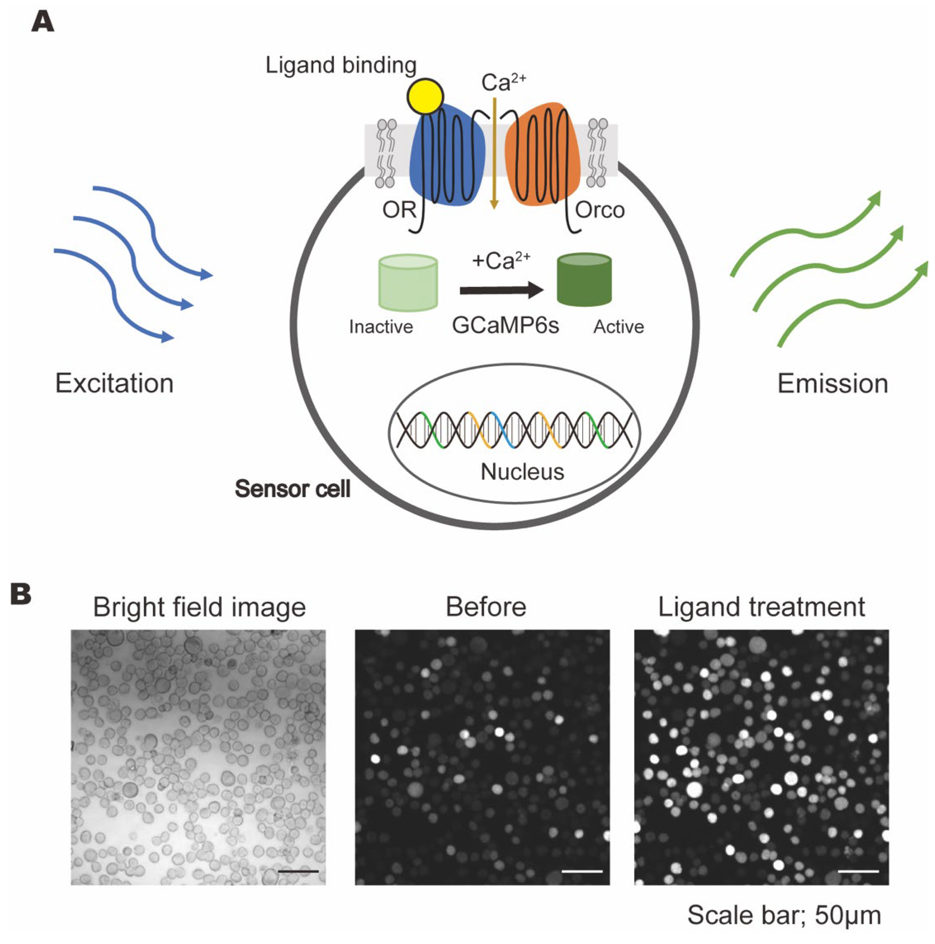
Figure 1. The generation of sensor cells. (A) A schematic illustration showing the generation of OR-, Orco-, and GCaMP6-expressing sensor cells. (B) The responses of Or47a-expressing sensor cells to pentyl acetate (100 µM).
Chemosensors 13 00273 g001
Figure 2. Ligand-induced fluorescence changes from microplate recordings using sensor cells. Fluorescence changes in Or98a-expressing cells in response to 6-methyl-5-hepten-2-one, including raw and adjusted fluorescence values.
Chemosensors 13 00273 g002
Figure 3. Evaluation of different affinity ligands. Four sensor cell lines were tested with four ligand classes, alcohol, aldehyde, ketone, and ester groups, comprising 19 ligands, all at a 1 mM concentration. Ligand affinity, selected according to data in DoOR, was designated as “high,” “medium”, and “low” and is shown in orange, yellow, and green, respectively. Ligand-induced responses were assessed by comparing the ΔF/F of each ligand to those with the control assay buffer (Ringer + DMSO) using one-way analysis of variance followed by Dunnett’s multiple comparisons testing. Data represent n = 5 for each sensor cell line; * p < 0.05, ** p < 0.001, and *** p < 0.0001.
Chemosensors 13 00273 g003
Figure 4. Dose responses of ligands with different affinities in sensor cell lines. (A) Heatmaps showing the mean-adjusted ΔF/F responses of each sensor cell line to the tested ligands (n = 5). (B) Dose–response curves of the most responsive ligand for Or13a-, Or47a-, and Or98a-expressing cells. Data represent n = 5 per cell line; error bars indicate standard deviation (SD).
Chemosensors 13 00273 g004
Figure 5. The selectivity of structurally similar alcohols. (A) The fluorescence patterns of structurally similar alcohols with different hydroxyl group positions and carbon chain lengths in a 1 mM concentration solution; “/” indicates that no ligands exist or none were used in this experiment. (B) Structurally similar alcohols with the same carbon chain length. Ligand-induced responses were assessed by comparing the normalized ΔF/F of each group. Groups labeled with different letters (e.g., a, b, c) are significantly different (p < 0.05) based on one-way analysis of variance followed by Tukey’s multiple comparisons testing (n = 5).
Chemosensors 13 00273 g005
Figure 6. The responses of structurally similar ligands in sensor cell lines. (A) Two types of structurally similar ligands with carbon chain lengths increasing from C6 to C9. Type A structures comprise unsaturated alkenyl alcohols with a hydroxy group at the tertiary position. Type B structures comprise saturated secondary alcohols with a hydroxy group at the tertiary position. (B) Odorant responses of Type A (green) and Type B (blue) structures in each sensor cell line under 1 mM. Data represent n = 5 for each sensor cell line.
Chemosensors 13 00273 g006
Disclaimer/Publisher’s Note: The statements, opinions and data contained in all publications are solely those of the individual author(s) and contributor(s) and not of MDPI and/or the editor(s). MDPI and/or the editor(s) disclaim responsibility for any injury to people or property resulting from any ideas, methods, instructions or products referred to in the content.