Review

Recent Advances on Fluorescent Sensors for Detection of Pathogenic Bacteria

1
Institute for Advanced Materials, School of Chemistry and Chemical Engineering, Jiangsu University, Zhenjiang 212013, China
2
National and Local Joint Engineering Laboratory of Municipal Sewage Resource Utilization Technology, School of Environmental Science and Engineering, Suzhou University of Science and Technology, Suzhou 215009, China
3
Department of Biology, Hong Kong Baptist University, Kowloon Tong, Hong Kong, China
4
School of Chemistry and Chemical Engineering, Hainan University, Haikou 572008, China
*
Author to whom correspondence should be addressed.
Chemosensors 2025, 13(5), 182; https://doi.org/10.3390/chemosensors13050182
Submission received: 19 March 2025 / Revised: 9 May 2025 / Accepted: 10 May 2025 / Published: 13 May 2025

Abstract

Pathogenic bacteria are one of the main causes of diseases and have become an important public health problem threatening human health and socio-economic development. Therefore, it is particularly important to develop an efficient and convenient detection method. Fluorescence detection has become a highly concerned analytical technology, which has gradually emerged in the aspect of pathogen detection, and is favored by researchers. In this review, we summarized a series of sensing strategies for pathogen detection based on fluorescence response signals in recent years, including single molecule fluorescent probes, biosensors, nanocomposite sensors and strategies for integrating different recognition elements with nanomaterials, along with the advantages and disadvantages of various design strategies. Based on the existing research reports, the existing problems and future research challenges of fluorescent sensor technology are proposed.

1. Introduction

The problem of food or medical equipment pollution caused by pathogenic bacteria is increasingly prominent. The number of food poisonings reported by the ministry of health is increasing year by year, and most of them are caused by pathogens. Pathogenic bacteria directly or indirectly pollute food or water sources. Human infection can lead to the occurrence of intestinal infectious diseases, poisoning and the prevalence of livestock and poultry infectious diseases [1,2,3]. Some proteins and endotoxins metabolized by bacteria can cause immune reaction and disease, and the continuous reproduction of bacteria will also invade normal tissues. Different kinds of bacteria can cause different degrees of pathological changes in the whole pathological process (Table 1). According to World Health Organization (WHO) surveillance data, foodborne diseases constitute a significant global health burden, with an estimated 600 million cases annually. Of these, diarrheal illnesses account for 550 million episodes worldwide, disproportionately affecting pediatric populations. Epidemiologic analyses indicate these infections result in approximately 3 million premature deaths annually among children under five, representing 12.5% of all-cause mortality in this age cohort. Crucially, microbial contamination of food supplies serves as the principal transmission mechanism, with enteropathogenic bacteria (e.g., Salmonella, Campylobacter and pathogenic E. coli) implicated in 70% of diarrheal cases according to WHO’s Global Foodborne Disease Network [4]. In addition to the disease problems caused by foodborne bacteria, clinical bacterial infections also bring great resistance to medical treatment. The challenge of antibacterial infection lies in how to detect these bacterial infections with a sufficiently sensitive and accurate method at the early stage of infection [5,6,7,8,9,10,11,12]. Therefore, there is an urgent need to develop accurate early screening methods to help reduce health problems caused by bacteria in food, drugs and the environment, and clinical bacterial infections. How to establish an effective pathogen detection system and quickly and accurately detect pathogens has become the key to ensure food safety, prevent disease transmission and prevent clinical antibacterial infection.
So far, researchers have spared no effort in the research of bacterial detection [13,14,15,16], such as the plate counting method [17,18,19,20,21], enzyme-linked immunosorbent assay [22,23], polymerase chain reaction (PCR) [24,25,26,27] and electrochemical immunosensor [28,29,30,31]; among which, the plate counting method and PCR method are the most commonly used. The plate counting method can estimate the number of viable bacteria in the sample, and the PCR method can detect specific DNA or RNA from target bacterial cells. These methods have a certain sensitivity and specificity in the detection process, but these methods have obvious shortcomings at the same time. They are not suitable for fast daily analysis and detection because of their time-consuming processes, expensive equipment and reagents, complex operation processes or strict experimental conditions.
Fluorescence detection is a newly developed high-efficiency detection technology, which has the advantages of good selectivity, high sensitivity, simple operation and easy rapid analysis. Researchers have designed a variety of sensing systems using fluorescence as the response signal to detect different targets [32,33,34,35,36,37,38,39,40,41,42,43,44,45]. In recent years, fluorescence detection technology has gradually emerged in the application of bacterial detection [46,47,48,49]. Fluorescent detection platforms for bacterial identification can be systematically categorized into two distinct operational paradigms. The first category encompasses systems utilizing luminescent nanomaterials (e.g., quantum dots, up-conversion nanoparticles) as photonic signal transducers, engineered to create biosensing architectures through specific biorecognition mechanisms. These systems typically employ antibody–antigen interactions or aptamer–target binding events to induce measurable fluorescence variations, enabling the quantitative detection of bacterial pathogens via mechanisms such as fluorescence resonance energy transfer (FRET) or surface-enhanced fluorescence (SEF). The other type is to use fluorescent dye small molecules to induce fluorescence signal changes by labeling or directly constructing fluorescent probes that react specifically with bacteria, achieving the visualization of bacterial detection. In addition, researchers have also tried to explore a variety of new biomolecular recognition elements for bacteria, including aptamers [50,51], antimicrobial peptides [52,53], phages [54,55], vancomycin [56] and lysozyme [57]. Meanwhile, combined with a series of inorganic functional nanomaterials with unique physical and chemical properties [58,59], researchers have constructed a variety of functional composite materials to detect the target bacteria by fluorescence.
This review will cover the construction of a series of fluorescent sensors and detection methods for bacteria in the past ten years (Figure 1). Fluorescent sensor detection systems can provide a sensitive and specific detection of bacteria, and can be used in a variety of applications and industries. Based on the different materials for constructing sensing systems, including single molecule fluorescent probes, quantum dots, up-conversion materials, gold nanoparticles and biomolecules, we will cover a wide range of advanced nanomaterials. However, due to the breadth of the field, not all the materials covered are included. In addition, this review will also put forward the direction of building fluorescent nanoprobes in the future to improve the detection and sensing of bacteria.

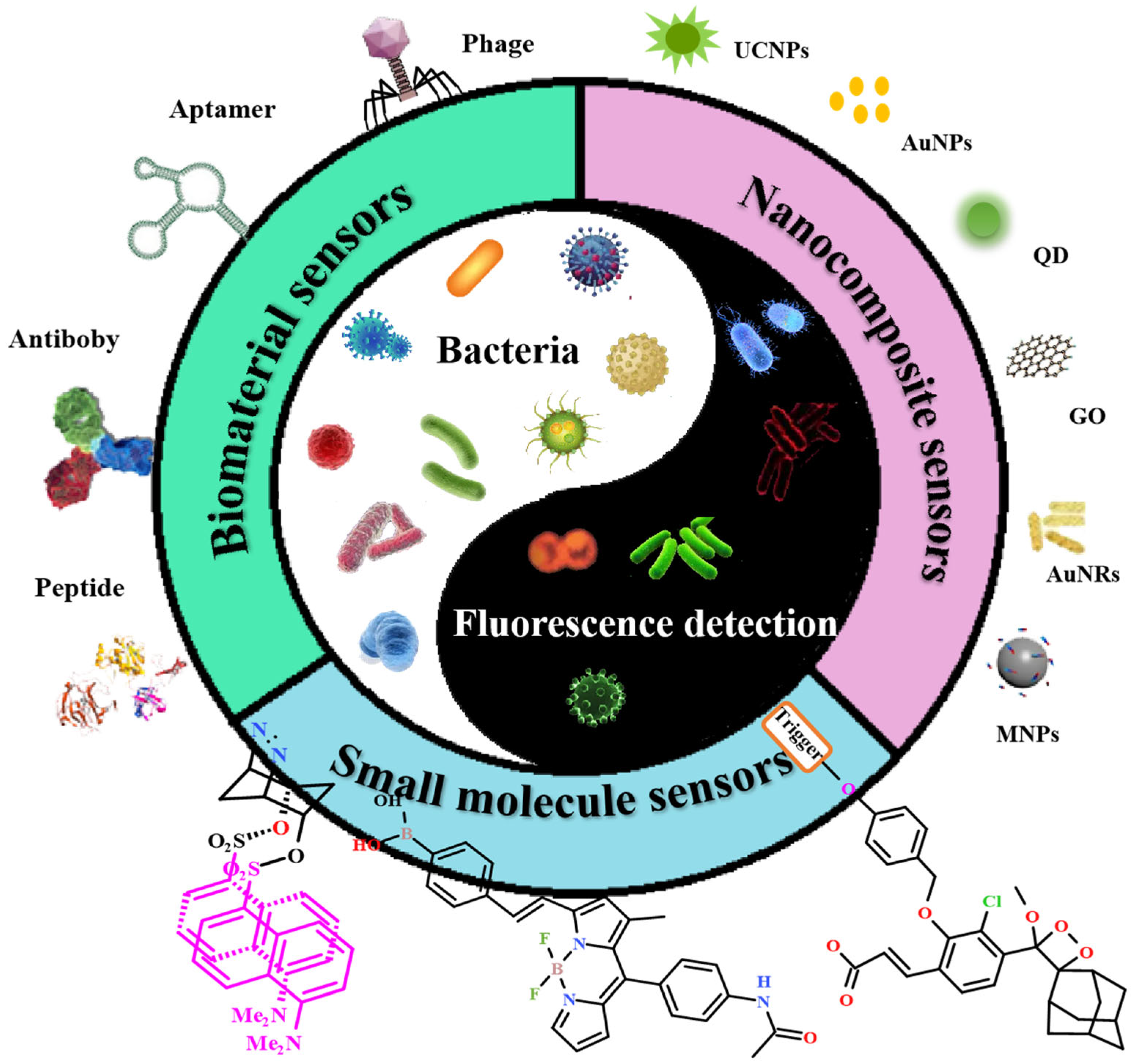
2. Fluorescent Sensing Systems for Bacteria Detection

2.1. Small Molecule-Based Fluorescent Probes

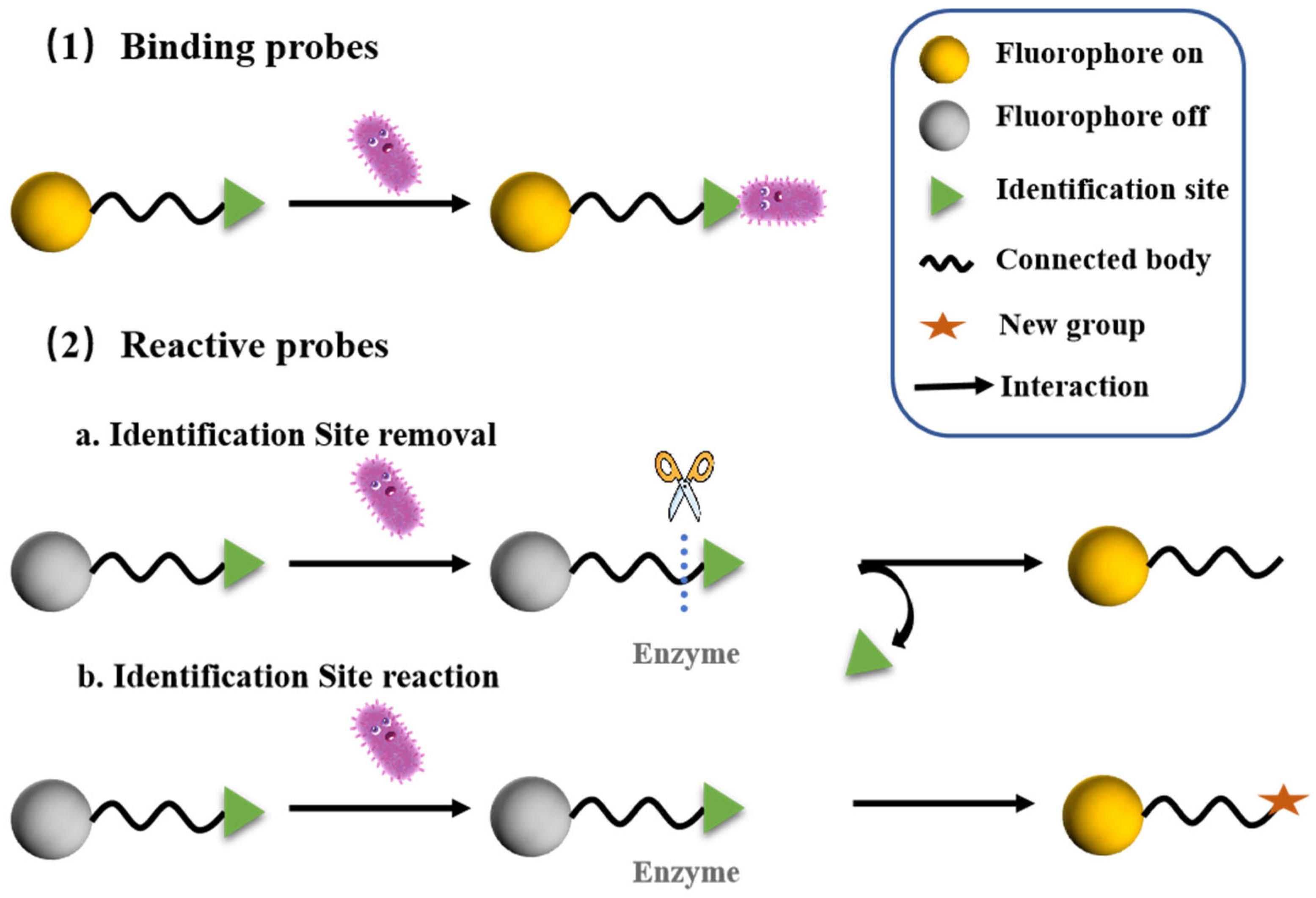
2.1.1. General Concepts

The simplest fluorescent detection method is to construct a fluorescent probe for the target object by small molecular fluorophores, which consists of three parts: identification group, linker and luminescent group. The fluorescent chromophores commonly used are coumarin, rhodamine, anthraquinone, cyanine, phenoxy dioxane, etc., which are the main luminescent parts of the probe. The recognition group is mainly the specific group that can provide the recognition site or the reaction site, while the linker is the bond bridge connecting the chromophore and the recognition group. For the construction of probes, one of the three is indispensable. When the recognition group of fluorescent probe binds the target or reacts with the target, it will lead to the change of the overall structure of the probe molecule, thus affecting the charge distribution of the whole molecule, which will induce the change of the luminescent performance of the system. Through the difference of optical signals before and after, the detection can be achieved. At present, fluorescent probes based on small molecule fluorophore have been widely used to detect all kinds of target substances, including heavy metal ions in environmental wastewater [60,61,62,63], pesticide residues in agricultural products [64,65,66,67], disease-related overexpressed substances in organisms [68,69,70,71,72,73], etc., showing certain application value in various fields.
Various fluorescent probes for bacteria have been developed with specific receptors to recognize biomarkers of targeted bacteria type, such as unique enzymes, mannose receptors or N-acetylglucosamine and N-acetylneuraminic acid residues on cell surfaces [74,75,76,77]. To date, small-molecule fluorescent probes for bacterial detection can be broadly classified into two categories: reactive probes and binding probes (Figure 2). Reactive probes typically target bacterial-secreted enzymes or other specific metabolites. The design strategy involves incorporating a recognition site at one end of the probe that can be selectively activated by these bacterial secretions. Upon activation, the fluorescence signal of the probe undergoes a detectable change (e.g., turn-on, ratiometric or spectral shift), enabling bacterial detection and analysis. The key challenge in developing reactive probes lies in the careful selection of activation sites. For instance, certain bacterial enzymes—such as β-lactamases or nitroreductases—exhibit catalytic activity and can cleave or modify specific chemical groups (e.g., ester or nitro moieties) on the probe, triggering the fluorescence response. Binding probes, on the other hand, rely on the introduction of capture groups that selectively bind to bacterial surface components (e.g., glycoproteins, lipopolysaccharides or peptidoglycan) through affinity interactions or electrostatic adsorption. These probes enable the direct fluorescence imaging of bacteria by physically adhering to their surfaces without requiring chemical activation. This classification highlights the distinct mechanisms—chemical reaction versus physical binding—that underpin bacterial detection strategies, each offering unique advantages in sensitivity, specificity and application scope.

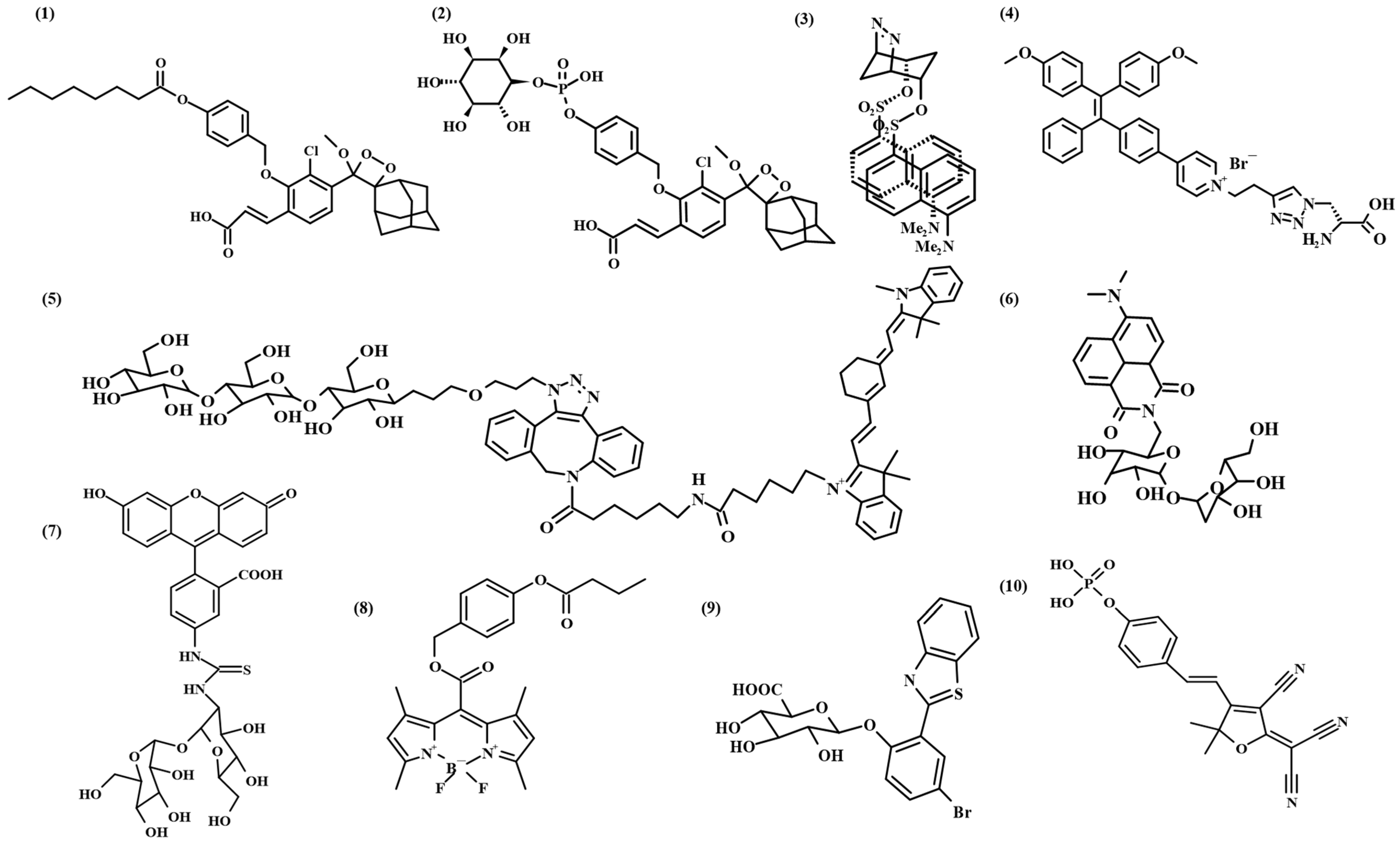
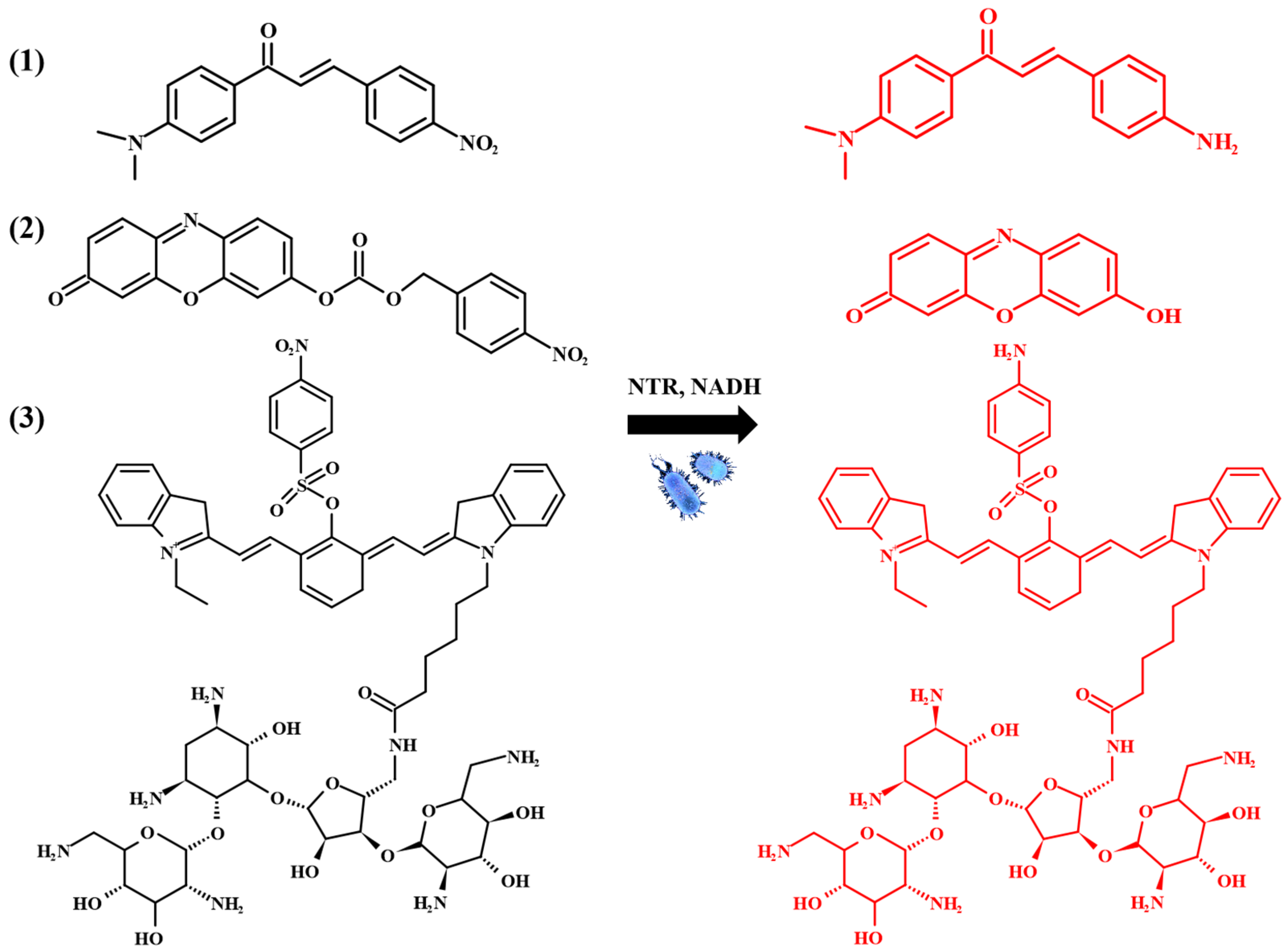
2.1.2. Reactive Probes

The classification and identification of bacteria are usually based on differences in cell composition, cell metabolism and cell structure. At present, microbiological detection based on microscope through morphological and colony testing is hindered by the high requirement and high cost for the slow culture time. Molecule probes that rely on DNA hybridization also have the problem of low sensitivity. Based on this, researchers have designed and constructed a series of small molecule fluorescent probes for the rapid identification and detection of target bacteria. The selective detection of bacterial specific antigen (including bacterial antigen/flagella antigen), cellular endocrine enzymes and various compounds can be used as a marker of bacterial existence. In particular, the development of highly specific enzyme-activated probes has become an alternative for the rapid detection of bacteria.
Recently, Doron Shabat et al. [78] reported the novel probe 1 based on the activation response of bacterial enzyme for the ultrasensitive direct detection of viable pathogenic bacteria (Figure 3). The probes are composed of a bright phenoxy-dioxetane luminophore masked by a triggering group, which is activated by a specific bacterial enzyme. The probe system could detect a minimum of ten Salmonella cells within 6 h incubation. This probe selects phenoxy-dioxetane as the luminescent group, which has conjugated electron withdrawing substituents. It can release benzoic acid derivatives in the process of chemical excitation, and has high emittance in water [79]. Similarly, Doron Shabat et al. also constructed probe 2 with a similar phenoxy-dioxetane structure by modifying the substrate of the virulence factor phosphatidylinositol-specific phospholipase C (PI-PLC) for the identification and detection of pathogenic L. monocytogenes. This design strategy provided a new idea for the construction of similar fluorescent probes for the detection of specific bacteria and other enzymes related to instant medical diagnosis. Sally Freeman et al. reported an optically responsive probe 3 based on cyclohexane [80]. The design of the probe is based on the cis-azo bridge as a bio-reduction trigger for conformational loop reversal. Vibhute et al. reported azidoacetamide-functionalized pseudaminic acid (Pse) derivatives for detecting the expression of Pse in bacteria, which successfully detected Bacillus cyuningiensist and Bacillus jejuni [81]. Hu et al. successfully developed a bacteria-metabolizable dual-functional probe 4, which is based on d-alanine, and a photosensitizer with aggregation-induced emission for fluorescence turn-on imaging of intracellular bacteria in living host cells and photodynamic ablation in situ [82]. Zlitni et al. developed a fluorescent derivative of maltotriose (probe 5), which is shown to be taken up in a variety of Gram-positive and Gram-negative bacteria strains in vitro. In vivo fluorescence and photoacoustic imaging studies highlight the ability of this probe to detect infection, assess infection burden and visualize the effectiveness of antibiotic treatment in E. coli-induced myositis and a clinically relevant S. aureus wound infection murine model [83]. Sahile and Backus et al. combined trehalose with fluorophore to construct novel probes 6 and 7 for detecting Mycobacterium tuberculosis [84,85]. Kim et al. addressed the issue of false positives in traditional clinical detection methods for Moraxella catarrhalis by developing a novel dual-mode probe, B-MC4 [86]. This probe, based on a 1,3,5,7-tetramethyl BODIPY scaffold, achieves the specific detection of C4-esterase activity via a meso-position butyrate modification. B-MC4 exhibits significantly enhanced resistance to nonspecific hydrolysis in buffer systems, effectively reducing false-positive interference. Wu et al. developed a chromogenic-fluorescent bifunctional medium (C-F E. coli agar) for the simultaneous detection of O157:H7 and non-O157:H7 Escherichia coli in foodborne disease prevention [87]. Utilizing a solid-state fluorophore BTBP, the team designed a BTBP-Gluc substrate that leverages excited-state intramolecular proton transfer (ESIPT) and aggregation-induced emission (AIE) effects to enable “off-on” fluorescence responses to β-glucuronidase (GUS) activity under acidic conditions (pH 3.0–7.0). Sedgwick et al. tackled the early monitoring of Staphylococcus aureus infections by designing a dual-mode probe, TCF-ALP, targeting alkaline phosphatase (ALP) as a biomarker [88]. TCF-ALP generates synchronous colorimetric and fluorescent signal changes through ALP-triggered phosphate hydrolysis, achieving visual pathogen detection with a limit of detection (LOD) of 3.7 × 10⁶ CFU/mL. Li et al. engineered a “detect-and-treat” theranostic photosensitizer, CySG-2, by covalently linking the photodynamic agent IR-780 with a cephalosporin intermediate GCLE [89]. This single-molecule probe enables the fluorescent labeling of enzyme-producing methicillin-resistant S. aureus (MRSA) and achieves potent antimicrobial photodynamic therapy (aPDT). Brönstrup et al. created tri-functional chemiluminescent probes leveraging bacterial iron acquisition systems [90]. These probes combine iron-chelating siderophores, enzyme-activatable dioxetanes and linker arms. Upon internalization, bacterial endogenous enzymes activate dioxetane cleavage, releasing chemiluminescent signals with an exceptional LOD of 9.1 × 10³ CFU/mL. Jokerst et al. designed a dual-modal (fluorescence/photoacoustic) imaging probe targeting the Porphyromonas gingivalis virulence factor arginine-specific gingipain (RgpB) [91]. Proteolytic cleavage by RgpB triggers > 100-fold fluorescence activation and 5-fold photoacoustic signal amplification, enabling dual-modal signal enhancement for pathogen detection. Xing et al. engineered a near-infrared dual-enzyme-activated probe, NO−AH, for uropathogenic E. coli (UPEC) detection [92]. The probe undergoes sequential activation. First, UPEC outer membrane protease OmpT cleaves NO−AH to release an intermediate, which is subsequently hydrolyzed by periplasmic aminopeptidase (APN), amplifying localized fluorescence through a biomimetic enzymatic cascade. Rivera-Fuentes et al. developed far-red fluorescent probes (650–900 nm) for high-contrast visualization of bacteria in human biopsy samples [93]. Operating in the far-red/NIR window, these probes minimize tissue autofluorescence while enhancing penetration depth, enabling the sensitive detection of deep-seated infections.
In addition to the characteristic enzymes produced by the target of bacterial infection, bacteria themselves have characteristic endogenous enzymes. Nitroreductase (NTR), a flavin-containing enzyme, mainly exists in bacteria, archaea and eukaryotes. NTR can selectively catalyze the reduction in nitro groups of aniline to hydroxylamines or amines [94]. Based on this, a series of fluorescent probe systems for detecting bacteria have been constructed using the catalytic properties of NTR. Wangngae et al. synthesized a new chalcone-based fluorescent turn-on probe responsive to NTR activity and successfully used it to identify infectious bacteria, including the ESKAPE pathogen [95]. The optical change of the probe is due to the NTR-mediated reduction in aromatic nitro groups to aniline, leading to fluorescence enhancement due to the push–pull effect of the probe molecular structure. Yoon et al. synthesized resorufin-based fluorescent turn-on probes that can detect bacterial NTR activity [96]. This is based on a NTR-mediated fluorogenic reaction leading to the production of highly fluorescent resorufin. It has been demonstrated that the NTR probe is applicable for the detection of various pathogenic bacteria. Wu et al. reported a near-infrared fluorescent probe that could be applied in the specific detection of NTR activity in bacterial pathogens [97]. Most remarkably, the probe was capable of noninvasively identifying bacterial infection sites in a mouse model (Figure 4).

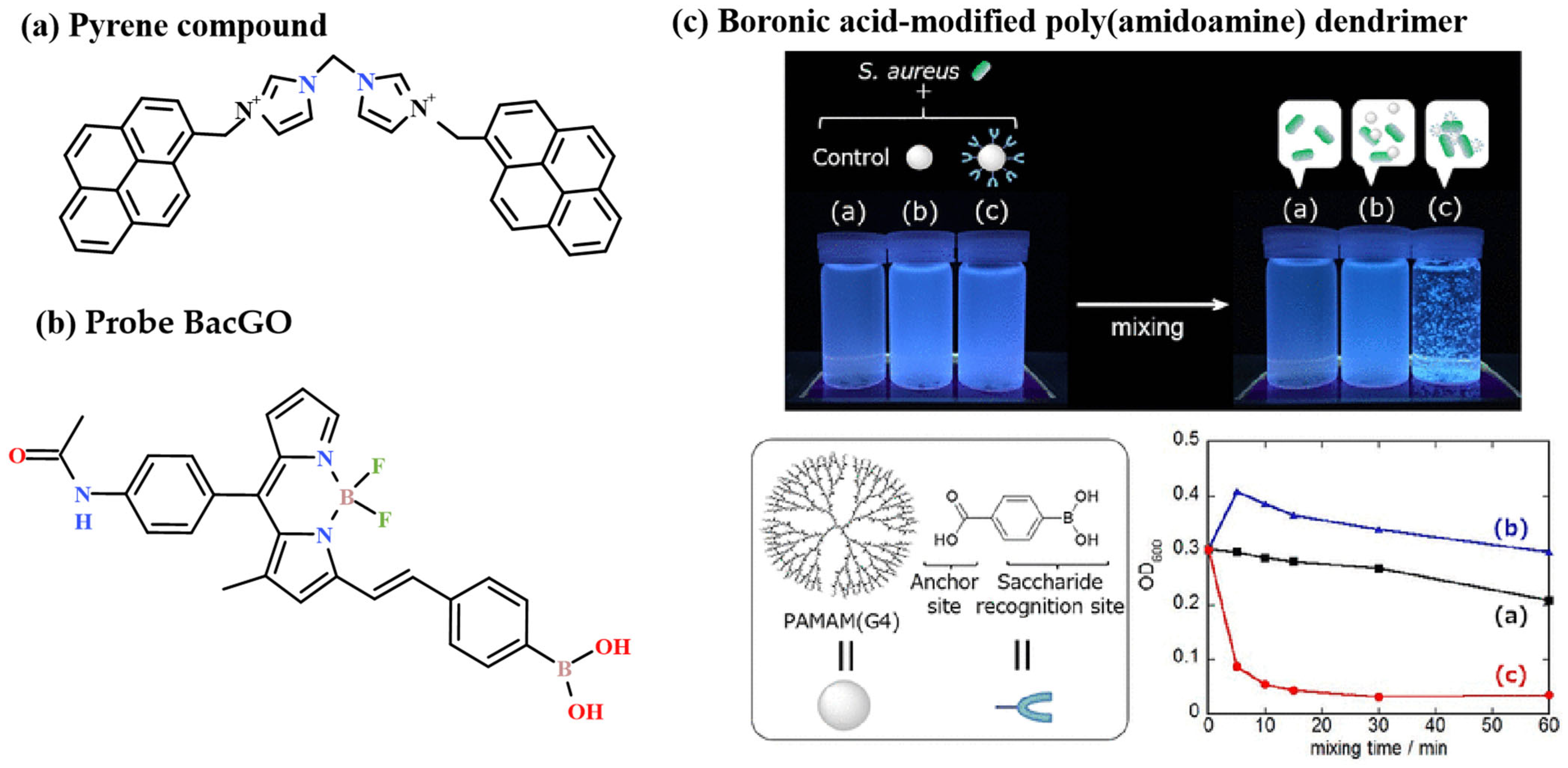
2.1.3. Binding Probes

Different types of bacteria have different biochemical properties on the cell wall. According to this characteristic, bacteria can be divided into two categories: Gram-positive bacteria and Gram-negative bacteria. Gram staining is a common method to identify bacteria by using this difference. It was first put forward by Danish bacteriologist Hans Christian Gram in 1884 [98,99,100]. It was initially used to identify the relationship between pneumococci and Klebsiella pneumoniae, and then extended to one of the gold standard techniques for bacterial classification. It has been applied to various forms of bacterial samples, including aquatic environment samples and infected tissue. In order to effectively replace standard Gram staining, selective Gram-positive bacterial fluorescent probes are necessary. Hexylamine iodide (HI) is the most well-known commercial probe and is known to bind to Gram-positive bacteria, but the reported Gram-positive bacteria have limited selectivity over Gram-negative bacteria.
In recent years, inspired by the Gram Staining technology, researchers have developed a series of binding fluorescent probes for bacteria by using the differences of the substances and charges on the surface of specific bacteria. Based on the fluorescent signal of the detection probe, the target bacteria can be labeled and detected. For instance, Wang et al. [101] realized the fluorescence detection of Escherichia coli, Bacillus subtilis and Staphylococcus aureus by using rhodamine 6G with a positive charge to fluorescently label negatively charged bacteria on the surface through electrostatic interaction. Xu et al. [102] reported a fluorescent probe based on a charge interaction to bind bacteria (Figure 5). After binding with bacteria, the probe was induced to depolymerize from aggregates and present a process of fluorescence from none to present, thus realizing the optical monitoring of target bacteria. The probe is constructed from pyrene compounds derived from imidazoline. Pyrene is a classic fluorophore that emits unique fluorescence transformed between pyrene monomer and excimer emissions [103]. The probe can self-assemble to form nanoparticles, and the aggregation effect can quench pyrene fluorescence. When imidazoline was used as the binding site to recognize the surface of anionic bacteria through electrostatic interaction, due to the synergistic effect of electrostatic interaction and hydrophobic force, the bacterial surface and imidazole compounds were competitively binding. This kind of binding led to the depolymerization of the aggregates. The pyrene fluorophore was in the state of pyrene excimer and pyrene monomer and displayed unique fluorescence, which gave a ratiometric signal. Then, according to the output signals (fluorescence increase and ratio change) of these two channels, a two-dimensional analysis chart could be established for the visual interpretation and identification of bacteria. Fluorescent probes rely on fluorescence spectra to identify bacterial species. Imaging based on fluorescence microscopy is also important [104,105,106,107,108]. And the probe also makes it possible to make two-photon ratio imaging of fluorescent labeled bacteria. Svechkarev et al. utilized DMAF fluorescent dye interacting with bacterial cell membranes to generate species-specific spectral fingerprints, and applied neural network classification algorithms for pattern recognition analysis and classification of spectral data. This sensor successfully differentiated eight representative pathogenic bacteria with species-level accuracy reaching 85.8%, while achieving 98.3% accuracy in distinguishing Gram-positive and Gram-negative bacteria. Breaking through the traditional “one-test-one-target” paradigm, this approach enables broad-spectrum pathogen detection through non-specific spectral fingerprinting combined with AI decoding, paving a new pathway for point-of-care diagnosis of infectious diseases [109].
Bacterial surfaces typically contain abundant metabolic compounds, and fluorescent probes can identify and recognize specific bacteria through metabolic labeling. Kwon et al. presented a novel fluorescent probe, BacGO, based on the BODIPY fluorophore, which showed good selectivity for Gram-positive bacteria [110]. The design idea is to modify the boric acid group on the basis of the BODIPY fluorophore and label the spores of Gram-positive bacteria with the boric acid group. The binding mechanism is based on the fact that the boric acid group can bind to the glycoprotein structure on the surface of bacteria [111,112]. BacGO could be used to assess the Gram status in the bacterial community from wastewater sludge. Furthermore, BacGO could sensitively and selectively detect a Gram-positive bacterial infection, not only in vitro but also using an in vivo keratitis mouse model. Tsuchido et al. prepared a phenylboronic acid-modified PAMAM(G4) (B-PAMAM(G4)) dendrimer, which could form large aggregates when mixed with bacteria in less than 5 min (Figure 5). The aggregates are visible to the naked eye and can be used to quickly and selectively distinguish between Gram-positive and Gram-negative bacteria [113].
Figure 5. (a) The fluorescent probe based on charge interaction to bind bacteria; (b) fluorescent probe BacGO based on BODIPY fluorophore; (c) boronic acid-modified poly(amidoamine) dendrimer. Reproduced with permission from ref. [113]. Copyright© 2019 Tsuchido, Y. et al. This is an open access article distributed under the Creative Commons Attribution License (CC BY 4.0), which permits unrestricted use, distribution and reproduction in any medium, provided the original work is properly cited.
Chemosensors 13 00182 g005
There are a large number of peptidoglycans in the cell walls of Gram-positive and Gram-negative bacteria. As a glycopeptide antibiotic, vancomycin has a high affinity for alanine at the end of the cell wall precursor peptidoglycan, which can inhibit the synthesis of high molecular weight peptidoglycan in bacterial cell walls, leading to cell wall defects and exerting antibacterial effects. It can quickly kill dividing and proliferating bacteria. It can also inhibit RNA synthesis in the cytoplasm. It can be used as a treatment for severe infections caused by penicillin-resistant and cephalosporin-resistant Gram-positive bacteria [114]. Mills et al. designed a no-wash red optical molecular imaging agent based on vancomycin and environmental merocyanine dyes, which can quickly and selectively detect Gram-positive bacteria [115]. In vivo experiments have shown that the imaging agent combined with a new imaging/delivery internal microscope device could specifically detect Gram-positive bacteria in human lungs. Andrew Wang et al. innovatively developed a dual-fluorescence probe synergistic targeting strategy based on the Staphylococcus aureus-specific siderophore-iron uptake system. The probes are preferentially internalized by the bacteria and activate fluorescence signals, enabling the preliminary differentiation of S. aureus from other bacterial species. By engineering vancomycin-derivative probes with optimized affinity, these probes selectively bind to the cell wall peptidoglycan of vancomycin-susceptible S. aureus (VSSA) and trigger fluorescence. The combined use of a siderophore-based probe (primary screening) and a vancomycin-based probe (secondary typing) achieves the high-specificity two-dimensional discrimination of vancomycin-resistant S. aureus (VRSA) and VSSA within complex microbial communities [116].
Although the aforementioned small-molecule fluorescent probes enable the real-time detection of target bacteria, their practical applications remain significantly challenged. First, limitations in physicochemical properties hinder utility. Most probes suffer from poor water solubility and insufficient photostability, leading to aggregation or quenching in physiological environments (e.g., blood or tissue fluids). Additionally, detection often relies on stringent conditions (e.g., specific pH, temperature or anaerobic environments), making them poorly adaptable to the diversity of complex clinical samples. Second, probe development and research are still in their infancy. Compared with well-established protein-based or nucleic acid-based probes, small-molecule probes lack systematic design strategies. For instance, (1) target selection is narrow, predominantly focusing on a limited set of biomarkers such as β-lactamases or lipopolysaccharides, and fails to cover drug-resistant bacteria or rare pathogens; (2) insufficient mechanistic studies hinder the real-time tracking of dynamic probe–bacteria interactions, such as binding kinetics or subcellular localization; (3) complex synthesis protocols impede scalable production. Future research could focus on the rational design of multifunctional probes by introducing hydrophilic groups (e.g., polyethylene glycol chains) or nano-carrier encapsulation strategies to improve water solubility; developing broad-spectrum activation mechanisms targeting conserved bacterial metabolic pathways (e.g., quorum-sensing signaling molecules or efflux pump systems) to enhance detection universality.

2.2. Biomaterial Sensors

There are also many traditional biological fluorescence detection technologies for bacteria, such as immunofluorescence technology. The basic reaction of immunology is the antigen antibody reaction. The antigen antibody reaction has high specificity. This technology is to label the fluorescent pigment that does not affect the activity of the antigen and antibody on the antibody (or antigen). After binding with the corresponding antigen (or antibody), the specific fluorescence reaction will appear under the fluorescence microscope. Then, the concentration of the measured substance can be calculated by measuring the fluorescence intensity with the instrument. The commonly used fluorescent pigments are fluorescein isothiocyanate (FITC), tetraethyl rhodamine (RIB200) and tetramethyl rhodamine isothiocyanate (TRITC). The main characteristics of this technique are high specificity, high sensitivity and high speed. The disadvantages are that the problem of nonspecific dyeing has not been solved completely, the objectivity of result judgment is not enough and the technical procedure is also complicated.
In recent years, the application of biomaterials (such as phages, aptamers and peptides) to bacterial fluorescence detection has gradually attracted the attention of researchers. Compared with the traditional biological fluorescence detection, the introduction of biomaterials into the fluorescence detection system can give full play to its own biological characteristics, improve the specificity and sensitivity of detection and effectively improve the performance of the detection system.

2.2.1. Phage-Based Fluorescence

Phage, the natural enemy of bacteria, has gradually returned to the view of researchers and become a research hotspot. Phage is a general term for viruses that parasitize bacteria, fungi and other microorganisms. Most of them are about 100 nm in diameter, which makes them become ideal biomaterial probes for bacterial detection. In addition to targeting, another key feature of phages is that they can distinguish living host bacterial cells from inactive host bacterial cells, because phages can only replicate and express enzymes in living bacterial cells. Therefore, it has an important application prospect in bacterial detection and bacterial resistance research.
The early potential applications for the bioluminescence detection of bacteria were initiated by the development of luciferase reporting phages (Ulitzur and Kuhn, 1987) [117]. Recent advances in biosensors have led to the ability to exploit certain enzymes, using photons as a by-product of their reactions. This bioluminescence phenomenon can be used to detect the existence and physiological status of cells. The mechanism is that phages specifically recognize the lipopolysaccharide receptor on the surface of bacteria through the tail silk protein on the tail silk, and then a small hole is dissolved in the peptidoglycan of the bacterial cell wall by using the tail thorn protein. With the contraction of the tail sheath, the head genetic material is injected into the bacterial cells, and then the offspring phages are synthesized by the bacterial metabolic system. Finally, under the interaction of porin and endolysin, the bacteria are lysed and the phages and intracellular substances are released. Based on the above principles, Yong and coworkers functionalized magnetic nanoparticles with phages [118]. Through the dual role of the capture and lysis of phage, a bioluminescence method was established for the detection of Pseudomonas aeruginosa based on phage recognition, and successfully applied to the rapid detection of the drug resistance of the bacteria. Wu et al. [119], Yang et al. [120] and Franche et al. [121] also reported the detection of Salmonella, Klebsiella and Escherichia coli by the phage-mediated bioluminescence method. The bioluminescence method described above has become a new and attractive method due to its high specificity and inherent ability to distinguish living cells from non-living cells. However, the main disadvantages are relatively long detection time and lack of sensitivity, which become obvious when low numbers of bacteria are detected.
In contrast, the detection method of fluorescent-labeled phage has the advantages of fast, intuitive and high sensitivity, and is gradually being favored by researchers. Goodridge et al. [122,123] labeled phage LG1 with YOYO-1 dye and realized the detection of Escherichia coli O157: H7. However, YOYO-1 dye made the phage lose the ability to inject nucleic acid into the host bacteria, thus showing a halo-like appearance. Mosier boss et al. [124] also prepared fluorescent-labeled phage P22 with SYBR gold dye and detected its host Salmonella typhimurium LT2. Jiang et al. [125] used SYBR gold to label Salmonella specific O-I phage to achieve rapid, intuitive, accurate and high-throughput detection of Salmonella in food. Low et al. [126] also established a new method of fluorescent phage labeling and used it for virus screening and interaction research. The DNA dye SYTO 13 is used to label the phage PMBT14. Fluorescent phage labeling can not only detect the host bacteria but can also prove to be helpful to analyze the interaction between the phage and the host bacteria.

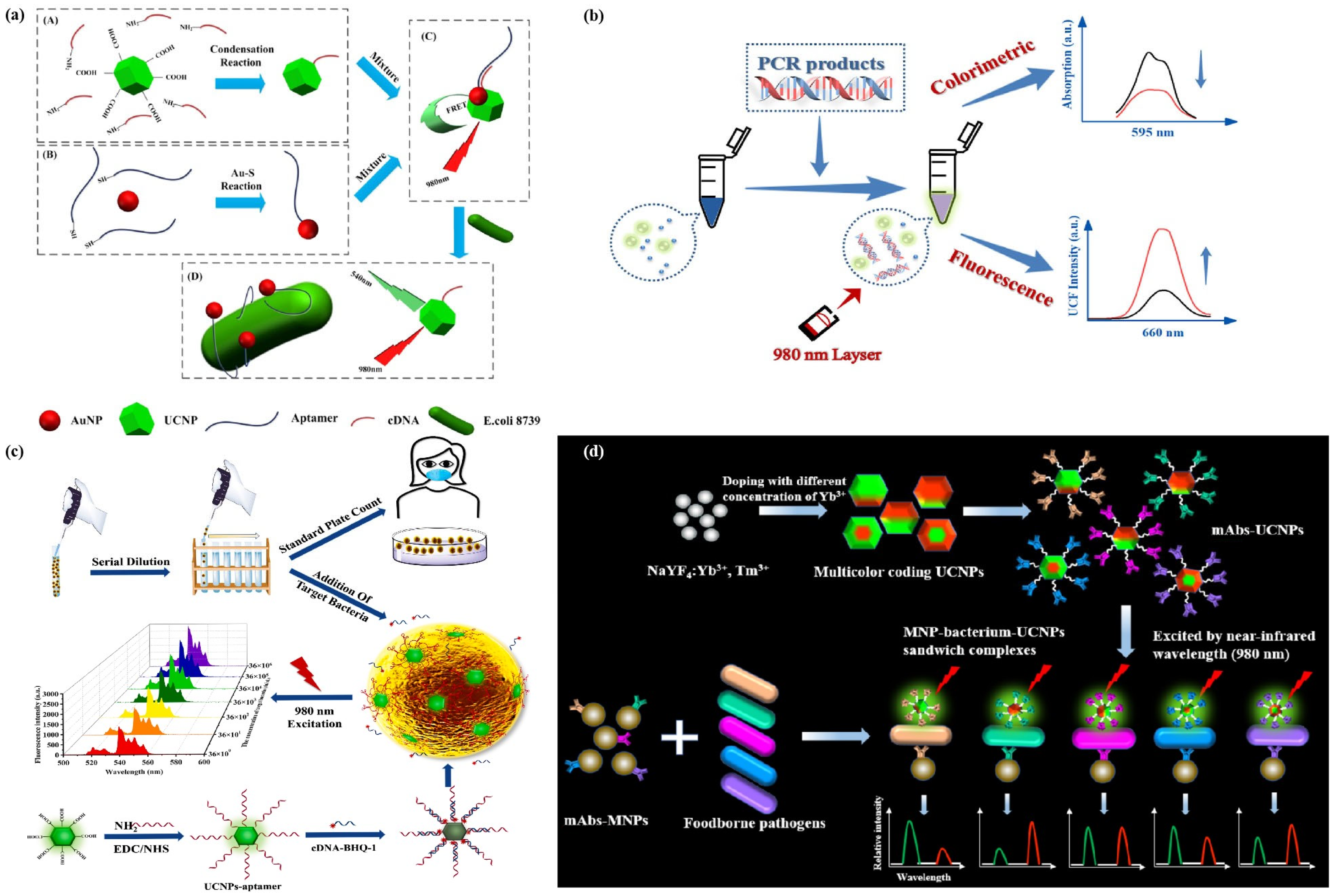
2.2.2. Aptamer-Based Fluorescence

Aptamers are oligonucleotides screened by repeated screening in vitro or systematic evolution of ligands by exponential enrichment (SELEX) technology, which have more advantages in capturing bacterial cells than antibody-based and phage-based recognition elements. They have the advantages of small volume (usually 3.5 nm), high specificity of target molecules, good applicability in vivo and in vitro, good biocompatibility, low production cost, high molecular stability and low detection limit. Aptamers can show a high binding affinity with target bacterial cells, based on their flexibility and folding ability when binding to the target. The application process can reduce the overall detection limit [127]. By using SELEX phylogenetic ligands, the aptamers suitable for a variety of target bacteria can be designed to bind fragments on the surface of bacterial cells, such as polysaccharides, proteins and flagella. Fluorescence-based aptamer assays include labeled apta-assay and unlabeled apta-assay [128]. Labeled apta-assays require at least one chromophore or fluorophore, and typically labeled fluorescence apta-assays are based on fluorescence resonance energy transfer (FRET) [129]. In addition, some nanomaterials with fluorescence and quenching properties can also replace the analysis of dye labeling. Label-free apta-assays means that the detection process does not require any chromophore or fluorophore. They are based on DNA intercalators, basic sites to bind dyes and metal nanomaterials based on fluorescent apta-assays [130].
At present, various types of organic and inorganic fluorescent materials have been used as labels to construct apta-assays. Maeng et al. [131] developed a labeled apta-assay method for the rapid detection of foodborne bacteria based on the change of RNA aptamer signal transduction intensity. Three RNA aptamers were screened to antagonize different components of the bacterial cell wall, including lipopolysaccharide (LPS) of Escherichia coli 157: H7, teichoic acid of Staphylococcus aureus and OmpC (a cell membrane protein) of Salmonella typhimurium. The aptamer with a poly A tail at the 3` end was hybridized with a sulfhydryl-modified DT linker probe on the surface of the silver membrane. Bacterial target cells can be captured by specific aptamers fixed on the surface of the silver membrane. When the second poly A aptamer binds to the captured bacterial target, a sandwich complex is formed. The dT-FAM probe was added and hybridized with the poly of a tail of the second aptamer to produce the fluorescence emission. Ahn, J. Y. et al. designed a fluorescent biosensor analysis method based on aptamers. The whole bacterium SELEX strategy was used to isolate the specific aptamer of Shigella sonnei. Among the 21 aptamers tested, the C (T) values of SS-3 and SS-4 aptamers were the lowest. Therefore, the sandwich complex of SS-3 and SS-4 was used to detect and identify Shigella sonnei cells. The constructed platform showed a significant ability to detect and distinguish Salmonella sonnei from other intestinal strains. The study also fully demonstrated the feasibility of an aptamer sensor platform for detecting Streptococcus sonnei in a variety of foods [132].
As precision molecular recognition elements, aptamers have been strategically integrated with nanoscale platforms to develop composite nanosystems for bacterial capture and detection. Through programmable surface functionalization, these hybrid systems combine the high specificity of aptamer-target binding with the unique optoelectronic properties of engineered nanomaterials. Representative configurations include quantum dots (QDs, semiconductor nanocrystals), plasmonic gold nanoparticles (AuNPs, 20–80 nm), superparamagnetic iron oxide nanoparticles (SPIONs, 10–100 nm) and lanthanide-doped up-conversion nanoparticles (UCNPs, e.g., NaYF4:Yb,Er). The structure–property relationships and biosensing applications of these aptamer–nanomaterial conjugates will be systematically analyzed in subsequent sections, with particular emphasis on their signal amplification mechanisms and limit of detection (LOD) optimization strategies.

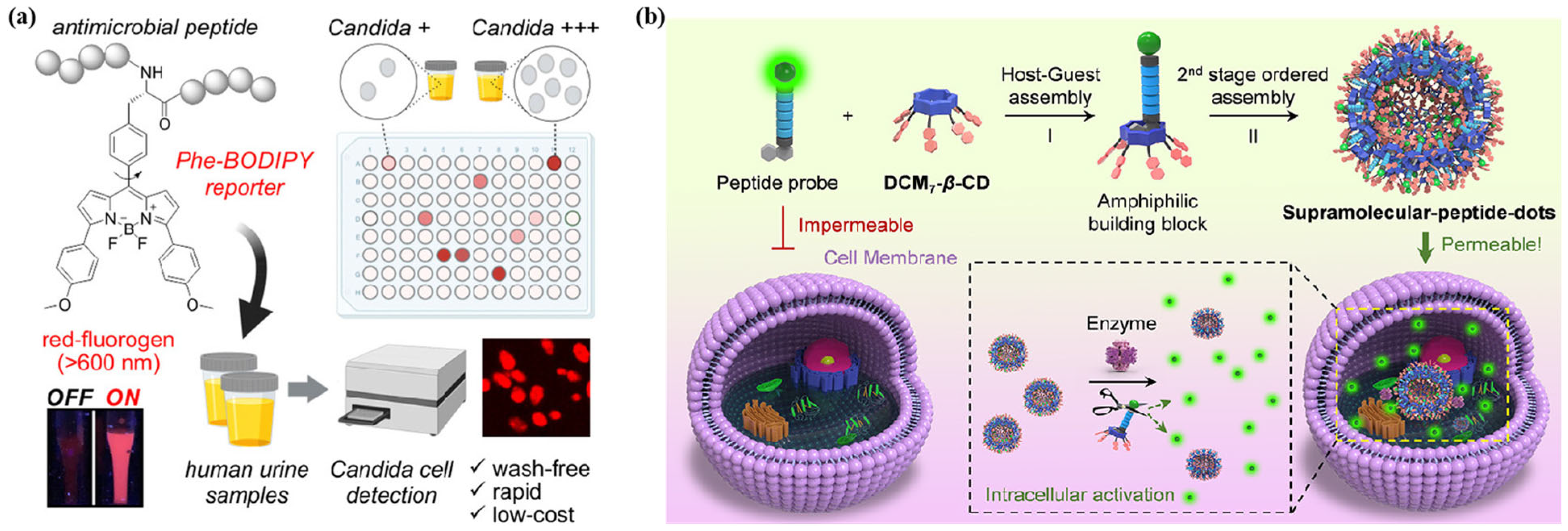
2.2.3. Peptide-Based Fluorescence

Biological polypeptide is a kind of substance with multiple biological functions. Polypeptide molecules participate in the regulation of cell proliferation and differentiation, energy metabolism, signal transduction and other life processes through high-affinity binding with receptors [133,134]. Researchers often use natural polypeptides or synthetic polypeptides to construct fluorescent dye-labeled polypeptide probes for biological analysis and imaging, disease diagnosis and adjuvant therapy [135,136,137,138,139,140]. Compared with molecular probes or other nuclear magnetic resonance imaging probes, they have the characteristics of low biological toxicity and easy degradation.
It is well known that a specific sequence of polypeptides can be specifically cleaved by the target enzyme, and bacteria will secrete a specific enzyme to take advantage of this characteristic. Liu et al. [141] reported an aggregation induced emission (AIE) bio-probe model. The probe (PyTPE-CRP) consists of two parts, an AIE molecule (PyTPE) and an enzyme reactive polypeptide chain (NEAYVHDAP). The biological probe can detect a bacterial infection and kill the living bacteria in macrophages through a dynamic process. The designed PyTPE-CRP can be specifically cleaved by casp-1 in bacterial-infected macrophages, and the generated residues are self-assembled into aggregates, which specifically accumulate on the phagosome-containing bacteria in the macrophages, resulting in the opening of intracellular fluorescence. At the same time, an early infection detection can also guide the use of similar antibiotics. Therefore, this method can be used as a sensitive detection platform for bacterial infection through fluorescence and intracellular bacterial clearance through photodynamic therapy. Mendive-Tapia et al. [142] designed a novel fluorinated amino acid (Phe-BODIPY) rational based on a phenylalanine (Phe) core, and used it to synthesize fluorescent antimicrobial peptides for the rapid labeling of Candida cells in urine (Figure 6a). The biological activity and confocal microscopy experiments conducted in different strains showed that the constructed antimicrobial peptides had good selectivity and high chemical stability towards bacterial cells. Jiao et al. created a hepta-dicyanomethylene-4H-pyran-appended β-cyclodextrin derivative, DCM7-β-CD. Modified cyclodextrin serves as an effective host for FITC-labeled 1-bromonaphthalene-modified peptides, and the interaction between the two can form supramolecular clusters, which then self-assemble into supramolecular peptide dots (Spds). Research has found that it exhibits better bacterial cellular uptake ability, providing enhanced labeling and therapeutic benefits for both Gram-positive and Gram-negative bacteria. The Spds platform reported in this article has the ability to promote the delivery of therapeutic peptides and provides an easy to implement strategy to enhance the antibacterial efficacy of known therapeutic peptides [143]. Fluorescent probes based on peptides for the specific detection of bacteria have better application value compared with organic small molecule probes, and are more suitable for monitoring and the subsequent adjuvant therapy of bacterial infections in organisms. However, there are also problems such as complex preparation, high cost and poor stability (Figure 6b).

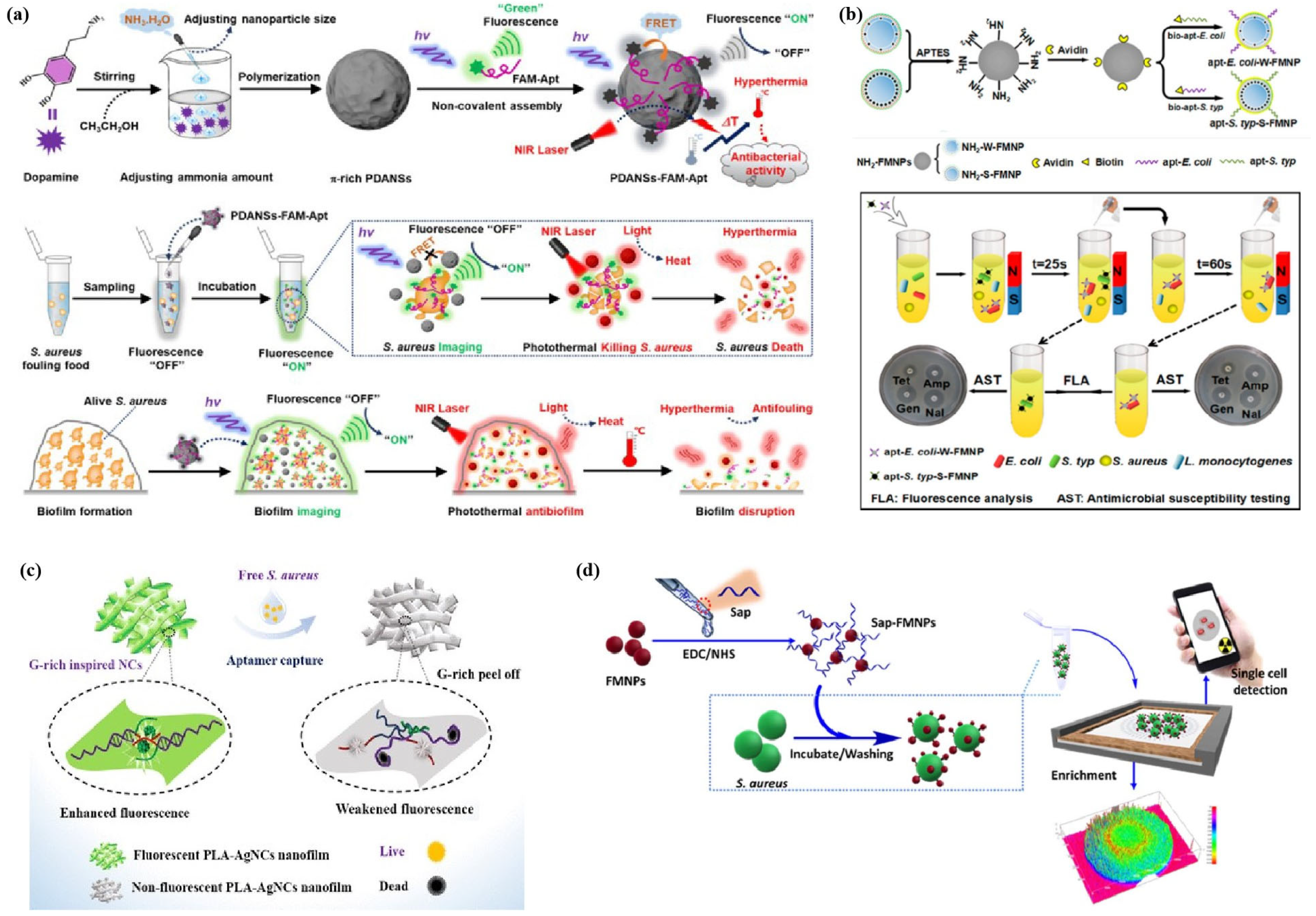
2.3. Nanocomposite Fluorescent Sensing System

In 1990, nanoscience and technology, which studies the preparation and properties of nano-sized materials, was officially born. Nanoscience is a new interdisciplinary, which is called the first-class important science and technology in the 21st century. With the vigorous development of nanoscience and technology, many scientists have also begun to apply this technology to the preparation of fluorescent chemical sensing materials, and have made remarkable achievements [144,145,146,147,148,149,150,151]. Due to the excellent properties of nanomaterials, such sensor probes generally have good biocompatibility, higher stability than photobleaching of fluorescent dyes and simple preparation. Nowadays, green production is becoming more and more mainstream, and fluorescent probes based on nanomaterials are more and more widely used. For pathogenic bacteria, the progress of nanotechnology has enabled new nano detection strategies to detect target pathogenic bacteria quickly and sensitively. Nanomaterials commonly used to construct fluorescent sensors mainly include gold nanoparticles (AuNPs), gold nanorods (AuNRs), magnetic nanoparticles (MNPs), graphene oxide (GO), quantum dots (QDs), carbon dots (CDs), up-conversion nanoparticles (UCNPs) and polydopamine nanospheres. Nanomaterials with excellent characteristics are combined with recognition elements such as antibodies, aptamers, phages or ligands based on electrostatic interaction to achieve the effective monitoring of target bacteria. At present, the nanoprobe system constructed by combining nanoparticles with aptamers is the most studied.
For some nanomaterials with luminescent properties, such as UCNPs, QDs and CDs, they have a higher extinction coefficient and good light stability, and have narrow and adjustable luminescent spectra, allowing researchers to accurately label target bacterial cells. Most importantly, fluorescent nanoparticles with different colors and modified by different recognition elements can be used to detect various types of bacterial cells at the same time.

2.3.1. UCNP-Based Nanoprobes

UCNPs are a new kind of luminescent material that emit UV visible fluorescence under near-infrared excitation. In recent years, they have been widely used in the fields of photodynamic therapy (PDT), biological imaging and biological detection [152,153,154,155,156]. Compared with traditional fluorescent probes (such as organic dyes, metal complexes or inorganic quantum dots), UCNPs have many advantages, such as non-spontaneous fluorescence, high chemical stability, large light penetration depth, long life, less damage to samples and almost no background light.
Compared with traditional down-conversion fluorescence imaging, anti-Stokes fluorescence imaging has unique advantages, especially in the application of biological imaging. Based on the performance advantages of UCNPs, Lin et al. [157] developed a new detection platform based on fluorescence resonance energy transfer (FRET) for rapid, ultra-sensitive and specific bacterial detection (Figure 7a). Among them, AuNPs as energy receptors bind to targeted aptamers, UCNPs as energy donors bind to the corresponding complementary DNA (cDNA). When the targeted aptamer hybridizes with cDNA, the spectral overlap between the fluorescence emission of UCNPs and the absorption of AuNPs leads to fret, which leads to UCNP fluorescence quenching. In the presence of target bacteria, aptamers preferentially combine with bacteria to form a three-dimensional structure in order to dissociate UCNP-cDNA from AuNP aptamers and restore UCNP fluorescence. E. coli ATCC 8739 was successfully detected based on this platform. Song et al. reported a novel sensing platform based on fluorescence quenching composed of alendronic acid (ADA)-coated up-conversion nanoparticles (UCNPs) and Nile Blue (NB) combined with polymerase chain reaction (PCR) for the rapid, sensitive and specific detection of Escherichia coli (E. coli) (Figure 7b) [158]. Chen et al. [159], based on the binding of UCNPs to aptamers and the fluorescence resonance energy transfer (FRET) between black hole quenching factor 1 (BHQ-1) linked to cDNA, developed a fluorescence sensing platform for the specific and sensitive detection of pathogens (Figure 7c). The fluorescence emission spectrum of UCNPs overlaps well with the absorption spectrum of BHQ-1, which makes FRET occur and leads to fluorescence quenching. In the presence of target bacteria, UCNP aptamers preferentially capture bacteria, thereby reducing the UCNP aptamer cDNA BHQ-1 complex and leading to fluorescence recovery. Zhao et al. [160] synthesized multicolor coding UCNPs and applied them to the rapid and simultaneous detection of five foodborne pathogens (Figure 7d). Multicolor coding UCNPs were obtained by doping different concentrations of sensitizers (Yb3+) into the shells of the synthesized NaYF4:Yb3+, Tm3+ (20%/2%)@NaYF4:Yb3+, Er3+ (x%/2%) and Er3+ (x%/2%) core–shell nanocrystals. Then, magnetic nanoparticles (MNP) modified with monoclonal antibodies against target bacteria were used to capture and isolate bacteria to obtain MNP bacterial complexes. Different UCNPs with multicolor coding as the signal probe were also modified by a monoclonal antibody and reacted with the MNP bacterial complex to form an MNP bacterial UCNP sandwich complex. After the sandwich complex was excited at 980 nm, the R/G ratio and green photoluminescence intensity (PL intensity) obtained can be used to distinguish and quantitatively detect foodborne pathogens, respectively.
The existing near-infrared-induced up-conversion nanoparticles are limited by issues such as insufficient fluorescence brightness. Wang et al. developed a triple-doped alkaline earth sulfide nanoparticle (AES NPs) with ultra bright near-infrared excitation fluorescence, high brightness and variable emission wavelength, consisting of red-emitting (CaS: Eu, Sm, Mn) and blue-emitting (SrS: Ce, Sm, Mn) alkaline earth sulfide nanoparticles. The nanoparticles are covalently linked to the bacterial targeting portion (teicoplanin) for in vivo and in situ imaging of Gram-positive/-negative bacteria. In vivo experiments have shown that functionalized composite nanoparticles can fully distinguish and differentiate between sterile inflammation and bacterial infection. It is worth noting that the strong fluorescence of this system under 980 nm light irradiation can achieve high sensitivity and deep tissue imaging [161].
The surface modified UCNPs have certain biocompatibility, and their biological toxicity depends more on the particle size and surface charge. The current research shows that the biological toxicity is small within the appropriate dosage range, but the long-term potential toxicity of UCNPs has not been monitored. For practical application, there is still a lot of systematic investigation work to be carried out.

2.3.2. QD-Based Nanoprobes

QDs, as semiconductor nanoparticles, are a class of zero-dimensional nanomaterials with significant fluorescence properties. The movement of electrons in the quantum dots in all directions is limited, so the quantum confinement effect is particularly significant. It is precisely because of this special property that the electronic and optical properties of QDs have a unique particle size dependence. At present, many scholars have systematically studied QDs with different sizes, shapes and components. In the field of fluorescent biomarkers, QDs can be combined with specific antibodies or small molecules [162,163,164]. Without changing their chemical properties, they can emit specific wavelength fluorescence after being excited by the light source in order to realize the recognition and detection of targets. Compared with traditional labeling materials, QDs have many advantages, such as high fluorescence intensity, diverse luminescence colors, suitability for multiple spatial and spectral transmission, effectively weakening or even eliminating the influence of background fluorescence and wide spectral range. These superior properties make QDs excellent fluorescent probes in many applications.
In recent years, QD fluorescent probes have been applied more and more to the fluorescent detection of pathogenic bacteria [165]. Fu et al. [166] reported a surface amino-modified CdSe/ZnS/SiO2 composite nanoparticle for the identification of target bacteria, in which CdSe/ZnS QDs are used as fluorescent markers. The nanoparticle has good dispersion, brightness and less non-specific binding in bacterial detection. At the same time, the method has high sensitivity and and 3 × 10² cell/mL bacteria were counted, which was lower than traditional plate counting and organic dye-based methods. Subsequently, Wang et al. [167] developed core–shell CdSe/ZnS quantum dots encapsulated in aminated silica nanoparticles (QDs@SiO2-NH2 FNPs) as advanced fluorescent probes for microbial sensing. Through the integration of spectral-encoded fluorescence labeling with dielectrophoresis (DEP) manipulation, this platform achieved the precise quantification of Salmonella typhimurium with a detection limit of 15 CFU/mL. Subsequently, in order to further reduce the detection limit and obtain a better visual detection performance, an integrated dielectric electrophoresis microfluidic chip and related microsystems were established. FNPs labeled bacteria can be enriched along the edge of interdigital microelectrodes in microchannels by positive DEP, and can be counted under fluorescence microscope.
Another common modification method of QDs is to modify groups or link biological molecules on the surface of QDs to make them better interact or specifically bind with bacteria. Xue et al. [168] prepared the QDs with carboxyl groups on the surface by modifying CdSe QDs with mercaptoacetic acid, and connected them with the amino group on the surface of the bacteria under the action of EDC/NHS, and detected Escherichia coli and Staphylococcus aureus with fluorescent labeling. Duan et al. [169] prepared two kinds of CdTe QDs modified by the carboxyl group, which emitted green fluorescence and red fluorescence, respectively. The two fluorescent QDs were connected with the Vibrio parahaemolyticus aptamer and Salmonella aptamer by amide construction, and incubated with the mixed samples containing the two bacteria for 1 h, and then flow cytometry was performed. The two fluorescent QDs were connected with the Vibrio parahaemolyticus aptamer and Salmonella aptamer, respectively, through amide. After incubation with mixed samples containing two kinds of bacteria for 1 h, and then were detected by flow cytometry. This method can realize the specific labeling and fluorescence detection of two kinds of bacteria at the same time. In addition to being specific recognition molecules, aptamers can also improve the efficiency of QD labeling bacteria and reduce the detection limit.
Although quantum dots have excellent fluorescence properties, studies have found that heavy metals such as chromium and selenium in quantum dots are highly toxic to organisms and cause great harm to the environment. Their applications in the biochemical field are limited. Therefore, researchers have begun to pay attention to fluorescent materials with better fluorescence efficiency, lower toxicity and better biocompatibility. In 2004, a new type of luminescent nanomaterial carbon dot (CD) was discovered. In addition to having similar optical properties with semiconductor quantum dots, it also has the advantages of good water solubility, surface functionalization, good biocompatibility, extensive carbon sources and low preparation cost [170]. It represents that the research of luminescent nanoparticles has entered a new stage.
CDS have been used in bacterial fluorescent labeling. Qi et al. [171] constructed a new fluorescent sensor array based on functional carbon dots (CDs) for the simultaneous detection of multiple bacteria. The sensor array is composed of three functionalized CdS carbon dots modified with different receptors (boric acid, polymixin and vancomycin). These three functionalized carbon dots can bind to all bacteria with different affinities. Taking advantage of the difference of bacterial binding ability, the three functionalized carbon dots will produce corresponding fluorescence response modes, and unique molecular fingerprints can be obtained for different bacteria. Xu et al. [172] successfully established a rapid and sensitive quantitative detection method for Salmonella typhimurium using aptamer-binding carbon dots (CDs-apt) as the fluorescent probe. Based on the fluorescence detection method with CDs-apt as the probe proposed in this study, a series of fluorescent probe systems for detecting other bacteria can be extended by changing the appropriate aptamer. Weng et al. [173] prepared mannose-modified carbon quantum dots (Man CDs) based on the reaction between ammonium citrate and mannose. Using the specific binding ability between mannose and E. coli, they detected and analyzed E. coli in tap water, apple juice and urine samples. Zhong et al. [174] used CDs with vancomycin surface modified to specifically detect Gram-positive bacteria (Figure 8a). When a large number of CDs are connected to the surface of the bacterial cell wall, it will cause a large number of labeled reagents to agglomerate, resulting in decreased fluorescence intensity, so as to selectively detect Staphylococcus aureus, Bacillus subtilis and Listeria. Similarly, Chandra et al. [175] used CDs with surface modified Amikacin to selectively label E. coli, and successfully detected E. coli from apple juice, orange juice and pineapple juice samples. Chen et al. [176] also used CDs modified by specific aptamers as energy donors (Ex 355 nm) and GO as energy receptors to build a biosensor with a fluorescence resonance energy transfer (FRET) effect, and realized the quantitative detection of E. coli O157:H7 pathogenic bacteria within 1 h. Lai et al. [177] prepared self-functionalized CQDs modified with mannose and folic acid using a dry heat method for the specific labeling and recognition of Escherichia coli (E. coli) and tumor cells, respectively (Figure 8b). Duan et al. [178] connected fluorescent dye-labeled aptamers to carbon nanoparticles, and achieved a sensitive and stable simultaneous detection of Vibrio parahaemolyticus, Staphylococcus aureus and Salmonella typhimurium through multiple FRET systems. Hu et al. prepared fluorescent probes (CQDs MNPs) for Escherichia coli 157: H7 (E. coli) based on adapter-labeled carbon quantum dots (CQDs) and complementary DNA-labeled magnetic nanoparticles (cDNA MNPs). The fluorescence intensity of CQDs MNPs decreases with the addition of E. coli. The sensitive detection of E. coli can be successfully achieved by utilizing the linear relationship between fluorescence intensity and E. coli concentration. This fluorescent probe shows great potential in ensuring food quality and safety [179].

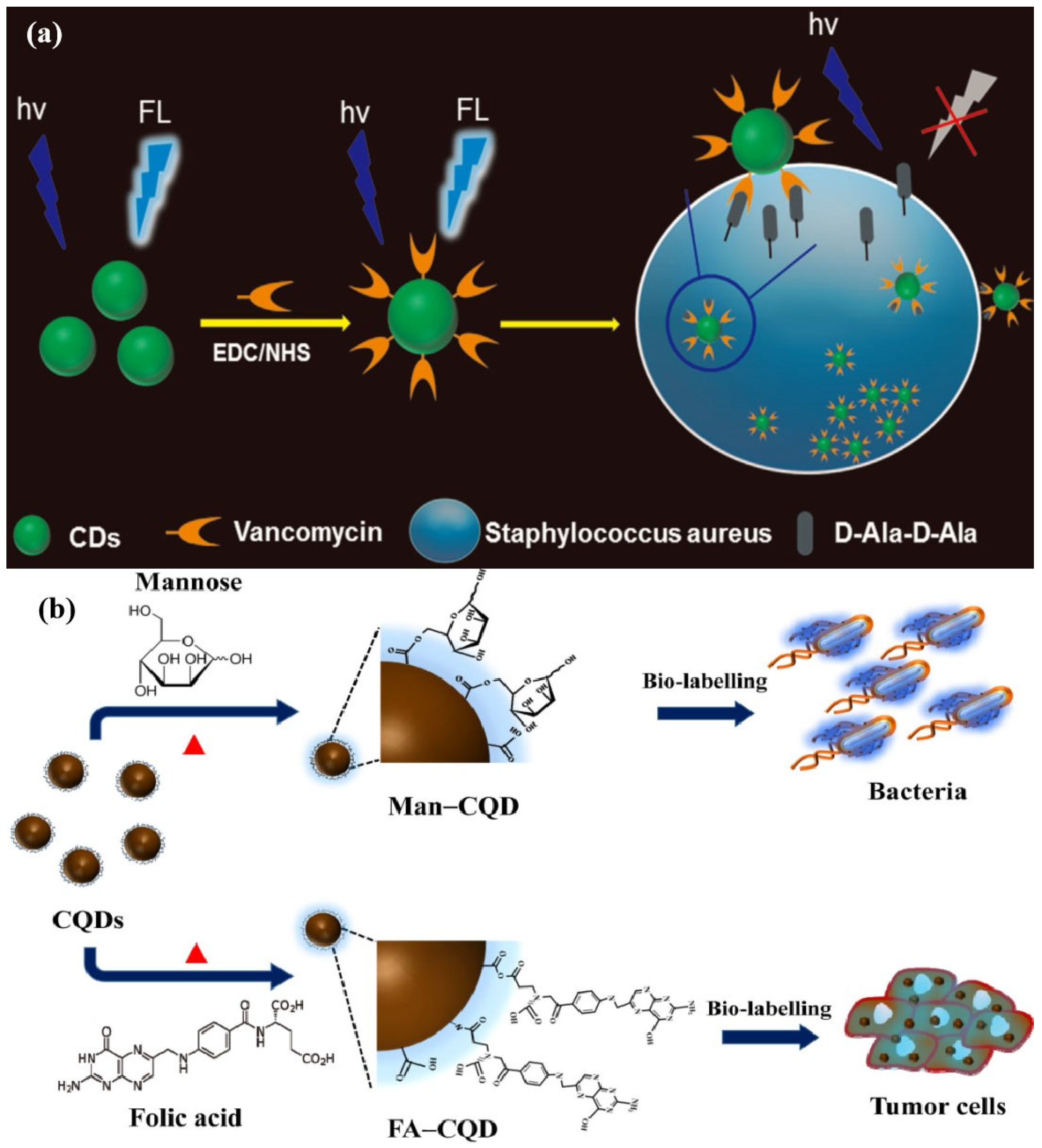
2.3.3. Other Types of Nanoprobes

Gold nanoclusters (Au NCs) are widely used as fluorescent probes in biomedical sensing and imaging due to their multifunctional optical properties and low cytotoxicity. Au NCs have precise energy states and unique geometric structures similar to chromophores. The regulation of kernel size and surface ligands can also manipulate their biochemical/biophysical properties [180]. Au NCs are a novel antibacterial agent with broad-spectrum antibacterial activity and no resistance to bacteria [181]. In recent years, Au NCs have also been widely used in the construction of antibacterial materials and bacterial detection systems. Cheng et al. [182] prepared vancomycin-stabilized fluorescent gold nanoclusters (AuNCs@Van) and aptamer-encapsulated magnetic beads for identifying and capturing Staphylococcus aureus. This dual cognitive strategy of vancomycin and aptamer greatly improves the specificity and sensitivity of detection. Yu et al. [183] also designed a vancomycin gold nanocluster AuNCs@Van and aptamer gold nanoparticles (Apt AuNPs) based on the dual recognition strategy, where AuNCs@Van acts as an energy donor, and Apt acts as an energy receptor. Staphylococcus aureus can be detected sensitively and selectively within 30 min by the FRET mechanism. Yan et al. [184] reported an on-off-on gold nanocluster-based (AuNC) fluorescent probe for the rapid differentiation and detection of Escherichia coli and bactericide screening. The AuNC can successfully achieve the rapid detection of trace amounts of Escherichia coli (about 100 CFU/mL) in artificially polluted water samples within 0.5 h, demonstrating great potential for the rapid point detection of pathogenic Escherichia coli in environmental monitoring and clinical bedside diagnosis. Qi et al. [185] successfully prepared SERS biosensors by combining teicoplanin (Tcp)-functionalized gold-coated nanomagnets as capture probes and Staphylococcus aureus Apt-functionalized silver-coated gold-coated nanomagnets as signal probes. Even in the presence of other bacteria, SERS biosensors, due to their dual recognition capabilities of Apt and Tcp, can specifically capture Staphylococcus aureus and form sandwich structures. This SERS biosensor has high selectivity towards Staphylococcus aureus and other bacteria. SERS biosensors can be used for the detection of Staphylococcus aureus in actual samples (Figure 9).
In addition, there are some nano-fluorescent probe systems based on polydopamine, magnetic nanoparticles and other frameworks for the labeling and detection of target bacteria. Shen et al. [186] selected polydopamine nanospheres (PDANSs) as energy receptors and a carboxyl fluorescein (FAM)-modified aptamer (FAM-Apt) as energy donors to construct a stimulus-responsive fluorescent nanoprobe (PDANSs-FAM-Apt) for the accurate detection of Staphylococcus aureus (S. aureus) (Figure 10a). The probe is suitable for S. aureus detection at the single cell level. In addition, based on the photothermal conversion performance of PDANSs, the probe can also be used for photothermal sterilization and biofilm removal. Li et al. [187] constructed a fluorescent magnetic multifunctional nanoprobe modified by a nucleic acid aptamer to capture E. coli or Salmonella typhimurium. The nanoprobe system can simultaneously detect a variety of pathogenic bacteria and effectively separate them according to the difference of the magnetic field response to the external magnetic field (Figure 10b). Shrivastava et al. [188] also used smart phones as auxiliary analysis tools to sensitively detect pathogenic bacteria through quantitative imaging. They used aptamers combined with fluorescent magnetic nanoparticles to capture target bacteria. Smart phones were equipped with light-emitting diodes as excitation sources for fluorescent imaging. This method can be used for the rapid field detection of Staphylococcus aureus, and can be applied to remote areas and environments with limited resources (Figure 10d). The previous studies on nanoprobe systems described above are mostly based on functional nanomaterials combined with aptamers. They have good detection and identification performances for target bacteria. At present, aptamer-based fluorescence detection technology is also developing rapidly, simply and conveniently to meet the needs of clinical diagnosis, environmental monitoring and food safety. As a new type of nanomaterial, the fluorescence and antibacterial properties of DNA AgNCs have attracted widespread attention. Yang et al. combined DNA-AgNCs with aptamers of bacteria to achieve a novel approach for the visual detection and effective elimination of bacteria [189]. DNA aptamers act as bridges, cleverly connecting the optical and antibacterial properties of DNA agncs. In addition, biodegradable DNA AgNC antibacterial nanofilms were prepared by electrospinning technology. This new type of nanofilm has broad application prospects in active packaging and biomedical engineering (Figure 10c). Zhao et al. developed a rapid multi-pathogen detection sensor based on integrated magnetic separation, fluorescent probes and smartphone imaging. By constructing three specific probes using aptamer-functionalized tetraphenylethylene derivatives, the system enables distinct fluorescent color differentiation for Staphylococcus aureus, Escherichia coli and Pseudomonas aeruginosa. A 3D-printed high-transparency resin microfluidic chip was engineered to integrate smartphone and external lens modules for RGB-based fluorescence signal analysis. The platform demonstrates a sensitivity of 102 CFU/mL with a 40-min detection time, offering a cost-effective solution for real-time detection across medical diagnostics, food safety and environmental monitoring scenarios [190]. Yi et al. developed a portable paper-based biochip integrated with smartphone technology for the on-site genetic detection of foodborne pathogens. Through a synergistic design combining material innovation (paper-based chip) and device miniaturization (smartphone integration), this technology addresses the dependency of traditional molecular diagnostics on laboratory settings. It establishes a scalable paradigm for implementing “on-site laboratory” concepts, demonstrating significant application prospects in public health emergency response and grassroots healthcare capacity building [191].

2.4. Mechanisms of Bacteria Detection

Based on the above discussion, it can be concluded that the most prevalent mechanisms for bacterial detection using fluorescence technology can be categorized into three primary types: enzyme activity-triggered response, cell membrane interaction and nucleic acid aptamer recognition. These methodologies are systematically characterized as follows: The enzyme activity mechanism encompasses both endogenous enzyme activation and exogenous enzyme mediation. This is typically achieved by targeting bacterial-specific enzymes (such as β-galactosidase and nitroreductase) to cleave fluorescent substrates, or by employing functionalized enzymes (e.g., horseradish peroxidase) to catalyze colorimetric or luminescent reactions; The cell membrane interaction mechanism involves membrane potential response and membrane component recognition. Specifically, cationic aggregation-induced emission (AIE) probes generate fluorescent signals by leveraging membrane potential gradients, while other probes achieve detection by specifically recognizing membrane biomarkers such as lipopolysaccharides (LPS) and phosphatidylcholine. The nucleic acid aptamer recognition mechanism primarily relies on two approaches: conformational transition (where ligand-target binding induces structural changes in molecular beacons) and competitive displacement (where complementary DNA strands are introduced to modulate fluorescence resonance energy transfer, FRET). In addition to these predominant mechanisms, emerging strategies reported in recent studies include immune response mediation, quorum sensing signal responsiveness and antibiotic resistance gene-specific targeting. We have conducted a systematic comparison of the advantages and limitations of common types of sensing systems, as summarized in Table 2. Among them, small molecule sensors are easy to operate, low-cost and suitable for on-site screening with limited resources. Biomaterial sensors have high specificity and are suitable for precision medicine. Nanocomposite material sensors have ultra-high sensitivity and are suitable for trace detection. In addition, the detection limits (LODs) of bacterial detection technologies vary significantly depending on the method’s underlying principles and applications. Traditional culture-based methods, while highly specific, typically achieve LODs of 10~102 CFU/mL, but require prolonged incubation. Molecular techniques like qPCR and digital PCR offer ultra-sensitive detection (down to 0.1–10 copies/mL) by amplifying bacterial DNA, making them ideal for clinical diagnostics but dependent on complex instrumentation. Isothermal amplification (e.g., LAMP) balances speed and sensitivity (10~102 CFU/mL), enabling point-of-care testing. Immunoassays (e.g., ELISA) are practical for high-throughput screening but often exhibit moderate sensitivity (103~105 CFU/mL). Biosensors (fluorescent, electrochemical) and nanomaterial-enhanced methods (e.g., SERS) bridge the gap between speed and sensitivity (1~105 CFU/mL), while microfluidic systems integrate sample processing to push LODs to 1–102 CFU/mL. While current technologies already cover a broad LOD spectrum, future advancements will focus on breaking the “single-cell barrier” through interdisciplinary innovations in nanotechnology, synthetic biology and AI. The ultimate goal is the universal, low-cost and rapid detection of bacteria at clinically and environmentally relevant concentrations, even in resource-limited settings.

3. Conclusions and Prospects

Pathogenic bacteria pose significant threats to public health by causing disease outbreaks, driving the need for advanced detection methods. Fluorescent probe systems for bacterial detection have evolved from small-molecule probes to nanomaterial-based probes, each offering distinct advantages and limitations. The integration of nanomaterials has enhanced signal transduction, enabling more sensitive bacterial detection. Currently, a key development trend in fluorescent sensors involves combining specific recognition elements with functional nanomaterials. However, most reported bacterial probe systems emit fluorescence in the ultraviolet or visible range, making them susceptible to background interference when analyzing complex matrices such as food and environmental samples. Additionally, many existing probes lack sufficient sensitivity and accuracy for trace-level bacterial detection. An effective fluorescent detection system must meet stringent analytical criteria, with analysis time and sensitivity being the most critical factors. Rapid and ultra-sensitive detection is essential, because even a single pathogenic unit in food or the human body can constitute an infectious dose. Moreover, pathogenic bacteria are often present in low concentrations within complex biological environments, necessitating highly selective detection methods to distinguish them from nonpathogenic organisms.
Thus, the major challenges in bacterial fluorescence detection include the following:
  • Designing high-performance probes that enable specific bacterial recognition while minimizing background interference and improving trace-level detection accuracy.
  • Optimizing detection conditions to ensure mild operation, cost-effective preparation and scalability for real-world applications in food safety, clinical diagnostics and environmental monitoring.

Author Contributions

Conceptualization, X.T. and Z.Z.; methodology, Q.Q.; software, J.L.; validation, L.L.; formal analysis, B.L.; investigation, Z.Z.; resources and data curation, Q.Q.; writing—original draft preparation, X.T.; writing—review and editing, X.T.; visualization, X.T.; supervision, Z.Z.; project administration, Z.Z.; funding acquisition, Z.Z. All authors have read and agreed to the published version of the manuscript.

Funding

This research was funded by the National Natural Science Foundation of China (No. 22208127), the Senior Talent Research Foundation of Jiangsu University (No. 23JDG030) and RGC Postdoctoral Fellowship Scheme of Hong Kong (RGC-PDFS-2324-2S04) and the Postgraduate Research & Practice Innovation Program of Jiangsu Province (KYCX24_3952, SJCX24_2419).

Conflicts of Interest

The authors declare that they have no known competing financial interests or personal relationships that could have appeared to influence the work reported in this paper.

References

  1. Yeni, F.; Yavaş, S.; Alpas, H.; Soyer, Y. Most common foodborne pathogens and mycotoxins on fresh produce: A review of recent outbreaks. Crit. Rev. Food Sci. Nutr. 2016, 56, 1532–1544. [Google Scholar] [CrossRef] [PubMed]
  2. Martinović, T.; Andjelković, U.; Gajdošik, M.Š.; Rešetar, D.; Josić, D. Foodborne pathogens and their toxins. J. Proteom. 2016, 147, 226–235. [Google Scholar] [CrossRef] [PubMed]
  3. Zhou, M.; Yang, Y.; Wu, M.; Ma, F.; Xu, Y.; Deng, B.; Zhang, J.; Zhu, G.; Lu, Y. Role of long polar fimbriae type 1 and 2 in pathogenesis of mammary pathogenic Escherichia coli. J. Dairy Sci. 2021, 104, 8243–8255. [Google Scholar] [CrossRef] [PubMed]
  4. Sugrue, I.; Tobin, C.; Ross, R.P.; Stanton, C.; Hill, C. Foodborne pathogens and zoonotic diseases. In Raw Milk Balance Between Hazards and Benefits; Academic Press: New York, NY, USA, 2019; pp. 259–272. [Google Scholar]
  5. Dai, J.; Bai, M.; Li, C.; Cui, H.; Lin, L. Advances in the mechanism of different antibacterial strategies based on ultrasound technique for controlling bacterial contamination in food industry. Trends Food Sci. Technol. 2020, 105, 211–222. [Google Scholar] [CrossRef]
  6. Bonah, E.; Huan, X.; Yi, R.; Aheto, J.H.; Osae, R.; Golly, M. Electronic nose classification and differentiation of bacterial foodborne pathogens based on support vector machine optimized with particle swarm optimization algorithm. J. Food Process Eng. 2019, 42, e13236. [Google Scholar] [CrossRef]
  7. Chen, H.; Kang, L.; Li, K.; Feng, T.; Ni, Z.; Gao, E.; Fang, Z. Accurate quantification on the change of some live pathogenic microbial flora in fermented feed. Int. J. Food Sci. Technol. 2023, 58, 3552–3559. [Google Scholar] [CrossRef]
  8. Bonah, E.; Huang, X.; Aheto, J.H.; Osae, R. Application of electronic nose as a non-invasive technique for odor fingerprinting and detection of bacterial foodborne pathogens: A review. J. Food Sci. Technol. 2019, 57, 1977–1990. [Google Scholar] [CrossRef]
  9. Bonah, E.; Huang, X.; Aheto, J.H.; Osae, R. Application of hyperspectral imaging as a nondestructive technique for foodborne pathogen detection and characterization. Foodborne Pathog. Dis. 2019, 16, 712–722. [Google Scholar] [CrossRef]
  10. Bonah, E.; Huang, X.; Yang, H.; Aheto, J.H.; Ren, Y.; Yu, S.; Tu, H. Detection of Salmonella Typhimuriumcontamination levels in fresh pork samples using electronic nose smellprints in tandem with support vector machine regression and metaheuristic optimization algorithms. J. Food Sci. Technol. 2020, 58, 3861–3870. [Google Scholar] [CrossRef]
  11. Xia, X.; Zhang, B.; Wang, J.; Li, B.; He, K.; Zhang, X. Rapid detection of Escherichia coli O157:H7 by loop-mediated isothermal amplification coupled with a lateral flow assay targeting the z3276 genetic marker. Food Anal. Methods 2021, 15, 908–916. [Google Scholar] [CrossRef]
  12. Long, L.; Cao, S.; Jin, B.; Yuan, X.; Han, Y.; Wang, K. Construction of a novel fluorescent probe for on-site measuring hydrogen sulfide levels in food samples. Food Anal. Methods 2019, 12, 852–858. [Google Scholar] [CrossRef]
  13. Zhu, A.; Ali, S.; Jiao, T.; Wang, Z.; Ouyang, Q.; Chen, Q. Advances in surface-enhanced Raman spectroscopy technology for detection of foodborne pathogens. Compr. Rev. Food Sci. Food Saf. 2023, 22, 1466–1494. [Google Scholar] [CrossRef] [PubMed]
  14. Duan, N.; Chang, B.; Zhang, H.; Wang, Z.; Wu, S. Salmonella typhimurium detection using a surface-enhanced Raman scattering-based aptasensor. Int. J. Food Microbiol. 2016, 218, 38–43. [Google Scholar] [CrossRef]
  15. Sun, Y.; Tang, H.; Zou, X.; Meng, G.; Wu, N. Raman spectroscopy for food quality assurance and safety monitoring: A review. Curr. Opin. Food Sci. 2022, 47, 100910. [Google Scholar] [CrossRef]
  16. Fakhlaei, R.; Babadi, A.A.; Sun, C.; Ariffin, N.M.; Khatib, A.; Selamat, J.; Zou, X. Application, challenges and future prospects of recent nondestructive techniques based on the electromagnetic spectrum in food quality and safety. Food Chem. 2024, 441, 138402. [Google Scholar] [CrossRef]
  17. Gorsuch, J.P.; Jones, Z.; Le Saint, D.; Kitts, C.L. Enumeration of industrial Bacillus assemblages in commercial products with customized plate-counting assays. J. Microbiol. Methods 2019, 164, 105682. [Google Scholar] [CrossRef]
  18. Van Nevel, S.; Koetzsch, S.; Proctor, C.R.; Besmer, M.D.; Prest, E.I.; Vrouwenvelder, J.S.; Knezevg, A.; Boon, N.; Hammes, F. Flow cytometric bacterial cell counts challenge conventional heterotrophic plate counts for routine microbiological drinking water monitoring. Water Res. 2017, 113, 191–206. [Google Scholar] [CrossRef]
  19. Ou, F.; Mcgoverin, C.; Swift, S.; Vanholsbeeck, F. Bacterial cell enumeration using flow cytometry. In Proceedings of the Asia-pacific Optical Sensors Conference, Shanghai, China, 11–14 October 2016; p. 18. [Google Scholar]
  20. Chiang, E.L.C.; Lee, S.; Medriano, C.A.; Li, L.; Bae, S. Assessment of physiological responses of bacteria to chlorine and UV disinfection using a plate count method, flow cytometry and viability PCR. J. Appl. Microbiol. 2022, 132, 1788–1801. [Google Scholar] [CrossRef]
  21. Lee, W.I.; Park, Y.; Shrivastava, S.; Jung, T.; Meeseepong, M.; Lee, J.; Jeon, B.; Yang, S.; Lee, N.E. A fully integrated bacterial pathogen detection system based on count-on-a-cartridge platform for rapid, ultrasensitive, highly accurate and culture-free assay. Biosens. Bioelectron. 2020, 152, 112007. [Google Scholar] [CrossRef]
  22. Santiago-Felipe, S.; Tortajada-Genaro, L.A.; Puchades, R.; Maquieira, A. Recombinase polymerase and enzyme-linked immunosorbent assay as a DNA amplification-detection strategy for food analysis. Anal. Chim. Acta 2014, 811, 81–87. [Google Scholar] [CrossRef]
  23. Umrao, P.D.; Kumar, V.; Kaistha, S.D. Enzyme-Linked Immunosorbent Assay Detection of Bacterial Wilt–Causing Ralstonia Solanacearum. In Experimental Protocols in Biotechnology; Humana: New York, NY, USA, 2020; pp. 1–18. [Google Scholar]
  24. Akkaya, O.; Guvenc, H.I.; Yuksekkaya, S.; Opus, A.; Guzelant, A.; Kaya, M.; Kurtoglu, M.G.; Kaya, N. Real-Time PCR detection of the most common bacteria and viruses causing meningitis. Clin. Lab. 2017, 63, 827–832. [Google Scholar] [CrossRef] [PubMed]
  25. Ohlsson, P.D.; Evander, M.; Petersson, K.; Mellhammar, L.; Lehmusvuori, A.; Karhunen, U.; Soikkeli, M.; Seppä, T.; Tuunainen, E.; Spangar, A.; et al. Integrated acoustic separation, enrichment, and microchip polymerase chain reaction detection of bacteria from blood for rapid sepsis diagnostics. Anal. Chem. 2016, 88, 9403–9411. [Google Scholar] [CrossRef] [PubMed]
  26. Athamanolap, P.; Hsieh, K.; O’Keefe, C.M.; Zhang, Y.; Yang, S.; Wang, T.H. Nanoarray digital PCR with high-resolution melt enables broad bacteria identification and pheno-molecular antimicrobial susceptibility test. Anal. Chem. 2019, 91, 12784–12792. [Google Scholar] [CrossRef] [PubMed]
  27. Shah, A.; Talley, N.J.; Koloski, N.; Macdonald, G.A.; Kendall, B.J.; Shanahan, E.R.; Walker, M.M.; Keely, S.; Jones, M.P.; Morrison, M.; et al. Duodenal bacterial load as determined by quantitative polymerase chain reaction in asymptomatic controls, functional gastrointestinal disorders and inflammatory bowel disease. Aliment. Pharmacol. Ther. 2020, 52, 155–167. [Google Scholar] [CrossRef]
  28. Pandey, C.M.; Tiwari, I.; Singh, V.N.; Sood, K.N.; Sumana, G.; Malhotra, B.D. Highly sensitive electrochemical immunosensor based on graphene-wrapped copper oxide-cysteine hierarchical structure for detection of pathogenic bacteria. Sens. Actuators B Chem. 2017, 238, 1060–1069. [Google Scholar] [CrossRef]
  29. Liébana, S.; Brandão, D.; Alegret, S.; Pividori, M.I. Electrochemical immunosensors, genosensors and phagosensors for Salmonella detection. Anal. Methods 2014, 6, 8858–8873. [Google Scholar] [CrossRef]
  30. Felix, F.S.; Angnes, L. Electrochemical immunosensors–a powerful tool for analytical applications. Biosens. Bioelectron. 2018, 102, 470–478. [Google Scholar] [CrossRef]
  31. Soares, R.R.; Hjort, R.G.; Pola, C.C.; Parate, K.; Reis, E.L.; Soares, N.F.; McLamore, E.S.; Claussen, J.C.; Gomes, C.L. Laser-induced graphene electrochemical immunosensors for rapid and label-free monitoring of Salmonella enterica in chicken broth. ACS Sens. 2020, 5, 1900–1911. [Google Scholar] [CrossRef]
  32. Zhao, S.; Jiao, T.; Wang, Z.; Adade, S.Y.S.S.; Wu, X.; Qin, O.; Chen, Q. On-line detecting soluble sugar, total acids, and bacterial concentration during kombucha fermentation based on the visible/near infrared combined meta-heuristic algorithm. J. Food Compos. Anal. 2023, 123, 105653. [Google Scholar] [CrossRef]
  33. Qiu, H.; Gao, L.; Wang, J.; Pan, J.; Yan, Y.; Zhang, X. A precise and efficient detection of Seta-Cyfluthrin via fluorescent molecularly imprinted polymers with ally fluorescein as functional monomer in agricultural products. Food Chem. 2017, 217, 620–627. [Google Scholar] [CrossRef]
  34. Li, Y.; Luo, S.; Sun, L.; Kong, D.; Sheng, J.; Wang, K.; Dong, C. A green, simple, and rapid detection for amaranth in candy samples based on the fluorescence quenching of nitrogen-doped graphene quantum dots. Food Anal. Methods 2019, 12, 1658–1665. [Google Scholar] [CrossRef]
  35. Zhao, S.; Jiao, T.; Adade, S.Y.S.S.; Wang, Z.; Wu, X. Based on vis-NIR combined with ANN for on-line detection of bacterial concentration during kombucha fermentation. Int. J. Food Microbiol. 2016, 218, 38–43. [Google Scholar] [CrossRef]
  36. Sharma, A.S.; Ali, S.; Sabarinathan, D.; Murugavelu, M.; Li, H.; Chen, Q. Recent progress on graphene quantum dots-based fluorescence sensors for food safety and quality assessment applications. Compr. Rev. Food Sci. Food Saf. 2021, 20, 5765–5801. [Google Scholar] [CrossRef] [PubMed]
  37. Huang, X.; Li, Z.; Zou, X.; Shi, J.; Mao, H.; Zhao, J.; Hao, L.; Holmes, M. Detection of meat-borne trimethylamine based on nanoporous colorimetric sensor arrays. Food Chem. 2016, 197, 930–936. [Google Scholar]
  38. Sharma, S.A.; Marimuthu, M.; Varghese, A.W.; Wu, J.; Xu, J.; Luo, X.; Devaraj, S.; Lan, Y.; Li, H.; Chen, Q. A review of biomolecules conjugated lanthanide up-conversion nanoparticles-based fluorescence probes in food safety and quality monitoring applications. Crit. Rev. Food Sci. Nutr. 2022, 64, 6129–6159. [Google Scholar] [CrossRef]
  39. Li, M.; Qiu, Y.; Liu, G.; Xiao, Y.; Tian, Y.; Fang, S. Plasmonic colorimetry and G-quadruplex fluorescence-based aptasensor: A dual-mode, protein-free and label-free detection for OTA. Food Chem. 2024, 448, 139115. [Google Scholar] [CrossRef]
  40. Zou, Y.; Shi, Y.; Wang, T.; Ji, S.; Zhang, X.; Shen, T.; Huang, X.; Xiao, J.; Farag, M.A.; Shi, J.; et al. Quantum dots as advanced nanomaterials for food quality and safety applications: A comprehensive review and future perspectives. Compr. Rev. Food Sci. Food Saf. 2024, 23, e13339. [Google Scholar] [CrossRef]
  41. Marimuthu, M.; Xu, K.; Song, W.; Chen, Q. Safeguarding food safety: Nanomaterials-based fluorescent sensors for pesticide tracing. Food Chem. 2024, 463, 141288. [Google Scholar] [CrossRef]
  42. Zhang, S.; Li, S.; Li, D.; Wu, J.; Jiao, T.; Wei, J.; Chen, X.; Chen, Q.; Chen, Q. Sulfadiazine detection in aquatic products using upconversion nanosensor based on photo-induced electron transfer with imidazole ligands and copper ions. Food Chem. 2024, 456, 139992. [Google Scholar] [CrossRef]
  43. Li, H.; Bei, Q.; Hassan, M.M.; Marimuthu, M.; Adade, S.Y.S.S.; Chen, Q.; Zareef, M. A Cu2+-modulated UCNPs@RBD sensor for sensitive detection of tetracyclines in food based on the spirolactam open-loop reaction. J. Food Compos. Anal. 2024, 133, 106374. [Google Scholar] [CrossRef]
  44. Liang, N.; Shi, B.; Hu, X.; Li, W.; Huang, X.; Li, Z.; Zhang, X.; Zou, X. A ternary heterostructure aptasensor based on metal-organic framework and polydopamine nanoparticles for fluorescent detection of sulfamethazine. Food Chem. 2024, 460, 140570. [Google Scholar] [CrossRef] [PubMed]
  45. Li, H.H.; Murugesan, A.; Shoaib, M.; Sheng, W.; Chen, Q.S. Functionalized metal-organic frameworks with biomolecules for sensing and detection applications of food contaminants. Crit. Rev. Food Sci. Nutr. 2024. [Google Scholar] [CrossRef]
  46. Xu, Y.; Hassan, M.M.; Sharma, A.S.; Li, H.; Chen, Q. Recent advancement in nano-optical strategies for detection of pathogenic bacteria and their metabolites in food safety. Crit. Rev. Food Sci. Nutr. 2021, 63, 486–504. [Google Scholar] [CrossRef] [PubMed]
  47. Wu, S.; Duan, N.; Qiu, Y.; Li, J.; Wang, Z. Colorimetric aptasensor for the detection of Salmonella enterica serovar typhimurium using ZnFe2O4-reduced graphene oxide nanostructures as an effective peroxidase mimetics. Int. J. Food Microbiol. 2017, 261, 42–48. [Google Scholar] [CrossRef] [PubMed]
  48. Zhang, B.; Li, H.; Pan, W.; Chen, Q.; Ouyang, Q.; Zhao, J. Dual-color upconversion nanoparticles (ucnps)-based fluorescent immunoassay probes for sensitive sensing foodborne pathogens. Food Anal. Methods 2017, 10, 2036–2045. [Google Scholar] [CrossRef]
  49. Li, H.; Sheng, W.; Haruna, S.A.; Hassan, M.; Chen, Q. Recent advances in rare earth ion-doped upconversion nanomaterials: From design to their applications in food safety analysis. Compr. Rev. Food Sci. Food Saf. 2023, 22, 3732–3764. [Google Scholar] [CrossRef]
  50. Song, S.H.; Gao, Z.F.; Guo, X.; Chen, G.H. Aptamer-based detection methodology studies in food safety. Food Anal. Methods 2019, 12, 966–990. [Google Scholar] [CrossRef]
  51. Zhu, L.; Li, S.; Shao, X.; Feng, Y.; Xie, P.; Luo, Y.; Huang, K.; Xu, W. Colorimetric detection and typing of E. coli lipopolysaccharides based on a dual aptamer-functionalized gold nanoparticle probe. Microchim. Acta 2019, 186, 111. [Google Scholar] [CrossRef]
  52. Wilson, D.; Materon, E.M.; Ibanez-Redin, G.; Faria, R.C.; Correa, D.S.; Oliveira, O.N., Jr. Electrical detection of pathogenic bacteria in food samples using information visualization methods with a sensor based on magnetic nanoparticles functionalized with antimicrobial peptides. Talanta 2019, 194, 611–618. [Google Scholar] [CrossRef]
  53. Zhou, C.; Zou, H.; Li, M.; Sun, C.; Ren, D.; Li, Y. Fiber optic surface plasmon resonance sensor for detection of E. coli O157:H7 based on antimicrobial peptides and AgNPs-rGO. Biosens. Bioelectron. 2018, 117, 347–353. [Google Scholar] [CrossRef]
  54. Richter, Ł.; Janczuk-Richter, M.; Niedziółka-Jönsson, J.; Paczesny, J.; Hołyst, R. Recent advances in bacteriophage-based methods for bacteria detection. Drug Discov. Today 2018, 23, 448–455. [Google Scholar] [CrossRef] [PubMed]
  55. Wang, D.; Hinkley, T.; Chen, J.; Talbert, J.N.; Nugen, S.R. Phage based electrochemical detection of Escherichia coli in drinking water using affinity reporter probes. Analyst 2019, 144, 1345–1352. [Google Scholar] [CrossRef] [PubMed]
  56. You, Q.; Zhang, X.; Wu, F.G.; Chen, Y. Colorimetric and test stripe-based assay of bacteria by using vancomycin-modified gold nanoparticles. Sens. Actuators B Chem. 2019, 281, 408–414. [Google Scholar] [CrossRef]
  57. Chen, J.; Andler, S.M.; Goddard, J.M.; Nugen, S.R.; Rotello, V.M. Integrating recognition elements with nanomaterials for bacteria sensing. Chem. Soc. Rev. 2017, 46, 1272–1283. [Google Scholar] [CrossRef]
  58. Li, H.; Ahmad, W.; Rong, Y.; Chen, Q.; Zuo, M.; Ouyang, Q.; Guo, Z. Designing an aptamer based magnetic and upconversion nanoparticles conjugated fluorescence sensor for screening Escherichia coli in food. Food Control 2020, 107, 106761. [Google Scholar] [CrossRef]
  59. Wang, L.; Huang, X.; Wang, C.; Tian, X.; Chang, X.; Ren, Y.; Yu, S. Applications of surface functionalized Fe3O4 NPs-based detection methods in food safety. Food Chem. 2021, 342, 128343. [Google Scholar] [CrossRef]
  60. Tang, X.; Zhu, Z.; Wang, Y.; Han, J.; Ni, L.; Wang, L.; Zhang, H.; Li, J.; Qiu, Y. A dual site-controlled probe for fluorescent monitoring of intracellular pH and colorimetric monitoring of Cu2+. Sens. Actuators B Chem. 2018, 270, 35–44. [Google Scholar] [CrossRef]
  61. Tang, X.; Wang, Y.; Han, J.; Ni, L.; Zhang, H.; Li, C.; Li, J.; Qiu, Y. A novel fluorescent probe based on biphenyl and rhodamine for multi-metal ion recognition and its application. Dalton Trans. 2018, 47, 3378–3387. [Google Scholar] [CrossRef]
  62. Tang, X.; Zhu, Z.; Liu, R.; Ni, L.; Qiu, Y.; Han, J.; Wang, Y. A novel OFF-ON-OFF fluorescence probe based on coumarin for Al3+ and F detection and bioimaging in living cells. Spectrochim. Acta Part A Mol. Biomol. Spectrosc. 2019, 211, 299–305. [Google Scholar] [CrossRef]
  63. De Acha, N.; Elosúa, C.; Corres, J.M.; Arregui, F.J. Fluorescent sensors for the detection of heavy metal ions in aqueous media. Sensors 2019, 19, 599. [Google Scholar] [CrossRef]
  64. Sekar, A.; Yadav, R.; Basavaraj, N. Fluorescence quenching mechanism and the application of green carbon nanodots in the detection of heavy metal ions: A review. New J. Chem. 2021, 45, 2326–2360. [Google Scholar] [CrossRef]
  65. Guo, W.Y.; Fu, Y.X.; Liu, S.Y.; Mei, L.C.; Sun, Y.; Yin, J.; Yang, W.C.; Yang, G.F. Multienzyme-targeted fluorescent probe as a biosensing platform for broad detection of pesticide residues. Anal. Chem. 2021, 93, 7079–7085. [Google Scholar] [CrossRef] [PubMed]
  66. Zhang, Q.; Fu, C.; Guo, X.; Gao, J.; Zhang, P.; Ding, C. Fluorescent determination of butyrylcholinesterase activity and its application in biological imaging and pesticide residue detection. ACS Sens. 2021, 6, 1138–1146. [Google Scholar] [CrossRef] [PubMed]
  67. Ma, S.; Ji, R.; Wang, X.; Yu, C.; Yu, Y.; Yang, X. Fluorescence detection of boscalid pesticide residues in grape juice. Optik 2019, 180, 236–239. [Google Scholar] [CrossRef]
  68. Chen, L.; Lu, J.; Luo, M.; Yu, H.; Chen, X.; Deng, J.; Hou, X.; Hao, E.; Wei, J.; Li, P. A ratiometric fluorescent sensing system for the selective and ultrasensitive detection of pesticide residues via the synergetic effects of copper nanoclusters and carbon quantum dots. Food Chem. 2022, 379, 132139. [Google Scholar] [CrossRef]
  69. Shang, Z.; Liu, J.; Hu, Z.; Meng, Q.; Wang, Y.; Zhang, R.; Zhang, Z. A near-infrared fluorescence probe for the detection of bisulfite in vivo and food samples. Dye. Pigment. 2022, 200, 110119. [Google Scholar] [CrossRef]
  70. Tang, X.; Zhu, Z.; Wang, Y.; Han, J.; Ni, L.; Zhang, H.; Li, J.; Yanli, M. A cyanobiphenyl based fluorescent probe for rapid and specific detection of hypochlorite and its bio-imaging applications. Sens. Actuators B Chem. 2018, 262, 57–63. [Google Scholar] [CrossRef]
  71. Jiang, L.; Chen, T.; Song, E.; Fan, Y.; Min, D.; Zeng, L.; Bao, G.M. High-performance near-infrared fluorescence probe for fast and specific visualization of harmful sulfite in food, living cells, and zebrafish. Chem. Eng. J. 2022, 427, 131563. [Google Scholar] [CrossRef]
  72. Zhang, Y.; Wang, X.; Bai, X.; Li, P.; Su, D.; Zhang, W.; Zhang, W.; Tang, B. Highly specific cys fluorescence probe for living mouse brain imaging via evading reaction with other biothiols. Anal. Chem. 2019, 91, 8591–8594. [Google Scholar] [CrossRef]
  73. Yuan, Z.; Xu, M.; Wu, T.; Zhang, X.; Shen, Y.; Ernest, U.; Gui, L.; Wang, F.; He, Q.; Chen, H. Design and synthesis of NQO1 responsive fluorescence probe and its application in bio-imaging for cancer diagnosis. Talanta 2019, 198, 323–329. [Google Scholar] [CrossRef]
  74. Wang, J.; Wang, X.; Li, Y.; Yan, S.; Zhou, Q.; Gao, B.; Peng, J.; Du, J.; Fu, Q.; Jia, S.; et al. A novel, universal and sensitive lateral-flow based method for the detection of multiple bacterial contamination in platelet concentrations. Anal. Sci. 2012, 28, 237–241. [Google Scholar] [CrossRef] [PubMed]
  75. Hossain, S.M.; Ozimok, C.; Sicard, C.; Aguirre, S.D.; Ali, M.M.; Li, Y.; Brennan, J.D. Multiplexed paper test strip for quantitative bacterial detection. Anal. Bioanal. Chem. 2012, 403, 1567–1576. [Google Scholar] [CrossRef] [PubMed]
  76. Ali, M.M.; Brown, C.L.; Jahanshahi-Anbuhi, S.; Kannan, B.; Li, Y.; Filipe, C.D.; Brennan, J.D. A printed multicomponent paper sensor for bacterial detection. Sci. Rep. 2017, 7, 12335. [Google Scholar] [CrossRef]
  77. Tominaga, T. Rapid detection of coliform bacteria using a lateral flow test strip assay. J. Microbiol. Methods 2019, 160, 29–35. [Google Scholar] [CrossRef]
  78. Roth-Konforti, M.; Green, O.; Hupfeld, M.; Fieseler, L.; Heinrich, N.; Ihssen, J.; Vorberg, R.; Wick, L.; Spitz, U.; Shabat, D. Ultrasensitive detection of Salmonella and Listeria monocytogenes by small-molecule chemiluminescence probes. Angew. Chem. Int. Ed. Engl. 2019, 131, 10469–10475. [Google Scholar] [CrossRef]
  79. Green, O.; Eilon, T.; Hananya, N.; Gutkin, S.; Bauer, C.R.; Shabat, D. Opening a gateway for chemiluminescence cell imaging: Distinctive methodology for design of bright chemiluminescent dioxetane probes. ACS Cent. Sci. 2017, 3, 349–358. [Google Scholar] [CrossRef]
  80. Rattray, N.J.; Zalloum, W.A.; Mansell, D.; Latimer, J.; Schwalbe, C.H.; Blake, A.J.; Bichenkova, E.V.; Freeman, S. Fluorescent probe for detection of bacteria: Conformational trigger upon bacterial reduction of an azo bridge. Chem. Commun. 2012, 48, 6393–6395. [Google Scholar] [CrossRef]
  81. Vibhute, A.M.; Tamai, H.; Logviniuk, D.; Jones, P.G.; Fridman, M.; Werz, D.B. Azide-functionalized derivatives of the virulence-associated sugar pseudaminic acid: Chiral pool synthesis and labeling of bacteria. Chemistry 2021, 27, 10595–10600. [Google Scholar] [CrossRef]
  82. Hu, F.; Qi, G.; Mao, D.; Zhou, S.; Wu, M.; Wu, W.; Liu, B. Visualization and In Situ Ablation of Intracellular Pathogenic bacteria through Metabolic Labeling. Angew. Chem. Int. Ed. Engl. 2020, 59, 9288–9292. [Google Scholar] [CrossRef]
  83. Zlitni, A.; Gowrishankar, G.; Steinberg, I.; Haywood, T.; Sam Gambhir, S. Maltotriose-based probes for fluorescence and photoacoustic imaging of bacterial infections. Nat. Commun. 2020, 11, 1250. [Google Scholar] [CrossRef]
  84. Sahile, H.A.; Rens, C.; Shapira, T.; Andersen, R.J.; Av-Gay, Y. DMN-Tre Labeling for Detection and High-Content Screening of Compounds against Intracellular Mycobacteria. ACS Omega 2020, 5, 3661–3669. [Google Scholar] [CrossRef]
  85. Backus, K.M.; Boshoff, H.I.; Barry, C.S.; Boutureira, O.; Patel, M.K.; D’Hooge, F.; Lee, S.S.; Via, L.E.; Tahlan, K.; Barry, C.E. Uptake of unnatural trehalose analogs as a reporter for Mycobacterium tuberculosis. Nat. Chem. Biol. 2011, 7, 228–235. [Google Scholar] [CrossRef]
  86. Lee, U.; Kim, Y.H.; Yoon, K.S.; Kim, Y. Selective butyrate esterase probe for the rapid colorimetric and fluorogenic identification of moraxella catarrhalis. Anal. Chem. 2020, 92, 16051–16057. [Google Scholar] [CrossRef] [PubMed]
  87. Wei, X.; Wu, Q.; Feng, Y.; Chen, M.; Zhang, S.; Chen, M.; Zhang, J.; Yang, G.; Ding, Y.; Yang, X.; et al. Off-on fluorogenic substrate harnessing ESIPT and AIE features for in situ and long-term tracking of β-glucuronidase in Escherichia coli. Sens. Actuators B Chem. 2020, 304, 127242. [Google Scholar] [CrossRef]
  88. Gwynne, L.; Williams, G.T.; Yan, K.C.; Patenall, B.L.; Gardiner, J.E.; He, X.P.; Maillard, J.Y.; James, T.D.; Sedgwick, A.C.; Jenkins, A.T.A. TCF-ALP: A fluorescent probe for the selective detection of Staphylococcus bacteria and application in “smart” wound dressings. Biomater. Sci. 2021, 9, 4433–4439. [Google Scholar] [CrossRef] [PubMed]
  89. Xu, Y.; Chen, H.; Xu, S.; Liu, J.; Chen, Y.; Gui, L.; Li, H.; Li, R.; Yuan, Z.; Li, B. β-Lactamase-Responsive Probe for Efficient Photodynamic Therapy of Drug-Resistant Bacterial Infection. ACS Sens. 2022, 7, 1361–1371. [Google Scholar] [CrossRef] [PubMed]
  90. Peukert, M.S.C.; Gholap, S.P.; Green, O.; Pinkert, L.; Heuvel, J.; Doron Shabat, M.H.; Brönstrup, M. Enzyme-activated, chemiluminescent siderophore-dioxetane probes enable the selective and highly sensitive detection of bacterial pathogens. Angew. Chem. Int. Ed. Engl. 2022, 61, e202201423. [Google Scholar] [CrossRef]
  91. Moore, C.; Cheng, Y.; Tjokro, N.; Zhang, B.; Kerr, M.; Hayati, M.; Chang, K.C.J.; Shah, N.; Chen, C.; Jokerst, J.V. A Photoacoustic-fluorescent imaging probe for proteolytic gingipains expressed by porphyromonas gingivalis. Angew. Chem. Int. Ed. Engl. 2022, 61, e202201843. [Google Scholar] [CrossRef]
  92. Xin, E.Y.H.; Kwek, G.; An, X.; Sun, C.; Liu, S.; Qing, N.S.; Lingesh, S.; Jiang, L.; Liu, G.; Xing, B. Enzymes in Synergy: Bacteria Specific Molecular Probe for Locoregional Imaging of Urinary Tract Infection in vivo. Angew. Chem. Int. Ed. Engl. 2024, 63, e202406843. [Google Scholar] [CrossRef]
  93. Jantarug, K.; Tripathi, V.; Morin, B.; Iizuka, A.; Kuehl, R.; Morgenstern, M.; Clauss, M.; Khanna, N.; Bumann, D.; Rivera-Fuentes, P. A Far-Red Fluorescent Probe to Visualize Gram-Positive Bacteria in Patient Samples. ACS Infect. Dis. 2024, 10, 1545–1551. [Google Scholar] [CrossRef]
  94. Qin, W.; Xu, C.; Zhao, Y.; Yu, C.; Shen, S.; Li, L.; Huang, W. Recent progress in small molecule fluorescent probes for nitroreductase. Chin. Chem. Lett. 2018, 29, 1451–1455. [Google Scholar] [CrossRef]
  95. Wangngae, S.; Pewklang, T.; Chansaenpak, K.; Ganta, P.; Worakaensai, S.; Siwawannapong, K.; Kluaiphanngam, S.; Nantapong, N.; Lai, R.-Y.; Kamkaew, A. A chalcone-based fluorescent responsive probe for selective detection of nitroreductase activity in bacteria. New J. Chem. 2021, 45, 11566–11573. [Google Scholar] [CrossRef]
  96. Yoon, J.W.; Kim, S.; Yoon, Y.; Lee, M.H. A resorufin-based fluorescent turn-on probe responsive to nitroreductase activity and its application to bacterial detection. Dye. Pigment. 2019, 171, 107779. [Google Scholar] [CrossRef]
  97. Wu, L.L.; Wang, Q.; Wang, Y.; Zhang, N.; Zhang, Q.; Hu, H.Y. Rapid differentiation between bacterial infections and cancer using a near-infrared fluorogenic probe. Chem. Sci. 2020, 11, 3141–3145. [Google Scholar] [CrossRef]
  98. Austrian, R. The Gram stain and the etiology of lobar pneumonia: An historical note. Bact. Rev. 1960, 24, 261–265. [Google Scholar] [CrossRef]
  99. Hucker, G.J.; Conn, H.J. Methods of Gram staining. N. Y. State Agric. Exp. Stn. 1923, 93, 3–37. [Google Scholar]
  100. Kass, E.D. History of the specialty of infectious diseases in the United States. Ann. Intern. Med. 1987, 106, 745–756. [Google Scholar] [CrossRef]
  101. Wang, Y.; Jiang, C.; Wen, G.; Zhang, X.; Luo, Y.; Qin, A.; Liang, A.; Jiang, Z. A sensitive fluorescence method for detection of E. coli using rhodamine 6G dyeing. Luminescence 2016, 31, 972–977. [Google Scholar] [CrossRef]
  102. Long, S.; Qiao, Q.; Miao, L.; Xu, Z. A self-assembly/disassembly two-photo ratiometric fluorogenic probe for bacteria imaging. Chin. Chem. Lett. 2019, 30, 573–576. [Google Scholar] [CrossRef]
  103. Li, Z.; Li, Y.; Wang, D.; Cui, Q.; Li, Z.; Wang, L.; Yang, H. Unique fluorescence properties of a self-assembling bis-pyrene molecule. Chin. Chem. Lett. 2018, 29, 1645–1647. [Google Scholar] [CrossRef]
  104. Leng, S.; Qiao, Q.L.; Gao, Y.; Miao, L.; Deng, W.G.; Xu, Z.C. SNAP-tag fluorogenic probes for wash free protein labeling. Chin. Chem. Lett. 2017, 28, 1911–1915. [Google Scholar] [CrossRef]
  105. Peng, L.; Xu, Y.; Zou, P. Genetically-encoded voltage indicators. Chin. Chem. Lett. 2017, 28, 1925–1928. [Google Scholar] [CrossRef]
  106. Xu, Z.; Chen, J.; Hu, L.L.; Tan, Y.; Liu, S.H.; Yin, J. Recent advances in formaldehyde-responsive fluorescent probes. Chin. Chem. Lett. 2017, 28, 1935–1942. [Google Scholar] [CrossRef]
  107. Ning, P.; Wang, W.; Chen, M.; Feng, Y.; Meng, X. Recent advances in mitochondria-and lysosomes-targeted small-molecule two-photon fluorescent probes. Chin. Chem. Lett. 2017, 28, 1943–1951. [Google Scholar] [CrossRef]
  108. Wu, D.; Shen, Y.; Chen, J.; Liu, G.; Chen, H.; Yin, J. Naphthalimide-modified near-infrared cyanine dye with a large stokes shift and its application in bioimaging. Chin. Chem. Lett. 2017, 28, 1979–1982. [Google Scholar] [CrossRef]
  109. Henry, J.; Endres, J.L.; Sadykov, M.R.; Bayles, K.W.; Svechkarev, D. Fast and accurate identification of pathogenic bacteria using excitation–emission spectroscopy and machine learning. Sens. Diagn. 2024, 3, 1253–1262. [Google Scholar] [CrossRef]
  110. Kwon, H.Y.; Liu, X.; Choi, E.G.; Lee, J.Y.; Choi, S.Y.; Kim, J.Y.; Wang, L.; Park, S.J.; Kim, B.; Lee, Y.A.; et al. Development of a universal fluorescent probe for gram-positive bacteria. Angew. Chem. Int. Ed. Engl. 2019, 131, 8514–8519. [Google Scholar] [CrossRef]
  111. Mader, H.S.; Wolfbeis, O.S. Boronic acid based probes for microdetermination of saccharides and glycosylated biomolecules. Microchim. Acta 2008, 162, 1–34. [Google Scholar] [CrossRef]
  112. Saito, S.; Massie, T.L.; Maeda, T.; Nakazumi, H.; Colyer, C.L. On-column labeling of gram-positive bacteria with a boronic acid functionalized squarylium cyanine dye for analysis by polymer-enhanced capillary transient isotachophoresis. Anal. Chem. 2012, 84, 2452–2458. [Google Scholar] [CrossRef]
  113. Tsuchido, Y.; Horiuchi, R.; Hashimoto, T.; Ishihara, K.; Kanzawa, N.; Hayashita, T. Rapid and selective discrimination of gram-positive and gram-negative bacteria by boronic acid-modified poly(amidoamine) dendrimer. Anal. Chem. 2019, 91, 3929–3935. [Google Scholar] [CrossRef]
  114. Guan, D.; Chen, F.; Xiong, L.; Tang, F.; Faridoon; Qiu, Y.; Zhang, N.; Gong, L.; Li, J.; Lan, L.; et al. Extra sugar on vancomycin: New analogues for combating multidrug-resistant Staphylococcus aureus and vancomycin-resistant Enterococci. J. Med. Chem. 2018, 61, 286–304. [Google Scholar] [CrossRef] [PubMed]
  115. Mills, B.; Megia-Fernandez, A.; Norberg, D.; Duncan, S.; Marshall, A.; Akram, A.R.; Quinn, T.; Young, I.; Bruce, A.M.; Scholefield, E.; et al. Molecular detection of Gram-positive bacteria in the human lung through an optical fiber-based endoscope. Eur. J. Nucl. Med. Mol. Imaging 2021, 48, 800–807. [Google Scholar] [CrossRef] [PubMed]
  116. Andrew Wang, T.S.; Chen, P.L.; Sarah Chen, Y.C.; Hung, H.M.; Huang, J.Y. Selectively targeting and differentiating vancomycin-resistant staphylococcus aureus via dual synthetic fluorescent probes. ACS Infect. Dis. 2021, 7, 2584–2590. [Google Scholar] [CrossRef]
  117. Carmi, O.A.; Stewart, G.S.; Ulitzur, S.; Kuhn, J. Use of bacterial luciferase to establish a promoter probe vehicle capable of nondestructive real-time analysis of gene expression in Bacillus spp. J. Bacteriol. 1987, 169, 2165–2170. [Google Scholar] [CrossRef]
  118. He, Y.; Wang, M.; Fan, E.; Ouyang, H.; Yue, H.; Su, X.; Liao, G.; Wang, L.; Lu, S.; Fu, Z. Highly specific bacteriophage-affinity strategy for rapid separation and sensitive detection of viable Pseudomonas aeruginosa. Anal. Chem. 2017, 89, 1916–1921. [Google Scholar] [CrossRef]
  119. Wu, Y.; Brovko, L.; Griffiths, M.W. Influence of phage population on the phage-mediated bioluminescent adenylate kinase (AK) assay for detection of bacteria. Lett. Appl. Microbiol. 2001, 33, 311–315. [Google Scholar] [CrossRef]
  120. Yang, X.; Wisuthiphaet, N.; Young, G.M.; Nitin, N. Rapid detection of Escherichia coli using bacteriophage-induced lysis and image analysis. PLoS ONE 2020, 15, 0233853. [Google Scholar] [CrossRef]
  121. Franche, N.; Vinay, M.; Ansaldi, M. Substrate-independent luminescent phage-based biosensor to specifically detect enteric bacteria such as E. coli. Environ. Sci. Pollut. Res. 2017, 24, 42–51. [Google Scholar] [CrossRef]
  122. Goodridge, L.; Chen, J.; Griffiths, M. Development and characterization of a fluorescent-bacteriophage assay for detection of Escherichia coli O157: H7. Appl. Environ. Microbiol. 1999, 65, 1397–1404. [Google Scholar] [CrossRef]
  123. Goodridge, L.; Chen, J.R.; Griffiths, M. The use of a fluorescent bacteriophage assay for detection of Escherichia coli O157:H7 in inoculated ground beef and raw milk. Int. J. Food Microbiol. 1999, 47, 43–50. [Google Scholar] [CrossRef]
  124. Mosier-Boss, P.A.; Lieberman, S.H.; Andrews, J.M.; Rohwer, F.L.; Wegley, L.E.; Breitbart, M. Use of fluorescently labeled phage in the detection and identification of bacterial species. Appl. Spectrosc. 2003, 57, 1138–1144. [Google Scholar] [CrossRef] [PubMed]
  125. Jiang, L.Y.; Jiang, Q.; Huang, K.H.; Zhang, C.Y.; Tang, T.S. Rapid detection of Salmonella in food by using fluorescently labeled phage O-I. Acta Microbiol. Sin. 2009, 49, 372–377. [Google Scholar]
  126. Low, H.Z.; Böhnlein, C.; Sprotte, S.; Wagner, N.; Fiedler, G.; Kabisch, J.; Franz, C.M. Fast and easy phage-tagging and live/dead analysis for the rapid monitoring of bacteriophage infection. Front. Microbiol. 2020, 11, 602444. [Google Scholar] [CrossRef] [PubMed]
  127. Majdinasab, M.; Hayat, A.; Marty, J.L. Aptamer-based assays and aptasensors for detection of pathogenic bacteria in food samples. TrAC Trends Anal. Chem. 2018, 107, 60–77. [Google Scholar] [CrossRef]
  128. Robati, R.Y.; Arab, A.; Ramezani, M.; Langroodi, F.A.; Abnous, K.; Taghdisi, S.M. Aptasensors for quantitative detection of kanamycin. Biosens. Bioelectron. 2016, 82, 162–172. [Google Scholar] [CrossRef]
  129. Feng, C.; Dai, S.; Wang, L. Optical aptasensors for quantitative detection of small biomolecules: A review. Biosens. Bioelectron. 2014, 59, 64–74. [Google Scholar] [CrossRef]
  130. Li, D.; Liu, L.; Huang, Q.; Tong, T.; Zhou, Y.; Li, Z.; Bai, Q.; Liang, H.; Chen, L. Recent advances on aptamer-based biosensors for detection of pathogenic bacteria. World J. Microbiol. Biotechnol. 2021, 37, 45. [Google Scholar] [CrossRef]
  131. Maeng, J.S.; Kim, N.; Kim, C.T.; Han, S.R.; Lee, Y.J.; Lee, S.W.; Lee, M.H.; Cho, Y.J. Rapid detection of food pathogens using RNA aptamers-immobilized slide. J. Nanosci. Nanotechnol. 2012, 12, 5138–5142. [Google Scholar] [CrossRef]
  132. Song, M.S.; Sekhon, S.S.; Shin, W.R.; Kim, H.C.; Min, J.; Ahn, J.Y.; Kim, Y.H. Detecting and discriminating Shigella sonnei using an aptamer-based fluorescent biosensor platform. Molecules 2017, 22, 825. [Google Scholar] [CrossRef]
  133. Karimzadeh, A.; Hasanzadeh, M.; Shadjou, N.; Guardia, M. Peptide based biosensors. TrAC Trends Anal. Chem. 2018, 107, 1–20. [Google Scholar] [CrossRef]
  134. Zhang, Q.; Lu, Y.; Li, S.; Wu, J.; Liu, Q. Peptide-based biosensors. Peptide Applications in Biomedicine. Biotechnol. Bioeng. 2018, 136, 565–601. [Google Scholar]
  135. Sun, H.; Gu, X.; Zhang, Q.; Xu, H.; Zhong, Z.; Deng, C. Cancer nanomedicines based on synthetic polypeptides. Biomacromolecules 2019, 20, 4299–4311. [Google Scholar] [CrossRef] [PubMed]
  136. Zhang, C.; Wu, W.; Li, R.Q.; Qiu, W.X.; Zhuang, Z.N.; Cheng, S.X.; Zhang, X.Z. Peptide-based multifunctional nanomaterials for tumor imaging and therapy. Adv. Funct. Mater. 2018, 28, 1804492–1804513. [Google Scholar] [CrossRef]
  137. Charron, C.L.; Hickey, J.L.; Nsiama, T.K.; Cruickshank, D.R.; Turnbull, W.L.; Luyt, L. GMolecular imaging probes derived from natural peptides. Nat. Prod. Rep. 2016, 33, 761–800. [Google Scholar] [CrossRef]
  138. Ahangarpour, M.; Kavianinia, I.; Harris, P.W.; Brimble, M.A. Photo-induced radical thiol–ene chemistry: A versatile toolbox for peptide-based drug design. Chem. Soc. Rev. 2021, 50, 898–944. [Google Scholar] [CrossRef]
  139. Qi, G.B.; Gao, Y.J.; Wang, L.; Wang, H. Self-assembled peptide-based nanomaterials for biomedical imaging and therapy. Adv. Mater. 2018, 30, 1703444. [Google Scholar] [CrossRef]
  140. Li, K.; Liu, C.J.; Zhang, X.Z. Multifunctional peptides for tumor therapy. Adv. Drug Deliv. Rev. 2020, 160, 36–51. [Google Scholar] [CrossRef]
  141. Qi, G.; Hu, F.; Shi, L.; Wu, M.; Liu, B. An AIEgen-peptide conjugate as a phototheranostic agent for phagosome-entrapped bacteria. Angew. Chem. Int. Ed. Engl. 2019, 58, 16229–16235. [Google Scholar] [CrossRef]
  142. Mendive-Tapia, L.; Mendive-Tapia, D.; Zhao, C.; Gordon, D.; Benson, S.; Bromley, M.J.; Wang, W.; Wu, J.; Kopp, A.; Ackermann, L. Rational design of phe-bodipy amino acids as fluorogenic building blocks for peptide-based detection of urinary tract candida infections. Angew. Chem. Int. Ed. Engl. 2022, 61, e202117218. [Google Scholar] [CrossRef]
  143. Jiao, J.B.; Wang, G.Z.; Hu, X.L.; Zang, Y.; Maisonneuve, S.; Sedgwick, A.C.; Sessler, J.L.; Xie, J.; Li, J.; He, X.P.; et al. Cyclodextrin-based peptide self-assemblies (Spds) that enhance peptide-based fluorescence imaging and antimicrobial efficacy. J. Am. Chem. Soc. 2020, 142, 1925–1932. [Google Scholar] [CrossRef]
  144. Han, W.S.; Lee, H.Y.; Jung, S.H.; Lee, S.J.; Jung, J.H. Silica-based chromogenic and fluorogenic hybrid chemosensor materials. Chem. Soc. Rev. 2009, 38, 1904–1915. [Google Scholar] [CrossRef] [PubMed]
  145. El-Safty, S.A.; Prabhakaran, D.; Ismail, A.A.; Matsunaga, H.; Mizukami, F. Nanosensor design packages: A smart and compact development for metal ions sensing responses. Adv. Funct. Mater. 2007, 17, 3731–3745. [Google Scholar] [CrossRef]
  146. Lee, S.J.; Lee, J.E.; Seo, J.; Jeong, I.Y.; Lee, S.S.; Jung, J.H. Optical sensor based on nanomaterial for the selective detection of toxic metal ions. Adv. Funct. Mater. 2007, 17, 3441–3446. [Google Scholar] [CrossRef]
  147. Wang, H.; He, Y. Recent advances in silicon nanomaterial-based fluorescent sensors. Sensors 2017, 17, 268. [Google Scholar] [CrossRef]
  148. Singh, H.; Bamrah, A.; Bhardwaj, S.K.; Deep, A.; Khatri, M.; Kim, K.H.; Bhardwaj, N. Nanomaterial-based fluorescent sensors for the detection of lead ions. J. Hazard. Mater. 2021, 407, 124379. [Google Scholar] [CrossRef]
  149. Wang, Y.; Xu, W.; Liu, H.; Jing, Y.; Zhou, D.; Ji, Y.; Widengren, J.; Bai, X.; Song, H. A multiband NIR upconversion core-shell design for enhanced light harvesting of silicon solar cells. Light Sci. Appl. 2024, 13, 312. [Google Scholar] [CrossRef]
  150. Li, H.; Wang, J.; Chen, S. Face-to-face integrated tandem quantum-dot LEDs with high performance and multifunctionality. Light Sci. Appl. 2025, 14, 171. [Google Scholar] [CrossRef]
  151. Zhao, J.; Liu, R.; Zou, G.; Ge, Z.; Zhang, Q.; Qiao, Y.; Ding, X.; Jiang, G.; Lou, Y.; Guo, Y.; et al. Deterministic resonance fluorescence improvement of single quantum dots by optimized surface passivation. Light Sci. Appl. 2025, 14, 170. [Google Scholar] [CrossRef]
  152. Wang, C.; Cheng, L.; Liu, Z. Upconversion nanoparticles for photodynamic therapy and other cancer therapeutics. Theranostics 2013, 3, 317–330. [Google Scholar] [CrossRef]
  153. Liu, B.; Li, C.; Xing, B.; Yang, P.; Lin, J. Multifunctional UCNPs@PDA-ICG nanocomposites for upconversion imaging and combined photothermal/photodynamic therapy with enhanced antitumor efficacy. J. Mater. Chem. B 2016, 4, 4884–4894. [Google Scholar] [CrossRef]
  154. Zhang, K.; Zhao, Q.; Qin, S.; Fu, Y.; Liu, R.; Zhi, J.; Shan, C. Nanodiamonds conjugated upconversion nanoparticles for bio-imaging and drug delivery. J. Colloid Interface Sci. 2019, 537, 316–324. [Google Scholar] [CrossRef] [PubMed]
  155. Tse, W.H.; Chen, L.; McCurdy, C.M.; Tarapacki, C.M.; Chronik, B.A.; Zhang, J. Development of biocompatible NaGdF4: Er3+, Yb3+ upconversion nanoparticles used as contrast agents for bio-imaging. Can. J. Chem. Eng. 2019, 97, 2678–2684. [Google Scholar] [CrossRef]
  156. Chen, Y.; Ma, T.; Liu, P.; Ren, J.; Li, Y.; Jiang, H.; Zhang, L.; Zhu, J. NIR-light-activated ratiometric fluorescent hybrid micelles for high spatiotemporally controlled biological imaging and chemotherapy. Small 2020, 16, 2005667. [Google Scholar] [CrossRef] [PubMed]
  157. Jin, B.; Wang, S.; Lin, M.; Jin, Y.; Zhang, S.; Cui, X.; Gong, Y.; Li, A.; Xu, F.; Lu, T.J. Upconversion nanoparticles based FRET aptasensor for rapid and ultrasenstive bacteria detection. Biosens. Bioelectron. 2017, 90, 525–533. [Google Scholar] [CrossRef]
  158. Song, Y.; Chen, M.; Han, L.; Yan, Z.; Pan, L.; Tu, K. A novel ADA-coated UCNPs@NB sensing platform combined with nucleic acid amplification for rapid detection of Escherichia coli. Anal. Chim. Acta 2023, 1239, 340751. [Google Scholar] [CrossRef]
  159. Liu, R.; Ali, S.; Haruna, S.A.; Ouyang, Q.; Li, H.; Chen, Q. Development of a fluorescence sensing platform for specific and sensitive detection of pathogenic bacteria in food samples. Food Control 2022, 131, 108419. [Google Scholar] [CrossRef]
  160. Hu, Q.; Wu, Q.; Huang, F.; Xu, Z.; Zhou, L.; Zhao, S. Multicolor coding up-conversion nanoplatform for rapid screening of multiple foodborne pathogens. ACS Appl. Mater. Interfaces 2021, 13, 26782–26789. [Google Scholar] [CrossRef]
  161. Wang, J.; Zhu, Y.; He, N.; Sun, Y.; Grimes, C.A.; Cai, Q. Tri-doped alkaline earth sulfide nanoparticles as a new class of highly efficient probe with near-IR stimulated fluorescence for in vivo and ultrasensitive bacteria targeted imaging. Sens. Actuators B Chem. 2020, 305, 127427. [Google Scholar] [CrossRef]
  162. Cheng, Y.; Da Ling, S.; Geng, Y.; Wang, Y.; Xu, J. Microfluidic synthesis of quantum dots and their applications in bio-sensing and bio-imaging. Nanoscale Adv. 2021, 3, 2180–2195. [Google Scholar] [CrossRef]
  163. Zhan, Y.; Liu, Z.; Liu, Q.; Huang, D.; Wei, Y.; Hu, Y.; Lian, X.; Hu, C. A facile and one-pot synthesis of fluorescent graphitic carbon nitride quantum dots for bio-imaging applications. New J. Chem. 2017, 41, 3930–3938. [Google Scholar] [CrossRef]
  164. Chinnathambi, S.; Shirahata, N. Recent advances on fluorescent biomarkers of near-infrared quantum dots for in vitro and in vivo imaging. Sci. Technol. Adv. Mater. 2019, 20, 337–355. [Google Scholar] [CrossRef] [PubMed]
  165. Duan, N.; Wu, S.; Dai, S.; Miao, T.; Chen, J.; Wang, Z. Simultaneous detection of pathogenic bacteria using an aptamer based biosensor and dual fluorescence resonance energy transfer from quantum dots to carbon nanoparticles. Microchim. Acta 2015, 182, 917–923. [Google Scholar] [CrossRef]
  166. Fu, X.; Huang, K.; Liu, S. A rapid and universal bacteria-counting approach using CdSe/ZnS/SiO2 composite nanoparticles as fluorescence probe. Anal. Bioanal. Chem. 2010, 396, 1397–1404. [Google Scholar] [CrossRef]
  167. Wang, R.; Xu, Y.; Jiang, Y.; Chuan, N.; Su, X.; Ji, J. Sensitive quantification and visual detection of bacteria using CdSe/ZnS@SiO2 nanoparticles as fluorescent probes. Anal. Methods 2014, 6, 6802–6808. [Google Scholar] [CrossRef]
  168. Xue, X.; Pan, J.; Xie, H.; Wang, J.; Zhang, S. Fluorescence detection of total count of Escherichia coli and Staphylococcus aureus on water-soluble CdSe quantum dots coupled with bacteria. Talanta 2009, 77, 1808–1813. [Google Scholar] [CrossRef]
  169. Duan, N.; Wu, S.; Yu, Y.; Ma, X.; Xia, Y.; Chen, X.; Huang, Y.; Wang, Z. A dual-color flow cytometry protocol for the simultaneous detection of Vibrio parahaemolyticus and Salmonella typhimurium using aptamer conjugated quantum dots as labels. Anal. Chim. Acta 2013, 804, 151–158. [Google Scholar] [CrossRef]
  170. Lim, S.Y.; Shen, W.; Gao, Z. Carbon quantum dots and their applications. Chem. Soc. Rev. 2015, 44, 362–381. [Google Scholar] [CrossRef]
  171. Zheng, L.; Qi, P.; Zhang, D. Identification of bacteria by a fluorescence sensor array based on three kinds of receptors functionalized carbon dots. Sens. Actuators B Chem. 2019, 286, 206–213. [Google Scholar] [CrossRef]
  172. Wang, R.; Xu, Y.; Zhang, T.; Jiang, Y. Rapid and sensitive detection of Salmonella typhimurium using aptamer-conjugated carbon dots as fluorescence probe. Anal. Methods 2015, 7, 1701–1706. [Google Scholar] [CrossRef]
  173. Weng, C.I.; Chang, H.T.; Lin, C.H.; Shen, Y.W.; Unnikrishnan, B.; Li, Y.J.; Huang, C.C. One-step synthesis of biofunctional carbon quantum dots for bacterial labeling. Biosens. Bioelectron. 2015, 68, 1–6. [Google Scholar] [CrossRef]
  174. Zhong, D.; Zhuo, Y.; Feng, Y.; Yang, X. Employing carbon dots modified with vancomycin for assaying Gram-positive bacteria like Staphylococcus aureus. Biosens. Bioelectron. 2015, 74, 546–553. [Google Scholar] [CrossRef] [PubMed]
  175. Chandra, S.; Chowdhuri, A.R.; Mahto, T.K.; Samui, A.; kumar Sahu, S. One-step synthesis of amikacin modified fluorescent carbon dots for the detection of Gram-negative bacteria like Escherichia coli. RSC Adv. 2016, 6, 72471–72478. [Google Scholar] [CrossRef]
  176. Pan, T.; Shan, X.; Jiang, D.; Qi, L.; Wang, W.; Chen, Z. Fluorometric aptasensor for determination of escherichia coli O157: H7 by FRET effect between aminated carbon quantum dots and graphene oxide. Anal. Sci. 2021, 37, 833–838. [Google Scholar] [CrossRef] [PubMed]
  177. Lai, I.P.J.; Harroun, S.G.; Chen, S.Y.; Unnikrishnan, B.; Li, Y.J.; Huang, C.C. Solid-state synthesis of self-functional carbon quantum dots for detection of bacteria and tumor cells. Sens. Actuators B 2016, 228, 465–470. [Google Scholar] [CrossRef]
  178. Duan, N.; Gong, W.; Wang, Z.; Wu, S. An aptasensor based on fluorescence resonance energy transfer for multiplexed pathogenic bacteria determination. Anal. Methods 2016, 8, 1390–1395. [Google Scholar] [CrossRef]
  179. Hu, X.; Li, Y.; Xu, Y.; Gan, Z.; Zou, X.; Shi, J.; Huang, X.; Li, Z.; Li, Y. Green one-step synthesis of carbon quantum dots from orange peel for fluorescent detection of Escherichia coli in milk. Food Chem. 2021, 339, 127775. [Google Scholar] [CrossRef]
  180. Zhang, H.; Liu, H.; Tian, Z.Q.; Lu, D.; Yu, Y.; Cestellos-Blanco, S.; Sakimoto, K.K.; Yang, P.D. Bacteria photosensitized by intracellular goldnanoclusters for solar fuel production. Nat. Nanotechnol. 2018, 13, 900–905. [Google Scholar] [CrossRef]
  181. Liu, N.W.; Wang, Y.C.; Wang, Z.P.; He, Q.X.; Liu, Y.; Dou, X.Y.; Yin, Z.M.; Li, Y.; Zhu, H.G.; Yuan, X. Conjugating AIE-featured AuAg nanoclusters with highly luminescent carbon dots for improved visible-light-driven antibacterial activity. Nanoscale 2022, 14, 8183–8191. [Google Scholar] [CrossRef]
  182. Cheng, D.; Yu, M.; Fu, F.; Han, W.; Li, G.; Xie, J.; Song, T.; Swihart Song, E. Dual recognition strategy for specific and sensitive detection of bacteria using aptamer-coated magnetic beads and antibiotic-capped gold nanoclusters. Anal. Chem. 2016, 88, 820–825. [Google Scholar] [CrossRef]
  183. Yu, M.; Wang, H.; Fu, F.; Li, L.; Li, J.; Li, G.; Song, Y.; Swihart, M.; Song, E. Dual-recognition förster resonance energy transfer based platform for one-step sensitive detection of pathogenic bacteria using fluorescent vancomycin–gold nanoclusters and aptamer–gold nanoparticles. Anal. Chem. 2017, 89, 4085–4090. [Google Scholar] [CrossRef]
  184. Yan, R.; Shou, Z.; Chen, J.; Wu, H.; Zhao, Y.; Qiu, L.; Jiang, P.; Mou, X.Z.; Wang, J.; Li, Y.Q. On-off-on gold nanocluster-based fluorescent probe for rapid Escherichia coli differentiation, detection and bactericide screening. ACS Sustain. Chem. Eng. 2018, 6, 4504. [Google Scholar] [CrossRef]
  185. Qi, X.; Ye, Y.; Wang, H.; Zhao, B.; Xu, L.; Zhang, Y.; Wang, X.; Zhou, N. An ultrasensitive and dual-recognition SERS biosensor based on Fe3O4@Au-Teicoplanin and aptamer functionalized Au@Ag nanoparticles for detection of Staphylococcus aureus. Talanta 2022, 250, 123648. [Google Scholar] [CrossRef] [PubMed]
  186. Ye, Y.; Zheng, L.; Wu, T.; Ding, X.; Chen, F.; Yuan, Y.; Fan, G.; Shen, Y. Size-dependent modulation of polydopamine nanospheres on smart nanoprobes for detection of pathogenic bacteria at single-cell level and imaging-guided photothermal bactericidal activity. ACS Appl. Mater. Interfaces 2020, 12, 35626–35637. [Google Scholar] [CrossRef]
  187. Li, L.; Li, Q.; Liao, Z.; Sun, Y.; Cheng, Q.; Song, Y.; Song, E.; Tan, W. Magnetism-resolved separation and fluorescence quantification for near-simultaneous detection of multiple pathogens. Anal. Chem. 2018, 90, 9621–9628. [Google Scholar] [CrossRef]
  188. Shrivastava, S.; Lee, W.I.; Lee, N.E. Culture-free, highly sensitive, quantitative detection of bacteria from minimally processed samples using fluorescence imaging by smartphone. Biosens. Bioelectron. 2018, 109, 90–97. [Google Scholar] [CrossRef]
  189. Yang, M.; Chen, X.; Zhu, L.J.; Lin, S.H.; Li, C.W.; Li, X.Y.; Huang, K.L.; Xu, W.T. Aptamer-functionalized DNA-silver nanocluster nanofilm for visual detection and elimination of bacteria. ACS Appl. Mater. Interfaces 2021, 13, 38647–38655. [Google Scholar] [CrossRef]
  190. Yin, P.; Wang, J.; Li, T.; Pan, Q.; Zhu, L.; Yu, F.; Zhao, Y.; Liu, H.B. A smartphone-based fluorescent sensor for rapid detection of multiple pathogenic bacteria. Biosens. Bioelectron. 2023, 242, 115744. [Google Scholar] [CrossRef]
  191. Zhou, Q.; Pan, J.; Mo, L.; Luo, Z.; Qin, Z.; Dai, Z.; Yi, C. Fluorescent on-site detection of multiple pathogens using smartphone-based portable device with paper-based isothermal amplification chip. Microchim. Acta 2022, 189, 333. [Google Scholar] [CrossRef]
Figure 1. Schematic diagram of different types of fluorescent sensors for detecting and sensing bacterial cells.
Chemosensors 13 00182 g001
Figure 2. Schematic diagram of two types of small molecule fluorescent probes for sensing bacteria: (1) Bingding probes and (2) Reactive probes.
Chemosensors 13 00182 g002
Figure 3. Molecular probe system for identifying bacteria with different response types.
Chemosensors 13 00182 g003
Figure 4. Fluorescent probe system activated by nitroreductase.
Chemosensors 13 00182 g004
Figure 6. (a) Fluorescent antimicrobial peptides constructed based on novel Phe-BODIPY amino acids and the schematic representation of the quantification of Candidacontent in urine samples by fluorescence emission upon incubationwith peptide 17 (10 μM) using a benchtop spectrophotometer. Reprinted with permission from ref. [142]. Copyright© 2022 Mendive-Tapia, L. et al. This is an open access article distributed under the Creative Commons Attribution License (CC BY 4.0), which permits unrestricted use, distribution and reproduction in any medium, provided the original work is properly cited. (b) Antimicrobial peptide probes. Reprinted with permission from ref. [143]. Copyright 2020 Jiao, J. B. et al.
Chemosensors 13 00182 g006
Figure 7. (a) A new detection platform based on fluorescence resonance energy transfer (FRET) for rapid, ultra-sensitive and specific bacterial detection, (A) The amino-modified complementary DNA of the aptamer is attached to the carboxyl-functionalized UCNPs by condensation reaction; (B) Conjugating thiol-modified aptamers to the AuNPs through Au-S chemistry; (C) The FRET system is established between a donor-acceptor pair: UCNPs-cDNA hybridized with AuNPs-aptamers; (D) By introducing target bacteria into the FRET system, aptamers preferentially bind to target bacteria resulting in the dissociation of cDNA, thereby aptamers-DNA pairs are destroyed and the green fluorescence recovers. Reprinted with permission from ref. [157]. Copyright 2017 Jin, B. et al. (b) A novel ADA-coated UCNPs@NB sensing platform combined with nucleic acid amplification for rapid detection of Escherichia coli. Reprinted with permission from ref. [158]. Copyright 2020 Song, Y. et al. (c) Schematic illustration of the developed sensing platform for the detection of pathogenic bacteria. Reprinted with permission from ref. [159]. Copyright 2021 Liu, R. et al. (d) Schematic of the proposed nanoplatform based on multicolor coding UCNPs for detection of five kinds of pathogenic bacteria. Reprinted with permission from ref. [160]. Copyright 2021 Hu, Q. et al.
Chemosensors 13 00182 g007
Figure 8. (a) Schematic illustration of our method for detecting Staphylococcus aureus. Reprinted with permission from ref. [174]. Copyright 2015 Zhong, D. et al. (b) Graphical representation of the synthesis of mannose-functionalized carbon quantum dots (Man–CQDs) and folic acid-functionalized carbon quantum dots (FA–CQDs) for the selective labeling of E. coli and tumor cells. Reprinted with permission from ref. [177]. Copyright 2016 Lai I.P.J. et al.
Chemosensors 13 00182 g008
Figure 9. Schematic illustration of (A) the synthesis of signal probe Au@Ag-DTNB-Apt NPs, (B,C) the synaptic-functionalized DNA-silver nanocluster nanofilm for visual detection and elimination of bacteria. Reprinted with permission from ref. [172]. Copyright 2022 Qi, X. et al.
Chemosensors 13 00182 g009
Figure 10. (a) General design of the smart nanoprobe PDANSs-FAM-Apt for accurate fluorescence detection and imaging-guided precise photothermal antibacterial activity. Reprinted with permission from ref. [186]. Copyright 2020 ACS Appl. Mater. Inter. (b) Bio-functionalization of FMNPs with aptamers and the schematic of sequential magnetic separation of target bacteria captured by apt-FMNPs from the mixture under an external magnetic field. Reprinted with permission from ref. [187]. Copyright 2018 Li, L. et al. (c) Mechanism of aptamer-enhanced fluorescence and antibacterial activity of dna-agncs in electrospinning film. Reprinted with permission from ref. [189]. Copyright 2021, Yang, M. et al. (d) An S. aureus specific aptamer (Sap) was covalently attached to fluorescent magnetic nanoparticles (FMNPs). Reprinted with permission from ref. [188]. Copyright 2018 Shrivastava, S. et al.
Chemosensors 13 00182 g010
Table 1. Pathogenic bacteria and their morbid substances and diseases they cause.
CategoriesBacterial KindsMorbid SubstanceClinical Disease
Pathogenic coccusStaphylococcusCoagulase, staphylococcal hemolytic toxin, leucovorin and enterotoxinSuppurative inflammation, food poisoning and staphylococcal enteritis
Group A streptococcusLipoteichoic acid, M protein, invasive enzymes, pyrogenic exotoxin and hemolytic toxinSuppurative infection, scarlet fever and allergic diseases (acute glomerulonephritis, rheumatism)
Streptococcus pneumoniaeCapsule, hemolysin and neuraminidaseLobar pneumonia
MeningococcusCapsule, pili and endotoxinEpidemic meningitis
EntericbacilliEscherichia coliAdhesin, endotoxin and enterotoxinSepsis, urinary tract infection, cholecystitis and diarrhoeal disease
Salmonella typhiHighly toxic endotoxin, invasiveness and a few exotoxinsIntestinal fever includes typhoid and paratyphoid
Shigella CastellaniInvasiveness, endotoxin and the production of a small number of exotoxinsBacterial dysentery
Anaerobic bacteriaClostridium tetaniHighly toxic exotoxinTetanus
Clostridium botulinumHighly toxic exotoxin (botulinum toxin)Food poisoning, traumatic infection poisoning and infantile botulism
ASPOROUS anaerobic bacteriaPili, capsule, toxins and enzymesAbdominal infection, oral infection, respiratory infection, septicemia, etc.
Vibrio and CampylobacterVibrio choleraeFlagella and pili, cholera enterotoxinCholera
CampylobacterUnclearAcute enteritis in infants
OtherMycoplasmaIt has no cell wall and is highly polymorphic. It is the smallest prokaryotic cell type microorganism that can be cultured and proliferatedMycoplasma
RickettsiaObligate living cell, a parasitic prokaryotic microbeEpidemic typhus and tsutsugamushi
ChlamydiaProkaryotic microorganisms with small volume, specific living cell parasitism and unique development cycleTrachoma and urinary tract infection
SpirocheteExotoxins and endotoxins similar to bacteriaTreponema pallidum can cause syphilis, and Leptospira can cause leptospirosis
Table 2. The advantages and disadvantages of different types of fluorescence sensors.
Different TypesLOD
(CFU/mL)
AdvantagesDisadvantages
Small molecule sensors103–105a. Real-time detection enables dynamic monitoring of bacterial activity.
b. Small molecular size facilitates penetration into tissues or biofilms.
a. Reaction-based probes rely on specific enzymes, resulting in limited species coverage.
b. Binding-based probes are susceptible to environmental interference.
c. Most probes exhibit poor water solubility and tend to aggregate in physiological environments.
Biomaterial-based sensors101–103a. Bio-recognition elements can precisely target bacterial surface biomarkers, significantly reducing cross-reactivity.
b. Integration with fluorescence signal amplification techniques achieves a LOD at the single-bacterium level.
c. Probes activated by the metabolic or enzymatic activity of live bacteria enable real-time tracking of bacterial proliferation or drug responses.
d. Flexible selection of biological components or coupling with nanomaterials allows customizable sensor design.
a. Biomolecules are prone to degradation by temperature, pH or proteases, requiring stringent storage and handling conditions.
b. Nonspecific adsorption or background fluorescence in complex samples may obscure the target signal.
c. Reliance on specific biomarkers complicates detection of mutated bacterial strains or those lacking known surface targets.
d. Antibody production is time-consuming and costly; aptamer screening requires SELEX technology; and nanomaterial synthesis involves complex processes.
Nanocomposite sensors1–10a. Nanomaterials exhibit large surface-to-volume ratios and quantum effects, enabling detection at ultra low bacterial concentrations.
b. Nanocomposites can combine target recognition, signal amplification and self-cleaning properties.
c. Fast electron transfer kinetics in nanomaterials allow real-time detection.
d. Surface modifications reduce nonspecific binding in complex matrices.
e. Nanocomposites are compatible with miniaturized devices (e.g., lab-on-a-chip, smartphone-based readers) for point-of-care testing.
a. Precise control of nanomaterial size, morphology and surface chemistry requires advanced techniques, increasing production costs.
b. Some nanomaterials exhibit cytotoxicity, limiting their use in live-cell imaging or in vivo applications.
c. Aggregation, oxidation or photobleaching of nanomaterials during storage or operation may degrade performance.
d. Nanocomposites often rely on antibodies/aptamers, making them ineffective against bacteria lacking target biomarkers.
Disclaimer/Publisher’s Note: The statements, opinions and data contained in all publications are solely those of the individual author(s) and contributor(s) and not of MDPI and/or the editor(s). MDPI and/or the editor(s) disclaim responsibility for any injury to people or property resulting from any ideas, methods, instructions or products referred to in the content.

