Article

Using BiOI/BiOCl Composite-Enhanced Cathodic Photocurrent and Amplifying Signal Variation in AgI for Developing a Highly Sensitive Photoelectrochemical Immunosensing Platform

by
Mengyang Zhang
1,†,
Weikang Wan
2,†,
Shurui Wang
1,†,
Huiyu Zeng
2,
Yang Wu
2,
Zhihui Dai
1,2 and
Wenwen Tu
1,*
1
School of Chemistry and Materials Science, Nanjing Normal University, Nanjing 210023, China
2
School of Chemistry and Molecular Engineering, Nanjing Tech University, Nanjing 211816, China
*
Author to whom correspondence should be addressed.
These authors contributed equally to this work.
Chemosensors 2025, 13(5), 164; https://doi.org/10.3390/chemosensors13050164
Submission received: 27 February 2025 / Revised: 24 April 2025 / Accepted: 2 May 2025 / Published: 5 May 2025
(This article belongs to the Special Issue Electrochemical Biosensors: Advances and Prospects)

Abstract

:
Photoelectrochemical (PEC) sensors have emerged as potential analysis techniques in recent years due to PEC’s benefits, which include straightforward operation, quick response times, and basic equipment. In this work, a new PEC sandwich immunoassay was fabricated, which was based on low-toxicity BiOI/BiOCl composites accompanied by enhanced signal detection via AgI-conjugated antibodies (Ab2-AgI). Specifically, the low-toxicity inorganic semiconductor BiOI/BiOCl composites were first utilized in PEC bioanalysis. Owing to the unique configuration of energy levels between BiOI and BiOCl, the photoelectric response was more excellent than those of BiOI or BiOCl alone. Moreover, the Ab2-AgI conjugates were utilized as signal amplification components through the specific antibody–antigen immunoreaction. In the presence of target Ag, the immobilized Ab2-AgI conjugates clearly improve the steric hindrance of the sensing electrode and effectively hinder the transfer of photo-induced holes; meanwhile, AgI NPs can competitively absorb excitation light. A new PEC immunosensing platform for detecting tumor markers at 0 V under visible light excitation was developed, and using carcinoembryonic antigen (CEA) as a model analyte demonstrated an ultra-low detection limit of 4.9 fg·mL−1. Meanwhile, it demonstrated excellent specificity and stability, potentially opening up a novel and promising platform for detecting other critical biomarkers.

1. Introduction

Photoelectrochemical (PEC) biosensors have gained significant attention in recent years for their high sensitivity, rapid detection capabilities, and cost-effectiveness, combining the advantages of electrochemical and optical analytical techniques to show great promise in biological applications. A PEC approach separates detection mechanisms from electrochemical backgrounds, effectively minimizing interference noise while substantially improving signal-to-noise ratios. Typically, the fundamental concept of PEC analysis lies in analyzing the biological interactions between recognition elements and their respective targets via the change in the photoelectric response of the photoelectrochemically active material [1,2,3]. Therefore, photoelectric materials that can generate strong and stable signals are very important in the PEC analysis. As a p-type semiconductor material, BiOI has been widely applied to photocatalysis [4,5], solar cells [6,7], PEC biological analysis [8,9], and other fields. It has excellent optical and electrical properties such as strong absorption of visible light, good electrical conductivity, and chemical stability. However, the small band gap (1.8 eV) of BiOI leads to the high recombination rate of photoelectron–hole pairs, which hinders its widespread application. To solve this problem, previous works have developed some methods, such as assembling BiOI with other semiconductor materials (BiOI/NiO [10], BiOI/C3N4 [11], Bi2S3/BiOI [12], etc.) or doping metal particles with BiOI (Au [13], Ag [14], Bi [15]). Inspired by this, we synthesized the BiOI/BiOCl composites, which combined a wide-band-gap semiconductor (BiOCl) with a narrow-band-gap sensitizer semiconductor, and they were able to make good use of the light energy, which helped to promote charge separation and, as a result, increased the efficiency of photocurrent conversion.
The PEC immunoassay is a vibrantly developing technique because of its great potential in biomedical applications [16,17]. According to whether there are markers, it could be divided into unlabeled or labeled modes [18]. The functions of an unlabeled PEC immunoassay were accomplished by steric hindrance produced through the formation of immunocomplexes [19,20,21,22]. Although this method is simple in design, the sensitivity needs to be improved due to the poor load capacity for analytes and the inability to perform amplification. Under these circumstances, enzyme amplification is usually performed on secondary antibodies in labeled PEC immunoassays [23,24,25]. However, incorporating the enzyme not only substantially raises increases the cost of sensor fabrication but also imposes stringent environmental requirements [26,27]. Fan et al. prepared sandwich-type PEC immunosensors by labeling nanoparticles on secondary antibodies and using their mechanisms to increase steric hindrance or competing electrons to quench photocurrents [28]. Inspired by this, we used AgI as a marker to construct a labeled PEC immunosensor on a secondary antibody to quench the photocurrent and greatly increase the sensitivity of the sensor.
In this work, the low toxicity BiOI/BiOCl semiconductor composite was used as a PEC active material for the first time to prepare a PEC bioanalytical platform; it cooperated with the electron acceptor H2O2 to promote the transfer of photogenerated electrons, significantly enhance the photoelectric response, and improve the sensitivity of the sensor. After the AgI-conjugated antibody (Ab2-AgI) complex was modified on the electrode by an immune reaction, the photocurrent was quenched due to the spatial obstruction of the Ab2-AgI complex and the competing electron acceptor, which hindered the transport of holes and further improved the sensitivity of the immunoassay. Herein, a novel sandwich PEC immunoassay approach was devised utilizing carcinoembryonic antigen (CEA) as the basis model (Scheme 1). This work shows super high sensitivity and good selectivity. At the same time, when using this method to analyze clinical samples, the results showed that the method also has good accuracy.

2. Materials and Methods

2.1. Preparation of BiOI/BiOCl Composites

The BiOI/BiOCl composites were synthesized via previous research with minor modifications [29,30]. Firstly, 4 mmol of Bi(NO3)3·5H2O was dissolved in 5 mL of glycol. In a separate container, a solution was prepared by dissolving 3.6 mmol of KI and 0.4 mmol of KCl in an additional 5 mL of glycol. Then, the above solution was mixed with vigorous mixing at ambient temperature for 60 min. The hydrothermal synthesis involved sealing the reaction mixture in a 20 mL Teflon-lined autoclave for 12 h at 160 °C. Post-synthesis processing included natural cooling, triple rinsing with ultrapure water and ethanol, and subsequent vacuum drying at 60 °C. The yields of the synthesis BiOI/BiOCl composites achieved 92.0%. The detailed yield calculations are shown in the Supplementary Information and the synthesis yields of the materials are displayed in Table S1.

2.2. Preparation of Ab2-AgI Conjugates

Chitosan (CS)-decorated AgI nanoparticles were synthesized following a previously reported method [31]. TheCS solution was prepared by dissolving 5 mg of CS powder in the 1 mL 1% (v/v) acetic acid aqueous solution. The mixture was stirred at 37 °C for 2 h until complete dissolution. Briefly, AgNO3 (10 mL, 0.1 M) aqueous solution was added into 10 mL of CS (0.5% m/v) solution with vigorous stirring for 30 min. Subsequently, 10 mL of KI (0.15 M) was added dropwise into the above mixture and vigorously stirred for another 3 h to obtain the CS-decorated AgI nanoparticles. The resulting precipitate was purified by washing with ultrapure water and ethanol three times and subsequently dried at a temperature of 60 °C. Ab2-AgI conjugates were prepared through the reported method with some modifications. Then, 0.2 mL of glutaraldehyde (5%) was added into 0.5 mL of Ab2 (0.1 mg·mL−1) for 1 h. Subsequently, 0.5 mL of AgI (5 mg·mL−1) solution was added to the above mixture and mildly stirred for 12 h. Finally, the solution was centrifuged and washed with the washing solution to obtain the Ab2-AgI conjugates. The Ab2-AgI conjugates were suspended in 0.5 mL of 10 mmol·L−1 Tris-HCl buffer (pH 7.4) and stored at 4 °C in the refrigerator for subsequent use.

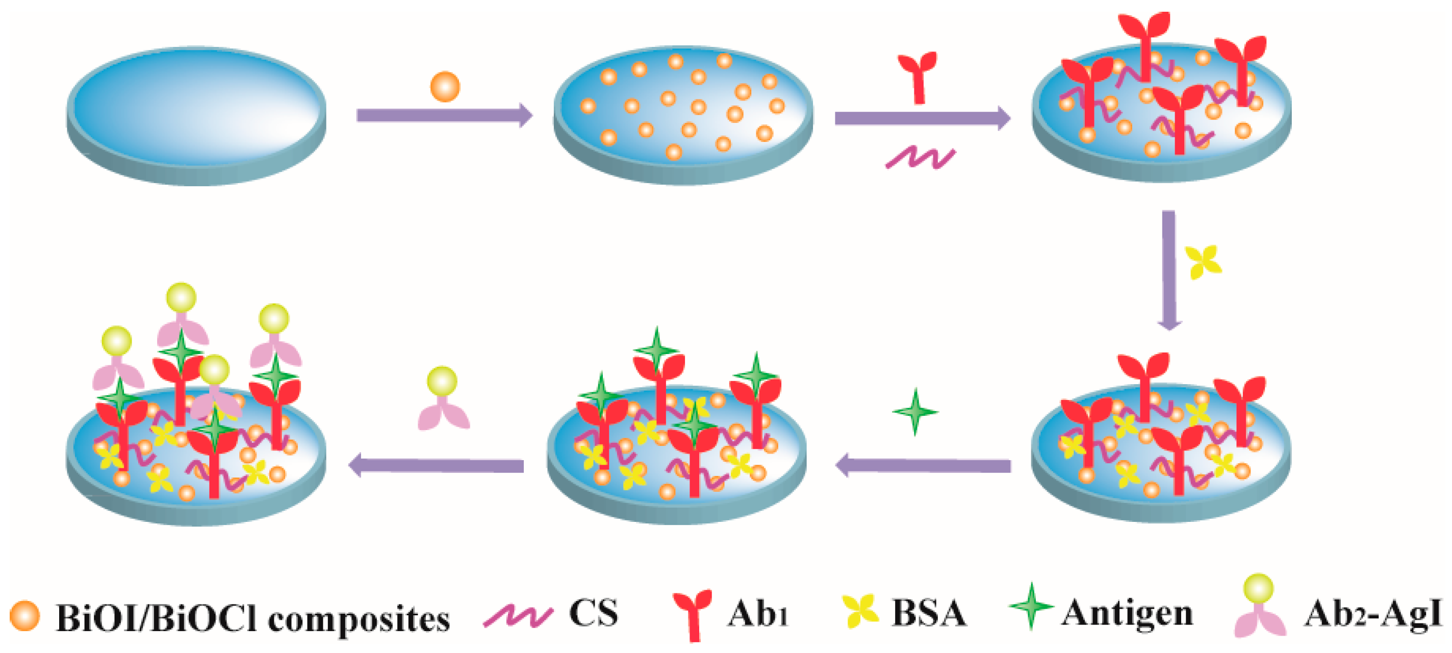
2.3. Fabrication of PEC Immunosensor

Before modification, the Indium tin oxide (ITO) glass was cleaned through sequential ultrasonic treatments in acetone, a 1:1 (v/v) mixture of ethanol and 1 mol·L−1 NaOH, and ultrapure water respectively. After being dried in the stove, 20 μL of the BiOI/BiOCl composite was modified on an ITO electrode with an exposed area of 0.09π cm2 and dried first. Then, 20 μL of CS (0.1 wt%) was dropped on the modified electrode and dried at 60 °C. The synthesized CS/BiOI/BiOCl/ITO electrode was subjected to multiple rinsing cycles using 0.1 M NaOH and deionized water. Next, the electrode was immersed in 20 μL of glutaraldehyde (5%) for 1 h and washed with the PBS (pH 7.4) solution. Whereafter, 20 μL of Capture CEA antibody (Ab1) with 50 μg·mL−1 was dropped on the above-modified electrode, and the obtained modified electrode was incubated at 4 °C overnight. After a gentle wash by the PBS (pH 7.4) solution, 20 μL of bovine serum albumin (BSA) with 1% aqueous solution was dropped on the electrode to incubate with blocking solution to prevent nonspecific binding for 60 min. Finally, the BSA/Ab1/CS/BiOI/BiOCl/ITO was obtained after rinsing with the washing solution. To rigorously evaluate detection selectivity, interference studies were systematically conducted by spiking the 10 pg mL−1 CEA solution with potential interferents (CA19-9, PSA, and AFP) at concentrations tenfold higher (100 pg mL−1), either individually or as a mixture.

2.4. PEC Measurement

The constructed biosensor underwent 1 h of incubation with gradient CEA concentrations at physiological temperature (37 °C) to allow a full immunoreaction between Ab1 and CEA. Then, 20 μL of Ab2-AgI conjugate solution was added and incubation was continued at the same temperature for another hour. Finally, the electrode was rinsed with the washing buffer and then immersed in a supporting electrolyte containing 0.02 mol·L−1 of H2O2. The PEC response for CEA detection was recorded under 450 nm irradiation at an applied potential of 0 V. All testing procedures were performed in an N2 atmosphere. The different electrodes were used for the continuous detection of CEA from low concentration to high value. All the error bars in this work originated from parallel measurement performed 6 times unless otherwise specified.

3. Results and Discussion

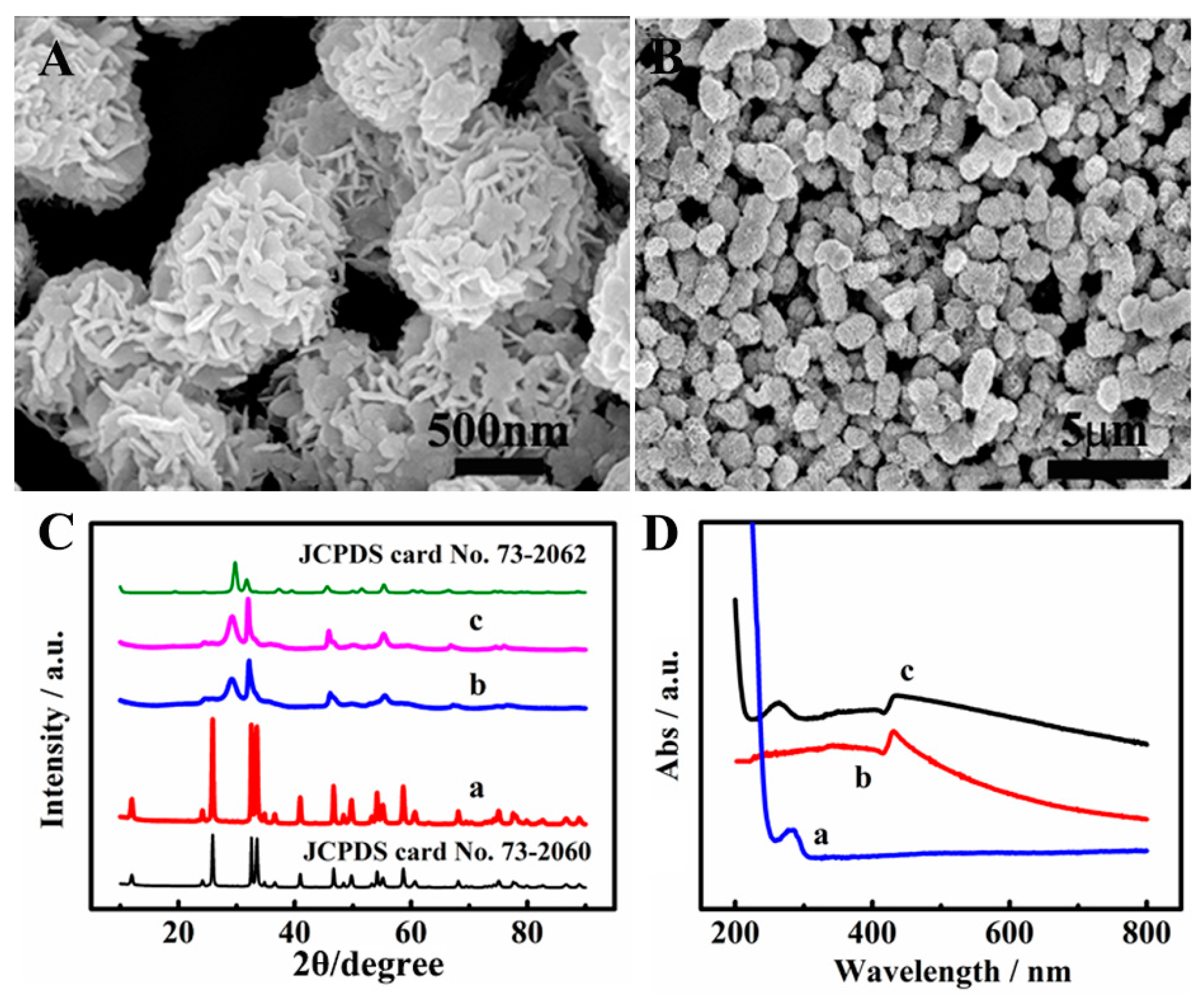
3.1. Morphological Characterization of BiOI/BiOCl

Scanning electron microscopy (SEM) was used to characterize the microstructures of BiOI/BiOCl composites. The BiOI/BiOCl composites appeared as loose, fluffy microsphere structures with diameters of about 1 μm in Figure 1A,B, which is composed of two-dimensional nanoplates [29]. In this way, the porous three-dimensional micro-hybrid structure may endow a large specific surface area and abundant electron carrier separation channels, which is favorable for the transmission of photogenerated electrons [10,32], thereby increasing the photocurrent and improving the sensitivity of the sensor. As shown in Figure S1A,B, BiOI exhibited flower-like microspheres (5–8 μm diameter) assembled from nanosheets, which was consistent with previous reports under hydrothermal conditions. In contrast, BiOCl (Figure S1C,D) displayed hierarchical 3D architectures composed of interlaced nanoplates. The X-ray diffraction (XRD) patterns of BiOI, BiOCl and BiOI/BiOCl composites are shown in Figure 1C, and the peaks match tetragonal BiOI (JCPDS 73-2062) and BiOCl (JCPDS 73-2060), confirming successful synthesis. The pattern of BiOI/BiOCl composites resembled BiOI due to weak BiOCl signals. The ultraviolet-visible (UV-vis) spectra of the Ab2-AgI conjugates were presented in the manner shown in Figure 1D. Ab2 showed a 280 nm peak (π-π* transition) in curve a, while AgI NPs exhibited a 430 nm peak in curve b. The Ab2-AgI conjugate spectrum (curve c) retained both peaks, confirming successful binding.

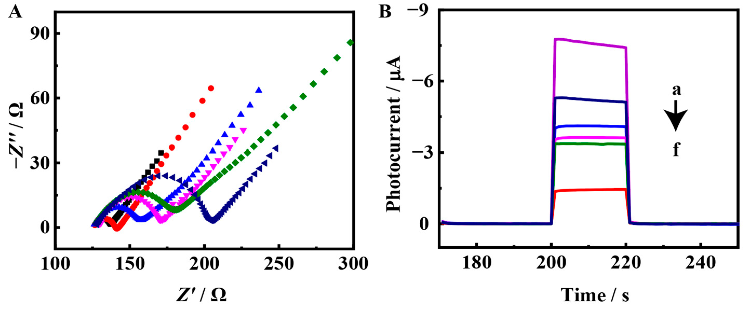
3.2. Characterization of the PEC Biosensor

Electrochemical impedance spectroscopy (EIS) was employed to evaluate the interfacial characteristics of the modified electrodes. As shown in the Nyquist diagram (Figure 2A), the semicircle diameter represents charge-transfer resistance (Rct), a key metric for assessing charge-transfer dynamics of the [Fe(CN)6]3−/4− redox probe across engineered electrode interfaces. When the BiOI/BiOCl composites were modified on the electrode, a clear semicircle (curve a) appeared on the EIS spectrum. Subsequently, the Rct increased stepwise with the gradual modification of CS, Ab1, BSA, and CEA (curves b, c, d, e); this is because the insulation of CS, Ab1, BSA, and CEA inhibited the transfer of the [Fe(CN)6]3−/4− redox probe to the electrode surface [33]. Finally, when the electrode was modified with the Ab2-AgI conjugates, Rct further increased (curve f). The sandwich immune complex formed between Ab2 and AgI significantly increased the steric hindrance, thus significantly obstructing the proximity of [Fe(CN)6]3−/4− to the electrode surface. The stepwise change in impedance indicates the successful construction of the PEC biosensors.

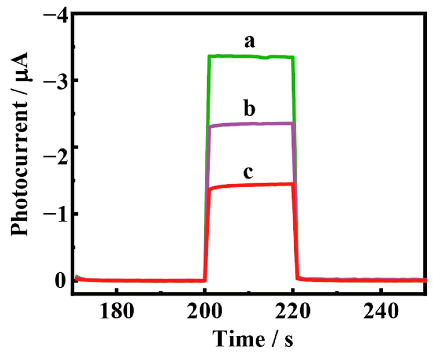
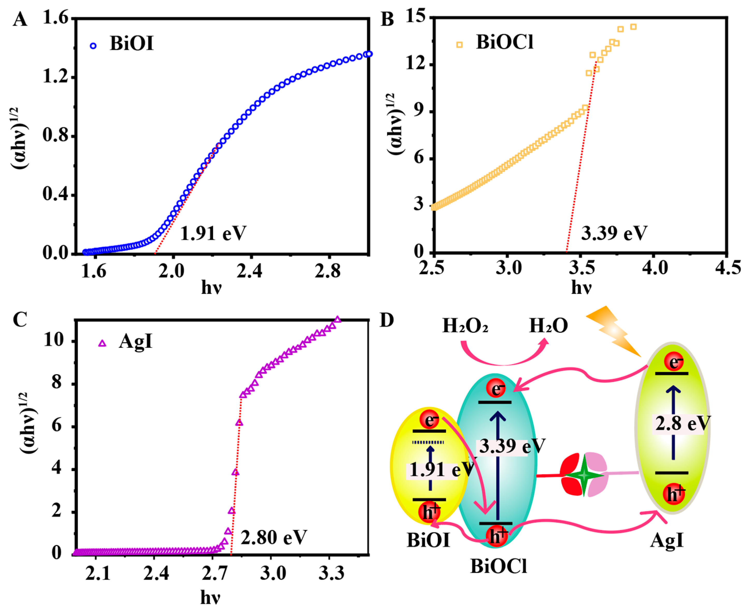
3.3. PEC Response of the Biosensing Platform

The proposed PEC biosensing approach was also systematically demonstrated through the photoelectrochemical response. As depicted in Figure 2B, the BiOI/BiOCl/ITO electrode presented a relatively considerable photocurrent response (curve a). When the CS was modified on the BiOI/BiOCl/ITO electrode, the photocurrent was significantly reduced, which was because the insulation of the CS obstructed the transmission of photoelectrons. Subsequently, the steric hindrance (curves c, d, e) in the presence of modified Ab1, BSA, and CEA further inhibited the transfer of electrons and the photocurrent was gradually reduced. Finally, the Ab2-CuS conjugates were modified on the electrode, and the photocurrent drastically decreased by 1.9 μA (curve f), indicating the development of an immune complex. This current change value was 1.9 times the improvement of that without AgI NPs (1.01 μA) (Figure 3, curve b), and the advantages of the labeled Ab2-AgI conjugates in signal amplification were confirmed. Consequently, the enhanced photocurrent response validated the successful development of the proposed sandwich immunoassay. The PEC response and stability tests of BiOI/BiOCl composites indicated that BiOI/BiOCl composites acted as PEC active materials that could generate strong and stable photoelectric signals to construct a PEC biosensing platform (Figure S2).
According to UV–vis diffuse reflectance spectra, the band gaps of BiOI, BiOCl, and AgI NPs were 1.91 eV, 3.39 eV and 2.80 eV (Figure 4A–C). AgI exhibited efficient light absorption and electron–hole pair generation. Given that the valence band (VB) potentials of AgI (2.50 eV) and BiOI (2.80 eV) were both significantly lower than the VB potential of BiOCl (3.32 eV), photogenerated holes in BiOCl’s VB demonstrated dual migration pathways, transferring not only to BiOI but also competitively to AgI’s VB. This competitive hole transfer mechanism accelerated electron acceptor consumption while simultaneously creating transport barriers for holes, ultimately resulting in significant deterioration of the PEC performance. Combined with the Mott–Schottky measurement results of BiOI, BiOCl, and AgI (Figure S3), the possible mechanism of the sandwich PEC biosensing is illustrated in Figure 4D. In the absence of a target, the BiOI/BiOCl composites could exhibit an evident photocurrent response because of the cascade band-edge levels. In the presence of a target, the Ab2-CS conjugates specifically bound with target Ag to form a sandwich structure. On one hand, the large steric hindrance of the Ab2-CS complex evidently hindered the transmission of electrons. On the other hand, AgI NPs could absorb light and generate electron–hole pairs, which would competitively consume electron acceptors and hinder the transport of holes, leading to a significantly decreased PEC response. Thus, the obvious change in photocurrent could contribute to the ultrahigh sensitivity of the proposed sandwich bioanalysis.

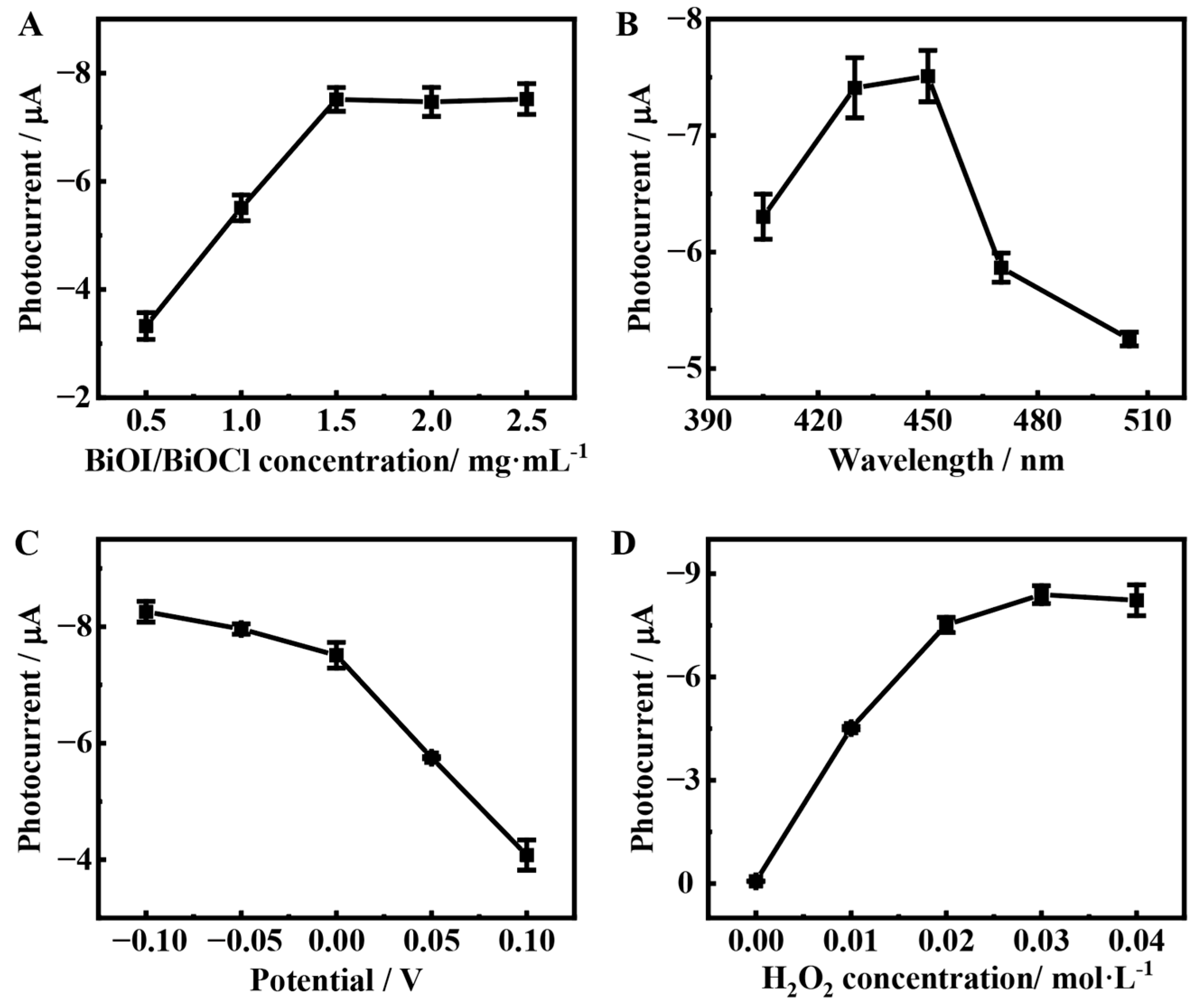
3.4. Optimization of the Detection Conditions

To acquire the intense PEC signal for promoting the sensitivity of the fabricated PEC biosensor, some experiment conditions are optimized in Figure 5. The concentration of BiOI/BiOCl composites had a great influence on the photocurrent intensity and the detection sensitivity. Figure 5A shows the effect of the BiOI/BiOCl composite concentration on the PEC response. With the concentration of BiOI/BiOCl composites rising from 0 to 1.5 mg·mL−1, the photocurrent gradually increased, and when the concentration was more than 1.5 mg·mL−1, the photocurrent tended to be stable. Accordingly, 1.5 mg·mL−1 was chosen as the optimum loading concentration of the BiOI/BiOCl composites. The irradiation wavelength is another important factor affecting the photoelectric behavior. As shown in Figure 5B, it was evident that the photoelectric response was the strongest at 450 nm, which was 7.51 μA. Although the photo-response at 430 nm (7.41 μA) was similar to that at 450 nm, the energy of the long-wavelength light was lower than that of short-wavelength light, and it caused less damage to biomolecules. Thus, 450 nm was selected as the irradiation wavelength for PEC bioanalysis.
In addition, the bias potential could influence the PEC performance. Figure 5C depicts the variation in photoelectronic response with the bias potential. Under the excitation of a 450 nm light source, the photocurrent intensity sharply increased when the bias potential changed from 0.1 V to 0 V and slowly increased between 0 V and −0.1 V. On the other hand, the photocurrent at 0 V was 7.51 μA, which was 91% of that at −0.1 V, sufficient to meet the requirements of sensitive detection. Moreover, without applying a potential, the interference from other substances in the solution was relatively small. Therefore, a potential of 0 V was selected for PEC measurements.
H2O2 as an electron donor facilitated the transmission of photogenerated electrons and intensified the PEC response. Figure 5D illustrated the photocurrent response to H2O2 concentration variations (0–0.04 M), showing a rapid enhancement at 0–0.02 M, followed by stabilization at higher concentrations (0.02–0.04 M). Thus, the 0.02 M H2O2 was used as the optimized concentration to maintain stability and enhanced PEC signal to meet the requirements of sensitive PEC detection.

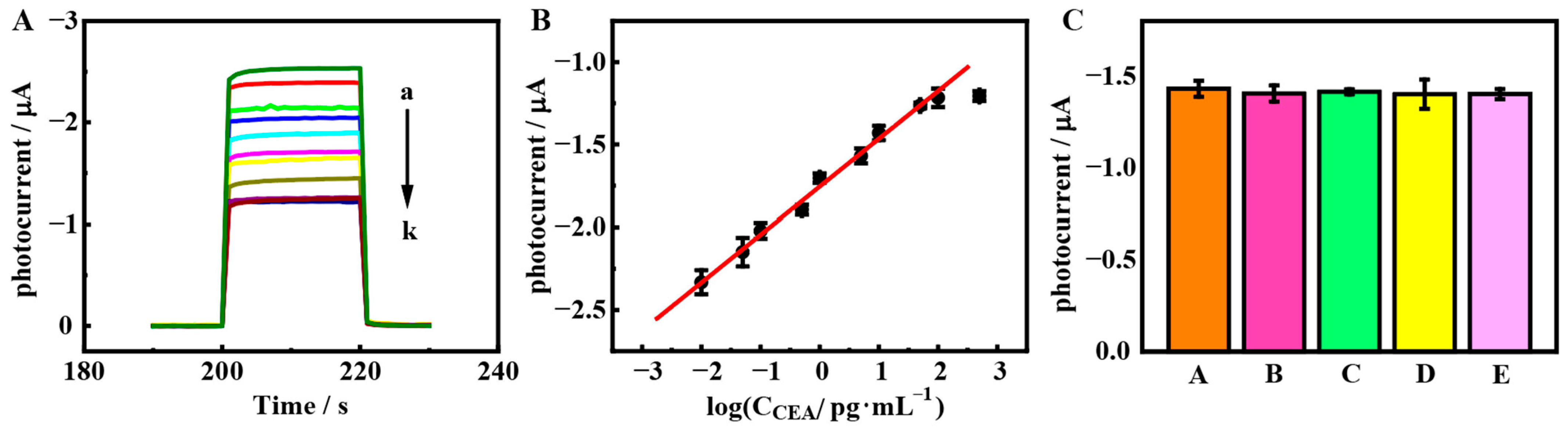
3.5. PEC Bioanalysis

Under tuned experimental settings, the developed PEC immunosensor was utilized for the quantitative analysis of CEA. As depicted in Figure 6A, an increase in the target concentration led to a decrease in the cathodic photocurrent (I). Moreover, within the range of 10 fg·mL−1 to 100 pg·mL−1, I demonstrated a robust linear correlation with the logarithm of the target concentration, as shown in Figure 6B. This range is broader compared to earlier methods such as fluorimetry [34] (20 fg·mL−1–100 pg·mL−1) and PEC biosensing [35] (8.0 fg·mL−1–50.0 pg·mL−1). The equation of linear regression was I = −1.75 + 0.29·log CCEA (pg·mL−1), featuring a correlation coefficient of 0.996 and a detection limit of 4.9 fg·mL−1 at a signal-to-noise ratio of 3σ (where σ was the relative standard deviation of 10 parallel measurements when the concentration of CEA was zero), which was lower than previous works, such as electrochemical analysis [36] (0.65 pg·mL−1), the electrochemiluminescence (ECL) method [37] (2.6 pg·mL−1), and PEC sensing [38] (1.3 pg·mL−1). As shown in Figure S4A, repeatability tests were conducted on BiOI/BiOCl/ITO electrodes using BiOI/BiOCl synthesized from a single batch. Error bars were included for all tested electrodes, with the results demonstrating excellent reproducibility, as evidenced by the RSD of 0.43%. To further investigate the reproducibility of the different batches of the materials, six batches of the replicate photocurrent response were performed. The test results displayed an RSD of 0.49% (Figure S4B). The above results indicated that the developed biosensor possessed ascendant repeatability and reproducibility. Additionally, to evaluate the detection selectivity, interference studies were carried out by adding some tumor markers including cancer antigen 19-9 (CA19-9), prostate-specific antigen (PSA), alpha fetoprotein (AFP), and their mixture in 10 pg·mL−1 of CEA solution, as shown in Figure 6C. The PEC response basically did not change; compared with that of CEA alone, the relative error of the variation in the photocurrent intensity was within 2%, which explained that the PEC bioanalysis platform had good selectivity.
To demonstrate the performance superiority of the sensor platform, we performed a comprehensive comparison of its analytical performance with those of other recently reported PEC and electrochemical immunosensors. The detailed results are summarized in Table 1, indicating the superior analytical performance (a significantly wider linear range and a substantially lower detection limit) of the proposed immunosensor.
Additionally, the long-term storage stability of the BiOI/BiOCl composites was evaluated by analyzing the variation in peak current response towards CEA before and after storage in a refrigerator at 4 °C for 21 days (Figure S5). After being stored for 21 days, the biosensor retained approximately 99% of its initial response values, demonstrating its excellent long-term stability.

3.6. PEC Assay of Human Serum Samples

To evaluate the feasibility of the designed PEC biosensing strategy, we conducted a systematic validation using clinical samples. Human serum samples were collected from patients in accordance with the guidelines approved by the Institutional Review and Ethics Boards at Jiangsu Province Hospital (code: 2021-SR-202). All specimens were obtained with the patients’ consent and accompanied by signed informed consent forms. Clinical serum samples were obtained by the centrifugation of blood for about 5 min with a rotation rate of 3000–4000 rpm (The Helsinki number was not needed in China.). Given that serum tumor marker concentrations may exceed the calibration range, all serum samples were subjected to a 1:100 dilution with Tris-HCl buffered saline (pH 7.4, 10 mM) prior to analysis. As can be seen (Table 2), compared with the reference value measured by the clinically available ECL analyzer, the relative error was less than 11.8%. This illustrated that the accuracy of the method was in an acceptable range, which verified the feasibility of the strategy. The applied potential required for PEC biosensing is significantly lower than that for ECL biosensing, leading to reduced interference in detection and minimized damage to biomolecules in the assay of tumor markers.
The recovery study of the developed immunosensor was evaluated by determining the concentration of CEA in serum samples. Known concentrations of CEA were spiked into the serum samples for direct analysis. As shown in Table S2, the constructed immunosensor exhibited a satisfactory recovery rate ranging from 95.4% to 108%, along with a low relative standard deviation (RSD) value (≤5.89%). These findings not only demonstrated satisfactory accuracy but also further substantiated the reliability of the sensing platform for analyzing real-world samples.
The commercial-scale implementation of this approach might encounter several critical challenges. The precise conjugation of AgI to Ab2 necessitated strict control over particle size, antibody orientation, and labeling density. Variability in nanoparticle–antibody coupling efficiency between batches could undermine assay reproducibility at scale. Additionally, the photosensitivity and potential oxidation of AgI under ambient conditions might require specialized storage solutions to ensure stability, thereby increasing storage complexity.

4. Conclusions

This work demonstrated a novel paradigm in PEC bioanalysis through the rational design of a BiOI/BiOCl heterojunction-based immunosensing platform. Hypotoxic BiOI/BiOCl semiconductor composites were utilized as PEC active materials for biological analysis, exhibiting significant photoelectric properties in the presence of H2O2. After the immune binding to the Ab2-AgI conjugates formed the sandwich structure, the photocurrent was significantly quenched due to the steric hindrance of the complexes, competitive consumption of electron donors, and the obstruction of hole transport by AgI nanoparticles. This phenomenon effectively enhanced the sensitivity of the immunoassay. Using CEA as a model analyte, the optimized biosensor achieved ultrasensitive CEA detection with a wide linear dynamic range, which was successfully applied to the assay of serum samples with satisfactory accuracy. This work not only establishes a generalizable platform for biomarker quantification but also provides fundamental insights into semiconductor interfacial engineering for advanced PEC biosensing applications.

Supplementary Materials

The following supporting information can be downloaded at https://www.mdpi.com/article/10.3390/chemosensors13050164/s1, Materials and Reagents, Apparatus, Figure S1: SEM images of (A) and (B) BiOI. SEM images of (C) and (D) BiOCl. Figure S2: (A) Photocurrent current performance of (a) BiOCl/ITO, (b) BiOI/ITO, and (c) BiOI/BiOCl/ITO electrodes in 0.1 mol·L−1 of Tris-HCl (pH = 7.4) containing 0.02 mol·L−1 of H2O2 at the applied potential of 0 V and 450 nm light excitation. (B) The test of photocurrent stability for BiOI/BiOCl/ITO electrodes. Figure S3: Mott–Schottky measurements of (A) BiOI, (B) BiOCl, and (C) AgI. Figure S4: (A) Repeatability and (B) reproducibility on BiOI/BiOCl/ITO electrodes with 6 experiment repetitions. Figure S5: The long-term storage stability of the biosensor. Table S1: The synthesis yields of the materials. Table S2: Detection of artificially added CEA in human serum.

Author Contributions

Conceptualization, M.Z., W.W. and S.W.; methodology, M.Z. and W.W.; software, S.W.; validation, M.Z., W.W. and S.W.; formal analysis, W.W. and H.Z.; investigation, Y.W.; resources, S.W. and Y.W.; data curation, M.Z. and S.W.; writing—original draft preparation, W.W., S.W. and W.T.; writing—review and editing, W.W., S.W. and W.T.; supervision, W.T. and Z.D.; funding acquisition, M.Z. and Z.D. All authors have read and agreed to the published version of the manuscript.

Funding

This research was financially supported by the National Natural Science Foundation of China (22234005 and 22494632), the Natural Science Foundation of Jiangsu Province of China (BK20222015), and the Postgraduate Research and Practice Innovation Program of Jiangsu Province (KYCX23_1689).

Institutional Review Board Statement

The research protocol was approved in China by the Institutional Review and Ethics Boards at Jiangsu Province Hospital, with approval number 2021-SR-202. All subjects gave their informed consent for inclusion before they participated in the study.

Informed Consent Statement

Written informed consent was obtained from all subjects involved in the study.

Data Availability Statement

The original contributions presented in the study are included in the article and the supplementary material, further inquiries can be directed to the corresponding author.

Acknowledgments

We would like to thank all those who contributed to this research, either in development or by taking part in the consultations.

Conflicts of Interest

The authors declare no conflict of interest.

References

  1. Ye, X.; Wang, X.; Kong, Y.; Dai, M.; Han, D.; Liu, Z. FRET modulated signaling: A versatile strategy to construct photoelectrochemical microsensors for in vivo analysis. Angew. Chem. Int. Ed. 2021, 60, 11774–11778. [Google Scholar] [CrossRef]
  2. Li, Z.; Lu, J.; Wu, F.; Tao, M.; Wei, W.; Wang, Z.; Wang, Z.; Dai, Z. Polarity conversion of the Ag2S/AgInS2 heterojunction by radical-induced positive feedback polydopamine adhesion for signal-switchable photoelectrochemical biosensing. Anal. Chem. 2023, 95, 15008–15016. [Google Scholar] [CrossRef] [PubMed]
  3. Petruleviciene, M.; Savickaja, I.; Juodkazyte, J.; Ramanavicius, A. Investigation of WO3 and BiVO4 photoanodes for photoelectrochemical sensing of xylene, toluene and methanol. Chemosensors 2023, 11, 552. [Google Scholar] [CrossRef]
  4. He, Q.; Zhang, Z.; Zhang, Q.; Zhang, Z. Synergistic modulation of BiOI by atomic-level vacancies and dominant facets for efficient photocatalytic degradation of bisphenol A. J. Mater. Chem. C 2024, 12, 17676–17686. [Google Scholar] [CrossRef]
  5. Zhang, L.; Gao, Y.; Teng, Z.; Liu, Y.; Liu, X.; Cong, C.; Lai, K.; Li, L.; Dong, J.; Li, N.; et al. Gas-liquid interface derived lightweight and twistable BiOI/In2O3/tape assembly for photocatalytic NO removal. Chem. Eng. J. 2024, 497, 154629. [Google Scholar] [CrossRef]
  6. Hoye, R.L.Z.; Lee, L.C.; Kurchin, R.C.; Huq, T.N.; Zhang, K.H.L.; Sponseller, M.; Nienhaus, L.; Brandt, R.E.; Jean, J.; Polizzotti, J.A.; et al. Strongly enhanced photovoltaic performance and defect physics of air-stable bismuth oxyiodide (BiOI). Adv. Mater. 2017, 29, 1702176. [Google Scholar] [CrossRef]
  7. Huq, T.N.; Lee, L.C.; Eyre, L.; Li, W.; Jagt, R.A.; Kim, C.; Fearn, S.; Pecunia, V.; Deschler, F.; MacManus-Driscoll, J.L.; et al. Electronic structure and optoelectronic properties of bismuth oxyiodide robust against percent-level Iodine-, oxygen-, and bismuth-related surface defects. Adv. Funct. Mater. 2020, 30, 1909983. [Google Scholar] [CrossRef]
  8. Yu, S.; Mei, L.; Xu, Y.; Xue, T.; Fan, G.; Han, D.; Chen, G.; Zhao, W. Liposome-mediated in situ formation of AgI/Ag/BiOI Z-Scheme heterojunction on foamed nickel electrode: A proof-of-concept study for cathodic liposomal photoelectrochemical bioanalysis. Anal. Chem. 2019, 91, 3800–3804. [Google Scholar] [CrossRef]
  9. Chen, Y.; Liang, J.; Xu, J.; Shan, L.; Lv, J.; Wu, C.; Zhang, L.; Li, L.; Yu, J. Ultrasensitive paper-based photoelectrochemical biosensor for acetamiprid detection enabled by spin-state manipulation and polarity-switching. Anal. Chem. 2024, 96, 12262–12269. [Google Scholar] [CrossRef]
  10. Yosefi, L.; Haghighi, M. Fabrication of nanostructured flowerlike p-BiOI/p-NiO heterostructure and its efficient photocatalytic performance in water treatment under visible-light irradiation. Appl. Catal. B 2018, 220, 367–378. [Google Scholar] [CrossRef]
  11. Du, Y.; Ma, R.; Wang, L.; Qian, J.; Wang, Q. 2D/1D BiOI/g-C3N4 nanotubes heterostructure for photoelectrochemical overall water splitting. Sci. Total Environ. 2022, 838, 156166. [Google Scholar] [CrossRef]
  12. Ju, P.; Zhang, Y.; Hao, L.; Cao, J.; Zhai, X.; Dou, K.; Jiang, F.; Sun, C. 1D Bi2S3 nanorods modified 2D BiOI nanoplates for highly efficient photocatalytic activity: Pivotal roles of oxygen vacancies and Z-scheme heterojunction. JMST 2023, 142, 45–59. [Google Scholar] [CrossRef]
  13. Wang, X.; Hu, X.; Yang, W.; Wang, F.; Liu, M.; Zhu, X.; Zhang, Y.; Yao, S. Exploitation of a turn-on photoelectrochemical sensing platform based on Au/BiOI for determination of copper(II) ions in food samples. J. Electroanal. Chem. 2021, 895, 115536. [Google Scholar] [CrossRef]
  14. Zheng, X.; Chu, Y.; Miao, B.; Fan, J. Ag-doped Bi2WO6/BiOI heterojunction used as photocatalyst for the enhanced degradation of tetracycline under visible-light and biodegradability improvement. J. Alloys Compd. 2022, 893, 162382. [Google Scholar] [CrossRef]
  15. Wang, J.; Wang, Y.; Cao, C.; Zhang, Y.; Zhang, Y.; Zhu, L. Decomposition of highly persistent perfluorooctanoic acid by hollow Bi/BiOI1−xFx: Synergistic effects of surface plasmon resonance and modified band structures. J. Hazard. Mater. 2021, 402, 123459. [Google Scholar] [CrossRef] [PubMed]
  16. Zhang, L.; Zhu, Y.-C.; Zhao, W.-W. Recent advances of nanostructured materials for photoelectrochemical bioanalysis. Chemosensors 2021, 10, 14. [Google Scholar] [CrossRef]
  17. Zhao, Z.; Han, D.; Xiao, R.; Wang, T.; Liang, Z.; Wu, Z.; Han, F.; Han, D.; Ma, Y.; Niu, L. An enzyme-free photoelectrochemical sensor platform for ascorbic acid detection in human urine. Chemosensors 2022, 10, 268. [Google Scholar] [CrossRef]
  18. Cancelliere, R.; Paialunga, E.; Grattagliano, A.; Micheli, L. Label-free electrochemical immunosensors: A practical guide. Trac-Trends Anal. Chem. 2024, 180, 117949. [Google Scholar] [CrossRef]
  19. Chen, Y.; Duan, W.; Xu, L.; Li, G.; Wan, Y.; Li, H. Nanobody-based label-free photoelectrochemical immunoassay for highly sensitive detection of SARS-CoV-2 spike protein. Anal. Chim. Acta 2022, 1211, 339904. [Google Scholar] [CrossRef]
  20. Dourbash, F.A.; Shestopalov, A.A.; Rothberg, L.J. Label-free immunoassay using droplet-based brewster’s angle straddle interferometry. Anal. Chem. 2021, 93, 4456–4462. [Google Scholar] [CrossRef]
  21. Han, Q.; Wang, R.; Xing, B.; Zhang, T.; Khan, M.S.; Wu, D.; Wei, Q. Label-free photoelectrochemical immunoassay for CEA detection based on CdS sensitized WO3@BiOI heterostructure nanocomposite. Biosens. Bioelectron. 2018, 99, 493–499. [Google Scholar] [CrossRef] [PubMed]
  22. Gubin, M.M.; Esaulova, E.; Ward, J.P.; Malkova, O.N.; Runci, D.; Wong, P.; Noguchi, T.; Arthur, C.D.; Meng, W.; Alspach, E.; et al. High-dimensional analysis delineates myeloid and lymphoid compartment remodeling during successful immune-checkpoint cancer therapy. Cell 2018, 175, 1014–1030. [Google Scholar] [CrossRef] [PubMed]
  23. Alan Saghatelian, K.M.G.; Desiree, A.T.; Ghadiri, M.R. DNA detection and signal amplification via an engineered allosteric enzyme. J. Am. Chem. Soc. 2003, 125, 344–345. [Google Scholar] [CrossRef]
  24. Li, Y.; Ding, J.; Qin, W. Enhanced selectivity in microdroplet-mediated enzyme catalysis. J. Am. Chem. Soc. 2024, 146, 24389–24397. [Google Scholar] [CrossRef]
  25. Jiang, Z.; Li, J.; Liu, G.; Qiu, Q.; Zhang, J.; Hao, M.; Ren, H.; Zhang, Y. A pH-sensitive glucose oxidase and Hemin coordination micelle for multi-enzyme cascade and amplified cancer chemodynamic therapy. Small 2024, 20, 2407674. [Google Scholar] [CrossRef]
  26. Zhao, Y.; Kong, W.; Wang, P.; Song, G.; Song, Z.L.; Yang, Y.; Wang, Y.; Yin, B.; Rong, P.; Huan, S.; et al. Tumor-specific multipath nucleic acid damages strategy by symbiosed nanozyme@enzyme with synergistic self-cyclic catalysis. Small 2021, 17, 2100766. [Google Scholar] [CrossRef] [PubMed]
  27. Zou, R.; Li, H.; Shi, J.; Sun, C.; Lu, G.; Yan, X. Dual-enhanced enzyme cascade hybrid hydrogel for the construction of optical biosensor. Biosens. Bioelectron. 2024, 263, 116613. [Google Scholar] [CrossRef]
  28. Fan, G.-C.; Han, L.; Zhu, H.; Zhang, J.-R.; Zhu, J.-J. Ultrasensitive photoelectrochemical immunoassay for matrix metalloproteinase-2 detection based on CdS:Mn/CdTe cosensitized TiO2 nanotubes and signal amplification of SiO2@Ab2 conjugates. Anal. Chem. 2014, 86, 12398–12405. [Google Scholar] [CrossRef]
  29. Wei, J.; Dong, H.; Gao, Y.; Su, X.; Tan, H.; Li, J.; Zhao, Q.; Guan, X.; Lu, Z.; Ouyang, J.; et al. Synergism of oxygen-iodine binary vacancies with the interfacial electric field: Enhancing CO2 photoreduction over VO–I-BiOCl/BiOI atomic-thin nanosheets. J. Mater. Chem. A 2023, 11, 4057–4066. [Google Scholar] [CrossRef]
  30. Sun, L.; Xiang, L.; Zhao, X.; Jia, C.-J.; Yang, J.; Jin, Z.; Cheng, X.; Fan, W. Enhanced visible-light photocatalytic activity of BiOI/BiOCl heterojunctions: Key role of crystal facet combination. ACS Catal. 2015, 5, 3540–3551. [Google Scholar] [CrossRef]
  31. Ren, H.; Han, Q.; Zhang, S.; Huang, Y.; Chen, Y.; Dai, H.; Yan, J.; Lin, Y. A photothermal assisted in situ signal-amplified electrochemical immunoassay based on multifunctional probe for detecting autoimmune hepatitis marker. Sens. Actuators B Chem. 2020, 309, 127823. [Google Scholar] [CrossRef]
  32. Sun, X.; Lu, J.; Wu, J.; Guan, D.; Liu, Q.; Yan, N. Enhancing photocatalytic activity on gas-phase heavy metal oxidation with self-assembled BiOI/BiOCl microflowers. J. Colloid Interface Sci. 2019, 546, 32–42. [Google Scholar] [CrossRef] [PubMed]
  33. Hallock, C.D.; Rose, M.J. Electrochemical impedance of well-passivated semiconductors reveals bandgaps, fermi levels, and interfacial density of states. J. Am. Chem. Soc. 2024, 146, 18989–18998. [Google Scholar] [CrossRef]
  34. Li, H.; Shi, L.; Sun, D.-E.; Li, P.; Liu, Z. Fluorescence resonance energy transfer biosensor between upconverting nanoparticles and palladium nanoparticles for ultrasensitive CEA detection. Biosens. Bioelectron. 2016, 86, 791–798. [Google Scholar] [CrossRef]
  35. Ge, L.; Wang, W.; Hou, T.; Li, F. A versatile immobilization-free photoelectrochemical biosensor for ultrasensitive detection of cancer biomarker based on enzyme-free cascaded quadratic amplification strategy. Biosens. Bioelectron. 2016, 77, 220–226. [Google Scholar] [CrossRef]
  36. Wei, Y.-p.; Liu, X.-P.; Mao, C.-j.; Niu, H.-L.; Song, J.-M.; Jin, B.-K. Highly sensitive electrochemical biosensor for streptavidin detection based on CdSe quantum dots. Biosens. Bioelectron. 2018, 103, 99–103. [Google Scholar] [CrossRef] [PubMed]
  37. Tang, Y.; Zhang, B.; Wang, Y.; Zhao, F.; Zeng, B. Electrochemiluminescence immunosensor for the detection of carcinoembryonic antigen based on oxygen vacancy-rich Co3O4 nanorods and luminol. ACS Appl. Nano Mater 2021, 4, 7264–7271. [Google Scholar] [CrossRef]
  38. Wei, Q.; Wang, C.; Li, P.; Wu, T.; Yang, N.; Wang, X.; Wang, Y.; Li, C. ZnS/C/MoS2 nanocomposite derived from metal–organic framework for high-performance photo-electrochemical immunosensing of carcinoembryonic antigen. Small 2019, 15, 1902086. [Google Scholar] [CrossRef]
  39. Wang, X.; Wang, H.; Wan, X.; Wei, Q.; Zeng, Y.; Tang, D. Smartphone-based point-of-care photoelectrochemical immunoassay coupling with ascorbic acid-triggered photocurrent-polarity conversion switching. Biosens. Bioelectron. 2025, 267, 116749. [Google Scholar] [CrossRef]
  40. Zhong, Z.; Ding, L.; Man, Z.; Zeng, Y.; Pan, B.; Zhu, J.-J.; Zhang, M.; Cheng, F. Versatile metal–organic framework incorporating Ag2S for constructing a photoelectrochemical immunosensor for two breast cancer markers. Anal. Chem. 2024, 96, 8837–8843. [Google Scholar] [CrossRef]
  41. Lu, L.; Zeng, R.; Lin, Q.; Huang, X.; Tang, D. Cation exchange reaction-mediated photothermal and polarity-switchable photoelectrochemical dual-readout biosensor. Anal. Chem. 2023, 95, 16335–16342. [Google Scholar] [CrossRef] [PubMed]
  42. He, S.; Chen, Y.; Lian, H.; Cao, X.; Liu, B.; Wei, X. Self-assembled DNA/SG-I nanoflower: Versatile photocatalytic biosensors for disease-related markers. Anal. Chem. 2025, 97, 4350–4358. [Google Scholar] [CrossRef] [PubMed]
  43. Qin, J.; Yu, Z.; Wu, D.; Li, M.; Tang, D. Target-induced oxygen vacancy on the etching WO3 photoanode for in-situ amplified photoelectrochemical immunoassay. Biosens. Bioelectron. 2025, 279, 117405. [Google Scholar] [CrossRef] [PubMed]
  44. Wang, H.; Tang, J.; Wan, X.; Wang, X.; Zeng, Y.; Liu, X.; Tang, D. Mechanism exploration of the photoelectrochemical immunoassay for the integration of radical generation with self-quenching. Anal. Chem. 2024, 96, 15503–15510. [Google Scholar] [CrossRef]
  45. Liang, H.; Luo, Y.; Li, Y.; Song, Y.; Wang, L. An immunosensor using electroactive COF as signal probe for electrochemical detection of carcinoembryonic antigen. Anal. Chem. 2022, 94, 5352–5358. [Google Scholar] [CrossRef]
  46. Yu, X.; Cao, Y.; Zhao, Y.; Xia, J.; Yang, J.; Xu, Y.; Zhao, J. Proximity amplification-enabled electrochemical analysis of tumor-associated glycoprotein biomarkers. Anal. Chem. 2023, 95, 15900–15907. [Google Scholar] [CrossRef]
  47. Chen, S.; Tian, S.; Wang, Y.; Li, M.; Tang, D. Harnessing bifunctional nanozyme with potent catalytic and signal amplification for innovating electrochemical immunoassay. Biosens. Bioelectron. 2025, 278, 117340. [Google Scholar] [CrossRef]
Scheme 1. The fabrication procedure of the PEC biosensing platform.
Chemosensors 13 00164 sch001
Figure 1. (A,B) SEM of BiOI/BiOCl composites. (C) XRD pattern of (a) BiOCl, (b) BiOI/BiOCl composites, and (c) BiOI. (D) The UV-vis spectra of (a) Ab2, (b) AgI, and (c) Ab2-AgI conjugates.
Chemosensors 13 00164 g001
Figure 2. (A) Nyquist plots. (B) Photoelectrochemical responses of (a) BiOI/BiOCl/ITO, (b) CS/BiOI/BiOCl/ITO, (c) Ab1/CS/BiOI/BiOCl/ITO, (d) BSA/Ab1/CS/BiOI/BiOCl/ITO, (e) CEA/BSA/Ab1/CS/BiOI/BiOCl/ITO, and (f) Ab2-AgI/CEA/BSA/Ab1/CS/BiOI/BiOCl/ITO electrodes.
Chemosensors 13 00164 g002
Figure 3. Photocurrent responses of (a) CEA/BSA/Ab1/CS/BiOI/BiOCl/ITO, (b) Ab2/CEA/BSA/Ab1/CS/BiOI/BiOCl/ITO, and (c) Ab2-AgI/CEA/BSA/Ab1/CS/BiOI/BiOCl/ITO electrodes.
Chemosensors 13 00164 g003
Figure 4. The UV–vis diffuse reflectance spectra of (A) BiOI, (B) BiOCl, and (C) AgI. (D) The photogenerated electron–hole transfer mechanism in the PEC immunosensing platform.
Chemosensors 13 00164 g004
Figure 5. The effects of the (A) concentration of BiOI/BiOCl composites, (B) excitation wavelength, (C) bias potential, and (D) the concentration of H2O2 on the PEC response.
Chemosensors 13 00164 g005
Figure 6. (A) The photoelectrochemical response for the detection of different concentrations of target Ag: (a) 0, (b) 10 fg·mL−1, (c) 50 fg·mL−1, (d) 100 fg·mL−1, (e) 500 fg·mL−1, (f) 1 pg·mL−1, (g) 5 pg·mL−1, (h) 10 pg·mL−1, (i) 50 pg·mL−1, (j) 100 pg·mL−1, (k) 500 pg·mL−1, and (B) the corresponding calibration curve. (C) The photocurrent responses of the immunoassay for 10 pg·mL−1 CEA detection (A) control (no interference), (B) coexistence with 100 U·mL−1 CA19-9, (C) 100 pg·mL−1 PSA, (D) 100 pg·mL−1 AFP, and (E) a mixed system.
Chemosensors 13 00164 g006
Table 1. Performance comparison with recently reported sensing platforms.
Detection MethodsLinear Response RangeLimit of DetectionRefs.
Photoelectrochemistry0.02 to 80 ng·mL−18.47 pg·mL−1[39]
Photoelectrochemistry0.01 to 32 ng·mL−12.3 pg·mL−1[40]
Photoelectrochemistry0.5 to 100 ng·mL−10.21 ng·mL−1[41]
Photoelectrochemistry0.5 to 80.0 ng·mL−10.5 ng·mL−1[42]
Photoelectrochemistry0.02 to 80 ng·mL−112.9 pg·mL−1[43]
Photoelectrochemistry0.02 to 50 ng·mL−18.9 pg·mL−1[44]
Electrochemistry0.11 to 80 ng·mL−10.034 ng·mL−1[45]
Electrochemistry0.001 to 100 ng·mL−10.419 pg·mL−1[46]
Electrochemistry0.020 to 100 ng·mL−10.013 ng·mL−1[47]
Photoelectrochemistry10 to 100 × 103 fg·mL−14.9 fg·mL−1this work
Table 2. The method accuracy of the developed PEC immunoassay for diluted human serum samples with the referenced CEA ELISA kit.
Simple Numberabcde
Developed method (ng·mL−1)2.060.817.0514.3527.63
Reference method (ng·mL−1)2.270.7786.7815.8931.34
Relative error (%)9.252.613.989.6911.8
Disclaimer/Publisher’s Note: The statements, opinions and data contained in all publications are solely those of the individual author(s) and contributor(s) and not of MDPI and/or the editor(s). MDPI and/or the editor(s) disclaim responsibility for any injury to people or property resulting from any ideas, methods, instructions or products referred to in the content.

