Communication

Electrochemical Assessment of Microbial Activity Using PEDOT:PSS-Immobilized Cells

1
Department of Genetics and Microbiology, Universitat Autònoma de Barcelona, 08193 Bellaterra, Spain
2
CERAMATHS, Université Polytechnique Hauts-de-France, 59313 Valencianes, France
*
Author to whom correspondence should be addressed.
Chemosensors 2025, 13(6), 211; https://doi.org/10.3390/chemosensors13060211
Submission received: 11 March 2025 / Revised: 31 May 2025 / Accepted: 5 June 2025 / Published: 9 June 2025
(This article belongs to the Special Issue Electrochemical Biosensors: Advances and Prospects)

Abstract

:
This study presents a microbial sensing device that employs a poly(3,4-ethylenedioxythiophene):polystyrene sulfonate (PEDOT:PSS) matrix to immobilize viable and metabolically Escherichia coli cells. This device enables the monitoring of microorganism metabolic activity in response to external stimuli such as variations in carbon sources or exposure to inhibitory or toxic compounds. PEDOT:PSS, a conductive and chemically stable polymer, was electrodeposited onto screen-printed electrodes, successfully entrapping approximately 1.26 × 10⁷ cells per electrode. The confocal microscopy of Live/Dead-stained samples confirmed a uniform cell distribution and an average viability of ~78%. Ferricyanide respirometry validated the metabolic activity of the immobilized cells. The biosensor’s performance was evaluated using 3,5-dichlorophenol (3,5-DCP) as a reference toxicant. The observed inhibition of microbial activity correlated with 3,5-DCP concentration, yielding a half-maximal effective concentration (EC50) of 9 ppm, consistent with the literature values.

1. Introduction

Microbial biosensors are sophisticated analytical tools that employ microorganisms as their recognition element [1,2,3]. These systems are engineered to immobilize microorganisms onto the surface of a transducer, enabling the generation of a measurable signal that is directly proportional to the concentration of the target analyte. Depending on the type of transducer and signal measured, biosensors are generally classified as either optical or electrochemical. Among electrochemical biosensors, amperometric types are the most commonly reported. In amperometric biosensors, a current is generated via the reduction or oxidation of electroactive substrates by the microorganisms immobilized on a working electrode [4]. Regardless of the immobilization strategy employed, it is crucial to preserve the viability and metabolic activity of the microorganisms while achieving high reproducibility and sensitivity [5].
Various methods exist for immobilizing microbial cells on electrodes, including adsorption, covalent bonding, encapsulation, and entrapment [6]. Among these, entrapment is the most widely used approach in microbial biosensor development [7,8]. Common polymers used for cell entrapment include polyvinyl alcohol (PVA) [9,10], polyvinylpyrrolidone (PVP) [11], agarose [12], and alginate [13]. The choice of polymer is critical for maintaining biosensor functionality.
The incorporation of conductive polymers, especially carbon-based materials, enhances the active surface area of sensors, thereby improving sensitivity. Originally developed for electronic applications [14], synthetic conductive polymers have recently been adapted for biosensor development [15]. Among them, polypyrrole (PPy), polythiophene (PTh), polyaniline (PANI), and poly(3,4-ethylenedioxythiophene) (PEDOT) have emerged as key materials [16,17], owing to their biocompatibility and electrical conductivity [18]. These monomers require polymerization via techniques such as chemical oxidation, electrodeposition, ultrasonication, electrospinning, vapor-phase polymerization, or photopolymerization. Polypyrrole, for example, can be synthesized in various forms—films, nanofibers, sponges, or nanoparticles—making it versatile for constructing different types of chemical and biological sensors [19].
PEDOT is synthesized via the chemical or electrochemical polymerization of 3,4-ethylenedioxythiophene (EDOT), a positively charged precursor stabilized by polystyrene sulfonate (PSS), a negatively charged surfactant [20,21,22]. PSS not only stabilizes the polymer but also improves its solubility [14,22]. The resulting PEDOT:PSS matrix exhibits high electrical conductivity, good chemical stability, and optical transparency [20,23].
In this study, we investigate the potential of PEDOT:PSS as a polymeric matrix for the immobilization of microorganisms in microbial biosensor development. Specifically, we assess the biocompatibility of the precursor solution and determine the optimal electrodeposition time to maximize the entrapment of viable microorganisms. The resulting biosensors are characterized using confocal laser scanning microscopy to evaluate the distribution and viability of cells within the polymer matrix. The metabolic activity of the immobilized microorganisms is further assessed using ferricyanide respirometry. Finally, we test the biosensors for their ability to detect toxicity using 3,5-dichlorophenol (3,5-DCP), observing a concentration-dependent response suitable for general toxicity assessment.
In summary, this work addresses a critical gap in the use of PEDOT:PSS as an immobilization matrix for whole-cell microorganisms. Although PEDOT and related conductive polymers have shown promise in biosensing applications, their direct application for the entrapment and functional preservation of live microbial cells remains underexplored. This study pioneers the evaluation of PEDOT:PSS for this purpose, leveraging its unique conductive and biocompatible properties to enhance overall biosensor performance. The motivation for this research lies in the growing demand for robust and efficient microbial biosensors in environmental monitoring and toxicity testing—applications where the effective immobilization and sustained activity of biological elements are essential. By optimizing entrapment conditions and characterizing biosensor performance in a real-world toxicity assay, this study contributes meaningfully to advancing microbial biosensor technologies.

2. Materials and Methods

2.1. Chemicals and Materials

Screen-printed electrodes (SPEs) featuring platinum (DRP-550) and carbon (DRP-110) working electrodes were acquired from DropSens (Asturias, Spain). Sodium poly(4-styrenesulfonate) (NaPSS), 3,4-ethylenedioxythiophene (EDOT, 97%), and lithium perchlorate (LiClO4, 99.99%) were obtained from Sigma-Aldrich (Burlington, MA, USA). Potassium hexacyanoferrate (III) (99%), potassium hexacyanoferrate (II) 3-hydrate (99%), and glucose were procured from Panreac (Barcelona, Spain). All chemicals were of analytical grade, and Milli-Q ultrapure water was used to prepare all solutions.
The PEDOT:PSS precursor solution was prepared following a method outlined elsewhere [23], consisting of 2.5% (v) EDOT, 5% (v) NaPSS, and 5% (v) LiClO4 0.1 M.

2.2. Microorganisms

Escherichia coli DSM 2613 was aerobically grown in 200 mL of Luria–Bertani (LB) broth for 18 h at 37 °C using an orbital shaker operating at 100 rpm. After incubation, cultures were centrifuged at 5000× g for 10 min (min) and resuspended in 2 mL of phosphate-buffered saline (PBS) or PEDOT:PSS precursor solution, yielding a suspension of 1010 cells/mL. Viable cell counts were determined by serial dilution and plating on LB agar. Viable cell counts were determined by serial dilution and plating on LB agar.

2.3. PEDOT:PSS Biosensor Fabrication

Platinum and carbon screen-printed electrodes (DRP-550 and DRP-110) were used for biosensor fabrication. Both the electrodeposition process and subsequent electrochemical measurements were performed using a µSTAT8000 multi-potentiostat (DropSens, Spain) connected to a PC running DropView 8400 software (DropSens, Spain).
A 100 µL aliquot of the PEDOT:PSS precursor solution (2.5% EDOT and 5% NaPSS) was applied to the SPE surface and subjected to electrodeposition at 100 µA for durations ranging from 1 to 12 min [24]. The resulting PEDOT:PSS-coated electrodes were analyzed via cyclic voltammetry to determine the optimal electrodeposition time. Measurements were conducted between −0.5 and +0.5 V in mixtures of ferricyanide (1 mM) and ferrocyanide (1 mM) in 0.1 M KNO3 at a scan rate of 0.05 V/s. Chronoamperometric readings were taken at 0.4 V for 5 min, and the current value at 80 s was used as the stable signal in all measurements.
To entrap E. coli in the PEDOT:PSS matrix, 200 mL of culture at 108 cells/mL was centrifuged, and the pellet was resuspended in 2 mL of PEDOT:PSS precursor solution. Electrodeposition of the microbial–polymer suspension onto Pt-SPEs was then carried out at 100 µA for 8 min.

2.4. Biosensor Characterization

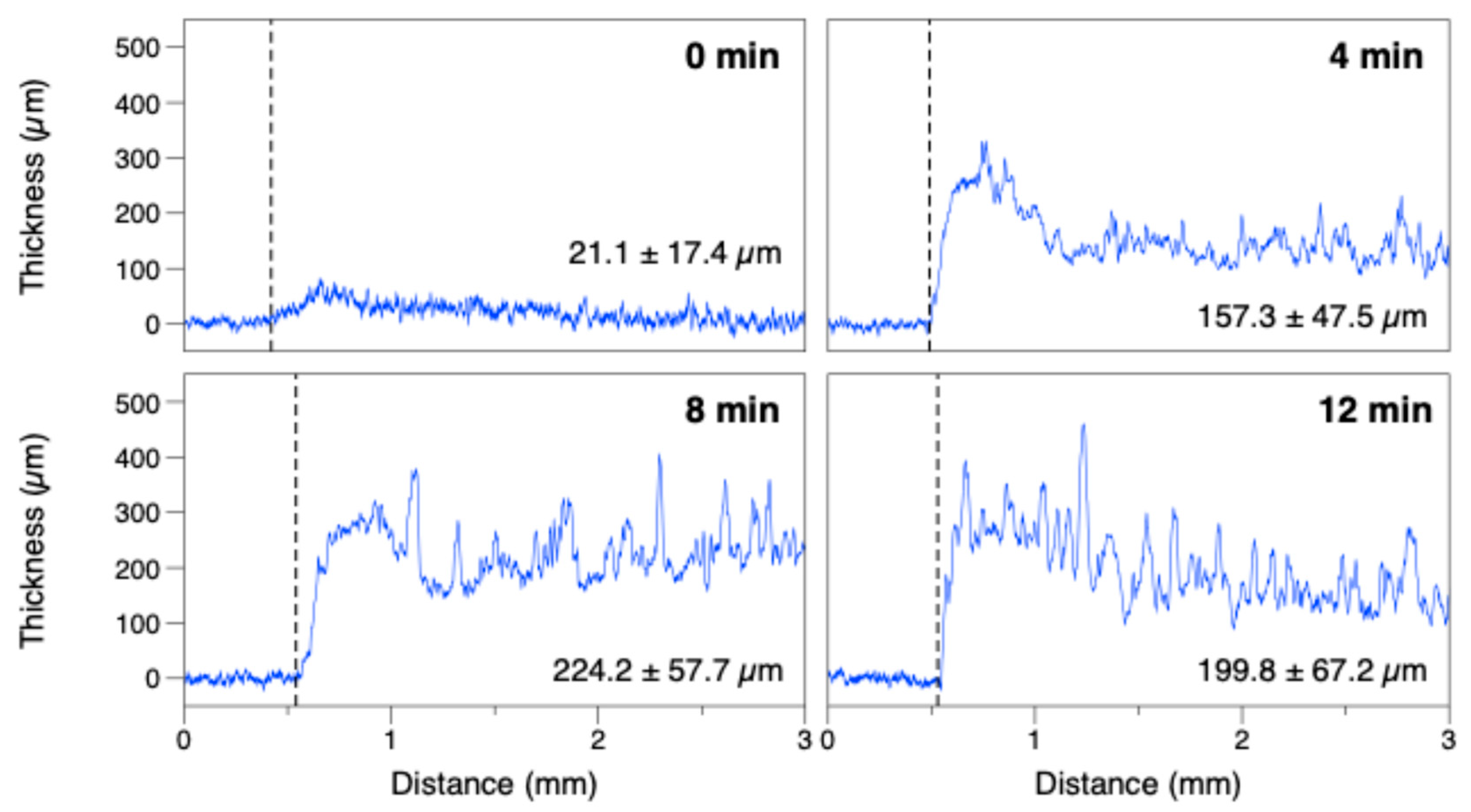
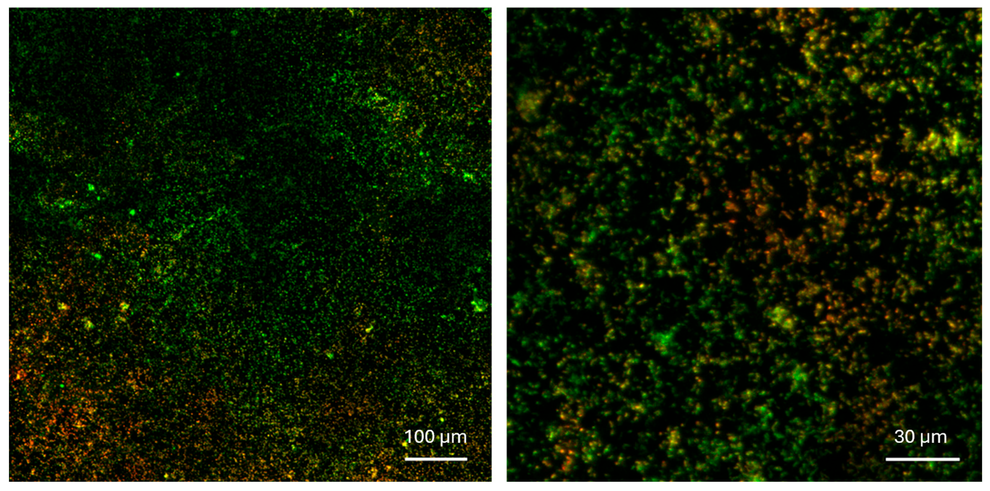
2.4.1. Confocal Imaging and Profilometry

Fluorescence microscopy was used to evaluate bacterial viability after immobilization, as well as to assess the spatial distribution of bacteria within the PEDOT:PSS matrix. Biosensors were stained with the Live/Dead BacLight kit (Invitrogen, Waltham, MA, USA), containing SYTO-9 and propidium iodide. SYTO-9 stains all bacteria, while propidium iodide selectively enters cells with compromised membranes. Both dyes share an excitation wavelength of 470 nm, with emission maxima at 530 nm (green) for SYTO-9 and 630 nm (red) for propidium iodide. The biosensor surface was covered with 75 µL of staining solution and incubated for 30 min, followed by rinsing with water and imaging with a confocal microscope (Leica TCS SP5; Leica Microsystems, Wetzlar, Alemania). The percentage of live (green) and dead (red) cells was quantified using ImageJ software 1.51 (2018) after applying image thresholding.
A KLA Tencor P-15 Profilometer, with a horizontal resolution of 200 nm, was used to determine the thickness of the electrodeposited layer.

2.4.2. Ferricyanide Respirometry

In ferricyanide respirometry, ferricyanide serves as an artificial electron acceptor, replacing oxygen in the microbial respiratory chain and being reduced to ferrocyanide by metabolically active cells [25]. The amount of ferrocyanide produced reflects microbial metabolic activity.
In this assay, 100 µL of 1 mM ferricyanide containing 0.2% glucose was applied to the surface of each biosensor. The sensors were incubated with this solution for 1.5, 3, and 4.5 h. During incubation, bacteria oxidized glucose and transferred electrons to Fe3+, producing Fe2+. The amount of Fe2+ generated was measured by chronoamperometry at 0.4 V, recording the current at 80 s. PEDOT:PSS-coated electrodes without bacteria served as negative controls to confirm the metabolic origin of the signal.

2.4.3. Toxicity Assay

To evaluate the biosensor’s potential for rapid toxicity screening, tests were conducted using 3,5-dichlorophenol (3,5-DCP) as a model toxicant. Microbial biosensors were exposed to concentrations ranging from 0.2 to 25 ppm of 3,5-DCP for 30 min. After exposure, the toxicant was removed, and the sensors were incubated for 3 h with 0.2% (w/v) glucose and 1 mM ferricyanide. Chronoamperometry at 0.4 V was performed to quantify the amount of Fe3+ reduced.
Simultaneously, two controls were prepared: one with bacteria and no toxicant (100% activity) and one without bacteria (0% activity).
The percentage of inhibition (I%) was determined by comparing the current generated by E. coli in the presence of a toxic agent (Toxic) with the current generated by a control sample containing E. coli without the toxic agent (Bacteria) and a Control without bacteria, as follows:
I % = B a c t e r i a T o x i c B a c t e r i a C o n t r o l × 100

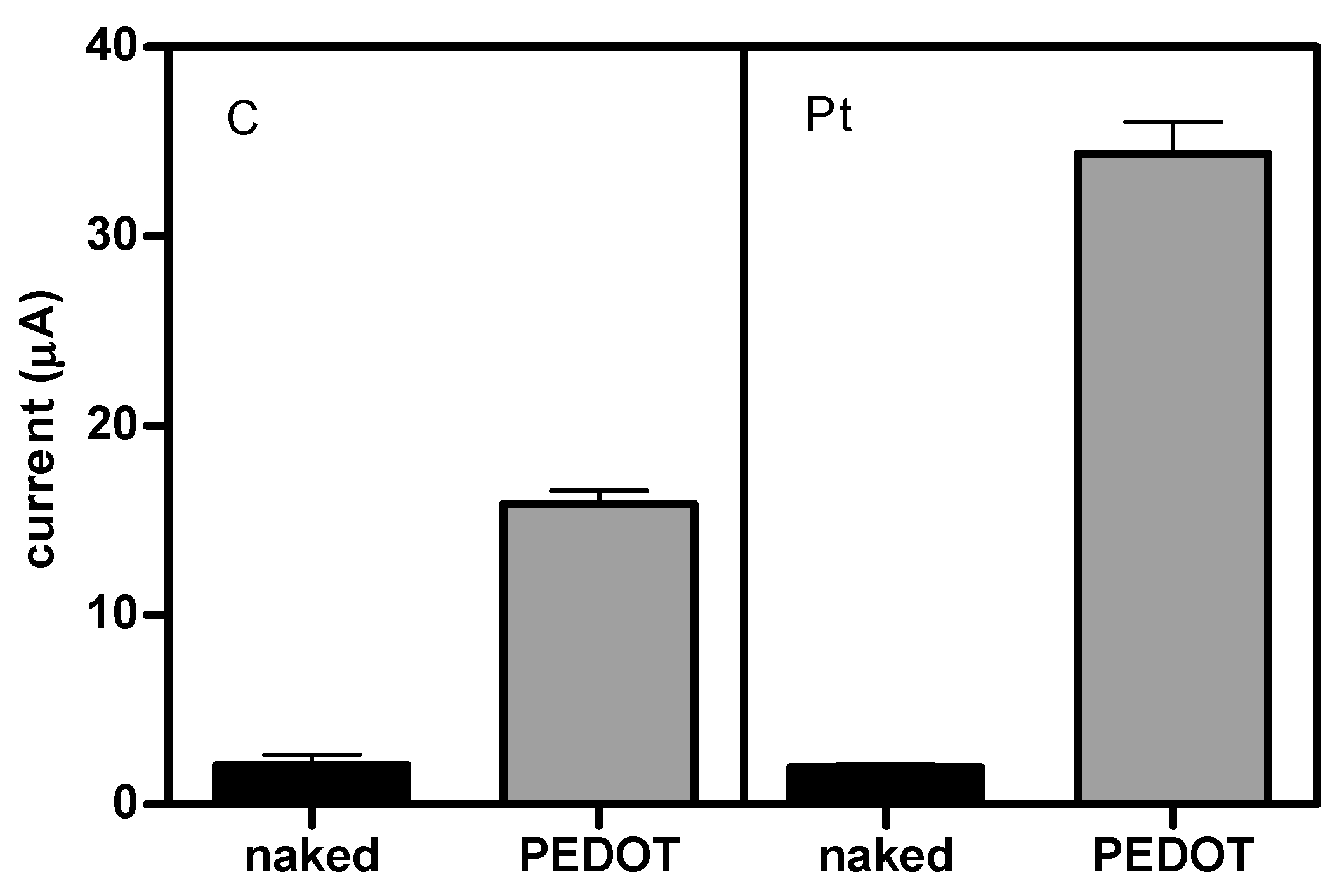
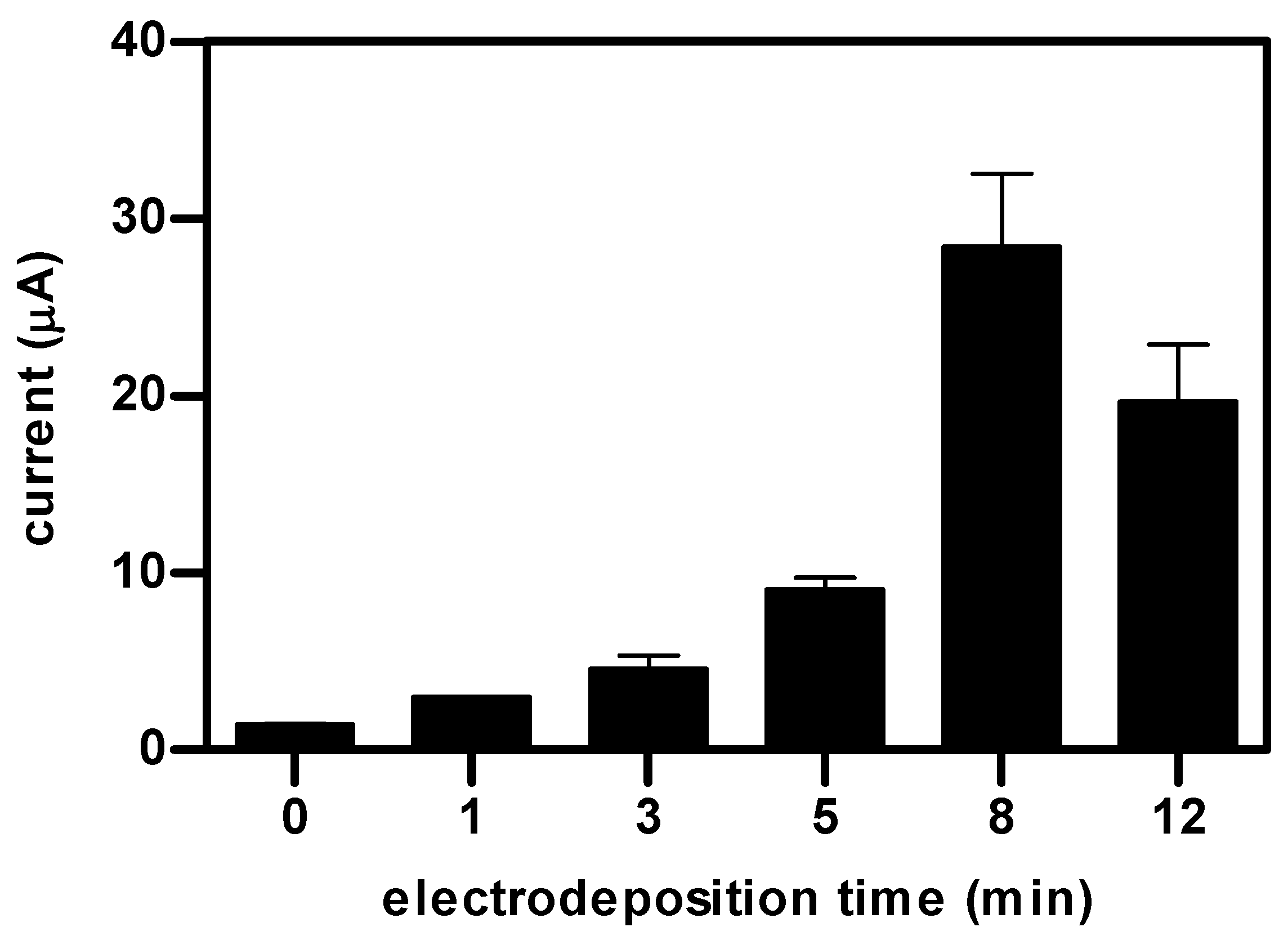
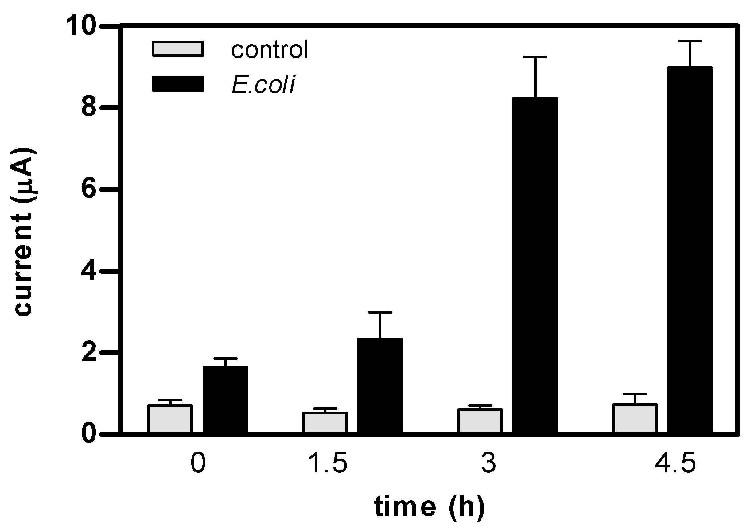
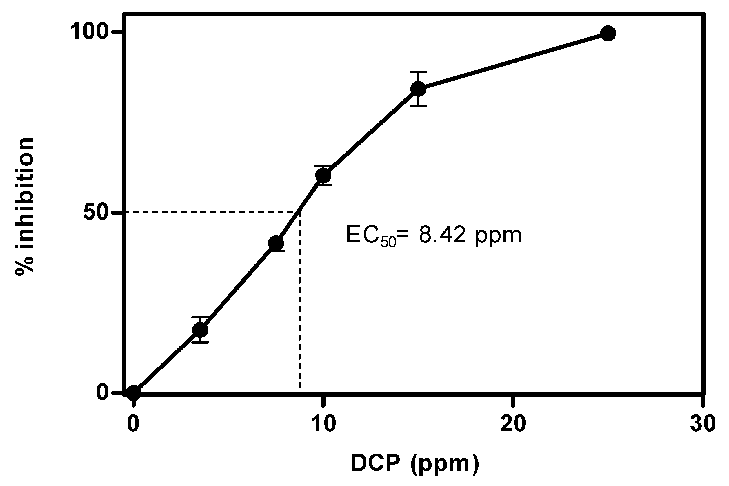
3. Results and Discussion

This study aimed to develop a novel microbial biosensor by entrapping bacterial cells within a 3,4-ethylenedioxythiophene:polystyrene sulfonate (PEDOT:PSS) matrix. To optimize biosensor performance and ensure maximum sensitivity, we evaluated both the working electrode material and the electrodeposition time.
To assess the effect of the electrode material, PEDOT:PSS-coated carbon and platinum screen-printed electrodes were exposed to a 1 mM ferro/ferricyanide solution for 60 s to allow adequate diffusion, after which cyclic voltammetry was performed between −0.5 and 0.5 V at a scan rate of 0.05 V/s. Figure 1 shows the current measured at 400 mV for each electrode/coating combination. While uncoated carbon and platinum electrodes yielded similar currents (2.1 and 2.0 µA, respectively), the application of a PEDOT:PSS coating significantly increased the current output to 15.9 µA in carbon electrodes (an 8-fold increase) and to 34.4 µA in platinum electrodes (an 18-fold increase). These enhanced currents reflect improved electron transfer kinetics and higher electrode conductivity, both critical for sensitive and reliable biosensor performance. Therefore, platinum was selected as the working electrode material for the subsequent experiments.
To optimize electrodeposition, Pt-PEDOT:PSS electrodes were fabricated as described in the Materials and Methods Section using deposition times of 0, 1, 3, 5, 8, and 12 min at 100 µA. Following a 60 s preincubation in 1 mM ferrocyanide, electrodes were analyzed via chronoamperometry at 400 mV. The stable current at 80 s is plotted in Figure 2 as a function of deposition time. Results show that the current increased with the deposition time, peaking at 8 min, which was chosen as the optimal electrodeposition time.
To further examine the impact of electrodeposition time, profilometry was used to measure the thickness and homogeneity of the PEDOT:PSS coating. Figure 3 shows thickness profiles as a function of radial scan distance from the electrode edge to the center. The coating was thicker near the edges (first 0.5 mm) than at the center. The thickness increased with the deposition time, from 21.7 ± 17.4 µm (uncoated) to 157.3 ± 47.5 µm (4 min) to 224.2 ± 57.7 µm (8 min), then slightly decreased to 199.8 ± 67.2 µm at 12 min. This decrease correlates with the reduction in electrochemical performance observed for 12 min coatings (Figure 2). Hence, 8 min was chosen as the optimal deposition time, balancing the maximum thickness with acceptable exposure to the potentially toxic precursor solution.
The confocal microscopy of PEDOT:PSS electrodes stained with Live/Dead reagents confirmed both the distribution and viability of immobilized E. coli cells (Figure 4). After 8 min of electrodeposition, cells were evenly distributed in the matrix, and 77.9 ± 12.7% remained viable. Despite the inherent toxicity of the precursor solution [26], the percentage of viable cells was sufficiently high for biosensor construction.
To assess the metabolic activity of the immobilized organisms, we conducted ferricyanide respirometry. In this assay, the production of Fe2+—resulting from microbial metabolic activity—is quantified by chronoamperometry following preincubation with a carbon source. The duration of this preincubation step significantly influences the accumulation of reduced iron in the medium. To determine the time required for optimal signal generation, we tested four preincubation times: 0, 1.5, 3, and 4.5 h. The results are shown in Figure 5.
In the absence of preincubation (0 h), biosensors containing bacteria produced a very low current (1.7 µA). However, electrode responses increased with longer incubation times, especially after 3 and 4.5 h, reaching 8.2 and 9.0 µA, respectively. Control sensors lacking bacteria were unaffected by the length of the preincubation and consistently produced low currents (0.7 µA) across all time points.
These findings indicate that the immobilized cells were not only viable, as confirmed by Live/Dead confocal microscopy, but also metabolically active, yielding a robust electrochemical signal after just 3 h of preincubation. In this context, PEDOT:PSS biosensors exhibited electrochemical performance comparable to previously reported ferricyanide respirometry-based biosensors [5,13], supporting their suitability for use as amperometric microbial biosensors.
As a final step to validate the applicability of the PEDOT:PSS microbial biosensors, we used them for the determination of toxicity in water, using 3,5-dichlorophenol (3,5-DCP), a standard toxicant in aquatic toxicity studies. Biosensors were exposed for 15 min to concentrations of DCP ranging from 0.1 to 25 ppm. After exposure, sensors were incubated for 3 h in a solution containing 1 mM ferricyanide and 0.2% glucose, and the degree of ferrocyanide accumulation was assessed by chronoamperometry at 400 mV.
The biosensor response was quantified as a percentage of inhibition (I%) relative to untreated controls. As shown in Figure 6, microbial inhibition increased with DCP concentration. No inhibition was detected at low concentrations, while measurable effects appeared at ≥2.5 ppm, reaching complete inhibition at 25 ppm. The EC50 was 8.42 ppm, in agreement with previously reported values [5,13,27,28,29,30].
These findings confirm the potential of PEDOT:PSS as an immobilizing matrix for microbial biosensors. The system exhibited sensitivity, a clear dose–response relationship, and rapid detection capabilities, making it a promising platform for environmental monitoring. Furthermore, this platform could be adapted for broader analytical applications by tailoring the biological component to target specific analytes or stressors.

4. Conclusions

In this study, we successfully developed a novel microbial biosensor based on the electrochemical entrapment of E. coli in a PEDOT:PSS matrix, enabling toxicity detection through metabolic activity monitoring. The optimization of the electrodeposition parameters revealed that platinum electrodes and an 8 min deposition time provided optimal performance. Under these conditions, electrodes were coated with a uniform PEDOT:PSS layer (224.2 ± 57.7 µm), entrapping approximately 1.26 × 10⁷ cells per electrode. The entrapped cells retained 77.9 ± 12.7% viability, as shown by the confocal imaging of Live/Dead-stained electrodes, and exhibited significant metabolic activity as confirmed by ferricyanide respirometry. Upon exposure to 3,5-dichlorophenol, the biosensor showed a clear concentration-dependent inhibition response, with a detection threshold of 2.5 ppm and an EC50 of 8.42 ppm.
These findings validate PEDOT:PSS as a functional immobilization matrix and highlight its potential for the rapid, low-cost toxicity screening of environmental samples. With the further tuning of the biological component, this platform could be adapted for the detection of diverse analytes or stressors in environmental or clinical settings.

Author Contributions

N.V. contributed to the conceptualization of the study, the execution of the experiments, and the drafting of the manuscript. C.C.-V. participated in the execution of the experiments, the data analysis, and the preparation of the manuscript. J.M. was involved in the conceptualization of the study, acquisition of funding, analysis of data, and final drafting of the manuscript. All authors have read and agreed to the published version of the manuscript.

Funding

This research was funded by research grant PID2021-125469NB-C32 (MICOLOR) from the Ministry of Science and Innovation of Spain.

Institutional Review Board Statement

Not applicable.

Informed Consent Statement

Not applicable.

Data Availability Statement

Data are contained within the article.

Conflicts of Interest

The authors declare no conflicts of interest.

Abbreviations

The following abbreviations are used in this manuscript:
PEDOT:PSSpoly(3,4-ethylenedioxythiophene):polystyrene
3,5-DCP3,5-dichlorophenol
EC50half-maximum effective concentration
PVApolyvinyl alcohol
PVPpolyvinylpyrrolidone
PPypolypyrrole
Pthpolythiophene
PANIpolyaniline
EDOT3,4-ethylenedioxythiophene
PSSsulfonated polystyrene
SPEscreen-printed electrodes
NaPSSsodium poly(4-styrenesulfonate)
LBLuria–Bertani
PBSphosphate-buffered saline
I%percentage of inhibition

References

  1. Lei, Y.; Chen, W. Microbial biosensors. Anal. Chem. Acta 2006, 568, 200–210. [Google Scholar] [CrossRef] [PubMed]
  2. Su, L.; Jia, W.; Hou, C.; Lei, Y. Microbial biosensors: A review. Biosens Bioelectron. 2011, 26, 1788–1799. [Google Scholar] [CrossRef] [PubMed]
  3. Bhalla, N.; Jolly, P.; Formisano, N.; Estrela, P. Introduction to biosensors. Essays Biochem. 2016, 60, 1–8. [Google Scholar]
  4. Ding, L.; Du, D.; Zhang, X.; Ju, H. Trends in cell-based electrochemical biosensors. Curr. Med. Chem. 2008, 15, 3160–3170. [Google Scholar] [CrossRef]
  5. Vigués, N.; Pujol-Vila, F.; Marquez-Maqueda, A.; Muñoz-Berbel, X.; Mas, J. Electro-addressable conductive alginate hydrogel for bacterial trapping and general toxicity determination. Anal. Chim. Acta 2018, 1036, 115–120. [Google Scholar] [CrossRef]
  6. Dolatabadi, S.; Manjulahumari, D. Microbial biosensors and bioelectronics. Res. J. Biotech. 2012, 7, 102–108. [Google Scholar]
  7. D’Souza, S.F. Microbial biosensors. Biosens. Bioelectron. 2001, 16, 337–353. [Google Scholar] [CrossRef]
  8. Trelles, J.A.; Rivero, C.W. Whole cell entrapment techniques. Methods Mol. Biol. 2013, 1051, 365–374. [Google Scholar] [PubMed]
  9. Efremenko, E.N.; Senko, O.V.; Aleskerova, L.E.; Alenina, K.A.; Mazhul, M.M.; Ismailov, A.D. Biosensors based on the luminous bacteria Photobaterium phosphoreum immobilized in polyvinyl alcohol cryogel for the monitoring of ecotoxicants. Appl. Biochem. Microbiol. 2014, 50, 477–482. [Google Scholar] [CrossRef]
  10. Ruan, B.; Wu, P.; Chen, M.; Lai, X.; Chen, L.; Yu, L.; Gong, B.; Kang, C.; Dang, Z.; Shi, Z.; et al. Immobilization of Shingomonas sp. GY2B in polyvinyl alcohol-alginate-kaolin beads for efficient degradation of phenol against unfavorable environmental factors. Ecotox. Environ. Saf. 2018, 162, 103–111. [Google Scholar] [CrossRef]
  11. Pourmadadi, M.; Shamsabadipour, A.; Aslani, A.; Eshaghi, M.M.; Rahdar, M.A.; Pandey, S. Development of polyvinylpyrrolidone-based nanomaterials for biosensors applications: A review. Inorg. Chem. Commun. 2023, 152, 110714. [Google Scholar] [CrossRef]
  12. Shin, H.J. Agarose-gel-immobilized recombinant bacterial biosensors for simple and disposable on-site detection of phenolic compounds. Appl. Microbiol. Biotechnol. 2012, 93, 1895–1904. [Google Scholar] [CrossRef] [PubMed]
  13. Vigués, N.; Pujol-Vila, F.; Macanás, J.; Muñoz, M.; Muñoz-Berbel, X.; Mas, J. Fast fabrication of reusable polyethersulfone microbial biosensors through biocompatible phase separation. Talanta 2020, 206, 120192. [Google Scholar] [CrossRef] [PubMed]
  14. Groenendaal, L.; Jonas, F.; Freitag, D.; Pielartzik, H.; Reynolds, J.R. Poly(3,4-ethylenedioxythiophene) and its derivatives: Past, present, and future. Adv. Mater. 2020, 12, 481–494. [Google Scholar] [CrossRef]
  15. Ramanavicius, S.; Ramanavicius, A. Conducting polymers in the design of biosensors and biofuel cells. Polymers 2021, 13, 49. [Google Scholar] [CrossRef]
  16. Apetrei, R.M.; Cârâc, G.; Bahrim, G.; Camurlu, P. Sensitivity enhancement for microbial biosensors through cell self-coating with polypyrrole. Int. J. Polym. Mater. 2019, 68, 1058–1067. [Google Scholar] [CrossRef]
  17. Canbay, E.; Habip, A.; Kara, G.; Eren, Z.; Akyilmaz, E. A microbial biosensor based on Lactobacillus delbruecki sp. bacterial cells for simultaneous determination of lactic and pyruvic acid. Food Chem. 2015, 169, 197–202. [Google Scholar] [CrossRef]
  18. Terán-Alcocer, A.; Bravo-Plascencia, F.; Cevallos-Morillo, C.; Palma-Cando, A. Electrochemical sensors based on conducting polymers for the aqueous detection of biologically relevant molecules. Nanomater 2021, 11, 252. [Google Scholar] [CrossRef]
  19. Zhang, X.; Tan, X.; Wang, P.; Qin, J. Application of polypyrrole-based electrochemical biosensor for the early diagnosis of colorectal cancer. Nanomater 2023, 13, 674. [Google Scholar] [CrossRef]
  20. Girtan, M.; Mallet, R.; Socol, M.; Stanculescu, A. On the physical properties PEDOT: PSS thin films. Mater. Today Commun. 2020, 22, 100735. [Google Scholar] [CrossRef]
  21. Hu, L.; Song, J.; Yin, X.; Su, Z.; Li, Z. Research progress on polymer solar cells based on PEDOT: PSS electrodes. Polymers 2020, 12, 145. [Google Scholar] [CrossRef] [PubMed]
  22. Kirchmeyer, S.; Reuter, K. Scientific importance, properties and growing applications of poly(3,4-ethylenedioxythiophene). J. Mater. Chem. 2005, 15, 2077–2088. [Google Scholar] [CrossRef]
  23. Hu, F.; Xue, Y.; Xu, J.; Lu, B. PEDOT-based conducting polymer actuators. Front. Robot. AI 2019, 6, 114. [Google Scholar] [CrossRef]
  24. Benoudjit, A.; Bader, M.M.; Wan Salim, W.W.A. Study of electropolymerized PEDOT: PSS transducers for application as electrochemical sensors in aqueous media. Sens. Bio-Sens. Res. 2018, 17, 18–24. [Google Scholar] [CrossRef]
  25. Pujol-Vila, F.; Vigués, N.; Díaz-González, M.; Muñoz-Berbel, X.; Mas, J. Fast and sensitive optical toxicity bioassay based on dual wavelength analysis of bacterial ferricyanide reduction kinetics. Biosens Bioelectron. 2015, 67, 272–279. [Google Scholar] [CrossRef]
  26. Zajdel, T.J.; Baruch, M.; Méhes, G.; Stavrinidou, E.; Berggren, M.; Maharbiz, M.M.; Simon, D.T.; Ajo-Franklin, C.M. PDOT:PSS-based Multilayer Bacterial-Composite Films for Bioelectronics. Sci. Rep. 2018, 8, 15293. [Google Scholar] [CrossRef] [PubMed]
  27. Tizzard, A.; Webber, J.; Goonertane, R.; John, R.; Hay, J.; Pasco, N. Micredox: Application for rapid biotoxicity assessment. Anal. Chim. Acta 2004, 522, 197–2005. [Google Scholar] [CrossRef]
  28. Zhai, J.; Yong, D.; Li, J.; Dong, A. A novel colorimetric biosensor for monitoring and detecting acute toxicity in water. Analyst 2012, 138, 702–707. [Google Scholar] [CrossRef]
  29. Qian, J.; Li, J.; Fang, D.; Yu, Y.; Zhi, J. A disposable biofilm-modified amperometric biosensor for the sensitive determination of pesticide biotoxicity in water. RSC Adv. 2014, 4, 55473–55482. [Google Scholar] [CrossRef]
  30. Gao, G.; Qian, J.; Fang, D.; Yu, Y.; Zhi, J. Development of mediated whole cell-based electrochemical biosensor for joint toxicity assessment of multi-pollutants using a mixed microbial consortium. Anal. Chim. Acta 2016, 924, 21–28. [Google Scholar] [CrossRef]
Figure 1. Current values at 400 mV of uncoated and PEDOT: PSS-coated carbon and platinum electrodes measured after a 60 s preincubation in an equimolar mixture of ferricyanide/ferrocyanide (1 mM). Electrodes were coated as described in Section 2.3, by applying a current of 100 µA for 8 min.
Chemosensors 13 00211 g001
Figure 2. Current intensities recorded by chronoamperometry at 80 s of platinum electrodes coated with PEDOT: PSS using different electrodeposition times. Chronoamperometries were carried out in 1 mM ferro/ferricyanide at 400 mV. Values represent the mean of five independent replicates; error bars indicate the standard error of the mean.
Chemosensors 13 00211 g002
Figure 3. Thickness profile of PEDOT: PSS coatings obtained after 0 min, 4 min, 8 min, and 12 min of electrodeposition at 100 µA. The initial (0 min) plot corresponds to the thickness profile of the screen-printed substrate. Measurements were carried out using a KLA Tencor P-15 Profilometer with a horizontal resolution of 200 nm. Data represent the average of two electrodes (Values shown at the bottom right of each graph indicate the average thickness of the coating (±standard deviation)).
Chemosensors 13 00211 g003
Figure 4. Confocal images of PEDOT:PSS-coated electrodes with electro-entrapped E. coli SYTO-9 (green) marks live cells; propidium iodide (red) marks dead cells. The images shown are representative of two independent electrodes.
Chemosensors 13 00211 g004
Figure 5. Current intensities recorded by chronoamperometry at 80 s and 400 mV of PEDOT:PSS biosensors preincubated in 1 mM Fe3+ and 0.2% glucose for 0, 1.5, 3, and 4.5 h. PEDOT:PSS-coated electrodes without bacteria were used as controls.
Chemosensors 13 00211 g005
Figure 6. Dose–response curve showing microbial activity inhibition (I%) versus 3,5-DCP concentration. EC50 (dotted line) was calculated as 8.42 ppm.
Chemosensors 13 00211 g006
Disclaimer/Publisher’s Note: The statements, opinions and data contained in all publications are solely those of the individual author(s) and contributor(s) and not of MDPI and/or the editor(s). MDPI and/or the editor(s) disclaim responsibility for any injury to people or property resulting from any ideas, methods, instructions or products referred to in the content.

