Review

A Review on Xanthine Oxidase-Based Electrochemical Biosensors: Food Safety and Quality Control Applications

Department of Organic Chemistry and Inorganic Chemistry, University of Food Technologies, 4002 Plovdiv, Bulgaria
Chemosensors 2025, 13(5), 159; https://doi.org/10.3390/chemosensors13050159
Submission received: 25 February 2025 / Revised: 27 April 2025 / Accepted: 28 April 2025 / Published: 1 May 2025

Abstract

:
Electrochemical biosensors are integrated bio-receptor–transducer devices that convert specific biological interactions into measurable electrical signals. Over the past decade, the use of novel nanomaterials, advanced enzyme immobilization techniques, and enhanced sensor architectures have been extensively studied, yielding significant progress in the design of highly sensitive, rapid, and reliable electrochemical biosensors. In the modern food industry various types of electrochemical biosensors are used, playing essential roles in the processes monitoring and optimization. This review highlights the strategies implemented to improve the analytical performance of electrochemical enzyme biosensors based on xanthine oxidase (XOx) for the quantitative detection of xanthine (X) and hypoxanthine (Hx), analytes relevant to the field of food quality control. The article covers recent developments (mainly original studies reported from 2010 to date) in the substrate materials, different electrode designs, working principles, advantages, limitations, and applications of XOx biosensors for meat freshness assessment. The article is meant to be a valuable resource that provides insights for improving design for the next generation bio-electroanalytical platforms to ensure food safety.

Graphical Abstract

1. Introduction

Recently, there has been an intensive research effort to develop novel reliable electrochemical biosensors with application in the analysis of various components of food, clinical, pharmaceutical, and environmental samples [1,2,3,4,5,6,7,8]. The high sensitivity of electrochemical transducers combined with the high enzyme specificity provides an opportunity to create sensitive and extremely selective analytical systems applicable in complex samples with variable composition.
The levels of xanthine (X) and hypoxanthine (Hx) are an index for evaluating meat/fish freshness and predict spoilage. X and Hx are heterocyclic purine derivatives generated by adenosine triphosphate (ATP) degradation in the post-mortem muscles of animals, fish, shellfish, etc. [9]. After death of an animal (or fish), ATP undergoes rapid degradation to Hx and subsequently to X, which increases and accumulates mostly in the muscles with the storage of meat. Thus, a close relationship between the postmortem nucleotide catabolism and meat spoilage can be established [10]. Keeping track of the X/Hx level in the meat/fish can be used as an index to determine the freshness of food products. According to previously reported studies for Hx/X, the acceptable limit in sea food is 4 mg/100 g (~26 µM) and levels above 6–9 mg/100 g (39.5–59 µM) indicate that the product is spoiled and unsafe for consumption [11,12].
On the other hand, the xanthine alkaloids, caffeine, theophylline, and theobromine, are known as mild stimulants. Caffeine is naturally found in coffee beans, tea leaves, mate, and guarana. Theophylline is present in tea while theobromine is found in cocoa beans and cola nuts [13].
Here, it should be noted that selective determination of X is relevant to both industrial and clinical applications. Xanthine levels in human blood and serum are an early indicator of abnormal purine metabolism. Xanthine and uric acid (final product of purine degradation) can penetrate cell membranes and cumulate in extracellular fluids. Their concentration levels in body fluids such as urine and serum are indicators of an abnormal purine profile and can be used as biomarkers for clinical diagnosis and medical management of various diseases like perinatal asphyxia, hyperuricemia, xanthinuria, gout, and renal failure [14,15]. Furthermore, research groups have quantified Hx concentration in vitreous humor to estimate the post-mortem interval in forensic cases [16,17].
Therefore, development of fast, economic, and reliable methods for quantitative detection of purine metabolites X and Hx is of great significance in food quality control and clinical diagnostics. Conventional X/Hx analytical techniques, such as HPLC, gas chromatography, and capillary electrophoresis, are sensitive and selective but fail to realize real-time on-site detection. Despite the significant advantages of these methods, they are time-consuming and labor-intensive, require cumbersome procedures for sample preparation, use highly expensive equipment, and require skilled personnel. Nowadays, electrochemical biosensors present an encouraging alternative as they offer a unique combination of key merits, including outstanding selectivity and low detection limits, fast response, simple instrumentation, and convenient operation. Hence, the elaboration of electrochemical biosensing devices is one of the most promising ways to solve issues concerning sensitive, reliable, and low-cost analysis [17,18,19,20,21].
By addressing the electrochemical aspects and challenges in the design of XOx-based biosensors, this article provides a broad picture of the current state of research in this field over the past 15 years. A critical overview of recent innovations and the most promising tools for Hx and X electrochemical biosensing is presented.

2. Electrochemical Biosensors—General Concepts

The intensive development of biosensor technologies is in response to the ever-growing need for highly selective, sensitive, reliable, and simplified methods for rapid, precise, and cost-effective quantitative analysis of substances important in the control of technological processes in the food industry, food quality, clinical analysis, and environmental protection.
Electrochemical biosensors, particularly amperometric sensors, may be used as portable hand-held tools, facilitating on-site quantification without cumbersome laboratory equipment and highly skilled personnel. Numerous electrochemical biosensor devices operate on minimal power, rendering them advantageous for battery-driven or wearable applications [22]. Moreover, recent advances in nanotechnologies, microfabrication, surface modification, and signal processing have aided the design of ultrasensitive microsensors enabling in situ detection in the actual microenvironment [23].
Current enzyme-based amperometric biosensing systems are categorized into three generations according to the analytical principle (Figure 1) [24]: (i) the electrical signal results from the electrochemical conversion of an electroactive substance generated (or consumed) in the enzyme-catalyzed reaction (first-generation principle); (ii) biosensors employing the second-generation principle, which utilize specific low-molecular-weight redox-active substances (mediators) that perform an electron “shuttle”, providing electron exchange between the electrode and the active site of the enzyme at comparatively low overpotential and monitor the reduced mediators thus formed; (iii) systems employing the third-generation principle—the enzyme molecule acts as an electrocatalyst, accelerating the electron transfer between the substrate molecule and electrode surface and the signal is generated as a result of direct electron transfer (DET). The DET-based analytical system is mediator less, label free, and offers fast response and high selectivity, since it operates at a potential close to the redox potential of the native enzyme. At the same time, the direct integration of the biorecognition component with the electrode surface implies a significant simplification of the sensor’s design [25].
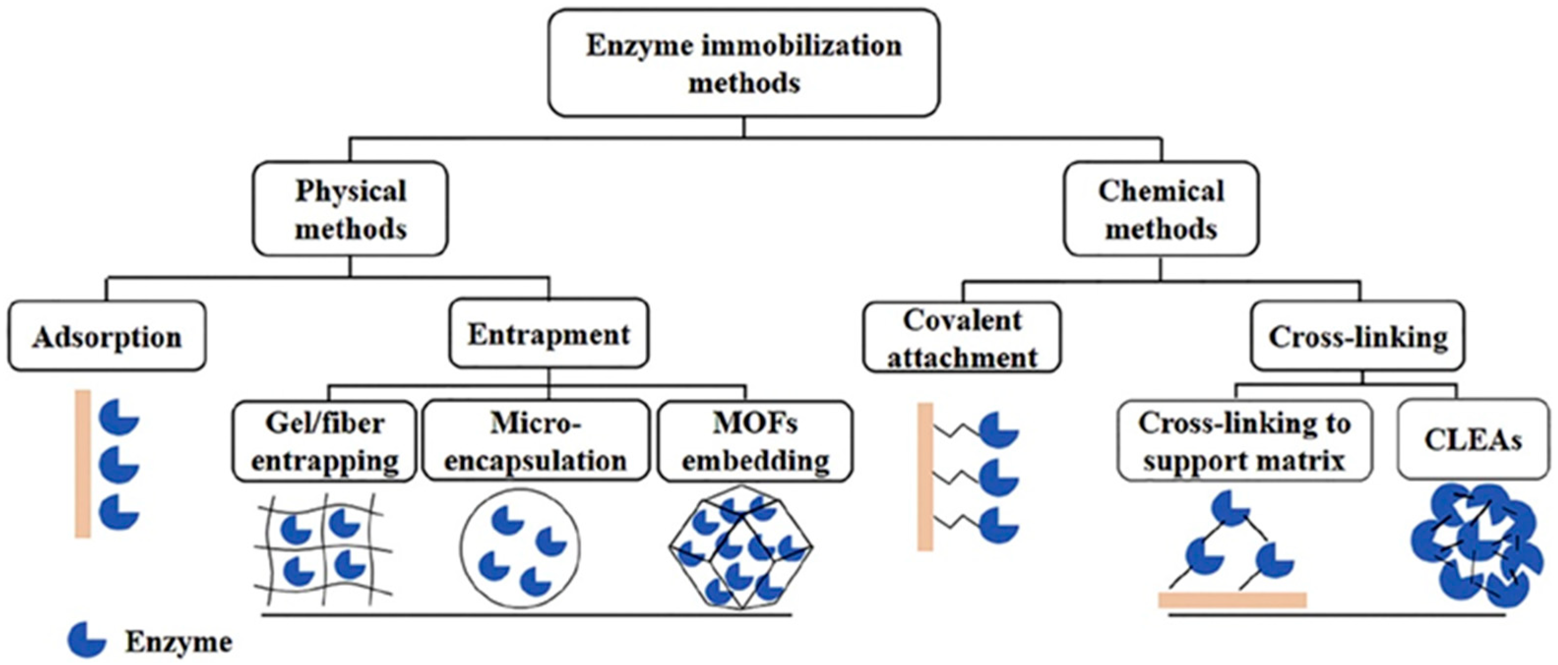
The choice of enzyme immobilization method and electrode material are crucial for analytical performance. Efficient enzyme immobilization is the key step in developing high-performance enzyme-based biosensing systems, since it will affect the loading as well as the enzyme activity. Immobilization is a complex process, which affects the biosensor’s characteristics in terms of long-term stability, sensitivity, and signal reproducibility. Various methods have been used for enzyme immobilization, each of which has its own advantages and disadvantages. Briefly, the main approaches for enzyme immobilization can be classified as follows (Figure 2):
  • Physical adsorption
Physical adsorption of enzyme molecules occurs directly on the electrode surface mainly driven by weak non-specific forces, such as van der Waals, hydrogen bonds, hydrophobic, and electrostatic interactions. This technique causes minimal conformational changes; thus, the immobilized enzyme molecules maintain a conformation that is closest to the native one. Physical adsorption is considered to be the simplest, most inexpensive, and most widely used method capable of high enzyme loading. High retention of the immobilized enzyme molecules activity is a key advantage of the method. Moreover, physical adsorption is reversible, which allows regeneration of the working electrode surface.
However, in this method, immobilization is accompanied by random orientation of enzyme molecules on the electrode surface. Other disadvantages of adsorption immobilization include non-specific binding, overloading of the matrix with enzyme molecules, and enzyme leaching. Concerning the electrochemical biosensor platforms, higher enzyme loadings cause signal decrease due to blocking of the electrode surface. This method is quite liable to change under certain conditions, such as temperature, pH, and the ionic strength of the supporting electrolyte, resulting in poor operational and storage stability of the enzyme electrode.
  • Physical entrapment or encapsulation
Enzyme molecules are entrapped within the 3D matrices through covalent or non-covalent bonds. Electropolymerization, sol-gel process, and microencapsulation are methods applicable to a large number of enzymes. These approaches protect the enzyme activity, provide a relatively high amount of biomaterial in a thin film very close to the electrode surface, and enhance stability. Among them, electropolymerization is distinguished by high reproducibility of the immobilization procedure. The method makes it possible to coat electrodes that have a small or uneven surface, and the thickness of the polymer coating can be controlled experimentally with a high degree of reproducibility.
The disadvantages of enzyme entrapment include low loading capacity, limited pore size, and mass transfer limitations of the substrate to the enzyme active site.
  • Chemical immobilization
Irreversible covalent binding is a commonly used technique based on direct attachment of the enzyme molecule to the transducer surface. As a result, biosensors are distinguished by strong resistance to environmental changes, as well as little enzyme leakage. Covalent coupling leads to better operational and long-term storage stability of modified enzyme electrodes as compared with others. The disadvantages include a complicated procedure, significant cost, steric hindrance, and conformational changes of enzyme molecules, and consequently, denaturation.
Cross-linking is based on the formation of intermolecular covalent cross-linkages between the enzyme molecules (or between enzyme molecules and functionally inert proteins) by means of multifunctional reagents that act as a linker to connect enzyme molecules in 3D cross-linked aggregates to the electrode surface. Cross-linking effectively prevents leakage and loss of the biocomponent. However, this method causes conformational changes in the enzyme molecules, which lead to a significant loss of catalytic activity.
In the case of third-generation biosensor systems, electrode material should provide an electron-conducting path for the realization of direct electron transfer between the prosthetic groups of the biomolecules and the electrode surface.
Both inorganic and organic conductive materials can be used as immobilization carriers—metals, metal oxides, various types of carbon materials (graphite, glassy carbon, pyrolytic graphite, activated carbon), polymers, etc. Generally, there are no known carriers that satisfy all the requirements regarding mechanical stability, biocompatibility, minimal water solubility, indifference to the substrate, etc. Here, it should be noted that the immobilization of enzymes on carbon matrices is not accompanied by noticeable losses of catalytic activity, which necessitates their advantageous use as carriers, especially in the development of bioelectrochemical systems (enzyme electrodes, biofuel cells). Carbon materials are widely used in applied bioelectrochemistry—they are distinguished by high electrical conductivity, biocompatibility, chemical and electrochemical stability, a wide potential range, relatively low cost, and accessibility. Key advantages are the possibility for surface modification, as well as the simplified mechanical and electrochemical pretreatment of the surface, through which a large number of oxygen-containing surface groups are quickly and easily generated, making them particularly suitable for the development of enzyme electrodes.
An effective approach to increasing the electrochemical activity of biosensor systems is the use of transition and platinum metal nanoparticles, metal oxide nanostructures, two- and three-component alloys, nanocomposites, etc. Recently, advances in nanotechnology have allowed for the design of novel improved biosensor systems.

3. Xanthine Oxidase-Based Electrochemical Biosensors

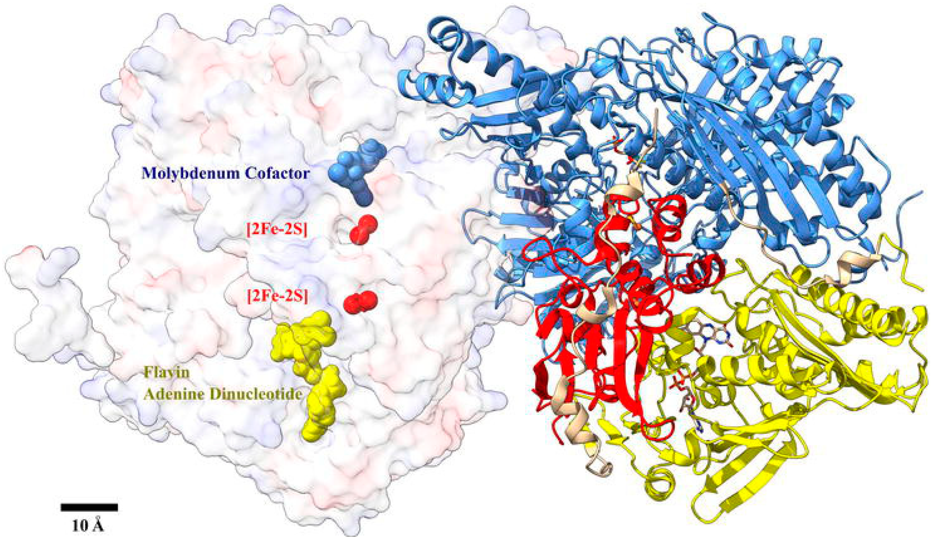
Xanthine oxidase (XOx) (EC 1.1.3.22) is a dimeric metalloflavoprotein (275 kDa) with a complex structural organization (Figure 3). Each monomer consists of three non-identical subunits linked together by disulfide bridges. The composition of one enzyme molecule includes two FAD molecules (one for each monomer) covalently bound to the protein part; two molybdenum atoms, which form the so-called “Mo-cofactor” (Moco), responsible for the transfer of electrons and protons from the substrate to the acceptor during the catalytic process; eight iron atoms, participating in iron–sulfur centers of the cluster type [2Fe-2S]. Moco lies close to the interfaces of both the FAD and the [2Fe-2S] domains. The FAD and the Mo-centers accept two electrons each, and the [2Fe-2S] clusters accept one electron each. The reactions through which Hx is converted to X, and X to uric acid both occur at the molybdenum active sites while oxygen reacts with the FAD [27].
XOx catalyzes the terminal two steps of purine degradation, converting hypoxanthine to xanthine and subsequent conversion of xanthine to uric acid and hydrogen peroxide (H2O2) using molecular oxygen (O2) as an electron acceptor (Figure 4):
Enzyme biosensors adopt immobilized XOx to convert Hx and X into uric acid and H2O2 in the presence of O2. Thus, the amount of Hx/X can be estimated by measuring:
(i)
the consumed O2;
(ii)
the produced uric acid;
(iii)
the produced H2O2. Generally, direct detection of H2O2 requires anodic potentials (around 0.6 V vs. Ag/AgCl) or cathodic potentials below 0.0 V vs. Ag/AgCl.
(iv)
the current generated as a result of DET between the redox-active sites of XOx and the electrode surface. Here, it should be noted that XOx is characterized by a pronounced spatial shielding effect of the active center due to its location at a significant depth (over 2 nm) in a hydrophobic cavity of the molecule, which hinders DET. Although various innovative strategies have been applied to realize DET, most often the turnover rate of electrons is considerably lower than the electron exchange rate between the redox-active site of the XOx macromolecule and O2 (the native electron acceptor).
XOx is one of the most fragile and short-lived enzymes. Hence, the immobilization method should be considered as a key step in a biosensor’s design. On the other hand, integrating XOx molecules with different nano-sized materials has resulted in increased use of this enzyme as a recognition element in biosensor systems. The use of nanostructured materials in the construction of XOx-based electrochemical biosensors showed significant improvement in their analytical performance, with great signal amplification, higher sensitivity, and enhanced selectivity and response time [28,29,30,31,32]. In general, innovations include the use of nanomaterials, conductive polymers, and nanohybrids, and shifting design towards miniaturized analytical devices for real-time on-site measurements [33,34].

3.1. First-Generation XOx Biosensors

At the first generation, amperometric biosensors-immobilized XOx reacts with the targeted substrate (Hx/X) in the presence of molecular oxygen to produce H2O2 that is subsequently oxidized (or reduced) at the electrode interface to produce an analytical current signal. The number of reports on biosensors based on measurement of oxygen consumption is limited [35,36].

3.1.1. Biosensors Based on the Electrooxidation of H2O2 and/or Uric Acid

In most cases, amperometric detection of Hx or X is based on the electrochemical oxidation of H2O2 and/or uric acid—products of their enzymatic oxidation. At conventional carbonaceous electrodes, the electrocatalytic oxidation typically takes place at potentials of 0.4 V (vs. Ag/AgCl) for uric acid and 0.6 V for H2O2, respectively. However, the co-oxidation of easily oxidizable substances, present in real samples, overestimates the analyte level. Therefore, in order to improve the electrochemical signal of H2O2 and selectivity, the applied potential must be effectively reduced.

Nanoparticle-Based Biosensors

The use of nanostructures, including nanofibers and nanoparticles (metal NPs, metal oxide NPs, CNTs, etc.) in the fabrication of transducers for biosensing is of great interest. Nano-sized materials accelerate the electron transfer, improving the analytical behavior of the transducers, and also act as immobilization matrices that significantly enhance the amount of the XOx enzyme loading.
A great number of nanomaterials with different morphologies have been synthesized and applied as novel electrode materials for XOx-based electroanalytical platforms, including the following: CuO nanoflake-like structures [37], Fe3O4 NPs [38,39], nano-CaCO3 [40], glassy carbon paste electrode modified with AuNPs (AuNPs–GCPE) [41], vertical columnar nanostructures of NiO [42], TiO2/c-MWCNT [43], CdONPs/c-MWCNT/Au [44], gold-coated iron nanoparticles Fe-NPs@Au [45], CPEs modified with electrodeposited AuNPs [46], porous silica nanomaterials [47], etc. These modifiers reduce the overpotential of H2O2 oxidation, enhance electron transfer rates, and possess good biocompatibility and strong adsorption ability, and therefore lead to high-performance sensing platforms.
When compared to other XOx-based biosensors, only the sensing platform based on Fe3O4 nanoparticles embedded on an Au working electrode showed current response for nanomolar concentration of X [38]. An extremely low detection limit in picomolar (pM/10−12 M) concentration was achieved using the enzyme electrode XOx/NanoFe3O4/Au; the LOD was found to be 2.5 pM and the LOQ 8.3 pM.
Here, the high long-term stability should be noted of XOx covalently immobilized onto chitosan-bound gold-coated iron nanoparticles, electrodeposited onto a pencil graphite electrode [45]. Surface derivatization of Fe-NPs with Au results in the formation of air stable nanoparticles with improved biocompatibility. The fabricated enzyme electrode XOx/CHIT/Fe-NPs@Au/PGE retains 75% of its activity after 100 uses over 100 days, which is one of the highest operational stabilities reported to date.

Biosensors Based on Polymers

The use of conducting polymers to improve the selectivity of an enzyme electrode was also reported. Conducting polymers offer effective matrices for enzyme macromolecules and enhance the electrokinetics rate [48]. Therefore, polymeric nanocomposites containing nanostructured materials have also been developed for XOx biosensing systems. Since 2020, various XOx electrochemical biosensors have been reported based on hybrid electrodes, such as a nanocomposite of carboxylated MWCNT and polyaniline (c-MWCNT/PANI) [49], pyrrole-polyvinylsulphonate film [10], zinc oxide nanoparticles–polypyrrole composite film (ZnO-NPs–PPy) [50], colloids of Au/polypyrrole (AuPPy) nanocomposites [51], citrate-capped AgNPs and L-cysteine (AgNPs/L-Cys) [52], poly(L-aspartic acid)/MWCNT composite film [11], nanoAg–ZnO/PPy/PGE [53], polyaniline-wrapped titanium dioxide (PANI@TiO2) nanohybrid [54], composite film ZnONPs/CHIT/c-MWCNTs/PANI) electrodeposited onto Pt-electrode [55], etc. A significant advantage of the electrochemical deposition of polymer films is that both the thickness and the chemical composition of the resulting polymer layer can be precisely regulated by controlling the potential values and the process duration.
Das and Mishra for the first time reported nanocellulose obtained from raw cotton as a matrix for XOx immobilization [56]. The proposed biosensor exhibits good performance in DPV mode (linear range 3–50 µM; LOD of 47.96 nM), selectivity, and high long-term stability (97% residual activity after 30 days storage). The analytical platform was used for quantification of X in fish meat stored for one month and the results were in good agreement with those obtained using a reference spectrophotometric method.
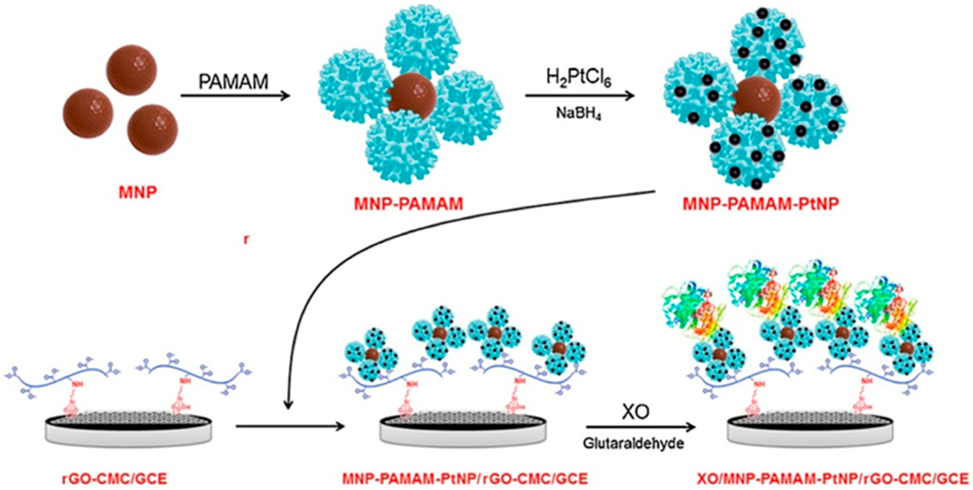
Borisova et al. described a novel electrode material based on ethylenediamine core polyamidoamine (PAMAM) dendrimers covalently attached to poly(dopamine)-coated magnetic NPs decorated with PtNPs [57]. As Figure 5 illustrates, the prepared nanohybrid was used to assemble an LbL architecture on the surface of GCE coated with nanomaterial synthesized by covalent attachment of carboxymethylcellulose to reduced graphene oxide (rGO-CMC). The electrode was employed as support to construct a bioelectroanalytical platform for the quantitative determination of X in fish meat. According to their studies, the presented sensing interface showed remarkable sensitivity towards X, demonstrating a broader than three orders of magnitude dynamic concentration range. At a potential of 0.6 V (vs. Ag/AgCl), the system showed a dynamic range between 50 nM and 12 μM and an extremely low detection limit of 13 nM. The as-prepared electrode was also tested in fish samples, giving quite similar results to those obtained by HPLC.
Biosensors based on AuNPs have attracted significant attention due to their fascinating properties, including the following: high conductivity, high volume-to-surface ratio, chemical stability, and biocompatibility. AuNPs facilitate the attachment of enzyme molecules via covalent thiol linkages, providing stable and robust immobilization strategies [58]. Thus, the presence of AuNPs ensures higher enzyme stability. For example, Dervisevic et al. reported a novel hybrid bio-nanocomposite film based on chitosan, PPy, and self-assembled AuNPs prepared by in situ chemical synthesis onto GCE [59]. The proposed biosensor shows good stability (85% at 18 days of storage), satisfactory recovery, and reproducibility in X determination in fish, chicken, and beef samples.
Zhang and co-workers fabricated a XOx–AuNPs–single-walled carbon nanohorn (XOx–AuNPs–SWCNH) biosensor with good responses at low applied potential (0.4 V vs. SCE) to both Hx and X with LOD of 0.61 and 0.72 µM, respectively [60]. The proposed system was used once daily for 7 days and the enzyme electrode showed 95% residual activity.
Three-dimensional xerogels are advanced, functional, porous materials used in biosensing systems, particularly for clinical applications [61,62]. The great surface area, synthetically tunable porosity, and an affordable preparation route make them suitable for biosensor applications. Dang et al. systematically examined the use of AuNPs-doped xerogels as part of a layer-by-layer (LbL) architecture of electrodes that operated as first-generation XOx biosensors [63].
Recently, organic mixed ionic-electronic conductors (OMIECs) have been introduced in biosensor design for a variety of applications in the food and agriculture industries [64,65]. Hydrophilic glycolated polymers in p-type OMIECs showed efficient ionic and electronic charge transport [66]. For the first time in 2025, the research group of Anna Herland developed an organic mixed ionic-electronic conductor (OMIEC)-based XOx biosensing system with a p-type conjugated polymer p(g42T-TT) (Figure 6) as the channel material [67]. The authors modified the gate with PtNPs as they possess excellent electrocatalytic activity towards H2O2 oxidation. The developed platform provides a response for both X and Hx and it is suitable for disposable application.

Biosensors Based on Metal-Organic Frameworks (MOFs)

Metal-organic frameworks (MOFs) have received significant attention as effective catalytic and sensing materials due to their remarkable structural and chemical characteristics, resulting in signal amplification and enhanced detection sensitivity [68]. The ability to easily modify pore sizes and surfaces within MOFs and to engineer electronic structure characteristics improved the specificity, conductivity, adsorption energy, and electron transfer mechanisms during H2O2 oxidation (or reduction).
Wang et al. successfully immobilized XOx onto a biocompatible Cu-MOF film and used it for construction of a biosensor for HX and X [69]. In the presence of X in the electrolyte (PBS, pH 7.5), a well-defined peak at 0.64 V (vs. SCE) was observed. In a parallel independent study, a DPV signal at 0.58 V was registered in the presence of Hx. The experimental results showed that the fabricated biosensor XOx@Cu-MOF/SA/GCE demonstrated high sensitivity, selectivity, and good recovery in the determination of seafood (squid and large yellow croaker) freshness, demonstrating its potential in practical applications.
Enzyme nanoparticles are protein clustered structures with 10–100 nm dimensions that improve enzyme-based biosensor performance by enhancing the surface area and sensitivity. Nanoparticles of XOx (XOxNPs) were fabricated using a desolvation method with C2H5OH and successive cross-linking with glutaraldehyde. XOxNPs were immobilized directly onto a gold electrode [70] and a good correlation was obtained between X levels in various fish meat measured by the presented biosensor and the standard bi-enzymic colorimetric method. The authors reported average storage stability—the electrode lost 50% of catalytic activity when it was used 200 times over a period of 2 months.
The main drawback of utilization of first-generation amperometric XOx biosensors based on electrooxidation of H2O2 is the necessity of permanent calibration as well as taking into account electroactive species present in biological samples (glucose, ascorbic acid, glutathione, neurotransmitters, drugs, etc.) that can contribute to the response output. Additionally, X and Hx itself are redox active compounds and could be oxidized electrochemically on enzymeless electrodes (on carbonaceous electrodes, at potentials even below 0.5 V vs. Ag/AgCl). In this regard, Dalkıran et al. demonstrated the synergetic signal amplification towards H2O2 oxidation of two different nanomaterials—graphene and Co3O4 nanoparticles [71]. The authors reported that the sensitivity of the enzyme electrode (Nafion/XOx/Co3O4/CHIT/GR/GCE) at an applied potential of 0.7 V (vs. Ag/AgCl) was found to be four times higher than the sensitivity of the non-enzyme electrode Nafion/Co3O4/CHIT/GR/GCE. These results clearly show that the direct oxidation of X has a significant contribution to biosensor response at such a positive working potential.

3.1.2. Biosensors Based on the Electroreduction of H2O2

Most research efforts seeking to overcome the problem identified have focused on the development of new electrocatalysts for H2O2 reduction. A selective assay of Hx and X could be achieved by using bioelectrochemical platforms based on the electrochemical reduction of enzymatically generated H2O2 at low working potentials near 0.0 V (vs. either an Ag/AgCl or SCE reference electrode). Detection of H2O2 at electroreduction mode is one of the most successful strategies in the development of biosensors based on oxidoreductases. This approach provides highly sensitive and selective analysis in the presence of easily oxidizable substances in real samples. Depending on the type of electrode material, the reduction current of H2O2 can be several hundred times higher than the reduction current of molecular oxygen and the interference of numerous electroactive components of real samples is eliminated.
Nanostructures of Au [46] and Fe3O4 [72] are utilized in the development of high-performance electrochemical XOx-based biosensors. Agüí et al. reported fabrication of an XOx biosensor system using AuNPs-modified CPE which demonstrated notably improved analytical characteristics compared to the previous colloidal Au-based biosensors [46]. The results indicate that detection of X/Hx through the current of H2O2 reduction at low working potentials provides a practically interference-free biosensor response.
Therefore, research on the development of new electrodes modified with nanostructures of noble metals, which are effective catalysts for the electroreduction of H2O2, is particularly relevant. Previous studies of our group already showed that graphite electrodes modified with platinum metals are promising transducers in amperometric biosensors [73,74,75,76,77,78,79]. A large-scale study of carbon electrodes electrochemically modified with microquantities of Pd, (Pd+Pt) and (Pd+Au) mixtures in different ratios has been carried out. Modified electrodes were found to be highly efficient catalysts for H2O2 electroreduction at potentials near 0 V (vs. Ag/AgCl). These electrodes show considerable potential in the improvement of the selectivity and sensitivity of amperometric H2O2 detection. Based on these transducers, laboratory prototypes of biosensors for X analysis have been successfully fabricated [76,77,78,79]. Moreover, the modified electrodes referred to could be easily functionalized with other H2O2-generating enzymes to construct highly sensitive and reliable biosensors for a variety of applications.
A comparison of first-generation biosensors designed for the quantitative detection of Hx and X in food samples is presented in Table 1.

3.2. Second-Generation XOx Biosensors

The selectivity of the bioelectrochemical assay of Hx/X can be improved by using redox mediators in order to reduce the overpotential and minimize interferences. Second-generation biosensor platforms introduce a redox-active substance capable of facilitating electron transfer between the enzyme’s active site and the electrode. They are small and mobile molecules, which are oxidized at the electrode surface and reduced at the active site of the enzyme or vice versa, requiring a lower overpotential to be realized amperometric measurement. Usually, a mediator is associated with the mono- or multilayer structures. For XOx, the following mediators are found to accelerate the electron transfer: cobalt phthalocyanine-ferricyanide [81], ferrocene and its derivates [82,83,84,85]. Among these materials, ferrocene (Fc) is the most commonly used in the fabrication of enzyme electrodes. Fc is a metal complex consisting of a central Fe-atom “sandwiched” between two parallel cyclopentadienyl ligands. Fc and Fc-polymers exhibited high stability and high-rate electron transfer in redox reactions, as well as facile derivative syntheses [86,87]. Prussian blue was also used as a mediator in XOx biosensors [88,89]. But it is questionable whether Prussian blue mediates the electron transport between the XOx active center and the electrode surface or catalyzes the reduction of enzymatically generated H2O2.
Dalkıran et al. developed two biosensors for X, based on the use of metal oxide NPs (Co3O4 or Fe3O4)-modified (c-MWCNTs)-7,7′,8,8′-tetracyanoquinodimethane (TCNQ)-chitosan hybrid [90]. The principle of operation of the proposed biosensors follows a well-known response mechanism: TCNQ(ox) accepts electrons from the reduced XOx macromolecule producing the TCNQ(red) form, then TCNQ(red) is re-oxidized on the electrode at 0.3 V. Both platforms responded rapidly (less than 10 s) and the generated current is proportional to the X concentration. The sensitivity of the system based on Fe3O4 was calculated to be 1.9-fold higher than the biosensor based on Co3O4. For the first biosensor, the calculated apparent Michaelis–Menten constant (KMapp) is three times lower, suggesting higher affinity towards the substrate molecule and high biocatalytic activity of XOx. The biosensors were used for the quantitative detection of X in coffee, and satisfactory results were obtained. Additionally, the proposed electrode system could be easily functionalized with other enzymes to fabricate reliable sensors for a variety of applications.
The same research team have used 1,4-benzoquinone (BQ) as an artificial electron transfer mediator [91]. At a potential of 0.25 V (vs. Ag/AgCl), H2BQ is re-oxidized to form BQ on the electrode surface. The fabricated biosensor was employed to detect X content in chicken and beef samples.
Dervisevic et al. presented an amperometric xanthine biosensor based on immobilization of XOx on novel nanocomposite mediator film poly(GMA-co-VFc)/MWCNT [92]. The authors reported extremely fast response (∼4 s), and good sensitivity and selectivity at a potential of 0.35 V vs. Ag/AgCl. The system was used to determine X in fish samples with satisfactory results. Long-term stability tests showed that the biosensor retained 70% activity after 25 days storage. According to the results, the proposed nanocomposite mediator film provides a novel platform for the development of various amperometric enzyme biosensors.
Table 2 presents a summary of second-generation XOx-based electrochemical biosensors for food analysis. Generally, second-generation systems are less commonly used than first-generation biosensor platforms as they have more complicated fabrication procedures and low stability under working conditions. Possible interactions between mediator and interference species in complex food samples reduce the accuracy of the sensing system in successive measurements.

3.3. Third-Generation XOx Biosensors

Direct electron transfer (DET) between XOx macromolecules and the electrode is the most promising approach for the development of electrochemical biosensors with superior response times. The DET principle is ideal as these systems do not require mediators, exogenous cofactors, or the use of high potentials.
In designing and constructing third-generation biosensor systems, an effective communication enzyme-electrode should be used [94]. Nowadays, the efforts of researchers are focused on the electrode material design and enzyme immobilization strategies in order to construct biosensors with improved sensitivity and stability [95]. The direct electrochemistry of XOx is a two-proton, coupled with a two-electron redox process:
[XOx–FAD] + 2e + 2H+ ⇄ [XOx–FADH2]
The process occurs due to both clusters [2Fe-2S] that establish connection between the Mo-cofactor and FAD for the electrons flow. The mechanism of DET in electrodes modified with XOx suggests that the catalytic transformation of the substrate molecule occurs first on the Moco site (Mo(IV), which is oxidized to Mo(VI), then the resulting 2e are transferred through [2Fe-2S] clusters to the FAD (being reduced to FADH2) and further to the electrode surface, where the XOx macromolecule is re-oxidized through a DET. Therefore, achieving DET depends on shortening the distance between the redox-active FAD cofactor and the conductive electrode material.
Quantitative detection of Hx and X based on the direct electrochemistry of XOx is problematic due to the deeply located electro-active centers. Careful research showed that a limited number of reports on the direct electrochemistry of XOx have appeared in the literature [80,96,97,98,99,100]. In these studies, the enzyme was immobilized on the following: graphite [96], CNT-modified carbon film electrode (CNTs/CF) [97], a gold electrode modified with SWCNTs [98], silica sol–gel (SG) thin film on CNTs-modified GCE [99], GCE with deposited laponite film [100], and graphene/titanium dioxide nanocomposite [80].
Measurements with XOx/CNTs/CF [97] in the presence and absence of dissolved oxygen clearly show that the biosensor response at a potential of −0.2 V vs. SCE is a result of the competition of both electron transfer pathways—regeneration of FAD and H2O2 reduction (third and first generation).
DET and the bioelectrocatalytic activity of XOx immobilized on GCE with colloidal laponite NPs was studied for the first time by Shan et al. [100]. The modified electrode exhibited a pair of well-defined reversible peaks attributed to the XOx–FAD cofactor at about −0.370 V (vs. SCE), with peak potential separation (ΔEp) of 29 mV. The data clearly show that laponite had a significant effect on the XOx kinetics and provided a suitable microenvironment for the enzyme molecules to transfer electrons directly with the underlying GCE. The apparent Michaelis–Menten constant KMapp value was found to be 6.4 × 10⁻⁵ M, which is close to that reported for the free XOx (4.5 × 10⁻⁵ M). A signal registered at a potential of 0.39 V shows that the analytical system achieved 95% of the steady-state current extremely rapidly (within 5 s). The electrode exhibited a linear concentration range of 39 nM to 210 µM xanthine (LOD = 10 nM) and excellent stability—after 20 days storage, the peak current retained 84% of the initial value.
Gao et al. fabricated a biosensor platform using XOx entrapped in silica sol–gel thin film [99]. Direct electrochemistry of XOx was achieved and the formal potential was −0.465 V (vs. SCE) with Δep = 63 mV. The linear range (from 0.2 µM to 10 µM) is shorter and the limit of detection of X (LOD = 0.1 µM) is higher compared to the previously commented third-generation biosensor [100]. However, the sensor exhibited excellent long-term stability, retaining 95% of its initial value after 90 days. The authors attribute the good electrode characteristics to large quantities of hydroxyl groups (-OH) and hydrogen bonds in the SG hybrid material providing a biocompatible microenvironment for the XOx macromolecules. Moreover, this assembly protocol can be extended to other enzymes for fabrication of robust biosensor platforms.
Albelda et al. reported a novel interface for Hx determination based on a graphene/titanium dioxide nanocomposite (TiO2-G) along with physical adsorption of XOx [80]. TiO2-G nanocomposite demonstrated good performance in the direct electrochemistry of XOx. The observed redox peaks (located at −0.44 V and −0.48 V vs. Ag/AgCl, respectively) are associated with the conversion of XOx-FAD to XOx-FADH2. The data indicate that the redox process is quasi-reversible. However, quantification of Hx at the proposed electrode XOx/TiO2-G was performed at a high potential of 0.8 V (vs. Ag/AgCl) measuring the current of H2O2 electrooxidation, i.e., the developed system operated as a first-generation biosensor (Table 1).
Here, it should be emphasized that studies with third-generation XOx-biosensor systems have so far been carried out only in model solutions and there are still no data on tests in real food samples.
There are some reports on novel biosensor systems in which the sensing mechanism is not fully clarified [101,102]. For example, Sharma et al. claimed that the MoS2/MoO3 nanocomposite facilitates the electron transfer between X and XOx, generating an electrochemical signal [101]. They drew this conclusion on the basis of DPV measurements at ambient aerobic conditions. The linear decrease in current observed with an increase in X concentration is attributed by the authors to direct electron transfer between X and the XOx/MoS2/MoO3/ITO electrode. However, in order to prove DET, voltammetric studies should be performed in the absence of oxygen, which is the second substrate in the enzyme-catalyzed process. If DET is realized, the CV of the XOx/MoS2/MoO3/ITO in deoxygenated buffer (Ar- or N2-saturated) should exhibit a couple of stable and well-defined redox peaks attributed to the conversion of XOx-FAD to the XOx-FADH2 redox center of the flavoenzyme. Thus, the article does not present evidence of independent electrochemical activity of the redox co-factor of XOx.
In the work of Sen et al., there is also no convincing evidence of a realized direct electrochemistry of XOx [102]. The authors presented a novel nanobiocomposite for XOx, developed by incorporating functionalized MWCNTs in nanogold-doped poly(o-phenylenediamine) (PPD) film on a glassy carbon electrode. The quantitative detection of X with the proposed XOx/fMWCNTs/Au-PPD/GCE was performed at a high potential of 0.625 V vs. Ag/AgCl, where the oxidation of H2O2 takes place, i.e., the system acts as a first-generation biosensor. The long-term storage stability of the developed modified electrode is outstanding—91% of its initial activity was retained after 210 measurements in 4 months. The high stability is attributed to the covalent bonding between free –NH2 groups of the enzyme macromolecules and carboxylated MWCNTs that prevented enzyme leakage.

3.4. Bi-Enzyme Biosensors

Bi-enzyme sensor platforms were developed by using either of two enzymes, XOx/horseradish peroxidase or XOx/uricase.
Horseradish peroxidase (HRP) (EC 1.11.1.7) is a heme-containing peroxidase used as an effective biocatalyst for reduction of H2O2. HRP demonstrates robust catalysis and stability upon immobilization.
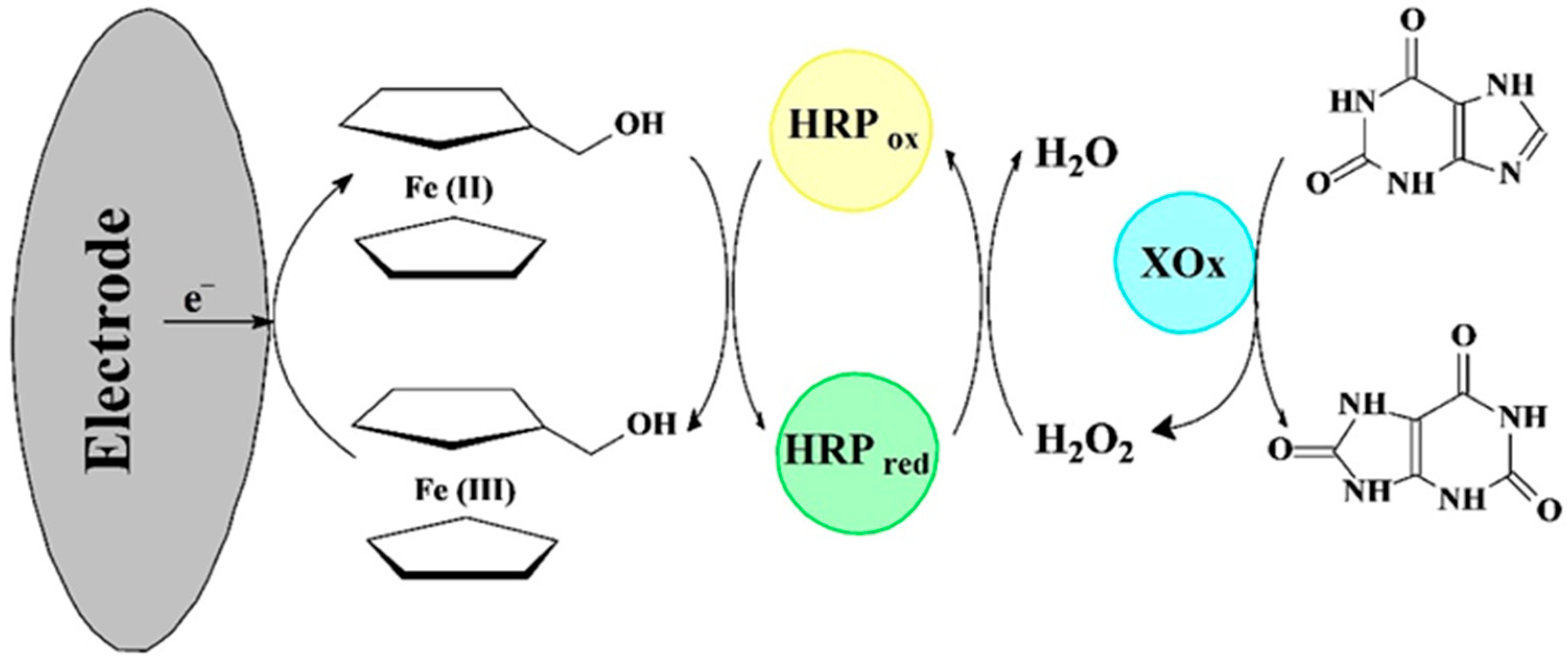
Biosensors based on GCE modified with CaCO3 nanostructures were developed by Shan et al. [40]. The hydrophilic CaCO3 NPs provide a convenient microenvironment for XOx, which retains its high native catalytic activity. Amperometric detection of X in the range from 2 to 250 μM was evaluated by holding the modified electrode XOx/Nano-CaCO3/GCE at 0.55V (vs. SCE). The authors developed a bienzymatic sensing system by the coupling of HRP with XOx (XOx/HRP/Nano-CaCO3-MeOHFc) to estimate the freshness of fish. In this configuration, H2O2 produced by XOx is detected through its HRP-MeOHFc-mediated reduction at low applied potential (−0.05 V vs. SCE) and can avoid the interference of the coexisting electroactive substances, providing excellent selectivity (Figure 7). The response is linear in the range 0.4–500 µM (LOD = 0.1 µM). The authors proved that postmortem concentration of X increases from 7 to 46 μM in the 4 to 48 h interval.
Dalkiran and co-workers constructed a new xanthine biosensor, combining the advantageous properties of MWCNTs, Co3O4 nanoparticles, and chitosan as a matrix for the assembly of enzyme molecules and nanoparticles [103]. The co-immobilization of XOx and HRP allows selective X determination at a low potential of −0.3 V vs. Ag/AgCl and the proposed bi-enzymatic system showed strong anti-interference ability. Amperometric detection of X using XOx/HRP/Co3O4/MWCNTs/CHIT/GCE was realized in the range from 0.2 to 160 µM.
Research efforts have been focused on the co-immobilization of XOx and uricase [104,105]. Uricase (EC 1.7.3.3) converts uric acid to 5-hydroxyisourate and H2O2, leading to the formation of allantoin.
Görgülü et al. proposed a novel approach—XOx and uricase were both immobilized via entrapment in polypyrrole-polyvinyl sulphonate on a platinum electrode [104]. Hypoxanthine determination by the fabricated Pt/PPy-PVS-XOx-U is based on the electrooxidation at 0.4 V of enzymatically generated H2O2. Two dynamic ranges were observed: 2.5 μM–10 μM and 25 μM–0.1 mM (LOD of 2.5 μM). The long-term stability is acceptable—after 33 days, the electrode lost 44% of its initial activity.
Erol et al. reported the integration of XOx and uricase at the surface obtained by electrochemical polymerization in the presence of PPypTS on the Pt-electrode [105]. The LOD for Hx was determined as 5 μM and the dynamic range was broader—from 5 μM to 5 mM. However, in this paper, the authors did not present any records from voltammetric and amperometric experiments, making it difficult to assess the electrochemical behavior of the commented electrode. The long-term stability is also problematic—the biosensor retained approximately 23% of the initial activity after 20 days. The biosensor retained 70% of the original response after 27 measurements, and the RSD was calculated as 17.49%.
The presented studies clearly show that such co-immobilization of two enzymes increases the complexity and cost of biosensors. The selected immobilization conditions should be a compromise and suitable for both enzymes used. On the other hand, the working buffer solution (composition, ionic strength, pH value) cannot be optimal for both biocatalysts; hence, it may decrease their activity. In these cases, the operational and long-term storage stability of the bi-enzyme layer should be enhanced by using new immobilization strategies.

4. Concluding Remarks

Nowadays, the increasing importance and prevalence of electrochemical enzyme-based biosensors as an area of research for analytical applications is due to their remarkable sensing performance, flexible design, and ease of miniaturization compared to other instrumental methods. Electrochemical biosensors have been proven to be reliable analytical tools with excellent operational features, including the following: extremely high specificity, sensitivity, low detection limits, relatively simple interpretation of the results, and improved direct visualization ability. These platforms have remarkable advantages in rapid quantitative detection and portability because they are generally designed as hand-held devices capable of on-site real-time analysis.
This review highlighted the main strategies in developing improved XOx-based electrochemical biosensors as powerful tools for Hx/X detection in foods. Biosensor platforms provide precise quantification of these analytes at low concentrations. The results examined clearly show that biosensors, based on various electrode designs, detect Hx and X with extremely high selectivity, sensitivity, and reproducibility. Most of them have been successfully applied to real food samples, revealing the ability of these bioelectroanalytical systems to provide fast and reliable control in the food industry, thus supporting public health.
Although there have been great achievements made, there are still challenges to overcome:
-
Extending the shelf life and stability of the biorecognition component. Reliable XOx immobilization should be the main focus in the innovative design of optimized biosensing platforms. Storage stability and operational stability can be improved by introducing novel nanomaterial-assisted enzyme immobilization techniques. At the same time, employing mild conditions, high quantities of enzyme molecules can be immobilized uniformly. For example, the biocompatibility and large surface area of gold nanowires provide high enzyme loading efficiency and a compatible microenvironment for XOx immobilization.
-
Improving low-level detection efficiency. Electrochemical enzyme biosensors have become more versatile, robust, and flexible with the induction of novel classes of nanocomposites. The analytical features in terms of repeatability and reproducibility may be adjusted, including functionalization or doping of the host matrices of biosensors, allowing fine control over biosensor performance. For example, chemical doping of heteroatoms (N, S, B, etc.) within carbon nanomaterials such as graphene and CNTs could substantially enhance their electrocatalytic properties. Utilization of recent advancements in quantum dots and dendrimers has also opened up new prospects for the development of efficient and high-performance XOx-based biosensors. There is increasing interest in nanocomposites with regular nanostructures in enzymatic biosensor interface design. Furthermore, the results obtained have shown that bimetallic nanocrystals with core-shell structures greatly affect the analytical performance of electrochemical biosensors. Introducing a third metal in the bimetallic structure may be a promising strategy for enhancing the catalytic activity and sensing performance of bimetallic nanocrystals.
-
Using synergies in material science, bioelectronics, and nanofabrication technologies, these devices should be miniaturized into smart hand-held analyzers. Electrochemical XOx-based biosensors need transducers assembled within a carefully designed sensing interface that can be fabricated into a portable unit. Thus, the electrochemical biosensor can be miniaturized into a compact device connected to a smartphone for powering, processing, data analysis, and visualization. In the foreseeable future, we expect artificial intelligence (AI) algorithms to be introduced to power bioelectroanalytical methods for Hx/X assay.
Although most electrochemical biosensors are still in the testing phase, some are routinely used in laboratory practice. By overcoming the existing challenges, XOx-based electrochemical biosensors have the potential to achieve breakthroughs in reliable food monitoring. Therefore, more multidisciplinary research should be undertaken in the design and optimization of biosensing platforms and we expect to see commercially available electrochemical biosensing devices for food control in the foreseeable future.

Funding

This research received no external funding.

Conflicts of Interest

The author declares no conflicts of interest.

Abbreviations

The following abbreviations are used in this manuscript:
ATPAdenosine triphosphate
BQ1,4-Benzoquinone
CHITChitosan
CLEAsCross-linked enzyme aggregates
CMCCarboxymethylcellulose
CPECarbon paste electrode
DETDirect electron transfer
FADFlavin adenine dinucleotide
FcFerrocene
GCEGlassy carbon electrode
GCPEGlassy carbon paste electrode
GMAGlycidyl methacrylate
GRGraphene
HMTESHydroxymethyltriethoxysilane
HPLCHigh-performance liquid chromatography
HRPHorseradish peroxidase
HxHypoxanthine
KMappApparent Michaelis–Menten constant
LbLLayer-by-layer
L-CysL-cysteine
LODLimit of detection
MeOHFcFerrocenemethanol
MNPMagnetic nanoparticles
MOFsMetal-organic frameworks
MVMethyl viologen
MWCNTsMulti-walled carbon nanotubes
NPsNanoparticles
OMIECOrganic mixed ionic-electronic conductor
PAMAMPolyamidoamine
PANIPolyaniline
PBSPhosphate buffer solution
PGEPencil graphite electrode
PPDPoly(o-phenylenediamine)
PPyPolypyrrole
PUPolyurethane
PVSPolyvinyl sulphonate
REGOReduced expanded graphene oxide
rGOReduced graphene oxide
SASodium alginate
SCESaturated calomel electrode
SGSilica sol–gel
SWCNHSingle-walled carbon nanohorn
SWCNTsSingle-walled carbon nanotubes
SWySodium montmorillonite
TCNQ7,7′,8,8′-Tetracyanoquinodimethane
XXanthine
XOxXanthine oxidase
VFcVinylferrocene

References

  1. Wijayanti, S.D.; Tsvik, L.; Haltrich, D. Recent Advances in Electrochemical Enzyme-Based Biosensors for Food and Beverage Analysis. Foods 2023, 12, 3355. [Google Scholar] [CrossRef]
  2. Singh, P.; Pandey, V.K.; Srivastava, S.; Singh, R. A systematic review on recent trends and perspectives of biosensors in food industries. J. Food Saf. 2023, 43, e13071. [Google Scholar] [CrossRef]
  3. Wang, K.; Lin, X.; Zhang, M.; Li, Y.; Luo, C.; Wu, J. Review of Electrochemical Biosensors for Food Safety Detection. Biosensors 2022, 12, 959. [Google Scholar] [CrossRef]
  4. Sumitha, M.S.; Xavier, T.S. Recent advances in electrochemical biosensors—A brief review. Hybrid Adv. 2023, 2, 100023. [Google Scholar] [CrossRef]
  5. Wu, J.; Liu, H.; Chen, W.; Ma, B.; Ju, H. Device integration of electrochemical biosensors. Nat. Rev. Bioeng. 2023, 1, 346–360. [Google Scholar] [CrossRef]
  6. Zhang, L.; Guo, W.; Lv, C.; Liu, X.; Yang, M.; Guo, M.; Fu, Q. Electrochemical biosensors represent promising detection tools in medical field. Adv. Sens. Energy Mater. 2023, 2, 100081. [Google Scholar] [CrossRef]
  7. Kim, J.; Jeong, J.; Ko, S.H. Electrochemical biosensors for point-of-care testing. Bio-Des. Manuf. 2024, 7, 548–565. [Google Scholar] [CrossRef]
  8. Baranwal, J.; Barse, B.; Gatto, G.; Broncova, G.; Kumar, A. Electrochemical Sensors and Their Applications: A Review. Chemosensors 2022, 10, 363. [Google Scholar] [CrossRef]
  9. Hong, H.; Regenstein, J.M.; Luo, Y. The importance of ATP-related compounds for the freshness and flavor of post-mortem fish and shellfish muscle: A review. Crit. Rev. Food Sci. Nutr. 2017, 57, 1787–1798. [Google Scholar] [CrossRef]
  10. Dolmacı, N.; Çete, S.; Arslan, F.; Yaşar, A. An amperometric biosensor for fish freshness detection from xanthine oxidase immobilized in polypyrrole-polyvinylsulphonate film. Artif. Cells Blood Substit. Biotechnol. 2012, 40, 275–279. [Google Scholar] [CrossRef]
  11. Yazdanparast, S.; Benvidi, A.; Abbasi, S.; Rezaeinasab, M. Enzyme-based ultrasensitive electrochemical biosensor using poly(l-aspartic acid)/MWCNT bio-nanocomposite for xanthine detection: A meat freshness marker. Microchem. J. 2019, 149, 104000. [Google Scholar] [CrossRef]
  12. Jeyasanta, I.; Giftson, K.H.; Patterson, J. Quality Indicator Hypoxanthine Compared with Other Volatile Amine Indicators of Sea Foods Stored in Refrigerator. Asian J. Anim. Vet. Adv. 2018, 13, 144–154. Available online: https://scialert.net/abstract/?doi=ajava.2018.144.154 (accessed on 10 February 2025). [CrossRef]
  13. Gunasekaran, S.; Sankari, G.; Ponnusamy, S. Vibrational spectral investigation on xanthine and its derivatives—Theophylline, caffeine and theobromine. Spectrochimica Acta Part A Mol. Biomol. Spectrosc. 2005, 61, 117–127. [Google Scholar] [CrossRef]
  14. Song, D.; Chen, Q.; Zhai, C.; Tao, H.; Zhang, L.; Jia, T.; Lu, Z.; Sun, W.; Yuan, P.; Zhu, B. Label-Free ZnIn2S4/UiO-66-NH2 Modified Glassy Carbon Electrode for Electrochemically Assessing Fish Freshness by Monitoring Xanthine and Hypoxanthine. Chemosensors 2022, 10, 158. [Google Scholar] [CrossRef]
  15. Kasare, P.K.; Wang, S.-F. Hydrothermally Synthesized Cerium Phosphate with Functionalized Carbon Nanofiber Nanocomposite for Enhanced Electrochemical Detection of Hypoxanthine. Chemosensors 2024, 12, 84. [Google Scholar] [CrossRef]
  16. Rognum, T.O.; Holmen, S.; Musse, M.A.; Dahlberg, P.S.; Stray-Pedersen, A.; Saugstad, O.D.; Opdal, S.H. Estimation of time since death by vitreous humor hypoxanthine, potassium, and ambient temperature. Forensic Sci. Int. 2016, 262, 160–165. [Google Scholar] [CrossRef]
  17. Liao, L.; Xing, Y.; Xiong, X.; Gan, L.; Hu, L.; Zhao, F.; Tong, Y.; Deng, S. An electrochemical biosensor for hypoxanthine detection in vitreous humor: A potential tool for estimating the post-mortem interval in forensic cases. Microchem. J. 2020, 155, 104760. [Google Scholar] [CrossRef]
  18. Dzyadevych, S.V.; Arkhypova, V.N.; Soldatkin, A.P.; El’skaya, A.V.; Martelet, C.; Jaffrezic-Renault, N. Amperometric enzyme biosensors: Past, present and future. IRBM 2008, 29, 171–180. [Google Scholar] [CrossRef]
  19. Fernández, H.; Zon, M.A.; Maccio, S.A.; Alaníz, R.D.; Di Tocco, A.; Carrillo Palomino, R.A.; Cabas Rodríguez, J.A.; Granero, A.M.; Arévalo, F.J.; Robledo, S.N.; et al. Multivariate Optimization of Electrochemical Biosensors for the Determination of Compounds Related to Food Safety—A Review. Biosensors 2023, 13, 694. [Google Scholar] [CrossRef]
  20. Hosseinikebria, S.; Khazaei, M.; Dervisevic, M.; Judicpa, M.A.; Tian, J.; Razal, J.M.; Voelcker, N.H.; Nilghaz, A. Electrochemical biosensors: The beacon for food safety and quality. Food Chem. 2025, 475, 143284. [Google Scholar] [CrossRef]
  21. Ghaani, M.; Azimzadeh, M.; Büyüktaş, D.; Carullo, D.; Farris, S. Electrochemical Sensors in the Food Sector: A Review. J. Agric. Food Chem. 2024, 72, 24170–24190. [Google Scholar] [CrossRef]
  22. Dube, A.; Malode, S.J.; Alodhayb, A.N.; Mondal, K.; Shetti, N.P. Conducting polymer-based electrochemical sensors: Progress, challenges, and future perspectives. Talanta Open 2025, 11, 100395. [Google Scholar] [CrossRef]
  23. Dodevska, T.; Hadzhiev, D.; Shterev, I. A Review on Electrochemical Microsensors for Ascorbic Acid Detection: Clinical, Pharmaceutical, and Food Safety Applications. Micromachines 2023, 14, 41. [Google Scholar] [CrossRef]
  24. Naresh, V.; Lee, N. A Review on Biosensors and Recent Development of Nanostructured Materials-Enabled Biosensors. Sensors 2021, 21, 1109. [Google Scholar] [CrossRef]
  25. Ruzgas, T.; Larpant, N.; Shafaat, A.; Sotres, J. Wireless, Battery-Less Biosensors Based on Direct Electron Transfer Reactions. ChemElectroChem 2019, 6, 5167. [Google Scholar] [CrossRef]
  26. Liu, D.-M.; Chen, J.; Shi, Y.-P. Advances on methods and easy separated support materials for enzymes immobilization. TrAC Trends Anal. Chem. 2018, 102, 332–342. [Google Scholar] [CrossRef]
  27. Seychell, B.C.; Vella, M.; Hunter, G.J.; Hunter, T. The Good and the Bad: The Bifunctional Enzyme Xanthine Oxidoreductase in the Production of Reactive Oxygen Species. In Reactive Oxygen Species-Advances and Developments; IntechOpen: London, UK, 2024. [Google Scholar] [CrossRef]
  28. Dervisevic, M.; Dervisevic, E.; Şenel, M. Recent progress in nanomaterial-based electrochemical and optical sensors for hypoxanthine and xanthine. A review. Microchim. Acta 2019, 186, 749. [Google Scholar] [CrossRef]
  29. Felicia, W.X.L.; Rovina, K.; ‘Aqilah, N.M.N.; Vonnie, J.M.; Yin, K.W.; Huda, N. Assessing Meat Freshness via Nanotechnology Biosensors: Is the World Prepared for Lightning-Fast Pace Methods? Biosensors 2023, 13, 217. [Google Scholar] [CrossRef]
  30. Wang, B.; Liu, K.; Wei, G.; He, A.; Kong, W.; Zhang, X. A Review of Advanced Sensor Technologies for Aquatic Products Freshness Assessment in Cold Chain Logistics. Biosensors 2024, 14, 468. [Google Scholar] [CrossRef]
  31. Ahlawat, J.; Sharma, M.; Pundir, C.S. Advances in xanthine biosensors and sensors: A review. Enzym. Microb. Technol. 2024, 174, 110377. [Google Scholar] [CrossRef]
  32. Garg, D.; Singh, M.; Verma, N.; Monika. Review on recent advances in fabrication of enzymatic and chemical sensors for hypoxanthine. Food Chem. 2022, 375, 131839. [Google Scholar] [CrossRef]
  33. Sakthivel, K.; Balasubramanian, S.; Chang-Chien, G.-P.; Wang, S.-F.; Ahammad; Billey, W.; Platero, J.; Soundappan, T.; Sekhar, P. Editors’ Choice—Review—Advances in Electrochemical Sensors: Improving Food Safety, Quality, and Traceability. ECS Sens. Plus 2024, 3, 020605. [Google Scholar] [CrossRef]
  34. Majer-Baranyi, K.; Székács, A.; Adányi, N. Application of Electrochemical Biosensors for Determination of Food Spoilage. Biosensors 2023, 13, 456. [Google Scholar] [CrossRef] [PubMed]
  35. Watanabe, E.; Ando, K.; Karube, I.; Matsuoka, H.; Suzuki, S. Determination of Hypoxanthine in Fish Meat with an Enzyme Sensor. J. Food Sci. 1983, 48, 496–500. [Google Scholar] [CrossRef]
  36. Boluda, A.; Casado, C.M.; Alonso, B.; García Armada, M.P. Efficient Oxidase Biosensors Based on Bioelectrocatalytic Surfaces of Electrodeposited Ferrocenyl Polycyclosiloxanes—Pt Nanoparticles. Chemosensors 2021, 9, 81. [Google Scholar] [CrossRef]
  37. Sriramulu, G.; Verma, R.; Singh, K.R.B.; Singh, P.; Chakra, C.S.; Mallick, S.; Singh, R.P.; Sadhana, K.; Singh, Y. Self-assembled copper oxide nanoflakes for highly sensitive electrochemical xanthine detection in fish-freshness biosensors. J. Mol. Struct. 2024, 1304, 137640. [Google Scholar] [CrossRef]
  38. Thandavan, K.; Gandhi, S.; Sethuraman, S.; Rayappan, J.B.B.; Krishnan, U.M. Development of electrochemical biosensor with nano-interface for xanthine sensing—A novel approach for fish freshness estimation. Food Chem. 2013, 139, 963–969. [Google Scholar] [CrossRef]
  39. Jain, U.; Narang, J.; Chauhan, N. Enhanced electrochemical performance of xanthine biosensor by core—Shell magnetic nanoparticles and carbon nanotube interface. Adv. Mater. Lett. 2016, 7, 472–479. [Google Scholar] [CrossRef]
  40. Shan, D.; Wang, Y.; Xue, H.; Cosnier, S. Sensitive and selective xanthine amperometric sensors based on calcium carbonate nanoparticles. Sens. Actuators B Chem. 2009, 136, 510–515. [Google Scholar] [CrossRef]
  41. Çubukçu, M.; Timur, S.; Anik, Ü. Examination of performance of glassy carbon paste electrode modified with gold nanoparticle and xanthine oxidase for xanthine and hypoxanthine detection. Talanta 2007, 74, 434–439. [Google Scholar] [CrossRef]
  42. Tripathi, A.; Elias, A.L.; Jemere, A.B.; Harris, K.D. Amperometric Determination of Xanthine Using Nanostructured NiO Electrodes Loaded with Xanthine Oxidase. ACS Food Sci. Technol. 2022, 2, 1307–1317. [Google Scholar] [CrossRef]
  43. Narang, J.; Malhotra, N.; Singhal, C.; Pundir, C.S. Evaluation of Freshness of Fishes Using MWCNT/TiO2 Nanobiocomposites Based Biosensor. Food Anal. Methods 2017, 10, 522–528. [Google Scholar] [CrossRef]
  44. Jain, U.; Narang, J.; Rani, K.; Burna, B.; Sunny, S.; Chauhan, N. Synthesis of cadmium oxide and carbon nanotube based nanocomposites and their use as a sensing interface for xanthine detection. RSC Adv. 2015, 5, 29675–29683. [Google Scholar] [CrossRef]
  45. Devi, R.; Yadav, S.; Naranga, J.; Pundir, C. Electrochemical biosensor based on gold coated iron nanoparticles/chitosan composite bound xanthine oxidase for detection of xanthine in fish meat. J. Food Eng. 2013, 115, 207–214. [Google Scholar] [CrossRef]
  46. Agüí, L.; Manso, H.; Yáñez-Sedeño, P.; Pingarrón, J.M. Amperometric biosensor for hypoxanthine based on immobilized xanthine oxidase on nanocrystal gold–carbon paste electrodes. Sens. Actuators B Chem. 2006, 113, 272–280. [Google Scholar] [CrossRef]
  47. Saadaoui, M.; Sánchez, A.; Díez, P.; Raouafi, N.; Pingarrón, J.M.; Villalonga, R. Amperometric xanthine biosensors using glassy carbon electrodes modified with electrografted porous silica nanomaterials loaded with xanthine oxidase. Microchim. Acta 2016, 183, 2023–2030. [Google Scholar] [CrossRef]
  48. Pundir, C.S.; Devi, R.; Narang, J.; Singh, S.; Nehra, J.; Chaudhry, S. Fabrication of an amperometric xanthine biosensor based on polyvinylchloride membrane. J. Food Biochem. 2011, 36, 21–27. [Google Scholar] [CrossRef]
  49. Devi, R.; Yadav, S.; Pundir, C.S. Electrochemical detection of xanthine in fish meat by xanthine oxidase immobilized on carboxylated multiwalled carbon nanotubes/polyaniline composite film. Biochem. Eng. J. 2011, 58–59, 148–153. [Google Scholar] [CrossRef]
  50. Devi, R.; Thakur, M.; Pundir, C.S. Construction and application of an amperometric xanthine biosensor based on zinc oxide nanoparticles–polypyrrole composite film. Biosens. Bioelectron. 2011, 26, 3420–3426. [Google Scholar] [CrossRef]
  51. Devi, R.; Yadav, S.; Pundir, C.S. Au-colloids–polypyrrole nanocomposite film based xanthine biosensor. Colloids Surf. A Physicochem. Eng. Asp. 2012, 394, 38–45. [Google Scholar] [CrossRef]
  52. Devi, R.; Batra, B.; Lata, S.; Yadav, S.; Pundir, C.S. A method for determination of xanthine in meat by amperometric biosensor based on silver nanoparticles/cysteine modified Au electrode. Process Biochem. 2013, 48, 242–249. [Google Scholar] [CrossRef]
  53. Sahyar, B.Y.; Kaplan, M.; Ozsoz, M.; Celik, E.; Otles, S. Electrochemical xanthine detection by enzymatic method based on Ag doped ZnO nanoparticles by using polypyrrole. Bioelectrochemistry 2019, 130, 107327. [Google Scholar] [CrossRef] [PubMed]
  54. Thakur, D.; Pandey, C.M.; Kumar, D. Highly Sensitive Enzymatic Biosensor Based on Polyaniline-Wrapped Titanium Dioxide Nanohybrid for Fish Freshness Detection. Appl. Biochem. Biotechnol. 2022, 194, 3765–3778. [Google Scholar] [CrossRef]
  55. Devi, R.; Yadav, S.; Pundir, C.S. Amperometric determination of xanthine in fish meat by zinc oxide nanoparticle/chitosan/multiwalled carbon nanotube/polyaniline composite film bound xanthine oxidase. Analyst 2012, 137, 754–759. [Google Scholar] [CrossRef]
  56. Das, J.; Mishra, H.N. Electrochemical biosensor for monitoring fish spoilage based on nanocellulose as enzyme immobilization matrix. Food Meas. 2023, 17, 3827–3844. [Google Scholar] [CrossRef]
  57. Borisova, B.; Sánchez, A.; Jiménez-Falcao, S.; Martín, M.; Salazar, P.; Parrado, C.; Pingarrón, J.M.; Villalonga, R. Reduced graphene oxide-carboxymethylcellulose layered with platinum nanoparticles/PAMAM dendrimer/magnetic nanoparticles hybrids. Application to the preparation of enzyme electrochemical biosensors. Sens. Actuators B Chem. 2016, 232, 84–90. [Google Scholar] [CrossRef]
  58. Siciliano, G.; Alsadig, A.; Chiriacò, M.S.; Turco, A.; Foscarini, A.; Ferrara, F.; Gigli, G.; Primiceri, E. Beyond traditional biosensors: Recent advances in gold nanoparticles modified electrodes for biosensing applications. Talanta 2024, 268 Pt 1, 125280. [Google Scholar] [CrossRef] [PubMed]
  59. Dervisevic, M.; Dervisevic, E.; Çevik, E.; Şenel, M. Novel electrochemical xanthine biosensor based on chitosan–polypyrrole–gold nanoparticles hybrid bio-nanocomposite platform. J. Food Drug Anal. 2017, 25, 510–519. [Google Scholar] [CrossRef]
  60. Zhang, L.; Lei, J.; Zhang, J.; Ding, L.; Ju, H. Amperometric detection of hypoxanthine and xanthine by enzymatic amplification using a gold nanoparticles–carbon nanohorn hybrid as the carrier. Analyst 2012, 137, 3126. [Google Scholar] [CrossRef]
  61. Labban, N.; Wayu, M.B.; Steele, C.M.; Munoz, T.S.; Pollock, J.A.; Case, W.S.; Leopold, M.C. First Generation Amperometric Biosensing of Galactose with Xerogel-Carbon Nanotube Layer-By-Layer Assemblies. Nanomaterials 2019, 9, 42. [Google Scholar] [CrossRef]
  62. Hughes, L.B.; Labban, N.; Conway, G.E.; Pollock, J.A.; Leopold, M.C. Adaptable Xerogel-Layered Amperometric Biosensor Platforms on Wire Electrodes for Clinically Relevant Measurements. Sensors 2019, 19, 2584. [Google Scholar] [CrossRef] [PubMed]
  63. Dang, Q.M.; Wemple, A.H.; Leopold, M.C. Nanomaterial-Doped Xerogels for Biosensing Measurements of Xanthine in Clinical and Industrial Applications. Gels 2023, 9, 437. [Google Scholar] [CrossRef]
  64. Salvigni, L.; Mariani, F.; Gualandi, I.; Decataldo, F.; Tessarolo, M.; Tonelli, D.; Fraboni, B.; Scavetta, E. Selective detection of liposoluble vitamins using an organic electrochemical transistor. Sens. Actuators B Chem. 2023, 393, 134313. [Google Scholar] [CrossRef]
  65. Gentile, F.; Vurro, F.; Janni, M.; Manfredi, R.; Cellini, F.; Petrozza, A.; Zappettini, A.; Coppedè, N. A Biomimetic, Biocompatible OECT Sensor for the Real-Time Measurement of Concentration and Saturation of Ions in Plant Sap. Adv. Electron. Mater. 2022, 8, 2200092. [Google Scholar] [CrossRef]
  66. Kim, H.; Won, Y.; Song, H.W.; Kwon, Y.; Jun, M.; Oh, J.H. Organic Mixed Ionic–Electronic Conductors for Bioelectronic Sensors: Materials and Operation Mechanisms. Adv. Sci. 2024, 11, 2306191. [Google Scholar] [CrossRef]
  67. Lin, Y.; Kroon, R.; Zeglio, E.; Herland, A. P-type accumulation mode organic electrochemical transistor biosensor for xanthine detection in fish. Biosens. Bioelectron. 2025, 269, 116928. [Google Scholar] [CrossRef]
  68. Kidanemariam, A.; Cho, S. Recent Advances in the Application of Metal–Organic Frameworks and Coordination Polymers in Electrochemical Biosensors. Chemosensors 2024, 12, 135. [Google Scholar] [CrossRef]
  69. Wang, Z.; Ma, B.; Shen, C.; Lai, O.-M.; Tan, C.-P.; Cheong, L.-Z. Electrochemical Biosensing of Chilled Seafood Freshness by Xanthine Oxidase Immobilized on Copper-Based Metal–Organic Framework Nanofiber Film. Food Anal. Methods 2019, 12, 1715–1724. [Google Scholar] [CrossRef]
  70. Joon, A.; Ahlawat, J.; Aggarwal, V.; Jaiwal, R.; Pundir, C.S. An improved amperometric determination of xanthine with xanthine oxidase nanoparticles for testing of fish meat freshness. Sens. Bio-Sens. Res. 2021, 33, 100437. [Google Scholar] [CrossRef]
  71. Dalkıran, B.; Erden, P.; Kılıç, E. Construction of an Electrochemical Xanthine Biosensor Based on Graphene/Cobalt Oxide Nanoparticles/Chitosan Composite for Fish Freshness Detection. J. Turk. Chem. Soc. Sect. A Chem. 2017, 4, 23–44. [Google Scholar] [CrossRef]
  72. Öztürk, F.; Erden, P.; Kacar, C.; Kilic, E. Amperometric biosensor for xanthine determination based on Fe3O4 nanoparticles. Acta Chim. Slov. 2014, 61, 19–26. Available online: https://acta-arhiv.chem-soc.si/61/61-1-19.pdf (accessed on 10 February 2025).
  73. Dodevska, T.; Horozova, E.; Dimcheva, N. Electrochemical characteristics and structural specifics of carbonaceous electrodes, modified with micro- and nanodeposits of platinum metals. Bulg. Chem. Commun. 2013, 45, 171–178. Available online: http://bcc.bas.bg/BCC_Volumes/Volume_45_Special_A_2013/BCC-45-SE-A-171-178.pdf (accessed on 10 February 2025).
  74. Horozova, E.; Dodevska, T.; Dimcheva, N. Modified graphite electrodes as catalysts for electroreduction of hydrogen peroxide. Bulg. Chem. Commun. 2008, 40, 233–239. Available online: http://www.bcc.bas.bg/BCC_Volumes/Volume_40_Number_3_2008/Volume_40_Number_3_2008_PDF/2817-RR.pdf (accessed on 10 February 2025).
  75. Dodevska, T.; Horozova, E.; Dimcheva, N. Electrocatalytic reduction of hydrogen peroxide on modified graphite electrodes: Application to the development of glucose biosensors. Anal. Bioanal. Chem. 2006, 386, 1413–1418. [Google Scholar] [CrossRef] [PubMed]
  76. Dimcheva, N.; Horozova, E.; Jordanova, Z. An amperometric xanthine oxidase enzyme electrode based on hydrogen peroxide electroreduction. Z. Naturforschung C 2002, 57, 883–889. [Google Scholar] [CrossRef]
  77. Horozova, E.; Dodevska, T.; Dimcheva, N. Modified with microquantities of platinum metals graphites: Application to the development of xanthine oxidase enzyme electrode. J. Univ. Chem. Technol. Metall. 2008, 43, 59–64. Available online: https://journal.uctm.edu/node/j2008-1/8_Horozova_Dodevska-59-64.pdf (accessed on 10 February 2025).
  78. Horozova, E.; Dodevska, T.; Dimcheva, N. Modified graphites: Application to the development of enzyme-based amperometric biosensors. Bioelectrochemistry 2009, 74, 260–264. [Google Scholar] [CrossRef]
  79. Dodevska, T.; Horozova, E.; Dimcheva, N. Design of an amperometric xanthine biosensor based on a graphite transducer patterned with noble metal microparticles. Cent. Eur. J. Chem. 2010, 8, 19–27. [Google Scholar] [CrossRef]
  80. Albelda, J.A.V.; Uzunoglu, A.; Santos, G.N.C.; Stanciu, L.A. Graphene-titanium dioxide nanocomposite based hypoxanthine sensor for assessment of meat freshness. Biosens. Bioelectron. 2017, 89, 518–524. [Google Scholar] [CrossRef]
  81. Kilinc, E.; Erdem, A.; Gokgunnec, L.; Dalbasti, T.; Karaoglan, M.; Ozsoz, M. Buttermilk based cobalt phthalocyanine dispersed ferricyanide mediated amperometric biosensor for the determination of xanthine. Electroanalysis 1998, 10, 273–275. [Google Scholar] [CrossRef]
  82. Arslan, F.; Yaşar, A.; Kılıç, E. An Amperometric Biosensor for Xanthine Determination Prepared from Xanthine Oxidase Immobilized in Polypyrrole Film. Artif. Cells Blood Substit. Biotechnol. 2006, 34, 113–128. [Google Scholar] [CrossRef] [PubMed]
  83. Erden, P.E.; Pekyardımcı, Ş.; Kılıç, E. Amperometric enzyme electrodes for xanthine determination with different mediators. Acta Chim. Slov. 2012, 59, 824–832. Available online: https://acta-arhiv.chem-soc.si/59/59-4-824.pdf (accessed on 10 February 2025). [PubMed]
  84. Dervisevic, M.; Custiuc, E.; Çevik, E.; Durmus, Z.; Şenel, M.; Durmus, A. Electrochemical biosensor based on REGO/Fe3O4 bionanocomposite interface for xanthine detection in fish sample. Food Control 2015, 57, 402–410. [Google Scholar] [CrossRef]
  85. Can, E.; Selvi, C.K.; Canel, E.; Erden, P.E.; Kılıç, E. Electrochemical xanthine biosensor based on carbon nanofiber and ferrocene carboxylic acid for the assessment of fish freshness. Ionics 2024, 30, 4215–4226. [Google Scholar] [CrossRef]
  86. Bollella, P. Enzyme-based amperometric biosensors: 60 years later … Quo Vadis? Anal. Chim. Acta 2022, 1234, 340517. [Google Scholar] [CrossRef]
  87. Takahashi, S.; Anzai, J.-I. Recent Progress in Ferrocene-Modified Thin Films and Nanoparticles for Biosensors. Materials 2013, 6, 5742–5762. [Google Scholar] [CrossRef]
  88. Teng, Y.; Chen, C.; Zhou, C.; Zhao, H.; Lan, M. Disposable amperometric biosensors based on xanthine oxidase immobilized in the Prussian blue modified screen-printed three-electrode system. Sci. China Chem. 2010, 53, 2581–2586. [Google Scholar] [CrossRef]
  89. Liu, Y.; Nie, L.; Tao, W.; Yao, S. Amperometric Study of Au-Colloid Function on Xanthine Biosensor Based on Xanthine Oxidase Immobilized in Polypyrrole Layer. Electroanalysis 2004, 16, 1271–1278. [Google Scholar] [CrossRef]
  90. Dalkıran, B.; Erden, P.E.; Kılıç, E. Amperometric biosensors based on carboxylated multiwalled carbon nanotubes-metal oxide nanoparticles-7,7,8,8-tetracyanoquinodimethane composite for the determination of xanthine. Talanta 2017, 167, 286–295. [Google Scholar] [CrossRef]
  91. Dalkıran, B.; Kaçar, C.; Erden, P.E.; Kılıç, E. Electrochemical xanthine biosensor based on zinc oxide nanoparticles–multiwalled carbon nanotubes–1,4-benzoquinone composite. J. Turk. Chem. Soc. Sect. A Chem. 2018, 5, 317–332. [Google Scholar] [CrossRef]
  92. Dervisevic, M.; Custiuc, E.; Çevik, E.; Şenel, M. Construction of novel xanthine biosensor by using polymeric mediator/MWCNT nanocomposite layer for fish freshness detection. Food Chem. 2015, 181, 277–283. [Google Scholar] [CrossRef] [PubMed]
  93. Hu, S.; Xu, C.; Luo, J.; Luo, J.; Cui, D. Biosensor for detection of hypoxanthine based on xanthine oxidase immobilized on chemically modified carbon paste electrode. Anal. Chim. Acta 2000, 412, 55–61. [Google Scholar] [CrossRef]
  94. Dodevska, T.; Horozova, E.; Dimcheva, N. Electrochemical behavior of ascorbate oxidase immobilized on graphite electrode modified with Au-nanoparticles. Mater. Sci. Eng. B 2013, 178, 1497–1502. [Google Scholar] [CrossRef]
  95. Schachinger, F.; Chang, H.; Scheiblbrandner, S.; Ludwig, R. Amperometric Biosensors Based on Direct Electron Transfer Enzymes. Molecules 2021, 26, 4525. [Google Scholar] [CrossRef]
  96. Horozova, E.; Dimcheva, N.; Jordanova, Z. Enzymatic and electrochemical reactions of xanthine oxidase immobilized on carbon materials. Z. Naturforschung C 1997, 52, 159–164. [Google Scholar] [CrossRef] [PubMed]
  97. Torres, A.C.; Ghica, M.E.; Brett, C.M.A. Design of a new hypoxanthine biosensor: Xanthine oxidase modified carbon film and multi-walled carbon nanotube/carbon film electrodes. Anal. Bioanal. Chem. 2013, 405, 3813–3822. [Google Scholar] [CrossRef]
  98. Wang, L.; Yuan, Z. Direct Electrochemistry of Xanthine Oxidase at a Gold Electrode Modified with Single-Wall Carbon Nanotubes. Anal. Sci. 2004, 20, 635–638. [Google Scholar] [CrossRef]
  99. Gao, Y.; Shen, C.; Di, J.; Tu, Y. Fabrication of amperometric xanthine biosensors based on direct chemistry of xanthine oxidase. Mater. Sci. Eng. C 2009, 29, 2213–2216. [Google Scholar] [CrossRef]
  100. Shan, D.; Wang, Y.-N.; Xue, H.-G.; Cosnier, S.; Ding, S.-N. Xanthine oxidase/laponite nanoparticles immobilized on glassy carbon electrode: Direct electron transfer and multielectrocatalysis. Biosens. Bioelectron. 2009, 24, 3556–3561. [Google Scholar] [CrossRef]
  101. Sharma, P.; Thakur, D.; Kumar, D. Novel Enzymatic Biosensor Utilizing a MoS2/MoO3 Nanohybrid for the Electrochemical Detection of Xanthine in Fish Meat. ACS Omega 2023, 8, 31962–31971. [Google Scholar] [CrossRef]
  102. Sen, S.; Sarkar, P. A novel third-generation xanthine biosensor with enzyme modified glassy carbon electrode using electrodeposited MWCNT and nanogold polymer composite film. RSC Adv. 2015, 5, 95911–95925. [Google Scholar] [CrossRef]
  103. Dalkiran, B.; Kaçar, C.; Erden, P.E.; Kiliç, E. Amperometric xanthine biosensors based on chitosan-Co3O4-multiwall carbon nanotube modified glassy carbon electrode. Sens. Actuators B Chem. 2014, 200, 83–91. [Google Scholar] [CrossRef]
  104. Görgülü, M.; Çete, S.; Arslan, H.; Yaşar, A. Preparing a new biosensor for hypoxanthine determination by immobilization of xanthine oxidase and uricase in polypyrrole-polyvinyl sulphonate film. Artif. Cells Nanomed. Biotechnol. 2013, 41, 327–331. [Google Scholar] [CrossRef] [PubMed]
  105. Erol, E.; Yildirim, E.; Cete, S. Construction of biosensor for hypoxanthine determination by immobilization of xanthine oxidase and uricase in polypyrrole-paratoluenesulfonate film. J. Solid State Electrochem. 2020, 24, 1695–1707. [Google Scholar] [CrossRef]
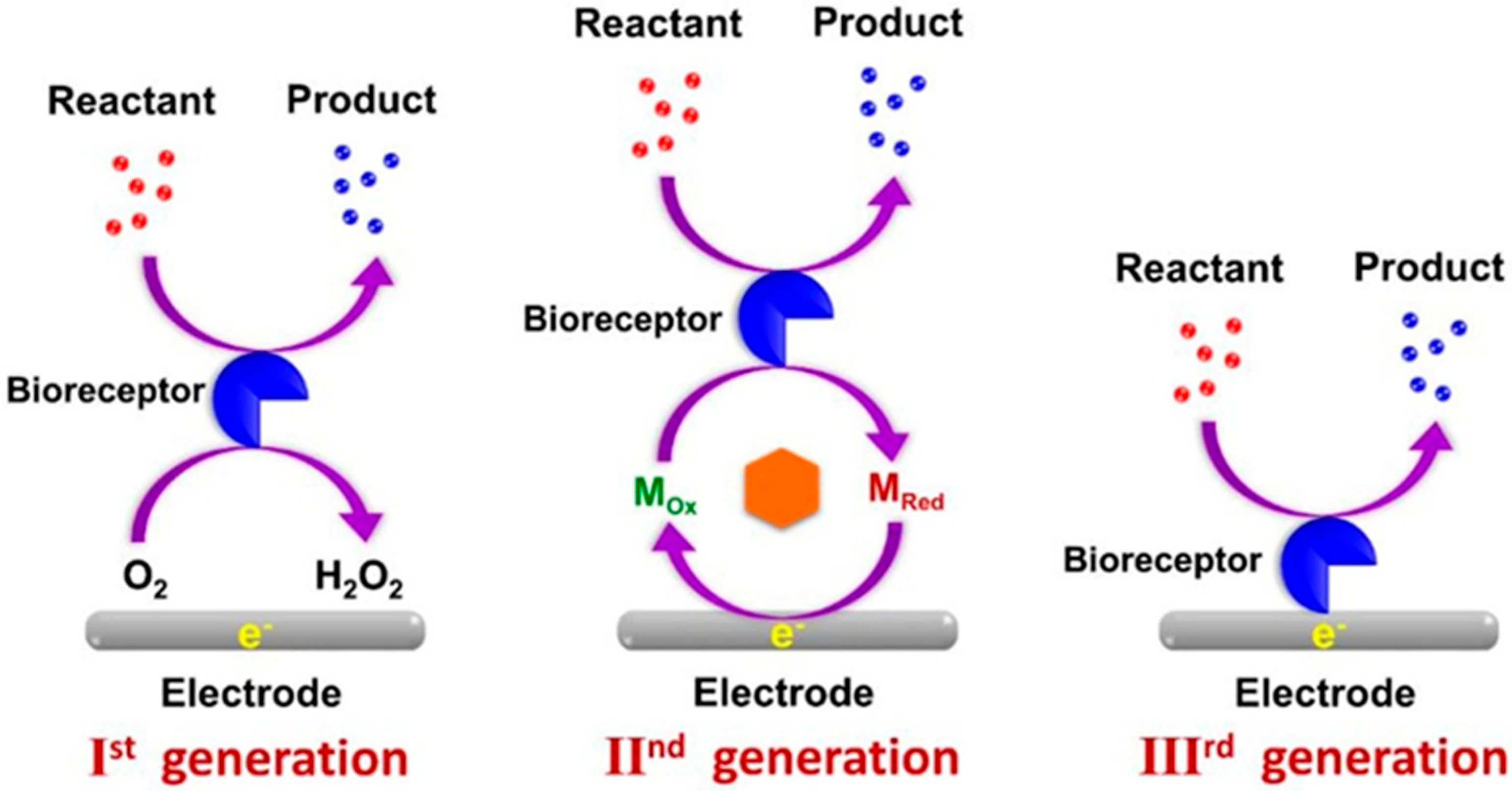
Figure 1. Three generations of biosensor construction (MOX: oxidized mediator; MRed: reduced mediator). Reproduced from Ref. [24]. Licensee 2021 MDPI.
Chemosensors 13 00159 g001
Figure 2. The methods for enzyme immobilization. MOFs—metal-organic frameworks; CLEAs—cross-linked enzyme aggregates. Reproduced with permission from [26]. Copyright 2018 Elsevier.
Chemosensors 13 00159 g002
Figure 3. Crystal structure of bovine xanthine oxidoreductase, (PDB 3UNC). The two subunits of the biologically active dimeric enzyme are shown as different representations. On the right-hand side is a cartoon of one monomer; blue represents the Moco domain (including the C terminus), yellow represents the FAD domain, and red represent the iron–sulfur domain (and includes the N-terminus). Cream represents the link portions and cofactors are also shown in stick form. On the left-hand-side the monomer is shown as a surface colored by electrostatic potential with cofactors as spheres colored as their domains in the cartoon and labeled accordingly. Reproduced from ref. [27]; permissions under Creative Commons Attribution 3.0 License. Copyright 2023 Seychell, Vella, Hunter, and Hunter.
Chemosensors 13 00159 g003
Figure 4. Reactions catalyzed by XOx.
Chemosensors 13 00159 g004
Figure 5. Schematic display of the preparation of the MNP-PAMAM-PtNP and the XOx/MNP-PAMAM-PtNP/rGO-CMC/GCE enzyme electrode. Reproduced with permission from [57]. Copyright 2016 Elsevier.
Chemosensors 13 00159 g005
Figure 6. Schematic diagram of the OECT-based xanthine biosensor. (1, 2) Illustration of the degradation of a salmon sample, causing xanthine accumulation. (3) Schematic overview of the OECT where the electrolyte (10 mM PBS) connects both channel (bottom) and gate (top). (4) Schematic overview of the OECT biosensor. All contacts (gate, source, and drain) are made of Au. The channel is p(g42T-TT). The reaction of xanthine is catalyzed by the enzyme XOx immobilized at the gate. (5) Expanded view of the gate layers including Au gate, PtNPs, and XOx and Nafion. (6) The oxidation of xanthine to uric acid. (7) Expanded view of the channel layers including glass substrate, p(g42T-TT), and XOD and Nafion matrix. (8) Chemical structure of p(g42T-TT). Reproduced with permission from [67]. Copyright 2025 Elsevier.
Chemosensors 13 00159 g006
Figure 7. Schematic illustration of the proposed reaction mechanism for the amperometric detection of xanthine by the XOx/HRP/Nano-CaCO3-MeOHFc-system. Reproduced with permission from [40]. Copyright 2009 Elsevier.
Chemosensors 13 00159 g007
Table 1. First-generation electrochemical biosensors for the detection of Hx and X in food samples.
ElectrodeAnalyteTechnique
(Potential)
Linear Range
(LOD)
StabilityReal SampleRef.
XOx-PVS-PPy/Pt 3XAmp.0.1–1000 µM49%fish[10]
(0.3 V **)(0.1 µM)(30 days)
XOx/PtNPs/FPP 4XAmp.10–1400 µM70%
(24 days)
fish[36]
(−0.1 V **)(45 nM)
XOx/PtNPs/FPP 1Amp.30–800 µM
(0.4 V **)(30 nM)
XOx/Nano Fe3O4/Au 1XAmp.0.4–2.4 nM80%
(11 days)
fish[38]
(0.5 V *)(2.5 pM)
XOx/Fe3O4-NPs/c-MWCNT/FTOXAmp.0.05–150 µM50%
(120 days)
fish[39]
(0.2 V *)(0.05 µM)
XOx/c-MWCNT/PANI/Pt 1XAmp.0.6–58 µM50%
(100 days)
fish[49]
(0.4 V *)(0.6 µM)
XOx/CHIT/Fe-NPs@Au/PGE 1XAmp.0.1–300 µM75%
(100 days)
fish[45]
(0.5 V *)(0.1 µM)
GA–BSA–XOx–AuNPs–CPE 2HxAmp.0.5–10 µM15 dayssardines, chicken[46]
(0.0 V *)(0.22 µM)
GA–BSA–XOx–AuNPs–CPE 1Amp.0.5–10 µM
(0.6 V *)(0.1 µM)
XOx/ZnO-NPs–PPy/Pt 1XAmp.0.8–40 µM60%
(100 days)
fish[50]
(0.38 V *)(0.8 µM)
XOx/AgNPs/L-Cys/Au 1XAmp.2–16 µM80%
(60 days)
chicken, beef, pork[52]
(0.5 V *)(0.15 µM)
XOx/nano Ag-ZnO/PPy/PGE 1XAmp.0.06-0.6 µM78%
(20 days)
fish[53]
(0.7 V *)(0.07 µM)
XOx/ZnO-NP/CHIT/c-MWCNT/PANI/Pt 1XCV1–100 µM70%
(30 days)
fish[55]
(0.5 V *)(0.1 µM)
XOx/PtNPs-PAMAM-MNP/GO-CMC/GCE 1XAmp.50 nM–12 µM73%
(28 days)
fish[57]
(0.6 V *)(13 nM)
CHIT–PPy/Au–XOx/GCE 1XAmp.1–200 µM85%
(18 days)
fish, chicken, beef[59]
(0.7 V *)(0.25 µM)
Pt/HMTES (XOx) + C6-MPCs/PU(75:25) 1XAmp.up to 600 µMfish[63]
(0.4 V *)(5.2 µM)
Amp.up to 600 µM
(0.65 V *)(3.1 µM)
XOx@Cu-MOF/SA/GCE 1XDPV0.01–10 µM80%
(20 days)
squid, large yellow croaker[69]
(0.579 V **)(6.4 nM)
HxDPV0.01–10 µM
(0.749 V **)(2.3 nM)
XOxNPs/Au 1XAmp.0.01–1 µM50%
(60 days)
fish[70]
(0.25 V *)(0.01 µM)
Nafion/XOx/Co3O4/CHIT/GR 1XAmp.0.5–80 µM83%
(60 days)
fish71
(0.7 V *)(0.2 µM)
Nafion/XOx/TiO2-G/GCE 1HxAmp.20–512 µM77%
(10 days)
pork[80]
(0.8 V *)(9.5 µM)
1 biosensor based on electrooxidation of H2O2; 2 biosensor based on electroreduction of H2O2; 3 biosensor based on electrooxidation of uric acid; 4 biosensor based on oxygen consumption; * Reference electrode Ag/AgCl; ** Reference electrode SCE.
Table 2. Second-generation electrochemical biosensors for detection of Hx and X in food samples.
ElectrodeMediatorAnalyteTechnique
(Potential)
Linear Range
(LOD)
StabilityReal SampleRef.
XOx/c-MWCNTs/Fe3O4/TCNQ/CHIT/GCETCNQXAmp.
(0.3 V *)
1.9–230 µM
(0.2 µM)
70%
(30 days)
coffee[90]
XOx/BQ-MWCNTs-ZnO-CHIT/GCEBQXAmp.
(0.25 V *)
0.9–110 µM
(0.21 µM)
95%
(25 days)
chicken, beef[91]
Poly(GMA-co-VFc)/REGO-Fe3O4/XOx/PGEVFcXAmp.
(0.35 V *)
2–36 µM
(0.17 µM)
70%
(25 days)
fish[92]
XOx/SWy-2-MV/CPEMVHxCV
(−0.72 V **)
1–400 µM
(0.8 µM)
60%
(5 weeks)
fish[93]
* Reference electrode Ag/AgCl; ** Reference electrode SCE; BQ—1,4-benzoquinone; MV—methyl viologen; TCNQ—7,7′,8,8′-tetracyanoquinodimethane; SWy-2—sodium montmorillonite; VFc—vinylferrocene.
Disclaimer/Publisher’s Note: The statements, opinions and data contained in all publications are solely those of the individual author(s) and contributor(s) and not of MDPI and/or the editor(s). MDPI and/or the editor(s) disclaim responsibility for any injury to people or property resulting from any ideas, methods, instructions or products referred to in the content.