Review

µ-NMR Technology for Biomedical Applications: A Review

by
Beatriz Sequeira-Antunes
1,2,3,* and
Hugo Alexandre Ferreira
1,2,*
1
Institute of Biophysics and Biomedical Engineering, Faculty of Sciences, University of Lisbon, Campo Grande, 1749-016 Lisbon, Portugal
2
Exotictarget, 4900-378 Viana do Castelo, Portugal
3
Instituto de Engenharia de Sistemas e Computadores-Microsistemas e Nanotecnologias (INESC-MN), 1000-029 Lisbon, Portugal
*
Authors to whom correspondence should be addressed.
Chemosensors 2024, 12(12), 248; https://doi.org/10.3390/chemosensors12120248
Submission received: 30 October 2024 / Revised: 24 November 2024 / Accepted: 25 November 2024 / Published: 27 November 2024
(This article belongs to the Special Issue Rapid Point-of-Care Testing Technology and Application)

Abstract

:
Nuclear magnetic resonance (NMR) is a versatile method that non-invasively provides detailed insights into the atomic and molecular information of samples containing non-zero spin nuclei, facilitating observations of their structure, dynamics, and interactions. By miniaturizing NMR systems, micro-NMR (µ-NMR) devices overcome the limitations of traditional bulky NMR instruments, making them more portable, cost-effective, and suitable for a wide range of applications. As such, this review aims to provide a comprehensive overview of the recent advancements and potential applications of µ-NMR in the field of biomedicine. Beginning with an overview of the principles underlying NMR, this paper explains the fundamental concepts essential for understanding µ-NMR technology. It then delves into miniaturization techniques, detailing advancements in microcoils and probes and the development and integration with microfluidics, which have enhanced the sensitivity, portability, and versatility of µ-NMR devices. Ultimately, this review discusses the current biomedical applications of µ-NMR, including molecular imaging, metabolomics, biomarker detection, and point-of-care diagnosis, and highlights the potential of this technology to revolutionize precision medicine and healthcare. Despite the promising applications, challenges such as sensitivity, spectral resolution, and integration with other technologies are discussed, along with recent advances and innovations aimed at addressing these limitations.

1. Introduction

Nuclear magnetic resonance (NMR) is a powerful tool for detecting and analyzing molecular information of materials and is widely used in medical diagnostics [1]. Nuclear magnetic resonance spectroscopy delves into molecules by manipulating the magnetic properties of their atomic nuclei. Initially, a powerful magnet aligns these nuclei, after which radio-frequency (RF) pulses perturb their orientation. Subsequently, the nuclei emit distinctive radio signals, providing insights into the structure and composition of the sample under study [2].
The NMR method has the advantage of allowing a wide range of chemical and physical properties on a whole range of structural levels to be studied using a single device. By analyzing just few key parameters, such as shifts in NMR peaks (chemical shifts), it is possible to identify complete chemical structures at the interatomic level [3], quickly identify molecules of interest in complex mixtures [4], and investigate molecular dynamics [5]. Moreover, NMR has become a proven instrument for metabolomic [6,7,8] and structural studies in biology [9,10].
NMR offers the significant benefit of being simple when it comes to identifying chemical structures in both liquid and solid samples [11]. However, achieving high sensitivity in standard NMR experiments is crucial. It is widely recognized that NMR’s inherent sensitivity is relatively low compared to other analytical methods [12]. One approach to compensate for NMR’s low sensitivity is to use larger sample quantities, lengthy acquisition times [13], and spinning the sample at very high rotations per minute at a particular angle, the known magic angle to the magnetic field [14]. Additionally, most current NMR instruments are bulky, heavy, and costly [13,15], limiting their broader application in medicine, biology, and industry. Consequently, these attributes render them impractical for rapid, real-time, and precise on-site analysis. This way, by reducing their size, weight, and cost through miniaturization, the potential applications of NMR can be significantly expanded [13].
Advancements in micro- and nano-manufacturing technology have facilitated the development of micro-NMR (µ-NMR) systems, offering a practical solution with benefits including affordability, improved portability, and reduced sample consumption [1,2,13]. These µ-NMR systems have the potential to overcome the limitations of traditional NMR systems by integrating into handheld devices or lab-on-a-chip (LoC) platforms. This enables rapid, portable, and user-friendly analysis, particularly in the field of point-of-care (PoC) diagnostics [2,16].
In this review paper, we aim to explore the potential of developing µ-NMR systems for biomedical applications. We begin by reviewing the principles of NMR, offering a foundational understanding of this technology. Next, we enumerate and describe the essential components of these systems: permanent magnets; microcoils; and transceiver electronic chips. Then, we discuss various biomedical applications, highlighting the practical uses and benefits of µ-NMR in medical and biological research through the development of PoC diagnosis devices. Then, some challenges and limitations related to sensitivity, resolution, and integration with other techniques are discussed, as well as recent efforts made to address these challenges. Finally, we present our conclusions and suggest future directions, summarizing the key points.

2. Principles of NMR

NMR is a non-invasive method that provides details about the atomic and molecular information of the samples [17]. Initially observed in 1946 by Purcell and Bloch [18], NMR operates by the interaction between the magnetic properties of atomic nuclei, specifically their spin, and externally applied magnetic fields [19].
Atomic nuclei exhibit a quantum mechanical property, known as spin, akin to the rotation of a charged particle around its axis. This spin generates a magnetic moment, a vector quantity indicating the magnitude and direction of the magnetic field linked with the nucleus [20]. Nuclei containing an even number of protons and neutrons possess zero nuclear spin, hence yielding zero magnetic moments. Contrarily, nuclei with an odd number of protons or neutrons (e.g., 1H, 13C, 15N, 31P, etc.) manifest non-zero magnetic moments due to their spin, thereby demonstrating magnetic properties [19]. Among these, hydrogen (1H) is the most studied nucleus due to its abundance in biological samples [2].
Figure 1 illustrates the fundamental principles of NMR, highlighting the sequence of events from nuclear spin alignment to signal detection. In the absence of an external magnetic field, nuclear spins are randomly oriented, resulting in zero net magnetization [2]. However, upon applying an external magnetic field ( B 0 ), according to their spin, nuclei align either parallel or anti-parallel to the field direction. This alignment generates a measurable net magnetization, M , which can be detected using radio-frequency techniques [19]. The interaction between the external magnetic field and the magnetic moment of atomic nuclei generates a force on the nuclei, causing them to precess around the direction of the magnetic field [17]. This precession occurs at a specific frequency known as the Larmor frequency [20].
In a state of equilibrium, the magnetization of the sample aligns with the direction of the static magnetic field, B 0 [19]. However, to detect this magnetization, it is necessary for it to precess at the Larmor frequency about the direction of the magnetic field [19]. To induce this precession, a radio-frequency (RF) magnetic field, B 1 , with a frequency equal to the Larmor frequency is applied perpendicular to the static magnetic field. The application of the B 1 field for a short time is called the RF pulse. It tilts the magnetization away from being parallel with the static magnetic field, initiating precession [20]. The RF pulse induces transitions between spin states, causing nuclei to absorb or emit energy in the form of electromagnetic radiation.
Upon cessation of the RF pulse, the nuclei return to their equilibrium state through relaxation processes. These include T1, or longitudinal relaxation, where nuclei realign with the magnetic field, and T2, or transverse relaxation, wherein coherence among the spins is lost due to interactions with their surroundings [19]. The precessing nuclei generate a weak electromagnetic signal, known as free induction decay (FID). This signal will be detected with an RF receiver coil, carrying information about the chemical environment and molecular interactions of the nuclei within the sample [2].
After the FID signal is detected by the RF receiver coil, several key steps are involved in processing this signal to obtain an NMR spectrum. First, the signal is converted into a digital signal using an analog-to-digital converter (ADC) [21]. Then, Fourier transformation (FT) is applied to the signal. The FT mathematically processes the complex time-domain FID signal to produce an NMR spectrum [22]. This spectrum displays the intensity of signals as a function of frequency, which corresponds to the chemical shifts of the nuclei [22]. The chemical shift indicates the resonant frequency of the nuclei relative to a standard reference compound [21]. Different chemical environments will cause, among other effects, shifts in the resonance frequency, allowing for the identification of different functional groups and molecular structures [21]. The integral of the signals in the NMR spectrum is proportional to the number of nuclei contributing to that resonance frequency [22]. This means that the area of a peak reflects the relative abundance of the species present in the sample. Consequently, signals can be used to quantify the concentration of each chemical species within the sample, allowing for detailed compositional analysis [21,22]. Together, these data enable the detailed analysis and interpretation of the sample’s molecular structure.

3. µ-NMR System

A typical NMR system comprises essential components that work together to generate, manipulate, and detect NMR signals [13]. µ-NMR systems primarily include a permanent magnet, a radio-frequency (RF) microcoil, and an NMR chip. The magnet creates a stable static magnetic field ( B 0 ) that aligns the magnetic moments of atomic nuclei in the sample [13]. The RF system, placed within the magnet and connected to the NMR chip, includes all the equipment for generating and manipulating RF signals. This system involves RF generators, like special coils placed near the sample to create another magnetic field ( B 1 ), and detectors (receivers) to pick up the signals [11]. Sometimes, extra electronics are needed to keep everything in sync and to store the data collected [11]. Making these parts smaller is essential for developing miniaturized NMR systems, often called µ-NMR systems. This section will discuss the most common parts of NMR systems.

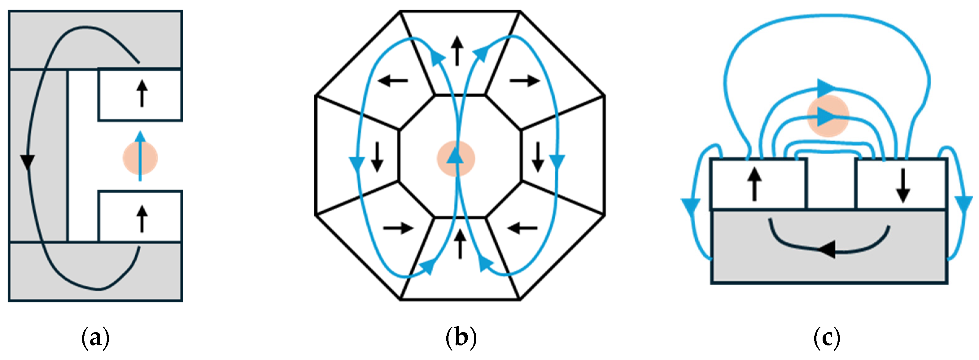
3.1. Permanent Magnet

Traditional NMR magnets, predominantly superconducting, offer high magnetic field strength and stability, crucial for generating high-resolution and sensitive NMR spectra [23,24]. These magnets are renowned for their robust performance but are typically large, heavy, and require cryogenic cooling using liquid helium and complex refrigeration systems [15,25]. Their size and weight make them expensive and impractical for portable NMR systems [11].
To mitigate these challenges, compact permanent magnets have gained prominence. Despite their lower magnetic field homogeneity and strength, ranging from 0.5 to 1.5 T, compared to superconducting magnets, compact permanent magnets offer simplicity, lower cost, portability, and reduced weight [15,23]. Additionally, compact permanent magnets have a small footprint and do not require power to generate the magnetic field, which explains their adequacy for portable μ-NMR applications [2].
In µ-NMR applications, permanent magnets are typically categorized into two main configurations: center-field magnets and stray-field magnets [26]. Center-field magnets are the most common type and have a hollow bore in the center where the sample is placed. This design offers good field homogeneity in the region of interest [25]. Various designs of permanent center-field magnets have been explored for NMR spectroscopy. One common design is the C-shaped magnet (Figure 2a), where both magnetized poles have equal strength and are separated by a gap for placing the sample [25,26]. This arrangement creates a large area of magnetic field inside the gap with a gentle gradient. The magnetic flux returns from one pole to the other through an iron yoke, completing the magnetic circuit. Another design is the Halbach magnet (Figure 2b) [25,26]. Halbach magnets work by arranging permanent magnets in a specific pattern to create a strong and uniform magnetic field in one direction while minimizing the field in the opposite direction. This is achieved by precisely adjusting each magnet’s orientation, resulting in a concentrated and efficient magnetic field. In contrast, stray-field magnets (Figure 2c) are designed to exploit the magnetic field that extends beyond the physical boundaries of the magnet [26]. Herein, two magnets are placed on an iron yoke, allowing the NMR signal collection from a shaped volume located up to a distance typically 5 to 25 mm away from the magnet surface [25,26]. The shape of this volume and the associated magnetic field distribution are defined by the arrangement of the magnet blocks, namely by the gap size between them [25].
To build a permanent magnet, two key elements must be considered: the material of the frame that supports the magnet block and the material used for the magnet block itself [25]. For the frame material, typical options include fiber-reinforced polymer resins or metals such as aluminum and titanium [25]. The choice of this material must consider its strength, lightness, and non-magnetic properties. On the other hand, the magnet block is characterized by properties such as field strength, Curie temperature, cost, corrosion resistance and homogeneity of the remnant magnetization [27]. The material used to construct is typically sintered from grains of alloys containing ferromagnetic elements like iron, cobalt, or nickel [25].
In µ-NMR applications, different materials for the development of permanent magnets can be used, such as aluminum–nickel–cobalt (AlNiCo), neodymium–iron–boron (NdFeB), and samarium–cobalt (SmCo). Table 1 summarizes their main characteristics, advantages, and disadvantages.
AlNiCo is an established magnet alloy family consisting primarily of aluminum (Al), nickel (Ni), cobalt (Co), and iron (Fe) along with copper (Cu) and titanium (Ti) [28]. These metals show good temperature stability and are resistant to corrosion; however, they have low remnant magnetization [25,28]. NdFeB and SmCo are rare earth permanent magnets [28]. The former is the most widely employed and commercially available permanent magnet [2,25]. This type of magnet has high remanence (i.e., residual magnetization in the absence of an external magnetic field) and coercivity (i.e., magnet resistance against demagnetization) properties [2,25,29,30]. Additionally, NdFeB magnets have good performance in small form-factor samples, making them suitable for µ-NMR devices, despite their low Curie temperature [2,31]. However, these permanent magnets are also susceptible to corrosion, often requiring a protective coating made of nickel [23,32]. SmCo permanent magnets, developed in the late 1960s, offer superior thermal stability and corrosion resistance compared to the NdFeB permanent magnets [33,34]. Furthermore, these magnets show high temperature performance and high resistance to demagnetization [34]. These magnets are inherently brittle and prone to fracturing during manufacturing, machining, and assembly [33]. Nowadays, most permanent magnets are constructed from either NdFeB or SmCo materials, with NdFeB being preferred when high field strength is a priority [25].

3.2. Types of Microcoils

Microcoils are essential components of µ-NMR systems, with various types and fabrication methods tailored to specific applications. Microcoils have been extensively studied for their potential to enhance signal-to-noise ratio (SNR) in NMR applications. Research has shown that SNR increases as coil size decreases, with the optimal wire diameter and turn count being crucial factors [35,36,37]. Various microcoil designs have been developed, including planar, solenoid, scroll, cone-shaped, Helmholtz, and stripline microcoils (Figure 3). Table 2 outlines the different types of microcoils, including their key characteristics, advantages, and drawbacks.

3.2.1. Planar Microcoils

Planar microcoils (Figure 3a) are flat coils fabricated on a substrate, typically using standard photolithographic techniques [38,39]. These microcoils can be categorized into spiral and non-spiral designs. Non-spiral geometries often offer advantages such as easier fabrication and lower secondary effects [40,41]. Among non-spiral planar microcoils, circular geometries demonstrate superior performance, exhibiting lower series resistance and more uniform magnetic field distribution [40]. Planar coils as a whole offer benefits such as an open-access design and the potential for integration into flow systems; however, they still generate relatively inhomogeneous B 1 fields compared to Helmholtz microcoils, which offer superior NMR performance [42]. Despite their limitations, planar microcoils remain significant in various biomedical applications due to their fabrication advantages and potential for miniaturization [38,43].

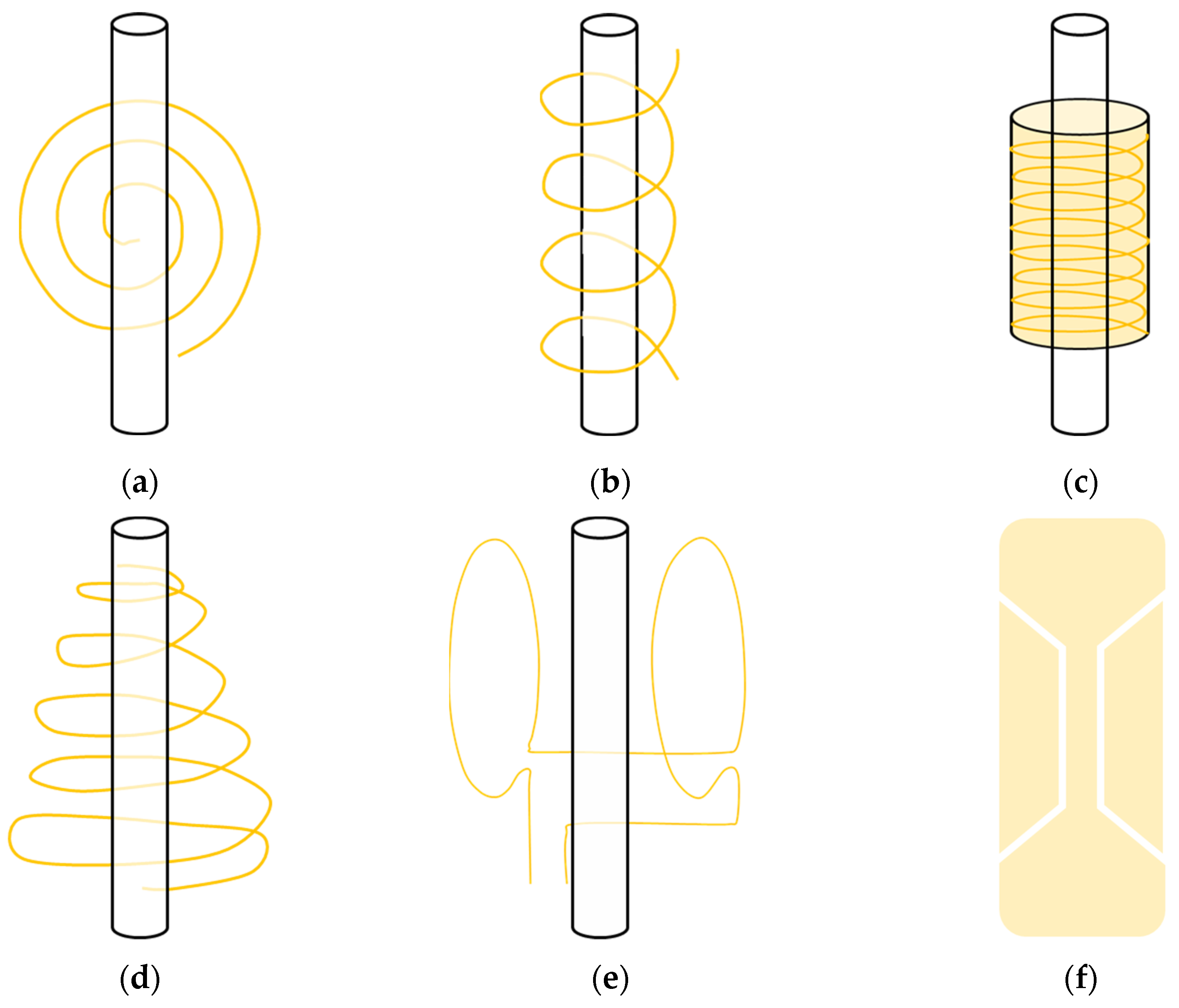
3.2.2. Solenoid Microcoils

Solenoid microcoils, as illustrated in Figure 3b, are three-dimensional helical coils that resemble traditional NMR coils but are miniaturized for µ-NMR applications [44]. These microcoils are typically fabricated through a combination of advanced microfabrication techniques such as wire bonding [45], laser micromachining [46], and electroplating [45,47]. They are known for their high magnetic field homogeneity and sensitivity, which make them ideal for high-resolution µ-NMR experiments [48]. When compared to planar microcoils, solenoid microcoils have the disadvantages of being expensive, not easily suitable for mass production, and more difficult to integrate with microfluidic systems [1]. Nevertheless, the helical design allows for a more uniform magnetic field compared to planar coils, significantly enhancing the quality of the NMR signal.

3.2.3. Scroll Microcoils

Scroll microcoils have emerged as a promising design for the NMR spectroscopy of nanoliter and sub-nanoliter samples [49]. Their microfabrication process includes wrapping a thin bilayer of copper or insulator foil around a wire, creating a scroll structure [49]. These coils are characterized by their cylindrical design, where the conductor is wound in a spiral pattern resembling a scroll (Figure 3c). This design is particularly beneficial for high-resolution NMR spectroscopy at very small coil dimensions, where conventional fabrication methods may be limited [50]. Scroll microcoils can generate strong and uniform magnetic fields within a small volume, making them suitable for specialized NMR experiments and compact analytical devices [51]. These coils offer advantages over traditional microsolenoid coils, including easier fabrication and improved B 1 homogeneity while maintaining comparable SNRs [51]. However, their cylindrical shape may pose integration challenges with planar microfluidic systems, limiting their versatility in certain applications.

3.2.4. Cone-Shaped Microcoils

Cone-shaped microcoils, represented in Figure 3d, feature a conical design, where the conductor is wound in a helical pattern along the surface of a cone [52]. This configuration allows for a focused magnetic field, which can enhance the sensitivity and resolution of samples placed within the cone’s narrow end. This way, one of the main advantages of cone-shaped microcoils is their ability to concentrate the magnetic field in a small region, making them highly effective for targeted NMR measurements and experiments requiring high spatial resolution [49,52]. However, the complex geometry of cone-shaped microcoils poses significant fabrication challenges, leading to higher production costs and reduced scalability compared to simpler coil designs.

3.2.5. Helmholtz Microcoils

Helmholtz microcoils are a type of electromagnetic coil configuration. They have two parallel, coaxial coils that generate a uniform magnetic field between them [53], as illustrated in Figure 3e. These coils are usually made by microfabrication methods involving photolithography and electroplating, to precisely control the coils’ dimensions and alignment. The primary advantage of Helmholtz microcoils is their capacity to create a uniform magnetic field in well-defined spatial regions, which are essential for applications requiring consistent field strength [42,53]. This uniformity enhances the accuracy and reproducibility of measurements compared to other coil configurations. However, Helmholtz microcoils are more complex to fabricate and integrate compared to simpler coil designs, and their bulkier size may limit their application in environments where space is constrained [42].

3.2.6. Stripline Microcoils

The stripline is a relatively new type of microcoil designed to address the challenges associated with planar spiral microcoils, such as inhomogeneous B 1 field distribution, as well as the difficulties of integrating solenoid microcoils into microfluidics chips [54]. These coils are often designed as a double-tuned H/X and fabricated using lithography processes and consist of a thin metal strip that is sandwiched between two ground planes (Figure 3f) [54]. This design leads to parallel field lines to the plane surface and thus results in a homogeneous B 1 field [55,56]. Stripline microcoils can easily be designed for a wide range of sample volumes [57,58], offer high sensitivity, good B 1 field homogeneity, and cost-effective construction for imaging small samples [55]. This type of microcoil requires a careful arrangement of the microfluidic device to match the sample with the detection zone, which can be a limitation [54].

3.3. NMR Chip: Transceiver Electronics

The NMR chip, also known as a transceiver electronic, is crucial for the operation of a µ-NMR system, being involved in the effective generation and detection of NMR signals [2,17]. In fact, the term “transceiver” is a portmanteau of “transmitter” and “receiver”, indicating that these electronics handle both the transmission and reception of RF signals.
The NMR chip (Figure 4) mainly comprises three modules: a pulse sequencer; an RF transmitter (TX) chain; and a low-noise receiver (RX) chain [59]. The pulse sequencer receives commands from the outside to generate the excitation pulse sequence according to the NMR experiment [2,17]. During excitation mode, the TX produces RF pulses, which are sent to the NMR microcoils to generate the B 1 field and excite the nuclear spins. The RX then captures the signal produced by the precession of these nuclear spins. The microcoil detects this small NMR signal, which is amplified to maintain SNR using a low-noise amplifier [2,17]. Next, a mixer, driven by a local oscillation signal, down-converts the signals to the audio frequency range. These down-converted signals are then filtered through low-pass or band-pass filters to remove out-of-band noise and high-frequency mixing artifacts. Finally, ADCs convert the filtered signals into digital format for storage and data processing.
These transceiver electronics chips are characterized by low power consumption, small die areas, and the potential for integration with on-chip microcoils and digital microfluidic devices. This integration facilitates electronic-automated sample management and measurement using portable magnets, highlighting their potential for diverse chemical and biological applications [59,60].

4. Point-Of-Care Applications Based on μ-NMR Systems

PoC devices have gradually become an area of major interest. Due to their ability to probe biomolecular information (proteins, enzymes, etc.) in a non-destructive and highly efficient detection manner, µ-NMR has emerged as a powerful tool for biomedical applications, offering rapid and sensitive analysis of biological samples, showing great potential for PoC applications [1]. These compact µ-NMR systems can be integrated with microfluidics chips, providing researchers with the convenience of using NMR to detect various biomolecules, pathogens, and cells [54]. This type of system is characterized by its small size, low cost, and minimal sample requirements, needing only a few microliters with minimal preparation steps.
One approach to creating μ-NMR systems involves the development of microcoils [61,62] and their integration with commercially high-field NMR spectrometers.
Grimes et al. [63] evaluated the microcoil NMR technique for metabolomics by comparing a 10 µL microcoil NMR probe with a standard 5 mm NMR probe [63]. The focus was on analyzing commonly used biofluids, including serum and urine. The results demonstrated that the microcoil probe offers a significant sensitivity advantage over the standard probe [63].
A study published by Garcia et al. [64] demonstrated the detection of early-stage ovarian cancer using a microflow NMR probe in combination with a high-resolution NMR spectrometer. This study analyzed 20 μL serum samples from patients, highlighting the potential of this method for early cancer detection [64]. Furthermore, Ryan et al. [65] reported the development of an NMR spectrometer on a chip, which integrated planar microcoils microfabricated using lithography techniques with a microfluidic device [65]. This system allowed for easy integration with NMR spectroscopy by simply attaching the planar microcoil to the surface of the microfluidic device. The design enabled the microfluidic chip to be easily inserted into an NMR probe, facilitating high-resolution NMR experiments [65]. Finch et al. [66] reported the development of a double-stripline NMR probe that can be integrated with a microfluidic device, enabling sample analysis with just 2 µL by combining the chip with an NMR spectrometer [66]. Anders et al. [67] presented a single-chip array of integrated NMR receivers for parallel spectroscopy and imaging. This array included a total of eight separated channels, with each one composed of a detection square planar microcoil, a tuning capacitor, and a low-noise amplifier [67]. Apart from these studies, other systems with simple planar microfluidic devices that can be inserted and removed from the µ-NMR probe have been reported [68,69,70,71].
Alternatively, a miniaturized NMR system can be developed. Huan et al. [72] created a quantitative PoC µ-NMR system with potential for clinical cancer diagnosis, by analyzing cells acquired through fine needle aspirations from suspected lesions [72]. The µ-NMR system was comprised of a solenoid microcoil, a portable NdFeB stray-field permanent magnet, and custom-built NMR hardware [72]. Years later, Gee et al. [73] employed µ-NMR technology to assess metastasis in melanoma patients using either fine needle aspirations or peripheral blood samples [73].
Lee et al. [74] introduced a novel, compact NMR system designed for multiplexed, quantitative, and rapid analysis, with the capability to perform eight simultaneous measurements using 5 to 10 µL sample volumes [74]. The system included four essential components: a µ-NMR chip containing a 2 × 4 planar microcoil array fabricated in a glass substrate using photolithography and electroplating; a microfluidic network for sample handling; on-board NMR electronics; and a small NdFeB stray-field permanent magnet [74]. A second prototype was reported by Lee and colleagues [75], featuring a diagnostic magnetic resonance sensor that combined a miniaturized NMR probe with targeted magnetic nanoparticles (MNPs) for the detection and molecular profiling of cancer cells [75]. While the portable permanent magnet remained the same between prototypes, the type of microcoils used changed. The first prototype [74] used planar microcoils, whereas the second employed solenoidal coils, which produced more homogeneous RF magnetic fields than the planar coils [75]. Additionally, the solenoid coil of the latter prototype was embedded in a microfluidic structure, microfabricated through molding techniques [75]. This system allowed for the detection of two cancer cell lines in a 1 µL sample volume of unprocessed fine needle aspirates of tumors and profiled the expression of several cellular markers in less than 15 min [75]. Despite the successful demonstration of μ-NMR prototypes, integrating the platform into routine clinical practice remained challenging [76]. To address this issue, Castro et al. [76] reported a third prototype, with the main difference being the assembly of electronic components using off-the-shelf integrated circuit parts and the capability to interface with a mobile terminal for system control [76].
Sun et al. [77] started by developing a 2 kg portable NMR system composed of a portable permanent magnet, a high-performance complementary metal-oxide semiconductor (CMOS) RF transceiver, and a separate high-quality planar microcoil [77,78]. In continuation of the work, these authors reported two new miniaturized NMR systems that exhibited a substantial reduction in size by several orders of magnitude, demonstrating a significant leap toward lab-on-a-chip functionality [79]. The transceiver architecture was essentially the same for both systems, with the primary differences being the size of the magnet and the type of microcoil used [79]. The first µ-NMR system, which they named the 0.1 kg palm NMR system, included a tiny permanent magnet the size of a ping-pong ball, and an off-chip solenoidal coil. A sample volume of 2 µL was placed inside the solenoidal coil and exposed to a static magnetic field of 0.56 T. The second system, called the one-chip NMR system, used the same magnet as a 2 kg NMR system but featured an on-chip planar microcoil, integrated as a planar spiral on the CMOS chip. The entire chip was protected by a package, except for the area above the microcoil, which could hold a 5 µL sample. Both of these systems demonstrated the capacity to identify biological entities such as avidin, human chorionic gonadotropin, and human bladder cancer cells [79].
Dreyer et al. [80] reported the development of a small, lightweight, easy-to-use, and portable chip-based NMR relaxometry system. This system utilized a custom-designed NMR-on-a-chip transceiver combined with a solenoid coil and a portable permanent H-shaped magnet 0.29 T, made of NdFeB (magnetic blocks) and iron (remaining components) [80]. The work published by Lei et al. [81] presented a µ-NMR system, composed of a permanent magnet and an on-chip planar coil, that was compatible with multi-type biological or chemical lab-on-a-chip assays [81]. Further, in a later study, Lei et al. [82] developed another µ-NMR system but now using a Halbach magnet and a solenoidal microcoil [82].
As a result of the research conducted by Ha et al. [83], a µ-NMR system was reported. They employed a solenoidal coil surrounding a capillary sample tube with an effective sample volume of 0.8 µL. The system featured a compact 0.51 T NdFeB permanent Halbach magnet and a 4 mm2 silicon chip containing an integrated spectrometer electronic chip, which included an RF receiver, an RF transmitter, and an arbitrary pulse sequencer [83]. Table 3 provides a summary of the aforementioned µ-NMR systems, detailing the type of magnet and microcoil used, the sample volume, and specific applications.
These strides made in µ-NMR point-of-care diagnosis systems have greatly expanded possibilities for rapid, point-of-care diagnostics and personalized healthcare. Furthermore, the incorporation of μ-NMR technology into clinical settings promises to deliver timely and accurate diagnosis. This improvement would not only make medical intervention more efficient and effective but would also set the stage for individualized care plans. The future may see μ-NMR systems becoming even more useful in point-of-care settings as this area continues to evolve, bringing advanced diagnostic tools for diverse healthcare environments and thus enhancing patient outcomes.

5. Challenges and Limitations

μ-NMR systems, while offering significant advantages in portability and the ability to analyze small sample volumes, face critical challenges and limitations, particularly in terms of sensitivity and resolution, being these two parameters critical in NMR. These limitations can significantly impact the accuracy and reliability of μ-NMR systems in complex biomedical and analytical applications, necessitating ongoing advancements in design and technology to overcome these obstacles. Furthermore, the integration of miniaturized NMR systems with other technologies can be challenging, requiring, for example, advanced and reproducible microfabrication techniques, precise sample handling, and effective shielding to avoid electromagnetic interferences.

5.1. Sensitivity and SNR

Sensitivity is a crucial factor in μ-NMR systems because it directly affects the system’s ability to detect and quantify low-concentration biomolecules. In biomedical applications, many important analytes, such as metabolites, proteins, or nucleic acids, are often present in very small quantities [84]. If the sensitivity of the μ-NMR system is insufficient, these low-concentration biomolecules may produce signals that are too weak to be accurately detected or distinguished from background noise. This limitation can lead to false negatives, reduced diagnostic accuracy, and a failure to capture critical biochemical information. Therefore, optimizing sensitivity is crucial to fully realizing the potential of μ-NMR systems in detecting and analyzing biomolecules at low concentrations.
One approach to increasing the sensitivity of a µ-NMR system is through microcoil design optimization. A key parameter in microcoil design is the quality factor (Q-factor), which indicates the resonator’s efficiency. High Q-factors are desirable for µ-NMR applications because they increase the coil’s ability to store energy, improving the sensitivity of the NMR signal [85,86]. Peck et al. [87] developed a solenoidal copper microcoil with an inner radius of 37.5 µm and five turns. For a sample volume of 0.3 nL at a Larmor frequency of 200 MHz, they achieved an SNR of 13.6 and a Q-factor of 36.3. Similarly, a solenoidal microcoil fabricated by Badilita et al. [88], designed for a sample volume of 0.31 µL, exhibited a Q-factor of 46 at 400 MHz and a mean SNR of 520 per voxel. Goloshevsky et al. [89] demonstrated a copper Helmholtz coil on a glass substrate, achieving a Q-factor of 6.6 at a Larmor frequency of 25 MHz with a sample volume of 2.45 µL. For planar microcoils, Renaud et al. [90] developed a rectangular planar copper microcoil with four turns, reporting a Q-factor of 10 at 85.13 MHz with a sample volume of 330 µL. Massin et al. [91] achieved a Q-factor of 24 at 300 MHz using a circular copper microcoil. Jiang et al. [85] described a process for fabricating high-Q-factor microcoils, producing a planar spiral microcoil with an inner diameter of 130 µm with three windings and an outer diameter of 240 µm. This coil achieved a Q-factor of 85 at a frequency of 1.6 GHz.
Additionally, the design of the microcoil itself and the placement of the sample relative to the coil are critical factors that influence system sensitivity. Optimal placement can concentrate the magnetic field, increasing the interaction between the sample and the field, which boosts sensitivity [92].
Beyond microcoil design optimization, advanced signal processing techniques, such as noise reduction algorithms, can be applied to isolate the NMR signal from background noise, thereby improving the system’s SNR [93,94,95]. Furthermore, hyperpolarization techniques combined with microfluidic systems can significantly enhance the sensitivity of NMR spectroscopy [16]. For instance, parahydrogen-induced hyperpolarization (PHIP) integrated into microfluidic chips has achieved picomole sensitivity for micromolar concentrations, enabling high-resolution NMR with unprecedented detection limits [96].

5.2. Resolution and Spectral Overlap

Resolution in NMR refers to the system’s ability to distinguish between different resonance frequencies in the NMR spectrum, which correspond to distinct chemical environments of the nuclei within the sample. High resolution allows for the clear separation of peaks in the NMR spectrum.
This is closely related to the homogeneity of the applied magnetic field [16]. In a magnetic field with reduced homogeneity, nuclear spins are subjected to slightly varying magnetic fields depending on their location within the sample [16]. These variations alter their Larmor frequencies, which in turn affect the observed chemical shifts. The main consequence is a broadening of the spectral peaks, which impacts both the width and intensity of these peaks, making it challenging to distinguish signals that are close together [16].
The miniaturization of NMR systems, while beneficial in many respects, presents a reduced magnetic field homogeneity, posing significant challenges to spectral resolution [2]. These factors can lead to broader peaks and increased spectral overlap, reducing the system’s effectiveness.
Some researchers have reported the spectral resolution achieved in their works, with these values varying depending on system design and conditions. For example, Ryan et al. [68] reported a spectral resolution of 3.5 Hz, while Finch et al. [66] and Lei et al. [82] achieved resolutions of 1.75 Hz and 3 Hz, respectively. Spengler et al. [97], using a Helmholtz microcoil with an active sample volume of 100 nL, reported a high spectral resolution of 0.62 Hz.
Recent advancements have focused on improving resolution, a critical challenge in the field. These include wavelet transform methods for resolving overlapping spectra [98], the intersection of non-redundant information on resonance groups (INIR) for reconstructing high-field spectra from lower-field data [99], and chemical shift upscaling to improve signal dispersion while preserving scalar couplings [100].

5.3. Integration with Other Technologies

The integration of µ-NMR systems with other technologies holds immense potential for advancements in various fields, especially in point-of-care diagnostics, environmental monitoring, and chemical analysis. However, several challenges must be addressed to realize this potential fully.
Integrating µ-NMR with microfluidics or lab-on-a-chip devices requires precise alignment and coordination between components that operate on different principles and scales [45]. The fabrication of microfluidic channels, for example, must be compatible with the dimensions and electromagnetic requirements of the µ-NMR coils. Achieving this level of precision in a compact system is technically challenging and requires advanced microfabrication techniques.
Maintaining consistent temperature and environmental conditions is critical for both µ-NMR and microfluidic systems, as fluctuations can affect the chemical shifts and relaxation times in NMR [101,102], as well as the flow and reaction rates in microfluidics [103]. One possible solution is to regulate the magnet-cooling water temperature and minimize fluctuations in room air temperature [104]. Additionally, identifying and suppressing sources of temperature sensitivity in these devices can lead to significant improvements in the signal-to-artifact ratio [105]. Moreover, the “T-lock” concept has been introduced to automatically compensate for RF-induced sample heating by continuously measuring the resonance frequency and adjusting the temperature control accordingly [106].
Furthermore, to combine µ-NMR with other technologies, some challenges need to be overcome. First, electromagnetic interference may occur, which can degrade the quality of the NMR signal, through the presence of noise or artifacts in the data, making it difficult to extract accurate and reliable information [107]. To overcome this limitation, although they increase the complexity of the system, effective shielding [108] and careful design [109] can be considered. Then, for applications that require real-time monitoring, such as PoC diagnostics or real-time metabolic analysis, the integrated system must process and analyze data quickly and accurately. Ensuring that the system can handle real-time data flow without lag while maintaining accuracy and sensitivity is technically demanding. As a solution, deep learning techniques can be used to improve data acquisition and analysis, increasing SNR ratios and speeding up nanoscale NMR spectroscopy [110]. Moreover, artificial intelligence-based tools have streamlined experimental processes, enabling novel NMR experiments and expanding the scope of biomolecular NMR [111]. Machine learning applications in NMR include peak-picking, compound identification, and structure determination, addressing challenges posed by large data sets [112].
Systems developed in response to these limitations will be more and more complete, becoming more sensitive, diagnostically accurate, versatile, fast, portable, and user-friendly. These benefits can significantly enhance patient care, especially in settings where rapid, reliable diagnostics are essential. The development of such advanced PoC devices can transform how and where healthcare is delivered, making it more accessible and responsive to patient needs.

6. Future Perspectives and Conclusions

μ-NMR technology represents a significant advancement in the field of nuclear magnetic resonance, as it provides miniaturized systems that enhance portability and reduce sample size requirements. It has a wide range of uses, especially in biomedical research where it can provide detailed molecular information with minimal invasiveness. The possibility of quickly and accurately examining patient-specific biomarkers using μ-NMR will open up avenues for practicing personalized medicine. This aspect enables more accurate diagnoses, monitoring, and treatment that would suit the individual’s molecular profile. In healthcare, µ-NMR could facilitate PoC testing, providing real-time diagnostic information that could lead to timely and effective clinical interventions.
To achieve miniaturization and perform reliable NMR sensing at a microscale, high-performance microcoils, compact permanent magnets, and advanced transceiver electronics need to be developed and integrated. The performance of μ-NMR systems in terms of sensitivity, Q-factor, and spectral resolution remains an area of active research, as these parameters are highly influenced by factors such as coil geometry, operating frequency, and experimental setup. While significant progress has been made, further advancements are needed to enhance resolution and sensitivity, which are crucial for accurately detecting low-abundance biomolecules in minimal sample volumes.
Additionally, integrating µ-NMR with other microfluidic and lab-on-a-chip technologies could lead to more versatile and multifunctional analytical platforms, capable of performing a variety of biomedical assays in a single and compact device. As such, future research should focus on developing cost-effective and scalable fabrication techniques for µ-NMR components so as to enable the wider adoption of the technology in both research and clinical settings.

Author Contributions

Conceptualization and methodology, B.S.-A. and H.A.F.; investigation, formal analysis, and writing—original draft preparation, B.S.-A.; writing—review and editing, B.S.-A. and H.A.F.; supervision, project administration, and funding acquisition, H.A.F. All authors have read and agreed to the published version of the manuscript.

Funding

This work was partially funded by Fundação para a Ciência e Tecnologia (FCT) under the project UIDB/00645/2020 (https://doi.org/10.54499/UIDB/00645/2020, accessed on 20 October 2024) and grant 22023.00474.BDANA, and also by Exotictarget Lda.

Institutional Review Board Statement

Not applicable.

Informed Consent Statement

Not applicable.

Data Availability Statement

Not applicable.

Conflicts of Interest

B.S.-A. and H.A.F., respectively, are a consultant for and a co-founder of Exotictarget Lda, a point-of-care urinalysis device company. The funders had no role in the design of the study; in the collection, analyses, or interpretation of data; in the writing of the manuscript; or in the decision to publish the results.

References

  1. Guo, J.; Jiang, D.; Feng, S.; Ren, C.; Guo, J. µ-NMR at the point of care testing. Electrophoresis 2020, 41, 319–327. [Google Scholar] [CrossRef] [PubMed]
  2. Dupré, A.; Lei, K.-M.; Mak, P.-I.; Martins, R.P.; Peng, W.K. Micro-and nanofabrication NMR technologies for point-of-care medical applications—A review. Microelectron. Eng. 2019, 209, 66–74. [Google Scholar] [CrossRef]
  3. Raman, S.; Lange, O.F.; Rossi, P.; Tyka, M.; Wang, X.; Aramini, J.; Liu, G.; Ramelot, T.A.; Eletsky, A.; Szyperski, T. NMR structure determination for larger proteins using backbone-only data. Science 2010, 327, 1014–1018. [Google Scholar] [CrossRef] [PubMed]
  4. Novoa-Carballal, R.; Fernandez-Megia, E.; Jimenez, C.; Riguera, R. NMR methods for unravelling the spectra of complex mixtures. Nat. Prod. Rep. 2011, 28, 78–98. [Google Scholar] [CrossRef]
  5. Casarini, D.; Lunazzi, L.; Mazzanti, A. Recent advances in stereodynamics and conformational analysis by dynamic NMR and theoretical calculations. Eur. J. Org. Chem. 2010, 2010, 2035–2056. [Google Scholar] [CrossRef]
  6. Robinette, S.L.; Brüschweiler, R.; Schroeder, F.C.; Edison, A.S. NMR in metabolomics and natural products research: Two sides of the same coin. Acc. Chem. Res. 2012, 45, 288–297. [Google Scholar] [CrossRef]
  7. Gowda, G.N.; Raftery, D. Advances in NMR-based metabolomics. Compr. Anal. Chem. 2014, 63, 187–211. [Google Scholar]
  8. Price, K.E.; Vandaveer, S.S.; Lunte, C.E.; Larive, C.K. Tissue targeted metabonomics: Metabolic profiling by microdialysis sampling and microcoil NMR. J. Pharm. Biomed. Anal. 2005, 38, 904–909. [Google Scholar] [CrossRef]
  9. Felli, I.C.; Pierattelli, R. Recent progress in NMR spectroscopy: Toward the study of intrinsically disordered proteins of increasing size and complexity. IUBMB Life 2012, 64, 473–481. [Google Scholar] [CrossRef]
  10. Markwick, P.R.; Malliavin, T.; Nilges, M. Structural biology by NMR: Structure, dynamics, and interactions. PLoS Comput. Biol. 2008, 4, e1000168. [Google Scholar] [CrossRef]
  11. Zalesskiy, S.S.; Danieli, E.; Blumich, B.; Ananikov, V.P. Miniaturization of NMR systems: Desktop spectrometers, microcoil spectroscopy, and “NMR on a chip” for chemistry, biochemistry, and industry. Chem. Rev. 2014, 114, 5641–5694. [Google Scholar] [CrossRef] [PubMed]
  12. Shapiro, E.M.; Skrtic, S.; Sharer, K.; Hill, J.M.; Dunbar, C.E.; Koretsky, A.P. MRI detection of single particles for cellular imaging. Proc. Natl. Acad. Sci. USA 2004, 101, 10901–10906. [Google Scholar] [CrossRef] [PubMed]
  13. Sun, N.; Liu, Y.; Qin, L.; Lee, H.; Weissleder, R.; Ham, D. Small NMR biomolecular sensors. Solid-State Electron. 2013, 84, 13–21. [Google Scholar] [CrossRef]
  14. Tilgner, M.; Vater, T.S.; Habbel, P.; Cheng, L.L. High-resolution magic angle spinning (HRMAS) NMR methods in metabolomics. In NMR-Based Metabolomics Methods Protocols; Humana: New York, NY, USA, 2019; pp. 49–67. [Google Scholar]
  15. Polishchuk, D.; Gardeniers, H. A compact permanent magnet for microflow NMR relaxometry. J. Magn. Reson. 2023, 347, 107364. [Google Scholar] [CrossRef]
  16. Plata, M. Protein NMR on a Chip: Development of an Integrated Microfluidic Platform for Studying Protein-Ligand Interactions by Nuclear Magnetic Resonance. Ph.D. Thesis, University of Southampton, Southampton, UK, 2023. [Google Scholar]
  17. Fan, S.; Zhou, Q.; Lei, K.-M.; Mak, P.-I.; Martins, R.P. Miniaturization of a nuclear magnetic resonance system: Architecture and design considerations of transceiver integrated circuits. IEEE Trans. Circuits Syst. I Regul. Pap. 2022, 69, 3049–3060. [Google Scholar] [CrossRef]
  18. Andrew, E.; Szczesniak, E. A historical account of NMR in the solid state. Prog. Nucl. Magn. Reson. Spectrosc. 1995, 28, 11–36. [Google Scholar] [CrossRef]
  19. Mlynárik, V. Introduction to nuclear magnetic resonance. Anal. Biochem. 2017, 529, 4–9. [Google Scholar] [CrossRef]
  20. Storey, P. Introduction to magnetic resonance imaging and spectroscopy. Magn. Reson. Imaging Methods Biol. Appl. 2006, 124, 3–57. [Google Scholar]
  21. Edwards, J.C. Principles of NMR; Process NMR Associates LLC: Danbury, CT, USA, 2009. [Google Scholar]
  22. Bank, S. Some principles of NMR spectroscopy and their novel application. Concepts Magn. Reson. Educ. J. 1997, 9, 83–93. [Google Scholar] [CrossRef]
  23. Danieli, E.; Perlo, J.; Blümich, B.; Casanova, F. Small magnets for portable NMR spectrometers. Angew. Chem. Int. Ed. 2010, 49, 4133–4135. [Google Scholar] [CrossRef]
  24. Moresi, G.; Magin, R. Miniature permanent magnet for table-top NMR. Concepts Magn. Reson. Part B Magn. Reson. Eng. Educ. J. 2003, 19, 35–43. [Google Scholar] [CrossRef]
  25. Blümich, B.; Rehorn, C.; Zia, W. Magnets for Small-Scale and Portable NMR; Wiley-VCH: Weinheim, Germany, 2018. [Google Scholar]
  26. Blümich, B.; Haber-Pohlmeier, S.; Zia, W. Compact NMR; Walter de Gruyter: Berlin, Germany, 2014. [Google Scholar]
  27. Trout, S. Material selection of permanent magnets, considering thermal properties correctly. In Proceedings of the Electrical Insulation Conference and Electrical Manufacturing and Coil Winding Conference (Cat. No. 01CH37264), Cincinnati, OH, USA, 28 October 2001; pp. 365–370. [Google Scholar]
  28. Kassen, A.G.; White, E.M.; Hu, L.; Tang, W.; Zhou, L.; Kramer, M.J.; Anderson, I.E. Novel mechanisms for solid-state processing and grain growth with microstructure alignment in alnico-8 based permanent magnets. AIP Adv. 2018, 8, 056206. [Google Scholar] [CrossRef]
  29. Crozier-Bioud, T.; Momeni, V.; Gonzalez-Gutierrez, J.; Kukla, C.; Luca, S.; Rolere, S. Current challenges in NdFeB permanent magnets manufacturing by Powder Injection Molding (PIM): A review. Mater. Today Phys. 2023, 34, 101082. [Google Scholar] [CrossRef]
  30. Cui, J.; Kramer, M.; Zhou, L.; Liu, F.; Gabay, A.; Hadjipanayis, G.; Balasubramanian, B.; Sellmyer, D. Current progress and future challenges in rare-earth-free permanent magnets. Acta Mater. 2018, 158, 118–137. [Google Scholar] [CrossRef]
  31. Calin, M.-D.; Helerea, E. Temperature influence on magnetic characteristics of NdFeB permanent magnets. In Proceedings of the 2011 7th International Symposium on Advanced Topics in Electrical Engineering (ATEE), Bucharest, Romania, 12–14 May 2011; pp. 1–6. [Google Scholar]
  32. Yan, M.; Jin, J.; Ma, T. Grain boundary restructuring and La/Ce/Y application in Nd–Fe–B magnets. Chin. Phys. B 2019, 28, 077507. [Google Scholar] [CrossRef]
  33. Cui, B.; Liu, X.; Nlebedim, I.C.; Cui, J. Mechanically robust high magnetic-performance Sm-Co sintered magnets through microstructure engineering. J. Alloys Compd. 2022, 926, 166869. [Google Scholar] [CrossRef]
  34. Liu, S. Sm–Co high-temperature permanent magnet materials. Chin. Phys. B 2019, 28, 017501. [Google Scholar] [CrossRef]
  35. Minard, K.R.; Wind, R.A. Solenoidal microcoil design—Part II: Optimizing winding parameters for maximum signal-to-noise performance. Concepts Magn. Reson. 2001, 13, 190–210. [Google Scholar] [CrossRef]
  36. Hoult, D.I.; Richards, R. The signal-to-noise ratio of the nuclear magnetic resonance experiment. J. Magn. Reson. 1969 1976, 24, 71–85. [Google Scholar] [CrossRef]
  37. Wu, N.; Peck, T.L.; Webb, A.G.; Magin, R.L.; Sweedler, J.V. 1H-NMR spectroscopy on the nanoliter scale for static and online measurements. Anal. Chem. 1994, 66, 3849–3857. [Google Scholar] [CrossRef]
  38. Van Bentum, P.; Janssen, J.; Kentgens, A. Towards nuclear magnetic resonance μ-spectroscopy and μ-imaging. Analyst 2004, 129, 793–803. [Google Scholar] [CrossRef] [PubMed]
  39. Badilita, V.; Meier, R.C.; Spengler, N.; Wallrabe, U.; Utz, M.; Korvink, J.G. Microscale nuclear magnetic resonance: A tool for soft matter research. Soft Matter 2012, 8, 10583–10597. [Google Scholar] [CrossRef]
  40. Krishnapriya, S.; Komaragiri, R.S.; Suja, K. Performance Evaluation of Via-free Non-spiral Planar Microcoils. In Proceedings of the 2020 IEEE 15th International Conference on Nano/Micro Engineered and Molecular System (NEMS), San Diego, CA, USA, 30 September 2020; pp. 247–250. [Google Scholar]
  41. Krishnapriya, S.; Chandrakar, H.; Komaragiri, R.S.; Suja, K. Performance analysis of planar microcoils for biomedical wireless power transfer links. Sādhanā 2019, 44, 187. [Google Scholar] [CrossRef]
  42. Bastawrous, M.; Gruschke, O.; Soong, R.; Jenne, A.; Gross, D.; Busse, F.; Nashman, B.; Lacerda, A.; Simpson, A.J. Comparing the potential of Helmholtz and planar NMR microcoils for analysis of intact biological samples. Anal. Chem. 2022, 94, 8523–8532. [Google Scholar] [CrossRef]
  43. Eroglu, S.; Gimi, B.; Roman, B.; Friedman, G.; Magin, R.L. NMR spiral surface microcoils: Design, fabrication, and imaging. Concepts Magn. Reson. Part B Magn. Reson.Eng. Educ. J. 2003, 17, 1–10. [Google Scholar] [CrossRef]
  44. Fratila, R.M.; Velders, A.H. Small-volume nuclear magnetic resonance spectroscopy. Annu. Rev. Anal. Chem. 2011, 4, 227–249. [Google Scholar] [CrossRef]
  45. Meier, R.C.; Höfflin, J.; Badilita, V.; Wallrabe, U.; Korvink, J.G. Microfluidic integration of wirebonded microcoils for on-chip applications in nuclear magnetic resonance. J. Micromech. Microeng. 2014, 24, 045021. [Google Scholar] [CrossRef]
  46. Jolic, K.; Ghantasala, M.; Hayes, J.; Jin, H. Fabrication of three-dimensional inductor coil using excimer laser micromachining. J. Micromech. Microeng. 2003, 13, 782. [Google Scholar] [CrossRef]
  47. Ehrmann, K.; Saillen, N.; Vincent, F.; Stettler, M.; Jordan, M.; Wurm, F.M.; Besse, P.-A.; Popovic, R. Microfabricated solenoids and Helmholtz coils for NMR spectroscopy of mammalian cells. Lab Chip 2007, 7, 373–380. [Google Scholar] [CrossRef]
  48. Kentgens, A.; Bart, J.; Van Bentum, P.; Brinkmann, A.; Van Eck, E.; Gardeniers, J.G.; Janssen, J.; Knijn, P.; Vasa, S.; Verkuijlen, M. High-resolution liquid-and solid-state nuclear magnetic resonance of nanoliter sample volumes using microcoil detectors. J. Chem. Phys. 2008, 128, 052202. [Google Scholar] [CrossRef]
  49. Barne, A.; Malhari, S.; Lakhani, T.; Mehendale, N. A Review on Types of NMR Micro-Coil Designs. SSRN 2022. [Google Scholar] [CrossRef]
  50. Grant, S.; Murphy, L.; Magin, R.; Friedman, G. Analysis of multilayer radio frequency microcoils for nuclear magnetic resonance spectroscopy. IEEE Trans. Magn. 2001, 37, 2989–2998. [Google Scholar] [CrossRef]
  51. Khelifa, M.; Mounier, D.; Yaakoubi, N. Design of high performance scroll microcoils for nuclear magnetic resonance spectroscopy of nanoliter and subnanoliter samples. Sensors 2020, 21, 170. [Google Scholar] [CrossRef]
  52. Inamura, T.; Dohi, T. Cone-shaped micro coil for magnetic resonance imaging. In Proceedings of the 2013 IEEE 26th International Conference on Micro Electro Mechanical Systems (MEMS), Taipei, Taiwan, 24 January 2013; pp. 335–338. [Google Scholar]
  53. Saqib, M.; Francis, S.; Francis, J. Design and development of Helmholtz coils for magnetic field. In Proceedings of the 2020 International Youth Conference on Radio Electronics, Electrical and Power Engineering (REEPE), Moscow, Russia, 12–14 March 2020; pp. 1–5. [Google Scholar]
  54. Li, Z.; Bao, Q.; Liu, C.; Li, Y.; Yang, Y.; Liu, M. Recent advances in microfluidics-based bioNMR analysis. Lab Chip 2023, 23, 1213–1225. [Google Scholar] [CrossRef]
  55. Jasiński, K.; Młynarczyk, A.; Latta, P.; Volotovskyy, V.; Węglarz, W.P.; Tomanek, B. A volume microstrip RF coil for MRI microscopy. Magn. Reson. Imaging 2012, 30, 70–77. [Google Scholar] [CrossRef]
  56. Jasinski, K.; Mlynarczyk, A.; Weglarz, W.; Latta, P.; Volotovskyy, V.; Tomanek, B. Application of microstrip RF coils to MRI microscopy. In Proceedings of the 37 Polish Seminar on Nuclear Magnetic Resonance and Its Applications, Cracow, Poland, 1–2 December 2010. [Google Scholar]
  57. Bart, J. Stripline-Based Microfluidic Devices for High-Resolution NMR Spectroscopy. Ph.D. Thesis, University of Twente, Enschede, The Netherlands, 2009; pp. 12–20. [Google Scholar]
  58. van Meerten, S.; van Zelst, F.; Tijssen, K.; Kentgens, A. An optimized NMR stripline for sensitive supercritical fluid chromatography-nuclear magnetic resonance of microliter sample volumes. Anal. Chem. 2020, 92, 13010–13016. [Google Scholar] [CrossRef]
  59. Lei, K.-M.; Mak, P.-I.; Law, M.-K.; Martins, R.P. A µ NMR CMOS Transceiver Using a Butterfly-Coil Input for Integration with a Digital Microfluidic Device Inside a Portable Magnet. IEEE J. Solid-State Circuits 2016, 51, 2274–2286. [Google Scholar] [CrossRef]
  60. Grisi, M.; Gualco, G.; Boero, G. A broadband single-chip transceiver for multi-nuclear NMR probes. Rev. Sci. Instrum. 2015, 86, 044703. [Google Scholar] [CrossRef]
  61. Olson, D.L.; Norcross, J.A.; O’Neil-Johnson, M.; Molitor, P.F.; Detlefsen, D.J.; Wilson, A.G.; Peck, T.L. Microflow NMR: Concepts and capabilities. Anal. Chem. 2004, 76, 2966–2974. [Google Scholar] [CrossRef]
  62. Schroeder, F.C.; Gronquist, M. Extending the scope of NMR spectroscopy with microcoil probes. Angew. Chem. Int. Ed. 2006, 45, 7122–7131. [Google Scholar] [CrossRef]
  63. Grimes, J.H.; O’Connell, T.M. The application of micro-coil NMR probe technology to metabolomics of urine and serum. J. Biomol. NMR 2011, 49, 297–305. [Google Scholar] [CrossRef] [PubMed]
  64. Garcia, E.; Andrews, C.; Hua, J.; Kim, H.L.; Sukumaran, D.K.; Szyperski, T.; Odunsi, K. Diagnosis of early stage ovarian cancer by 1H NMR metabonomics of serum explored by use of a microflow NMR probe. J. Proteome Res. 2011, 10, 1765–1771. [Google Scholar] [CrossRef] [PubMed]
  65. Ryan, H.; Song, S.-H.; Zaß, A.; Korvink, J.; Utz, M. Contactless NMR spectroscopy on a chip. Anal. Chem. 2012, 84, 3696–3702. [Google Scholar] [CrossRef] [PubMed]
  66. Finch, G.; Yilmaz, A.; Utz, M. An optimised detector for in-situ high-resolution NMR in microfluidic devices. J. Magn. Reson. 2016, 262, 73–80. [Google Scholar] [CrossRef]
  67. Anders, J.; Chiaramonte, G.; SanGiorgio, P.; Boero, G. A single-chip array of NMR receivers. J. Magn. Reson. 2009, 201, 239–249. [Google Scholar] [CrossRef]
  68. Ryan, H.; Song, S.-H.; Lamson, K.; Elliot, J.; Zaß, A.; Wang, K.; Korvink, J.; Reed, M.; Landers, J.; Utz, M. Metabolomic NMR on a chip via inductive coupling. In Proceedings of the 15th International Conference on Miniaturized Systems for Chemsitry and Life Sciences, Seattle, WA, USA, 1–5 October 2011. [Google Scholar]
  69. Sharma, M.; Utz, M. Modular transmission line probes for microfluidic nuclear magnetic resonance spectroscopy and imaging. J. Magn. Reson. 2019, 303, 75–81. [Google Scholar] [CrossRef]
  70. Spengler, N.; Moazenzadeh, A.; Meier, R.C.; Badilita, V.; Korvink, J.; Wallrabe, U. Micro-fabricated Helmholtz coil featuring disposable microfluidic sample inserts for applications in nuclear magnetic resonance. J. Micromech. Microeng. 2014, 24, 034004. [Google Scholar] [CrossRef]
  71. Wensink, H.; Benito-Lopez, F.; Hermes, D.C.; Verboom, W.; Gardeniers, H.J.; Reinhoudt, D.N.; van den Berg, A. Measuring reaction kinetics in a lab-on-a-chip by microcoil NMR. Lab Chip 2005, 5, 280–284. [Google Scholar] [CrossRef]
  72. Haun, J.B.; Castro, C.M.; Wang, R.; Peterson, V.M.; Marinelli, B.S.; Lee, H.; Weissleder, R. Micro-NMR for rapid molecular analysis of human tumor samples. Sci. Transl. Med. 2011, 3, ra16–ra71. [Google Scholar] [CrossRef]
  73. Gee, M.S.; Ghazani, A.A.; Haq, R.; Wargo, J.A.; Sebas, M.; Sullivan, R.J.; Lee, H.; Weissleder, R. Point of care assessment of melanoma tumor signaling and metastatic burden from μNMR analysis of tumor fine needle aspirates and peripheral blood. Nanomed. Nanotechnol. Biol. Med. 2017, 13, 821–828. [Google Scholar] [CrossRef]
  74. Lee, H.; Sun, E.; Ham, D.; Weissleder, R. Chip–NMR biosensor for detection and molecular analysis of cells. Nat. Med. 2008, 14, 869–874. [Google Scholar] [CrossRef] [PubMed]
  75. Lee, H.; Yoon, T.-J.; Figueiredo, J.-L.; Swirski, F.K.; Weissleder, R. Rapid detection and profiling of cancer cells in fine-needle aspirates. Proc. Natl. Acad. Sci. USA 2009, 106, 12459–12464. [Google Scholar] [CrossRef] [PubMed]
  76. Castro, C.M.; Ghazani, A.A.; Chung, J.; Shao, H.; Issadore, D.; Yoon, T.-J.; Weissleder, R.; Lee, H. Miniaturized nuclear magnetic resonance platform for detection and profiling of circulating tumor cells. Lab Chip 2014, 14, 14–23. [Google Scholar] [CrossRef] [PubMed]
  77. Sun, N.; Liu, Y.; Lee, H.; Weissleder, R.; Ham, D. CMOS RF biosensor utilizing nuclear magnetic resonance. IEEE J. Solid-State Circuits 2009, 44, 1629–1643. [Google Scholar] [CrossRef]
  78. Liu, Y.; Sun, N.; Lee, H.; Weissleder, R.; Ham, D. CMOS mini nuclear magnetic resonance system and its application for biomolecular sensing. In Proceedings of the 2008 IEEE International Solid-State Circuits Conference-Digest of Technical Papers, San Francisco, CA, USA, 7 February 2008; pp. 140–602. [Google Scholar]
  79. Sun, N.; Yoon, T.-J.; Lee, H.; Andress, W.; Weissleder, R.; Ham, D. Palm NMR and 1-chip NMR. IEEE J. Solid-State Circuits 2010, 46, 342–352. [Google Scholar] [CrossRef]
  80. Dreyer, F.; Yang, Q.; Alnajjar, B.; Krüger, D.; Blümich, B.; Anders, J. A portable chip-based NMR relaxometry system with arbitrary phase control for point-of-care blood analysis. IEEE Trans. Biomed. Circuits Syst. 2023, 17, 831–842. [Google Scholar] [CrossRef]
  81. Lei, K.-M.; Heidari, H.; Mak, P.-I.; Law, M.-K.; Maloberti, F.; Martins, R.P. A handheld high-sensitivity micro-NMR CMOS platform with B-field stabilization for multi-type biological/chemical assays. IEEE J. Solid-State Circuits 2016, 52, 284–297. [Google Scholar] [CrossRef]
  82. Lei, K.-M.; Ha, D.; Song, Y.-Q.; Westervelt, R.M.; Martins, R.; Mak, P.-I.; Ham, D. Portable NMR with parallelism. Anal. Chem. 2020, 92, 2112–2120. [Google Scholar] [CrossRef]
  83. Ha, D.; Paulsen, J.; Sun, N.; Song, Y.-Q.; Ham, D. Scalable NMR spectroscopy with semiconductor chips. Proc. Natl. Acad. Sci. USA 2014, 111, 11955–11960. [Google Scholar] [CrossRef]
  84. Finch, G. Optimised Detectors for Integration of NMR with Microfluidic Devices. Ph.D. Thesis, University of Southampton, Southampton, UK, 2017. [Google Scholar]
  85. Jiang, Y.; Ono, T.; Esashi, M. High aspect ratio spiral microcoils fabricated by a silicon lost molding technique. J. Micromech. Microeng. 2006, 16, 1057. [Google Scholar] [CrossRef]
  86. Li, Y.; Ahmad, M.M.; Hand, J.W.; Syms, R.R.; Gilderdale, D.; Collins, D.J.; Young, I.R. Microcoils on structured silicon substrates for magnetic resonance detection. IEEE Sens. J. 2007, 7, 1362–1369. [Google Scholar] [CrossRef]
  87. Peck, T.L.; Magin, R.L.; Lauterbur, P.C. Design and analysis of microcoils for NMR microscopy. J. Magn. Reson. Ser. B 1995, 108, 114–124. [Google Scholar] [CrossRef] [PubMed]
  88. Badilita, V.; Kratt, K.; Baxan, N.; Mohmmadzadeh, M.; Burger, T.; Weber, H.; Elverfeldt, D.v.; Hennig, J.; Korvink, J.G.; Wallrabe, U. On-chip three dimensional microcoils for MRI at the microscale. Lab Chip 2010, 10, 1387–1390. [Google Scholar] [CrossRef] [PubMed]
  89. Goloshevsky, A.; Walton, J.; Shutov, M.; De Ropp, J.; Collins, S.; McCarthy, M. Development of low field nuclear magnetic resonance microcoils. Rev. Sci. Instrum. 2005, 76, 024101. [Google Scholar] [CrossRef]
  90. Renaud, L.; Armenean, M.; Berry, L.; Kleimann, P.; Morin, P.; Pitaval, M.; O’Brien, J.; Brunet, M.; Saint-Jalmes, H. Implantable planar rf microcoils for NMR microspectroscopy. Sens. Actuators A Phys. 2002, 99, 244–248. [Google Scholar] [CrossRef]
  91. Massin, C.; Boero, G.; Vincent, F.; Abenhaim, J.; Besse, P.-A.; Popovic, R. High-Q factor RF planar microcoils for micro-scale NMR spectroscopy. Sens. Actuators A Phys. 2002, 97, 280–288. [Google Scholar] [CrossRef]
  92. Evelhoch, J.L.; Crowley, M.G.; Ackerman, J.J. Signal-to-noise optimization and observed volume localization with circular surface coils. J. Magn. Reson. 1969 1984, 56, 110–124. [Google Scholar] [CrossRef]
  93. Noorbehesht, B.; Lee, H.; Enzmann, D.R. Signal-to-noise ratio enhancement in NMR spectroscopy using a vector-space technique. J. Magn. Reson. 1969 1987, 73, 423–435. [Google Scholar] [CrossRef]
  94. Taylor, H.; Haiges, R.; Kershaw, A. Increasing Sensitivity in Determining Chemical Shifts in One Dimensional Lorentzian NMR Spectra. J. Phys. Chem. A 2013, 117, 3319–3331. [Google Scholar] [CrossRef]
  95. Kolar, P.; Grbić, M.S.; Hrabar, S. Sensitivity enhancement of NMR spectroscopy receiving chain used in condensed matter physics. Sensors 2019, 19, 3064. [Google Scholar] [CrossRef]
  96. Eills, J.; Hale, W.; Sharma, M.; Rossetto, M.; Levitt, M.H.; Utz, M. High-resolution nuclear magnetic resonance spectroscopy with picomole sensitivity by hyperpolarization on a chip. J. Am. Chem. Soc. 2019, 141, 9955–9963. [Google Scholar] [CrossRef] [PubMed]
  97. Spengler, N.; Hoefflin, J.; Moazenzadeh, A.; Mager, D.; MacKinnon, N.; Badilita, V.; Wallrabe, U.; Korvink, J.G. Heteronuclear micro-Helmholtz coil facilitates µm-range spatial and sub-Hz spectral resolution NMR of nL-volume samples on customisable microfluidic chips. PLoS ONE 2016, 11, e0146384. [Google Scholar] [CrossRef] [PubMed]
  98. Shao, X.; Gu, H.; Wu, J.; Shi, Y. Resolution of the NMR spectrum using wavelet transform. Appl. Spectrosc. 2000, 54, 731–738. [Google Scholar] [CrossRef]
  99. Lorieau, J.L.; Malooley, A.; Banerjee, I. Super-Resolution NMR Spectroscopy using the Intersection of Non-Redundant Information on Resonance Groups. bioRxiv 2022. [Google Scholar] [CrossRef]
  100. Zeng, Q.; Chen, J.; Lin, Y.; Chen, Z. Boosting resolution in NMR spectroscopy by chemical shift upscaling. Anal. Chim. Acta 2020, 1110, 109–114. [Google Scholar] [CrossRef]
  101. Petrakis, L.; Sederholm, C.H. Temperature-Dependent Chemical Shifts in the NMR Spectra of Gases. J. Chem. Phys. 1961, 35, 1174–1178. [Google Scholar] [CrossRef]
  102. Muller, N.; Reiter, R.C. Temperature dependence of chemical shifts of protons in hydrogen bonds. J. Chem. Phys. 1965, 42, 3265–3269. [Google Scholar] [CrossRef]
  103. Miralles, V.; Huerre, A.; Malloggi, F.; Jullien, M.-C. A review of heating and temperature control in microfluidic systems: Techniques and applications. Diagnostics 2013, 3, 33–67. [Google Scholar] [CrossRef]
  104. Slomp, G. Temperature Effects in Nuclear Magnetic Resonance Spectroscopy. Rev. Sci. Instrum. 1959, 30, 1024–1027. [Google Scholar] [CrossRef]
  105. Bowyer, P.J.; Swanson, A.G.; Morris, G.A. Analyzing and correcting spectrometer temperature sensitivity. J. Magn. Reson. 2001, 152, 234–246. [Google Scholar] [CrossRef]
  106. Hiller, S.; Arthanari, H.; Wagner, G. The T-lock: Automated compensation of radio-frequency induced sample heating. J. Biomol. NMR 2009, 44, 69–76. [Google Scholar] [CrossRef] [PubMed]
  107. Kaur, M.; Kakar, S.; Mandal, D. Electromagnetic interference. In Proceedings of the 2011 3rd International Conference on Electronics Computer Technology, Kanyakumari, India, 8–11 April 2011; pp. 1–5. [Google Scholar]
  108. Chung, D. Materials for electromagnetic interference shielding. J. Mater. Eng. Perform. 2000, 9, 350–354. [Google Scholar] [CrossRef]
  109. Parsa, J.; O’Reilly, T.; Webb, A. A single-coil-based method for electromagnetic interference reduction in point-of-care low field MRI systems. J. Magn. Reson. 2023, 346, 107355. [Google Scholar] [CrossRef]
  110. Kong, X.; Zhou, L.; Li, Z.; Yang, Z.; Qiu, B.; Wu, X.; Shi, F.; Du, J. Artificial intelligence enhanced two-dimensional nanoscale nuclear magnetic resonance spectroscopy. npj Quantum Inf. 2020, 6, 79. [Google Scholar] [CrossRef]
  111. Shukla, V.K.; Heller, G.T.; Hansen, D.F. Biomolecular NMR spectroscopy in the era of artificial intelligence. Structure 2023, 31, 1360–1374. [Google Scholar] [CrossRef]
  112. Kuhn, S. Applications of machine learning and artificial intelligence in NMR. Magn. Reson. Chem. 2022, 60, 1019–1020. [Google Scholar] [CrossRef]
Figure 1. The principle of nuclear magnetic resonance (NMR). RF—radio-frequency; FID—free induction decay; ADC—analog-to-digital converter; FT—Fourier transformation.
Chemosensors 12 00248 g001
Figure 2. Types of NMR permanent magnets: (a) C-shaped center-field magnet; (b) Halbach magnet; and (c) stray-field magnet. The orange dots represent the location of the sample, and the blue lines indicate the magnetic flux lines, showing the direction and distribution of the magnetic field.
Chemosensors 12 00248 g002
Figure 3. Types of NMR microcoils positioned around a sample channel (cylinder) through which the sample flows: (a) planar; (b) solenoid; (c) scroll; (d) cone-shaped; (e) Helmholtz and (f) stripline.
Chemosensors 12 00248 g003
Figure 4. Schematic of different modules of an NMR chip.
Chemosensors 12 00248 g004
Table 1. Comparison of AlNiCo, NdFeB, and SmCo permanent magnets—descriptions, advantages, and drawbacks.
MagnetDescriptionAdvantagesDrawbacks
AlCiNoOne of the oldest types of permanent magnets, known for temperature stability.Excellent temperature stability
Resistant to corrosion
Lower magnetic field strength compared to NdFeB and SmCo
Easily demagnetized if not handled properly
NdFeBThe most used and commercially available permanent magnets.High magnetic field strength
High remanence and coercivity
Excellent performance in small sizes
Prone to corrosion
Less effective at high temperatures
SmCoHigh-performing magnet made from an alloy of samarium and cobalt.High resistance to demagnetization
Stable performance at elevated temperatures
Brittle and prone to cracking
Table 2. Summary of different types of microcoils.
MicrocoilDescriptionAdvantagesDrawbacks
PlanarFlat coils fabricated on a substrate. Includes spiral and non-spiral designs.Ease of fabrication
Integration with microfluidics
Miniaturization
Open-access design
Relatively inhomogeneous B 1 fields
Reduced NMR performance compared to other designs
SolenoidThree-dimensional helical coils resembling traditional NMR coils but on a smaller scale.High magnetic field homogeneity
High sensitivity
More complex fabrication
Difficult to integrate with microfluidics
ScrollCylindrical coils with a spiraled conductor forming a scroll-like structure.Compact design
Good sensitivity and field homogeneity
Complex fabrication
Difficult to integrate with microfluidics
Cone-shapedConical coils designed to fit specific sample geometries.Focused magnetic field
Adaptable to specific sample shapes
Complex fabrication
Limited to specific applications
HelmholtzTwo parallel coils placed on opposite sides of the sample, generating a uniform magnetic field.High magnetic field homogeneity
Suitable for uniform field distribution
Bulky design
More complex integration
StriplineTwo parallel microstrip elements conducting RF currents in opposite directions, creating a homogeneous RF field between them.Homogeneous   B 1 field
Flexible design
High sensitivity
Complex integration with microfluidics
Table 3. Summary of point-of-care applications.
ReferenceMagnetMicrocoilSample VolumeApplicationTarget Biomarkers
Grimes et al. [63]Varian INOVA spectrometerNot available10 µLMetabolomicsUrine (e.g.,): hippurate; lactate; creatinine; citrate
Serum (e.g.,): citrate; creatinine; lactate; glucose
Ryan et al. [68]Varian VNMR spectrometerPlanar1.2 µLLab-on-chip developmentD-glucose
Finch et al. [66]Bruker AVANCE III spectrometerDouble-stripline2 µLNot availableNot available
Huan et al. [72]Stray-field NdFeB magnetSolenoid Not availableClinical cancer diagnosisEpCAM; MUC-1; HER2; EGFR; B7-H3; Ki-67; p53; vimentin
Lee et al. [74]NdFeB Planar5 to 10 µLDetection and profiling of circulating tumor cellsHER2; EGFR; EpCAM; VEGF; AFP; CA125; glucose; folic acid
Lee et al. [75]NdFeBSolenoid1 µLDetection and profiling of circulating tumor cellsEpCAM; EGFR; HER2
Castro et al. [76]NdFeBPlanar 10 µLDetection and profiling of circulating tumor cellsEpCAM; EGFR; HER2; MUC-1; vimentin
Sun et al. [77]Permanent magnetPlanar 5 µLBiomolecular sensingVEGF; PSA; CEA; AFP
Sun et al. [79]Permanent magnetSolenoid2 µLBiomolecular sensingNot available
Dreyer et al. [80]NdFeB H-shaped magnetSolenoid90 µLPoint-of-care blood analysisNot available
Lei et al. [81]Permanent magnetPlanar2.5 µLBiological/chemical assayAvidin/CuSO4
Lei et al. [82]Halbach magnetSolenoid0.8 µLAnalysis of small molecules for chemistry and biologyβ-lactoglobulin
Ha et al. [83]NdFeB Halbach magnetSolenoid0.8 µLNot availableNot available
EpCAM—epithelial cell adhesion molecule; MUC-1—mucin 1; HER2—human epidermal growth factor receptor 2; EGFR—epidermal growth factor receptor; B7-H3—B7 homolog 3; CK18—cytokeratin 18; VEGF—vascular endothelial growth factor; AFP—alpha-fetoprotein; CA125—cancer antigen 125; PSA—prostate-specific antigen; CEA—carcinoembryonic antigen.
Disclaimer/Publisher’s Note: The statements, opinions and data contained in all publications are solely those of the individual author(s) and contributor(s) and not of MDPI and/or the editor(s). MDPI and/or the editor(s) disclaim responsibility for any injury to people or property resulting from any ideas, methods, instructions or products referred to in the content.

