Article

Cleavage Reaction Lateral Flow Assays for Salivary Pepsin Measurement Using a Pepsin-Susceptible Peptide Substrate

1
Department of Medical Engineering, Graduate School, Kyung Hee University, Seoul 02447, Republic of Korea
2
Department of Biomedical Engineering, College of Medicine, Kyung Hee University, Seoul 02447, Republic of Korea
3
Textile Innovation R&D Department, Korea Institute of Industrial Technology, Ansan 15588, Republic of Korea
4
Department of Otolaryngology–Head and Neck Surgery, Kyung Hee University College of Medicine, Kyung Hee University Medical Center, Seoul 02447, Republic of Korea
5
Department of Precision Medicine, Graduate School, Kyung Hee University, Seoul 02447, Republic of Korea
*
Author to whom correspondence should be addressed.
Chemosensors 2024, 12(11), 241; https://doi.org/10.3390/chemosensors12110241
Submission received: 1 October 2024 / Revised: 30 October 2024 / Accepted: 12 November 2024 / Published: 20 November 2024
(This article belongs to the Special Issue Rapid Point-of-Care Testing Technology and Application)

Abstract

:
In this study, we introduce a novel cleavage reaction lateral flow assay (LFA) based on pepsin activity against a pepsin-susceptible peptide (PSP) substrate to detect salivary pepsin. Two types of cleavage reaction LFAs, the within-tube and on-strip cleavage reactions, were prepared based on the PSP and pepsin reaction location. In the within-tube cleavage reaction LFA, samples were treated in the microtube within a heating block for 30 min separately and subsequently developed with running buffer in the LFA. For the on-strip cleavage reaction, samples were treated on the reaction zone of the strip within the heating zone of the multifunctional strip cassette for 10 min. After developing the running buffer in the LFA, the assay image was obtained using a universal mobile reader with a multifunctional strip cassette. The within-tube cleavage reaction LFA showed high sensitivity (limit of detection [LOD] 1.9 ng/mL), good specificity, and high reproducibility. This assay exhibited better linearity in the log concentration range of pepsin (4–500 ng/mL) than a commercially available dipstick assay. The on-strip cleavage reaction LFA showed a similar sensitivity (LOD 1.4 ng/mL) to that of the within-tube reaction assay. Therefore, we expect these cleavage reaction LFAs using PSP to be utilized as simple and effective tools to detect salivary pepsin.

1. Introduction

Pepsin is secreted by the gastric chief cells of the stomach and is a primary component of gastric juice. The variable composition of gastric refluxate causes components such as acid or bile salts to be present or absent; however, all refluxates contain pepsin [1]. Pepsin is known to be particularly involved in non-acid or weak-acid reflux [2]. As pepsin is activated in an acidic environment of pH 2.0 and is inactivated again at pH > 6.0, it may remain inactive when refluxed to the laryngopharynx at pH 6.8. However, it can be reactivated by subsequent acid reflux events, increasing proteolytic activity [3]. Pepsin can enter the oral cavity with reflux fluid and mix with the saliva when reflux occurs [4]. Relevant studies have reported that pepsin analysis in saliva can be a simple, noninvasive, and reliable diagnostic tool for laryngopharyngeal reflux (LPR) and gastroesophageal reflux disease (GERD) [5,6,7,8,9]. Two analytical methods, enzyme-linked immunosorbent assay (ELISA) [7,8,10] and the PeptestTM lateral flow device (RD Biomed Ltd., Hull, UK) [4,5,9,11], have been used to analyze pepsin levels in saliva. However, pepsin ELISA requires a prolonged analysis time, complex experimental processes, and expensive equipment. PeptestTM is based on a pair of pepsin-specific monoclonal antibodies (mAbs) against human pepsin A (isozymes 1, 3a, 3b, and 3c) [12] and is simple and easy to use. However, it has some limitations, including low detection sensitivity (limit of detection [LOD] = 16 ng/mL) and the high cost of two highly specific human pepsin mAbs as recognition units. Some studies have suggested 16 ng/mL as a gold standard threshold for clinically significant pepsin levels [13,14], while others have reported cut-off values of 4.21 ng/mL or 5 ng/mL [8,15]. In particular, Sabry et al. reported that fasting salivary pepsin level with a cut-off value of >5 ng/mL is a reliable, noninvasive method for detecting LPR, especially in resource-limited settings [15]. Therefore, there is a need to develop more sensitive and simple detection tools for diagnosing reflux disease.
Recently, new detection strategies based on fluorescence modulation via electrostatic interactions or enzymatic digestion have been reported for pepsin detection without pepsin-specific antibodies (Abs). Zhou et al. [16] developed a convenient fluorescence assay for pepsin at neutral pH, utilizing electrostatic interactions between positively charged SYBR Green and negatively charged pepsin. Owing to the low isoelectric point and large molecular size of pepsin, the molecular rotation of SYBR Green dye is limited, which increases its fluorescence intensity. In contrast, the fluorescence quenching of bovine serum albumin (BSA) and squaraine dye (SQ) assembly via pepsin digestion has been utilized for pepsin detection [17]. Pepsin catalyzes BSA hydrolysis under acidic conditions (pH 2.6), which aggregates SQ and quenches BSA-SQ fluorescence. In addition, lysozyme-stabilized gold nanoclusters (AuNCs@Lyz) have also been utilized for the scissor-based fluorescent detection of pepsin [18]. The enzymatic digestion of AuNCs@Lyz with pepsin significantly decreases fluorescence intensity under acidic conditions (pH 3.0). However, these methods require specific equipment for fluorescence analysis. In a previous study, we developed a pepsin-susceptible peptide (PSP) substrate that was specifically cleaved by pepsin and verified its PSP-based detection using a commercially available dipstick, HybriDetect™, with a streptavidin test line [19]. As the dipstick is a half-strip without a sample pad or conjugate pad of lateral flow assay (LFA), it is deemed necessary for the early stages of LFA development [20]. And the dipstick, which is dipped into a solution inside a microtube, cannot add the sample in a controlled fashion. In addition, streptavidin immobilized on the nitrocellulose (NC) membrane may be prone to detach when the flow passes, depending on the type of running buffer or concentration of surfactants.
In this study, the simple and effective cleavage reaction LFAs based on pepsin activity against a PSP substrate were introduced. We prepared two types of cleavage reaction LFAs, these being the within-tube and on-strip cleavage reactions. The within-tube cleavage reaction LFA uses a general LF strip, consisting of four parts: a sample pad, conjugate pad, NC membrane, and adsorption pad, together with a commercial strip cassette. Polystreptavidin R (pSA), a streptavidin polymer, is used for the test line on the NC membrane. The PSP is cleaved by a pepsin sample in the tube within heating block for 30 min, after which the mixture of cleaved PSP and running buffer is dropped onto the sample pad of the LF strip through the injection port of the cassette. Then, it flows along the LF strip, passing through the conjugate pad into the NC membrane and then onto the absorption pad. After 20 min, color changes in the test and control lines can be visually verified and also analyzed quantitatively using a scanned image analysis.
The on-strip cleavage reaction LFA comprises an extended LF strip and multifunctional strip cassette for a more rapid and equipment-free detection of pepsin. The extended LF strip has an additional reaction zone for the cleavage reaction between PSP and pepsin. The multifunctional strip cassette is equipped with a small heater for the cleavage reaction and a mobile reader for image analysis. The mixture of the sample and PSP is dropped onto the reaction zone of the extended LF strip and reacted on the heating zone of the multifunctional strip cassette. After 10 min, the strip is developed with running buffer for 20 min. Images of two lines are obtained using a smartphone camera. In the presence of pepsin in samples, the PSP substrate is cleaved by pepsin and forms cleaved fragments. As these fragments cannot display any color on the test line, the peak area of the test line is inversely proportional to the pepsin concentration (Figure 1). To maximize the LFA sensing performance, we first optimized the experimental conditions of the general LFA, such as concentrations of pSA and PSP reporter and the running buffer composition. We evaluated the analytical performance of the PSP-based LFA for sensitivity, specificity, and reproducibility of pepsin detection. Finally, the performance of the new LFA in an on-strip cleavage reaction was examined.

2. Materials and Methods

2.1. Chemicals and Materials

Gold nanoparticles (Au NPs, diameter 30 nm), potassium carbonate (K2CO3), sodium hydrogen carbonate (NaHCO3), sodium chloride (NaCl), Tween-20, sucrose, pepsin (from porcine gastric mucosa), citric acid monohydrate (C6H8O37·H2O), BSA fraction V, trypsin, α-amylase (from human saliva), lysozyme, and mucin (from porcine stomach) were purchased from Sigma-Aldrich (St. Louis, MO, USA). Fluorescein isothiocyanate (FITC) polyclonal Ab and goat anti-rabbit IgG Ab were purchased from Invitrogen (Waltham, MA, USA) and Thermo Fisher Scientific (Waltham, MA, USA), respectively. The pSA was obtained from BioTez (Berlin, Germany). A PSP substrate with a 10-amino-acid-sequence GDFLMGRDMR (G: glycine, D: Aspartic acid, F: phenylalanine, L: leucine, M: methionine, R: arginine), with biotin and FITC at both termini (FITC-PSP-B, PSP reporter) [19] was commissioned for synthesis at Peptron, Inc. (Daejeon, Republic of Korea). And tris-HNO3 buffer solution (20 mM, pH 8.0) and phosphate-buffered saline (PBS, 10×) were purchased from BioSolution (Seoul, Republic of Korea). The Hi-Flow Plus HF 135 NC membrane, glass fiber diagnostic pad (GFDX103000), and cellulose fiber sample pad (CFSP203000) were purchased from Millipore (Burlington, MA, USA). LFA cassettes were obtained from DCN Dx (Carlsbad, CA, USA). All reagents were of analytical grade and were used without further purification. All aqueous solutions were prepared using distilled water with 18.2 MΩ·cm resistivity.

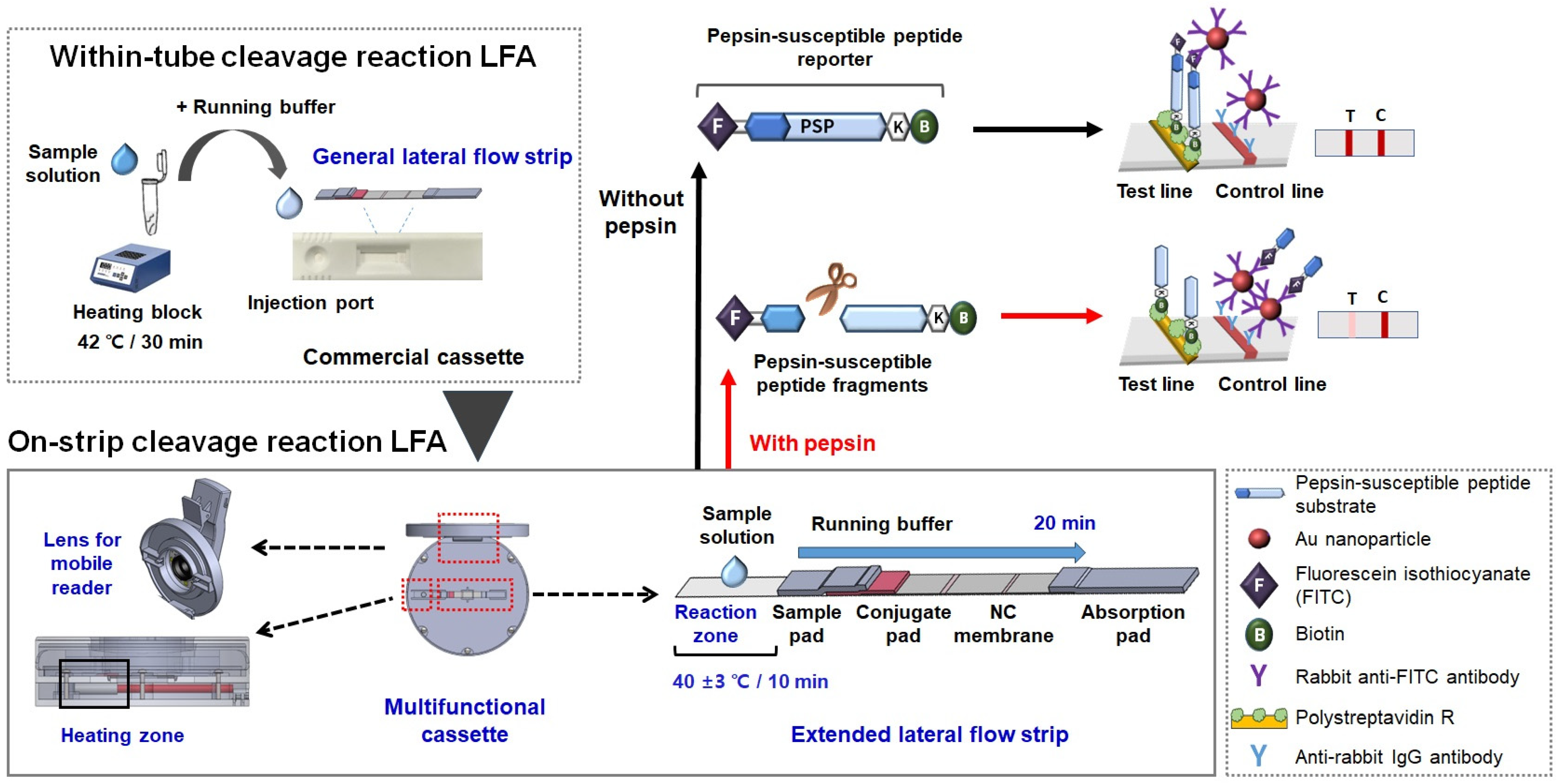
2.2. Preparing the Two Types of Lateral Flow Strips

To prepare Au NP-FITC Ab conjugates, 1 mL of Au NP solution was adjusted to pH 9 with 0.1 M K2CO3 (40 μL). Then, 48 μL of FITC Ab (0.25 mg/mL) was added to this solution and incubated via rocking for 1 h at room temperature. The prepared Au-FITC Ab conjugate was blocked with 120 μL of BSA (10% in distilled water) with shaking for 10 min. The Au-FITC Ab conjugate was collected via centrifuging (4000× g for 30 min, 4 °C). The pellet was resuspended in conjugate buffer consisting of 1× PBS (pH 7.2) and 1% BSA.
Sample and conjugate pads (cellulose fiber) were pretreated via dipping into 1× PBS (pH 7.2) containing 10% sucrose, 2% BSA, and 0.05% Tween-20, followed by drying at 37 °C for 2 h. Then, 30 μL of the Au-FITC Ab conjugates (optical density [OD] at 530 nm = 0.2) was dropped onto the conjugate pad and dried at 37 °C for 1 h. The pSA (3 mg/mL) and anti-rabbit IgG Ab (0.8 mg/mL) were striped at the test line and control line, respectively, onto a NC membrane, using an automated lateral flow reagent dispenser (dispense rate: 0.25 mL/min, Claremont BioSolutions, LLC, Upland, CA, USA). After drying at 30 °C for 4 h, it was stored in a plastic box with desiccant and light-blocking foil at 4 °C for further use.
To prepare the strip for a general LFA, the NC membrane was placed on top of the backing film (60 mm in length) with double-sided adhesive tape and cut to a width of 5 mm. The sample pad, conjugate pad containing Au NP-FITC Ab conjugates, and absorbent pad were assembled sequentially, overlapping each other by 2.5 mm, as illustrated in Figure S1a in the Supporting Information (SI). The general lateral flow strip was placed into the commercial strip cassette such that a sample solution flowed constantly (Figure S1b). For the on-strip cleavage reaction LFA using a multifunctional strip cassette, the strip consisted of the same four parts and an additional reaction zone that extended the length of the backing film (70 mm). The dimensions of this strip are shown in Figure S2a.

2.3. Detecting Pepsin Using the Within-Tube Cleavage Reaction LFAs

The PSP-based within-tube cleavage reaction LFA was performed in two consecutive steps: (1) PSP substrate cleavage by pepsin and (2) PSP or PSP fragment capture on the NC membrane of the strip, as previously reported [19]. First, 9 μL of the reaction solution containing citric acid (0.1 M, pH 2.0) and artificial saliva (pH 7.0) at a ratio of 1:1 (v/v) was added to the microtube. Artificial saliva was prepared by dissolving 50 mM NaHCO3, 1.4 mM K2CO3, and 8.6 mM NaCl in distilled water, in accordance with the standards and specifications for utensils, containers, and packages by the Ministry of Food and Drug Safety of Korea [21]. Then, 1 μL of pepsin (final 0, 4, 20, 100, 500 ng/mL) was injected in the reaction solution and mixed well. And 1 μL of FITC-PEP-B (final 20 μM) was added to the pepsin solution, mixed well, and the mixture was immediately incubated for 30 min at 42 °C using a heating block. After incubation, 90 μL of running buffer (tris-HNO3 buff solution [pH 8.0] and 0.1% Tween-20) was added to each reaction tube and gently mixed. The mixture was added dropwise to the sample pad of the LF strip through the injection port of the cassette and allowed to flow for 20 min. Each strip was scanned immediately using an Epson perfection V700 photo flatbed scanner (Seiko Epson, Nagano, Japan), and the scanned images were analyzed using ImageJ software (version 1.54a). In ImageJ, the intensities were converted into profile plots, with each peak in the plot representing one red line on the LF strip. As a result, the intensity of the test line was expressed as the peak area in the square pixels, representing the numerically integrated area under each peak [22,23]. The appearance of the test and control lines indicated a negative result. A higher pepsin concentration resulted in a paler or thinner test line color.
To preliminarily evaluate the feasibility of the PSP-based within-tube LFA cleavage reaction, pepsin was detected in human saliva samples (n = 3). Saliva collection was approved by the Ethics Committee of the Kyung Hee University Medical Center (IRB No. 2018-06-046), and all participants provided written informed consent prior to the commencement of the study. Pepsin levels in each sample were analyzed using a PSP-based LFA and a Human pepsin ELISA kit (E0922Hu, BT LAB, Brimingham, UK). ELISA was performed as per manufacturer’s instructions.

2.4. Fabricating the Multifunctional Strip Cassette Integrated with a Heating Pad and Mobile Reader

For the rapid, equipment-free, and on-site detection of pepsin, a multifunctional strip cassette was designed, characterized by a heating pad for PSP cleavage by pepsin, and a universal mobile reader for the image acquisition and simple analysis of color change without a scanner. It was fabricated using a three-dimensional (3D) printer (3DISON S, ROKIT Healthcare Inc., Seoul, Republic of Korea). As shown in Figure S3, this mainly consists of three parts. The first part is a main circular body (diameter: 80 mm) and a central window (diameter: 35 mm). The LF strip is placed on the base plate; the open window of the top facilitates checking the strip result with the naked eye, similar to a general strip cassette. The second part is a clip-type mobile camera lens with an additional macrolens. It can be connected with the open window of the main body to analyze the lateral flow strip using a mobile camera in a dark room. As a mobile camera lens has several light-emitting diodes, it can supply constant and homogenous light. A small macrolens is used to obtain an image of the detection zone in the strip, which shows both the test and control lines. The third part is a heating zone with a heating element (positive temperature coefficient) underneath it. It constantly maintains the temperature at 40 ± 3 °C for the cleavage of the PSP reporter by pepsin in the sample using a power supply (5.5 V). It does not require a heating block, hence allowing simple and easy pepsin detection.
After the extended strip was placed in the multifunctional strip cassette, the power was increased to increase the heating zone temperature. Then, the mixture of the sample (10 μL) containing the reaction solution with pepsin and FITC-PEP-B (1 μL) was dropped onto the reaction zone of the strip (Figure S2b). After reacting for 10 min, 100 μL running buffer was added onto a cross marking region within the heating zone, which flowed into the NC membrane. Each result was evaluated at 20 min. A picture of each test and control line was acquired using a multifunctional strip cassette and a Samsung smartphone camera. The peak area of the test line was quantitatively analyzed using ImageJ software.

3. Results and Discussion

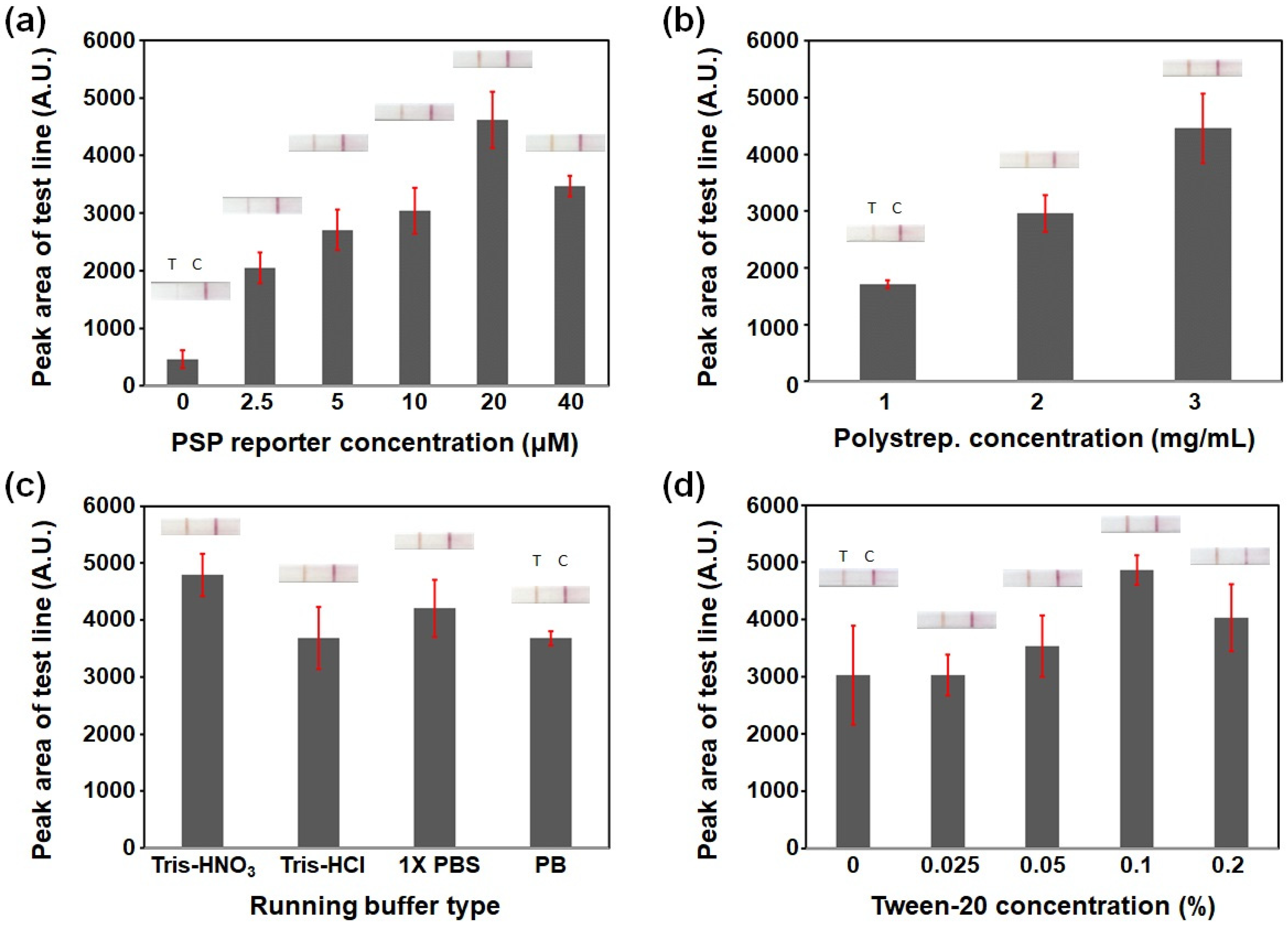
3.1. Optimizing the PSP-Based Cleavage Reaction LFA Conditions: pSA and PSP Reporter Concentrations and Running Buffer Composition

In recent years, LFAs have attracted considerable attention as tools for point-of-care testing (POCT) because of their numerous advantages, including a low cost, simplicity, portability, minimal sample preparation, rapid response, and convenience [24]. Typically, LFAs are based on the immobilization of a recognition unit conjugated to a colorimetric reporter, such as Au NPs, on a test line in the presence of a target analyte. However, developing highly specific human pepsin mAbs is time consuming and expensive. In addition, immune reactions between antigens and Abs may not work effectively under acidic conditions because saliva is collected using citric acid (0.1 M, pH 2.0), hence resulting in incorrect results. Here, we utilized a PSP substrate specifically cleaved by pepsin as a reporter which directly bonded with the test line. For this, PSP was modified with FITC and biotin at the N- and C-terminals. PSP has several advantages over Abs, including ease of synthesis, stability, and low cost. In addition, PSP can be more effectively cleaved by pepsin at a low pH. The detection principle of the PSP-based cleavage reaction for pepsin is as follows: in the absence of pepsin in samples, the PSP reporter can bind to gold anti-FITC Ab conjugates and displays a strong red color in the test line. In contrast, when the PSP reporter reacts with pepsin, the cleaved fragments cannot display any color on the test line. Therefore, the peak area of the test line is inversely proportional to pepsin concentration.
To maximize the sensing performance of this PSP-based cleavage reaction LFA, optimizing the concentrations of the PSP reporter and pSA for the test line and the running buffer composition is important. First, for the within-tube cleavage reaction LFA, we optimized the PSP reporter concentration for pepsin detection. The cleavage reaction temperature of 42 °C and time of 30 min was fixed in all experiments based on the optimization from our previous study [19]. As shown in Figure 2a, the peak area of the test line increased depending on the PSP reporter concentration by up to 20 μM (4623 ± 489). However, it decreased at 40 μM of PSP (3470 ± 178), which was attributed to the high-dose hook effect [25]. Hence, we selected 20 μM as the PSP reporter concentration because it showed the highest peak area. The concentration of pSA for test line was optimized at a fixed PSP concentration of 20 μM. A higher pSA concentration resulted in a stronger color intensity and larger peak area of the test line (Figure 2b) because of the increased biotin-binding sites for the PSP reporter. As a streptavidin polymer, pSA has been used as a protein anchor for biotinylated nucleic acids and proteins. It is known to have a good protein adsorption affinity for nitrocellulose [26]. As shown in Figure S4, the color intensity of the pSA-immobilized test line was higher than that of the same quantity of the streptavidin-immobilized line, indicating a good adsorption onto the NC membrane. Considering the fabrication cost, a pSA concentration of 3 mg/mL was selected.
The LFA running buffer is important for maintaining the sample pH, minimizing nonspecific binding, controlling flow speed, and allowing a perfect flow through the strip [27]. In particular, the running buffer is added to the sample in the reaction buffer (pH 2.8) after the cleavage reaction between the PSP reporter and pepsin. Hence, the buffer must maintain the pH at which the PSP reporter or cleaved fragments bind to the LFA test line. To better visualize the test line on the strip for accurately reading LFA results, its composition and concentration should be optimized. We evaluated the performance of four buffer types: tris-HNO3 (pH 8.0), tris-HCl (pH 8.0), PBS (pH 7.2), and phosphate buffer (PB; pH 7.2) supplemented with 0.1% (v/v) Tween-20. As both tris-buffer and PBS are generally used in LFAs, the peak areas of the test line for tris-HNO3 (4795 ± 373) and PBS (4208 ± 501) were not statistically different (Figure 2c). However, the peak areas of the test line for tris-HCl (3684 ± 543) and PB (3682 ± 126) were significantly lesser than that of tris-HNO3. Therefore, we selected the optimal running buffer as tris-HNO3 (pH 8.0). We then tested the color change in the test line depending on the Tween-20 concentration. The role of the detergent was to facilitate the dragging of elements along the strip and provide a clean white background for better result readings. As the Tween-20 concentration increased up to 0.1%, the peak area of the test line increased (Figure 2d). However, 0.2% Tween-20 showed a weaker color intensity and larger standard deviation (4028 ± 587) than 0.1% Tween-20 (4865 ± 257). High Tween-20 concentrations may cause the pSA of the test line to move toward the control line and hinder the PSP reporter binding to the test line because of the fast flow rate. Therefore, the optimal concentration of Tween-20 in tris-HNO3 (pH 8.0) running buffer was selected to be 0.1%.

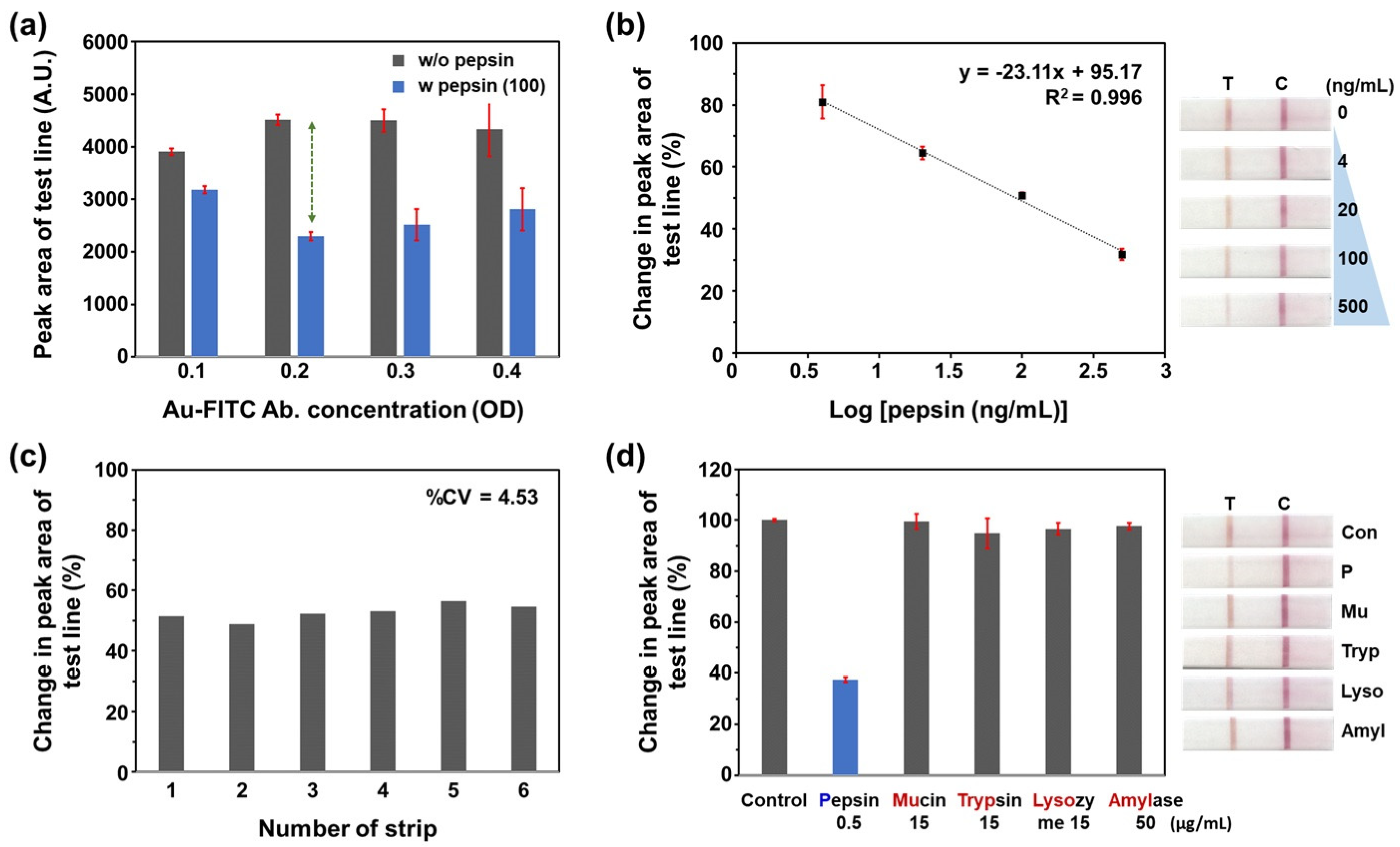
3.2. Analytical Performance of the PSP-Based Within-Tube Cleavage Reaction LFA for Pepsin Detection

We evaluated the analytical performance of the PSP-based within-tube cleavage reaction LFA. First, PSP (20 μM) was cleaved using various concentrations of pepsin (0, 4, 20, 100, and 500 ng/mL) at 42 °C for 30 min using a heating block. The LFA test line peak area was measured after developing each sample for 20 min. Although there are differences among studies, the reported cut-off values of salivary pepsin concentration for detecting reflux disease range from 4.21 to 216 ng/mL [8,13,14,15,28]. Therefore, the concentration range of pepsin was chosen to be 4–500 ng/mL to adequately cover both the cut-off values and higher pepsin levels. Figure 3a shows the differences between the peak areas of the test line with and without pepsin (100 ng/mL) according to the concentration of Au-FITC Ab conjugates (based on OD at 520 nm). At an OD > 0.2, the test line peak areas without pepsin were similar. However, pepsin decreased the peak area of the test line the most at an OD of 0.2. In cases with and without pepsin, a higher OD resulted in a larger standard deviation. Therefore, we selected the optimal OD for the Au-FITC Ab conjugate to be 0.2, which showed the greatest color change in the LFA test line.
Figure 3b shows that the pepsin concentration changed the test line color in the LF strip. As the pepsin concentration increased, the peak area of the test line decreased. To obtain a simple linear equation in a wide range of pepsin concentration, we performed a logarithmic processing of concentration data and constructed a single logarithmic plot [29]. As a result, the change (%) in the test line peak area of the strip showed a linear relationship with the log concentration of pepsin, ranging from 4 to 500 ng/mL (R2 = 0.996, n = 3). The limit of detection was found to be 1.9 ng/mL, according to the standard deviation of the blank and slope methods (3 sbl/slope) [30]. In addition, we investigated the reproducibility of the PSP-based LFA via measuring peak area changes in response to pepsin (100 ng/mL) in six assays on different dates. As shown in Figure 3c, the coefficient of variation (CV) was 4.53%. This result indicated that the PSP-based LFA was reproducible. To evaluate the specificity of the PSP-based LFA, we tested the change in the test line peak areas of the LFA after adding four kinds of enzymes, including mucin (15 μg/mL), trypsin (15 μg/mL), lysozyme (15 μg/mL), and amylase (50 μg/mL). Figure 3d shows that only pepsin (500 ng/mL) significantly changed the test line peak area on the LF strip. The other enzymes showed colors and test line peak areas similar to those of the control (n = 3). This result clearly demonstrates that the PSP-based LFA has a high specificity for pepsin. In addition, we preliminarily tested the long-term stability of the pSA-immobilized NC membrane by evaluating test line peak area changes in response to pepsin (100 ng/mL) after 30 days of storage at 4 °C without light and moisture. As shown in Figure S5, there was no significant change in the test line peak areas (99% of initial response at 30 days) of the LF strip.
Table S1 presents a comparison of the sensing performance of the PSP-based LFA with other methods for salivary pepsin detection. The PSP-based LFA showed the highest sensitivity and could detect a wider range of salivary pepsin compared to the commercial PeptestTM, which may be attributed to the sensitive reaction of PSP cleavage by pepsin. Additionally, we compared its analytical performance with other Au NPs-based LFAs that detect protein biomarkers (Table S2). Most of the developed LFAs exhibit adequate sensitivity for detecting target biomarkers. However, other detection methods typically use a pair of target-specific Abs to recognize the target protein, which increases manufacturing costs. Furthermore, methods such as localized surface plasmon resonance or fluorescence resonance energy transfer offer better performance than colorimetry, but require large and expensive readers. In contrast, the PSP-based LFA is easy to use, simple, and cost-effective because it utilizes peptides instead of high-cost Abs and does not require a specific reader.
To evaluate the preliminary feasibility of our PSP-based LFA, we first applied it to real sample analysis using the standard addition method. Table S3 shows the recovery results of pepsin in human saliva samples obtained with PSP-based LFA for pepsin-spiked samples. This PSP-based LFA produced recoveries ranging from 94.5 to 99.7% for pepsin. Next, we quantified the pepsin concentration in human saliva (n = 3). The obtained results were compared with those obtained using ELISA as a reference method. As shown in Table S4, the results showed good agreement between the two methods, with a maximum relative error of 12.7%. Therefore, the PSP-based LFA may be a useful tool for quantifying salivary pepsin levels.

3.3. Comparison with a Commercially Available Dipstick Assay for Pepsin Detection

HybriDetect™ is a universal lateral flow dipstick in which a gold-labeled polyclonal (rabbit) anti-FITC Ab is immobilized in the sample application area. It consists of a streptavidin test line for biotin binding and an anti-rabbit IgG Ab control line for the binding of gold anti-FITC Ab conjugates. As we confirmed the feasibility of the PSP-based pepsin assay using this dipstick in our previous study, we compared the sensing performance of the PSP-based LFA with that of the dipstick assay. Figure 4a shows the relative changes (%) in the results for both the LF strip and dipstick according to the pepsin concentration. LFA is presented as the change in the test line peak area compared to 0 ng/mL pepsin. The dipstick is presented as two lines that show the changes in color intensity of test line and the color intensity ratio between the test and control lines (IT−line/IC−line) compared to 0 ng/mL pepsin. As shown in Figure 4a, the relative change in IT−line/IC−line of the dipstick (blue line) showed the highest slope (35.65% change/Log[ng/mL]). However, both the test and control lines had to be analyzed. The percentage change in the test line color intensity (orange line) on the dipstick did not show a linear relationship with the log concentration of pepsin from 4 to 500 ng/mL. In contrast, the percentage change in the results of LFA (black line) showed the best linearity in the same concentration range. In addition, LFA is simpler than the dipstick assay because it only uses a test line for analysis. Therefore, we expect that this LFA with a simpler reader can be applied for POCT.

3.4. Evaluation of On-Strip Cleavage Reaction LFA with a Multifunctional Strip Cassette

Although the PSP-based within-tube cleavage reaction LFA is highly sensitive and cost-effective, it requires the cleavage reaction of the PSP reporter to occur separately in a microtube using a heating block. A heating block is commonly used in laboratories for treating samples at a constant temperature. However, it may not be available in low-resource settings for POCT. The LFA must meet the World Health Organization criteria for POC diagnostics to be affordable, sensitive, specific, user-friendly, rapid, robust, equipment-free, and deliverable to end users (ASSURED) [31]. This limitation may act as a hurdle in moving from a specialized laboratory to POCT. Therefore, we designed and developed an on-strip cleavage reaction LFA consisting of an extended LF strip with a reaction zone, a multifunctional strip cassette with a heating zone, and a mobile reader. The PSP reporter can be cleaved by pepsin directly on the reaction zone of the LF strip within the multifunctional strip cassette without additional heat treatment. As the optimal temperature for the cleavage reaction between pepsin and PSP was 42 °C in our previous result, the heating zone of the multifunctional strip cassette was maintained to 40 ± 3 °C using a small heater.
To evaluate the sensing performance of the on-strip cleavage reaction LFA, we examined the changes in the test line peak area in LFA with varying pepsin concentrations after the cleavage reaction for 10 min within a multifunctional strip cassette. A reaction time of 10 min was selected to prevent the small sample volume (10 μL) from evaporating. The results were analyzed with the LFA images using a scanner and mobile reader, respectively.
Figure 4b,c show the photographic images of the multifunctional strip cassette, the calibration plot, and scanned images of the on-strip cleavage reaction LFA using a multifunctional strip cassette. Similar to the LFA using PSP that was cleaved within the heating block, the change (%) in the test line peak area of the strip showed a linear relationship with the log concentration of pepsin, ranging from 4 to 500 ng/mL (R2 = 0.987, n = 3). The LOD was found to be 1.4 ng/mL, which was similar to that of the tube reaction within the heating block. From this result, we inferred that the heating zone in the multifunctional strip cassette could be effective for the cleavage reaction between PSP and pepsin, despite the shorter reaction time.
Next, we evaluated the performance of the multifunctional strip cassette integrated into a mobile phone. After developing with the running buffer for 20 min, a multifunctional strip cassette was placed on the lid to block light. A photograph of the LFA detection zone was captured using a mobile phone. The color intensity of images, such as grayscale, red, green, and blue scales, was then analyzed using a color picker application. Among them, green color intensity showed the best linearity (R2 = 0.991) and the highest slope (Figure S6). This may be attributed to the fact that the complementary color of the pinkish red is green. Therefore, the color mostly absorbed may be the green component [32,33]. Based on the results of the preliminary test, we suggest that the on-strip cleavage reaction of LFA with a multifunctional strip cassette can be utilized as an effective tool for PSP-based pepsin detection in POCT. In future studies, we plan to upgrade the multifunctional strip cassette to a more user-friendly device using a universal serial bus (USB) rechargeable heater or a film heater.

4. Conclusions

We developed two types of simple, sensitive, and effective LFAs for detecting pepsin using a PSP substrate that was specifically cleaved by pepsin. The within-tube and on-strip cleavage reaction LFAs depend on the location of the reaction between PSP and pepsin. The general and the extended LF strips which had the pSA-immobilized test line were used for each cleavage reaction LFAs. And the multifunctional strip cassette, consisting of a heating zone for the cleavage reaction and a universal mobile reader for image acquisition, was utilized for the on-strip cleavage reaction LFA. Both the within-tube and on-chip cleavage reaction LFAs showed high sensitivity, with a linear range of 4–500 ng/mL pepsin and LODs of 1.9 ng/mL (within-tube) and 1.4 ng/mL (on-chip). And they also represented good specificity, with high reproducibility. These PSP-based cleavage reaction LFAs can be sensitive to pepsin because of the capability of the PSP substrate and optimized cleavage conditions. And they are cost-effective because they do not use high-cost human pepsin mAbs as recognition units. In particular, these newly developed LFAs have several advantages over our previously developed dipstick assay; (1) in terms of design, the LF strip is extended to add a reaction zone, and the multifunctional cassette block integrated with a heating pad and mobile reader is prepared to run cleavage reactions directly on the strip without using an external heating block. Additionally, the pSA-immobilized test line on the NC membrane of LF strips is effective for detecting the PSP substrate functionalized with biotin. (2) In terms of performance, the LOD of the newly developed LFA is similar to the dipstick assay in the previous study. However, it shows better linearity within a similar concentration range. Additionally, the total time to confirm the results is 30 min, which is half the time of the dipstick assay (60 min). (3) In terms of practical application, the multifunctional strip cassette for the on-strip cleavage reaction LFA can provide an effective heating zone and mobile reader for the POCT of salivary pepsin. Therefore, the PSP-based cleavage reaction LFAs can be utilized as simple, user-friendly, and reliable tools for the on-site detection of salivary pepsin. In addition, these LFAs have great potential for the noninvasive diagnosis and/or screening of LPR and GERD using saliva.

Supplementary Materials

The following supporting information can be downloaded at https://www.mdpi.com/article/10.3390/chemosensors12110241/s1. Figure S1: (a) A design illustrating the lateral flow strip for the within-tube cleavage reaction lateral flow assay (LFA). (b) Photographic images of the prepared strip and commercial strip cassette.; Figure S2: (a) A design illustrating the extended lateral flow strip for the on-strip cleavage reaction LFA. (b) Photographic images of the prepared strip set on the multifunctional strip cassette.; Figure S3: Schematic diagram and photograph of a multifunctional strip cassette fabricated using a 3D printer.; Figure S4: Comparison of strip color change between polystreptavidin R and streptavidin-immobilized test lines.; Figure S5: Change in test line peak area of the LF strip response to pepsin (100 ng/mL) during 30 days of storage at 4 °C without light and moisture.; Figure S6: Comparison between calibration plots of gray, red, green, and blue scale intensities in the photograph of the detection zone in the on-strip cleavage reaction LFA obtained using the smartphone.; Table S1. Comparison of the analytical performance of PSP-based cleavage reaction LFA with other methods for salivary pepsin detection. Reproduced with permission from [19], Lee, Y.J. et al., Microchim Acta 2024; 191, 117.; Table S2. Comparison of the analytical performance of PSP-based cleavage reaction LFA with other Au nanoparticle-based LFA for detection of protein biomarker.; Table S3. The recovery test results of pepsin in human saliva samples obtained with PSP-based LFA for pepsin-spiked samples. The measurements were performed three times using different batches of strips.; Table S4. Pepsin levels in saliva samples (n = 3) obtained from LFA and ELISA. References [34,35,36,37,38,39,40,41,42] are cited in supplementary file.

Author Contributions

Conceptualization, G.-J.L., J.-C.L. and Y.J.L.; methodology, Y.J.L., S.-W.K., J.-C.L. and G.-J.L.; validation, S.-W.K., Y.-G.E. and G.-J.L.; formal analysis, S.-W.K. and Y.J.L.; investigation, Y.J.L. and S.-W.K.; resources, Y.-G.E.; writing—original draft preparation, G.-J.L.; writing—review and editing, Y.J.L., J.-C.L., Y.-G.E. and G.-J.L.; visualization, S.-W.K., J.-C.L. and G.-J.L.; supervision, G.-J.L.; project administration, G.-J.L.; funding acquisition, G.-J.L. and Y.-G.E. All authors have read and agreed to the published version of the manuscript.

Funding

This research was supported by a grant from the Korea Health Technology R&D Project through the Korea Health Industry Development Institute (KHIDI), funded by the Ministry of Health & Welfare, Korea (grant number: HI18C1039), and a National Research Foundation grant funded by the Korean government (No. NRF-2021R1A2C1093825).

Institutional Review Board Statement

The study design and sample collection were approved by the Ethics Committee of Kyung Hee University Medical Center (IRB No. 2018-06-046). The study was conducted in accordance with the tenets of Declaration of Helsinki and written informed consent was obtained from all the participants prior to the commencement of the study.

Informed Consent Statement

Informed consent was obtained from all subjects involved in the study.

Data Availability Statement

Data are contained within the article and supplementary materials.

Conflicts of Interest

The authors declare no conflicts of interest.

References

  1. Samuels, T.L.; Johnston, N. Pepsin as a marker of extraesophageal reflux. Ann. Otol. Rhinol. Laryngol. 2010, 119, 203–208. [Google Scholar] [CrossRef] [PubMed]
  2. Wood, J.M.; Hussey, D.J.; Woods, C.M.; Watson, D.I.; Carney, A.S. Biomarkers and laryngopharyngeal reflux. J. Laryngol. Otol. 2011, 125, 1218–1224. [Google Scholar] [CrossRef] [PubMed]
  3. Johnston, N.; Dettmar, P.W.; Bishwokarma, B.; Lively, M.O.; Koufman, J.A. Activity/stability of human pepsin: Implications for reflux attributed laryngeal disease. Laryngoscope 2007, 117, 1036–1039. [Google Scholar] [CrossRef] [PubMed]
  4. Wang, Y.J.; Lang, X.Q.; Wu, D.; He, Y.Q.; Lan, C.H.; Xiao, X.; Wang, B.; Zou, D.W.; Wu, J.M.; Zhao, Y.B.; et al. Salivary Pepsin as an Intrinsic Marker for Diagnosis of Sub-types of Gastroesophageal Reflux Disease and Gastroesophageal Reflux Disease-related Disorders. J. Neurogastroenterol. Motil. 2020, 26, 74–84. [Google Scholar] [CrossRef]
  5. Divakaran, S.; Rajendran, S.; Thomas, R.M.; Jacob, J.; Kurien, M. Laryngopharyngeal rReflux: Symptoms, signs, and Presence of Pepsin in Saliva—A Reliable Diagnostic Triad. Int. Arch. Otorhinolaryngol. 2021, 25, e273–e278. [Google Scholar]
  6. Lee, Y.J.; Kwon, J.; Shin, S.; Eun, Y.G.; Shin, J.H.; Lee, G.J. Opti mization of Saliva Collection and Immunochromatographic Detection of Salivary Pepsin for Point-of-Care Testing of Laryn gopharyngeal Reflux. Sensors 2020, 20, 325. [Google Scholar] [CrossRef]
  7. Na, S.Y.; Kwon, O.E.; Lee, Y.C.; Eun, Y.G. Optimal timing of saliva collection to detect pepsin in patients. Laryngoscope 2016, 126, 2270–2273. [Google Scholar] [CrossRef]
  8. Guo, Z.; Wu, Y.; Chen, J.; Zhang, S.; Zhang, C. The Role of Salivary Pepsin in the Diagnosis of Gastroesophageal Reflux Disease (GERD) Evaluated Using High-Resolution Manometry and 24-Hour Multichannel Intraluminal Impedance-pH Monitoring. Med. Sci. Monit. 2020, 26, e927381. [Google Scholar] [CrossRef]
  9. Du, X.; Wang, F.; Hu, Z.; Wu, J.; Wang, Z.; Yan, C.; Zhang, C.; Tang, J. The diagnostic value of pepsin detection in saliva for gastro-esophageal reflux disease: A preliminary study from China. BMC Gastroenterol. 2017, 17, 107. [Google Scholar] [CrossRef]
  10. Raja Nhari, R.M.H.; Muhammad Zailani, A.N.; Khairil Mokhtar, N.F.; Hanish, I. Detection of porcine pepsin in model cheese using polyclonal antibody-based ELISA. Food Addit. Contam. Part A Chem. Anal. Control Expo. Risk Assess 2020, 37, 561–567. [Google Scholar] [CrossRef]
  11. Guo, Z.; Jiang, J.; Wu, H.; Zhu, J.; Zhang, S.; Zhang, C. Salivary peptest for laryngopharyngeal reflux and gastroesophageal reflux disease. Medicine 2021, 100, e26756. [Google Scholar] [CrossRef] [PubMed]
  12. Strugala, V.; Woodcock, A.D.; Dettmar, P.W.; Faruqi, S.; Morice, A.H. Detection of pepsin in sputum: A rapid and objective measure of airways reflux. Eur. Respir. J. 2016, 47, 339–341. [Google Scholar] [CrossRef] [PubMed]
  13. Lechien, J.R.; Bobin, F. Diagnostic Value of Fasting and Bedtime Saliva Pepsin Measurements in Laryngopharyngeal Reflux. Biomedicines 2024, 12, 398. [Google Scholar] [CrossRef] [PubMed]
  14. Luebke, K.E.; Samuels, T.L.; Johnston, N. The Role of Pepsin in LPR: Will It Change Our Diagnostic and Therapeutic Approach to the Disease? Curr. Otorhinolaryngol. Rep. 2016, 4, 55–62. [Google Scholar] [CrossRef]
  15. Sabry, M.; Yosef, T.M.; Mahmoud, A.M.; George Michael, T.M.A. Fasting salivary pepsin level as a reliable non-invasive method of screening for laryngopharyngeal reflux in Egyptian patients. Egypt. J. Intern. Med. 2022, 34, 16. [Google Scholar] [CrossRef]
  16. Zhou, J.; Chen, X.; Wei, Y.; Lu, R.; Wei, Z.; Huang, K.; Luo, H.; Zhang, J.; Zheng, C. Portable and Rapid Fluorescence Turn-On Detection of Total Pepsin in Saliva Based on Strong Electrostatic Interactions. Anal. Chem. 2023, 95, 18303–18308. [Google Scholar] [CrossRef]
  17. Jiang, Q.; Chen, Z.; Huang, Y.; Gao, Q.; Luo, C.; Mehdi, M.; Xu, Y.; Li, H.; Sun, S. A bovine serum albumin and squaraine dye assembly fluorescent probe for pepsin detection. Microchem. J. 2023, 186, 108361. [Google Scholar] [CrossRef]
  18. Li, W.; Gao, Z.; Su, R.; Qi, W.; Wang, L.; He, Z. Scissor-based fluorescent detection of pepsin using lysozyme-stabilized Au nanoclusters. Anal. Methods 2014, 6, 6789–6795. [Google Scholar] [CrossRef]
  19. Lee, Y.J.; Noh, J.K.; Woo, S.R.; Kang, S.W.; Eun, Y.G.; Lee, G.J. Determination of pepsin in human saliva using pepsin-susceptible peptide reporter and colorimetric dipstick assay: A prospective, crosssectional study. Microchim. Acta 2024, 191, 117. [Google Scholar] [CrossRef]
  20. Domfe, T.; Njengele-Tetyana, Z.; Mhlanga, N.; Tetyana, P.; Skepu, A.; Ngila, J.C.; Sikhwivhilu, L.M. Development of a Versatile Half-Strip Lateral Flow Assay toward the Detection of Rift Valley Fever Virus Antibodies. Diagnostics 2022, 12, 2664. [Google Scholar] [CrossRef]
  21. 2019-2, 20190109; Standards and Specifications for Utensils, Containers and Packages. Ministry of Food and Drug Safety of Korea: Cheongju, Republic of Korea, 2019; Part II. p. 208.
  22. Jacinto, M.J.; Trabuco, J.R.C.; Vu, B.V.; Garvey, G.; Khodadady, M.; Azevedo, A.M.; Aires-Barros, M.R.; Chang, L.; Kourentzi, K.; Litvinov, D.; et al. Enhancement of lateral flow assay performance by electromagnetic relocation of reporter particles. PLoS ONE 2018, 13, e0186782. [Google Scholar] [CrossRef] [PubMed]
  23. Yrad, F.M.; Castañares, J.M.; Alocilja, E.C. Visual Detection of Dengue-1 RNA Using Gold Nanoparticle-Based Lateral Flow Biosensor. Diagnostics 2019, 9, 74. [Google Scholar] [CrossRef] [PubMed]
  24. Liu, Y.; Zhan, L.; Qin, Z.; Sackrison, J.; Bischof, J.C. Ultrasensitive and highly specific lateral Flow assays for point-of-care diagnosis. ACS Nano 2021, 15, 3593–3611. [Google Scholar] [CrossRef] [PubMed]
  25. Ross, G.M.S.; Filippini, D.; Nielen, M.W.F.; Salentijn, G.I.J. Unraveling the Hook Effect: A Comprehensive Study of High Antigen Concentration effects in Sandwich lateral Flow immunoassays. Anal. Chem. 2020, 92, 15587–15595. [Google Scholar] [CrossRef] [PubMed]
  26. Hallerbach, K.; Khederlou, K.; Wentland, L.; Senten, L.; Brentano, S.; Keefe, B.; Fu, E. Protein-Based Anchoring Methods for Nucleic Acid Detection in Lateral Flow Format Assays. Micromachines 2023, 14, 1936. [Google Scholar] [CrossRef]
  27. Machado, I.; Goikoetxea, G.; Alday, E.; Jiménez, T.; Arias-Moreno, X.; Hernandez, F.J.; Hernandez, L.I. Ultra-Sensitive and Specific Detection of S. aureus Bacterial Cultures Using an Oligonucleotide Probe Integrated in a Lateral Flow-Based Device. Diagnostics 2021, 11, 2022. [Google Scholar] [CrossRef]
  28. Weitzendorfer, M.; Antoniou, S.A.; Schredl, P.; Witzel, K.; Weitzendorfer, I.C.; Majerus, A.; Emmanuel, K.; Koch, O.O. Pepsin and oropharyngeal pH monitoring to diagnose patients with laryngopharyngeal reflux. Laryngoscope 2020, 130, 1780–1786. [Google Scholar] [CrossRef]
  29. Kong, D.; Zhao, J.; Tang, S.; Shen, W.; Lee, H.K. Logarithmic Data Processing Can Be Used Justifiably in the Plotting of a Calibration Curve. Anal. Chem. 2021, 93, 12156–12161. [Google Scholar] [CrossRef]
  30. Taverniers, I.; De Loose, M.; Van Bockstaele, E. Trends in quality in the analytical laboratory. II. Analytical method validation and quality assurance. Trac-Trends Anal. Chem. 2004, 23, 535–552. [Google Scholar] [CrossRef]
  31. Sena-Torralba, A.; Álvarez-Diduk, R.; Parolo, C.; Piper, A.; Merkoç, I. Toward Next Generation Lateral Flow Assays: Integration of Nanomaterials. Chem. Rev. 2022, 122, 14881–14910. [Google Scholar] [CrossRef]
  32. Puangpila, C.; Anukulkich, N.; Chiapleam, S.; Intajan, B.; Jakmunee, J.; Pencharee, S. Development of lectin-based lateral flow assay for fucosylated alpha-fetoprotein. J. Cell Biochem. 2023, 124, 1546–1556. [Google Scholar] [CrossRef] [PubMed]
  33. Charbaji, A.; Heidari-Bafroui, H.; Anagnostopoulos, C.; Faghri, M. A New Paper-Based Microfluidic Device for Improved Detection of Nitrate in Water. Sensors 2021, 21, 102. [Google Scholar] [CrossRef] [PubMed]
  34. Zheng, Z.; Geng, W.C.; Li, H.B.; Guo, D.S. Sensitive fluorescence detection of saliva pepsin by a supramolecular tandem assay enables the diagnosis of gastroesophageal reflux disease. Supramol. Chem. 2021, 33, 80–87. [Google Scholar] [CrossRef]
  35. Lee, D.; Lee, Y.J.; Eun, Y.G.; Lee, G.J. Label-free detection of salivary pepsin using gold Nanoparticle/Polypyrrole Nanocoral Modified screen-printed Electrode. Sensors 2018, 18, 1685. [Google Scholar] [CrossRef] [PubMed]
  36. Kim, D.; Kim, J.; Kwak, C.H.; Heo, N.S.; Oh, S.Y.; Lee, H.; Lee, G.W.; Ezhil Vilian, A.T.; Han, Y.K.; Kim, W.S.; et al. Rapid and label-free bioanalytical method of alpha fetoprotein detection using LSPR chip. J. Cryst. Growth 2017, 469, 131–135. [Google Scholar] [CrossRef]
  37. Li, R.; Feng, F.; Chen, Z.-Z.; Bai, Y.-F.; Guo, F.-F.; Wu, F.-Y.; Zhou, G. Sensitive detection of carcinoembryonic antigen using surface plasmon resonance biosensor with gold nanoparticles signal amplification. Talanta 2015, 140, 143–149. [Google Scholar] [CrossRef]
  38. Hosu, O.; Ravalli, A.; Lo Piccolo, G.M.; Cristea, C.; Sandulescu, R.; Marrazza, G. Smartphone-based immunosensor for CA125 detection. Talanta 2017, 166, 234–240. [Google Scholar] [CrossRef]
  39. Mohammadi, S.; Salimi, A.; Hamd-Ghadareh, S.; Fathi, F.; Soleimani, F. A FRET immunosensor for sensitive detection of CA 15-3 tumor marker in human serum sample and breast cancer cells using antibody functionalized luminescent carbon-dots and AuNPs-dendrimer aptamer as donor–acceptor pair. Anal. Biochem. 2018, 557, 18–26. [Google Scholar] [CrossRef]
  40. Pham, X.-H.; Hahm, E.; Huynh, K.-H.; Son, B.S.; Kim, H.-M.; Jun, B.-H. Sensitive colorimetric detection of prostate specific antigen using a peroxidase-mimicking anti-PSA antibody coated Au nanoparticle. BioChip J. 2020, 14, 158–168. [Google Scholar] [CrossRef]
  41. Kim, J.; Oh, S.Y.; Shukla, S.; Hong, S.B.; Heo, N.S.; Bajpai, V.K.; Chun, H.S.; Jo, C.-H.; Choi, B.G.; Huh, Y.S.; et al. Heteroassembled gold nanoparticles with sandwich-immunoassay LSPR chip format for rapid and sensitive detection of hepatitis B virus surface antigen (HBsAg). Biosens. Bioelectron. 2018, 107, 118–122. [Google Scholar] [CrossRef]
  42. Zhang, X.; Fishlock, S.; Sharpe, P.; McLaughlin, J. Development of colorimetric lateral flow assays with gold nanostructures for Cystatin C detection. Sens. Actuators Rep. 2022, 4, 100121. [Google Scholar]
Figure 1. A schematic demonstrating the detection principle of the PSP-based cleavage reaction LFA. The cleavage reaction of PSP by pepsin was performed in a microtube within a heating block (within-tube cleavage reaction LFA) and the reaction zone of the strip within a multifunctional strip cassette (on-strip cleavage reaction LFA), respectively. The black dashed arrow indicates a magnification of the area marked with a red box. After the reaction, samples were developed with a running buffer for 20 min. Increased pepsin quantity indicates a greater decrease in the test line color intensity (black solid arrow: without pepsin, red solid arrow: with pepsin).
Chemosensors 12 00241 g001
Figure 2. Optimization of PSP-based cleavage reaction lateral flow assay conditions for pepsin detection: concentrations of (a) PSP reporter and (b) polystreptavidin R, (c) running buffer types, and (d) Tween-20 concentration added to the running buffer. Error bars represent standard deviation of the mean (n = 3).
Chemosensors 12 00241 g002
Figure 3. (a) The differences between test line peak areas without (black bar) and with pepsin (100 ng/mL, blue bar) according to the concentration of Au-FITC Ab conjugates in the PSP-based within-tube cleavage reaction LFA. The green dashed arrow indicates the greatest color change in the test line with and without pepsin. (b) Calibration plots of test line peak area changes in the general type, PSP-based LFA versus log concentration of pepsin (4–500 ng/mL, n = 3). (c) The reproducibility of PSP-based LFA, determined via measuring the changes in test line peak areas to pepsin (100 ng/mL) in six assays on different dates. (d) Changes in test line peak areas of PSP-based LFA after reaction with 500 ng/mL of pepsin and four different enzymes: mucin (Mu, 15 μg/mL), trypsin (Tryp, 15 μg/mL), lysozyme (Lyso, 15 μg/mL), and amylase (Amyl, 50 μg/mL).
Chemosensors 12 00241 g003
Figure 4. (a) Comparison between the relative changes (%) in results versus the pepsin concentration in the within-tube cleavage reaction LFA and commercially available HybriDetectTM dipstick assay. (b) Photographic images of multifunctional strip cassette. The LF strip is placed on the base plate with a heating zone and a mobile camera lens with light-emitting diodes can supply constant and homogenous light. A clip-type multifunctional strip cassette is integrated with a mobile phone. (c) Calibration plot of changes in the test line peak area versus the pepsin concentration of the on-strip cleavage reaction LFA using a multifunctional strip cassette.
Chemosensors 12 00241 g004
Disclaimer/Publisher’s Note: The statements, opinions and data contained in all publications are solely those of the individual author(s) and contributor(s) and not of MDPI and/or the editor(s). MDPI and/or the editor(s) disclaim responsibility for any injury to people or property resulting from any ideas, methods, instructions or products referred to in the content.

