Review

Methods and Analysis of Biological Contaminants in the Biomanufacturing Industry

1
Department of Biomedical Engineering, University of Calgary, Calgary, AB T2N 1N4, Canada
2
Department of Chemical Engineering, University of Calgary, Calgary, AB T2N 1N4, Canada
3
Hotchkiss Brain Institute, University of Calgary, Calgary, AB T2N 1N4, Canada
*
Author to whom correspondence should be addressed.
Chemosensors 2023, 11(5), 298; https://doi.org/10.3390/chemosensors11050298
Submission received: 30 March 2023 / Revised: 3 May 2023 / Accepted: 14 May 2023 / Published: 18 May 2023

Abstract

:
The advent of bioprocessing has revolutionized the biomanufacturing industry, leading to the rise of biotherapeutics derived from biologic products such as chimeric antigen receptor (CAR) T-cells used for targeted cancer treatment and the Vero cell line for the production of viral vectors and vaccines. Despite these promising developments, most biologic products are characterized by fragile macromolecular structures that are heterogenous with a purity profile that varies with each batch making them susceptible to microorganism contamination. Regulatory oversight of biologic products is imperative to ensure adherence to good manufacturing practices and compliance with quality management systems. Current quality assurance protocols during production include monoclonality during cell line development, real-time monitoring of process parameters, flow cytometry for microbial monitoring, polymerase chain reaction, and immunoassay techniques to amplify DNA sequences related to bacterial or biological contaminants. FDA guidance recommends the implementation of process analytical technology within biomanufacturing production to measure critical quality parameters, which includes screening for potential biological contamination. Future advancements in bioprocess monitoring and control should capitalize on providing cheap, real-time, and sensitive detection. Biosensors, mass spectrometry, and polymerase chain reaction present robust, rapid, and real-time capabilities for multiplexed detection of contaminant analytes and have shown promise in meeting these needs. This review discusses the main biological contaminants of bioprocesses, European Union and FDA regulatory guidelines for monitoring and control within biologics production, existing methods and their limitations, and future advancements for biological contamination detection.

1. Introduction

Cells are now grown and differentiated for life-saving medical applications including cancer treatment (dendritic cells, CAR-T cells), hematopoiesis (hematopoietic stem cells), and vaccines for infectious diseases (Vero cells) [1]. For the patients, they present numerous interesting new therapy choices. However, many of these biologic products comprise macromolecules that are orders of magnitude more complex than the conventional tiny molecules (such as aspirin, insulin, diphenhydramine, antihistamines, etc.) produced through chemical synthesis [2,3]. Most biologic products are biosynthesized by live cells, except for DNA/RNA and peptides [3]. These bio-syntheses are more naturally variable than chemical synthesis, which makes them more difficult to characterize and manage properly [3]. These cell culture products are grown and expanded under a controlled environment inside medium to large-volume bioreactors [4]. Generally, good manufacturing practices and strict quality control are applied to eliminate any contaminations that could not only set back the production line but, if unchecked, can have fatal outcomes for patients treated with contaminated cells [4]. Due to these factors, it is essential to characterize and monitor these macromolecules in a highly precise and sensitive manner during development and manufacturing to effectively regulate the safety and efficacy of biologic products [3].
The World Health Organization (WHO) defines adventitious biological contaminants as microorganisms that might have been accidentally introduced during the production of a biologic product, such as transmissible spongiform encephalopathy (TSE), viruses, bacteria, fungi, mycoplasma, and more [5]. Starting materials used in manufacturing biopharmaceutical products, such as cell substrates, porcine trypsin, bovine serum, or any other source materials of animal or human origin, environmental exposure, such as equipment, handling, and personnel, may unintentionally introduce adventitious agents [5,6]. Mammalian cell culture contamination continues to be a significant issue in the development and production of bioprocesses, resulting in time, money, and effort losses [7]. Contamination happens when undesirable microorganisms or higher eukaryotic cells infiltrate the culture. This could prevent the target cell line from growing and make it more difficult to identify certain cell lines. Cell culture consumers in the US alone are thought to incur annual costs of millions of dollars [7]. A person being affected as a result of a contaminated product has the greatest immediate impact. As contaminated products must be pulled off the market, contamination might result in shortages of products or vaccines [8]. Therefore, the issue of unidentified contamination concerns associated with live or vectored vaccinations necessitates stricter safety control [9].
There are many current methods that have been continuously used for monitoring different biological contaminants that affect the yield of the bioprocess. These range from gold-standard microbial monitoring [10,11] to evolving reagent-based tests for protein and nucleic acid markers of biological contaminants [12], as well as the collection of sophisticated tools such as process analytical technologies [13]. Many emerging methods are also finding foot in the industrial process by offering facile operations, rapid results, and automation [14,15]. Additional review articles only focus on specific bioprocess contaminants or only examine particular biomonitoring processes [16,17]. Therefore, an overview addressing all necessary biomonitoring-related topics would greatly benefit researchers, policymakers, and practitioners. It provides an outline of the subject, highlighting the most significant aspects and recent advancements in the field of biomonitoring, and serves as a valuable resource for individuals developing and utilizing biomonitoring systems. Such an article bridges the gap between research and practice, thereby encouraging the establishment of more comprehensive biomonitoring strategies and enhancing our ability to protect human health.
In this review, we discuss the main biological contaminants of bioprocesses such as bacteria, viruses, and fungi. We also introduce the regulatory requirements of different regulatory bodies in the world. Many gold-standard tools and screening methods for each type of contamination are discussed in detail while outlining their advantages and shortcomings. Finally, emerging methods for detecting biological contaminants and their advantages over the current methods are reviewed. While there are a few reviews that comprehensively discuss different biological contaminants such as mycoplasma [18], bacteria [5], viruses [9], and fungi [19], this review enables researchers to understand the challenges and advances in designing novel screening methods for biological contaminants in biomanufacturing.

2. Biological Contaminants

Any microorganism that is accidentally introduced during the production of a biological product in the biomanufacturing process is considered a biological contaminant [5]. Indoor air settings include a complex variety of biological contaminants, which, if not controlled, may contaminate the finished product and cause adverse consequences in people, such as allergies, infections, or inflammatory reactions [20]. Controlling biological pollution in indoor air is primarily centered on avoiding circumstances that offer a substrate for the development of viable particles, followed by the containment and removal of such growth. It is feasible to plan preventative measures to avoid indoor environmental contaminations by employing air filters and boosting ventilation effectiveness [20].
Biological contaminants can easily contaminate any biomanufacturing process since they occur everywhere in the environment. The majority of these microorganisms can be classified into bacteria [5], fungi [19], and viruses [21] and will be briefly discussed here. Other preventative actions, such as assessing the indoor environment for potential biological pollutants, as well as chemical pollutants, are also necessary. Chemical pollutants can exist in various shapes and can contaminate the bioprocess depending on the type of exposure. Dust and particles are one of the most common types of these contaminants that can originate from skin cells, hair, or other organic matter [22].

2.1. Bacteria

Due to the abundance of bacteria in the environment, they are very likely to contaminate biomanufacturing processes. Aerosol generation on the surfaces of instruments, or even water, is a significant mode of bacterial contamination. These airborne microbes can invade cell cultures and outgrow the desired cells [16]. In addition, some bacteria release toxic chemicals into the environment. More specifically, in the outer membrane of Gram-negative bacteria, a highly toxic chemical called endotoxin exists, which is a potential contaminant of many bioprocesses. This chemical is released during different phases of bacterial growth, and even in small amounts, can cause various health issues such as including septic shock, inflammation, fever, and organ dysfunction if entered into the human body [23,24]. Bacteria are simpler to detect compared to viruses and mycoplasma as the cloudiness, pH, and color changes of the media could indicate contamination; however, this detection occurs rather late in the process [7]. Among various contaminations, bacterial contamination, specifically by mycoplasmas, is a grave concern in the biomanufacturing of these cells [25]. Mycoplasmas are the smallest self-replicating bacteria that lack cell walls, making them resistant to common antibiotics [26]. Mycoplasmas can be introduced throughout the manufacturing process including through starting materials and via human intervention and can influence almost every parameter of the cell culture system [27]. It is estimated that ~5 to 30% of the world’s cell lines are infected with mycoplasma causing an estimated average of USD 350 million in economic loss each year [18,28].
There are currently around 190 species of mycoplasma that are found in humans, animals, insects, and plants, yet only six of them are thought to be in charge of almost 95% of cell culture contamination incidents: Mycoplasma orale, Mycoplasma arginini, Mycoplasma hyorhinis, Mycoplasma fermentans, Mycoplasma hominis, or Acholeplasma laidlawii [29]. M. orale, M. fermentans, and M. hominis, present in the human oropharyngeal tract, are mainly introduced through personnel, and account for more than half of mycoplasma infections in cell cultures [30]. M. arginini and A. laidlawii are commonly isolated from fetal bovine serum (FBS) or newborn bovine serum (NBS). Swine trypsin solutions are a major source of M. hyorhinis [30]. One of the tiniest free-living bacterium species, Mycoplasma can be found in sizes between 0.15 and 0.3 μm [31]. Special tip organelles, containing high concentrations of adhesins, allow mycoplasmas to attach to and penetrate their host cells (Figure 1a). Moreover, the lack of a stiff wall in mycoplasma may facilitate their fusion with the host cell’s membrane and the exchange of membrane and cytoplasmic components [32,33]. Because of their small size, mycoplasma strains can accumulate to large concentrations in cell culture without causing media turbidity or other overt signs of infection. They can also bypass numerous filtration devices. Furthermore, mycoplasma contamination can have a major financial impact due to the time and costs involved with the loss of batches that were contaminated, the search for the source of the contamination, and the facility’s decontamination [32].

2.2. Viruses

Viruses may enter cell cultures from contaminated mammalian cells, tissues, and blood materials such as serum. The detection of viruses is often more difficult than that of other microorganisms, and they can potentially replicate human pathogens in mammalian cells [36]. Like mycoplasma, a normal light microscope cannot detect viral contamination, unlike bacterial and fungal contamination. Virus contamination can only be suspected when it causes morphological changes in the cultured cells, such as a cytopathic effect. Viruses that transmit silently without modifying the morphology of the infected cell are clearly more dangerous. Moreover, it is possible for cells that have been infected with some viruses to become more susceptible to infection by other viruses [21]. Moreover, some viruses can be as small as 20 nm; thus, they can pass through filtration devices [36]. Viruses that infect bacteria are called bacteriophages [37,38]. They can be found in almost any environment, including soil, food, groundwater, and surface water, and suboptimal environmental conditions do not affect their stability. Bacteriophages are involved in antibiotic resistance spreading. Therefore, rapid phage detection is important in bioprocesses that involve bacterial cultures [39]. Figure 1b illustrates how a virus attacks cell culture. Viral DNA replicates when it gets inside a cell, hijacking the cellular processes to produce proteins encoded with viral DNA. Proteins and genetic material can be translocated from cells and assembled into new virus particles using viral mechanisms. Pharmaceutical medicines still include some adventitious substances despite advanced detection measures [5]. The inclusion of porcine circovirus 1 (PCV1) in a rotavirus vaccination was a recent noteworthy occurrence. Early cases of biological contamination (such as tetanus contamination of diphtheria anti-toxin) predate contemporary immunization and resulted in the development of regulatory monitoring in the early 1900s. An important manufacturing transition from primary rhesus monkey kidney cells to African Green monkey kidney cultures occurred from the revelation that the early polio vaccine was tainted by simian virus 40 (SV40) due to rhesus monkey infection. Reverse transcriptase and bacteriophages were found in measles and polio vaccines, and the emergence of bovine spongiform encephalopathy (BSE), also known as “mad cow disease”, and ultimately the human version variant Creutzfeldt–Jakob disease (vCJD), in the 1990s, resulted in extensive regulatory deliberations as well as recommendations on the use of bovine vaccines [9].

2.3. Fungi

Like bacteria, fungi are also found abundantly in the environment. Therefore, bioprocesses are prone to contamination by fungi [36]. Aspergillus species dominate fungus contamination in cell culture. M. morale is the most common source of contamination that can spread from the oral cavity. We may be able to detect fungal contamination by direct microscopic imaging of a culture if it cannot be seen visually. Biochemical tests detect 1,3-d-glucan or galactomannan from fungus cells. The polysaccharide antigen galactomannan is found primarily in the cell walls of Aspergillus species, which sometimes affects cell cultures [36]. Figure 1c shows the infection mechanism of a specific fungus. The invasion of a host cell involves two steps: adhesion and invasion. Host cell invasion can be categorized into two general mechanisms, induced endocytosis and active penetration. A fungus may also gain nutrients from the invaded host cell.
Considering the safety issues, past instances, risks imposed on public health as the result of consumption of contaminated products, and the financial losses associated with such products, it is essential to understand the nature of the contamination, its sources, prevention, and inactivation methods. Most importantly, it is necessary to acknowledge the limitations of current screening methods and move toward the development of technologies that maximize safety, while minimizing cost, time requirement, and labor.

3. Regulatory Requirements for Bioprocess Monitoring

Regulatory requirements are critical in ensuring the safety, effectiveness, and quality of products produced through bioprocessing, usually by emphasizing Quality by Design (QbD) principles and adherence to Good Manufacturing Practices (GMPs) [40]. Since these products are often used in biomedical contexts, it is essential to establish regulatory guidelines to ensure that they meet rigorous quality and safety standards. The bioprocessing sector is heavily regulated, and companies are required to observe strict adherence to several laws and policies established by different regulatory bodies, such as the US Food and Drug Administration (FDA) and the European Medicines Agency (EMA) [41]. Harmonization of such regulatory requirements is critical for the development and manufacture of drug products while ensuring that quality, safety, and efficacy standards are upheld to meet the regulatory requirements prioritizing public health [42].

3.1. Regulations in the United States

The regulatory landscape in the field of bioprocess monitoring in the United States is governed primarily by the Food and Drug Administration (FDA), whose scope of activities includes surveillance of pharmaceutical manufacturer compliance to current Good Manufacturing Practices (cGMPs) among many others [43]. The FDA enforces Title 21 of the Code of Federal Regulations; for monitoring bioprocess specifically, Part 210 (cGMP in Manufacturing, Processing, Packaging, or Holding of Drugs) and Part 211 (cGMP for Finished Pharmaceuticals) outline the minimum standards used in the production, processing, and packaging of drug products [44]. Regulations require written procedures for preventing undesired microbial contamination to be established and adhered to. Given this general requirement, pharmaceutical manufacturers have the responsibility to generate a complete list of microorganisms for which to test; although, Part 317.2 is considered a logical but incomplete starting point as it does not outline potential opportunistic pathogens [45]. To introduce some guidance on industry compendial standards harmonization, the United States Pharmacopeia (USP) stated that a 10 CFU/100 mL target for pre-filtration bioburden is suitable for general cases, with a maximum acceptable count of 20 CFU/100 mL [46].

3.2. Regulations in the European Union (EU)

In the European Union (EU), the European Medicines Agency (EMA) harmonizes bioprocessing activities and coordinates compliance with Good Manufacturing Practices (GMPs) [47]. Specifically, the EudraLex, Volume 4—Good Manufacturing Practice (GMP) guidelines uphold the rigorous standards of Quality Management with regard to the development, production, and oversight of drug products in the pharmaceutical industry of the EU [48]. Unlike the FDA guidelines, Annex 1—Manufacture of Sterile Medicinal Products outlines the approaches to be taken regarding aseptic and terminal sterilization processes, which include the specific techniques and thresholds for the maximum permitted microbial contamination level during qualification according to area grades [49]. However, despite providing more specific guidance compared to the regulatory requirements in the US, the annex does not specify the species of bacteria to test against, unless otherwise a model organism is stipulated for standardized testing, e.g., bacterial retention testing.

3.3. Challenges and Trends in Meeting Regulatory Requirements

With the difference in the specific regulatory requirements across different global regions of interest due to their high pharmaceutical production capacity, there is a need for technologies that can adapt to the proprietary processes of various pharmaceutical entities while still adhering to the stipulated regulations. Furthermore, there is recent interest in the field of bioprocess intensification in the context of continuous production [50]. Hence, in order to develop, analyze, and control a continuous pharmaceutical manufacturing process, it is necessary to put new developments in the field of process analytical technology (PAT), specifically bioprocess monitoring, into practice [51].

4. Current Methods

Bioprocess monitoring is a critical component of manufacturing biological products such as pharmaceuticals, cell therapy products, gene therapy products, and therapeutics. Hence, it becomes crucial to ensure that the final product meets the desired specifications and is safe for use [52]. Monitoring bacterial or biological contamination in bioprocesses requires detecting and analyzing factors such as pH, temperature, dissolved oxygen, nutrient levels, and cell counts during manufacturing. Along with contamination prevention, these details are used to control and optimize the process, detect and repair problems, and assure product quality and process efficiency [53]. In recent years, several FDA-approved procedures for detecting biological contaminants have been approved to be applied in the industry. Immunoassay procedures, such as Enzyme-Linked Immunosorbent Assay (ELISA), assess the presence and concentration of specific proteins or antibodies that may indicate bacterial contamination [54]. ELISA is a powerful method for detecting and measuring proteins and antibodies frequently used in producing medicines, vaccines, and diagnostic kits, which, if utilized correctly, can also aid in the detection of bacterial contamination [54]. However, alternative procedures may be more appropriate depending on the application and product. For example, Polymerase chain reaction (PCR) can detect bacterial and virus contaminants, and cell analysis can evaluate if there is any aberrant cell development that could indicate bacterial or other contamination [54]. The technique employed is often decided by the unique needs of the process and the product being manufactured, with the ultimate goal of preventing and detecting biological contamination [11].

4.1. Process Analytical Technology (PAT)

Process Analytical Technology (PAT) is a systematic approach to designing, analyzing, and controlling biomanufacturing processes that can help prevent contamination [13]. PAT allows for real-time monitoring and control of critical process parameters such as temperature, pressure, and pH, as well as rapid and precise measurement of the final product’s quality attributes [55]. Potential issues can be identified early by continuously monitoring the process and analyzing data using advanced statistical and mathematical techniques [55]. Proactive adjustments can be made to prevent or correct deviations from the desired specifications, consequently aiding in improving process efficiency, reducing waste, and improving product quality and consistency while ensuring patient safety [56]. One of the primary benefits of PAT is its ability to provide continuous monitoring and control, allowing for detecting and correcting process deviations that could result in biological contamination [57]. PAT implementation, conversely, can be difficult and costly, necessitating the use of specialized analytical equipment and personnel, as well as an investment in process understanding, data analysis, and process control expertise [58]. Despite these challenges, the advantages of PAT make it a valuable method for ensuring consistent product quality and process performance in the bioprocess industry, especially when it comes to preventing biological contamination [53].
PAT has been extensively used for contamination detection in bioprocesses, some exploring combinations of two or more methods, and some modifying and enhancing the current methods [3,59,60]. For instance, a light microscope and/or molecular probes are traditionally used to detect microbial contamination in cell culture media. Some of these methods are incompatible with certain cell lines, require human intervention, and take a long time to test. Using an integrated microfluidic device (IMD), a research group demonstrated that (E. coli) could be detected in BPH-1 cell culture. A sensor with continuous monitoring capability was used to monitor glucose metabolism in cell culture media, and an optofluidic sensor was used to measure acidity in cell culture media (Figure 2a). In contrast to light microscopes and plate counting methods, this IMD detects bacterial contamination at a stage where it is undetectable. A continuous cell culture monitoring platform reduces the risk of undetected contamination by reducing even the short-term (less than 8 h) presence of E. coli changing cellular metabolism in the long term (at least up to 24 h). This increases the reliability of experiments conducted with no interference with cell culture. The glucose detection range was 50–950 µM, with a sensitivity of 3.81 ± 0.08 µA mM−1, and a detection limit of 27.7 ± 1.3 µM. This method provides continuous and sensitive pH monitoring within the physiological range of pH 6.4–8.1. Designed to allow easy surface modification using drop casting, the sensor has a microfluidic chamber that allows continuous media analysis and allows the electrodes to be accessed [61]. In another study, FTIR imaging with a focal plane array detector (FPA) was used to identify mycoplasma in a human brain glioblastoma cell culture due to its advantages of quick acquisition and high spectral quality. By FTIR micro-analysis, it was possible to distinguish control cells from mycoplasma-infected cells because of molecular changes in the host cells. The FTIR data were compared to those from PCR and a biochemical test. Since the detection level was just on the boundary of what was acceptable, the extremely sensitive biochemical test was unable to identify the positive contamination. As with PCR, FTIR analysis was able to identify mycoplasma contamination. In addition, this study demonstrates the ability of FTIR to detect early metabolic changes caused by mycoplasma infection, though it will be necessary to conduct an additional study and analyze samples to test the specificity in various mycoplasma species [60].
Integrating the Modular Automated Sampling Technology (MAST) based aseptic sampling, multi-function Sequential Injection Analysis (SIA) sample preparation, UHPLC separation, and high-resolution mass spectrometry (HRMS) analysis Liu et al. obtained real-time monitoring of multiple product quality attributes (Figure 3). During a 17-day 3 L Chinese Hamster Ovary (CHO) bioreactor cell culture process, continuous measurements of titer, size variants, glycosylation, and post-translational modifications yielded quality profiles comparable to traditional offline analyses. The online workflow provided fast cycle time, automated end-to-end analysis, and easy application to a variety of bioreactor scales for batch and perfusion processes. Additionally, the automation platform supported the online multi-attribute method, removing a major barrier to the broad implementation of high-resolution, high-sensitivity procedures. Automated real-time monitoring and feedback control will be part of the future biomanufacturing process, thereby improving yield, robustness, and product quality. The minimum 25 mL volume requirement should be addressed in future work. While multi-day repeated sampling may not have a significant impact on pilot or commercial processes, it might during bench-scale development [3].

4.2. Microbial Monitoring

Microbial control and quality assurance rely heavily on microbial monitoring. Microbial monitoring methods are used to ensure that the bioprocess environment is free of contaminants and that the final product is free of microorganisms that could be harmful to patients [56]. The FDA has established guidelines for the use of microbial monitoring methods such as viable cell counting and flow cytometry for the detection and enumeration of microorganisms in bioprocesses [61]. The principle of viable cell counting is based on the measurement of microbial growth via agar plate culture of microorganisms. Microorganisms in the sample are transferred to agar plates and incubated under optimal conditions for microorganism growth [56]. After growing the microorganisms in a suitable medium, and counting the number of cultured colonies, the number of colony-forming units (CFUs) in the sample is counted to determine the number of viable cells [56]. One of the critical applications of microbial monitoring in bioprocesses is in the production of biopharmaceuticals, where it is essential to ensure that the final product is free of microbial contamination [56]. This can be performed by monitoring the bioprocess at various stages, such as during fermentation or downstream processing, to detect and quantify any microorganisms that may be present. This information can then be used to make adjustments to the process to reduce or eliminate microbial contamination [56].
However, there are some disadvantages to using microbial monitoring for bioprocess monitoring. Microbial monitoring can be time-consuming and resource-intensive, requiring specialized equipment and trained personnel. This includes preparing the sample, cultivating it, and identifying the present microorganisms. Specificity is also limited since microbial monitoring is not as precise as other techniques, such as PCR or qPCR. The method relies on microorganism growth and does not allow for the identification of specific microorganisms [56].
Subsequently, microbial monitoring is a popular method for monitoring bioprocesses. However, it has downsides including being time demanding and having limited real-time monitoring [61]. Other methods, such as PCR or qPCR, can be used in conjunction with microbial monitoring to provide more specific and accurate information about the presence of microorganisms in the process.

4.3. Polymerase Chain Reaction

PCR is widely used in bioprocess monitoring to amplify specific DNA sequences of interest, such as those of bacterial or biological contaminants [61]. PCR can be used to detect and quantify the presence of specific microorganisms in a bioprocess by tracing the expression of specific genes linked to microbes [11]. The method entails repeatedly heating and cooling a nucleic acid sample to denature and re-anneal distinct DNA strands, followed by strand extension by a polymerase enzyme. This procedure duplicates the target DNA sequence, allowing detection and quantification via gel electrophoresis or real-time PCR techniques [62]. There are several types of PCR, including conventional PCR, quantitative PCR (qPCR), and reverse transcription PCR (RT-PCR) [10]. Conventional PCR is the most basic technique to amplify a specific DNA sequence of interest. qPCR is used to quantify the amount of a specific DNA sequence in a sample. RT-PCR detects and quantifies RNA molecules in a sample, which is useful for monitoring gene expression [10]. One of the primary advantages of PCR is its high sensitivity and specificity, which enables the detection of very low levels of target DNA or RNA. PCR is also a quick and straightforward technique, making it ideal for routine monitoring in a bioprocess [63]. However, PCR has some limitations, such as the possibility of false positive results, the need for meticulous sample preparation, and the possibility of cross-contamination between samples. Furthermore, PCR requires specialized equipment and reagents, which can be costly [62]. Overall, PCR is regarded as a valuable tool for detecting genetic changes in cell lines used in bioprocessing and monitoring the presence and growth of bacterial or biological contaminants during fermentation or cell culture processes.

4.4. Immunoassay Techniques

Immunoassay techniques are a group of analytical methods used to detect and quantify the presence of specific molecules in a sample, such as bacterial proteins or antibodies. These techniques use the specific binding of an antigen or antibody to a labeled reagent, such as a fluorescent marker, to detect the presence of the target molecule [64]. These techniques can be used to quantitatively and qualitatively measure the bacterial target, and ensure that the final product meets the desired specifications. The FDA has approved immunoassay techniques such as ELISA for bioprocess monitoring [65]. ELISA is a plate-based assay that uses a specific antibody–antigen binding to detect the presence of a target molecule. The sample is added to a plate coated with a particular antibody, and any target molecule in the sample will bind to the antibody. A labeled reagent is then added to the plate to detect the presence of the target molecule [54]. Another example is sandwich ELISA, which utilizes a capture antibody and a detection antibody to detect the target molecule. This method allows for higher sensitivity and specificity in detecting the target molecule [54].
Immunoassay techniques have several advantages for bioprocess monitoring, including high specificity and sensitivity, the ability to detect a wide range of biomolecules, and the ability to measure both qualitative and quantitative characteristics of the product [65]. However, these techniques also have some limitations, such as the need for a highly pure and specific antibody, the potential for interference from other molecules in the sample, and the need for specialized equipment and trained personnel to perform the assay [54]. One disadvantage of using immunoassay techniques to monitor bioprocesses is that they may not be as sensitive as other techniques. Furthermore, using specific antibodies or probes can be expensive and may not be available for all biomolecules [54].

4.5. Limulus Amebocyte Lysate

Limulus Amebocyte Lysate (LAL) is a routinely used test for detecting endotoxins in bioprocesses, which are toxic substances released by Gram-negative bacteria that can cause undesirable effects on people, such as fever, inflammation, and shock [66]. Thus, detecting and controlling endotoxins in bioprocesses is crucial for guaranteeing product quality and patient safety. The test uses horseshoe crab blood that primarily includes enzymes that bind to endotoxins, causing a coagulation reaction that can be detected visually or spectrophotometrically [66]. One of the primary benefits of LAL is its incredible sensitivity, which allows for detecting endotoxins at low concentrations. It is also quick and simple to perform, creating further possibilities for more automated high-throughput tests [66]. However, this technique has its own limitations. The test is endotoxin-specific and will not detect different types of bacterial contamination, such as Gram-positive bacteria or fungi [66]. Furthermore, LAL is sensitive to interference from specific sample matrix components, such as surfactants or chelating agents, which might result in false negative or false-positive results. As a result, thorough sample preparation and test procedure validation are crucial for reliable results [66].
Various biological contamination detection methods have distinct benefits as well as drawbacks. PAT is an all-encompassing method for real-time monitoring and controlling manufacturing processes. For instance, mass spectrometry is an effective analytical method that can be used to detect and quantify biological contaminants as part of PAT. Different methods, such as targeted mass spectrometry, can be used for analysis depending on the analyte of interest and the required specificity and sensitivity. Microbial monitoring involves finding and counting microorganisms in a given environment or product [61]. Culture-based techniques, such as plate counting and membrane filtration, have low LODs and help detect various microorganisms but require a lengthy incubation period. For instance, the limit of detection for detecting E.coli in water with the assistance of a culture-based technique is 0.01 cfu/mL [67]. Non-culture-based techniques, such as PCR and immunoassay approaches, have higher LODs but are more rapid and specific. PCR is a highly sensitive and specific method for determining specific DNA fragments, making it helpful in spotting various microorganisms [63]. Depending on the type of the PCR analysis, this method can have a limit of detection as low as 9 genome copies/mL [68]. Immunoassay techniques are useful for detecting particular pathogens in food, clinical samples, and environmental samples due to their high specificity [54]. The limit of detection for this technique can vary depending on the specific sample and assay used; however, for instance, this technique is capable of detecting aerobic Bactria with an LOD of 25 cfu/mL [69]. LAL is a highly sensitive assay that can identify endotoxins in pharmaceuticals, medical devices, and biological products; however, it can only detect endotoxins produced by Gram-negative bacteria [66]. This technique can be used for the detection of endotoxins with a detection limit of 0.01 Endotoxin Unit/mL [70]. Depending on the specific application and the type of microorganisms being targeted, it may be necessary to apply a combination of various methodologies for effective biological contamination recognition. For instance, it is possible to increase the sensitivity and specificity of microbial monitoring by integrating the process with PCR techniques. PCR can be used to confirm the existence of certain microorganisms detected by culture-based methods, whereas immunoassays can detect particular pathogens not detected by culture-based methods. In addition, PAT can be combined with microbial monitoring techniques to monitor manufacturing processes in real time and detect potential deviations before product failures, guaranteeing the reliability and quality of the overall product [71].

5. Emerging Methods

As discussed in the previous sections, the established methods are not ideal for the dynamically progressing biomanufacturing process, as they fail to provide cheap, real-time, and sensitive detection. Moreover, in the early 2000s, as bioprocesses and their products seemed to be more and more complex, FDA published a guideline that involved the use of process analytical technology (PAT) as “a framework for innovative pharmaceutical development, manufacturing, and quality assurance” [3]. It encourages manufacturers to incorporate in-process, rather than end-of-process testing, monitoring, and control. In the context of biological contaminant detection, among the studied PAT tools, biosensors offer many advantages such as ease of operation, sensitivity, affordability, as well as fast and label-free online detection. Biosensors are analytical tools used for detection of one or multiple analytes [39]. The biosensors explored for bioprocess monitoring can be divided into two major categories based on their transduction mechanism: optical and electrochemical. Other methods that utilize one or more transduction mechanisms in combination to detect biological contaminants are also discussed here. A list of methods and their performance are included in Table 1.

5.1. Optical

Optical biosensors incorporate a biorecognition element with a transducer as a compact analytical device. In optical biosensors, the optical signal produced by the interaction of light with the analyte directly correlates with the concentration of the analyte. In biosensors, biorecognition elements include enzymes, antibodies, antigens, receptors, nucleic acids, whole cells, and tissues [75]. As compared to traditional analytical techniques, optical biosensors provide a unique combination of highly sensitive, specific, and cost-effective detection of biological and chemical substances in real time [76] as compared with the conventional techniques such as culturing and PCR. Additionally, optical biosensing is supported by multidisciplinary approaches such as microelectronics, microelectromechanical systems (MEMS), nanotechnology, molecular biology, and biochemistry [76]. There are many applications of optical biosensors, including those in healthcare, environmental analysis, and biotechnology. Several advantages are available with these applications, including a low concentration and reduced amount of analyte required, a fast analysis completion time, and reusability of the sensor chip [76].
Optical biosensors use different methods to generate an optical signal [15]. Among them, plasmonic optical biosensors have been employed to detect biological contaminants [14,72]. Essentially, they work by interacting light with thin films and nanoparticles to generate an optical phenomenon [77]. Two types of plasmonic optical biosensors exist: one uses a thin metal film and the other uses nanostructure-based inorganic plasmon resonance. Surface plasmon resonance (SPR), a metal-based film sensor, is the most common plasmonic biosensor. Noble metal nanoparticles possess Localized Surface Plasmon Resonance (LSPR) as a consequence of collective oscillations of conduction electrons at their surface [15]. Interaction between biomolecules at the surface of the sensor alters the refractive index of the surrounding medium in LSPR biosensors. Subsequently, the LSPR peak wavelength of the extinction spectrum changes, and this wavelength shift indicates the concentration of the target [78]. Zandieh et al. used this phenomenon to develop a biosensor based on silver nanocolumns for endotoxin detection [14]. Many biopharmaceutical products are contaminated by this toxic component in Gram-negative bacteria’s outer membranes. As the sensing layer of the biosensors must be in contact with biological or chemical liquors, their stability is a major concern. In order to fabricate stable and oxidation-resistant nanostructures, the silver nanostructures were first covered by a self-assembled monolayer of 3-Mercaptopropionic acid (MPA). Polymyxin B (PmB), a poly cationic peptide, was then immobilized on the MPA SAM, to act as the sensing probe (Figure 4a). Compared to conventional peptides and aptamers, PmB is significantly cost-effective, and offers better stability and high affinity with endotoxin. Their biosensor could selectively detect endotoxin concentrations as low as 340 pg/mL. For selectivity studies, two PmB-coated substrates were incubated in 10 μg/mL solution of BSA and HBs-ag, which respectively resulted in 0.23 nm and 0.58 nm LSPR peak shift, both less than the 10% effect concentration value of biosensor (3.96 nm). Furthermore, PmB functionalized substrate was incubated in a solution of endotoxin B. abortus. The biosensor was found to be insensitive to endotoxin molecules specific to a particular bacterium. Although the Limulus Amebocyte Lysate (LAL) test, which is conventionally used to detect endotoxin, is more sensitive, this biosensor offers fast, label-free, and low-cost detection without the need for multiple preparation steps. Moreover, interfering molecules such as EDTA, glucans, proteases, and phenols, as well as temperature and pH, interfere with LAL, further limiting their application. Additionally, ELISAs are rarely available due to the difficulty in developing specific antibodies against the endotoxin-producing bacteria [14].
In another work to detect endotoxin using plasmonic biosensors, Manoharana et al. developed a biosensor using Ubent fiber optic probes (UFOPs) (Figure 4b,c). Optical fiber probes were functionalized with biomimetic hydrophobic layers of octadecyltrichlorosilanes (OTS) to entrap endotoxin hydrophobically. To enhance specificity and signal amplification, PmB-coated gold nanoparticles were sandwiched between the bound endotoxin molecules (PmB-AuNPs). A silver reduction step of endotoxin resulted in a 36-fold increase in sensitivity and a lower limit of detection of 0.4 ng/mL. Several biopharmaceutical interferents, including amino acids such as threonine and tyrosine, lactic acid, human immunoglobulin (IgG), normal saline (NS), and dextrose normal saline (DNS), were used to evaluate the specificity of the assay. There was a 10–100-fold increase in the concentration of these contaminants over endotoxin. In addition to detecting endotoxin sensitively, the assay was highly specific, suggesting it could serve as an efficient platform during therapeutic development to detect endotoxins [72]. The setup and fabrication steps are shown in Figure 4.

5.2. Electrochemical

Another class of biosensors used for bioprocess monitoring uses electrochemical transduction mechanisms. An electrochemical biosensor measures the conductance, resistance, or capacitance of a surface in response to biological binding events. Biorecognition molecules are attached to one of the electrodes of these devices. Sensor signals are generated by oxidation and reduction reactions triggered by the binding of a target to a biological recognition element [79].
Numnuama et al. developed a flow injection capacitive biosensor for detecting trace amounts of DNA using affinity binding between immobilized histone and DNA. It is critical to quantify residual cellular DNA from host cells in the purification process since biopharmaceutical products must meet specific requirements for contaminating cellular DNA, which is less than 10 ng per dose. Gold electrodes were coated with thioctic acid self-assembled monolayer (SAM) (Figure 5a,b). A lower detection limit of 10−5 ngL−1 for DNA from calf thymus, shrimp, and E. coli was achieved. As a real sample, a particle-free homogenate of shrimp protein was used to determine residual DNA. To test the possibility that the protein in the extract might interfere with the detection of residual DNA by non-specifically binding to the electrode surface, bovine serum albumin (BSA) was used. As compared to DNA, the BSA responses were nearly constant when using an electrode immobilized with calf thymus histone. Thus, non-specific bindings from proteins do not contribute to the response. Although electrode preparation takes two days, one electrode can be reused up to 40 times by using the appropriate regeneration solution, which helps to reduce the cost of analysis [73].
A bacteriophage (phage) is a virus that infects host bacteria. Since bacteriophages are resistant to suboptimal environmental conditions, rapid and sensitive detection of bacteriophages is crucial during biotechnological and biopharmaceutical processes. A double agar overlay plaque assay is the most commonly used biological assay to detect bacteriophages. However, this method is laborious and fails to provide rapid detection. To address the need for hassle-free and rapid phage detection, Ertürk and Lood developed a molecularly imprinted polymer (MIP) integrated capacitive biosensor to detect an E. coli phage (Figure 5c,d). MIPs, unlike common recognition elements, are robust and stable in harsh environments. The relationship between biosensor response and phage concentration varied linearly between 101 and 105 pfu/mL with a LOD value of 10 pfu/mL. When tested against other phages with similar morphology, the sensor showed a high selectivity, as a 1000-fold higher concentration of non-E. coli phage failed to generate an equal signal. Furthermore, they investigated the ability of phage imprinted cavities resembling phage receptors to detect E. coli, which resulted in a linear response to the concentration ranges of 1.0 × 10²–1.0 × 10⁷ cfu/mL with a LOD value of 1.0 × 10² cfu/mL, selectivity toward E. coli compared to common bacteria, and no sample preparation requirement. To investigate the detection ability of the developed sensor from real complex samples, water from a local river was used since phages are commonly isolated from water samples. To reach the desired final concentration, river samples were spiked with known phage concentrations (101–105 pfu/mL) and diluted appropriately with phosphate buffer. Even in complex media, the sensor could detect the phage. The rapid and real-time detection capabilities of this sensor, along with its stability for up to two months when used daily, renders it promising for biotechnological and biopharmaceutical process monitoring [39].
Aptamers, single-stranded oligonucleotides that bind specifically and with high affinity to a wide variety of targets, such as proteins, small molecules, and living cells, are easy to synthesize and chemically modify and integrate into various analytical designs. They are developed through a process called systematic evolution of ligands by exponential enrichment (SELEX), which involves the separation of target-bound aptamers from unbound aptamers and PCR amplification of the bound aptamers. Kim et al. used a modified SELEX process to obtain an endotoxin-specific aptamer for endotoxin detection in biological liquors, using an electrochemical sensor. The impedance-based aptasensor was fabricated by functionalizing a gold electrode with endotoxin aptamer and MCH SAM. A detection range of 0.01–1 ng/mL was achieved, with minimal response to interfering components commonly found in biological media, such as proteins, saccharides, plasmid DNA, RNA, and lipids. As representative model analytes for endotoxin-rich environments, pDNA, RNA, BSA, glucose, sucrose, and cholesterol were selected to assess sensor selectivity. The 15 min detection time of this sensor is significantly lower than the LAL method conventionally used for endotoxin detection [74].

6. Challenges of Emerging Methods

Nevertheless, the use of optical or electrochemical biosensors for contamination monitoring is subject to some restrictions.
The sensitivity and specificity of biosensors is one of their drawbacks. These electrochemical biosensors can be developed to recognize certain analytes, but they might not be able to detect all potential contaminants. For example, some biosensors can only detect small molecules, while others are designed to recognize protein targets. The sensitivity of biosensors can also be impacted by interfering substances, which can lead to false-positive results [80]. Proteins, lipids, and other metabolites may be present in complex mixtures in bioprocess samples, which may affect the biosensor’s response. Some of the biosensor’s limitations in bioprocess monitoring can be overcome in several ways. Utilizing both conventional laboratory-based techniques and biosensors is one strategy. A biosensor, for instance, can be used as a screening tool to quickly identify potential contaminations, which are then confirmed using more precise laboratory-based techniques [81]. Consistency and repeatability of the biosensor response over time are two other limitations. Environmental variables such as temperature, pH, and humidity can have an impact on the sensitivity and accuracy of biosensors. Variations in the manufacturing process, such as different bioreceptor or immobilization methods, can also impact the repeatability of the biosensor response [81]. The repeatability and accuracy of the biosensor response, for instance, can be increased by using reference biosensors. Reference biosensors are biosensors created to identify a well-known analyte or to offer a benchmark signal for comparison with the biosensor under test [82]. The sensitivity and detection limit of the biosensor can also be increased by using signal amplification techniques such as enzyme amplification and nanoparticle amplification [82]. By using reference biosensors, signal amplification methods, and careful optimization of the biosensor design and manufacturing process, these limitations can be lifted.
Despite these drawbacks, these emerging methods have several advantages over conventional laboratory-based techniques for monitoring bioprocesses. Real-time measurements from these methods facilitate quicker and more precise decision-making [80]. Additionally, they require less sample handling and preparation, which can lower the risk of error and contamination [81].

7. Conclusions and Future Outlook

Testing for biological contaminants in a biomanufacturing plant has a market size of USD 10 billion with 350 million tests run annually [83]. Among other contaminants, extraneous biological materials do not just contaminate the biomanufacturing but also make the products fatal to consumers. The bio-contaminants in biomanufacturing can be categorized broadly into bacteria, viruses, fungi, and their related products. While the current gold standard technologies provide sensitive and specific detection of these biological contaminants, they lack convenience as they are still limited to batch monitoring by a trained professional. One of the key challenges in detecting biological contaminants is the ability to identify them quickly and accurately. Advancements in technologies such as mass spectrometry, PCR, and microfluidics have led to the development of rapid and sensitive detection methods that can detect even low levels of contaminants. Furthermore, other relevant technologies can be used. Such as Acoustic ejection mass spectrometry (AEMS), which can transfer small volumes of liquid samples from a surface to the inlet of the mass spectrometer using acoustic waves [84]. Real-time monitoring systems allow for the continuous monitoring of biomanufacturing processes, providing real-time data on the presence of contaminants. This can help identify potential sources of contamination early on, preventing contamination from spreading and reducing the risk of product recalls. Artificial intelligence and machine learning algorithms can be used to analyze large datasets and recognize patterns that may be indicative of contamination. These technologies can also help predict potential contamination events, allowing for proactive measures to be taken. Single-use systems are becoming increasingly popular in biomanufacturing due to their convenience and reduced risk of cross-contamination. These systems also make it easier to trace the source of any contamination, simplifying the detection process. Many emerging methods such as optical and electrochemical sensors and biosensors that are integrated with technologies such as microfluidics can provide the aforementioned benefits and enable continuous and timely monitoring of biological contaminants in biomanufacturing industries.

Funding

This research was funded by VPR catalyst grant (NSERC GRF), University of Calgary grant number [10016280] and Startup grant, University of Calgary grant number [10034788].

Institutional Review Board Statement

Not applicable.

Informed Consent Statement

Not applicable.

Data Availability Statement

Not applicable.

Conflicts of Interest

The authors declare no conflict of interest.

References

  1. Jarrige, M.; Frank, E.; Herardot, E.; Martineau, S.; Darle, A.; Benabides, M.; Domingues, S.; Chose, O.; Habeler, W.; Lorant, J.; et al. The Future of Regenerative Medicine: Cell Therapy Using Pluripotent Stem Cells and Acellular Therapies Based on Extracellular Vesicles. Cells 2021, 10, 240. [Google Scholar] [CrossRef] [PubMed]
  2. Govardhanagiri, S.; Bethi, S.; Nagaraju, G.P. Small Molecules and Pancreatic Cancer Trials and Troubles. In Breaking Tolerance to Pancreatic Cancer Unresponsiveness to Chemotherapy; Elsevier: Amsterdam, The Netherlands, 2019; pp. 117–131. [Google Scholar]
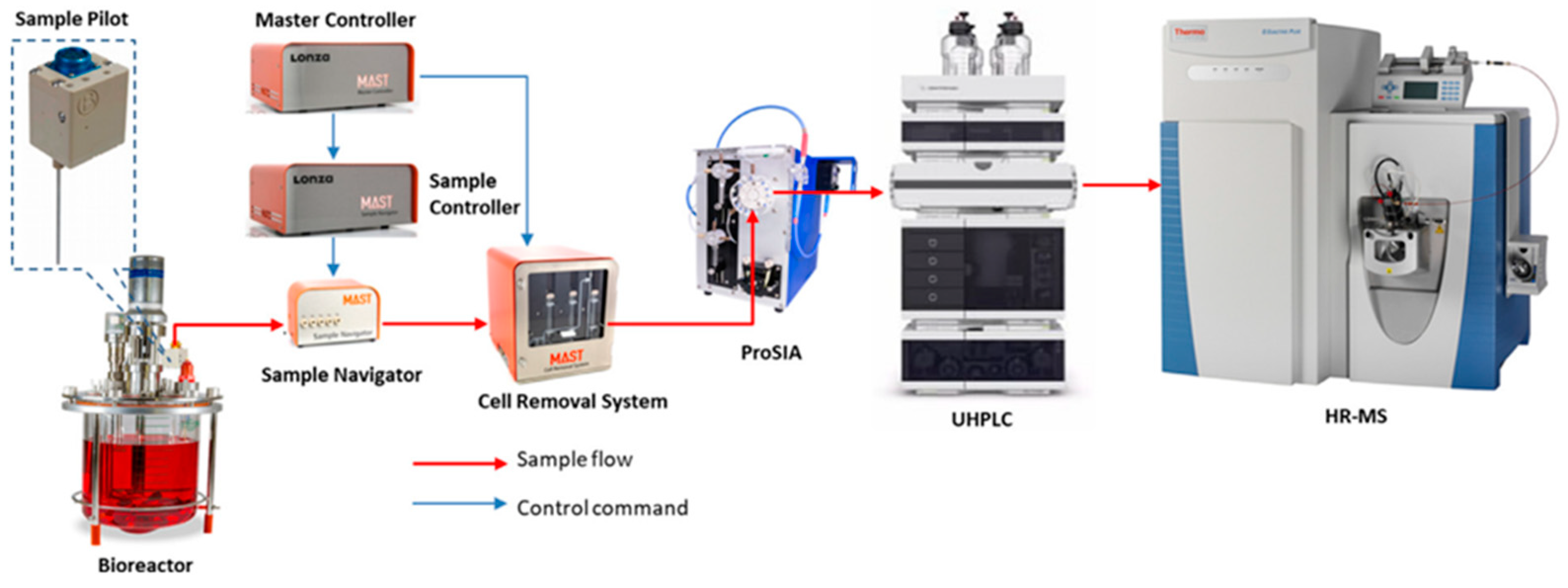
  3. Liu, Y.; Zhang, C.; Chen, J.; Fernandez, J.; Vellala, P.; Kulkarni, T.A.; Aguilar, I.; Ritz, D.; Lan, K.; Patel, P.; et al. A Fully Integrated Online Platform for Real Time Monitoring of Multiple Product Quality Attributes in Bio-Pharmaceutical Processes for Monoclonal Antibody Therapeutics. J. Pharm. Sci. 2022, 111, 358–367. [Google Scholar] [CrossRef] [PubMed]
  4. Zmievskaya, E.; Valiullina, A.; Ganeeva, I.; Petukhov, A.; Rizvanov, A.; Bulatov, E. Application of CAR-T Cell Therapy beyond Oncology: Autoimmune Diseases and Viral Infections. Biomedicines 2021, 9, 59. [Google Scholar] [CrossRef]
  5. Morris, C.; Lee, Y.S.; Yoon, S. Adventitious Agent Detection Methods in Bio-Pharmaceutical Applications with a Focus on Viruses, Bacteria, and Mycoplasma. Curr. Opin. Biotechnol. 2021, 71, 105–114. [Google Scholar] [CrossRef] [PubMed]
  6. Cleveland, M.H.; Anekella, B.; Brewer, M.; Chin, P.-J.; Couch, H.; Delwart, E.; Huggett, J.; Jackson, S.; Martin, J.; Monpoeho, S.; et al. Report of the 2019 NIST-FDA Workshop on Standards for next Generation Sequencing Detection of Viral Adventitious Agents in Biologics and Biomanufacturing. Biologicals 2020, 64, 76–82. [Google Scholar] [CrossRef] [PubMed]
  7. Gustavsson, R.; Mandenius, C.F.; Löfgren, S.; Scheper, T.; Lindner, P. In Situ Microscopy as Online Tool for Detecting Microbial Contaminations in Cell Culture. J. Biotechnol. 2019, 296, 53–60. [Google Scholar] [CrossRef]
  8. Valiant, W.G.; Cai, K.; Vallone, P.M. A History of Adventitious Agent Contamination and the Current Methods to Detect and Remove Them from Pharmaceutical Products. Biologicals 2022, 80, 6–17. [Google Scholar] [CrossRef]
  9. Klug, B.; Robertson, J.S.; Condit, R.C.; Seligman, S.J.; Laderoute, M.P.; Sheets, R.; Williamson, A.-L.; Gurwith, M.; Kochhar, S.; Chapman, L.; et al. Adventitious Agents and Live Viral Vectored Vaccines: Considerations for Archiving Samples of Biological Materials for Retrospective Analysis. Vaccine 2016, 34, 6617–6625. [Google Scholar] [CrossRef]
  10. Cao, Y.; Yu, M.; Dong, G.; Chen, B.; Zhang, B. Digital PCR as an Emerging Tool for Monitoring of Microbial Biodegradation. Molecules 2020, 25, 706. [Google Scholar] [CrossRef]
  11. Shmidt, A.; Egorova, T. PCR-Based Analytical Methods for Quantification and Quality Control of Recombinant Adeno-Associated Viral Vector Preparations. Pharmaceuticals 2021, 15, 23. [Google Scholar] [CrossRef]
  12. Bilitewski, U. Chapter 11 Biosensors for Bioprocess Monitoring. In Comprehensive Analytical Chemistry; Elsevier: Amsterdam, The Netherlands, 2005; Volume 44, pp. 539–578. ISBN 978-0-444-50715-0. [Google Scholar]
  13. Guidance for Industry: PAT—A Framework for Innovative Pharmaceutical Development, Manufacturing and Quality Assurance; US Department of Health and Human Services, Food and Drug Administration: Silver Spring, MD, USA, 2004.
  14. Zandieh, M.; Hosseini, S.N.; Vossoughi, M.; Khatami, M.; Abbasian, S.; Moshaii, A. Label-Free and Simple Detection of En-Dotoxins Using a Sensitive LSPR Biosensor Based on Silver Nanocolumns. Anal. Biochem. 2018, 548, 96–101. [Google Scholar] [CrossRef]
  15. Khansili, N.; Rattu, G.; Krishna, P.M. Label-Free Optical Biosensors for Food and Biological Sensor Applications. Sens. Actuators B Chem. 2018, 265, 35–49. [Google Scholar] [CrossRef]
  16. Barone, P.W.; Wiebe, M.E.; Leung, J.C.; Hussein, I.T.M.; Keumurian, F.J.; Bouressa, J.; Brussel, A.; Chen, D.; Chong, M.; Dehghani, H.; et al. Viral Contamination in Biologic Manufacture and Implications for Emerging Therapies. Nat. Biotechnol. 2020, 38, 563–572. [Google Scholar] [CrossRef] [PubMed]
  17. Cheng, D.L.; Ngo, H.H.; Guo, W.S.; Liu, Y.W.; Zhou, J.L.; Chang, S.W.; Nguyen, D.D.; Bui, X.T.; Zhang, X.B. Bioprocessing for Elimination Antibiotics and Hormones from Swine Wastewater. Sci. Total Environ. 2018, 621, 1664–1682. [Google Scholar] [CrossRef]
  18. Armstrong, S.E.; Mariano, J.A.; Lundin, D.J. The Scope of Mycoplasma Contamination within the Biopharmaceutical Industry. Biol. J. Int. Assoc. Biol. Stand. 2010, 38, 211–213. [Google Scholar] [CrossRef]
  19. Vijayakumar, R.; Sandle, T. A Review on Fungal Contamination in Pharmaceutical Products and Phenotypicidentification of Contaminants by Conventional Methods. Eur. J. Parenter. Pharm. Sci. 2012, 17, 4–18. [Google Scholar]
  20. Kumar, P.; Kausar, M.A.; Singh, A.B.; Singh, R. Biological Contaminants in the Indoor Air Environment and Their Impacts on Human Health. Air Qual. Atmos. Health 2021, 14, 1723–1736. [Google Scholar] [CrossRef]
  21. Merten, O.-W. Virus contaminations of cell cultures—A biotechnological view. Cytotechnology 2002, 39, 91–116. [Google Scholar] [CrossRef]
  22. Seguel, J.M.; Merrill, R.; Seguel, D.; Campagna, A.C. Indoor Air Quality. Am. J. Lifestyle Med. 2017, 11, 284–295. [Google Scholar] [CrossRef]
  23. Zhang, C.; Tian, F.; Zhang, M.; Zhang, Z.; Bai, M.; Guo, G.; Zheng, W.; Wang, Q.; Shi, Y.; Wang, L. Endotoxin Contamination, a Potentially Important Inflammation Factor in Water and Wastewater: A Review. Sci. Total Environ. 2019, 681, 365–378. [Google Scholar] [CrossRef]
  24. Rasuli, L.; Dehghani, M.H.; Aghaei, M.; Mahvi, A.H.; Mubarak, N.M.; Karri, R.R. Occurrence and Fate of Bacterial Endotoxins in the Environment (Air, Water, Wastewater) and Remediation Technologies: An Overview. Chemosphere 2022, 303, 135089. [Google Scholar] [CrossRef] [PubMed]
  25. Barile, M.F. Mycoplasma Infections of Cell Cultures. Isr. J. Med. Sci. 1981, 17, 555–562. [Google Scholar] [CrossRef] [PubMed]
  26. Uphoff, C.C.; Drexler, H.G. Elimination of Mycoplasmas from Infected Cell Lines Using Antibiotics. Cancer Cell Cult. Methods Protoc. 2011, 731, 105–114. [Google Scholar]
  27. Hay, R.J.; Macy, M.L.; Chen, T.R. Mycoplasma Infection of Cultured Cells. Nature 1989, 339, 487–488. [Google Scholar] [CrossRef]
  28. Drexler, H.G.; Uphoff, C.C. Mycoplasma contamination of cell cultures: Incidence, sources, effects, detection, elimination, prevention. Cytotechnology 2002, 39, 75–90. [Google Scholar] [CrossRef]
  29. Fratz-Berilla, E.J.; Angart, P.; Graham, R.J.; Powers, D.N.; Mohammad, A.; Kohnhorst, C.; Faison, T.; Velugula-Yellela, S.R.; Trunfio, N.; Agarabi, C. Impacts on Product Quality Attributes of Monoclonal Antibodies Produced in CHO Cell Bioreactor Cul-Tures during Intentional Mycoplasma Contamination Events. Biotechnol. Bioeng. 2020, 117, 2802–2815. [Google Scholar] [CrossRef]
  30. Nikfarjam, L.; Farzaneh, P. Prevention and detection of Mycoplasma contamination in cell culture. Cell J. 2012, 13, 203–212. [Google Scholar]
  31. Becherucci, V.; Curini, L.; Ceccantini, R.; Bisin, S.; Gori, V.; Gentile, F.; De Rienzo, E.; Piccini, L.; Bindi, B.; Pavan, P.; et al. A Practical Approach for Gmp-Compliant Validation of Real-Time PCR Method for Mycoplasma Detection in Human Mesenchymal Stromal Cells as Advanced Therapy Medicinal Product. Biologicals 2021, 73, 31–40. [Google Scholar] [CrossRef]
  32. Kahane, I.; Banai, M.; Razin, S.; Feldner, J. Attachment of Mycoplasmas to Host Cell Membranes. Clin. Infect. Dis. 1982, 4, 185–192. [Google Scholar] [CrossRef]
  33. Benedetti, F.; Curreli, S.; Zella, D. Mycoplasmas–Host Interaction: Mechanisms of Inflammation and Association with Cellular Transformation. Microorganisms 2020, 8, 1351. [Google Scholar] [CrossRef]
  34. Majdoul, S.; Compton, A.A. Lessons in Self-Defence: Inhibition of Virus Entry by Intrinsic Immunity. Nat. Rev. Immunol. 2022, 22, 339–352. [Google Scholar] [CrossRef] [PubMed]
  35. Casadevall, A.; Coelho, C.; Alanio, A. Mechanisms of Cryptococcus neoformans-Mediated Host Damage. Front. Immunol. 2018, 9, 855. [Google Scholar] [CrossRef]
  36. Malik, P.; Mukherjee, S.; Mukherjee, T.K. Microbial Contamination of Mammalian Cell Culture. In Practical Approach to Mammalian Cell and Organ Culture; Mukherjee, T.K., Malik, P., Mukhopadhyay, S., Eds.; Springer Nature: Singapore, 2023; pp. 1–45. [Google Scholar]
  37. Moineau, S. Bacteriophage. In Brenner’s Encyclopedia of Genetics; Elsevier: Amsterdam, The Netherlands, 2013; pp. 280–283. [Google Scholar]
  38. Abedon, S.T.; Duffy, S.; Turner, P.E. Bacteriophage Ecology. In Encyclopedia of Microbiology; Elsevier: Amsterdam, The Netherlands, 2009; pp. 42–57. [Google Scholar]
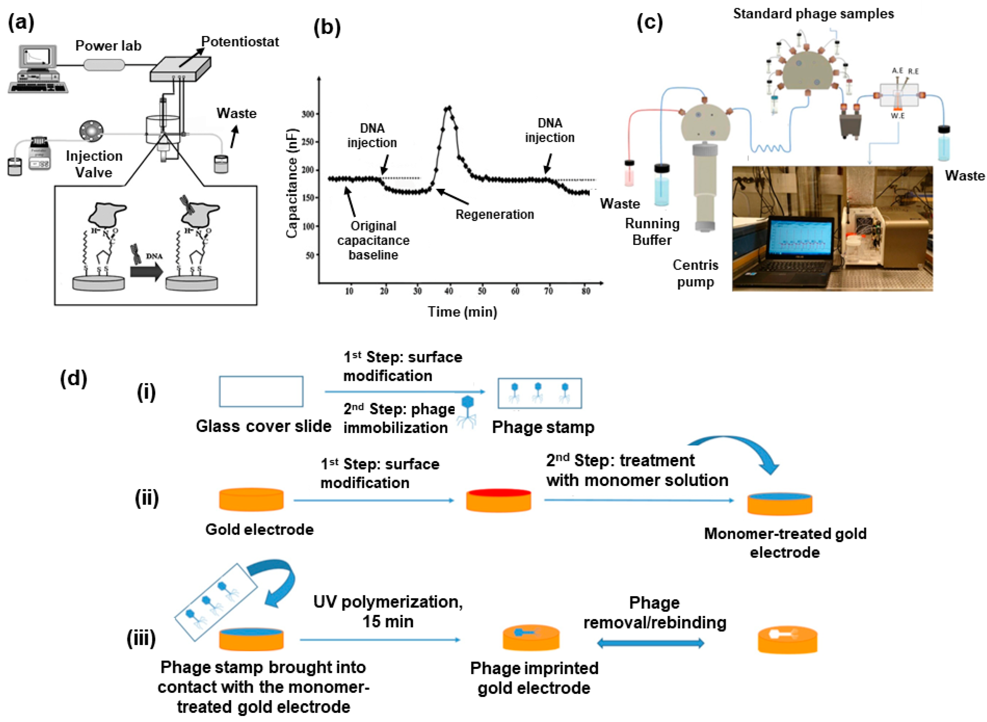
  39. Ertürk, G.; Lood, R. Bacteriophages as Biorecognition Elements in Capacitive Biosensors: Phage and Host Bacteria Detection. Sens. Actuators B Chem. 2018, 258, 535–543. [Google Scholar] [CrossRef]
  40. Rao, G.; Moreira, A.; Brorson, K. Disposable Bioprocessing: The Future Has Arrived. Biotechnol. Bioeng. 2009, 102, 348–356. [Google Scholar] [CrossRef]
  41. Närhi, M.; Nordström, K. National GMP Regulations and Codes and International GMP Guides and Guidelines: Corre-Spondences and Differences. In Pharmaceutical Sciences Encyclopedia; John Wiley & Sons, Inc.: Hoboken, NJ, USA, 2010; p. 378. [Google Scholar]
  42. Haleem, R.M.; Salem, M.Y.; Fatahallah, F.A.; Abdelfattah, L.E. Quality in the Pharmaceutical Industry—A Literature Review. Saudi Pharm. J. 2015, 23, 463–469. [Google Scholar] [CrossRef] [PubMed]
  43. Rathore, A.S.; Li, Y.; Chhabra, H.; Lohiya, A. FDA Warning Letters: A Retrospective Analysis of Letters Issued to Phar-Maceutical Companies from 2010–2020. J. Pharm. Innov. 2022. [Google Scholar] [CrossRef]
  44. Bunn, G.P. (Ed.) Good Manufacturing Practices for Pharmaceuticals, 7th ed.; CRC Press, Taylor & Francis Group: Boca Raton, FL, USA, 2019. [Google Scholar]
  45. Cundell, T. Exclusion of Objectionable Microorganisms from Non-sterile Pharmaceutical Drug Products. In Pharmaceutical Microbiological Quality Assurance and Control; Roesti, D., Goverde, M., Eds.; Wiley: Hoboken, NJ, USA, 2019; pp. 371–400. [Google Scholar]
  46. Jornitz, M.W. (Ed.) Filtration and Purification in the Biopharmaceutical Industry, 3rd ed.; CRC Press: Boca Raton, FL, USA, 2019. [Google Scholar]
  47. Seet, W.T.; Afandi, M.A.M.; Shamsuddin, S.A.; Lokanathan, Y.; Ng, M.H.; Maarof, M. Current Good Manufacturing Practice (CGMP) Facility and Production of Stem Cell; Stem Cell Production, Ed.; Springer: Singapore, 2022. [Google Scholar]
  48. Andriolo, G. GMP-Grade Methods for Cardiac Progenitor Cells: Cell Bank Production and Quality Control. In Stem Cells and Good Manufacturing Practices; Turksen, K., Ed.; Springer: New York, NY, USA, 2020; Volume 2286, pp. 131–166. [Google Scholar]
  49. EU GMP. The Rules Governing Medicinal Products in the European Union. Volume 4 EU Guidelines to Good Manufacturing PRACTICE—Medicinal Products for Human and Veterinary Use, Annex 1–2008. Available online: https://health.ec.europa.eu/system/files/2022-08/20220825_gmp-an1_en_0.pdf (accessed on 3 March 2023).
  50. Whitford, W.G. Bioprocess Intensification: Technologies and Goals. In Process Control, Intensification, and Digitalisation in Continuous Biomanufacturing; Subramanian, G., Ed.; Wiley: Hoboken, NJ, USA, 2022; pp. 93–136. [Google Scholar]
  51. Gerzon, G.; Sheng, Y.; Kirkitadze, M. Process Analytical Technologies—Advances in Bioprocess Integration and Future Perspectives. J. Pharm. Biomed. Anal. 2022, 207, 114379. [Google Scholar] [CrossRef]
  52. Rathore, A.S.; Mishra, S.; Nikita, S.; Priyanka, P. Bioprocess Control: Current Progress and Future Perspectives. Life 2021, 11, 557. [Google Scholar] [CrossRef]
  53. Gomes, J.; Chopda, V.R.; Rathore, A.S. Integrating systems analysis and control for implementing process analytical technology in bioprocess development: Integrating systems analysis and control. J. Chem. Technol. Biotechnol. 2015, 90, 583–589. [Google Scholar] [CrossRef]
  54. Vashist, S.K.; Luong, J.H.T. Immunoassays. In Handbook of Immunoassay Technologies; Elsevier: Amsterdam, The Netherlands, 2018; pp. 1–18. [Google Scholar]
  55. Rathore, A.S.; Bhambure, R.; Ghare, V. Process analytical technology (PAT) for biopharmaceutical products. Anal. Bioanal. Chem. 2010, 398, 137–154. [Google Scholar] [CrossRef]
  56. Shintani, H. Validation Studies for Microbial Contamination and Control of Contaminants. Biocontrol Sci. 2015, 20, 161–170. [Google Scholar] [CrossRef] [PubMed]
  57. Abu-Absi, N.R. Real Time Monitoring of Multiple Parameters in Mammalian Cell Culture Bioreactors Using an In-Line Raman Spectroscopy Probe. Biotechnol. Bioeng. 2011, 108, 1215–1221. [Google Scholar] [CrossRef] [PubMed]
  58. Read, E.K.; Shah, R.B.; Riley, B.S.; Park, J.T.; Brorson, K.A.; Rathore, A.S. Process Analytical Technology (PAT) for Bio-Pharmaceutical Products: Part II. Concepts and Applications. Biotechnol. Bioeng. 2010, 105, 285–295. [Google Scholar] [CrossRef]
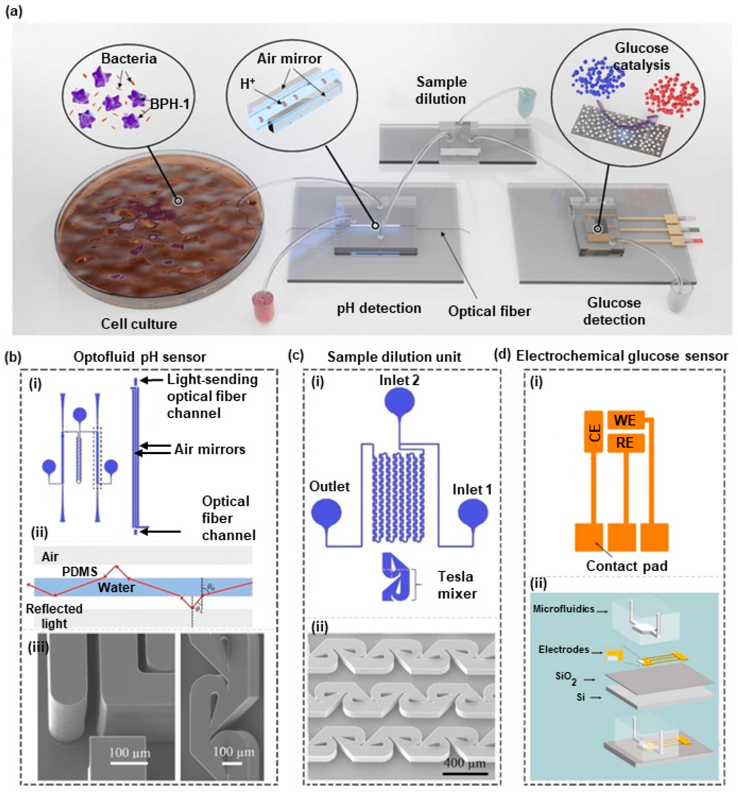
  59. Dervisevic, E.; Dervisevic, M.; Ang, B.; Carthew, J.; Tuck, K.L.; Voelcker, N.H.; Cadarso, V.J. Integrated Microfluidic Device to Monitor Unseen Escherichia Coli Contamination in Mammalian Cell Cul-Ture. Sens. Actuators B Chem. 2022, 359, 131522. [Google Scholar] [CrossRef]
  60. Wehbe, K.; Vezzalini, M.; Cinque, G. Detection of Mycoplasma in Contaminated Mammalian Cell Culture Using FTIR Mi-Crospectroscopy. Anal. Bioanal. Chem. 2018, 410, 3003–3016. [Google Scholar] [CrossRef]
  61. Lowder, J.N.; Whelton, P. Microbial Contamination of Cellular Products for Hematolymphoid Transplantation Therapy: Assessment of the Problem and Strategies to Minimize the Clinical Impact. Cytotherapy 2003, 5, 377–390. [Google Scholar] [CrossRef]
  62. Bustin, S.A. The MIQE Guidelines: Minimum Information for Publication of Quantitative Real-Time PCR Experiments. Clin. Chem. 2009, 55, 611–622. [Google Scholar] [CrossRef]
  63. Kleinschmidt, K.; Wilkens, E.; Glaeser, S.P.; Kaempfer, P.; Staerk, A.; Roesti, D. Development of a Qualitative Real-Time PCR for Microbiological Quality Control Testing in Mammalian Cell Culture Production. J. Appl. Microbiol. 2017, 122, 997–1008. [Google Scholar] [CrossRef]
  64. Davies, C. Immunoassay Performance Measures 1 1This Chapter Is from the First Edition of The Immunoassay Handbook, with Few Changes, Because of the High Regard in Which Chris Davies’ Original Material Is Held. The Theory Still Applies but Some of the Examples given Are Dated. In The Immunoassay Handbook; Elsevier: Amsterdam, The Netherlands, 2013; pp. 11–26. [Google Scholar]
  65. Findlay, J.W.A. Validation of Immunoassays for Bioanalysis: A Pharmaceutical Industry Perspective. J. Pharm. Biomed. Anal. 2000, 21, 1249–1273. [Google Scholar] [CrossRef]
  66. Novitsky, T.J. Biomedical Applications of Limulus Amebocyte Lysate. In Biology and Conservation of Horseshoe Crabs; Tanacredi, J.T., Botton, M.L., Smith, D., Eds.; Springer: Boston, MA, USA, 2009; pp. 315–329. [Google Scholar]
  67. Brown, J.; Bir, A.; Bain, R.E.S. Novel Methods for Global Water Safety Monitoring: Comparative Analysis of Low-Cost, Field-Ready E. Coli Assays. npj Clean. Water 2020, 3, 9. [Google Scholar] [CrossRef]
  68. Arnaout, R.; Lee, R.A.; Lee, G.R.; Callahan, C.; Yen, C.F.; Smith, K.P.; Arora, R.; Kirby, J.E. SARS-CoV-2 Testing: The Limit of Detection Matters. bioRxiv 2020. [Google Scholar] [CrossRef]
  69. Maturin, L. Aerobic Plate Count. In Bacteriological Analytical Manual Online. 2001. Available online: https://cir.nii.ac.jp/all?q=http://www.cfsan.fda.gov/%E3%80%9Cebam/bam-3.html (accessed on 3 March 2023).
  70. Xiong, X.L.; Wang, S.M.; Zhang, Y.; Chen, L.C. Detection of Endotoxin Concentration Using Piezoelectric Based Biosensor System. AMM 2012, 195–196, 874–878. [Google Scholar] [CrossRef]
  71. Feng, P.; Weagant, S.; Grant, M. Enumeration of Escherichia coli and the Coliform Bacteria. In Bacteriological Analytical Manual, 8th ed.; FDA/Center for Food Safety & Applied Nutrition: College Park, MD, USA. Available online: http://www.cfsan.fda.gov/~ebam/bam4.html (accessed on 3 March 2023).
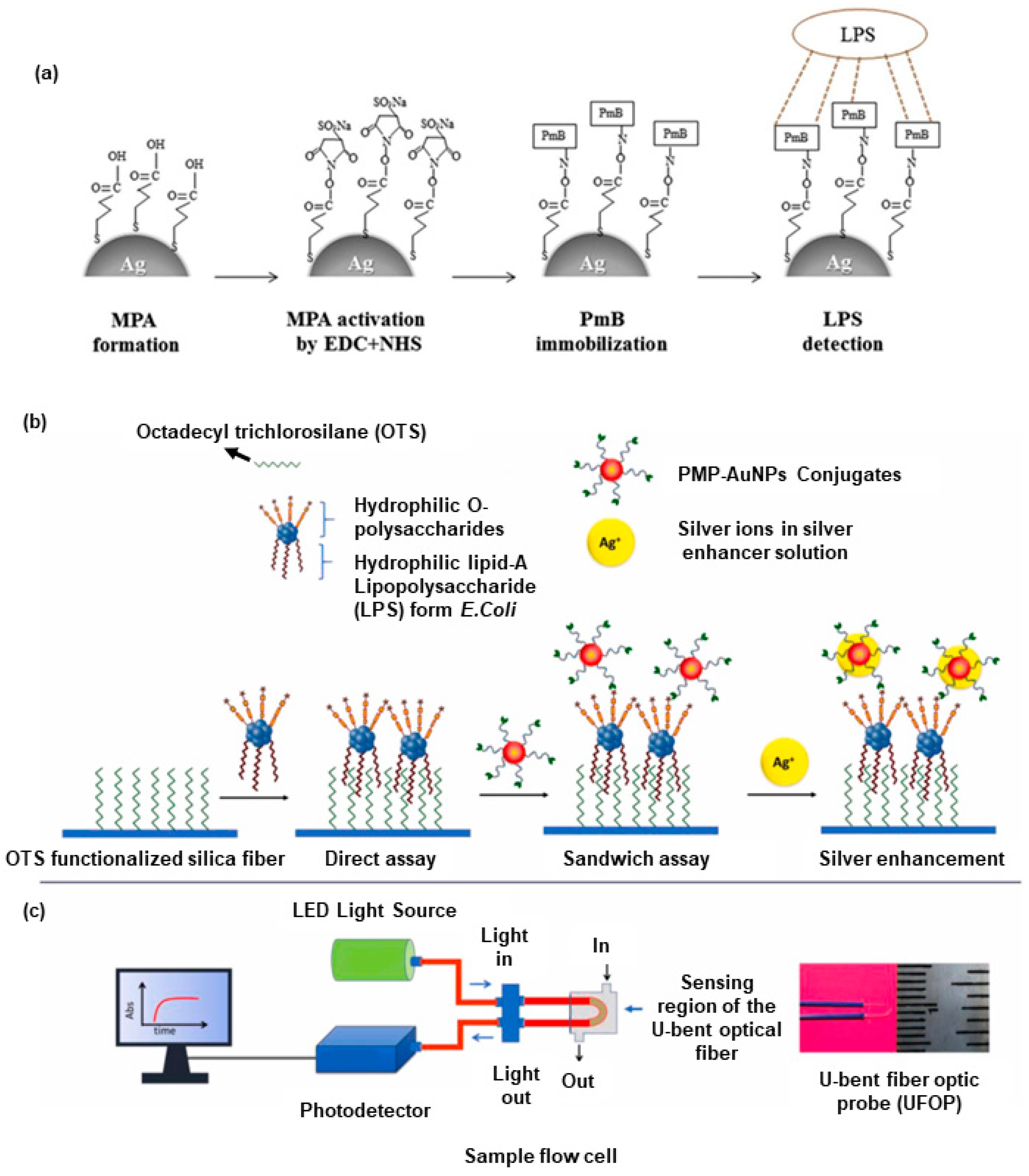
  72. Manoharan, H.; Kalita, P.; Gupta, S.; Sai, V.V.R. Plasmonic biosensors for bacterial endotoxin detection on biomimetic C-18 supported fiber optic probes. Biosens. Bioelectron. 2019, 129, 79–86. [Google Scholar] [CrossRef] [PubMed]
  73. Numnuam, A.; Kanatharana, P.; Mattiasson, B.; Asawatreratanakul, P.; Wongkittisuksa, B.; Limsakul, C.; Thavarungkul, P. Capacitive Biosensor for Quantification of Trace Amounts of DNA. Biosens. Bioelectron. 2009, 24, 2559–2565. [Google Scholar] [CrossRef]
  74. Kim, S.-E.; Su, W.; Cho, M.; Lee, Y.; Choe, W.-S. Harnessing Aptamers for Electrochemical Detection of Endotoxin. Anal. Biochem. 2012, 424, 12–20. [Google Scholar] [CrossRef] [PubMed]
  75. Damborský, P.; Švitel, J.; Katrlík, J. Optical biosensors. Essays Biochem. 2016, 60, 91–100. [Google Scholar] [CrossRef]
  76. Singh, P. Surface Plasmon Resonance: A Boon for Viral Diagnostics. In Reference Module in Life Sciences; Elsevier: Amsterdam, The Netherlands, 2017; p. 9780128096338123000. [Google Scholar]
  77. Balbinot, S.; Srivastav, A.M.; Vidic, J.; Abdulhalim, I.; Manzano, M. Plasmonic biosensors for food control. Trends Food Sci. Technol. 2021, 111, 128–140. [Google Scholar] [CrossRef]
  78. Hamza, M.E.; Othman, M.A.; Swillam, M.A. Plasmonic Biosensors: Review. Biology 2022, 11, 621. [Google Scholar] [CrossRef] [PubMed]
  79. Srivastava, K.R.; Awasthi, S.; Mishra, P.K.; Srivastava, P.K. Biosensors/Molecular Tools for Detection of Waterborne Pathogens. In Waterborne Pathogens; Elsevier: Amsterdam, The Netherlands, 2020; pp. 237–277. [Google Scholar]
  80. Mulchandani, A.; Bassi, A.S. Principles and Applications of Biosensors for Bioprocess Monitoring and Control. Crit. Rev. Biotechnol. 1995, 15, 105–124. [Google Scholar] [CrossRef]
  81. Carpenter, A.; Paulsen, I.; Williams, T. Blueprints for Biosensors: Design, Limitations, and Applications. Genes 2018, 9, 375. [Google Scholar] [CrossRef]
  82. Gavrilaș, S.; Ursachi, C.Ș.; Perța-Crișan, S.; Munteanu, F.-D. Recent Trends in Biosensors for Environmental Quality Monitoring. Sensors 2022, 22, 1513. [Google Scholar] [CrossRef]
  83. Reardon, K.F. Practical Monitoring Technologies for Cells and Substrates in Biomanufacturing. Curr. Opin. Biotechnol. 2021, 71, 225–230. [Google Scholar] [CrossRef] [PubMed]
  84. Liu, C. Acoustic Ejection Mass Spectrometry: Fundamentals and Applications in High-Throughput Drug Discovery. Expert Opin. Drug Discov. 2022, 17, 775–787. [Google Scholar] [CrossRef] [PubMed]
Figure 1. Infection mechanism of different cell culture contaminants: (a) Bacteria/mycoplasma [33] (reprinted with permission from [33] Copyright 2020 MDPI), (b) viruses [34] (reprinted with permission from [34] Copyright 2019 Elsevier), (c) fungi [35] (reprinted with permission from [35] Copyright 2022 Nature).
Chemosensors 11 00298 g001
Figure 2. (a) An illustration of the cell culture media sample analysis characteristics and sequence for the IMD. (b) Optofluidic pH sensor, (i) complete device detailing the microchannel dimensions and configuration of the optical waveguide, (ii) illustration of the light propagation alongside the optical waveguide, and (iii) SEM micrographs of waveguide and micromixer areas. (c) (i) Sample dilution unit detailing one modified Tesla unit and (ii) SEM micrograph of micromixer area. (d) (i) Design and (ii) components of the electrochemical microfluidic glucose sensor (reprinted with permission from [59] Copyright 2022, Elsevier).
Chemosensors 11 00298 g002
Figure 3. Schematics of the fully automated online UHPLC-MS for real-time CQA monitoring of cell culture process (reprinted with permission from [3] Copyright 2021 Pharmaceutical Biotechnology).
Chemosensors 11 00298 g003
Figure 4. Silver-nanocolumn LSPR biosensor for endotoxin detection: (a) Silver-nanocolumn surface modification steps [14] (reprinted with permission from [14] Copyright 2018 Elsevier), (b) UFOP-based biosensor for endotoxin detection bioassay on an OTS-functionalized UFOP surface, (c) optical setup with a photograph of an actual UFOP with a bend diameter of 1 mm (reprinted with permission from [72] Copyright 2018 Elsevier).
Chemosensors 11 00298 g004
Figure 5. Schematic of the biosensor setup: (a) flow injection capacitive biosensor system to detect trace amounts DNA. The total capacitance measured at the working electrode/solution interface comes from the capacitance of self-assembled thioctic acid monolayer, the capacitance of the histone layer, and the capacitance DNA analyte interaction [73]. (b) Capacitance vs. time plot. The binding of histone and DNA causes the capacitance to decrease (ΔC1) with subsequent signal increase due to dissociation under regeneration conditions. After regeneration, the system can be reused to detect DNA in a new injection (ΔC2) [73]. (Reprinted with permission from [73] Copyright 2009 Elsevier.) (c) Schematic of the capacitive biosensor with an automated flow injection system [39] (reprinted with permission from [39] Copyright 2009 Elsevier). (d) Schematic of the phage imprinted capacitive biosensor. (i) Preparation of the glass coverslips (phage stamps), (ii) preparation of the capacitive gold electrodes, (iii) imprinting of phage onto the gold electrode surface via UV-polymerization and removal/rebinding of phage to the electrode surface [39] (reprinted with permission from [39] Copyright 2009 Elsevier).
Chemosensors 11 00298 g005
Table 1. Summary of analytical parameters of the biological contaminant’s detection methods.
Method TypeTarget BiocontaminantTime Required to Do the TestLODSpecificityReference
LSPREndotoxin1 to 24 h340 pg/mLSelectively detects endotoxin[14]
UFOPsEndotoxin25 min0.4 ng/mLHighly specific[72]
CapacitiveResidual DNA14 min10−5 ng/LSpecific to DNA regardless of source[73]
MIP/CapacitiveE. coli phage
E. coli
Real-time10 pfu/mL
1.0 × 10² cfu/mL
Selective for E. coli phage in river water
Selective for E. coli but recognized other bacteria
[39]
Impedance-based aptasensorEndotoxin15 min0.01 ng/mLMinimal response to other media components (pDNA, RNA, proteins, saccharides, and lipids)[74]
Integrated microfluidic deviceE. coliLess than 8 h27.7 ± 1.3 µM of glucoseHighly selective toward glucose[59]
FTIR microspectroscopyMycoplasma bovis6 min (data acquisition)NANot validate for specificity[60]
Disclaimer/Publisher’s Note: The statements, opinions and data contained in all publications are solely those of the individual author(s) and contributor(s) and not of MDPI and/or the editor(s). MDPI and/or the editor(s) disclaim responsibility for any injury to people or property resulting from any ideas, methods, instructions or products referred to in the content.

