
A Highly Sensitive and Selective Near-Infrared Fluorescent Probe for Detecting Peroxynitrite in Living Cells and Drosophila Brains

Abstract
1. Introduction
2. Materials and Methods
2.1. Spectral Characterization of the Probe
2.2. Cytotoxicity Assay
2.3. Fluorescence Imaging of HeLa Cells
2.4. Fluorescence Imaging of Drosophila Brains
3. Results and Discussion
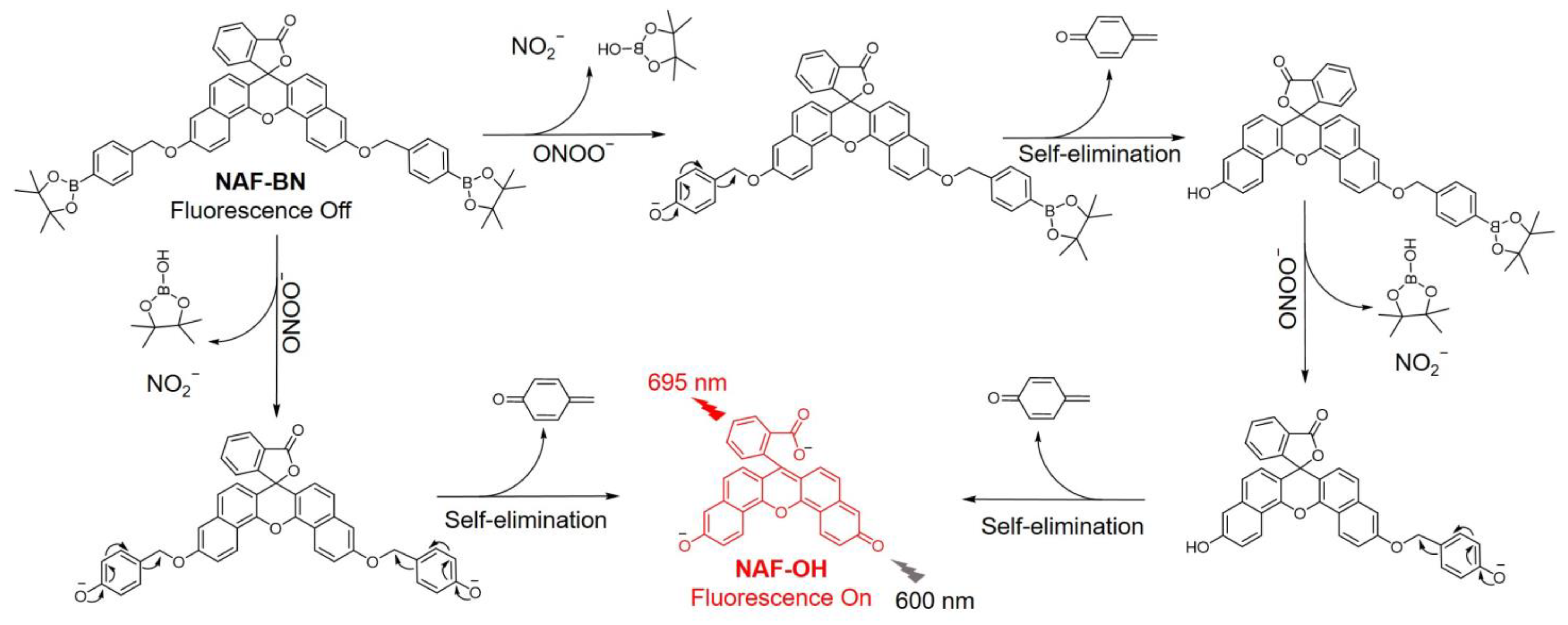
3.1. Design and Synthesis
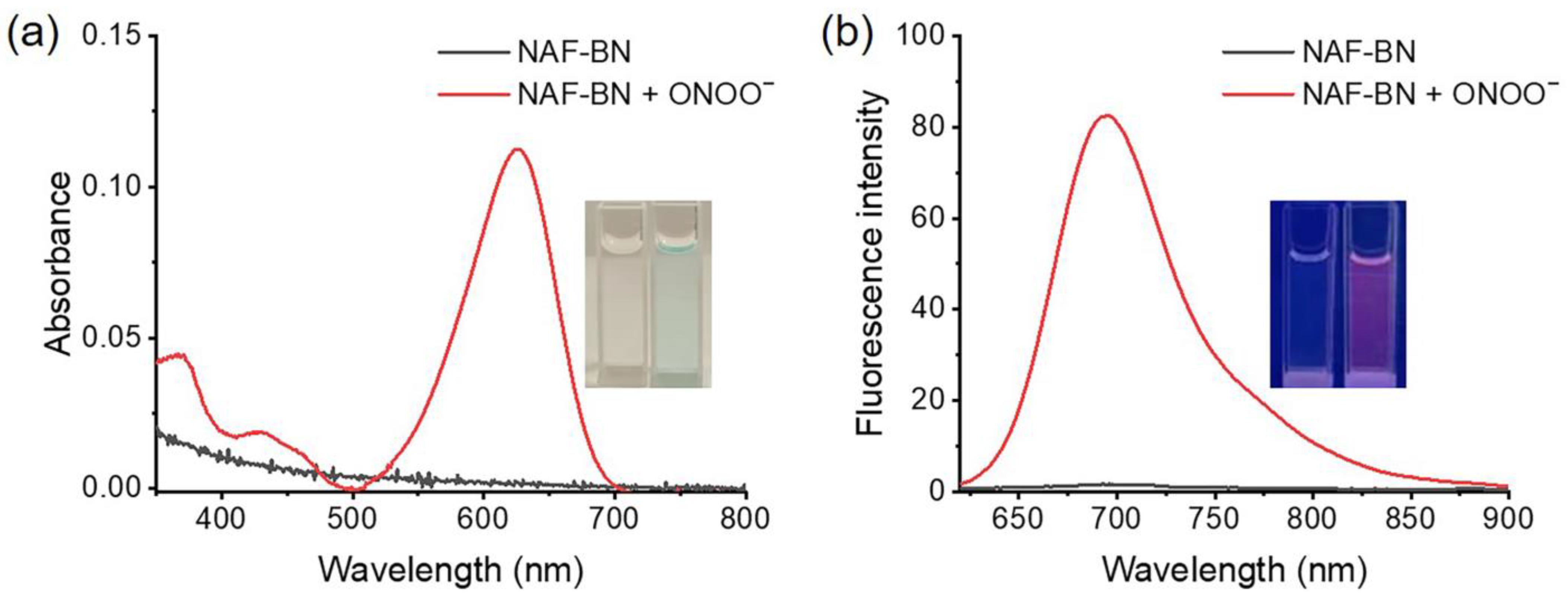
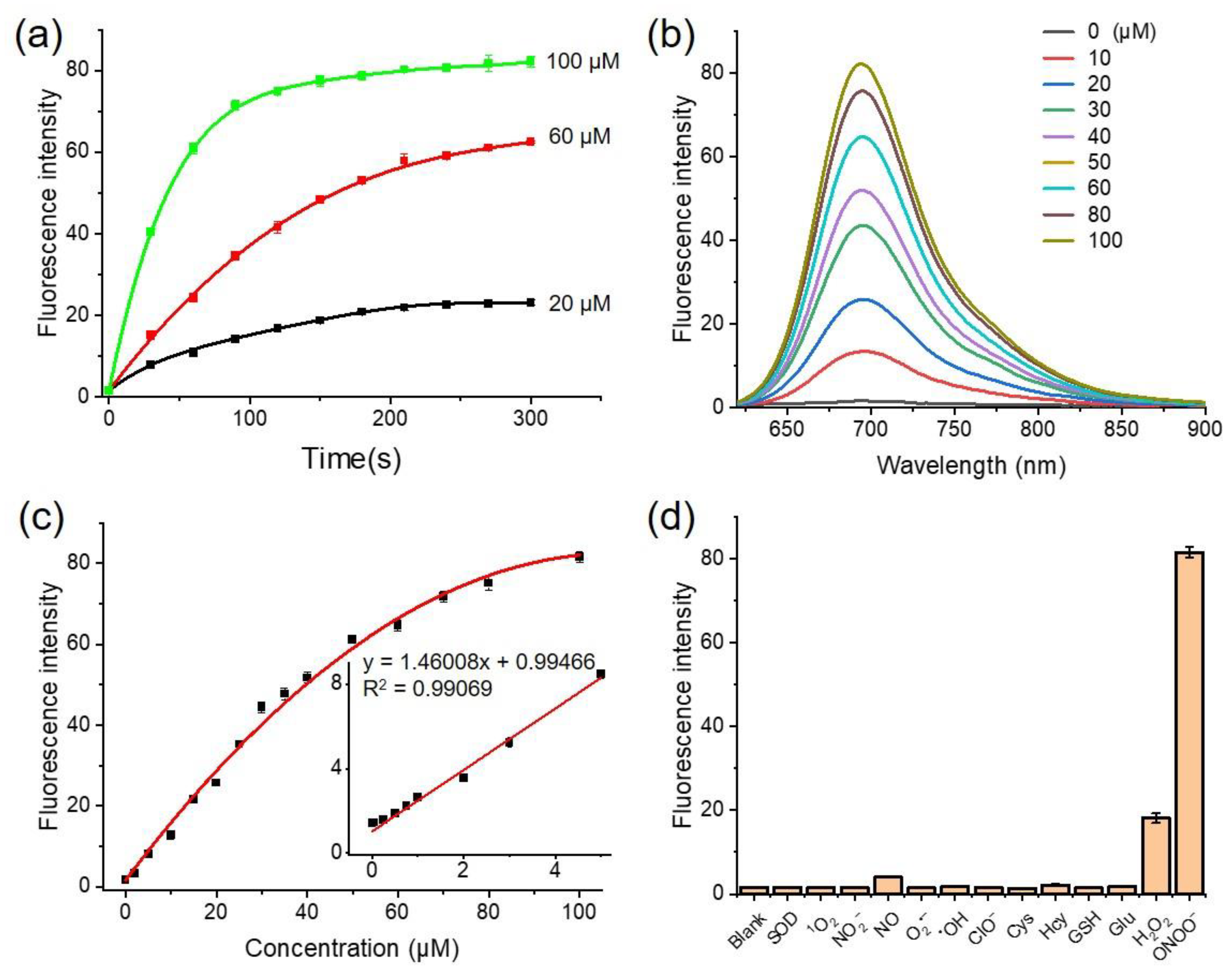
3.2. Sensing Abilities of NAF-BN
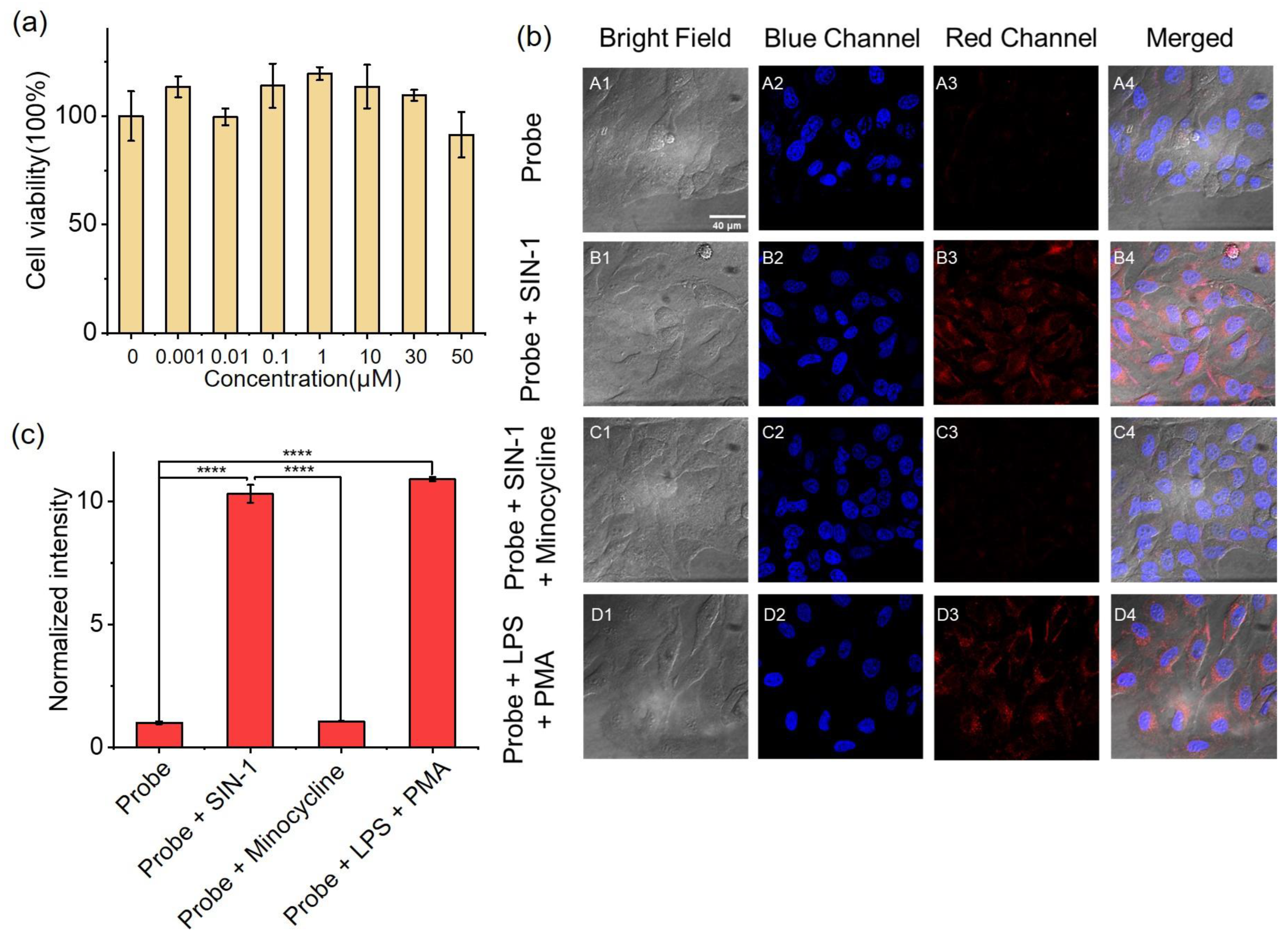
3.3. Bioimaging of ONOO− in HeLa Cells
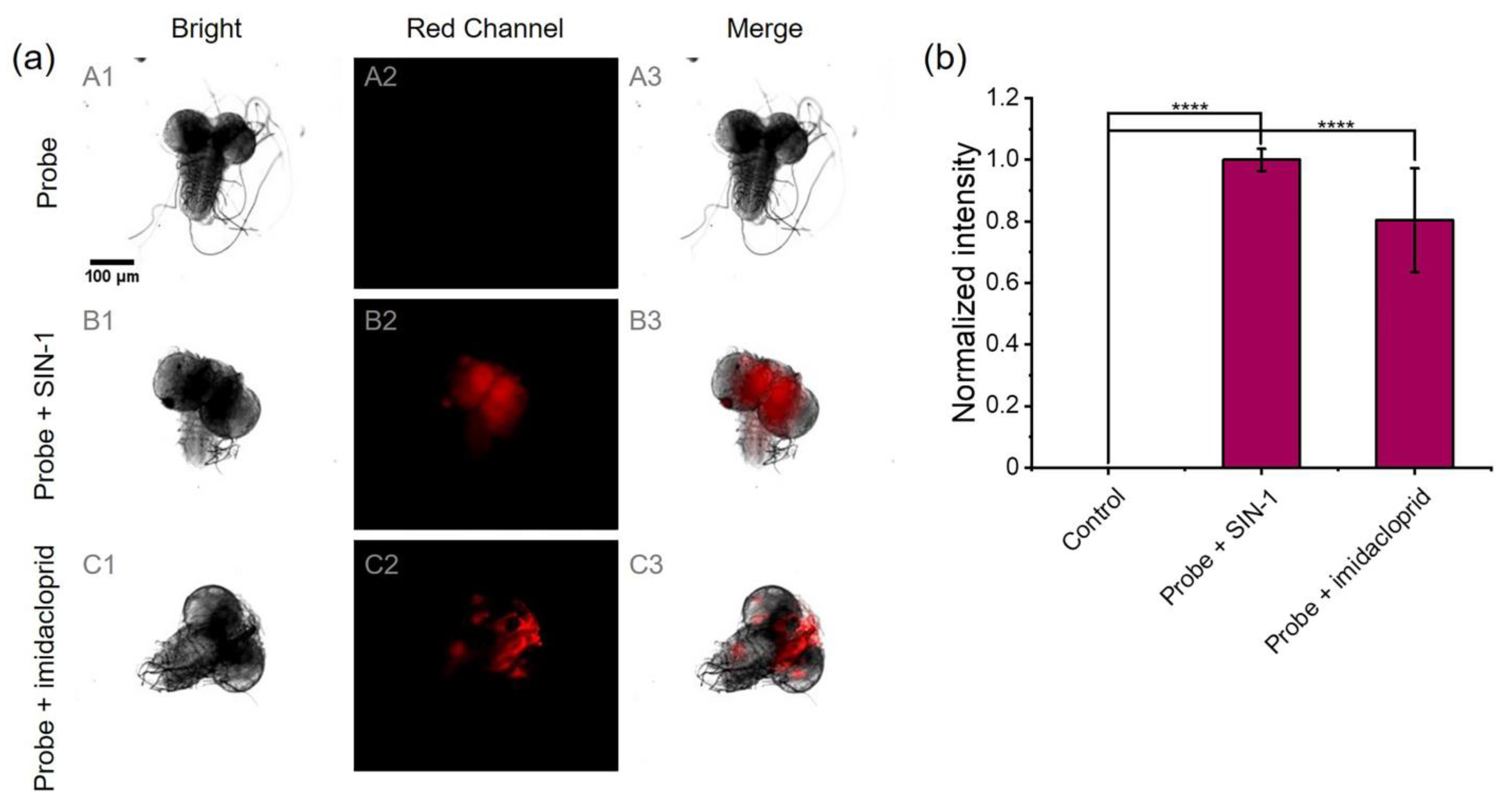
3.4. Bioimaging of ONOO− in Drosophila Brains
4. Conclusions
Supplementary Materials
Author Contributions
Funding
Institutional Review Board Statement
Informed Consent Statement
Data Availability Statement
Acknowledgments
Conflicts of Interest
References
- Blough, N.V.; Zafiriou, O.C. Reaction of Superoxide with Nitric Oxide to Form Peroxonitrite in Alkaline Aqueous Solution. Inorg. Chem. 1985, 24, 3502–3504. [Google Scholar] [CrossRef]
- Botti, H.; Moller, M.N.; Steinmann, D.; Nauser, T.; Koppenol, W.H.; Denicola, A.; Radi, R. Distance-Dependent Diffusion-Controlled Reaction of *NO and O2*− at Chemical Equilibrium with ONOO−. J. Phys. Chem. B 2010, 114, 16584–16593. [Google Scholar] [CrossRef]
- Radi, R. Peroxynitrite, a Stealthy Biological Oxidant. J. Biol. Chem. 2013, 288, 26464–26472. [Google Scholar] [CrossRef]
- Ma, Q.; Xu, S.; Zhai, Z.; Wang, K.; Liu, X.; Xiao, H.; Zhuo, S.; Liu, Y. Recent Progress of Small-Molecule Ratiometric Fluorescent Probes for Peroxynitrite in Biological Systems. Chemistry 2022, 28, e202200828. [Google Scholar] [CrossRef]
- Ferrer-Sueta, G.; Radi, R. Chemical Biology of Peroxynitrite: Kinetics, Diffusion, and Radicals. ACS Chem. Biol. 2009, 4, 161–177. [Google Scholar] [CrossRef]
- Lefer, A.M. Peroxynitrite as a Cytoprotective Agent in Myocardial Ischemia/Reperfusion Injury. Pathophysiology 1998, 5, 38. [Google Scholar] [CrossRef]
- Sies, H.; Jones, D.P. Reactive Oxygen Species (ROS) as Pleiotropic Physiological Signalling Agents. Nat. Rev. Mol. Cell Biol. 2020, 21, 363–383. [Google Scholar] [CrossRef]
- Vana, L.; Kanaan, N.M.; Hakala, K.; Weintraub, S.T.; Binder, L.I. Peroxynitrite-Induced Nitrative and Oxidative Modifications Alter Tau Filament Formation. Biochemistry 2011, 50, 1203–1212. [Google Scholar] [CrossRef]
- Szabo, C. Multiple Pathways of Peroxynitrite Cytotoxicity. Toxicol. Lett. 2003, 140–141, 105–112. [Google Scholar] [CrossRef]
- Szabo, C. DNA Strand Breakage and Activation of Poly-Adp Ribosyltransferase: A Cytotoxic Pathway Triggered by Peroxynitrite. Free Radic. Biol. Med. 1996, 21, 855–869. [Google Scholar] [CrossRef]
- Pandey, V.K.; Amin, P.J.; Shankar, B.S. G1-4a, a Polysaccharide from Tinospora Cordifolia Induces Peroxynitrite Dependent Killer Dendritic Cell (KDC) Activity against Tumor Cells. Int. Immunopharmacol. 2014, 23, 480–488. [Google Scholar] [CrossRef] [PubMed]
- Xia, H.; Zhang, L.; Dai, J.; Liu, X.; Zhang, X.; Zeng, Z.; Jia, Y. Effect of Selenium and Peroxynitrite on Immune Function of Immature Dendritic Cells in Humans. Med. Sci. Monit 2021, 27, e929004. [Google Scholar] [CrossRef] [PubMed]
- Greten, F.R.; Grivennikov, S.I. Inflammation and Cancer: Triggers, Mechanisms, and Consequences. Immunity 2019, 51, 27–41. [Google Scholar] [CrossRef]
- Salvemini, D.; Doyle, T.M.; Cuzzocrea, S. Superoxide, Peroxynitrite and Oxidative/Nitrative Stress in Inflammation. Biochem. Soc. Trans. 2006, 34, 965–970. [Google Scholar] [CrossRef] [PubMed]
- Dedon, P.C.; Tannenbaum, S.R. Reactive Nitrogen Species in the Chemical Biology of Inflammation. Arch Biochem. Biophys. 2004, 423, 12–22. [Google Scholar] [CrossRef]
- Cobb, C.A.; Cole, M.P. Oxidative and Nitrative Stress in Neurodegeneration. Neurobiol. Dis. 2015, 84, 4–21. [Google Scholar] [CrossRef]
- Barnham, K.J.; Masters, C.L.; Bush, A.I. Neurodegenerative Diseases and Oxidative Stress. Nat. Rev. Drug Discov. 2004, 3, 205–214. [Google Scholar] [CrossRef]
- Li, J.; Su, J.; Li, W.; Liu, W.; Altura, B.T.; Altura, B.M. Peroxynitrite Induces Apoptosis in Canine Cerebral Vascular Muscle Cells: Possible Relation to Neurodegenerative Diseases and Strokes. Neurosci. Lett. 2003, 350, 173–177. [Google Scholar] [CrossRef]
- Mungrue, I.N.; Gros, R.; You, X.; Pirani, A.; Azad, A.; Csont, T.; Schulz, R.; Butany, J.; Stewart, D.J.; Husain, M. Cardiomyocyte Overexpression of Inos in Mice Results in Peroxynitrite Generation, Heart Block, and Sudden Death. J. Clin. Investig. 2002, 109, 735–743. [Google Scholar] [CrossRef]
- van der Loo, B.; Labugger, R.; Skepper, J.N.; Bachschmid, M.; Kilo, J.; Powell, J.M.; Palacios-Callender, M.; Erusalimsky, J.D.; Quaschning, T.; Malinski, T.; et al. Enhanced Peroxynitrite Formation Is Associated with Vascular Aging. J. Exp. Med. 2000, 192, 1731–1744. [Google Scholar] [CrossRef]
- Shuhendler, A.J.; Pu, K.; Cui, L.; Uetrecht, J.P.; Rao, J. Real-Time Imaging of Oxidative and Nitrosative Stress in the Liver of Live Animals for Drug-Toxicity Testing. Nat. Biotechnol. 2014, 32, 373–380. [Google Scholar] [CrossRef] [PubMed]
- Peng, J.; Samanta, A.; Zeng, X.; Han, S.; Wang, L.; Su, D.; Loong, D.T.; Kang, N.Y.; Park, S.J.; All, A.H.; et al. Real-Time in vivo Hepatotoxicity Monitoring through Chromophore-Conjugated Photon-Upconverting Nanoprobes. Angew. Chem. Int. Ed. Engl. 2017, 56, 4165–4169. [Google Scholar] [CrossRef] [PubMed]
- Graham, P.M.; Li, J.Z.; Dou, X.; Zhu, H.; Misra, H.P.; Jia, Z.; Li, Y. Protection against Peroxynitrite-Induced DNA Damage by Mesalamine: Implications for Anti-Inflammation and Anti-Cancer Activity. Mol. Cell Biochem. 2013, 378, 291–298. [Google Scholar] [CrossRef] [PubMed]
- Vasilescu, A.; Gheorghiu, M.; Peteu, S. Nanomaterial-Based Electrochemical Sensors and Optical Probes for Detection and Imaging of Peroxynitrite: A Review. Microchim. Acta 2017, 184, 649–675. [Google Scholar] [CrossRef]
- Hulvey, M.K.; Frankenfeld, C.N.; Lunte, S.M. Separation and Detection of Peroxynitrite Using Microchip Electrophoresis with Amperometric Detection. Anal. Chem. 2010, 82, 1608–1611. [Google Scholar] [CrossRef]
- Eliasson, M.J.; Huang, Z.; Ferrante, R.J.; Sasamata, M.; Molliver, M.E.; Snyder, S.H.; Moskowitz, M.A. Neuronal Nitric Oxide Synthase Activation and Peroxynitrite Formation in Ischemic Stroke Linked to Neural Damage. J. Neurosci. 1999, 19, 5910–5918. [Google Scholar] [CrossRef]
- Imaram, W.; Gersch, C.; Kim, K.M.; Johnson, R.J.; Henderson, G.N.; Angerhofer, A. Radicals in the Reaction between Peroxynitrite and Uric Acid Identified by Electron Spin Resonance Spectroscopy and Liquid Chromatography Mass Spectrometry. Free Radic. Biol. Med. 2010, 49, 275–281. [Google Scholar] [CrossRef]
- Jiao, X.; Li, Y.; Niu, J.; Xie, X.; Wang, X.; Tang, B. Small-Molecule Fluorescent Probes for Imaging and Detection of Reactive Oxygen, Nitrogen, and Sulfur Species in Biological Systems. Anal. Chem. 2018, 90, 533–555. [Google Scholar] [CrossRef]
- Cui, W.L.; Wang, M.H.; Yang, Y.H.; Wang, J.Y.; Zhu, X.Z.; Zhang, H.T.; Ji, X.X. Recent Advances and Perspectives in Reaction-Based Fluorescent Probes for Imaging Peroxynitrite in Biological Systems. Coord. Chem. Rev. 2023, 474, 214848. [Google Scholar] [CrossRef]
- Sedgwick, A.C.; Dou, W.T.; Jiao, J.B.; Wu, L.; Williams, G.T.; Jenkins, A.T.A.; Bull, S.D.; Sessler, J.L.; He, X.P.; James, T.D. An Esipt Probe for the Ratiometric Imaging of Peroxynitrite Facilitated by Binding to Abeta-Aggregates. J. Am. Chem. Soc. 2018, 140, 14267–14271. [Google Scholar] [CrossRef]
- Wu, L.; Wang, Y.; Weber, M.; Liu, L.; Sedgwick, A.C.; Bull, S.D.; Huang, C.; James, T.D. Esipt-Based Ratiometric Fluorescence Probe for the Intracellular Imaging of Peroxynitrite. Chem. Commun. 2018, 54, 9953–9956. [Google Scholar] [CrossRef] [PubMed]
- Shu, W.; Wu, Y.L.; Zang, S.P.; Su, S.; Kang, H.; Jing, J.; Zhang, X.L. A Mitochondria-Targeting Highly Specific Fluorescent Probe for Fast Sensing of Endogenous Peroxynitrite in Living Cells. Sens. Actuators B Chem. 2020, 303, 127284. [Google Scholar] [CrossRef]
- Xia, L.; Tong, Y.; Li, L.; Cui, M.; Gu, Y.; Wang, P. A Selective Fluorescent Turn-on Probe for Imaging Peroxynitrite in Living Cells and Drug-Damaged Liver Tissues. Talanta 2019, 204, 431–437. [Google Scholar] [CrossRef] [PubMed]
- Odyniec, M.L.; Park, S.J.; Gardiner, J.E.; Webb, E.C.; Sedgwick, A.C.; Yoon, J.; Bull, S.D.; Kim, H.M.; James, T.D. A Fluorescent Esipt-Based Benzimidazole Platform for the Ratiometric Two-Photon Imaging of ONOO− in vitro and ex vivo. Chem. Sci. 2020, 11, 7329–7334. [Google Scholar] [CrossRef] [PubMed]
- Qu, W.; Niu, C.; Zhang, X.; Chen, W.; Yu, F.; Liu, H.; Zhang, X.; Wang, S. Construction of a Novel Far-Red Fluorescence Light-up Probe for Visualizing Intracellular Peroxynitrite. Talanta 2019, 197, 431–435. [Google Scholar] [CrossRef]
- Li, Y.; Wu, Y.; Chen, L.; Zeng, H.; Chen, X.; Lun, W.; Fan, X.; Wong, W.Y. A Time-Resolved near-Infrared Phosphorescent Iridium(Iii) Complex for Fast and Highly Specific Peroxynitrite Detection and Bioimaging Applications. J. Mater. Chem. B 2019, 7, 7612–7618. [Google Scholar] [CrossRef]
- Zhang, K.; Wang, Z.; Hu, X.; Meng, J.; Bao, W.; Wang, X.; Ding, W.; Tian, Z. A Long-Wavelength Turn-on Fluorescent Probe for Intracellular Nanomolar Level Peroxynitrite Sensing with Second-Level Response. Talanta 2020, 219, 121354. [Google Scholar] [CrossRef]
- Zhou, Y.; Li, P.; Fan, N.; Wang, X.; Liu, X.; Wu, L.; Zhang, W.; Zhang, W.; Ma, C.; Tang, B. In Situ Visualization of Peroxisomal Peroxynitrite in the Livers of Mice with Acute Liver Injury Induced by Carbon Tetrachloride Using a New Two-Photon Fluorescent Probe. Chem. Commun. 2019, 55, 6767–6770. [Google Scholar] [CrossRef]
- Yan, M.; Fang, H.X.; Wang, X.Q.; Xu, J.J.; Zhang, C.W.; Xu, L.; Li, L. A Two-Photon Fluorescent Probe for Visualizing Endoplasmic Reticulum Peroxynitrite in Parkinson’s Disease Models. Sens. Actuators B Chem. 2021, 328, 129003. [Google Scholar] [CrossRef]
- Liu, X.; Gu, F.; Zhou, X.; Zhou, W.; Zhang, S.; Cui, L.; Guo, T. A Naphthalimide-Based Turn-on Fluorescence Probe for Peroxynitrite Detection and Imaging in Living Cells. RSC Adv. 2020, 10, 38281–38286. [Google Scholar] [CrossRef]
- Li, M.; Gong, X.; Li, H.W.; Han, H.; Shuang, S.; Song, S.; Dong, C. A Fast Detection of Peroxynitrite in Living Cells. Anal. Chim. Acta 2020, 1106, 96–102. [Google Scholar] [CrossRef] [PubMed]
- Zhu, B.C.; Zhang, M.; Wu, L.; Zhao, Z.Y.; Liu, C.Y.; Wang, Z.K.; Duan, Q.X.; Wang, Y.W.; Jia, P. A Highly Specific Far-Red Fluorescent Probe for Imaging Endogenous Peroxynitrite in the Mitochondria of Living Cells. Sens. Actuators B Chem. 2018, 257, 436–441. [Google Scholar] [CrossRef]
- Guria, U.N.; Gangopadhyay, A.; Ali, S.S.; Maiti, K.; Samanta, S.K.; Manna, S.; Ghosh, A.K.; Uddin, M.R.; Mandal, S.; Mahapatra, A.K. A Benzothiazole-Conjugated Hemicyanine Dye as a Ratiometric Nir Fluorescent Probe for the Detection and Imaging of Peroxynitrite in Living Cells. Anal. Methods 2019, 11, 5447–5454. [Google Scholar] [CrossRef]
- Fang, Y.; Chen, R.-X.; Qin, H.-F.; Wang, J.-J.; Zhang, Q.; Chen, S.; Wen, Y.-H.; Wang, K.-P.; Hu, Z.-Q. A Chromene Based Fluorescence Probe: Accurate Detection of Peroxynitrite in Mitochondria, Not Elsewhere. Sens. Actuators B Chem. 2021, 334, 129603. [Google Scholar] [CrossRef]
- Yuan, R.Q.; Ma, Y.H.; Du, J.Y.; Meng, F.X.; Guo, J.J.; Hong, M.; Yue, Q.L.; Li, X.; Li, C.Z. A Novel Highly Selective near-Infrared and Naked-Eye Fluorescence Probe for Imaging Peroxynitrite. Anal. Methods 2019, 11, 1522–1529. [Google Scholar] [CrossRef]
- Wu, Y.; Shi, A.; Li, Y.; Zeng, H.; Chen, X.; Wu, J.; Fan, X. A near-Infrared Xanthene Fluorescence Probe for Monitoring Peroxynitrite in Living Cells and Mouse Inflammation Model. Analyst 2018, 143, 5512–5519. [Google Scholar] [CrossRef]
- Zhou, D.Y.; Juan, O.Y.; Li, Y.; Jiang, W.L.; Yang, T.; Yi, Z.M.; Li, C.Y. A Ratiometric Fluorescent Probe for the Detection of Peroxynitrite with Simple Synthesis and Large Emission Shift and Its Application in Cells Image. Dye. Pigment. 2019, 161, 288–295. [Google Scholar] [CrossRef]
- Zhang, J.; Kan, J.; Sun, Y.; Won, M.; Kim, J.H.; Zhang, W.; Zhou, J.; Qian, Z.; Kim, J.S. Nanoliposomal Ratiometric Fluorescent Probe toward ONOO(−) Flux. ACS Appl. Bio. Mater. 2021, 4, 2080–2088. [Google Scholar] [CrossRef]
- An, Q.; Su, S.Z.; Chai, L.; Wang, Y.Y.; Wang, X.M.; Li, X.C.; Liang, T.; Hu, W.; Song, X.J.; Li, C.Y. Imaging of Peroxynitrite in Mitochondria by a near-Infrared Fluorescent Probe with a Large Stokes Shift. Talanta 2023, 253, 124073. [Google Scholar] [CrossRef]
- Yu, F.; Li, P.; Wang, B.; Han, K. Reversible Near-Infrared Fluorescent Probe Introducing Tellurium to Mimetic Glutathione Peroxidase for Monitoring the Redox Cycles between Peroxynitrite and Glutathione in vivo. J. Am. Chem. Soc. 2013, 135, 7674–7680. [Google Scholar] [CrossRef]
- Yu, F.; Li, P.; Li, G.; Zhao, G.; Chu, T.; Han, K. A near-Ir Reversible Fluorescent Probe Modulated by Selenium for Monitoring Peroxynitrite and Imaging in Living Cells. J. Am. Chem. Soc. 2011, 133, 11030–11033. [Google Scholar] [CrossRef] [PubMed]
- Hou, T.; Zhang, K.; Kang, X.; Guo, X.; Du, L.; Chen, X.; Yu, L.; Yue, J.; Ge, H.; Liu, Y.; et al. Sensitive Detection and Imaging of Endogenous Peroxynitrite Using a Benzo[D]Thiazole Derived Cyanine Probe. Talanta 2019, 196, 345–351. [Google Scholar] [CrossRef] [PubMed]
- Zhang, W.; Liu, Y.; Gao, Q.; Liu, C.; Song, B.; Zhang, R.; Yuan, J. A Ruthenium(Ii) Complex-Cyanine Energy Transfer Scaffold Based Luminescence Probe for Ratiometric Detection and Imaging of Mitochondrial Peroxynitrite. Chem. Commun. 2018, 54, 13698–13701. [Google Scholar] [CrossRef]
- Xue, X.L.; Zhang, H.; Chen, G.H.; Yu, G.H.; Hu, H.R.; Niu, S.Y.; Wang, K.P.; Hu, Z.Q. Coumarin-Cyanine Hybrid: A Ratiometric Fluorescent Probe for Accurate Detection of Peroxynitrite in Mitochondria. Spectrochim. Acta A Mol. Biomol. Spectrosc. 2023, 292, 122443. [Google Scholar] [CrossRef]
- Johnson, R.E.; van der Zalm, J.M.; Chen, A.; Bell, I.J.; Van Raay, T.J.; Al-Abdul-Wahid, M.S.; Manderville, R.A. Unraveling the Chemosensing Mechanism by the 7-(Diethylamino) Coumarin-Hemicyanine Hybrid: A Ratiometric Fluorescent Probe for Hydrogen Peroxide. Anal. Chem. 2022, 94, 11047–11054. [Google Scholar] [CrossRef] [PubMed]
- Bolland, H.R.; Hammond, E.M.; Sedgwick, A.C. A Fluorescent Probe Strategy for the Detection and Discrimination of Hydrogen Peroxide and Peroxynitrite in Cells. Chem. Commun. 2022, 58, 10699–10702. [Google Scholar] [CrossRef] [PubMed]
- Albers, A.E.; Dickinson, B.C.; Miller, E.W.; Chang, C.J. A Red-Emitting Naphthofluorescein-Based Fluorescent Probe for Selective Detection of Hydrogen Peroxide in Living Cells. Bioorg. Med. Chem. Lett. 2008, 18, 5948–5950. [Google Scholar] [CrossRef]
- Hou, J.; Qian, M.; Zhao, H.; Li, Y.; Liao, Y.; Han, G.; Xu, Z.; Wang, F.; Song, Y.; Liu, Y. A near-Infrared Ratiometric/Turn-on Fluorescent Probe for in vivo Imaging of Hydrogen Peroxide in a Murine Model of Acute Inflammation. Anal. Chim. Acta 2018, 1024, 169–176. [Google Scholar] [CrossRef]
- Wang, K.; Ma, W.; Xu, Y.; Liu, X.; Chen, G.; Yu, M.; Pan, Q.; Huang, C.; Li, X.; Mu, Q.; et al. Design of a Novel Mitochondria Targetable Turn-on Fluorescence Probe for Hydrogen Peroxide and Its Two-Photon Bioimaging Applications. Chin. Chem. Lett. 2020, 31, 3149–3152. [Google Scholar] [CrossRef]
- Wang, W.X.; Jiang, W.L.; Mao, G.J.; Tan, M.; Fei, J.; Li, Y.; Li, C.Y. Monitoring the Fluctuation of Hydrogen Peroxide in Diabetes and Its Complications with a Novel Near-Infrared Fluorescent Probe. Anal. Chem. 2021, 93, 3301–3307. [Google Scholar] [CrossRef]
- Sikora, A.; Zielonka, J.; Lopez, M.; Joseph, J.; Kalyanaraman, B. Direct Oxidation of Boronates by Peroxynitrite: Mechanism and Implications in Fluorescence Imaging of Peroxynitrite. Free Radic. Biol. Med. 2009, 47, 1401–1407. [Google Scholar] [CrossRef]
- Jourden, J.L.; Daniel, K.B.; Cohen, S.M. Investigation of Self-Immolative Linkers in the Design of Hydrogen Peroxide Activated Metalloprotein Inhibitors. Chem. Commun. 2011, 47, 7968–7970. [Google Scholar] [CrossRef] [PubMed]
- Daniel, K.B.; Agrawal, A.; Manchester, M.; Cohen, S.M. Readily Accessible Fluorescent Probes for Sensitive Biological Imaging of Hydrogen Peroxide. Chembiochem 2013, 14, 593–598. [Google Scholar] [CrossRef] [PubMed]
- Zielonka, J.; Sikora, A.; Hardy, M.; Joseph, J.; Dranka, B.P.; Kalyanaraman, B. Boronate Probes as Diagnostic Tools for Real Time Monitoring of Peroxynitrite and Hydroperoxides. Chem. Res. Toxicol. 2012, 25, 1793–1799. [Google Scholar] [CrossRef] [PubMed]
- Martin-Romero, F.J.; Gutierrez-Martin, Y.; Henao, F.; Gutierrez-Merino, C. Fluorescence Measurements of Steady State Peroxynitrite Production Upon Sin-1 Decomposition: Nadh Versus Dihydrodichlorofluorescein and Dihydrorhodamine 123. J. Fluoresc. 2004, 14, 17–23. [Google Scholar] [CrossRef] [PubMed]
- Dai, C.; Ciccotosto, G.D.; Cappai, R.; Wang, Y.; Tang, S.; Xiao, X.; Velkov, T. Minocycline Attenuates Colistin-Induced Neurotoxicity via Suppression of Apoptosis, Mitochondrial Dysfunction and Oxidative Stress. J. Antimicrob. Chemother. 2017, 72, 1635–1645. [Google Scholar] [CrossRef]
- Tyagi, S.R.; Tamura, M.; Burnham, D.N.; Lambeth, J.D. Phorbol Myristate Acetate (PMA) Augments Chemoattractant-Induced Diglyceride Generation in Human Neutrophils but Inhibits Phosphoinositide Hydrolysis. Implications for the Mechanism of PMA Priming of the Respiratory Burst. J. Biol. Chem. 1988, 263, 13191–13198. [Google Scholar] [CrossRef]
- Chung, T.H.; Wu, Y.P.; Chew, C.Y.; Lam, C.H.; Tan, K.T. Imaging and Quantification of Secreted Peroxynitrite at the Cell Surface by a Streptavidin-Biotin-Controlled Binding Probe. Chembiochem 2018, 19, 2584–2590. [Google Scholar] [CrossRef]
- Deng, Y.; Feng, G. Visualization of ONOO− and Viscosity in Drug-Induced Hepatotoxicity with Different Fluorescence Signals by a Sensitive Fluorescent Probe. Anal. Chem. 2020, 92, 14667–14675. [Google Scholar] [CrossRef]
- Lu, J.; Li, Z.; Zheng, X.; Tan, J.; Ji, Z.; Sun, Z.; You, J. A Rapid Response near-Infrared Ratiometric Fluorescent Probe for the Real-Time Tracking of Peroxynitrite for Pathological Diagnosis and Therapeutic Assessment in a Rheumatoid Arthritis Model. J. Mater. Chem. B 2020, 8, 9343–9350. [Google Scholar] [CrossRef]
- Wang, P.; Yu, L.; Gong, J.; Xiong, J.; Zi, S.; Xie, H.; Zhang, F.; Mao, Z.; Liu, Z.; Kim, J.S. An Activity-Based Fluorescent Probe for Imaging Fluctuations of Peroxynitrite (ONOO−) in the Alzheimer’s Disease Brain. Angew. Chem. Int. Ed. Engl. 2022, 61, e202206894. [Google Scholar] [CrossRef] [PubMed]
- Mao, G.J.; Gao, G.Q.; Dong, W.P.; Wang, Q.Q.; Wang, Y.Y.; Li, Y.; Su, L.; Zhang, G. A Two-Photon Excited near-Infrared Fluorescent Probe for Imaging Peroxynitrite during Drug-Induced Hepatotoxicity and Its Remediation. Talanta 2021, 221, 121607. [Google Scholar] [CrossRef] [PubMed]
- Xin, F.; Zhao, J.; Shu, W.; Zhang, X.; Luo, X.; Tian, Y.; Xing, M.; Wang, H.; Peng, Y.; Tian, Y. A Thiocarbonate-Caged Fluorescent Probe for Specific Visualization of Peroxynitrite in Living Cells and Zebrafish. Analyst 2021, 146, 7627–7634. [Google Scholar] [CrossRef] [PubMed]
- Liu, Y.; Ma, Y.; Lin, W. Construction of a Bi-Functional Ratiometric Fluorescent Probe for Detection of Endoplasmic Reticulum Viscosity and ONOO− in Cells and Zebrafish. Sens. Actuators B Chem. 2022, 373, 132742. [Google Scholar] [CrossRef]
- Sun, Q.; Xu, J.; Ji, C.; Shaibani, M.S.S.; Li, Z.; Lim, K.; Zhang, C.; Li, L.; Liu, Z. Ultrafast Detection of Peroxynitrite in Parkinson’s Disease Models Using a near-Infrared Fluorescent Probe. Anal. Chem. 2020, 92, 4038–4045. [Google Scholar] [CrossRef]
- Azuma, E.; Nakamura, N.; Kuramochi, K.; Sasamori, T.; Tokitoh, N.; Sagami, I.; Tsubaki, K. Exhaustive Syntheses of Naphthofluoresceins and Their Functions. J. Org. Chem. 2012, 77, 3492–3500. [Google Scholar] [CrossRef]
- Lee, L.G.; Berry, G.M.; Chen, C.H. Vita Blue: A New 633-Nm Excitable Fluorescent Dye for Cell Analysis. Cytometry 1989, 10, 151–164. [Google Scholar] [CrossRef]
- Salerno, M.; Ajimo, J.J.; Dudley, J.A.; Binzel, K.; Urayama, P. Characterization of Dual-Wavelength Seminaphthofluorescein and Seminapthorhodafluor Dyes for Ph Sensing under High Hydrostatic Pressures. Anal. Biochem. 2007, 362, 258–267. [Google Scholar] [CrossRef]
- Yamaguchi, M.; Yoshida, H. Drosophila as a Model Organism. In Drosophila Models for Human Diseases; Yamaguchi, M., Ed.; Springer: Singapore, 2018; pp. 1–10. [Google Scholar]
- Yucel, M.S.; Kayis, T. Imidacloprid Induced Alterations in Oxidative Stress, Biochemical, Genotoxic, and Immunotoxic Biomarkers in Non-Mammalian Model Organism Galleria mellonella L. (Lepidoptera: Pyralidae). J. Environ. Sci. Health B 2019, 54, 27–34. [Google Scholar] [CrossRef]
- Li, Z.; Yu, T.; Chen, Y.; Heerman, M.; He, J.; Huang, J.; Nie, H.; Su, S. Brain Transcriptome of Honey Bees (Apis mellifera) Exhibiting Impaired Olfactory Learning Induced by a Sublethal Dose of Imidacloprid. Pestic. Biochem. Physiol. 2019, 156, 36–43. [Google Scholar] [CrossRef]
- Martelli, F.; Zhongyuan, Z.; Wang, J.; Wong, C.O.; Karagas, N.E.; Roessner, U.; Rupasinghe, T.; Venkatachalam, K.; Perry, T.; Bellen, H.J.; et al. Low Doses of the Neonicotinoid Insecticide Imidacloprid Induce Ros Triggering Neurological and Metabolic Impairments in Drosophila. Proc. Natl. Acad. Sci. USA 2020, 117, 25840–25850. [Google Scholar] [CrossRef] [PubMed]
- Li, H.; Shi, W.; Li, X.; Hu, Y.; Fang, Y.; Ma, H. Ferroptosis Accompanied by •OH Generation and Cytoplasmic Viscosity Increase Revealed Via Dual-Functional Fluorescence Probe. J Am Chem Soc 2019, 141, 18301–18307. [Google Scholar] [CrossRef] [PubMed]
- Massey, V.; Komai, H.; Palmer, G.; Elion, G.B. On the Mechanism of Inactivation of Xanthine Oxidase by Allopurinol and Other Pyrazolo[3,4-D]Pyrimidines. J. Biol. Chem. 1970, 245, 2837–2844. [Google Scholar] [CrossRef] [PubMed]
- Suo, F.T.; Chen, X.W.; Fang, H.X.; Gong, Q.Y.; Yu, C.M.; Yang, N.D.; Li, S.; Wu, Q.; Li, L.; Huang, W. Hybrid Fluorophores-Based Fluorogenic Paper Device for Visually High-Throughput Detection of Cu2+ in Real Samples. Dyes Pigm. 2019, 170, 107639. [Google Scholar] [CrossRef]


Disclaimer/Publisher’s Note: The statements, opinions and data contained in all publications are solely those of the individual author(s) and contributor(s) and not of MDPI and/or the editor(s). MDPI and/or the editor(s) disclaim responsibility for any injury to people or property resulting from any ideas, methods, instructions or products referred to in the content. |
© 2023 by the authors. Licensee MDPI, Basel, Switzerland. This article is an open access article distributed under the terms and conditions of the Creative Commons Attribution (CC BY) license (https://creativecommons.org/licenses/by/4.0/).
Share and Cite
Wang, W.; Deng, J.-B.; Jin, L.; Guan, B.-O. A Highly Sensitive and Selective Near-Infrared Fluorescent Probe for Detecting Peroxynitrite in Living Cells and Drosophila Brains. Chemosensors 2023, 11, 286. https://doi.org/10.3390/chemosensors11050286
Wang W, Deng J-B, Jin L, Guan B-O. A Highly Sensitive and Selective Near-Infrared Fluorescent Probe for Detecting Peroxynitrite in Living Cells and Drosophila Brains. Chemosensors. 2023; 11(5):286. https://doi.org/10.3390/chemosensors11050286
Chicago/Turabian StyleWang, Wei, Jian-Bin Deng, Long Jin, and Bai-Ou Guan. 2023. "A Highly Sensitive and Selective Near-Infrared Fluorescent Probe for Detecting Peroxynitrite in Living Cells and Drosophila Brains" Chemosensors 11, no. 5: 286. https://doi.org/10.3390/chemosensors11050286
APA StyleWang, W., Deng, J.-B., Jin, L., & Guan, B.-O. (2023). A Highly Sensitive and Selective Near-Infrared Fluorescent Probe for Detecting Peroxynitrite in Living Cells and Drosophila Brains. Chemosensors, 11(5), 286. https://doi.org/10.3390/chemosensors11050286