
Chemists Focus on Probes, Biologists on Cells—But Who Talks about Probe-Cell Interactions? A Critical Account of the Suboptimal Reporting of Novel Fluorescent Imaging Probes, Using Lipid Droplet Stains as a Case Study


Abstract
1. Problems with the Reporting of Novel Fluorescent Imaging Probes
2. Using Publications Describing Novel LD Probes as a Case Study
3. Why Are the Technical Factors concerning Probe–Cell Interactions So Important?
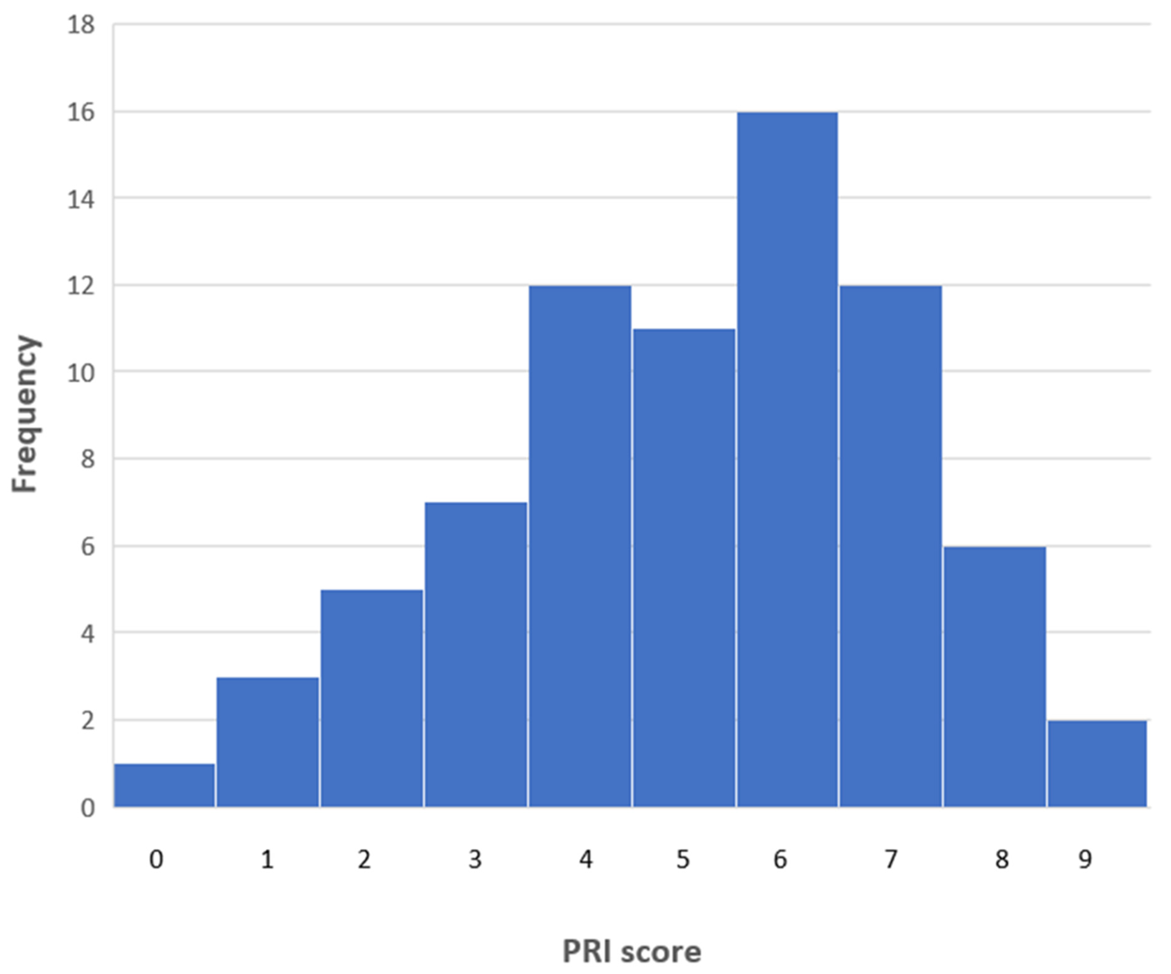
4. The Protocol Reporting Index (PRI) as an Indicator of Quality
5. How Good Is the Reporting of Key Protocol Factors?
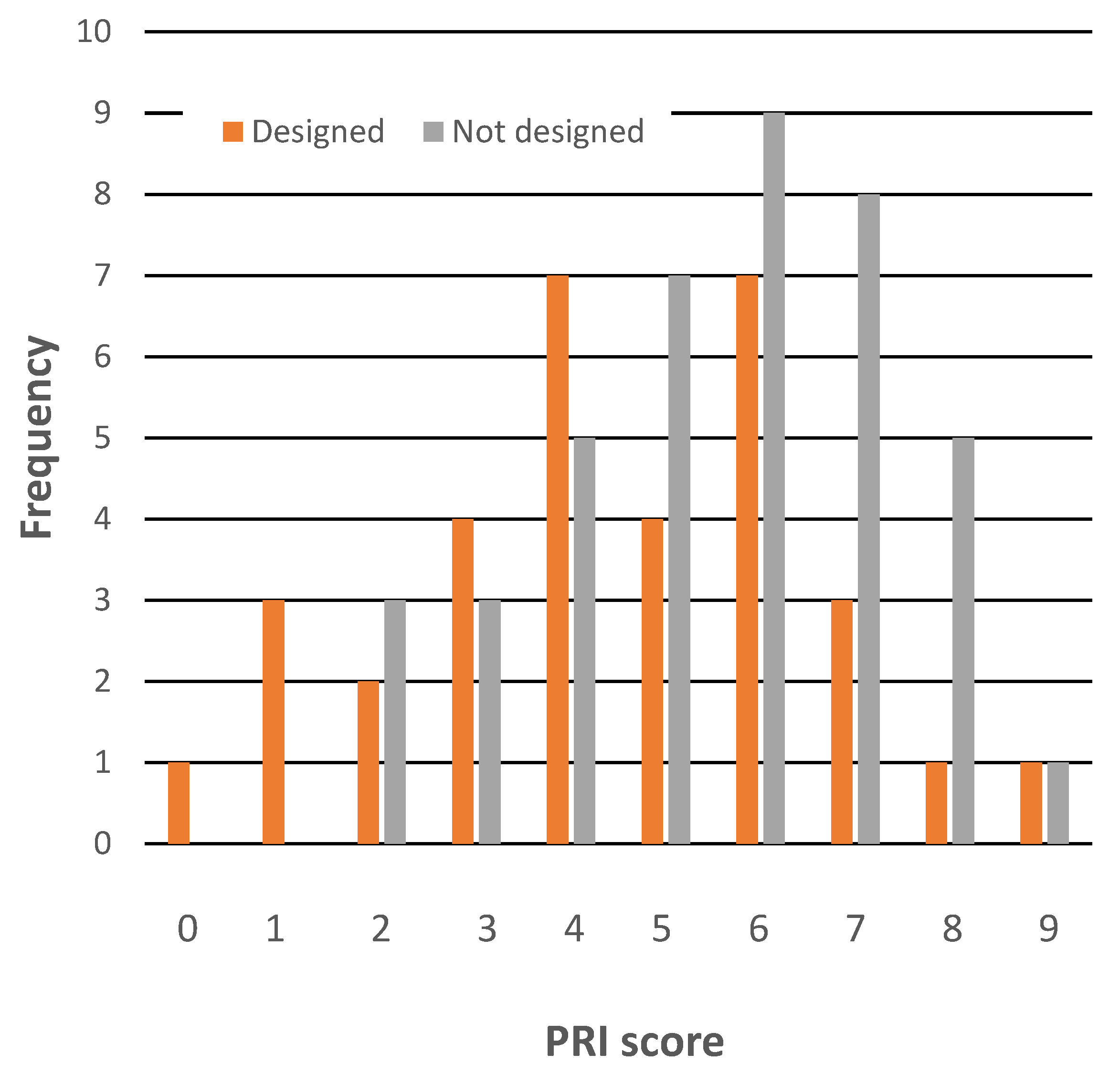
6. Do High-Quality Journals or Papers from Better-Known Research Groups Provide Better Staining Protocol Reporting?
7. The Oddly Biased Use of the Term “Probe Design”
8. A Possible Explanation for the Poor Reporting of Protocol Information
9. Some Suggestions for Remedial Action
- □
- Was the application of the novel probe evaluated using more than one cell type?
- □
- Is a stand-alone staining protocol provided, with a clear heading?
- □
- Is the probe concentration in the staining solution specified?
- □
- Is the staining time specified?
- □
- Is the staining temperature specified?
- □
- Is the solvent for the staining solution specified?
- □
- Are cosolutes in the staining solution specified?
- □
- Was post-staining washing required? If Y, respond to the following queries.
- □
- Were washing solvents and any cosolutes specified?
- □
- Were wash times, number of washes, and washing temperature specified?
10. Conclusions
- Approximately three quarters of scientific reports describing novel fluorescent probes for LDs do not provide sufficient technical information (e.g., concentration, solvent and cosolute, time, post-stain washing procedure) to permit the direct replication of the recommended staining process by another investigator.
- Approximately two thirds of such reports also fail to provide a full account of the supposed staining mechanism of the probe.
- Consequently, a trial application of such probes by biomedical investigators is made harder than need be the case.
- This suboptimal reporting is anomalous, as extensive accounts are typically provided of syntheses and, where appropriate, of the optical properties of the probes. Moreover, the technical protocol information was known to the authors as they carried out the work described.
- A possible explanation for such inadequate reporting is that chemists focus on probe chemistry, biologists on cell properties. The failure of either party to address the interaction of probe and cell is thus a “not my problem” problem.
- It is very unlikely that the omissions discussed are restricted to probes for LDs, and consequently, such omissions are probably of general concern for the fluorescent imaging probe field.
- Correcting such errors of omission requires authors to alter their reporting habits; facilitating such changes is a task for journal editors.
- One way to achieve this would be to adopt the best practice already in place in many chemistry and biology journals. Namely, to devise and make use of checklists of required content, for use by authors, editors, and reviewers.
Funding
Institutional Review Board Statement
Informed Consent Statement
Data Availability Statement
Acknowledgments
Conflicts of Interest
Appendix A
- Appelqvist, H.; Stranius, K.; Börjesson, K,; Nilsson, R.; Dyrager, C. Specific imaging of intracellular lipid droplets using a benzothiazole derivative with solvatochromic properties. Bioconjugate Chem. 2017, 28, 1363–1370. https://doi.org/10.1021/acs.bioconjchem.7b00048.
- Becerra-Ruiz, M.; Vargas, V.; Jara, P.; Tirapegui, C.; Carrasco, C.; Nuñez, M.; Lezana, N.; Galdámez, A.; Vilches-Herrera, M. Blue-fluorescent probes for lipid droplets based on dihydrochromeno-fused pyrazolo- and pyrrolopyridines. Eur. J. Org. Chem. 2018, 4795–4801. https://doi.org/10.1002/ejoc.201701633.
- Benčić, P.; Mandić, L.; Džeba, I.; Bujak, I.T.; Biczók, N.; Mihaljević, B.; Mlinarić-Majerski, K.; Weber, I.; Kralj, M.; Basarić, N. Application of 4-amino-N-adamantylphthalimde solvatochromic dye for fluorescence microscopy in selective visualization of lipid droplets and mitochrondria. Sens Actuators B Chem. 2019, 286, 52–61. https://doi.org/10.1016/j.snb.2019.01.102.
- Chen, H.; Zhao, J.; Lin, J.; Dong, B.; Li, H.; Geng, B.; Yan, M. Two-photon fluorescent probes for detecting the viscosity of lipid droplets and its application in living cells. RSC Adv. 2001, 11, 8250–8254. https://doi.org/10.1039/D0RA09683K.
- Choi, JY.; Kim, G.-H.; Guo, Z.; Lee, H.Y.; Swamy, K.M.K.; Pai, J.; Shin, S.; Yoon, J. Highly selective ratiometric fluorescent probe for Au3+ and its application to bioimaging. Biosens. Bioelectron. 2013, 49, 438–441. https://doi.org/10.1016/j.bios.2013.05.033.
- Chun, H.-S.; Jeon, J.H.; Pagire, H.S.; Lee, J.H.; Chung, H.-C.; Park, M.J.; So, J.-H.; Ryu, J.-H.; Kim, C.-H.; Ahn, J.H.; Bae, M.A. Synthesis of LipidGreen2 and its application in lipid and fatty liver imaging. Mol. BioSyst. 2013, 9, 630–633. https://doi.org/10.1039/C3MB70022D.
- Collot, M.; Bou, S.; Fam, T.K.; Richert, L.; Mély, Y.; Danglot, L.; Klymchenko, A.S. Probing polarity and heterogeneity of lipid droplets in live cells using a push-pull fluorophore. Anal. Chem. 2019, 91, 1928–1935. https://doi.org/10.1021/acs.analchem.8b04218.
- de Moliner, F.; King, A.; Dias, G.G.; de Lima, G.F.; de Simone, C.A.; da Silva, E.N.; Vendrell, M. Quinone-derived π-extended phenazines as new fluorogenic probes for live-cell imaging of lipid droplets. Front. Chem. 2018, 6. 339. https://doi.org/10.3389/fchem.2018.00339.
- Fan, L.; Wang, X.; Zan, Q.; Fan, L.; Li, F.; Yang, Y.; Zhang, C.; Shuang, S.; Dong, C. Lipid droplet-specific fluorescent probe for in vivo visualization of polarity in fatty liver, inflammation, and cancer models. Anal. Chem. 2021, 93, 8019–8026. https://doi.org/10.1021/acs.analchem.1c01125.
- Fernandus. Tan, J.R.; Lim, J.H.; Arai, S.; Sou, K.; Lee, C.-L.K. Squaraine probes for the bimodal staining of lipid droplets and endoplasmic reticulum imaging in live cells. Analyst 2022, 147, 3570–3577. https://doi.org/10.1039/D2AN00803C.
- Gao, M.; Su, H.; Li, S.; Lin, Y.; Ling, X.; Qin, A.; Tang, B.Z. An easily accessible aggregation-induced emission probe for lipid droplet-specific imaging and movement tracking. Chem. Commun. 2017, 53, 921–924. https://doi.org/10.1039/C6CC09471F.
- Gao, M.; Su, H.; Lin, Y.; Ling, X.; Qin, A.; Tang, B.Z. Photoactivatable aggregation-induced emission probes for lipid droplets-specific live cell imaging. Chem. Sci. 2017, 8, 1763–1768. https://doi.org/10.1039/C6SC04842K.
- Goel, A.; Sharma, A.; Kathuria, M.; Bhattacharjee, A.; Verma, A.; Mishra, P.R.; Nazir, A.; Mitra, K. New fluoranthene FLUN-550 as a fluorescent probe for selective staining and quantification of intracellular lipid droplets. Org. Lett. 2014, 16, 756–759. https://doi.org/10.1021/ol403470d.
- Guo, R.; Yin, J.; Ma, Y.; Li, G.; Wang, Q.; Lin, W. A novel NIR probe for detection of viscosity in cellular lipid droplets, zebra fishes and living mice. Sens. Actuators B Chem. 2018, 271, 321–328. https://doi.org/10.1016/j.snb.2018.05.055.
- Jiang, G.; Li, C.; Liu, X.; Chen, Q.; Li, X.; Gu, X.; Zhang, P.; Lai, Q.; Wang, J. Lipid droplet-targetable fluorescence guided photodynamic therapy of cancer cells with an activatable AIE-active fluorescent probe for hydrogen peroxide. Adv. Optical. Mater. 2020, 8, 2001119. https://doi.org/10.1002/adom.202001119.
- Jiang, M.; Gu, X.; Lam, J.W.Y.; Zhang, Y.; Kwok, R.T.K.; Wong, K.S.; Tang, B.Z. Two-photon AIE bio-probe with large Stokes shift for specific imaging of lipid droplets. Chem. Sci. 2017, 8, 5440–5446. https://doi.org/10.1039/C7SC01400G.
- Jiang, W.; Chen, J.; An, K.; Bao, P.; Qiao, Q.; Liu, X.; Xu, Z. Constructing D-π-A-π dye to obtain red-emission probe for structured illumination microscopy imaging of lipid droplet dynamics. Green Chem. Eng. 2022, in press. https://doi.org/10.1016/j.gce.2022.07.002.
- Kang, M.; Gu, X.; Kwok, R.T.K.; Leung, C.W.T.; Lam, J.W.Y.; Li, F.; Tang, B.Z. A near-infrared AIEgen for specific imaging of lipid droplets. Chem. Commun. 2016, 52, 5957–5960. https://doi.org/10.1039/C6CC01797E.
- Kuntam, S.; Puskás, L.G.; Ayadin, F. Characterization of a new class of blue-fluorescent lipid droplet markers for live-cell imaging in plants. Plant Cell Rep. 2015, 34, 655–665. https://doi.org/10.1007/s00299-015-1738-4.
- Lai, Z.-L.; Chang, J.-S.; Chan, Y.-C.; Chang, C.-C.; Li, C.-Y. Tumor tissues diagnosis with PIEE lipid droplet vesicles. Sens. Actuators B Chem. 2021, 330, 129269. https://doi.org/10.1016/j.snb.2020.129269.
- Lee, H.W.; Lee, I.-J.; Lee, S.-J.; Kim, Y.R.; Kim, H.M. Highly sensitive two-photon lipid droplet tracker for in vivo screening of drug induced liver injury. ACS Sens. 2022, 7.4, 1027–1035. Doi.org/10.1021/acssensors.1c02679.
- Lee, J.H.; So, J.-H.; Jeon, J.H.; Choi, E.B.; Lee, Y.-R.; Chang, Y.-T.; Kim, C.-H.; Bae, M.A.; Ahn, J.H. Synthesis of a new fluorescent small molecule probe and its use for in vivo lipid imaging. Chem. Commun. 2011, 47, 7500–7502. https://doi.org/10.1039/C1CC11253H.
- Lee, Y.; Na, S.; Lee, S.; Jeon, N.L.; Park, S.B. Optimization of Seou-Fluor-based lipid droplet bioprobes and their application in microalgae for bio-fuel study. Mol. BioSyst. 2013, 9, 952–956. https://doi.org/10.1039/C2MB25479D.
- Li, C.; Zhuang, W.; Wang, Y.; Li, S.; Chen, J.; Zhou, L.; Liao, Y.; Chen, M.; You, J. Specific lipid droplet imaging of atherosclerotic plaques and fatty liver using an imidazole-based fluorescent probe. Dyes Pigm. 2022, 204, 110439. Doi.org/10.1016/j.dyepig.2022.110439.
- Li, S.; Ling, X.; Lin, Y.; Qin, A.; Gao, M.; Tang, B.Z. In situ generation of photoactivatable aggregation-induced emission probes for organelle-specific imaging. Chem. Sci. 2018, 9, 5730–5735. https://doi.org/10.1039/C8SC01887A.
- Li, X.; Long, C.; Cui, Y.; Tao, F.; Yu, X.; Lin W. Charge-dependent strategy enables a single fluorescent probe to study the interaction relationships between mitochondria and lipid droplets. ACS Sens. 2021, 6, 1595–1603. https://doi.org/10.1021/acssensors.0c02677.
- Li, Z.; Yang, Y.; Yin, P.; Yang, Z.; Zhang, B.; Zhang, S.; Han, B.; Lv, J.; Dong, F.; Ma, H. A new lipid-droplets targeted fluorescence probe with dual-reactive sites for specific detection of ClO– in living cells. ChemistrySelect 2022, 7, e202104525. https://doi.org/10.1002/slct.202104525.
- Liu G.; Wang J.; Zhang G.; Zhang H.; Zhu Y.; Xu H.; Kong L.; Tian Y.; Zhu X.; Zhou H. Dynamic cyclic behaviors of lipid droplets monitoried by two-photon fluorescence probe with high photostability. Spectrochim. Acta A Mol. Biol. Spectrosc. 2020, 228, 117766. https://doi.org/10.1016/j.saa.2019.117766.
- Lu, B.; Yin, J.; Liu, C.; Lin, W. NIR fluorescence imaging of lipid drops viscosity in liver organs of diabetic mice. Dyes Pigm. 2021. 187, 109120. Foi: 10.1016/j.dyepig.2020.109120.
- Mota, A.R.R.; Correa, J.R.; de Andrade, L.P.; Assumpção, J.A.F. From live cells to Caenorhabditis elegans: Selective staining and quantification of lipid structures using a fluorescent hybrid benzothiadiazole derivative. ACS Omega 2018, 3, 3874–3881. https://doi.org/10.1021/acsomega.8b00434.
- Ni, J.-S.; Lee, M.M.S.; Zhang, P.; Gui, C.; Chen, Y.; Wang, D.; Yu, Z.-Q.; Kwok, R.T.K.; Lam, J.W.Y.; Tang, B.Z. SwissKnife-inspired multifunctional fluorescence probes for cellular organelle targeting based on simple AIEgens. Anal. Chem. 2019, 91, 2169–2176. https://doi.org/10.1021/acs.analchem.8b04736.
- Niko, Y.; Didier, P.; Mély, Y.; Konishi, G.-i.; Klymchenko, A.S. Bright and photostable push-pull pyrene dye visualizes lipid order variation between plasma and intracellular membranes. Sci. Rep. 2016, 6. 18870. https://doi.org/10.1038/srep18870.
- Niu, G.; Zhang, R.; Kwong, J.P.C.; Lam, J.W.Y.; Chen, C.; Wang, J.; Chen, Y.; Feng, X.; Kwok, R.T.K.; Sung, H.H.-Y.; Williams, I.D.; Elsegood, M.R.J.; Qu, J.; Ma, C.; Wong, K.S.; Yu, X.; Tang, B.Z. Specific two-photon imaging of live cellular and deep-tissue lipid droplets by lipophilic AIEgens at ultralow concentration. Chem. Mater. 2018, 30, 4778–4787. https://doi.org/10.1021/acs.chemmater.8b01943.
- Öberg, E.; Appelqvist, H.; Nilsson, K.P.R. Non-fused phospholes as fluorescence probes for imaging of lipid droplets in living cells. Front Chem 2017, 5, 28. https://doi.org/10.3389/fchem.2017.00028.
- Park, H.; Li, S.; Niu, G.; Zhang, H.; Song Z.; Lu, Q.; Zhang, J.; Ma, C.; Kwok, R.T.K.; Lam, J.W.Y.; Wong, K.S.; Yu, X.; Xiong, Q.; Tang, B.Z. Diagnosis of fatty liver disease by a multiphoton-active and lipid-droplet-specific AIEgen with nonaromatic rotors. Mater. Chem. Front. 2021, 5, 18531862. https://doi.org/10.1039/d0qm00877j.
- Peng, M.; Yin, J.; Lin, W. A two-photon fluorescent probe for detecting lipid droplet viscosity in living cells and zebra fish. New J. Chem. 2018, 42, 18521–18525. https://doi.org/10.1039/C8NJ04918A.
- Puskás, LG.; Fehér, LZ.; Vizler, C.; Ayadin, F.; Ráso, E.; Molnár, E.; Magyary, I.; Kanizsal, I.; Gyuris, M.; Madácsi, R.; Fábián, G.; Farkas, K.; Hegyi, P.; Baska, F.; Ózsvári, B.; Kitajka, K. Polyunsaturated fatty acids synergize with lipid droplet binding thalidomide analogs to induce oxidative stress in cancer cells. Lipids Health Dis. 2010, 9, 56. https://doi.org/10.1186/1476-511X-9-56.
- Ren, W.; Wang, D.; Huang, W.; Li, J.; Tian, X.; Liu, Z.; Han, G.; Liu, B.; Han, M.-Y.; Zhang, Z.; Zhang, R. Real-time tracking of lipid droplets interactions with other organelles by high signal/noise probe. Dyes Pigm. 2021, 191, 109336. https://doi.org/10.1016/j.dyepig.2021.109366.
- Sambol, M.; Košćak, M.; Uzelac, L.; Kralj, M.; Piantanida, I.; Basarić, N. Simultaneous staining of endoplasmic reticulum and lipid droplets by naphthol-aminonaphalimide conjugates and photoinduced antiproliferative effects. Dyes Pigm. 2022, 206, 110651. https://doi.org/10.1016/j.dyepig.2022.110651.
- Sharma, A.; Umar, S.; Kar, P.; Singh, K.; Sachdev, M.; Goel, A. A new type of biocompatible fluorescent probe AFN for fixed and live cell imaging of intracellular lipid droplets. Analyst 2016, 141, 137–143. https://doi.org/10.1039/C5AN01623A.
- Shi, J.; Tian, Y.; Guo, B.; Wu, Y.; Jing, J.; Zhang, R.; Zhang, X. An AIEgen-based fluorescent probe for highly selective and specific imaging of lipid droplets in L02 and HepG2 cells. Sens. Actuators B Chem. 2019, 284, 545–552. https://doi.org/10.1016/j.snb.2018.12.162.
- Shi, L.; Li, K.; Li, L.-L.; Chen, S.-Y.; Li, M.-Y.; Zhou, Q.; Wang, N.; Yu, X.-Q.Novel easily available purine-based AIEgens with colour tunability and applications in lipid droplet imaging. Chem. Sci. 2018, 9, 8969–8974. https://doi.org/10.1039/C8SC03369B.
- Sk, B.; Thakre, P.K.; Tomar, R.S.; Patra, A. A pyridoindole-based multifunctional bioprobe: pH-induced fluorescence switching and specific targeting of lipid droplets. Chem. Asian J. 2017, 12, 2501–2509. https://doi.org/10.1002/asia.201700898.
- Song, C.W.; Tamima, U.; Reo, Y.J.; Dai, M.; Sarkar, S.; Ahn, K.H. A rationally designed polarity-viscosity sensitive probe for imaging lipid droplets. Dyes Pigm. 2019, 171, 107718. https://doi.org/10.1016/j.dyepig.2019.107718.
- Spandl, J.; White, D.J.; Peychl, J.; Thiele, C. Live cell multicolour imaging of lipid droplets with a new dye, LD540. Traffic 2009, 10, 1579–1584. https://doi.org/10.1111/j.1600-0854.2009.00980.x.
- Sun, Z.; Liu, Y.; Guan, P.; Yang, B.; Liu, B. Near-infrared dual-functional AIEgens for lipid droplets imaging in multispecies and photodynamic therapy. Dyes Pigm. 2021, 185, 108884. https://doi.org/10.1016/j.dyepig.2020.108884.
- Tan, P.; Li, C.; Wang, Y.; Zhuang, W.; Chen, M.; Zhou, L.; Zhang, J.; Gong, Q.; Wei, Q.; You, J. A biheteroaryl-bridged fluorescence probe enables lipid droplets-specific bioimaging and photodynamic therapy in clinical clear cell renal cell carcinoma. Dyes Pigm. 2021, 188, 109215. https://doi.org/10.1016/j.dyepig.2021.109215.
- Tan, P.; Zhuang, W.; Li, S.; Zhang, J.; Xu, H.; Yang, L.; Liao, Y.; Chen, M.; Wei, Q. A lipid droplet targeted fluroescent probe for high-efficiency image-guided photodynamic therapy of renal cell carcinoma. Chem. Commun. 2021, 57, 1046–1049. https://doi.org/10.1039/D0CC07336A.
- Tang, J.; Zhang, Y.; Yin, H.-Y.; Xu, G.; Zhang, J.-L. Precise labeling and tracking of lipid droplets in adipocytes using a luminescent ZnSalem complex. Chem. Asian J. 2017, 12, 2533–2338. https://doi.org/10.1002/asia.201701010.
- Tatenaka, Y.; Kato, H.; Ishiyama, M.; Sasamoto, K.; Shiga, M.; Nishitoh, H.; Ueno, Y. Monitoring lipid droplet dynamics in living cells by using fluorescent probes. Biochemistry 2019, 58, 499–503. https://doi.org/10.1021/acs.biochem.8b01071.
- Wang, E.; Zhao, E.; Hong, Y.; Lam, J.W.Y.; Tang, B.Z. A highly selective AIE fluorogen for lipid droplet imaging in live cells and green algae. J. Mater. Chem. B 2014, 2, 2013–2019. https://doi.org/10.1039/c3tb21675f.
- Wang, K.-N.; Peng, X.-J.; Tang, H.-T.; Pan, Y.;-M. He, L. Photostable fluorescent probes for 3D imaging and monitoring the metabolism of lipid droplets. Dyes Pigm. 2020, 180, 108502. https://doi.org/10.1016/j.dyepig.2020.108502.
- Wang, L.; Chen, X.; Xiong, W.; Ran, X.; Tang, H. Cao D. Design and synthesis of an AIEgen with multiple functions: Solvatochromism, chromism, lipid droplet imaging. Dyes Pigm. 2020, 181, 108537. https://doi.org/10.1016/j.dyepig.2020.108537.
- Wang, X.; Gou, Z.; Lv, J.-J.; Zuo, Y. A novel coumarin-TPA based fluorescent probe for turn-on hypochlorite detection and lipid-droplet-polarity bioimaging in cancer cells. Spectrochim. Acta A Mol. Biomol. Spectrosc. 2022, 279, 121481. https://doi.org/10.1016/j.saa.2022.121481.
- Wang, Z.; Gui, C.; Zhao, E.; Wang, J.; Li, X.; Qin, A.; Zhao, Z.; Yu, Z.; Tang, B.Z. Specific fluorescence probes for lipid droplets based on simple AIEgens. ACS Appl Mater Interfaces 2016, 8, 10193–10200.
- Wei, X.; Zhang, H.; Sun, Y.; Liu, J.; Li, Z. Engineering a lipid droplet targeting fluorescent probe with a large Stokes shift through ester substituent rotation for in vivo tumor imaging. Analyst 2021, 146, 495–501. https://doi.org/10.1039/D0AN01925A.
- Wu, C.-J.; Li, X.-Y.; Zhu, T.; Zhao, M.; Song, Z.; Li, S.; Shan, G.-G.; Niu, G. Exploiting the twisted intramolecular charge transfer effect to construct a wash-free solvatochromic fluorescent lipid droplet probe for fatty liver disease diagnosis. Anal. Chem. 2022, 94, 3881–3887. https://doi.org/10.1021/acs.analchem.1c04847.
- Wu, M.-Y.; Leung, J.-K.; Kam, C.; Chou, T.Y.; Wang, D.; Feng, S.; Chen, S. A near-infrared AIE probe for super-resolution imaging and nuclear lipid droplets dynamic study. Mater. Chem. Front. 2021, 5, 3043–3049. https://doi.org/10.1039/D0QM00914H.
- Wu, W.-L.; Ma, H.-L.; Xi, L.-L.; Huang, M.-F.; Wang, K.-M.; Miao, J.-Y.; Zhao, B.-X. A novel lipid droplets-targeting ratiometric fluorescent probe for hypochlorous acid in living cells. Talanta 2019, 194, 308–313. https://doi.org/10.1016/j.talanta.2018.10.006.
- Xia, Y.; Yu, T.; Li, F.; Zhu, W.; Ji, Y.; Kong, S.; Li, C.; Huang, B.; Zhang, X.; Tian, Y.; Zhou, H. A lipid droplet-targeted fluorescent probe for visualizing exogenous copper(II) based on LLCT and LMCT. Talanta 2018, 188, 178–182. https://doi.org/10.1016/j.talanta.2018.05.080.
- Xu, W.; Lee, M.M.S.; Zhang, Z.; Sung, H.H.Y.; Williams, I.D.; Kwok, R.T.K.; Lam, J.W.Y.; Wang, D.; Tang, B.Z. Facile synthesis of AIEgens with wide color tunability for cellular imaging and therapy. Chem. Sci. 2019, 10, 3494–3501. https://doi.org/10.1039/C8SC05805A.
- Yamaguchi, E.; Wang, C.; Fukazawa, A.; Taki, M.; Sato, Y.; Sasaki, T.; Ueda, M.; Sasaki, N.; Higashiyama, T.; Yamaguchi, S. Environment-sensitive fluorescent probe: A benzophosphole oxide with an electron-donating substituent. Angew. Chem. Int. Ed. 2015, 54, 4539–4543. https://doi.org/10.1002/anie.201500229.
- Yang, D.; Ning, J.-Y.; Wu, X.-T.; Yao, W.; Shi, H.-N.; Miao, J.-Y.; Zhao, B.-X.; Lin, Z.-M. Ratiometric fluorescence sensing of endogenous sulfur dioxide derivatives: Bio-imaging application in lipid droplets. Dyes Pigm. 2021, 192, 109457. https://doi.org/10.1016/j.dyepig.2021.109457.
- Yang, H.-J.; Hsu, C.-H.; Yang, J.-Y.; Yang, W.Y. Monodansylpentane as a blue-fluorescent lipid-droplet marker for multi-color live-cell imaging. PLoS One 2012, 7.3, e32693. https://doi.org/10.1371/journal.pone.0032693.
- Yang, L.; Wang, J.; Liu, B.; Han, G.; Wang, H.; Yang, L.; Zhao, J.; Han, M.-Y.; Zhang, Z. Tracking lipid droplet dynamics for the discrimination of cancer cells by a solvatochromic fluorescent probe. Sens. Actuators B Chem. 2021, 333, 129541. https://doi.org/10.1016/j.snb.2021.129541.
- Yin, J.; Peng, M.; Ma, Y.; Guo, R.; Lin, W. Rational design of a lipid-droplet-polarity based fluorescent probe for potential cancer diagnosis. Chem. Commun. 2018, 54, 12093–12096. https://doi.org/10.1039/C8CC07398H.
- Yin, W.; Li, Y.; Li, N.; Yang, W.; An, H.; Gao, J.; Bi, Y.; Zhao, N. Hybridization of triphenylamine and sa-licylaldehyde: A facile strategy to construct aggregation-induced emission luminogens with excited-state intramolecular proton transfer for specific lipid droplets and Gram-positive bacteria imaging. Adv. Opt. Mater. 2020, 8, 1902027. https://doi.org/10.1002/adom.201902027.
- Yu, C.; Guo, X.; Fang, X.; Chen, N.; Wu, Q.; Hao, E. Efficiently emissive, strongly solvatochromic and lipid droplet-specific, fluorescent probes for mapping polarity in vitro. Dyes Pigm. 2022, 197, 109838. https://doi.org/10.1016/j.dyepig.2021.109838.
- Yu, F.; Jing, X.; Lin, W. A unique amphipathic polyethylene glycol-based fluorecent probe for the visualization of lipid droplets and discrimination of living and dead cells in biological systems. Sens Actuators B Chem. 2020, 302, 127207. https://doi.org/10.1016/j.snb.2019.127207.
- Zhai, J.; Zhang, Y.; Yang, C.; Xu, Y.; Qin, Y. A long wavelength hydrophobic probe for intracellular lipid droplets. Analyst 2014, 139, 52–54. https://doi.org/10.1039/C3AN01461D.
- Zhang, F.; Liu, Y.; Yang, B.; Wen, G.; Liu, B. Near-infrared AIEgens for lipid droplets imaging in corpus adiposum or trachea of Locusta migratoria and its application in photodynamic therapy. Sens. Actuators B Chem. 2020, 322, 128589. https://doi.org/10.1016/j.snb.2020.128589.
- Zhang, S.; Yang, Z.; Li, M.; Zhang, Q.; Tian, X.; Li, D.; Li, S.; Tian, Y. A multi-photon fluorescence probe based on quinoline groups for the highly selective and sensitive detection of lipid droplets. Analyst 2020, 145, 7941–7945. https://doi.org/10.1039/D0AN01847C.
- Zhao, N.; Ma, C.; Yang, W.; Yin, W.; Wei, J.; Li, N. Facile construction of boranil complexes with aggregation-induced emission characteristics and their specific lipid droplet imaging applications. Chem. Commun. 2019, 55, 8494–8497. https://doi.org/10.1039/C9CC04041B.
- Zheng, X.; Zhu, W.; Ni, F.; Ai, H.; Gong, S.; Zhou, X.; Sessler, J.L.; Yang, C. Simultaneous dual-colour tracking lipid droplets and lysosomes dynamics using a fluorescent probe. Chem. Sci. 2019, 10, 2342–2348. https://doi.org/10.1039/C8SC04462G.
- Zheng, Z.; Zhang, T.; Liu, H.; Chen, Y.; Kwok, R.T.K.; Ma, C.; Zhang, P.; Sung, H.H.Y.; Williams, I.D.; Lam, J.W.Y.; Wong, K.S.; Tang, B.Z. Bright near-infrared aggregation-induced emission luminogens with strong two-photon absorption, excellent organelle specificity, and efficient photodynamics therapy potential. ACS Nano 2018, 12, 8145–8159.
References
- Rajapaksha, A.A.; Fu, Y.-X.; Guo, W.Y.; Liu, S.-Y.; Li, Z.-W.; Xiong, C.-Q.; Yang, W.-C.; Yang, G.-F. Review on the recent progress in the development of fluorescent probes targeting enzymes. Methods Appl. Fluoresc. 2021, 9, 032001. [Google Scholar] [CrossRef] [PubMed]
- Qiao, L.; Shao, X.; Gao, S.; Ming, Z.; Fu, X.; Wei, Q. Research on endoplasmic reticulum targeting fluorescent probes and endoplasmic reticulum stress-mediated nanoanticancer strategies: A review. Colloids Surf. B Biointerfaces 2021, 208, 112046. [Google Scholar] [CrossRef] [PubMed]
- Zhang, J.; Wang, N.; Ji, X.; Tao, Y.; Wang, J.; Zhao, W. BODIPY-based fluorescent probes for biothiols. Chemistry 2020, 26, 4172–4192. [Google Scholar] [CrossRef] [PubMed]
- Chen, J.Y.; Cheung, N.H.; Fung, M.C.; Wen, J.M.; Leung, W.N.; Mak, N.K. Subcellular localization of merocyanine 450 (MC450) and induction of apoptosis in murine myeloid leukemia cells. Photochem. Photobiol. 2000, 72, 114–120. [Google Scholar] [CrossRef] [PubMed]
- Horobin, R.W.; Rashid-Doubell, F. Predicting small-molecule fluorescent probe localization in living cells using QSAR modelling. 2. Specifying probe, protocol and cell factors; selecting QSAR models; predicting entry and localization. Biotech. Histochem. 2013, 88, 461–476. [Google Scholar] [CrossRef] [PubMed]
- Mason, W.T. (Ed.) Fluorescent and Luminescent Probes for Biological Activity, 2nd ed.; Academic Press: San Diego, CA, USA, 1999. [Google Scholar]
- Stockert, J.C.; Blázquez-Castro, A. Fluorescence Microscopy in Life Sciences; Bentham Life Sciences: Sharjah, United Arab Emirates, 2017. [Google Scholar]
- Leo, A.; Hansch, C.; Elkins, D. Partition coefficients and their uses. Chem. Rev. 1971, 71, 525–616. [Google Scholar] [CrossRef]
- Rim, S.; Audus, K.L.; Borchardt, R.T. Relationship of octanol/buffer and octanol/water partition coefficients to transcellular diffusion across brain microvessel endothelial cell monolayers. Int. J. Pharm. 1986, 32, 79–84. [Google Scholar] [CrossRef]
- Borrirukwisitsak, S.; Keenan, H.E.; Gauchotte-Lindsay, C. Effects of salinity, pH and temperature on the octanol-water partition coefficient of bisphenol A. Int. J. Environ. Sci. Dev. 2012, 3, 460. [Google Scholar] [CrossRef]
- Greenspan, P.; Mayer, E.P.; Fowler, S.D. Nile red: A selective fluorescent stain for intracellular lipid droplets. J. Cell Biol. 1985, 100, 965–973. [Google Scholar] [CrossRef] [PubMed]
- Yin, W.; Li, Y.; Li, N.; Yang, W.; An, H.; Gao, J.; Bi, Y.; Zhao, N. Hybridization of triphenylamine and salicylaldehyde: A facile strategy to construct aggregation-induced emission luminogens with excited-state intramolecular proton transfer for specific lipid droplets and Gram-positive bacteria imaging. Adv. Opt. Mater. 2020, 8, 1902027. [Google Scholar] [CrossRef]
- Zheng, X.; Zhu, W.; Ni, F.; Ai, H.; Gong, S.; Zhou, X.; Sessler, J.L.; Yang, C. Simultaneous dual-colour tracking lipid droplets and lysosomes dynamics using a fluorescent probe. Chem. Sci. 2019, 10, 2342–2348. [Google Scholar] [CrossRef] [PubMed]
- Guo, R.; Yin, J.; Ma, Y.; Li, G.; Wang, Q.; Lin, W. A novel NIR probe for detection of viscosity in cellular lipid droplets, zebra fishes and living mice. Sens. Actuators B Chem. 2018, 271, 321–328. [Google Scholar] [CrossRef]
- Dougherty, M.R.; Horne, Z. Citation counts and journal impact factors do not capture some indicators of research quality in the behavioural and brain sciences. R. Soc. Open Sci. 2022, 9, 220334. [Google Scholar] [CrossRef] [PubMed]
- Kim, H.J.; Grofman, B. Who creates a Google Scholar profile? PS Political Sci. Politics 2020, 53, 515–520. [Google Scholar] [CrossRef]
- Horobin, R.W. Histochemistry: An Explanatory Outline of Histochemistry and Biophysical Staining; Fischer: Stuttgart, Germany; Butterworth: London, UK, 1982. [Google Scholar]
- Stockert, J.C.; Durantini, E.N.; Gonzalez Lopez, E.J.; Durantini, J.E.; Villanueva, A.; Horobin, R.W. Fluorescence labeling of mitochondria in living cells by the cationic photosensitizer ZnTM2,3PyPz, and the possible roles of redox processes and pseudobase formation in facilitating dye uptake. Biotech. Histochem. 2022, 97, 473–479. [Google Scholar] [CrossRef] [PubMed]


| Number of Cell Lines Studied | Information Regarding Staining System | Post-Staining Treatment | Explicit Staining Protocol Provided | PRI Score | |||||||
|---|---|---|---|---|---|---|---|---|---|---|---|
| Probe Conc. | Staining Time | Temp. | Solvent | Solutes | No Wash | Wash, but No Details Given | Wash, with Details Given | ||||
| Add individual score if criterion met | 1 if >1 | 1 | 1 | 1 | 1 | 1 | 2 | 1 | 2 | 1 | Sum of individual item scores |
Disclaimer/Publisher’s Note: The statements, opinions and data contained in all publications are solely those of the individual author(s) and contributor(s) and not of MDPI and/or the editor(s). MDPI and/or the editor(s) disclaim responsibility for any injury to people or property resulting from any ideas, methods, instructions or products referred to in the content. |
© 2023 by the author. Licensee MDPI, Basel, Switzerland. This article is an open access article distributed under the terms and conditions of the Creative Commons Attribution (CC BY) license (https://creativecommons.org/licenses/by/4.0/).
Share and Cite
Horobin, R.W. Chemists Focus on Probes, Biologists on Cells—But Who Talks about Probe-Cell Interactions? A Critical Account of the Suboptimal Reporting of Novel Fluorescent Imaging Probes, Using Lipid Droplet Stains as a Case Study. Chemosensors 2023, 11, 282. https://doi.org/10.3390/chemosensors11050282
Horobin RW. Chemists Focus on Probes, Biologists on Cells—But Who Talks about Probe-Cell Interactions? A Critical Account of the Suboptimal Reporting of Novel Fluorescent Imaging Probes, Using Lipid Droplet Stains as a Case Study. Chemosensors. 2023; 11(5):282. https://doi.org/10.3390/chemosensors11050282
Chicago/Turabian StyleHorobin, Richard W. 2023. "Chemists Focus on Probes, Biologists on Cells—But Who Talks about Probe-Cell Interactions? A Critical Account of the Suboptimal Reporting of Novel Fluorescent Imaging Probes, Using Lipid Droplet Stains as a Case Study" Chemosensors 11, no. 5: 282. https://doi.org/10.3390/chemosensors11050282
APA StyleHorobin, R. W. (2023). Chemists Focus on Probes, Biologists on Cells—But Who Talks about Probe-Cell Interactions? A Critical Account of the Suboptimal Reporting of Novel Fluorescent Imaging Probes, Using Lipid Droplet Stains as a Case Study. Chemosensors, 11(5), 282. https://doi.org/10.3390/chemosensors11050282