1. Introduction
Occupational noise exposure remains a significant global public health concern and is recognised as one of the most prevalent workplace hazards worldwide. The World Health Organisation (WHO) estimates that over 1.5 billion people globally live with some degree of hearing loss, with a substantial proportion attributable to preventable environmental and occupational exposures [
1]. Noise induced hearing loss (NIHL) continues to rank among the most common occupational diseases internationally, accounting for a considerable share of work related morbidity and long-term disability [
2]. Although industrial and manufacturing sectors have traditionally received regulatory attention, hazardous noise exposure within healthcare settings remains comparatively under-recognised despite increasing evidence of risk.
Healthcare environments such as dental clinics generate intermittent noise levels capable of inducing temporary and permanent auditory effects. Dental professionals are routinely exposed to airborne acoustic energy and structure-borne vibration produced by high-speed hand-pieces, ultrasonic scalers, micro-motors, and suction systems [
3,
4]. Measured “A” weighted sound pressure levels (LAeq) at the operator’s ear frequently range between 80 and 97 dB SPL [
3,
4]. The simultaneous operation of multiple devices, such as a hand-piece and high-volume suction, can elevate the cumulative noise level to between 94 and 96 dB(A) [
5,
6]. These levels are of significant occupational concern when considered against international safety standards [
7,
8]. Applying the 3 dB exchange rate principle, permissible exposure duration is halved for every 3 dB increase in sound level. Consequently, while exposure at 88 dB(A) is limited to 4 h, the 94–96 dB(A) range associated with combined instrument use would limit safe, unprotected exposure to approximately 1 h, underscoring the potential occupational relevance of dental equipment noise. Available evidence suggests that active exposure to dental instruments ranges from 30 to 120 min per day. Under a 3-dB exchange rate (NIOSH), levels of 94–96 dB(A) correspond to permissible durations of 1 h to 30 min. However, dental noise exposure is intermittent, occurring in short, task-dependent bursts rather than continuously. Accordingly, cumulative exposure is better represented by time-weighted average (TWA) metrics. Personal dosimetry studies report 8 h TWA levels of 60–75 dB(A), including values of 64.3–68.9 dB(A) in dental settings, despite transient peaks exceeding 120–130 dB. Thus, even when average exposure remains below occupational limits, repeated high-intensity peaks and cumulative dose may still pose a risk to cochlear function [
9,
10].
Although dental noise exposure is often intermittent, cumulative daily exposure over years of clinical practice may pose substantial auditory risk. A systematic review of dental professionals reported a positive association between years of clinical experience and hearing impairment, particularly at high frequencies [
11]. Clinical surveys in paediatric dental settings have further shown that average operator noise levels exceed the 85 dB(A) recommended limit for a substantial proportion of practitioners [
6]. Professional organisations, including the American Dental Association, have cautioned that even moderate but repeated exposure across a multi-decade career may contribute to noise-induced hearing loss and tinnitus [
12].
Repeated daily exposure at these levels, even when below regulatory limits for industrial settings, may contribute to subclinical auditory dysfunction over time. Cochlear outer hair cells (OHCs) are essential for maintaining auditory sensitivity and frequency resolution [
13,
14]. Functional alterations in OHCs may occur before measurable threshold shifts are detected using conventional pure-tone audiometry. The WHO has emphasised that early cochlear changes may remain undetected when screening programs rely solely on behavioural threshold measures [
1]. Otoacoustic emissions (OAEs), generated by OHC-related cochlear processes, provide a sensitive and objective indicator of cochlear function [
15]. Distortion product otoacoustic emissions (DPOAEs), in particular, have demonstrated utility in detecting early exposure-related cochlear changes in individuals with clinically normal audiometric thresholds [
16,
17,
18]. Both occupational and experimental studies have shown that short-term noise exposure can produce measurable reductions in DPOAE amplitudes even when permanent hearing loss is not evident [
19,
20,
21]. Accordingly, DPOAEs serve as an early functional biomarker of cochlear status and may reveal exposure-related changes prior to clinically apparent threshold elevation.
In parallel, WHO reports that more than one billion young people worldwide are at risk of hearing loss due to unsafe recreational listening practices, particularly through personal listening devices (PLDs) [
1,
22]. Many young healthcare professionals regularly use PLDs at levels ranging between 75–90 dB(A) [
23], creating the potential for additive exposure when combined with occupational dental noise. This combined or cumulative exposure represents an emerging concern within occupational epidemiology, particularly in younger workforces.
The present study focused on young dental practitioners to examine early cochlear changes in a population with relatively limited cumulative occupational exposure. This approach minimizes the confounding effects of long-term noise exposure, aging, and pre-existing auditory deficits, thereby allowing clearer identification of short-term, exposure-related changes in cochlear function. Young dental practitioners with clinically normal hearing thresholds typically exhibit robust outer hair cell (OHC) function, making them a sensitive model for detecting subtle, transient alterations using otoacoustic emissions [
24,
25]. In contrast, individuals with greater than 5 years of dental practitioners are more likely to exhibit permanent threshold shifts on audiometry, particularly at high frequencies, reflecting cumulative cochlear damage [
9,
25]. Moreover, experimental and translational studies have demonstrated that repeated noise exposure can induce cochlear changes that are not immediately reflected in audiometric thresholds but can be detected using objective measures such as otoacoustic emissions [
26]. By selecting a younger cohort with shorter exposure histories, the study aims to capture early, potentially reversible changes in cochlear function prior to the development of measurable audiometric hearing loss.
From a healthcare systems standpoint, early identification of reversible cochlear stress is critical. Subclinical auditory dysfunction may compromise communication in clinical environments, increase cognitive listening effort, and, if cumulative, contribute to long-term hearing impairment and associated healthcare costs. The WHO advocates strengthening preventive and monitoring frameworks for hearing health within occupational settings as part of integrated primary healthcare models [
1]. Distortion product otoacoustic emissions (DPOAEs) offer frequency-specific assessment of OHC function, demonstrate robust test–retest reliability in adults, and are less influenced by transient physiological variability compared to transient evoked OAEs [
27,
28]. Their sensitivity to early cochlear changes makes them particularly suitable for occupational hearing surveillance among healthcare professionals exposed to moderate but repeated acoustic stress.
Accordingly, the present study aimed to investigate acute and short-term changes in distortion product otoacoustic emission amplitudes measured before, immediately after, and 48 h following dental procedures in young dental doctors with clinically normal hearing sensitivity. Additionally, the study examined whether combined exposure to dental noise and regular personal listening device use influences the magnitude of cochlear response changes. By integrating objective cochlear assessment within a healthcare occupational framework, this study seeks to contribute evidence toward strengthening hearing conservation strategies in dental practice.
2. Materials and Methods
The study followed a prospective observational study with repeated measures. A total of 40 (20 male and 20 female) individuals participated in the study. All participants were dental practitioners (operators) and not patients receiving treatment and they were recruited from dental college, King Khalid University, Abha, Saudi Arabia.
2.1. Participants
The study involved a total of 40 participants (80 ears) in the age range of 19–26 years, grouped into two groups. Group-1 includes dentists without using PLD’s, and group-2 includes dentists using PLD’s. To minimise observer bias, the investigator analysing the DPOAE recordings was blinded to the participants’ group identity during data processing and statistical analysis. The sample size determination was conducted using the G*Power3.1.24. In the present study, “normative DPOAE” refers to emission amplitudes reported in individuals with normal hearing sensitivity and no otological pathology, as described in established literature [
24]. Sample size estimation was informed by prior studies examining short-term noise exposure, which report small reductions in DPOAE amplitude (typically in the range of 1–3 dB) following acoustic exposure [
29]. Based on these findings, the study was powered to detect small-to-moderate changes in DPOAE amplitude. A sample size of N = 40 was estimated (comprising both groups), aiming for a statistical significance (
) of 0.05 and a power (
) of 0.80. Thus, a sample size of 40 (20 in each group) was considered appropriate for this study.
2.1.1. Group I: Dentists with Using PLD’s
The dentists using PLDs group consisted of 18 individuals (10 males, 8 females) aged 19 to 27 years (mean age: 23.88 ± 5.2 years). All participants had pure-tone thresholds below 15 dB HL at octave frequencies and 100% speech identification scores at 40 dB SL (re: Speech Recognition Threshold). Tympanometry results were normal (Type A), and both DPOAEs and TEOAEs were present. Participation in the study was entirely voluntary. PLD use was assessed using a structured self-report questionnaire administered prior to testing. Participants reported the frequency and duration of use, type of device, and typical listening volume. All subjects in this group reported regular PLD use, with an average daily usage of approximately 6–8 h. All participants predominantly used earphones for recreational listening.
Estimated listening levels (in dBA) were derived based on reported volume settings and corresponding ear-canal output levels as described in World Health Organization (WHO) guidelines [
22]. The estimated output levels ranged from 80 to 90 dBA, with a majority of participants clustered around approximately 90 dBA.
2.1.2. Group II: Dentists Without Using PLDs
The normal hearing (NH) group consisted of 22 individuals (11 males, 11 females) aged 19 to 25 years (mean age: 21.88 ± 3.2 years). All participants had pure-tone thresholds below 15 dB HL at octave frequencies and 100% speech identification scores at 40 dB SL (re: Speech Recognition Threshold). Tympanometry results were normal (Type A), and both DPOAEs and TEOAEs were present. The presence of DPOAEs was determined based on a signal-to-noise ratio (SNR) of at least 6 dB, along with an absolute DPOAE amplitude greater than −10 dB SPL. TEOAEs were considered present when reproducibility exceeded 70% and the SNR was at least 6 dB across the specified frequency bands. Participation in the study was entirely voluntary, and this group reported rare use of personal listening devices (PLDs).
2.1.3. Ethical Considerations
The study involved non-invasive procedures conducted on dentists working at university dental clinic. Prior to enrolment, participants were informed in detail about the primary objectives of the study, the procedures involved, the duration required for testing, and the number and timing of repeated measurements. Written informed consent was obtained from all participants. Ethical clearance for the study was obtained from the King Khalid University ethical review committee, with approval number IRB/KKUCOD/ETH/2023-24/044.
2.2. Procedure
A detailed case history was obtained from all participants before audiological evaluation, with specific probing into the presence or history of hearing loss and any related otological symptoms. Individuals who reported any active condition or history of risk factors with potential audiological impact were excluded from the study.
All enrolled participants underwent pure-tone audiometry, which included air-conduction and bone-conduction threshold measurements at standard clinical audiometric frequencies (250–8k Hz). All participants demonstrated clinically normal hearing sensitivity, with audiometric thresholds and pure-tone averages (computed for test frequencies 500, 1k, 2k, and 4k Hz) less than 15 dB HL. Speech audiometry revealed speech identification scores 100%, and uncomfortable loudness level (UCL) measures were ≥100 dB HL for all participants. Middle ear function was assessed using 226 Hz probe tone tympanometry, which revealed Type A tympanograms bilaterally, along with the presence of both ipsilateral and contralateral acoustic reflexes in both ears at test frequencies of 500, 1k, 2k, and 4k Hz.
2.2.1. Measurements of Noise Levels
Noise measurements were conducted in the clinical operatory (dental chair area) under semi-isolated conditions to minimize interference from ambient clinic noise. These measurements were obtained during the same dental procedures in which DPOAE recordings were performed. The primary target of the measurements was the operator (dentist), the noise levels of the equipment were measured in two microphone position are (1) noise level was measured at equipment where the microphone was kept 1 cm from equipment and (2) operator ear level, which is at a distance of 15 cm from a main noise source to simulate the auditory position of the operator (dentists). The noise levels were measured over entire dental procedure. The same procedure was repeated 6 times sequentially in the same day, giving a total of recorded 6 measurements for each piece of equipment. The mean of the values was determined and the overall value was recorded. The noise levels for the ultrasonic scaler, turbine, contra angle hand-piece, micro motor hand-piece, low volume suction pump, high volume suction pump were measured in clinical areas. The measurements were taken with the equipment only turned on and without cutting.
The sound levels were measured with a precision sound level meter (BEHA UNITEST 93517, Germany). The noise measurement equipment (sound level meter, noise dosimeter, and acoustic calibrator) was calibrated using an external accredited laboratory to ISO/IEC 17,025 standards and issued calibration certificates. Sound levels were measured in A-weighted sound levels in decibels dB(A). The sound level is measured on the A scale, which was designed to mimic the response of the human ear.
2.2.2. Measurement of DPOAEs
DPOAEs were recorded in a quiet cabin, proximal to the clinical area, using the Otoacoustics’ Titan system (firmware version 1.10.14). DP-grams were obtained at f2 frequencies of 1000, 1501, 2002, 3174, 4004, and 6384 Hz. Stimuli were presented at L1 = 65 dB SPL, L2 = 55 dB SPL, with an ratio of 1.22. The distortion product () served as the outcome measure, providing frequency-specific information on outer hair cell function. For a DPOAE to be considered present and reproducible at a given f2 frequency, the typical criterion used for this type of research protocol is a signal-to-noise ratio (SNR) of >6 dB (meaning the DPOAE amplitude must be at least >6 dB above the noise floor). The distortion product amplitude itself is commonly measured within a narrow frequency band (filter band), typically a or octave band centred around frequency. The absolute DPOAE amplitude measurement was considered in dB SPL. DPOAE presence was defined using an SNR ≥ 6 dB. Absolute DPOAE amplitude (dB SPL) was used as the primary outcome measure for statistical analysis. This measure was preferred over the signal-to-noise ratio (SNR) because the study aimed to evaluate changes in cochlear output (outer hair cell function) independent of variations in background noise that can influence SNR.
Distortion product otoacoustic emissions (DPOAEs) were recorded at three time points. Baseline measurements (pre-exposure) were obtained prior to the dental procedure. Participants performed routine dental procedures as part of their clinical duties, with procedure durations varying between approximately 45 min and 1 h. Immediately following completion of the procedure, post-exposure DPOAE recordings were obtained within 3–5 min. Subsequently, all participants returned for a follow-up DPOAE assessment conducted 48 h after the dental procedure. This repeated-measures design allowed evaluation of immediate and short-term changes in cochlear outer hair cell function associated with dental procedure-related noise exposure. The experimental timeline of DPOAE measurements is given in
Figure 1, below.
Distortion product otoacoustic emissions (DPOAEs) were recorded at three time points. Baseline measurements (Pre-exposure) were obtained prior to the dental procedure. Participants performed routine dental procedures as part of their clinical duties, with procedure durations varying between approximately 45 min and 1 h. Immediately following completion of the procedure, post-exposure DPOAE recordings were obtained within 3–5 min. Subsequently, all participants returned for a follow-up DPOAE assessment conducted 48 h after the dental procedure. The selection of these measurement intervals was based on the time-dependent nature of cochlear outer hair cell (OHC) responses to acoustic exposure. The immediate post-exposure measurement (3–5 min) was intended to capture acute, reversible changes associated with early metabolic stress, while the 48-h follow-up was chosen to evaluate recovery of OHC function and distinguish transient effects from potential longer-lasting alterations. Previous studies have demonstrated that noise-induced changes in otoacoustic emissions and cochlear function evolve over time and may recover within hours to days depending on exposure characteristics [
26]. This repeated-measures design allowed evaluation of immediate and short-term changes in cochlear outer hair cell function associated with dental procedure-related noise exposure. The experimental timeline of DPOAE measurements is given in
Figure 1 below.
2.3. Statistical Analysis
All statistical analyses were performed in R (version 4.4.0; R Core Team, Vienna, Austria, 2025). Mixed-effects modelling was performed using the lme4 package [
30]. For DPOAE levels, a linear mixed-effects model (LMM) with a was fitted. The fixed-effects structure included group and time. To account for individual variability, a random intercept was specified for participant. The statistical significance of fixed effects was evaluated using Type III ANOVA with Satterthwaite approximation as implemented in the car package [
31]. For the measured LEAeq levels of the noise at two locations, a paired-sample
t-test was performed.
3. Results
3.1. Noise Levels of Dental Equipment
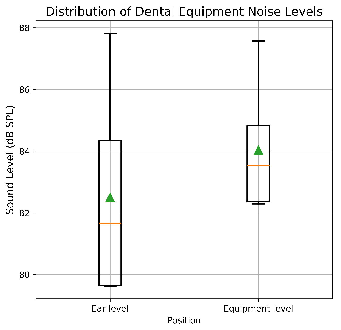
Figure 2 illustrates the mean noise levels (±1 SD) of dental equipment measured in the present study. The overall A-weighted equivalent continuous sound pressure level (LAeq) at the equipment level was 83.0 dB SPL, whereas the corresponding level at the operator’s ear position was 81.5 dB SPL. These values are comparable to those reported in earlier investigations of dental clinical noise exposure [
11,
32].
Comparison of noise levels measured at the equipment level and at ear level indicated modest attenuation with distance; equipment-level LAeq values varied within ±3 dB, whereas ear-level measurements varied within ±4 dB across repeated trials. Statistical comparison of six paired measurements revealed no significant difference between equipment-level and ear-level noise exposure (paired t-test, p > 0.05).
3.2. DPOAE Levels Before Exposure and After Exposure
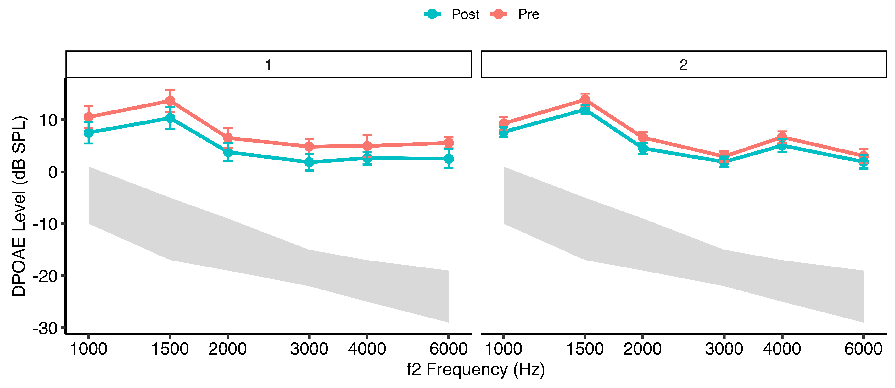
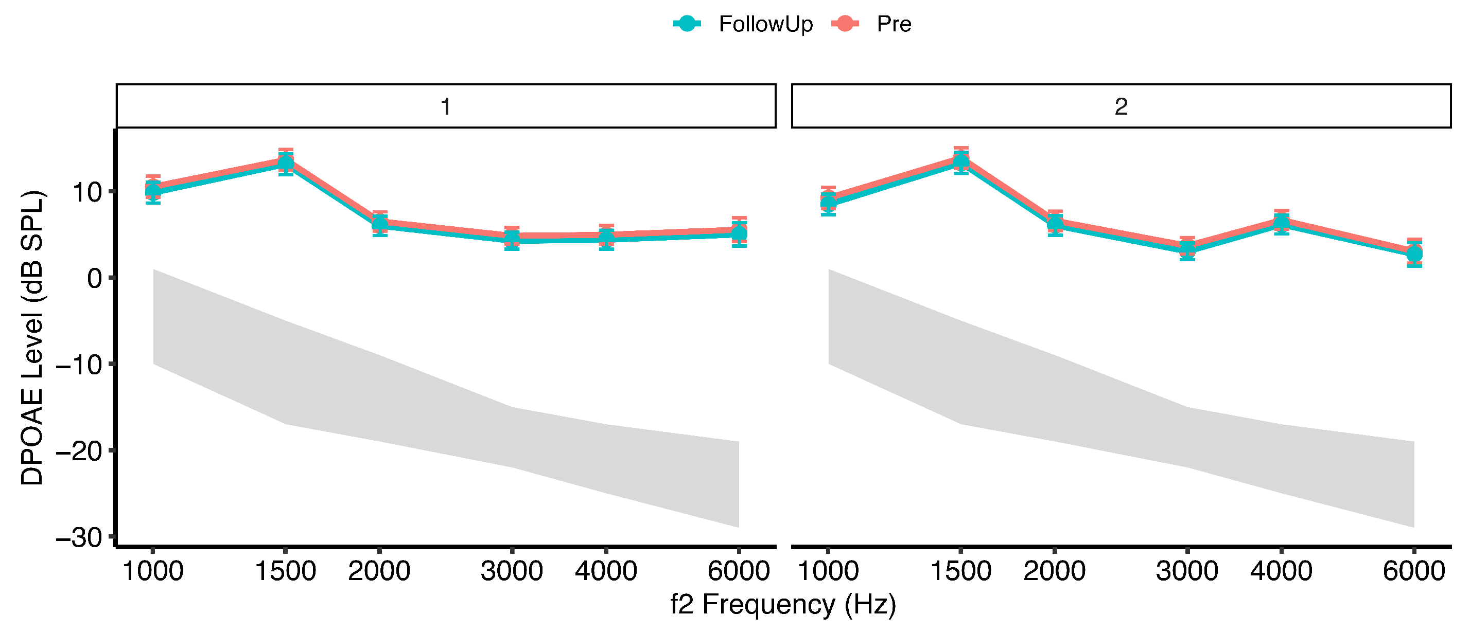
Figure 3 and
Figure 4 illustrate mean distortion product otoacoustic emission (DPOAE) levels (±95% confidence intervals) across
frequencies measured at pre-exposure, post-exposure, and follow-up time points in the two groups. At baseline (pre-exposure), DPOAE amplitudes were comparable between Group 1 (dentists exposed to dental equipment noise only) and Group 2 (dentists additionally using personal listening devices), indicating similar outer hair cell (OHC) function prior to exposure.
Immediately following the dental procedure (post-exposure), both groups demonstrated a reduction in DPOAE amplitudes, consistent with acute exposure-related cochlear stress. The magnitude of reduction was greater in Group 2 than in Group 1. Across frequencies, the most prominent decreases were observed in the mid-to-high range (approximately 3–6 kHz). At the follow-up assessment (48 h), DPOAE levels in both groups returned to values comparable to baseline, demonstrating recovery of cochlear responses.
A linear mixed-effects model with subject-specific random intercepts (Type III ANOVA, Satterthwaite approximation) revealed a significant main effect of frequency, , , , indicating moderate frequency-dependent variation in DPOAE amplitudes. A significant main effect of time was also observed, , , , reflecting small-to-moderate exposure-related changes across measurement intervals. The main effect of group was not statistically significant, , , suggesting comparable overall DPOAE amplitudes between groups when averaged across time and frequency.
Importantly, a significant interaction was identified, , , . Although the associated effect size was small, this interaction indicates differential temporal response patterns between groups. No significant , , or three-way interactions were observed (all ), suggesting that temporal exposure effects were broadly consistent across the tested frequency range.
Post hoc comparisons of estimated marginal means (Kenward–Roger degrees of freedom, Tukey-adjusted) averaged across frequencies revealed significant Pre–Post reductions in DPOAE amplitudes in both groups. In Group 1, DPOAE levels decreased significantly from pre- to post-exposure (mean difference = 3.12 dB, SE = 0.55, , ). In Group 2, a smaller but statistically significant reduction was observed (mean difference = 1.58 dB, SE = 0.40, , ).
Comparisons between pre-exposure and follow-up measurements demonstrated recovery of cochlear function in both groups. In Group 1, follow-up levels did not differ significantly from baseline (mean difference = −0.17 dB, ), and in Group 2, no significant pre-to-follow-up difference was observed (mean difference = −0.21 dB, ). Significant post-to-follow-up contrasts in both groups confirmed reversibility of the acute reduction (both ).
The intraclass correlation coefficient (ICC) indicated substantial between-subject variability in DPOAE amplitudes. The adjusted ICC was 0.695, suggesting that approximately 70% of total variance was attributable to stable inter-individual differences after accounting for fixed effects, supporting the appropriateness of mixed-effects modelling and the stability of DPOAE measurements across repeated assessments.




 ,
,