Comparison Study of Diagnosis and Treatment Planning for Dental Infections between Dental Students and Practitioners


Abstract
:1. Introduction
2. Materials and Methods
2.1. Ethical Approval
2.2. Developing a Survey Questionnaire
2.3. Study Participants
2.4. Statistical Analysis
3. Results
4. Discussion
5. Conclusions
Supplementary Materials
Author Contributions
Funding
Institutional Review Board Statement
Informed Consent Statement
Data Availability Statement
Acknowledgments
Conflicts of Interest
References
- Herrera, D.; Retamal-Valdes, B.; Alonso, B.; Feres, M. Acute Periodontal Lesions (Periodontal Abscesses and Necrotizing Periodontal Diseases) and Endo-Periodontal Lesions. J. Clin. Periodontol. 2018, 45, S78–S94. [Google Scholar] [CrossRef] [PubMed] [Green Version]
- Glickman, G.N. AAE Consensus Conference Recommended Diagnostic Terminology. J. Endod. 2009, 35, 1634. [Google Scholar] [CrossRef]
- Herrera, D.; Alonso, B.; de Arriba, L.; Santa Cruz, I.; Serrano, C.; Sanz, M. Acute Periodontal Lesions. Periodontology 2000, 65, 149–177. [Google Scholar] [CrossRef] [Green Version]
- Herrera, D.; Roldán, S.; Sanz, M. The Periodontal Abscess: A Review. J. Clin. Periodontol. 2000, 27, 377–386. [Google Scholar] [CrossRef]
- Oh, S.L.; Fouad, A.F.; Park, S.H. Treatment Strategy for Guided Tissue Regeneration in Combined Endodontic-Periodontal Lesions: Case Report and Review. J. Endod. 2009, 35, 1331–1336. [Google Scholar] [CrossRef]
- Oh, S.L.; Jones, D. Sophomore-Senior Case Studies: A Case-Based Learning Exercise with Peer Teaching. J. Dent. Educ. 2021, 85, 1117–1119. [Google Scholar] [CrossRef] [PubMed]
- Oh, S.L.; Yang, J.S.; Kim, Y.J. Discrepancies in Periodontitis Classification among Dental Practitioners with Different Educational Backgrounds. BMC Oral Health 2021, 21, 39. [Google Scholar] [CrossRef] [PubMed]
- Diagnosis and Treatment Planning Using the 2017 Classification of Periodontal Diseases among Three Dental Schools. Available online: https://pubmed.ncbi.nlm.nih.gov/35644870/ (accessed on 29 May 2022).
- McGuire, M.K.; Nunn, M.E. Prognosis Versus Actual Outcome. II. The Effectiveness of Clinical Parameters in Developing an Accurate Prognosis. J. Periodontol. 1996, 67, 658–665. [Google Scholar] [CrossRef] [PubMed]
- Kwok, V.; Caton, J. Prognosis Revisited: A System for Assigning Periodontal Prognosis. J. Periodontol. 2007, 78, 2063–2071. [Google Scholar] [CrossRef] [PubMed]
- The Jamovi Project. Jamovi (Version 1.6). Available online: https://www.jamovi.org (accessed on 21 January 2022).
- Nagata, M.; Nagata, M.; Kanie, T.; Shima, K. Clinical and Histologic Aspects of Cervical Cemental Tear as a Risk for Periodontal Diseases. Clin. Adv. Periodontics 2016, 6, 167–174. [Google Scholar] [CrossRef]
- Lin, H.J.; Chan, C.P.; Yang, C.Y.; Wu, C.T.; Tsai, Y.L.; Huang, C.C.; Yang, K.D.; Lin, C.C.; Chang, S.H.; Jeng, J.H. Cemental Tear: Clinical Characteristics and Its Predisposing Factors. J. Endod. 2011, 37, 611–618. [Google Scholar] [CrossRef]
- Tang, L.; Zhou, X.; Wang, Y.; Zhang, L.; Zheng, Q.; Huang, D. Detection of Vertical Root Fracture Using Cone Beam Computed Tomography: Report of Two Cases. Dent. Traumatol. 2011, 27, 484–488. [Google Scholar] [CrossRef] [PubMed]
- Chavda, R.; Mannocci, F.; Andiappan, M.; Patel, S. Comparing the In Vivo Diagnostic Accuracy of Digital Periapical Radiography with Cone-Beam Computed Tomography for the Detection of Vertical Root Fracture. J. Endod. 2014, 40, 1524–1529. [Google Scholar] [CrossRef] [PubMed]
- Khasnis, S.A.; Kidiyoor, K.H.; Patil, A.B.; Kenganal, S.B. Vertical Root Fractures and Their Management. J. Conserv. Dent. 2014, 17, 103–110. [Google Scholar] [CrossRef]
- Walton, R.E. Vertical Root Fracture: Factors Related to Identification. J. Am. Dent. Assoc. 2017, 148, 100–105. [Google Scholar] [CrossRef] [PubMed]
- Moons, K.G.M.; Royston, P.; Vergouwe, Y.; Grobbee, D.E.; Altman, D.G. Prognosis and Prognostic Research: What, Why, and How? Bmj 2009, 338, b375. [Google Scholar] [CrossRef]
- Miller, P.D., Jr.; McEntire, M.L.; Marlow, N.M.; Gellin, R.G. An Evidenced-Based Scoring Index to Determine the Periodontal Prognosis on Molars. J. Periodontol. 2014, 85, 214–225. [Google Scholar] [CrossRef] [Green Version]
- Samet, N.; Jotkowitz, A. Classification and Prognosis Evaluation of Individual Teeth--A Comprehensive Approach. Quintessence Int. 2009, 40, 377–387. [Google Scholar] [PubMed]
- Azarpazhooh, A.; Sgro, A.; Cardoso, E.; Elbarbary, M.; Laghapour Lighvan, N.; Badewy, R.; Malkhassian, G.; Jafarzadeh, H.; Bakhtiar, H.; Khazaei, S.; et al. A Scoping Review of 4 Decades of Outcomes in Nonsurgical Root Canal Treatment, Nonsurgical Retreatment, and Apexification Studies—Part 2: Outcome Measures. J. Endod. 2022, 48, 29–39. [Google Scholar] [CrossRef]
- Azarpazhooh, A.; Cardoso, E.; Sgro, A.; Elbarbary, M.; Laghapour Lighvan, N.; Badewy, R.; Malkhassian, G.; Jafarzadeh, H.; Bakhtiar, H.; Khazaei, S.; et al. A Scoping Review of 4 Decades of Outcomes in Nonsurgical Root Canal Treatment, Nonsurgical Retreatment, and Apexification Studies—Part 1: Process and General Results. J. Endod. 2022, 48, 15–28. [Google Scholar] [CrossRef] [PubMed]
- Imura, N.; Pinheiro, E.T.; Gomes, B.P.F.A.; Zaia, A.A.; Ferraz, C.C.R.; Souza-Filho, F.J. The Outcome of Endodontic Treatment: A Retrospective Study of 2000 Cases Performed by a Specialist. J. Endod. 2007, 33, 1278–1282. [Google Scholar] [CrossRef] [PubMed]
- Weston, M.; Haudek, K.C.; Prevost, L.; Urban-Lurain, M.; Merrill, J. Examining the Impact of Question Surface Features on Students’ Answers to Constructed-Response Questions on Photosynthesis. CBE Life Sci. Educ. 2015, 14, ar19. [Google Scholar] [CrossRef] [PubMed]


| General Information | |
|---|---|
| Gender |
|
| |
| Postgraduate training |
|
| |
| |
| |
| |
| Case A | |
| 1. Based on the clinical and radiographical evaluation presented, what is the most appropriate diagnosis of the maxillary right central incisor? | |
| 2. Based on the clinical and radiographical evaluation, assign the prognosis for the maxillary right central incisor using McGuire and Nunn prognosis system. | |
| 3. What was your primary determinant for assigning the prognosis for the maxillary right central incisor? | |
| 4. In your opinion, what is the most appropriate treatment option for this patient’s maxillary right central incisor? | |
| Case B | |
| 1. Based on the clinical and radiographical evaluation presented, what is the most appropriate diagnosis of the mandibular right second molar? | |
| 2. What is the most appropriate prognosis for the mandibular right second molar based on the clinical and radiographical evaluation? Use the Kwok and Caton prognosis scheme. | |
| 3. What was your primary determinant for assigning the prognosis for the mandibular right second molar? | |
| 4. In your opinion, what is the most appropriate treatment option for this patient’s mandibular right second molar? | |
| Senior 1 (n1 = 63) | Senior 2 (n2 = 63) | Dental Practitioner (n3 = 59) |
|---|---|---|
| Male (29) | Male (29) | Male (39) |
| Female (34) | Female (34) | Female (20) |
| Final grade in the third-year periodontics (mean ± SD) | Postgraduate training | |
| 80.3 ± 12.2 | 79.2 ± 10 | None (23) AGD/GPR (17) Endodontics (9) Periodontics (8) Others (2) |
| Case A | Senior 1 (n1 = 63) | DP (n3 = 59) | Fisher’s Exact Test |
|---|---|---|---|
| Diagnosis | |||
| Periodontal abscess * | 28 (44%) | 10 (17%) | p < 0.001 |
| Root fracture | 1 (2%) | 32 (54%) | |
| Cyst | 24 (38%) | 8 (14%) | |
| Other | 10 (16%) | 9 (15%) | |
| Periapical abscess | 7 | 0 | |
| Endodontic-periodontal combined lesion | 1 | 3 | |
| Invasion of biologic width | 2 | 2 | |
| Gingival abscess | 0 | 1 | |
| Necrotic pulp | 0 | 2 | |
| Periodontitis | 0 | 1 | |
| Treatment | |||
| Root canal therapy | 1 (2%) | 1 (2%) | p < 0.001 |
| Enucleation | 17 (27%) | 8 (13%) | |
| Extraction | 4 (6%) | 37 (63%) | |
| GTR/Surgical debridement | 41 (65%) | 7 (12%) | |
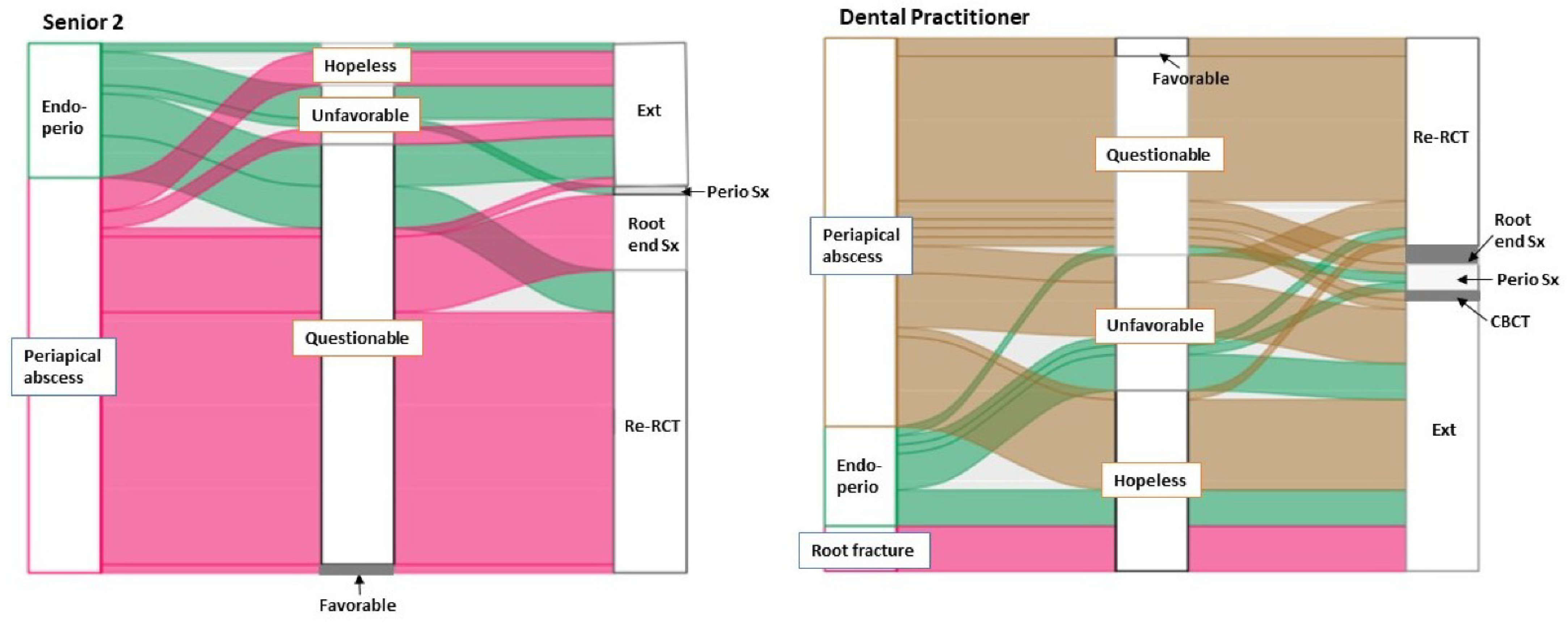
| Case B | Senior 2 (n2 = 63) | DP (n3 = 59) | |
| Diagnosis | |||
| Periapical abscess * | 47 (75%) | 43 (73%) | p = 0.05 |
| Endodontic-periodontal combined lesion | 16 (25%) | 11 (19%) | |
| Root fracture | 0 (0%) | 5 (8%) | |
| Treatment | |||
| Re-root canal therapy | 36 (57%) | 24 (41%) | p = 0.008 |
| Extraction | 17 (27%) | 30 (51%) | |
| Periodontal Surgery | 1 (2%) | 3 (5%) | |
| Root-end Surgery | 9 (14%) | 2 (3%) |
| Case A | Senior 1 (n1 = 63) | DP (n3 = 59) |
|---|---|---|
| Periodontal support (% BL/ CAL/ PD/ Periodontal Dx) | 58 (92%) | 27 (46%) |
| Restorability | 2 (3%) | 10 (17%) |
| Signs and symptom/ mobility | 2 (3%) | 8 (13.5%) |
| Root surface morphology | 1 (2%) | 7 (11.8%) |
| Root fracture | 6 (10%) | |
| Unknown pulpal Dx | 1 (1.7%) | |
| Case B | Senior 2 (n2 = 63) | DP (n3 = 59) |
| Change in the size of PARL | 29 (46%) | 22 (37.3%) |
| Restorability | 13 (20.6%) | 4 (6.8%) |
| Periodontal support | 11 (17.5%) | 6 (10.2%) |
| Pulpal/periapical Dx | 5 (7.9%) | 17 (28.8%) |
| Success rate of treatment option | 4 (6.3%) | 6 (10.2%) |
| Failed RCT/ root fracture | 1 (1.6%) | 4 (6.8%) |
Publisher’s Note: MDPI stays neutral with regard to jurisdictional claims in published maps and institutional affiliations. |
© 2022 by the authors. Licensee MDPI, Basel, Switzerland. This article is an open access article distributed under the terms and conditions of the Creative Commons Attribution (CC BY) license (https://creativecommons.org/licenses/by/4.0/).
Share and Cite
Oh, S.-L.; Jones, D.; Kim, J.R.; Choi, S.K.; Chung, M.-K. Comparison Study of Diagnosis and Treatment Planning for Dental Infections between Dental Students and Practitioners. Healthcare 2022, 10, 1393. https://doi.org/10.3390/healthcare10081393
Oh S-L, Jones D, Kim JR, Choi SK, Chung M-K. Comparison Study of Diagnosis and Treatment Planning for Dental Infections between Dental Students and Practitioners. Healthcare. 2022; 10(8):1393. https://doi.org/10.3390/healthcare10081393
Chicago/Turabian StyleOh, Se-Lim, Deborah Jones, Jong Ryul Kim, Seung Kee Choi, and Man-Kyo Chung. 2022. "Comparison Study of Diagnosis and Treatment Planning for Dental Infections between Dental Students and Practitioners" Healthcare 10, no. 8: 1393. https://doi.org/10.3390/healthcare10081393
APA StyleOh, S.-L., Jones, D., Kim, J. R., Choi, S. K., & Chung, M.-K. (2022). Comparison Study of Diagnosis and Treatment Planning for Dental Infections between Dental Students and Practitioners. Healthcare, 10(8), 1393. https://doi.org/10.3390/healthcare10081393