1. Introduction
Computed tomography (CT) enables non-invasive visualization of internal anatomical structures and has been widely used in clinical applications such as diagnosis, surgical planning, and image-guided intervention [
1]. In CT imaging, a three-dimensional attenuation volume is reconstructed from a set of two-dimensional X-ray projection images acquired multiple projection angles [
2,
3]. However, acquiring a dense set of X-ray projections inevitably involves radiation exposure, which motivates reconstruction methods that reduce the number of acquired projections while maintaining image quality [
4,
5]. Sparse-view CT reconstruction addresses this requirement by recovering a complete three-dimensional volume from a limited number of X-ray projection images acquired over a scan trajectory. Although these projections cover the overall scanning range, the reduced number of views provides insufficient angular measurement constraints for recovering the full attenuation field. This makes the reconstruction problem inherently ill-posed and often leads to streak artifacts, blurred anatomical boundaries, and structural ambiguity.
To mitigate these degradations caused by sparse angular sampling, various reconstruction methods have been developed for sparse-view CT reconstruction. Analytic methods, such as the Feldkamp-Davis-Kress (FDK) [
6], have the advantage of high computational efficiency. However, direct back-projection from sparsely sampled measurements often produces severe streak artifacts because the missing projection information is directly propagated into the reconstructed volume. Iterative algebraic methods, such as the simultaneous algebraic reconstruction technique (SART) [
7], aim to improve reconstruction quality by repeatedly enforcing projection consistency. However, when only a small number of measured views is available, the solution can remain unstable because multiple attenuation distributions can satisfy similar projection constraints. Deep learning-based methods have also shown strong potential for sparse-view CT reconstruction. Supervised convolutional neural networks [
8,
9,
10] can learn task-specific mappings that suppress sparse-view artifacts, but acquiring paired sparse-view and high-quality CT data remains challenging, and generalization across different anatomical regions or acquisition settings is often limited. Adversarial learning can improve perceptual realism, but it may introduce hallucinated structures when the measured projections do not provide enough evidence to support the generated anatomy.
Recently, Neural Radiance Fields (NeRFs) [
11] have been investigated as a promising optimization-based alternative for sparse-view CT reconstruction. Compared with deep learning-based methods, NeRF-based reconstruction does not require ground-truth CT volumes for training and can optimize an attenuation field directly from the projection images of each target case. This property is valuable for sparse-view CT because reconstruction can be performed from available measurements without relying on volume-level supervision. Despite these advantages, existing NeRF-based methods still face important limitations under sparse-view conditions. In the projection domain, the available supervision is inherently indirect because each projection value is obtained by accumulating attenuation coefficients along a ray. Minimizing the projection loss therefore enforces consistency between rendered and measured projections, but it does not uniquely determine how attenuation should be distributed within the reconstructed volume. Different three-dimensional attenuation distributions can explain similar sparse-view measurements, allowing anatomically inaccurate structures to produce comparable projection values. This non-uniqueness constitutes geometric ambiguity, which is an inherent property of the under-constrained sparse-view reconstruction problem.
The effect of this geometric ambiguity may appear as increased epistemic uncertainty when the model has insufficient evidence to determine the local attenuation distribution. Unlike geometric ambiguity, epistemic uncertainty is a model-dependent estimate of insufficient knowledge about a specific region. A volumetric region may be intersected by measured rays but still exhibit high epistemic uncertainty when the available projection evidence is insufficient or geometrically ambiguous. Conversely, a region with relatively sparse ray coverage may be reconstructed with low epistemic uncertainty if its attenuation distribution is consistently supported by the available measurements. Therefore, the estimated epistemic uncertainty should be interpreted as a model-dependent proxy for insufficient projection constraints rather than as a direct measurement of geometric ambiguity or reconstruction error. Most existing NeRF-based CT reconstruction methods do not explicitly estimate or use this volume-domain uncertainty during optimization. As a result, training samples may be selected without considering whether they pass through regions that remain insufficiently constrained, leaving structurally uncertain regions inadequately optimized.
These limitations suggest that sparse-view NeRF-based CT reconstruction requires a mechanism that identifies where the attenuation field is insufficiently constrained by the measured projections and actively uses this information during optimization. Epistemic uncertainty provides a suitable signal for this purpose because it reflects the model’s lack of knowledge arising from insufficient or geometrically ambiguous projection evidence [
12,
13]. Previous NeRF studies have investigated evidential uncertainty estimation and uncertainty-guided view or ray selection [
14,
15,
16]. However, these approaches have mainly been formulated for radiance-field rendering or have used uncertainty as a criterion for sample selection. They do not directly address how evidential uncertainty should be aggregated under the X-ray CT line-integral formulation or how volumetric uncertainty can jointly guide feature learning and sample allocation throughout reconstruction.
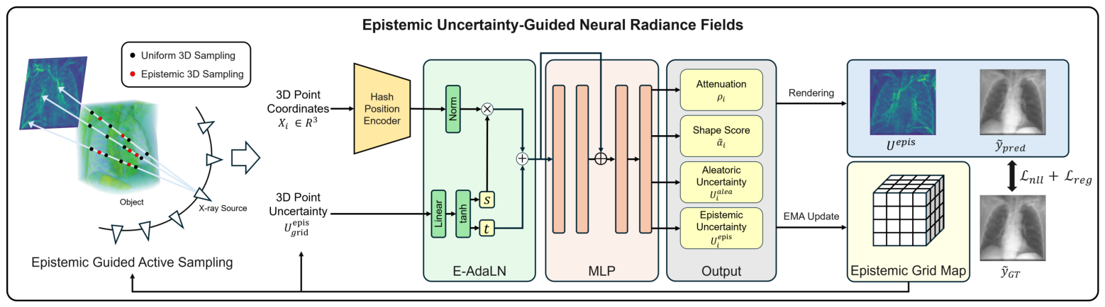
In this work, we propose EpiC-NeRF, an uncertainty-aware NeRF-based framework that incorporates epistemic uncertainty as an active feedback signal within a CT-specific closed loop. EpiC-NeRF adapts evidential uncertainty estimation and aggregation to line-integral attenuation modeling by jointly predicting the attenuation coefficient and evidential parameters at sampled spatial points. The resulting epistemic uncertainty is accumulated in a persistent three-dimensional Epistemic Grid Map, which maintains its spatial distribution throughout optimization. The accumulated uncertainty is queried by Epistemic-Adaptive Layer Normalization (E-AdaLN) to modulate intermediate features and is also used by a dual active sampling strategy to guide the allocation of projection rays and spatial points. The newly estimated uncertainty subsequently updates the Epistemic Grid Map, and influences feature modulation and sample allocation in the following iterations. Through this unified feedback mechanism, EpiC-NeRF progressively directs optimization toward volumetric regions that remain under-constrained by the available projections.
The main contributions of this work are summarized as follows:
CT-specific evidential attenuation modeling: We adapt the point-to-ray aggregation of evidential uncertainty to the X-ray CT line-integral formulation, enabling attenuation estimates and their associated uncertainties to be jointly optimized from sparse projection measurements.
Closed-loop uncertainty-guided reconstruction: We maintain volumetric epistemic uncertainty in a persistent Epistemic Grid Map and feed it back into subsequent reconstruction iterations. This feedback loop combines E-AdaLN for uncertainty-dependent feature modulation with dual active sampling for ray- and point-level sample allocation.
Validation across diverse anatomical structures: We evaluate EpiC-NeRF on four soft- and hard-tissue CT datasets. Quantitative, qualitative, uncertainty, and ablation analyses demonstrate the effectiveness of the proposed framework and the complementary contributions of its uncertainty-guided components.
4. Experiments
4.1. Datasets
We evaluated the proposed framework using four medical image datasets that represent different anatomical structures and reconstruction challenges commonly encountered in sparse-view CT reconstruction. The Chest dataset was obtained from the LIDC-IDRI dataset [
30] and was selected to evaluate the reconstruction of soft tissues and fine vascular-like structures with relatively low contrast. The Abdomen dataset was obtained from the 3D-IRCADb dataset [
31] and contains multiple organs with similar attenuation coefficients, which makes the accurate separation of adjacent structures challenging. The Foot and Jaw datasets were obtained from the Open Scientific Visualization Datasets [
32] and were used to evaluate hard-tissue reconstruction with dense and irregular contour shapes. The Foot dataset contains thin bone structures, whereas the Jaw dataset includes complex dental structures with sharp boundaries.
These datasets were selected to cover both soft-tissue and hard-tissue reconstruction scenarios. Soft-tissue datasets are challenging because adjacent anatomical regions often exhibit similar attenuation values, while hard-tissue datasets require accurate preservation of fine boundaries and high-contrast structures. Therefore, the four datasets provide complementary conditions for assessing reconstruction fidelity, structural preservation, and robustness under sparse-view settings.
To prepare the projection data, we used the TIGRE tomography toolbox to generate 50 X-ray projections for each volume. Following the experimental setting of NAF, the scanning range was set from to , and 3% Gaussian noise was added as a simplified robustness perturbation to ensure a fair comparison under the same simulation conditions. The generated projection data were used as sparse-view inputs, and the original CT volumes were used as ground truth for quantitative and qualitative evaluation.
4.2. Experimental Settings
We implemented the proposed framework using PyTorch 1.13.1 [
33] and trained it using the Adam [
34] optimizer with
and
for 500 epochs. The initial learning rate was set to
, and the batch size was set to 1024 rays per iteration. For the Epistemic Grid Map, the uncertainty values were updated using an exponential moving average to reduce unstable fluctuations during training. The momentum parameter
was set to 0.99. This update strategy allowed the grid map to maintain a stable spatial record of epistemic uncertainty while still reflecting changes in the reconstruction state as training progressed. The number of sampled points along each ray was determined according to the CT resolution of each dataset. We sampled 256 points for the Chest dataset, 576 points for the Abdomen dataset, 320 points for the Jaw and Foot dataset. The proposed framework allocated three quarters of the total sampling points to coarse sampling and the remaining points to fine sampling. The regularization parameter
was set to 1, 1, 0.5, and 5 for the Chest, Abdomen, Faw, and Foot datasets, respectively. All experiments were conducted on a server equipped with an Intel i9-9900X CPU (Intel, Santa Clara, CA, USA), 64 GB of RAM, and an NVIDIA RTX Titan GPU with 24 GB of VRAM (NVIDIA, Santa Clara, CA, USA). The same hardware environment was used for all compared methods whenever possible to ensure a fair comparison of reconstruction quality and computational efficiency.
4.3. Experimental Procedures
We compared the proposed framework with five baseline methods: the traditional filtered back-projection algorithm (FDK) [
6], the iterative reconstruction method SART [
7], the compressed-sensing-based method ASD-POCS [
18], and two neural implicit reconstruction methods, NAF [
20] and SAX-NeRF [
21]. These methods were selected because they represent complementary categories of sparse-view CT reconstruction. FDK provides a classical analytic baseline, SART and ASD-POCS represent iterative reconstruction methods that enforce projection consistency and sparsity priors, and NAF and SAX-NeRF represent recent neural implicit approaches for sparse-view CT reconstruction. In addition, these methods either provide publicly available source codes or contain sufficient implementation details for replication.
For quantitative evaluation, we compared all methods using 3D PSNR and 3D SSIM between the reconstructed volume and the ground-truth CT volume [
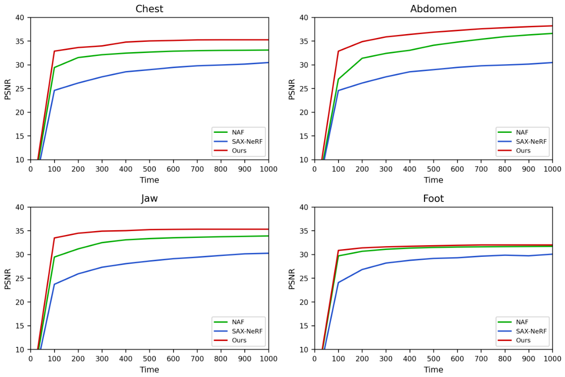
35]. These two metrics were used to assess signal fidelity and structural similarity at the volumetric level. In addition, we measured the convergence behavior of neural implicit methods by reporting PSNR as a function of training time. This evaluation was conducted for NAF, SAX-NeRF, and the proposed framework to assess reconstruction efficiency during optimization.
For qualitative evaluation, we visually compared the reconstructed volumes across all four datasets. The qualitative analysis focused on the preservation of anatomical boundaries, the reduction of streak artifacts, and the recovery of fine structures under sparse-view conditions.
We also conducted an ablation study to evaluate the contribution of each proposed component. Starting from the baseline configuration, we incrementally introduced the epistemic uncertainty estimation, E-AdaLN, 2D active ray sampling, and 3D active point sampling. The ablation experiments were performed across all four datasets, and reconstruction quality was measured using 3D PSNR. This experiment was designed to quantify the individual contribution of each component and to verify whether the proposed uncertainty-guided components provide complementary improvements.
5. Results
5.1. Quantitative Evaluation
Table 1 presents the quantitative comparison of our framework with five baseline methods across four CT volume datasets using 3D PSNR and 3D SSIM. Overall, our framework achieved the highest PSNR and SSIM values across all datasets. Compared with the second-best result in each metric, our framework achieved consistent improvements across all datasets. On the Chest dataset, our framework achieved 35.25 dB PSNR and 0.9737 SSIM, improving over the second-best method, SAX-NeRF, by 2.28 dB and 0.0118, respectively. On the Abdomen dataset, our framework achieved 38.23 dB PSNR and 0.9813 SSIM. This corresponds to a 2.73 dB improvement over the second-best PSNR result obtained by NAF and a 0.0083 improvement over the second-best SSIM result obtained by SAX-NeRF. On the Jaw dataset, our framework achieved 35.28 dB PSNR and 0.9510 SSIM, improving over the second-best PSNR result obtained by NAF by 1.92 dB and the second-best SSIM result obtained by ASD-POCS by 0.0199. On the Foot dataset, our framework achieved 32.00 dB PSNR and 0.9368 SSIM, improving over the second-best method, SAX-NeRF, by 0.32 dB and 0.0038, respectively.
These results indicate that the proposed uncertainty-aware reconstruction is effective across both soft-tissue and hard-tissue datasets. The improvement was particularly notable in the Chest and Abdomen datasets, where low contrast, similar attenuation coefficients, and ambiguous anatomical boundaries make accurate reconstruction more challenging. This tendency suggests that epistemic uncertainty is useful for identifying regions where projection evidence is insufficient and for guiding optimization toward such ambiguous areas. In contrast, the performance gain on the Foot dataset was relatively modest, suggesting that high-contrast bone structures are already better constrained by existing neural implicit baselines. Nevertheless, our framework still achieved the best quantitative performance, indicating that uncertainty-guided optimization can provide stable improvement without degrading reconstruction quality in strongly constrained structures.
5.2. Qualitative Evaluation
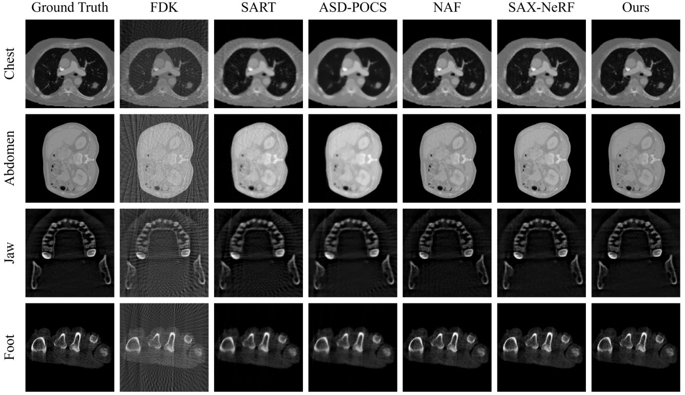
Figure 2 presents the qualitative comparison of reconstructed slices across all evaluated methods and datasets. FDK produced severe streak artifacts in all cases, which substantially obscured anatomical structures. SART reduced the dominant streak artifacts, but residual noise and local degradation were still observed. ASD-POCS further suppressed artifacts, but tended to over-smooth fine structural details and attenuate local boundaries. NAF and SAX-NeRF achieved higher visual quality than conventional reconstruction methods, showing clearer anatomical structures and fewer global artifacts. However, they still produced local structural errors, especially around ambiguous regions with weak projection evidence. In contrast, our framework generated reconstructed slices that were visually closer to the ground truth, with clearer boundaries and better preservation of anatomical details across both soft-tissue and hard-tissue datasets.
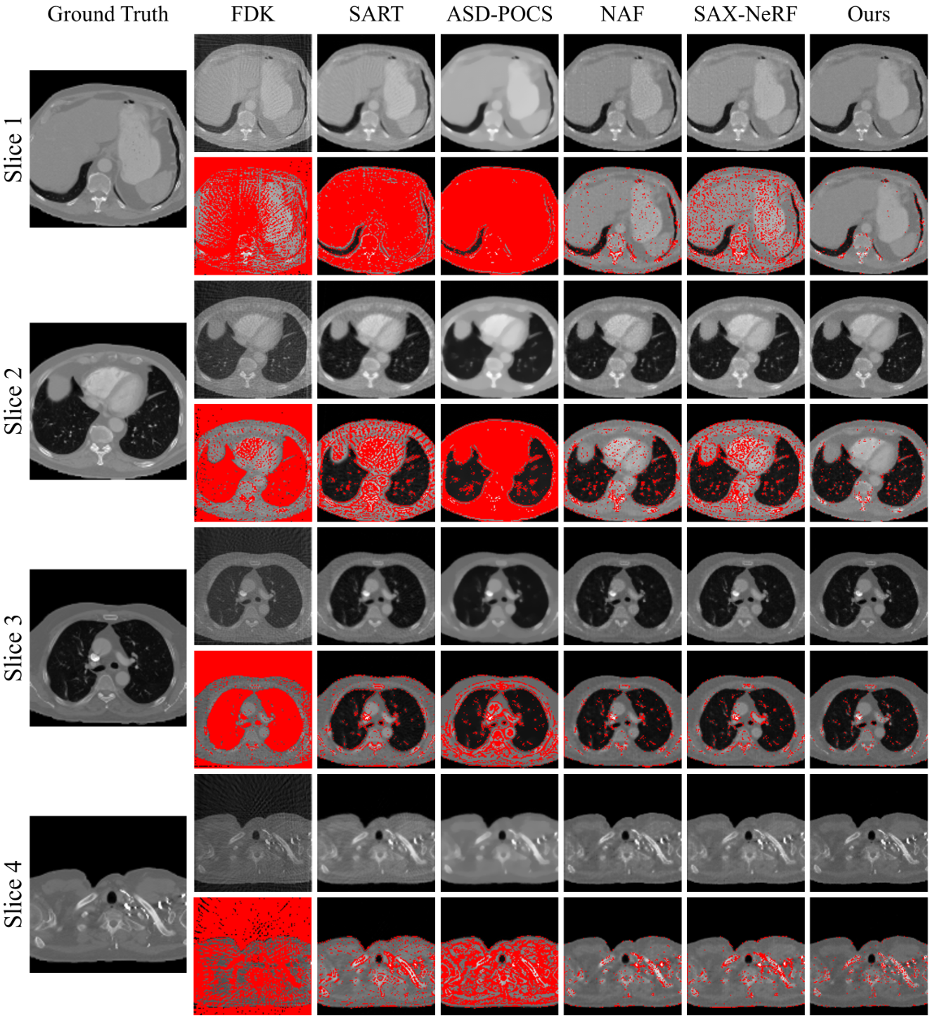
Figure 3 provides a detailed visual comparison of different reconstruction methods using selected internal CT slices and their corresponding local error maps. Conventional analytic and iterative methods, including FDK, SART, and ASD-POCS, exhibit widespread reconstruction errors, particularly around anatomical boundaries and low-contrast soft-tissue regions. Although neural implicit methods such as NAF and SAX-NeRF reduce these artifacts, their error maps still show noticeable residual errors near complex anatomical structures, organ boundaries, and high-frequency details. In contrast, our framework produces reconstructed slices that are visually closer to the ground truth and shows sparser and more localized error distributions. The results suggest that epistemic uncertainty helps identify volumetric regions that remain difficult to reconstruct under sparse-view conditions. By incorporating this uncertainty into feature normalization and active sampling, our framework reduces persistent structural artifacts and improves local reconstruction fidelity while preserving the overall anatomical appearance.
5.3. Epistemic Uncertainty Analysis
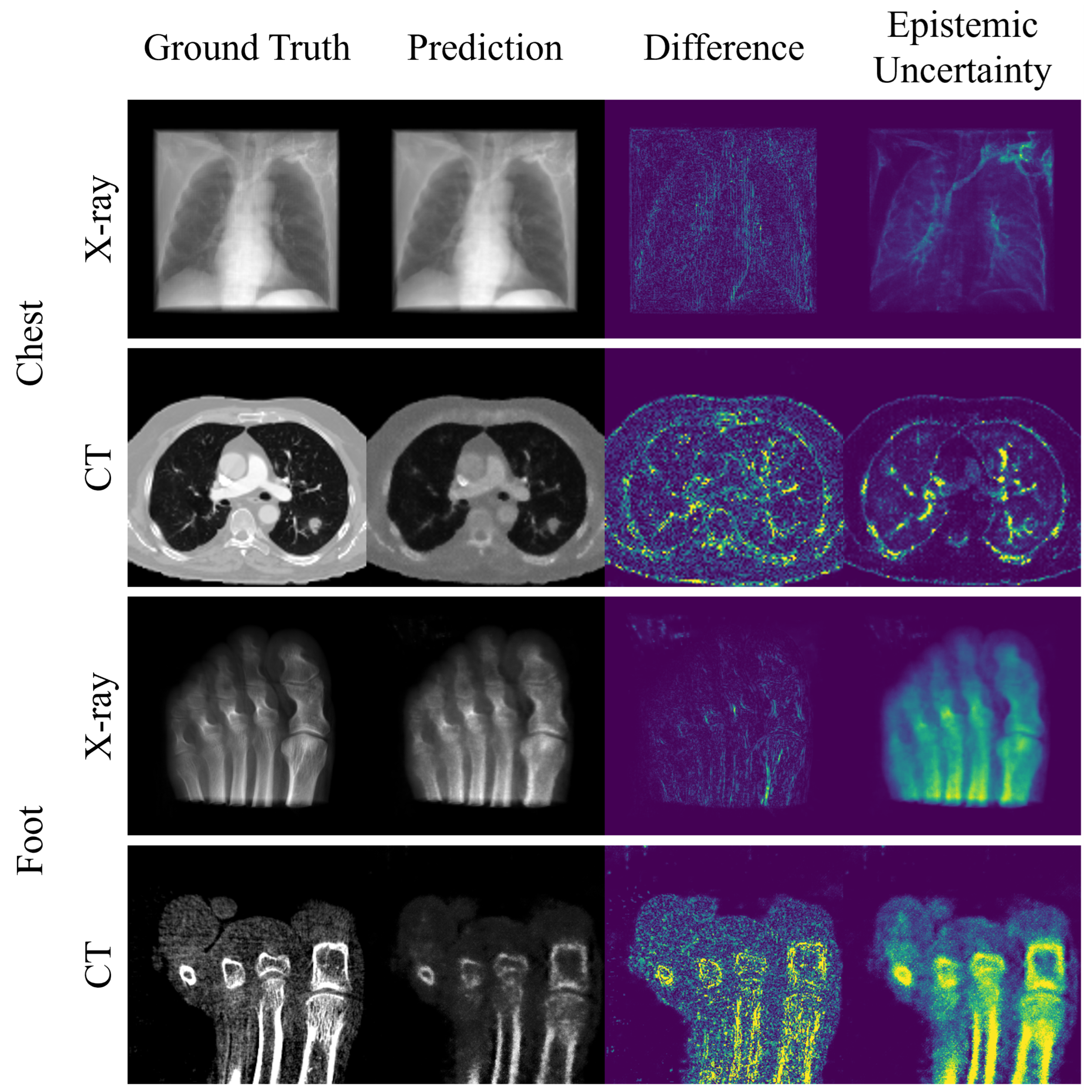
Figure 4 presents the relationship between reconstruction errors and epistemic uncertainty in the projection and CT domains using representative examples from the Chest and Foot datasets. In the X-ray projection domain, the epistemic uncertainty map represents ray-level uncertainty accumulated along each projection ray, whereas the CT-domain uncertainty map corresponds to a slice extracted from the Epistemic Grid Map. Regions with high epistemic uncertainty largely coincided with areas showing relatively large reconstruction errors in the difference maps. In the Chest dataset, uncertainty was mainly concentrated around low-contrast anatomical boundaries, vascular-like structures, and internal soft-tissue regions, where the attenuation distribution was insufficiently constrained by the sparse projection measurements. In the Foot dataset, uncertainty was more localized around overlapping bone structures, thin cortical boundaries, and high-frequency anatomical details.
Geometric ambiguity and epistemic uncertainty are closely related in these regions but represent different concepts. Geometric ambiguity is an inherent property of the sparse-view reconstruction problem, in which different attenuation distributions can produce similar projection measurements. Epistemic uncertainty instead represents the model’s lack of knowledge about the local attenuation distribution under the available measurements. Therefore, the estimated uncertainty does not directly quantify geometric ambiguity or reconstruction error. Although the uncertainty maps did not exactly match the difference maps at the pixel level, they consistently emphasized regions that were more likely to remain insufficiently constrained during reconstruction. These results indicate that the estimated epistemic uncertainty provides a model-dependent indication of insufficient knowledge and can serve as a useful signal for uncertainty-aware optimization in EpiC-NeRF.
5.4. Time Efficiency Comparison
Figure 5 illustrates the convergence behavior of NAF, SAX-NeRF, and our framework across the four datasets. The 3D PSNR was measured at 100-s intervals up to 1000 s. Overall, our framework achieved higher PSNR values than the two neural implicit baselines throughout the training process. In the Chest dataset, our framework reached a PSNR of approximately 33 within the first 100 s, whereas NAF required a longer training time to approach a similar range and SAX-NeRF remained substantially lower even after full convergence. Similar tendencies were observed in the Abdomen and Jaw datasets, where our framework showed both faster early improvement and higher final PSNR. In the Foot dataset, the performance gap was smaller, but our framework still maintained the highest PSNR during most of the training process. These results indicate that our framework achieves higher reconstruction quality within the same wall-clock training budget. NAF showed relatively fast convergence, but its final PSNR remained lower than that of our framework. SAX-NeRF improved more gradually and consistently produced lower PSNR values across all datasets. This suggests that the proposed uncertainty-guided optimization is effective not only for improving final reconstruction quality but also for guiding the training process toward informative regions from the early stage. In particular, the use of epistemic uncertainty allows the framework to prioritize ambiguous projection rays and spatial points, which helps the model achieve better reconstruction fidelity within the same training budget.
5.5. Ablation Study
Table 2 presents the quantitative ablation study of the proposed components across the four datasets. We evaluated four configurations, starting from a baseline model that retained the same attenuation-field backbone and evidential output layer but excluded E-AdaLN and uncertainty-guided active sampling. The baseline model achieved PSNRs of 32.50 dB on Chest, 35.33 dB on Abdomen, 34.59 dB on Jaw, and 31.49 dB on Foot. Although this configuration provided a reasonable representation for sparse-view CT reconstruction, it did not explicitly use epistemic uncertainty during feature processing or sample allocation. Adding E-AdaLN consistently improved the reconstruction performance across all datasets, yielding PSNR gains of 1.17 dB on Chest, 2.08 dB on Abdomen, 0.67 dB on Jaw, and 0.26 dB on Foot. The larger gains observed on Chest and Abdomen indicate that uncertainty-dependent feature modulation is particularly beneficial for under-constrained regions with subtle attenuation variations. The subsequent integration of 2D active sampling further increased the PSNR by 1.01 dB on Chest, 0.69 dB on Abdomen, 0.12 dB on Jaw, and 0.14 dB on Foot. This improvement suggests that prioritizing projection rays with high epistemic uncertainty provides more informative supervision during optimization. Finally, incorporating 3D active sampling achieved the highest performance on all four datasets, reaching 35.25 dB on Chest, 38.23 dB on Abdomen, 35.47 dB on Jaw, and 32.00 dB on Foot. Compared with 2D active sampling alone, point-level sampling provided additional improvements across all anatomical regions, with the largest gain observed on Chest. This result indicates that allocating samples to uncertain spatial regions provides complementary refinement that cannot be fully achieved through ray-level selection alone. Although the improvements were more pronounced on the soft-tissue datasets, the consistent performance gains on Jaw and Foot demonstrate that the proposed components are also applicable to high-contrast hard-tissue structures. Overall, these results show that E-AdaLN, 2D active sampling, and 3D active sampling play complementary roles in using epistemic uncertainty to guide feature modulation and sample allocation.
5.6. Regularization Parameter Sensitivity Analysis
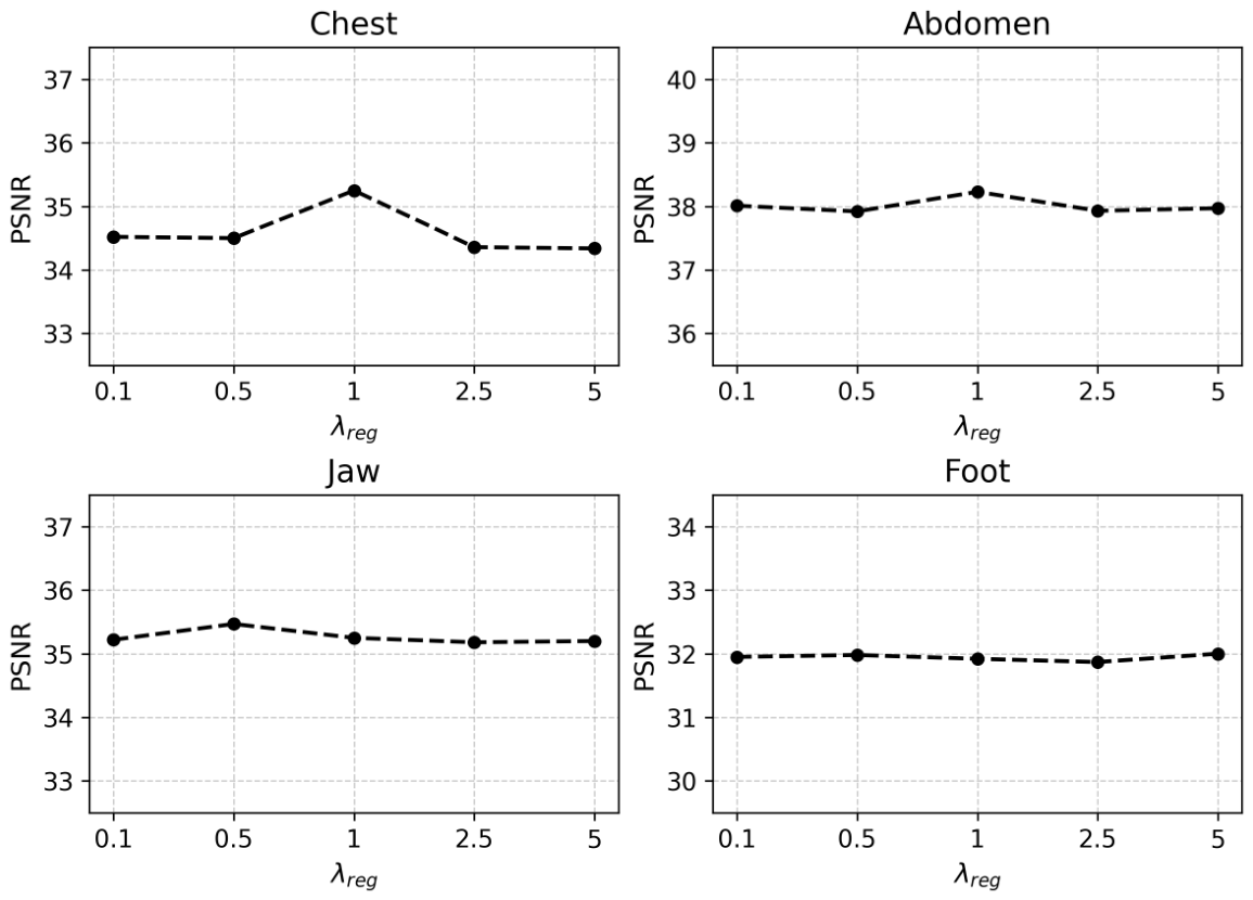
The regularization parameter controls the balance between projection reconstruction and the suppression of excessive evidence assigned to inaccurate predictions. A small value weakens the evidential regularization, whereas a large value increases its influence on the optimization. We therefore evaluated the sensitivity of the reconstruction performance to and examined whether a common parameter value was suitable for datasets with different anatomical and attenuation characteristics.
Figure 6 presents the 3D reconstruction PSNR obtained using
. The same candidate values were evaluated for all four datasets, while the remaining experimental settings were kept fixed. The Chest and Abdomen datasets achieved their highest PSNR at
, while the Jaw and Foot datasets achieved their highest PSNR at
and
, respectively. The Chest dataset showed a relatively clear performance peak at
, indicating that both insufficient and excessive evidential regularization can degrade its reconstruction performance. In contrast, the PSNR variations were relatively small for the Abdomen, Jaw, and Foot datasets, demonstrating that the proposed method remains stable over a broad range of
. In particular,
produced competitive performance across all four datasets and may therefore serve as a practical default value. Nevertheless, the small performance gains obtained through dataset-specific selection suggest that the preferred regularization strength can vary depending on the attenuation distribution and anatomical characteristics of each volume. Based on these results, we selected
values of 1 for Chest, 1 for Abdomen, 0.5 for Jaw, and 5 for Foot.
6. Conclusions
In this paper, we proposed EpiC-NeRF, an uncertainty-aware NeRF-based framework for sparse-view CT reconstruction. EpiC-NeRF incorporates epistemic uncertainty into attenuation field optimization to address spatially uneven reconstruction difficulty under limited projection views. The proposed framework estimates epistemic uncertainty through an evidential formulation and maintains its spatial distribution in a persistent Epistemic Grid Map. This uncertainty is then used for Epistemic-Adaptive Layer Normalization and dual active sampling in projection and volume spaces. Quantitative evaluations on four medical image datasets demonstrated that EpiC-NeRF achieved the highest PSNR and SSIM across all datasets compared with analytic, iterative, and neural implicit baselines. Qualitative results further showed that EpiC-NeRF reduced streak artifacts and local structural errors, particularly in ambiguous regions with insufficient projection evidence. In addition, convergence analysis and ablation results confirmed that uncertainty-guided normalization and sampling provide complementary benefits for reconstruction fidelity and training efficiency.
Nevertheless, several limitations remain. First, the performance gain was not uniform across anatomical structures. The improvement was more pronounced in soft-tissue datasets, such as Chest and Abdomen, whereas the performance margin was smaller in hard-tissue datasets with high contrast and clearly defined boundaries. This suggests that the current uncertainty-guided strategy is particularly effective for ambiguous, low-contrast regions but may require further adaptation for structures in which intensity contrast already provides strong structural constraints. Second, the current framework employs a monoenergetic and scatter-free log-domain line-integral forward model. Although this formulation is consistent with the simulated projection data used in this study, it does not explicitly account for physical effects encountered during real X-ray acquisition, such as beam hardening and scattering. Future work will investigate adaptive uncertainty weighting, structure-aware feature representations [
36], anatomy-dependent sampling strategies, and more realistic X-ray forward modeling to improve signal fidelity, structural preservation, and robustness under real acquisition conditions. Finally, because EpiC-NeRF relies on per-volume optimization, reducing its computational cost remains important for practical deployment. We will therefore investigate faster optimization strategies, compact uncertainty representations, and the integration of anatomical priors to improve robustness and computational efficiency across a broader range of sparse-view CT settings.