Rapid Liquid AP-MALDI MS Profiling of Lipids and Proteins from Goat and Sheep Milk for Speciation and Colostrum Analysis

 , ,
, ,  and
and 
Abstract

1. Introduction
2. Materials and Methods
2.1. Sample Collection
2.2. Milk Sampling for Goat/Sheep Speciation
2.3. Colostrum Sampling
2.4. Conventional Milk Sample Analysis
2.5. Sample Preparation for MS Analysis
2.6. MS Analysis
2.7. MS Profile Data Analysis
2.8. MS/MS Analysis
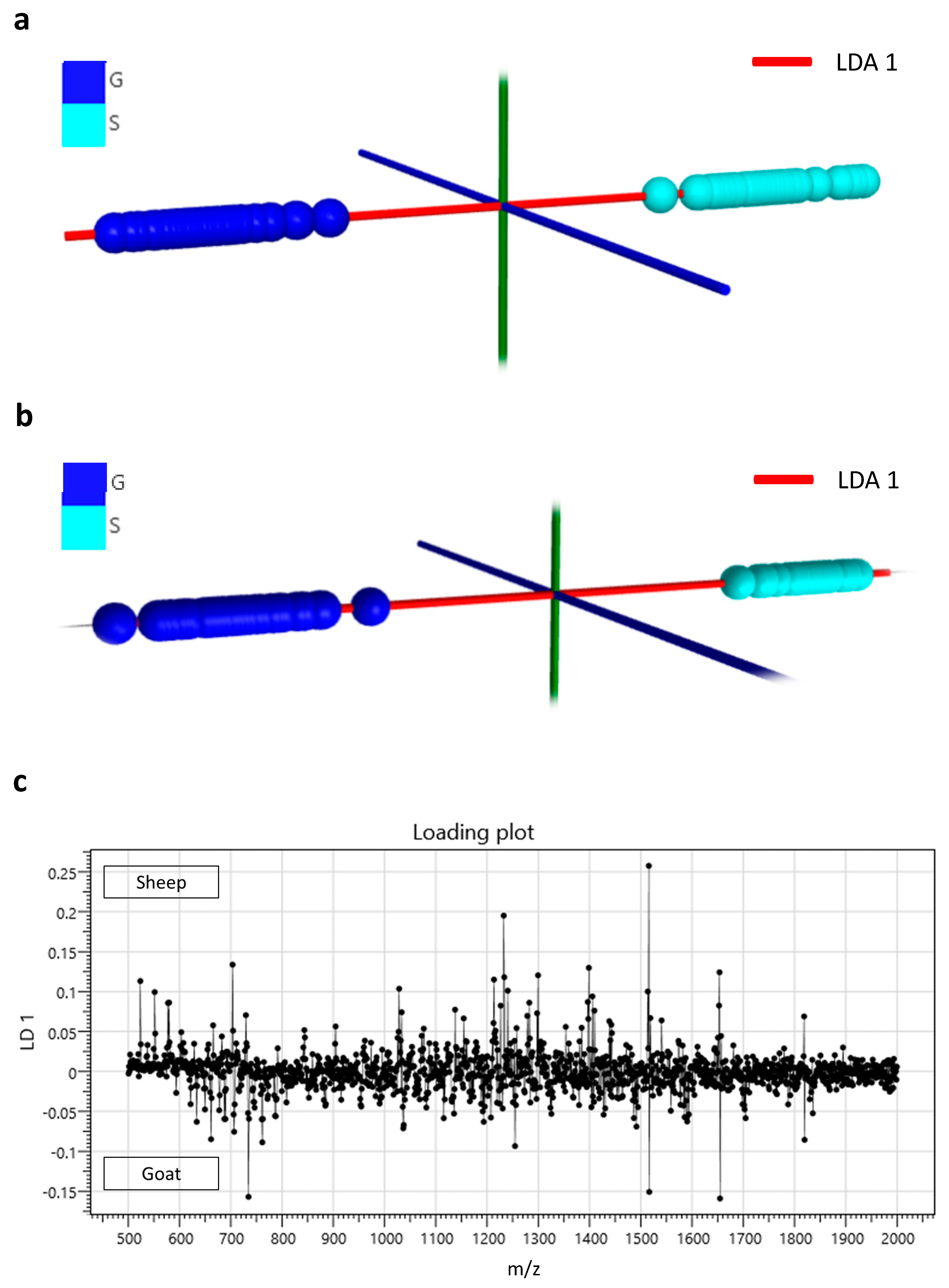
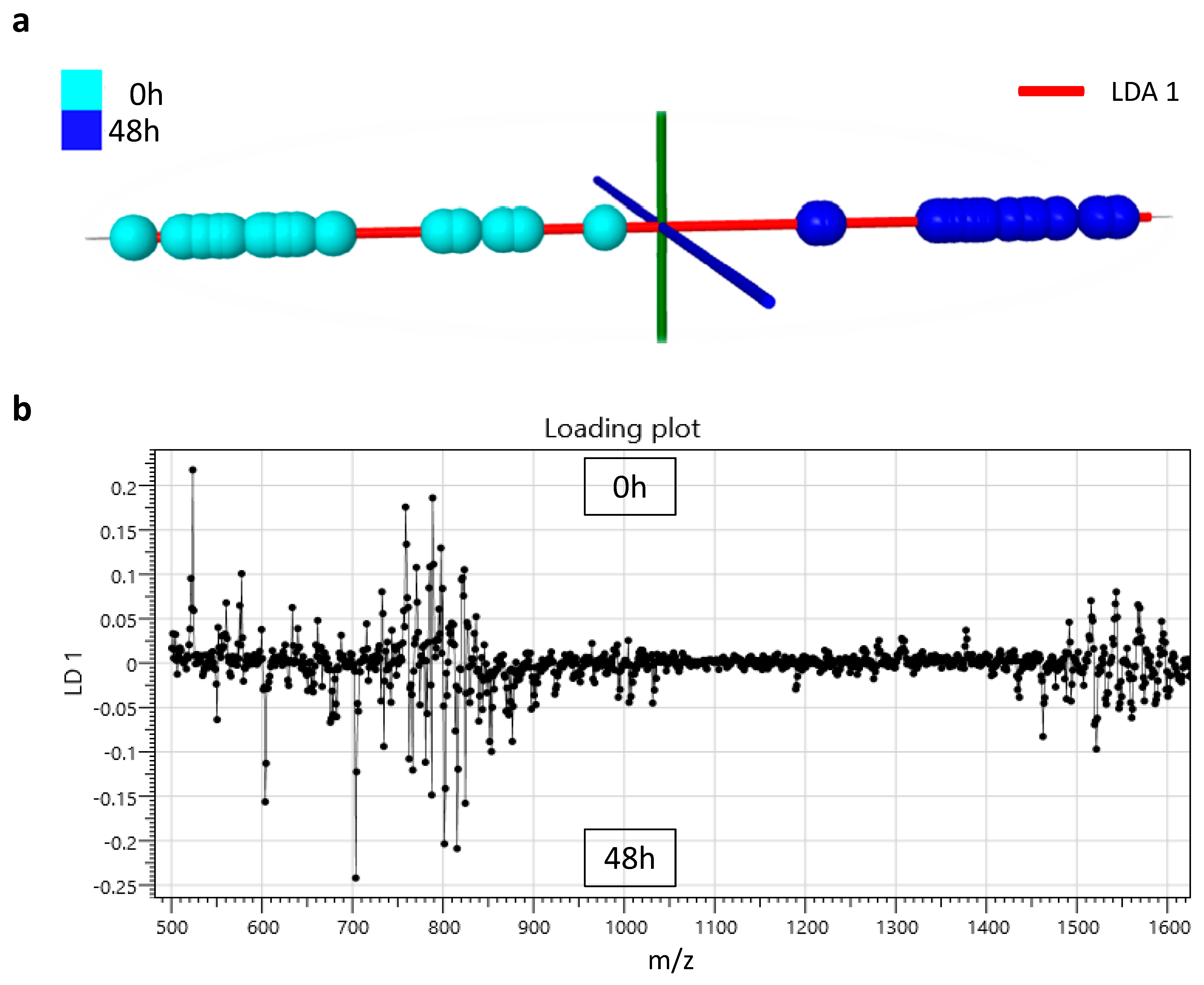
3. Results
4. Discussion
5. Conclusions
Author Contributions
Funding
Acknowledgments
Conflicts of Interest
References
- Pandya, A.; Ghodke, K. Goat and sheep milk products other than cheeses and yoghurt. Small Rumin. Res. 2007, 68, 193–206. [Google Scholar] [CrossRef]
- López-Calleja, I.; González, I.; Fajardo, V.; Martín, I.; Hernández, P.; García, T.; Martín, R. Quantitative detection of goats’ milk in sheep’s milk by real-time PCR. Food Control 2007, 18, 1466–1473. [Google Scholar] [CrossRef]
- Roncada, P.; Piras, C.; Soggiu, A.; Turk, R.; Urbani, A.; Bonizzi, L. Farm animal milk proteomics. J. Proteom. 2012, 75, 4259–4274. [Google Scholar] [CrossRef] [PubMed]
- De La Fuente, M.A.; Juárez, M. Authenticity assessment of dairy products. Crit. Rev. Food Sci. Nutr. 2005, 45, 563–585. [Google Scholar] [CrossRef] [PubMed]
- Soggiu, A.; Roncada, P.; Piras, C. Proteomics in milk and dairy products. In Proteomics in Domestic Animals: From Farm to Systems Biology; Springer: Cham, Switzerland, 2018; pp. 169–193. [Google Scholar]
- Molina, E.; Martín-Álvarez, P.J.; Ramos, M. Analysis of cows’, ewes’ and goats’ milk mixtures by capillary electrophoresis: Quantification by multivariate regression analysis. Int. Dairy J. 1999, 9, 99–105. [Google Scholar] [CrossRef]
- Chianese, L.; Laezza, P.; Smaldone, L.; Stingo, C.; Del Giovine, L.; Addeo, F. Evaluation of bovine milk in the buffalo mozzarella cheese by two-dimensional electrophoresis. Sci. Tec. Latt.-Casearia 1990, 41, 315–326. [Google Scholar]
- Ferreira, I.M.; Caçote, H. Detection and quantification of bovine, ovine and caprine milk percentages in protected denomination of origin cheeses by reversed-phase high-performance liquid chromatography of beta-lactoglobulins. J. Chromatogr. A 2003, 1015, 111–118. [Google Scholar] [CrossRef]
- Sánchez-Macías, D.; Moreno-Indias, I.; Castro, N.; Morales-delaNuez, A.; Argüello, A. From goat colostrum to milk: Physical, chemical, and immune evolution from partum to 90 days postpartum. J. Dairy Sci. 2014, 97, 10–16. [Google Scholar] [CrossRef]
- Argüello, A.; Castro, N.; Alvarez, S.; Capote, J. Effects of the number of lactations and litter size on chemical composition and physical characteristics of goat colostrum. Small Rumin. Res. 2006, 64, 53–59. [Google Scholar] [CrossRef]
- Nocek, J.; Braund, D.; Warner, R. Influence of neonatal colostrum administration, immunoglobulin, and continued feeding of colostrum on calf gain, health, and serum protein. J. Dairy Sci. 1984, 67, 319–333. [Google Scholar] [CrossRef]
- Marnila, P.; Korohnen, H. Colostrum. In Encyclopedia of Dairy Sciences; Elsevier: Amsterdam, The Netherlands, 2002; ISBN 978-0-12-227235-6. Available online: https://www.sciencedirect.com/referencework/9780122272356/encyclopedia-of-dairy-sciences#book-info (accessed on 18 August 2020).
- McMartin, S.; Godden, S.; Metzger, L.; Feirtag, J.; Bey, R.; Stabel, J.; Goyal, S.; Fetrow, J.; Wells, S.; Chester-Jones, H. Heat treatment of bovine colostrum. I: Effects of temperature on viscosity and immunoglobulin G level. J. Dairy Sci. 2006, 89, 2110–2118. [Google Scholar] [CrossRef]
- Gapper, L.W.; Copestake, D.E.; Otter, D.E.; Indyk, H.E. Analysis of bovine immunoglobulin G in milk, colostrum and dietary supplements: A review. Anal. Bioanal. Chem. 2007, 389, 93–109. [Google Scholar] [CrossRef] [PubMed]
- Gelsinger, S.; Smith, A.; Jones, C.; Heinrichs, A.J. Comparison of radial immunodiffusion and ELISA for quantification of bovine immunoglobulin G in colostrum and plasma. J. Dairy Sci. 2015, 98, 4084–4089. [Google Scholar] [CrossRef] [PubMed]
- Lopreiato, V.; Ceniti, C.; Trimboli, F.; Fratto, E.; Marotta, M.; Britti, D.; Morittu, V.M. Evaluation of the capillary electrophoresis method for measurement of immunoglobulin concentration in ewe colostrum. J. Dairy Sci. 2017, 100, 6465–6469. [Google Scholar] [CrossRef]
- Quigley, J.D.; Lago, A.; Chapman, C.; Erickson, P.; Polo, J. Evaluation of the Brix refractometer to estimate immunoglobulin G concentration in bovine colostrum. J. Dairy Sci. 2013, 96, 1148–1155. [Google Scholar] [CrossRef]
- Bartier, A.; Windeyer, M.; Doepel, L. Evaluation of on-farm tools for colostrum quality measurement. J. Dairy Sci. 2015, 98, 1878–1884. [Google Scholar] [CrossRef]
- Abraham, R.S.; Barnidge, D.R.; Lanza, I.R. Assessment of proteins of the immune system. In Clinical Immunology: Principles and Practice, 4th ed.; Elsevier Inc.: Philadelphia, PA, USA, 2012; pp. 1145–1159. [Google Scholar]
- Calvano, C.D.; De Ceglie, C.; Aresta, A.; Facchini, L.A.; Zambonin, C.G. MALDI-TOF mass spectrometric determination of intact phospholipids as markers of illegal bovine milk adulteration of high-quality milk. Anal. Bioanal. Chem. 2013, 405, 1641–1649. [Google Scholar] [CrossRef]
- Cramer, R.; Pirkl, A.; Hillenkamp, F.; Dreisewerd, K. Liquid AP-UV-MALDI enables stable ion yields of multiply charged peptide and protein ions for sensitive analysis by mass spectrometry. Angew. Chem. Int. Ed. 2013, 52, 2364–2367. [Google Scholar] [CrossRef]
- Wiangnon, K.; Cramer, R. Liquid MALDI MS analysis of complex peptide and proteome samples. J. Proteome Res. 2016, 15, 2998–3008. [Google Scholar] [CrossRef]
- Ryumin, P.; Cramer, R. The composition of liquid atmospheric pressure matrix-assisted laser desorption/ionization matrices and its effect on ionization in mass spectrometry. Anal. Chim. Acta 2018, 1013, 43–53. [Google Scholar] [CrossRef]
- Ryumin, P.; Brown, J.; Morris, M.; Cramer, R. Investigation and optimization of parameters affecting the multiply charged ion yield in AP-MALDI MS. Methods 2016, 104, 11–20. [Google Scholar] [CrossRef]
- Marty, M.T.; Baldwin, A.J.; Marklund, E.G.; Hochberg, G.K.; Benesch, J.L.; Robinson, C.V. Bayesian deconvolution of mass and ion mobility spectra: From binary interactions to polydisperse ensembles. Anal. Chem. 2015, 87, 4370–4376. [Google Scholar] [CrossRef]
- European Commission. Commission regulation (EC) No 273/2008 of 5 March 2008 laying down detailed rules for the application of council regulation (EC) no. 1255/1999 as regards methods for the analysis and quality evaluation of milk and milk products. OJEC 2008, 88, 1–115. [Google Scholar]
- Morand-Fehr, P.; Fedele, V.; Decandia, M.; Le Frileux, Y. Influence of farming and feeding systems on composition and quality of goat and sheep milk. Small Rumin. Res. 2007, 68, 20–34. [Google Scholar] [CrossRef]
- Park, Y.; Juárez, M.; Ramos, M.; Haenlein, G. Physico-chemical characteristics of goat and sheep milk. Small Rumin. Res. 2007, 68, 88–113. [Google Scholar] [CrossRef]
- Hale, O.J.; Morris, M.; Jones, B.; Reynolds, C.K.; Cramer, R. Liquid atmospheric pressure matrix-assisted laser desorption/ionization mass spectrometry adds enhanced functionalities to MALDI MS profiling for disease diagnostics. ACS Omega 2019, 4, 12759–12765. [Google Scholar] [CrossRef] [PubMed]
- Palmblad, M.; BindschedLer, L.V.; Cramer, R. Quantitative proteomics using uniform 15N-labeling, MASCOT, and the trans-proteomic pipeline. Proteomics 2007, 7, 3462–3469. [Google Scholar] [CrossRef] [PubMed]
- Towers, M.W.; Mckendrick, J.E.; Cramer, R. Introduction of 4-chloro-α-cyanocinnamic acid liquid matrices for high sensitivity UV-MALDI MS. J. Proteome Res. 2010, 9, 1931–1940. [Google Scholar] [CrossRef]
- Krenkel, H.; Hartmane, E.; Piras, C.; Brown, J.; Morris, M.; Cramer, R. Advancing Liquid Atmospheric Pressure Matrix-Assisted Laser Desorption/Ionization Mass Spectrometry Toward Ultrahigh-Throughput Analysis. Anal. Chem. 2020, 92, 2931–2936. [Google Scholar] [CrossRef]
- Calvano, C.D.; Jensen, O.N.; Zambonin, C.G. Selective extraction of phospholipids from dairy products by micro-solid phase extraction based on titanium dioxide microcolumns followed by MALDI-TOF-MS analysis. Anal. Bioanal. Chem. 2009, 394, 1453–1461. [Google Scholar] [CrossRef]
- Lellman, S.E.; Cramer, R. Bacterial identification by lipid profiling using liquid atmospheric pressure matrix-assisted laser desorption/ionization mass spectrometry. Clin. Chem. Lab. Med. (CCLM) 2019, 58, 930–938. [Google Scholar] [CrossRef] [PubMed]
- Li, M.; Li, W.; Wu, J.; Zheng, Y.; Shao, J.; Li, Q.; Kang, S.; Zhang, Z.; Yue, X.; Yang, M. Quantitative lipidomics reveals alterations in donkey milk lipids according to lactation. Food Chem. 2020, 310, 125866. [Google Scholar] [CrossRef] [PubMed]
- Martín, M.J.; Martín-Sosa, S.; Hueso, P. Bovine milk gangliosides: Changes in ceramide moiety with stage of lactation. Lipids 2001, 36, 291–298. [Google Scholar] [CrossRef] [PubMed]
- Contarini, G.; Povolo, M.; Pelizzola, V.; Monti, L.; Bruni, A.; Passolungo, L.; Abeni, F.; Degano, L. Bovine colostrum: Changes in lipid constituents in the first 5 days after parturition. J. Dairy Sci. 2014, 97, 5065–5072. [Google Scholar] [CrossRef] [PubMed]
- Cui, N.; Wen, P.; Liang, Q.; Liu, H.; Zhang, W.; Wang, P.; Guo, H.; Ren, F. Chemical composition of yak colostrum and transient milk. J. Anim. Physiol. Anim. Nutr. 2015, 99, 825–833. [Google Scholar] [CrossRef] [PubMed]
- Mancini, G.J.A.J.; Carbonara, A.T.; Heremans, J.F. Immunochemical quantitation of antigens by single radial immunodiffusion. Immunochemistry 1965, 2, 235–254. [Google Scholar] [CrossRef]


| Samples | Data Description | Fat (%) | Crude Protein (%) | Lactose (%) | Casein (%) | Cryoscopy (mC) | Urea (mg/dL) | True Protein (%) | Acetone (mM) | β-hydroxy-butyrate (mM) |
|---|---|---|---|---|---|---|---|---|---|---|
| Goat | Mean | 5.49 | 3.97 | 4.66 | 3.05 | 540 | 52.75 | 3.68 | 0.06 | 0.02 |
| (n = 43) | SD * | 1.43 | 0.53 | 0.21 | 0.44 | 9 | 13.77 | 0.52 | 0.08 | 0.03 |
| Sheep | Mean | 6.6 | 5.35 | 4.71 | 4.03 | 564 | 40.67 | 4.97 | 0.27 | 0.15 |
| (n = 44) | SD * | 0.98 | 0.6 | 0.4 | 0.46 | 20 | 9.97 | 0.59 | 0.26 | 0.17 |
| Ion Bin(m/z) | Peak m/z Values (Monoisotopic) | Theoretical m/z Values (Monoisotopic) | Putative Lipid Assignment |
|---|---|---|---|
| 523.5 | 523.47 | 523.48 | [Cer(d32:3) + NH4]+ |
| 675.5 | 675.51 | 675.53 | [SM(D16:1/16:0) + H]+ |
| 703.5 | 703.57 | 703.57 | [SM(34:1;O2) + H]+ |
| 706.5 | 706.55 | 706.54 | [PC(30:0) + H]+ |
| 734.5 | 734.57 | 734.57 | [PE(34:4) + Na]+ |
| 787.5 | 787.63 | 787.63 | [SM(40:1;O2) + H]+ |
| 788.5 | 788.62 | 788.62 | [PC(36:1) + H]+ |
| 800.5 | 801.68 | 801.67 | [SM(d41:1) + H]+ |
| 815.5 | 815.69 | 815.70 | [SM(42:1)+ H]+ |
© 2020 by the authors. Licensee MDPI, Basel, Switzerland. This article is an open access article distributed under the terms and conditions of the Creative Commons Attribution (CC BY) license (http://creativecommons.org/licenses/by/4.0/).
Share and Cite
Piras, C.; Ceniti, C.; Hartmane, E.; Costanzo, N.; Morittu, V.M.; Roncada, P.; Britti, D.; Cramer, R. Rapid Liquid AP-MALDI MS Profiling of Lipids and Proteins from Goat and Sheep Milk for Speciation and Colostrum Analysis. Proteomes 2020, 8, 20. https://doi.org/10.3390/proteomes8030020
Piras C, Ceniti C, Hartmane E, Costanzo N, Morittu VM, Roncada P, Britti D, Cramer R. Rapid Liquid AP-MALDI MS Profiling of Lipids and Proteins from Goat and Sheep Milk for Speciation and Colostrum Analysis. Proteomes. 2020; 8(3):20. https://doi.org/10.3390/proteomes8030020
Chicago/Turabian StylePiras, Cristian, Carlotta Ceniti, Evita Hartmane, Nicola Costanzo, Valeria Maria Morittu, Paola Roncada, Domenico Britti, and Rainer Cramer. 2020. "Rapid Liquid AP-MALDI MS Profiling of Lipids and Proteins from Goat and Sheep Milk for Speciation and Colostrum Analysis" Proteomes 8, no. 3: 20. https://doi.org/10.3390/proteomes8030020
APA StylePiras, C., Ceniti, C., Hartmane, E., Costanzo, N., Morittu, V. M., Roncada, P., Britti, D., & Cramer, R. (2020). Rapid Liquid AP-MALDI MS Profiling of Lipids and Proteins from Goat and Sheep Milk for Speciation and Colostrum Analysis. Proteomes, 8(3), 20. https://doi.org/10.3390/proteomes8030020