Article

HRAMS Proteomics Insights on the Anti-Filarial Effect of Ocimum sanctum: Implications in Phytochemical-Based Drug-Targeting and Designing

1
Department of Biochemistry, Institute of Science, Banaras Hindu University, Varanasi 221005, India
2
Department of Molecular Biology, National Aids Research Institute, Pune 411026, India
*
Author to whom correspondence should be addressed.
Proteomes 2025, 13(1), 2; https://doi.org/10.3390/proteomes13010002
Submission received: 22 October 2024 / Revised: 2 December 2024 / Accepted: 9 December 2024 / Published: 27 December 2024

Abstract

:
Lymphatic filariasis (LF) continues to impact 657 million individuals worldwide, resulting in lifelong and chronic impairment. The prevalent anti-filarial medications—DEC, albendazole, and ivermectin—exhibit limited adulticidal efficacy. Despite ongoing LF eradication programs, novel therapeutic strategies are essential for effective control. This study examines the mechanism of action of Ocimum sanctum on the filarial parasites Setaria cervi via a synergistic biochemical and proteomics methodology. The ethanolic extract of Ocimum sanctum (EOS) demonstrated potential anti-filarial action in the MTT reduction experiment, with an LC50 value of 197.24 µg/mL. After EOS treatment, an elevation in lipid peroxidation (51.92%), protein carbonylation (48.99%), and NADPH oxidase (88.88%) activity, along with a reduction in glutathione (GSH) (−39.23%), glutathione reductase (GR) (−60.17%), and glutathione S transferase (GST) (−50.48%) activity, was observed. The 2D gel electrophoresis identified 20 decreased and 11 increased protein spots in the EOS-treated parasites relative to the control group. Additionally, in drug docking analysis, the EOS bioactive substances ursolic acid, rutin, and rosmarinic acid show a significant binding affinity with the principal differentially expressed proteins. This paper demonstrates, for the first time, that the anti-filarial efficacy of EOS is primarily facilitated by its impact on energy metabolism, antioxidant mechanisms, and stress response systems of the parasites.

1. Introduction

Lymphatic filariasis (LF) is a neglected tropical disease referred to as elephantiasis. At present, 657 million individuals are at risk of lymphatic filariasis infection throughout 39 endemic countries [1]. The lymphatic-dwelling parasite roundworms, namely, Wuchereria bancrofti, Brugia malayi, and Brugia timori, are the etiological agents of the disease. LF is responsible for severe morbidity, especially in the tropical and sub-tropical regions of the world. The disease accounts for an estimated 5.5 million disability adjusted life years (DALY) globally [2]. LF leads to chronic and long-term disability and poses a substantial obstacle to the socioeconomic advancement of affected individuals in endemic areas. In 2020, the WHO reset the deadline of the Global Programme to Eliminate Lymphatic Filariasis (GPELF) from 2020 to 2030 by stopping the spread of infection. The WHO recommends preventive chemotherapy via mass drug administration (MDA) of anti-filarial medicines to the community at risk of LF infection. The MDA program includes annual doses of the anti-filarial drugs diethylcarbamazine (DEC), ivermectin, and albendazole, either in combination or alone. However, these drugs have only microfilaricidal activity and are unable to kill the adult worms, which typically reside inside the host lymphatics [3]. In addition to the absence of microfilaricidal action, anti-filarial medications are linked to several side effects. DEC and ivermectin can cause adverse effects in microfilaremic patients, i.e., fever, headache, myalgia, fatigue, and malaise. Additionally, DEC and ivermectin are not recommended when LF infection is co-endemic with onchocerciasis or loiasis, as loss of vision and fatal encephalopathy has been reported in certain instances [4]. Administration of albendazole is associated with vomiting, headache, temporary hair loss, nausea, fever, and dizziness [5], which deter patients from subsequent doses. Compounding these adversities is the emergence of anti-helminthic drug resistance in parasitic nematodes, which has now become a major concern for LF treatment [6]. Consequently, there is an urgent necessity for the formulation of effective macrofilaricidal agents that are non-toxic and exhibit minimum adverse effects.
Plants serve as a rich source of active components and secondary metabolites; moreover, phytomedicines against many chronic and parasitic diseases are already in use or under clinical trial [7]. Recently, more traditional medicinal plants and their active ingredients are being explored for development of new drugs for chronic infections and illnesses due to their reduced toxicity and adverse effects. Ocimum sanctum (OS), an aromatic plant belonging to the Lamiaceae family, is prevalent in tropical and subtropical regions of Asia, including India, and is extensively utilized in traditional medicines. OS has antioxidant, antimicrobial, anti-inflammatory, and immunomodulatory properties [8,9]. The major OS bioactive compounds are eugenol, rosmarinic acid, ursolic acid, and kaempferol, which contribute to its pharmacological effects [10,11].
In a previous study, the anti-filarial effect of OS was evaluated on lymphatic filarial parasites Setaria cervi [12]. The present study was undertaken to investigate the molecular mechanism of the anti-filarial effect of OS using a combined proteomics, biochemical, and in silico approach. Although S. cervi is a bovine filarial parasite, its opportunistic and accidental infections in humans leads to neurological syndromes, lung infections, abscess, and allergic reactions [13]. In the present study, the effect of ethanolic extract of Ocimum sanctum (EOS) was evaluated on parasite viability, augmentation of ROS parameters, antioxidant enzymes/proteins like GST, GR, GSH, and NADPH oxidase. Further, 2D electrophoresis and high-resolution accurate mass spectrometry (HRAMS) analysis were used to evaluate the impact of EOS on the S. cervi proteoforms profile. This research is the first study elucidating the molecular mechanism underlying the anti-filarial effects of Ocimum sanctum, which may be further investigated for the formulation of novel anti-filarial treatments.

2. Materials and Methods

2.1. Ethanolic Extract Preparation of Ocimum sanctum

Ethanolic extract of Ocimum sanctum (EOS) was prepared according to Mishra et al. [12]. The leaves were washed, shade dried, prepared to a fine powder with a mortar and pestle, and stored at 4 °C for extract extraction. A 10% ethanol extract was made from OS leaf powder using a Soxhlet device. The ethanol was evaporated with a rotary evaporator, and the dried extract was kept at −20 °C. The dried OS leaf extract was weighed and solubilized in DMSO (100 mg/mL concentration) before each application.

2.2. Parasite Collection and Culture

S. cervi nematodes were obtained from the peritoneal folds of Indian water buffaloes slaughtered for non-vegetarian cuisines. Adult live female S. cervi were transported to the laboratory in Krebs’s Ringer bicarbonate buffer (KRB), containing 100 U/mL streptomycin, 100 µg/mL penicillin, 2 mM glutamine, and 0.5% glucose (maintenance medium). Worms were rinsed with KRB and incubated in a maintenance medium at 37 °C in a water bath for one hour prior to any subsequent treatments [14]. The S. cervi worms (n = 20) were subjected to varying concentrations of EOS, i.e., 125 μg/mL, 250 μg/mL, and 375 μg/mL, for a duration of 6 h at 37 °C in a CO2 incubator in triplicates. Worms incubated in maintenance medium containing DMSO (0.37%) acted as the vehicle control.

2.3. Preparation of S. cervi Homogenate

A total of 10% w/v homogenate of adult female S. cervi was prepared in 100 mM Tris-HCl, pH 7.0, including 1 mM EDTA and 1 mM phenylmethylsulphonyl fluoride (PMSF), utilizing a motor-driven homogenizer (REMI type RQ127A, Mumbai, India) at 4 °C [15]. The control and EOS-treated parasites were separately homogenized in 100 mM Tris–HCl, pH 7.0, with 1 mM PMSF and 1 mM EDTA, using a REMI homogenizer model RQ 127A at 4 °C, followed by centrifugation at 5000× g and subsequently at 12,000× g for 30 min. Further, centrifugation at 1000× g for 10 min at 4 °C was performed. The resultant clear supernatant was preserved at −20 °C in aliquots after the addition of protease inhibitors PMSF and 1 mM EDTA. Protein quantification was conducted using Bradford’s method [16], with bovine serum albumin (BSA) serving as the reference.

2.4. Estimation of Viability

The viability of control and EOS-treated S. cervi parasites was assessed using the MTT assay [17] in three independent sets. S. cervi worms (n = 5) were cultured in PBS medium with 0.5 mg/mL MTT (3-(4, 5-dimethylthiazol-2-yl)-2, 5-diphenyl tetrazolium bromide) for 2 h at 37 °C in darkness. Subsequently, the worms were introduced into 200 µL of dimethyl sulfoxide (DMSO) to solubilize the formazan crystals. After 1 h, the OD of the solution was assessed at 540 nm using a microplate reader (BioRad, Hercules, CA, USA).

2.5. Estimation of Glutathione (GSH) Level

The GSH level was assessed thrice in S. cervi worm extract using Ellman’s technique [18], with few changes. Equal amounts of S. cervi extract and 5% meta phosphoric acid (MPA) were combined and centrifuged at 1000× g at 4 °C for 10 min. The supernatant was subsequently isolated, and 100 µL of it was combined with 1.88 mL of 0.1 M potassium phosphate buffer (pH 8.0) and 0.02 mL of 4% DTNB (5,5′-dithiobis-(2-nitrobenzoic acid)), resulting in a final volume of 2 mL. The reaction mixture was incubated for 15 min at ambient temperature, and the absorbance was spectrophotometrically measured at 412 nm. GSH concentrations were determined using a standard curve of reduced glutathione. The experiment was run in triplicates and repeated thrice.

2.6. Estimation of Glutathione-S-Transferase (GST) Activity

The GST activity was assessed using the method of Habig et al. [19] in both control and treated S. cervi worms. In summary, 50 µL of extract was combined with 0.1 M phosphate buffer (pH 6.5), 2 mM GSH, and 1 mM CDNB (1-chloro-2,4-dinitrobenzene) to achieve a final volume of 1 mL at 25 °C. The absorbance was recorded for 3 min at 340 nm. One unit of GST enzyme activity is defined as the quantity of enzyme that catalyzes the oxidation of 1 mM of substrate per mL per minute at 25 °C. The experiment was run in triplicate and repeated thrice.

2.7. Estimation of Glutathione Reductase (GR) Activity

The GR activity was evaluated by combining 0.1 mM NADPH with 50 µL of S. cervi extract in a 50 mM potassium phosphate buffer (pH 7.0), 0.2 mM EDTA, and 0.5 mM oxidized glutathione (GSSG) at ambient temperature. The variation in absorbance was observed with a Systronics UV-Vis spectrophotometer for 3 min at 340 nm. One unit of GR enzyme activity is defined as the quantity of enzyme that catalyzes the reduction of 1 µmol of NADPH per minute (ε 340 nm for NADPH is 6.22 mM−1 cm−1) [20]. Independent experiments were performed in three sets and repeated thrice.

2.8. Estimation of Thioredoxin Reductase (TrxR) Activity

The activity of the TrxR enzyme in the EOS-treated extract was assessed in triplicates in three independent experiments via the reduction of DTNB in the presence of NADPH [21]. The assay mixture comprised 0.1 M potassium phosphate buffer (pH 7.0) and 2 mM EDTA, in a final volume of 1 mL, combined with 10 μM NADPH and 50 μM DTNB. The reaction commenced with the addition of NADPH, and the absorbance increase at 412 nm was recorded for 3 min at 25 °C. One unit of enzyme activity is defined as the NADPH-dependent synthesis of 2 μmol of 2-nitro-5-thiobenzoate (ε412 nm 13.6 mM−1 cm−1) per min.

2.9. Estimation of Protein Carbonyl Content

The parasites were monitored by assessing the protein carbonyl concentration in three independent sets [22]. A total of 10% cold trichloroacetic acid (TCA) solution was combined with worm extract in a 1:1 ratio and centrifuged at 6000× g for 5 min at 4 °C. Subsequently, the pellet was reconstituted in 10 mM DNPH (2,4-Dinitrophenylhydrazine) formulated in 2 N HCl and incubated at ambient temperature for 1 h with intermittent vortexing. Centrifugation was conducted at 6000× g for 5 min at 4 °C, after which 20% TCA was added to the pellet, followed by a second centrifugation at 6000× g for 5 min at 4 °C. The pellet was subsequently washed three times with a combination of ethanol and ethyl acetate (1:1), after which it was resuspended in 800 µL of 6 M guanidium hydrochloride, and absorbance was measured at 370 nm. The molar extinction coefficient of 22,000 × 106 μM−1 cm−1 was utilized for computations.

2.10. Estimation of Lipid Peroxidation

Lipid peroxidation was assessed in three replicates by quantifying malondialdehyde (MDA) levels [23]. In summary, 100 µL of 10% SDS was combined with 300 µL of both control and treated S. cervi extract, followed by a 5 min incubation at room temperature. Subsequently, 600 µL of 20% acetic acid was incorporated into the reaction mixture, and after 2 min, 600 µL of 0.8% 2-thiobarbituric acid (TBA) was pipetted, resulting in a final volume of 3 mL. The reaction mixture was subsequently heated for 1 h in a water bath and then cooled to 4 °C. Next, it was centrifuged at 10,000× g for 5 min at 4 °C, and the absorbance of the supernatant containing active TBA reactive substances was measured at 532 nm. The molar extinction coefficient of 1.53 × 10⁵ M−1 cm−1 for MDA was utilized in the computations.

2.11. Estimation of NADPH Oxidase Activity

The NADPH oxidase activity experiment was conducted in three sets by homogenizing five worms in a 50 mM phosphate buffer (pH 7.2) with 0.25% SDS. The mixture underwent centrifugation at 600× g for 10 min at 4 °C. Additionally, 100 µL of the supernatant was combined with 10 mM phosphate buffer (pH 7.2) containing 1 mM MgCl2, 80 µM cytochrome C, and 2 mM sodium azide. A total of 0.2 mM NADPH was included in the reaction mixture, after which the absorbance was measured at 550 nm [24].

2.12. Two-Dimensional Gel Electrophoresis of S. cervi Protein Samples After EOS Treatment

2.12.1. Sample Preparation

The homogenate of treated and control samples, in two different sets, was combined with ten times its volume of ice-cold acetone and incubated at −20 °C for 3 h. The protein was precipitated via centrifugation at 5500× g for 10 min at 4 °C. The pellet was subsequently dissolved in rehydration solution (40 mM Tris-HCl pH 7.5, 7 M Urea, 2 M Thiourea, 2% CHAPS, 15 mM DTT, 0.5% v/v IPG buffer, 1 mM PMSF) and subjected to isoelectric focusing (IEF).

2.12.2. Two-Dimensional Electrophoresis

Initially, pre-cast ReadyStripsTM (BioRad), 11 cm with a pH range of 3–10, were rehydrated using a solution containing 400 µg of protein and 0.2% bromophenol blue. Next, strips were immersed in solution containing pharmalyte and incubated for 10 h at 25 °C. Further, isoelectric focusing (IEF) was conducted using the PROTEAN IEF cell (BioRad) at 250 V (linear gradient) for 25 min, 8000 V (linear gradient) for 2.5 h, 8000 V (Rapid) for 7 h, and 500 V used for storage. Subsequently, IEF strips were incubated in equilibration buffer containing DTT and iodoacetamide for reduction and alkylation, respectively (50 mM Tris-HCl pH 8.8, 6 M Urea, 30% (v/v) glycerol, 2% (v/v) SDS, 1% DTT or 2.5% iodoacetamide) for 15 min for each. The strips were further loaded and subjected to a 12.5% SDS-PAGE [25]. The proteins were separated via electrophoresis, and the gel was stained with colloidal Coomassie solution (0.12% Coomassie brilliant blue G 250, 10% (NH4)2SO4, 10% orthophosphoric acid, and 20% methanol).

2.13. Image Analysis and Quantitation

The PDQuest image analysis software basic version 8.0.1 was utilized for the quantitative examination of protein spots. Two independent experiments were undertaken, and a scatter plot signed ranks test was executed to assess the statistically significant differences among protein spots. Protein spot volume intensity with changes greater than 1.5-fold were only documented as significantly increased or decreased.

2.14. Reduction and Trypsin Digestion of Differentially Expressed Spots from 2D Gels

The differentially expressed protein spots were washed several times with water after being manually excised. A total of 15 mM K3[Fe (CN)6] and 50 mM NH4HCO3 in 1:1 ratio was used for destaining of the Coomassie brilliant blue-stained spots via gentle shaking for 10 min. This step was repeated again till the blue color was not visible. Dehydration was achieved by covering the gel pieces with 100% acetonitrile (ACN) for 30 min at 4 °C. A total of 10 mM DDT in 100 mM of NH4HCO3 was added to the gel pieces, and reduction was carried out for 45 min. The accesses liquid was removed, and gel pieces were washed with 100% ACN and air dried for 10–15 min. For trypsin digestion, gel spots were hydrated with 50 µL of 0.01 mg/mL trypsin (Sigma) solution for 30 min at 4 °C. The samples were incubated overnight at 37 °C; the next day, formic acid was added to a final concentration of 5%, followed by the addition of 100% ACN. The supernatant was pipetted into a new vial, and two more extractions with gel pieces were performed using 1:1 ACN/water and 1% formic acid in ACN. The digested samples were desalted utilizing C18 cartridges and completely dried using SpeedVac. The peptides were ultimately preserved at −80 °C for further proteomics investigation.

2.15. High-Resolution Accurate Mass Spectrometry (HRAMS) Analysis

The samples underwent analysis using the high-resolution accurate mass spectrometer at the Central Discovery Centre, Banaras Hindu University, for protein identification. A peptide analysis was conducted using an Orbitrap Eclipse Tribrid mass spectrometer equipped with nano LC and UHPLC (Thermo Fisher Scientific, Waltham, MA, USA). A total of 1 μg of the digested and desalted peptide mixture was introduced into a reverse-phase C18 trap column (Acclaim PepMap100; 75 μm × 2 cm, nanoViper) linked to a reverse-phase C18 analytical column (PepMapTM RSLC C18; 2 μm, 100 Å, 75 μm × 50 cm) using buffer A (2% acetonitrile in water + 0.1% formic acid). Peptides were separated at a flow rate of 300 nL/min utilizing a linear gradient of buffer B (85% acetonitrile in water + 0.1% formic acid) as follows: 0 to 5 min—2% of buffer B; 5 to 30 min—2–20% of buffer B for; 30 to 85 min—20–50% of buffer B; 85 to100 min—50–100% buffer B; 101 to 113 min—100% of buffer B; followed by 2% buffer B to 120 min. Comprehensive mass spectrometry scans were conducted within the range of 200–1600 m/z, utilizing Thermo Scientific™ Proteome Discoverer™ software v 3.0 (Thermo Fisher Scientific) for peptide identification. The MS/MS spectra of individual peptides were aligned with the database sequence using Proteome Discoverer™ software.

2.16. Statistical Analysis

All in vitro tests were conducted in triplicate (n = 3) and repeated thrice unless stated otherwise. The data are shown as mean ± SD, derived using Graph Pad Prism 8.1 software (GraphPad Software, La Jolla, CA). The statistical significance between control and EOS-treated parasites was determined using Student’s t-test. Statistical significance was determined as * p < 0.05, ** p < 0.01, and *** p < 0.001.

2.17. String Analysis of Differentially Expressed Proteins

Protein–protein interaction network analysis was used to forecast the functional connections of differentially expressed proteins (DEPs) upon EOS treatment. The STRING tool v12.0 was utilized, and the interaction format was designated exclusively for “Brugia malayi” (string-db.org accessed on 15 March 2022). The gene ontology terms, biological processes, and pathway analyses for each dataset were generated using STRING program [26].

2.18. Retrieval of Targeted Protein Structures

The three-dimensional structure of Wuchereria bancrofti Glyceraldehyde 3-phosphate dehydrogenase (GAPDH) (4K9D, 10.2210/pdb4K9D/pdb) was obtained from the Protein Data Bank [27]. The three-dimensional structures of filarial heat shock protein 70 (HSP70), adenylate Kinase (ADK), enolase, and phosphoglycerate kinase (PGK) were absent from the PDB databases. Consequently, the template sequences of W. bancrofti HSP 70 (accession no. AAF32254.1), ADK (accession no. EJW76745.1), enolase (accession no. AHI18146.1), and PGK (accession no. EJW85467.1) were chosen for protein structure modeling from the NCBI database based on optimal score and query coverage. LOMETS, a meta server methodology for template-based protein structure prediction, was utilized for retrieving the three-dimensional structures of proteins.

2.19. Protein Model Validation

The validation of protein models was conducted using the SAVES v6.0 system (saves.mbi.ucla.edu accessed on 20 March 2022). The quality evaluation and hydrogen bond parameters of the HSP70, ADK, enolase, GAPDH, and PGK models were assessed using the VADAR 1.8 server [28]. The active sites of the selected 3D structures of HSP70, ADK, enolase, GAPDH, and PGK were predicted using Discovery Studio 3.5.

2.20. Retrieval of Ligand

The compounds eugenol, kaempferol, luteolin, rosmarinic acid, rutin, and ursolic acid, identified as the principal bioactive constituents of OS (Mishra et al., 2022 [12]), were chosen for docking study. Ligand structures were obtained from the PubChem Compound Database [29] in SDF format and subsequently converted to PDB format using Discovery Studio 3.5. The Lipinski filter [30,31] and admetSAR software v2.0 [32] were employed to predict drug-like properties.

2.21. Molecular Docking

Docking study was conducted using the principal differentially expressed proteins of filarial parasites, namely, HSP70, ADK, enolase, GAPDH, and PGK, against the bioactive constituents of Ocimum sanctum (OSBCs), specifically, eugenol, kaempferol, luteolin, rosmarinic acid, rutin, and ursolic acid. The PatchDock server version Beta 1.3 and YASARA tools version 15.11.18 were employed to achieve the optimal docking configuration of OSBC with the differentially expressed proteins [33]. Visualization of complexes was performed using Biovia Discovery Studio 3.5.

3. Results

3.1. Viability

Adult female S. cervi worms were incubated in KRB maintenance medium for 6 h at 37 °C and 5% CO2 with various concentrations of EOS. The EOS treatment was given at 125, 250, and 375 µg/mL concentrations, and the viability of S. cervi parasites was assessed over the entire period of 6 h by MTT assay. A gradual reduction in the viability was observed with increasing concentration of EOS, and maximum reduction (90.84%) was observed at 375 µg/mL concentration (Figure 1A). The LC50 value of EOS was determined using Origin pro 2024 software and was calculated to be 197.24 µg/mL.

3.2. Effect of EOS on Oxidative Stress Markers

The effect of EOS on adult female S. cervi was observed after 6 h of treatment. The glutathione (GSH) level was decreased (39.23%, p = 0.025) in worms treated with 375 µg/mL EOS. The GST activity was significantly diminished in treated parasites relative to control worms after 6 h of incubation. It was observed that the GST activity of EOS-treated S. cervi decreased by 11.02% (125 µg/mL), 31.93% (250 µg/mL), and 50.48% (375 µg/mL) after 6 h of EOS incubation. A dose-dependent inhibition in enzymatic activity of GR was also seen. A reduction of 25.78% in 125 µg/mL, 40.44% in 250 µg/mL, and 60.1% in 375 µg/mL EOS-treated S. cervi, was observed. Thioredoxin reductase activity was reduced by 37.36%, 67.88%, and 77.98% in EOS-treated parasites at EOS concentrations of 125 µg/mL, 250 µg/mL, and 375 µg/mL, respectively (Table 1). The protein oxidation following EOS treatment was measured by quantitating the protein carbonyl content. Significant increments of 21.68% (125 µg/mL), 34.98% (250 µg/mL), and 48.99% (375 µg/mL) in the protein carbonyl content of EOS-treated worms as compared with control worms was observed (Figure 1B). Lipid peroxidation, as indicated by MDA levels, was assessed in control and EOS-treated parasites. The MDA level was significantly elevated to 14.58%, 36.25%, and 51.92% in parasites subjected to 125, 250, and 375 µg/mL compared to the control worms (Figure 1C). Our experimental evidence showed that EOS treatment led to an increase in NADPH oxidase activity by 26.66%, 60.00%, and 88.88% after treatment with 125, 250, and 375 µg/mL EOS extract, respectively (Figure 1D).

3.3. Proteforms Profile of S. cervi After EOS Treatment

The observation of viability and oxidative stress markers confirmed that S. cervi parasites can tolerate EOS concentrations of 250 µg/mL up to 6 h. Hence, this concentration was chosen for analyzing the alteration in the proteforms profile of filarial parasites. The adult female S. cervi worms exposed to 250 µg/mL EOS exhibited notable changes in their proteforms profile relative to the control worms (Figure 2). In 2D gel, a total of 122 spots were observed in the control and 102 spots in EOS-treated parasites, respectively. PD quest analysis showed that 20 decreased and 11 increased protein spots were present in EOS-treated worms. The Pearson correlation coefficient between the treated and control samples was observed at 0.647 (Supplementary Figure S1). The differentially expressed spots were further identified via HRAMS analysis (Table 2). The identified proteins spots that were increased were mostly stress-responsive proteins and chaperons like heat-shock protein 70 (HSP 70), as well as thioredoxin domain containing protein-3 homolog. The decreased proteins were adenylate kinase, enolase, glyceraldehyde 3 phosphate dehydrogenase, and phosphoglycerate kinase, which were mostly related to energy metabolism.

3.4. Protein Networks and Functional Analysis

Among the 16 DEPs, 9 proteins exhibited interrelations, 1 protein was isolated with no connections to any nodes, and 7 proteins were absent from the STRING database (Figure 3). The interaction network showed a local clustering coefficient of 0.455 and a PPI enrichment p-value of 7.11 × 10⁻⁶. Functional pathway analysis of nine DEPs was investigated via a biological process and KEGG. The nine interrelated DEPs were involved in eight biological processes, including nucleoside diphosphate phosphorylation (17%), glycolytic process (15%), ATP metabolism process (17%), glucose metabolic process (9%), organic substance catabolic process (18%), cellular response to heat (6%), gluconeogenesis (6%), protein refolding (6%), and chaperone cofactor-dependent protein refolding (6%). The KEGG pathways study indicated that the major DEPs were associated with glycolysis/gluconeogenesis, amino acid biosynthesis pathways, carbon metabolism, and metabolic processes.

3.5. Target Protein Retrieval and Validation

The major differentially expressed proteins (DEPs) identified in the 2D proteome analysis of EOS-treated S. cervi parasites, i.e., ADK, enolase, GAPDH, HSP 70, and PGK, were chosen as drug targets for in silico study. Based on earlier reports, major OSBC like eugenol, kaempferol, luteolin, rosmarinic acid, rutin, and ursolic acid were selected for molecular docking against S. cervi DEPs (Pandey et al., 2015; Mishra et al., 2022 [10,12]). The 3D structures of ADK, enolase, HSP70, and PGK were modeled by the LOMET server, and the GAPDH protein structure was retrieved from PDB. The structural properties and H-bond statistics of all five targeted proteins were analyzed via a Vadar 1.8 server (Supplementary Table S1A, S1B). The Ramachandran plot for stereochemical quality of targeted proteins was checked by the Procheck server available on SAVES v6.0 (Figure 4). The ψ (psi) and φ (phi) distribution of the ADK structure showed 96.6% amino acid (a.a.) residues in the core region and 2.8% residues in the allowed region. Similarly, 86.4% a.a. of enolase were in the core region, and 11.8% were in the allowed region. The structures of GAPDH and HSP 70 had 93.5% and 90.4% a.a. residues in the core region, whereas they had 6.1% and 8.3% in the allowed region, respectively. PGK has 86.9% in the core region and 12.6% in the allowed region. (Supplementary Table S2).

3.6. Molecular Docking Analysis

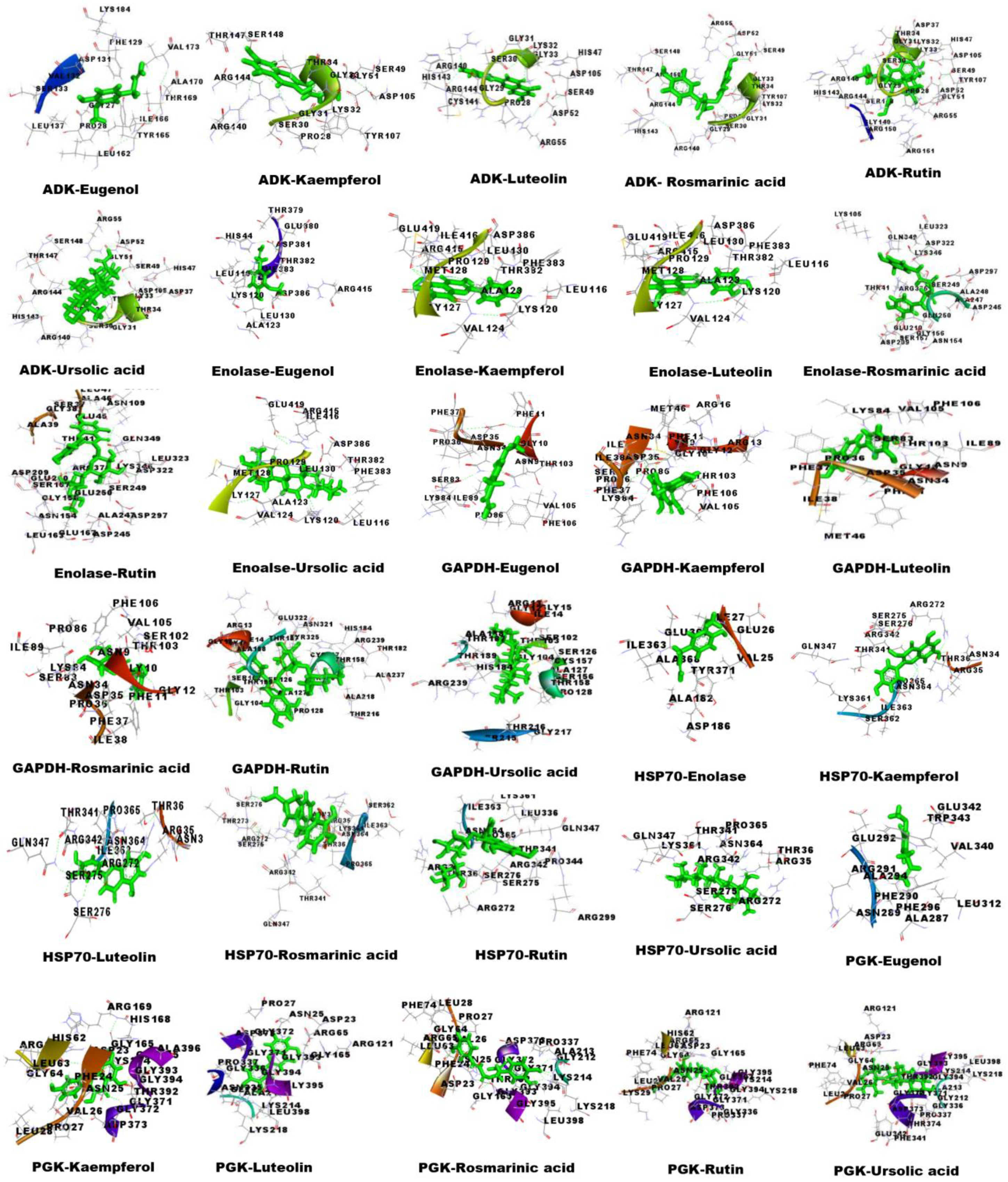
The structures of OSBC eugenol, kaempferol, luteolin, rosmarinic acid, rutin, and Ursolic acid were retrieved from the PubChem database (Supplementary Figure S2). Molecular docking of the aforementioned OSBCs and S. cervi DEPs was via by YASARA software version 15.11.18 and the PatchDock server version Beta 1.3 (Table 3). The amino acid interactions of the docked complexes were visualized by Discovery studio 3.5 (Figure 5). The highest scoring OSBC in molecular docking were rutin, luteolin, and ursolic acid. Among these, rutin formed the maximum number of hydrogen bonds with GAPDH (7 bonds) and enolase (7 bonds), whereas rosmarinic acid formed the maximum number of hydrogen bonds with ADK (5 bonds) and PGK (11 bonds). Eugenol formed hydrogen bonds only with HSP 70 (one bond) and did not form any hydrogen bonds with other DEPs. The anti-filarial drugs, DEC and albendazole, formed hydrogen bonds with PGK (Albendazole-1, DEC-2) and ADK (DEC-3) (Supplementary Table S3). Rutin showed the highest binding energy with ADK (9.0130 Kcal/mol), enolase (8.7070 Kcal/mol), and HSP 70 (7.9120 Kcal/mol). Ursolic acid had the strongest interaction with GAPDH (9.2360 Kcal/mol) and PGK (10.2220 Kcal/mol). The GSC score and AI area, as predicted by the PatchDock server version Beta 1.3, were highest for rutin and ursolic acid. The OSBC eugenol had the lowest binding energy and dissociation constant against S. cervi DEPs. The drug-likeness properties and ADME prediction of OSBCs used in this study is based upon a previous report from the laboratory (Mishra et al., 2022 [12]).

4. Discussion

Modern drugs used to treat infectious diseases are either directly or indirectly derived from the bioactive components of plants [34]. In the past few decades, traditional medicinal herbs and phytochemicals have gained great interest for the development of innovative and effective pharmaceuticals with fewer side effects than allopathic treatments. Additionally, the intake of dietary phytochemicals as chemoprevention agents for the detoxification of reactive species, and as nutraceuticals, is becoming popular even in developed countries of Europe and America too [35]. Herbal medicines are a time-tested and cost-effective alternative therapy for developing countries, where huge populations are living at the risk of LF infections. Numerous in vitro and in vivo studies have already demonstrated that crude extracts from plants, vital oils, and extracted active ingredients/compounds possess anti-filarial activity, which can be scientifically explored to discover new anti-filarial lead compounds [36].
The therapeutic potential of OS is immense, and the herb is already much explored as a potent antibacterial, antiviral, cardio-, hepato-, and nephro-protective [8]. OS has diverse medicinal properties and has pharmacological activities against lung cancer, malaria, Leishmaniasis, HIV, neurodegenerative, and lung diseases [37,38,39,40,41,42]. The ethanolic extract of OS can induce apoptosis in lung carcinoma, and its essential oil promotes cytotoxicity and apoptosis in colorectal adenocarcinoma [43,44]. Several OS formulations, like aqueous, ethanolic, and methanolic leaf extracts, as well as seed oil and fresh leaf paste, have been tested for their therapeutic potential [8].
In our study, the treatment of worms with ethanolic extract of O. sanctum (EOS) reduced the viability of S. cervi parasites in a dose- and time-dependent manner. It has been shown previously that anti-filarial medication—DEC, albendazole, and Ivermectin—do does not affect the viability of adult S. cervi, even after exposures for 4–8 h [45,46]. Nevertheless, the EOS showed significant macrofilaricidal activity within 6 h of treatment, as assessed via the MTT reduction assay. The reported LC50 values of some other herbal crude extracts tested against adult female S. cervi were 945.4 µg/mL for Andrographis paniculata and 47.12 µg/mL for Azadirachta indica [13,47]. The LC50 value of EOS is 197.24 µg/mL, which is much lower than the reported LC50 value of A. paniculata crude extract. Although the LC50 of EOS was higher than A. indica extract, the exposure of worms to A. indica was for 24 h, which is much longer than the EOS exposure in this study. It was observed that the EOS was more potent than the aqueous OS leaf extract (unpublished data). Earlier OS extract was reported as ovicidal and larvicidal in gastrointestinal nematodes, though the mechanism of anti-helminthic action of OS remained unclear [48]. As a result, the current study attempts to analyze the mechanism of anti-filarial action of OS and its bioactive components.
The filarial parasites survive easily for several years by successfully evading the host’s immune attacks. The parasites employ a battery of enzymatic and non-enzymatic defense proteins/enzymes such as glutathione-S-transferase, glutathione (GSH), GR, and NADPH oxidase as their defense system [49]. GSH regulates cellular homeostasis via the oxidation of SH groups of proteins involved in various functions and is also important as a scavenger of free radicals, thus maintaining a reducing environment within the cells. Additionally, equally important are the enzymes GST and GR, which act as free radicals scavengers, hence protecting the parasites from the hosts’ immune attacks [50]. A significant decrease in the free glutathione levels of EOS-treated S. cervi parasites were observed after exposure to 375 µg/mL of EOS (−39.23%). The antioxidant enzymes GST and GR showed similar patterns of decrease in enzymatic activity following EOS treatment. The effect of EOS on the antioxidant defense mechanism of the filarial parasites was pronounced and could be a major factor contributing to its anti-filarial activity.
The disruption of the antioxidant defense mechanism of S. cervi parasites prompted an investigation of oxidative stress parameters like lipid peroxidation, NADPH oxidase activity, and protein carbonyl content. An increase in protein carbonyl content, lipid peroxidation, and NADPH oxidase activity was observed after EOS exposure. An increase of almost 52% in levels of lipid peroxidation strongly indicates alteration in membrane permeability and fluidity, leading to irreversible membrane damage [51]. The NADPH oxidase has been implicated in varied functions, such as cellular signaling, regulation of gene expression, and host defense against viral and bacterial infections. An increase in NADPH oxidase activity leads to the transport of electrons to oxygen in the extracellular environment, resulting in the formation of superoxide anions [52]. The superoxide anion, so generated, augments the levels of hydrogen peroxide and other reactive oxygen species in the extracellular milieu of the filarial parasites. Once formed, the reactive oxygen species promote varieties of oxidative damage, such as the oxidation of proteins, which is estimated by measuring the increase in the protein carbonyl content [53]. The protein carbonyl content of the EOS-treated parasites was significantly higher than the control S. cervi worms (p = 0.0043), which could be due to the generation of ROS and oxidative stress.
The proteoforms profile of S. cervi adult female parasites after EOS treatment (250 µg/mL) exhibited a reduction in the number of protein spots as compared to the control worms. The gel analysis report showed that the differentially expressed proteins participated in several biological processes, including drug detoxification, redox regulation, energy metabolism, stress response, signaling, and structural and cytoskeleton. An increase in the levels of stress response and heat-shock proteins like HSP 70 and HSP 18 was also observed after EOS treatment. The molecular chaperone HSP 70 is an efficient redox scavenger and is overexpressed under stress conditions like hypoxia, nutrient depletion, and oxidative stress [54]. The sentinel heat shock protein, HSP 70, protects the cells from different stresses and imbalances that may occur due to aging or pathophysiological changes [55]. It is quite possible that the S. cervi parasites perceived EOS exposure as a stress stimulus and responded by overexpressing the heat-shock proteins HSP 70 and HSP 18.
Additionally, EOS treatment had a significant effect on the enzymes of energy metabolism like enolase and GAPDH, which were decreased in the treated parasites as compared to control worms. These enzymes are involved in the glycolytic pathway, and a decrease in the expression can be correlated with the decrease in the energy generation [56] during EOS exposure. ADK was significantly decreased in the treated parasites, which would impact the AMP-driven metabolic signaling, intracellular energetic communication, as well as the ATP supply [57]. The antioxidant proteins thioredoxin domain containing protein-3 homolog was increased, while GST was decreased following exposure to EOS. Decreased in GST in filarial parasites is associated with the loss of metabolic homeostasis, culminating in parasites’ death [58]. The EOS-treated S. cervi worms also showed diminished levels of the p27 molecule, which is an inhibitor of CDK (cyclin dependent kinases). p27 is associated with cell viability, and its reduced expression could render the parasites vulnerable to death [53].
The in silico approach offers a considerable advantage over in vivo lab research in terms of time and resource involvement. The effectiveness of molecular docking in the realms of drug discovery and design is now widely recognized. In particular, the molecular targets of natural compounds used in disease management are initially verified through the use of molecular docking. In natural compounds/nutraceutical research, molecular docking studies are employed to provide crucial information prior to in vitro experiments. In order to identify the anti-filarial effect of O. sanctum bioactive compounds, molecular docking was performed. The O. sanctum bioactive compounds used in this report are either reported as major components of OS leaf extract in the literature or have proven pharmacological and therapeutics activities. The computational evaluation of the ligand–receptor electrostatics can be predicted via docking studies [59]. The molecular docking technique predicts the orientation of the ligand in a complex that it may form with proteins or enzymes. The form and structure of the docked complex, as well as the Van der Waal forces, ionic bonds, and hydrogen bonds between ligand and receptor, are taken into account to quantify the molecular interactions [60].
The three best bioactive compounds based on molecular docking analysis were ursolic acid, rutin, and rosmarinic acid. Ursolic acid, a natural pentacyclic triterpenoid carboxylic acid, has proven anti-filarial activity against B. malayi and S. cervi microfilariae and adult parasites [61,62]. In our investigation, ursolic acid has given high binding energies against the differentially expressed proteins, ADK, enolase, GAPDH, HSP 70, and PGK. Another in silico study has also reported the high binding affinity of ursolic acid (UA) with Brugia malayi GST; hence, UA may be considered as a potential lead compound for designing novel anti-filarial compounds/drugs [61].
The second-best OSBC with anti-filarial activity identified via molecular docking is the flavonoid rutin. It has a wide range of pharmaceutical benefits, including antibacterial, anticancer, antifungal, antiretroviral, and antimalarial activities [63]. In pancreatic cancer cells, rutin has been shown to promote apoptosis by upregulating miR-877-3p expression, which, in turn, represses bcl-2 transcription [64]. However, the anti-filarial effect of rutin has not been much explored, except in a solitary report on B. malayi worms, where it was shown to have macro and microfilaricidal activities [65]. In molecular docking, rutin showed high binding energies and low dissociation constant with ADK, enolase, GAPDH, HSP 70, and PGK. Based on the earlier findings and our work, the bioactive compound rutin can certainly be explored further for its anti-filarial effects. The phenolic compound rosmarinic acid also showed good docking scores against the targeted S. cervi DEPs. Rosmarinic acid is an anticancer, antiangiogenic, anti-inflammatory, and antimicrobial agent [66]. We observed that rosmarinic acid could bind strongly to several proteins, such as ADK, enolase, GAPDH, HSP 70, and PGK. The antiparasitic and anti-filarial activities of rosmarinic acid have not been investigated so far, and more experimental validations can delineate its mode of anti-filarial action.
Although the study was meticulously planned, the present investigation still has some limitations. Despite the substantial benefits of our 2D profiling, it has a drawback in terms of throughput; on average, a 2D study takes around two–three days. Also, solubilization is a major concern because the hydrophobic and membrane-bound proteins in the worm tegument need to be solubilized separately using detergents; hence, the proteoform profile of this research does not include the impact of EOS on these proteoforms. Additionally, the alterations in proteomes were quantified using 2D image analysis tools, which required a number of replicates for comparison and spot matching [67] since we have used protease inhibitors such as PMSF and EDTA, which may be a reason for the loss of certain proteins and/or their absence from the 2D PAGE gels.
Thus, in essence, this work proves the macrofilaricidal effect of OS by mediating its action on filarial antioxidant enzymes/proteins, lipid peroxidation, and protein carbonylation. Following this, the proteoforms analysis successfully identified putative filarial drug targets that were validated in silico via molecular docking against OS bioactive compounds.

5. Conclusions

The ex vivo experiments demonstrated that EOS had significantly affected the viability and biochemical markers of adult female S. cervi parasites. The effect of EOS was mainly due to the increase in reactive oxygen production evidenced by the decrease in GST, GR, and GSH levels. Proteomic analysis and biochemical assays confirmed that EOS could induce apoptosis in S. cervi. Molecular docking with OS bioactive compounds proved that eugenol, kaempferol, luteolin, rosmarinic acid, rutin, and ursolic acid could stably interact with DEPs of S. cervi. In conclusion, the overall outcomes from our study provide insight into the effect of OS and its bioactive compounds on S. cervi. Further studies on the multi-inhibitory potential of OS and its derivatives can lead to the development of better anthelminthic drugs in future.

Supplementary Materials

The following supporting information can be downloaded at: https://www.mdpi.com/article/10.3390/proteomes13010002/s1, Table S1A: Hydrogen bond statics of Protein models analyzed by VADAR; Table S1B: Protein structure elements of Protein model depicted by VADAR server v1.8; Table S2: Ramachandran plot analysis of targeted protein models by Procheck server; Table S3: The docked complex of GAPDH, Enolase, ADK, HSP70, and PGK with different bioactive compounds of OS showing interacting receptor residue, and ligand and protein complex involved in H-bonding; Figure S1: Correlation graph of spot matching. Analysis of protein spots in adult female S. cervi parasites after EOS treatment. Light blue region: upregulated spots and red region: downregulated spots; Figure S2: Structures of Ocimum sanctum bioactive compounds used in molecular docking study as retrieved from PubChem database.

Author Contributions

Conceptualization, A.M. and A.S.; methodology, A.M. and V.K; software, A.M., S.K. and V.K.; validation, A.M., V.K. and A.S.; formal analysis, A.M. and V.K.; investigation, A.M. and A.S.; resources, A.S.; data curation, A.M. and A.S.; writing—original draft preparation, A.M. and A.S.; writing—review and editing, A.M., V.K., A.S. and H.S.; visualization, A.M., S.K. and V.K; supervision, A.S.; project administration, A.S. All authors have read and agreed to the published version of the manuscript.

Funding

This research received no external funding.

Institutional Review Board Statement

This study uses Setaria cervi bovine filarial parasites obtained from slaughter houses in Varanasi city. Varanasi municipal corporation does not require the study to be reviewed or approved by an ethics committee because the Setaria cervi parasites are the by-products of local slaughter houses.

Data Availability Statement

The 2D electrophoresis gel images and proteomics data have been deposited to Zenodo, available at https://doi.org/10.5281/zenodo.13968369.

Acknowledgments

Ayushi Mishra expresses gratitude to the Council of Scientific and Industrial Research (CSIR), India (09/013(0832)/2018-EMR-I), for awarding a Senior Research Fellowship (SRF). For financial support as an SRF, Vipin Kumar is grateful to the Department of Biotechnology (DBT), India (DBT/JRF/BET-18/I/2018/AL/19). Sunil Kumar is grateful to the NBCFDC JRF (Ref. No. 231620090211), Junior Research Fellowship, University Grant Commission (UGC), New Delhi. We acknowledge the HRAMS Facility, DST-Sophisticated Analytical and Technical Help Institute (SATHI), Banaras Hindu University, Varanasi, India, for providing the proteomics central facility. We also express our gratitude to the Centre for Bioinformatics, School of Biotechnology for YASARA software, and to the DBT-BHU Interdisciplinary School of Life Sciences, Banaras Hindu University for laboratory space.

Conflicts of Interest

The authors declare no conflict of interest.

References

  1. Available online: https://www.who.int/news-room/fact-sheets/detail/lymphatic-filariasis (accessed on 2 December 2024).
  2. Ton, T.G.; Mackenzie, C.; Molyneux, D.H. The burden of mental health in lymphatic filariasis. Infect. Dis. Poverty 2015, 4, 34. [Google Scholar] [CrossRef] [PubMed]
  3. Wadhawan, M.; Singh, N.; Rathaur, S. Inhibition of cathepsin B by E-64 induces oxidative stress and apoptosis in filarial parasite. PLoS ONE 2014, 9, e93161. [Google Scholar] [CrossRef] [PubMed]
  4. Budge, P.J.; Herbert, C.; Andersen, B.J.; Weil, G.J. Adverse events following single dose treatment of lymphatic filariasis: Observations from a review of the literature. PLoS Negl. Trop. Dis. 2018, 12, e0006454. [Google Scholar] [CrossRef] [PubMed]
  5. Behera, D.R.; Bhatnagar, S. Filariasis: Role of medicinal plant in lymphatic filariasis. Int. J. Herb. Med. 2018, 6, 40–46. [Google Scholar]
  6. Cobo, F. Determinants of parasite drug resistance in human lymphatic filariasis. Rev. Española Quimioter. 2016, 29, 288–295. [Google Scholar]
  7. Singh, A.; Mishra, A.; Chaudhary, R.; Kumar, V. Role of herbal plants in prevention and treatment of parasitic diseases. J. Sci. Res. 2020, 64, 50–58. [Google Scholar] [CrossRef]
  8. Pattanayak, P.; Behera, P.; Das, D.; Panda, S.K. Ocimum sanctum Linn. A reservoir plant for therapeutic applications: An overview. Pharmacogn. Rev. 2010, 4, 95–105. [Google Scholar] [CrossRef]
  9. Baliga, M.S.; Jimmy, R.; Thilakchand, K.R.; Sunitha, V.; Bhat, N.R.; Saldanha, E.; Rao, S.; Rao, P.; Arora, R.; Palatty, P.L. Ocimum sanctum L. (Holy Basil or Tulsi) and its phytochemicals in the prevention and treatment of cancer. Nutr. Cancer 2013, 65 (Suppl. S1), 26–35. [Google Scholar] [CrossRef]
  10. Pandey, R.; Chandra, P.; Srivastava, M.; Mishra, D.K.; Kumar, B. Simultaneous quantitative determination of multiple bioactive markers in Ocimum sanctum obtained from different locations and its marketed herbal formulations using UPLC-ESI-MS/MS combined with principal component analysis. Phytochem. Anal. 2015, 26, 383–394. [Google Scholar] [CrossRef]
  11. Chaudhary, A.; Sharma, S.; Mittal, A.; Gupta, S.; Dua, A. Phytochemical and antioxidant profiling of Ocimum sanctum. J. Food Sci. Technol. 2020, 57, 3852–3863. [Google Scholar] [CrossRef]
  12. Mishra, A.; Kumar, V.; Singh, A. Deciphering the anti-filarial potential of bioactive compounds from Ocimum sanctum: A combined experimental and computational study. Pharm. Biol. 2022, 60, 2237–2252. [Google Scholar] [CrossRef] [PubMed]
  13. Mukherjee, N.; Mukherjee, S.; Saini, P.; Roy, P.; Sinha Babu, S.P. Antifilarial effects of polyphenol rich ethanolic extract from the leaves of Azadirachta indica through molecular and biochemical approaches describing reactive oxygen species (ROS) mediated apoptosis of Setaria cervi. Exp. Parasitol. 2014, 136, 41–58. [Google Scholar] [CrossRef] [PubMed]
  14. Singh, A.; Rathaur, S. Identification and characterization of a selenium dependent glutathione peroxidase in Setaria cervi. Biochem. Biophys. Res. Commun. 2005, 331, 1069–1074. [Google Scholar] [CrossRef] [PubMed]
  15. Sharma, S.; Ahmad, F.; Singh, A.; Rathaur, S. Identification of glucose regulated protein94 (GRP94) in filarial parasite S. cervi and its expression under ER stress. Comp. Biochem. Physiol. Part B Biochem. Mol. Biol. 2022, 258, 110683. [Google Scholar] [CrossRef]
  16. Bradford, M.M. A rapid and sensitive method for the quantitation of picogram quantities of protein utilizing the principle of protein dye binding. Anal. Biochem. 1970, 72, 248–254. [Google Scholar] [CrossRef]
  17. Mosmann, T.R.; Coffman, R.L. TH1 and TH2 cells: Different patterns of lymphokine secretion lead to different functional properties. Annu. Rev. Immunol. 1989, 7, 145–173. [Google Scholar] [CrossRef]
  18. Ellman, G.L. Tissue sulfhydryl groups. Arch. Biochem. Biophys. 1959, 82, 70–77. [Google Scholar] [CrossRef]
  19. Habig, W.H.; Pabst, M.J.; Jakoby, W.B. Glutathione-S-transferases: The first enzymatic step in mercapturic acid formation. J. Biol. Chem. 1974, 249, 7130–7139. [Google Scholar] [CrossRef]
  20. Carlberg, I.; Mannervik, B. Purification and characterization glutathione reductase from calf liver: An improved procedure for affinity chromatography on 29 59-ADP sepharose 4B. Anal. Biochem. 1981, 116, 531–536. [Google Scholar] [CrossRef]
  21. Holmgren, A.; Björnstedt, M. Thioredoxin and thioredoxin reductase. Methods Enzymol. 1995, 252, 199–208. [Google Scholar]
  22. Levine, R.L.; Williams, J.A.; Stadtman, E.R.; Shacter, E. Carbonyl assay for determination of oxidatively modified proteins. Methods Enzymol. 1994, 233, 346–357. [Google Scholar] [PubMed]
  23. Ohkawa, H.; Ohishi, N.; Yagi, K. Assay for lipid peroxides in animal tissues by thiobarbituric acid reaction. Anal. Biochem. 1979, 95, 351–358. [Google Scholar] [CrossRef] [PubMed]
  24. Heyneman, R.A.; Vercauteren, R.E. Activation of a NADPH oxidase from horse polymorphonuclear leukocytes in a cell-free system. J. Leukoc. Biol. 1984, 36, 751–759. [Google Scholar] [CrossRef]
  25. Kumar, V.; Mishra, A.; Yadav, A.K.; Rathaur, S.; Singh, A. Lymphatic filarial serum proteome profiling for identification and characterization of diagnostic biomarkers. PLoS ONE 2022, 17, e0270635. [Google Scholar] [CrossRef]
  26. Szklarczyk, D.; Gable, A.L.; Lyon, D.; Junge, A.; Wyder, S.; Huerta-Cepas, J.; Simonovic, M.; Doncheva, N.T.; Morris, J.H.; Bork, P.; et al. STRING v11: Protein-protein association networks with increased coverage, supporting functional discovery in genome-wide experimental datasets. Nucleic Acids Res. 2019, 47, D607–D613. [Google Scholar] [CrossRef]
  27. Burley, S.K.; Bhikadiya, C.; Bi, C.; Bittrich, S.; Chen, L.; Crichlow, G.V.; Christie, C.H.; Dalenberg, K.; Costanzo, L.D.; Duarte, J.M.; et al. RCSB Protein Data Bank: Powerful new tools for exploring 3D structures of biological macromolecules for basic and applied research and education in fundamental biology, biomedicine, biotechnology, bioengineering and energy sciences. Nucleic Acids Res. 2021, 49, D437–D451. [Google Scholar] [CrossRef]
  28. Willard, L.; Ranjan, A.; Zhang, H.; Monzavi, H.; Boyko, R.F.; Sykes, B.D.; Wishart, D.S. VADAR: A web server for quantitative evaluation of protein structure quality. Nucleic Acids Res. 2003, 31, 3316–3319. [Google Scholar] [CrossRef]
  29. Kim, S.; Chen, J.; Cheng, T.; Gindulyte, A.; He, J.; He, S.; Li, Q.; Shoemaker, B.A.; Thiessen, P.A.; Yu, B.; et al. PubChem in 2021: New data content and improved web interfaces. Nucleic Acids Res. 2021, 49, D1388–D1395. [Google Scholar] [CrossRef]
  30. Lipinski, C.A. Lead- and drug-like compounds: The rule-of-five revolution. Drug Discov. Today Technol. 2004, 1, 337–341. [Google Scholar] [CrossRef]
  31. Jayaram, B.; Singh, T.; Mukherjee, G.; Mathur, A.; Shekhar, S.; Shekhar, V. Sanjeevini: A freely accessible web-server for target directed lead molecule discovery. BMC Bioinform. 2012, 13, S7. [Google Scholar] [CrossRef]
  32. Cheng, F.; Li, W.; Zhou, Y.; Shen, J.; Wu, Z.; Liu, G.; Lee, P.W.; Tang, Y. Correction to “admetSAR: A Comprehensive Source and Free Tool for Assessment of Chemical ADMET Properties”. J. Chem. Inf. Model. 2019, 59, 4959. [Google Scholar] [CrossRef] [PubMed]
  33. Kumar, V.; Mishra, A.; Singh, V.K.; Singh, A. Targeting HIV-1 Tat and Human Tat Protein Complex through Natural Products: An In silico Docking and Molecular Dynamic Simulation Approach. Lett. Drug Des. Discov. 2022, 19, 982–995. [Google Scholar] [CrossRef]
  34. Musthaba, M.; Baboota, S.; Athar, T.M.; Thajudeen, K.Y.; Ahmed, S.; Ali, J. Patented herbal formulations and their therapeutic applications. Recent Pat. Drug Deliv. Formul. 2010, 4, 231–244. [Google Scholar] [CrossRef] [PubMed]
  35. Ekor, M. The growing use of herbal medicines: Issues relating to adverse reactions and challenges in monitoring safety. Front. Pharmacol. 2014, 4, 177. [Google Scholar] [CrossRef]
  36. Al-Abd, N.M.; Nor, Z.M.; Al-Adhroey, A.H.; Suhaimi, A.; Sivanandam, S. Recent advances on the use of biochemical extracts as filaricidal agents. Evid. Based Complement. Altern. Med. 2013, 2013, 986573. [Google Scholar] [CrossRef]
  37. Kustiati, U.; Ratih, T.S.D.; Agung, N.D.A.; Kusindarta, D.L.; Wihadmadyatami, H. In silico molecular docking and in vitro analysis of ethanolic extract Ocimum sanctum Linn.: Inhibitory and apoptotic effects against non-small cell lung cancer. Vet. World 2021, 14, 3175–3187. [Google Scholar] [CrossRef]
  38. Inbaneson, S.J.; Sundaram, R.; Suganthi, P. In vitro antiplasmodial effect of ethanolic extracts of traditional medicinal plant Ocimum species against Plasmodium falciparum. Asian Pac. J. Trop. Med. 2012, 5, 103–106. [Google Scholar] [CrossRef]
  39. Kaur, S.; Bhardwaj, K.; Sachdeva, H. Antileishmanial efficacy of Boerhaavia diffusa L. and Ocimum sanctum L. against experimental visceral leishmaniasis. Indian J. Exp. Biol. 2015, 53, 522–529. [Google Scholar]
  40. Sonar, V.P.; Corona, A.; Distinto, S.; Maccioni, E.; Meleddu, R.; Fois, B.; Floris, C.; Malpure, N.V.; Alcaro, S.; Tramontano, E.; et al. Natural product-inspired esters and amides of ferulic and caffeic acid as dual inhibitors of HIV-1 reverse transcriptase. Eur. J. Med. Chem. 2017, 130, 248–260. [Google Scholar] [CrossRef]
  41. Nandini, H.S.; Krishna, K.L.; Apattira, C. Combination of Ocimum sanctum extract and Levetiracetam ameliorates cognitive dysfunction and hippocampal architecture in rat model of Alzheimer’s disease. J. Chem. Neuroanat. 2022, 120, 102069. [Google Scholar] [CrossRef]
  42. Venuprasad, M.P.; Kandikattu, H.K.; Razack, S.; Amruta, N.; Khanum, F. Chemical composition of Ocimum sanctum by LC-ESI–MS/MS analysis and its protective effects against smoke induced lung and neuronal tissue damage in rats. Biomed. Pharmacother. 2017, 91, 1–12. [Google Scholar] [CrossRef] [PubMed]
  43. Magesh, V.; Lee, J.C.; Ahn, K.S.; Lee, H.J.; Lee, H.J.; Lee, E.O.; Shim, B.S.; Jung, H.J.; Kim, J.S.; Kim, D.K.; et al. Ocimum sanctum induces apoptosis in A549 lung cancer cells and suppresses the in vivo growth of Lewis lung carcinoma cells. Phytother. Res. 2009, 23, 1385–1391. [Google Scholar] [CrossRef] [PubMed]
  44. Sharma, M.; Agrawal, S.K.; Sharma, P.R.; Chadha, B.S.; Khosla, M.K.; Saxena, A.K. Cytotoxic and apoptotic activity of essential oil from Ocimumviride towards COLO 205 cells. Food Chem. Toxicol. 2010, 48, 336–344. [Google Scholar] [CrossRef] [PubMed]
  45. Singh, A.; Rathaur, S. Combination of DEC plus aspirin induced mitochondrial mediated apoptosis in filarial parasite Setaria cervi. Biochimie 2010, 92, 894–900. [Google Scholar] [CrossRef]
  46. Sharma, S.; Ahmad, F.; Singh, A.; Rathaur, S. Role of anti-filarial drugs in inducing ER stress mediated signaling in bovine filarial parasitosis Setaria cervi. Vet. Parasitol. 2021, 290, 109357. [Google Scholar] [CrossRef]
  47. Yadav, S.; Sharma, S.; Ahmad, F.; Rathaur, S. Antifilarial efficacy of green silver nanoparticles synthesized using Andrographis paniculate. J. Drug Deliv. Sci. Technol. 2020, 56, 101557. [Google Scholar] [CrossRef]
  48. Kanojiya, D.; Shanker, D.; Sudan, V.; Jaiswal, A.K.; Parashar, R. Anthelmintic activity of Ocimum sanctum leaf extract against ovine gastrointestinal nematodes in India. Res. Vet. Sci. 2015, 99, 165–170. [Google Scholar] [CrossRef]
  49. Chiumiento, L.; Bruschi, F. Enzymatic antioxidant systems in helminth parasites. Parasitol. Res. 2009, 105, 593. [Google Scholar] [CrossRef]
  50. Gupta, S.; Srivastava, A.K. Glutathione metabolism of filarial worms: A vulnerable target for the design and synthesis of new antifilarial agents. Med. Sci. Monit. 2006, 12, HY1–HY9. [Google Scholar]
  51. Tiwari, S.; Wadhawan, M.; Singh, N.; Rathaur, S. Effect of CDNB on filarial thioredoxin reductase: A proteomic and biochemical approach. J. Proteom. 2015, 113, 435–446. [Google Scholar] [CrossRef]
  52. Taylor, J.P.; Tse, H.M. The role of NADPH oxidases in infectious and inflammatory diseases. Redox Biol. 2021, 48, 102159. [Google Scholar] [CrossRef] [PubMed]
  53. Singh, N.; Wadhawan, M.; Tiwari, S.; Kumar, R.; Rathaur, S. Inhibition of Setaria cervi protein tyrosine phosphatases by Phenylarsine oxide: A proteomic and biochemical study. Acta Trop. 2016, 159, 20–28. [Google Scholar] [CrossRef] [PubMed]
  54. Srivastava, S.; Srikanth, E.; Liebau, E.; Rathaur, S. Identification of Setaria cervi heat shock protein 70 by mass spectrometry and its evaluation as diagnostic marker for lymphatic filariasis. Vaccine 2010, 28, 1429–1436. [Google Scholar] [CrossRef] [PubMed]
  55. Kim, J.Y.; Barua, S.; Huang, M.Y.; Park, J.; Yenari, M.A.; Lee, J.E. Heat Shock Protein 70 (HSP70) Induction: Chaperonotherapy for Neuroprotection after Brain Injury. Cells 2020, 9, 2020. [Google Scholar] [CrossRef] [PubMed]
  56. Zinsser, V.L.; Hoey, E.M.; Trudgett, A.; Timson, D.J. Biochemical characterisation of glyceraldehyde 3-phosphate dehydrogenase (GAPDH) from the liver fluke, Fasciola hepatica. Biochim. Biophys. Acta 2014, 1844, 744–749. [Google Scholar] [CrossRef]
  57. Dzeja, P.; Terzic, A. Adenylate kinase and AMP signaling networks: Metabolic monitoring, signal communication and body energy sensing. Int. J. Mol. Sci. 2009, 10, 1729–1772. [Google Scholar] [CrossRef]
  58. Rathaur, S.; Yadav, M.; Singh, N.; Singh, A. Effect of diethylcarbamazine, butylated hydroxy anisole and methyl substituted chalcone on filarial parasite Setaria cervi: Proteomic and biochemical approaches. J. Proteom. 2011, 74, 1595–1606. [Google Scholar] [CrossRef]
  59. Sahoo, R.N.; Pattanaik, S.; Pattnaik, G.; Mallick, S.; Mohapatra, R. Review on the use of Molecular Docking as the First Line Tool in Drug Discovery and Development. Indian J. Pharm. Sci. 2022, 84, 1334–1337. [Google Scholar] [CrossRef]
  60. Agu, P.C.; Afiukwa, C.A.; Orji, O.U.; Ezeh, E.M.; Ofoke, I.H.; Ogbu, C.O.; Aja, P.M. Molecular docking as a tool for the discovery of molecular targets of nutraceuticals in diseases management. Sci. Rep. 2023, 13, 13398. [Google Scholar] [CrossRef]
  61. Kalani, K.; Kushwaha, V.; Sharma, P.; Verma, R.; Srivastava, M.; Khan, F.; Murthy, P.K.; Srivastava, S.K. In Vitro, In Silico and In Vivo Studies of Ursolic Acid as an Anti-Filarial Agent. PLoS ONE 2014, 9, e11124. [Google Scholar] [CrossRef]
  62. Saini, P.; Gayen, P.; Kumar, D.; Nayak, A.; Mukherjee, N.; Mukherjee, S.; Pal, B.C.; Babu, S.P. Antifilarial effect of ursolic acid from Nyctanthes arbortristis: Molecular and biochemical evidences. Parasitol. Int. 2014, 63, 717–728. [Google Scholar] [CrossRef] [PubMed]
  63. Ganeshpurkar, A.; Saluja, A.K. The Pharmacological Potential of Rutin. Saudi Pharm. J. 2017, 25, 149–164. [Google Scholar] [CrossRef] [PubMed]
  64. Huo, M.; Xia, A.; Cheng, W.; Zhou, M.; Wang, J.; Shi, T.; Cai, C.; Jin, W.; Zhou, M.; Liao, Y.; et al. Rutin Promotes Pancreatic Cancer Cell Apoptosis by Upregulating miRNA-877-3p Expression. Molecules 2022, 27, 2293. [Google Scholar] [CrossRef]
  65. Lakshmi, V.; Joseph, S.K.; Srivastava, S.; Verma, S.K.; Sahoo, M.K.; Dube, V.; Mishra, S.K.; Murthy, P.K. Antifilarial activity in vitro and in vivo of some flavonoids tested against Brugia malayi. Acta Trop. 2010, 116, 127–133. [Google Scholar] [CrossRef]
  66. Swamy, M.K.; Sinniah, U.R.; Ghasemzadeh, A. Anticancer potential of rosmarinic acid and its improved production through biotechnological interventions and functional genomics. Appl. Microbiol. Biotechnol. 2018, 102, 7775–7793. [Google Scholar] [CrossRef]
  67. Jain, M.; Dhariwal, R.; Patil, N.; Ojha, S.; Tendulkar, R.; Tendulkar, M.; Dhanda, P.S.; Yadav, A.; Kaushik, P. Unveiling the Molecular Footprint: Proteome-Based Biomarkers for Alzheimer’s Disease. Proteomes 2023, 11, 33. [Google Scholar] [CrossRef]
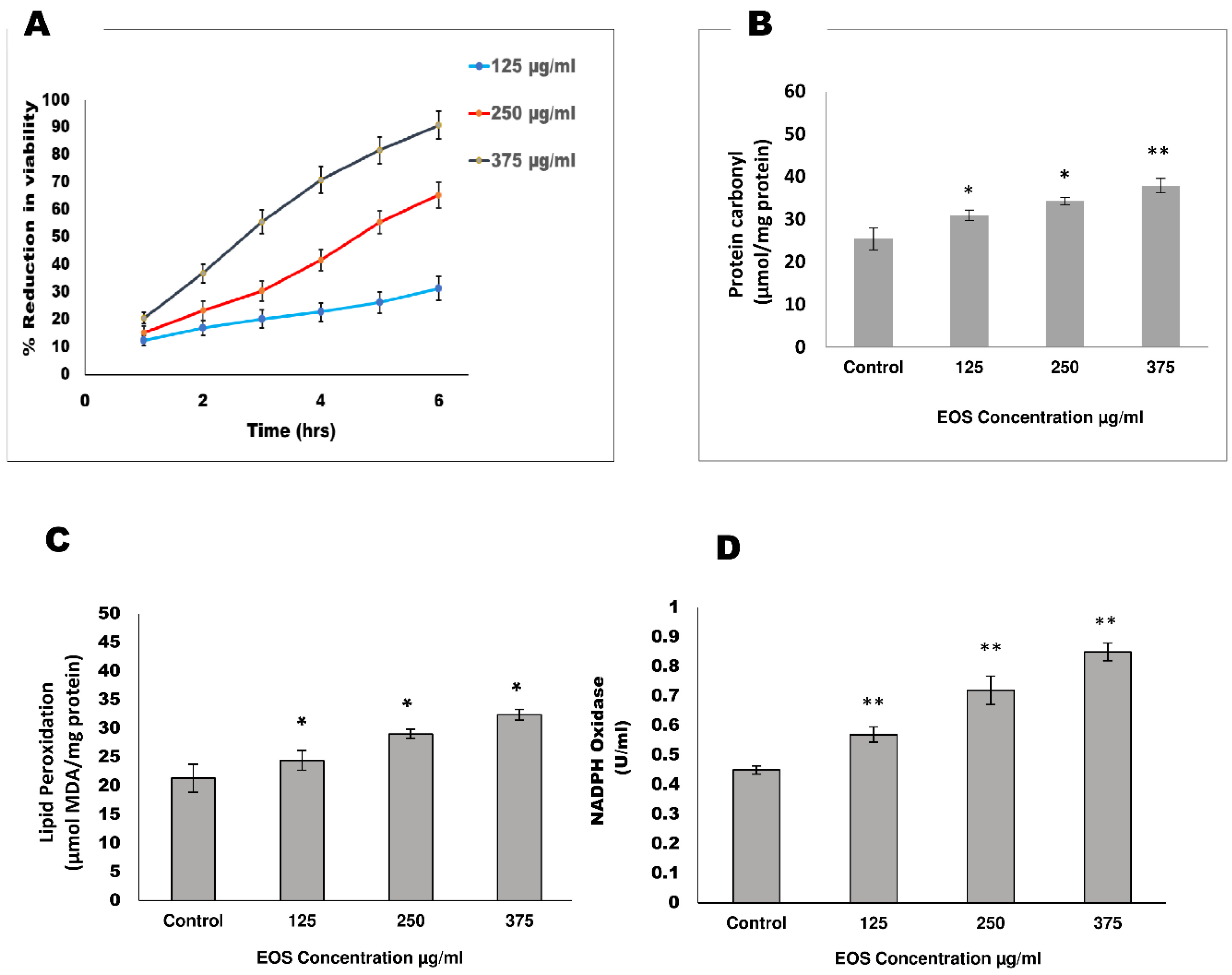
Figure 1. Effect of EOS on the adult female S. cervi worms. Adult worms (n = 5) of equal size were exposed at 125 µg/mL, 250 µg/mL, and 375 µg/mL concentration of ethanolic extract of OS in 20 mL of KRB maintenance medium at 37 °C. (A) The viability of parasites was determined via MTT assay after 6 h of incubation; percent reduction in viability was calculated with respect to the control group of S. cervi worms. (B) Protein carbonyl content is expressed in terms of μmol/mg protein. (C) Lipid peroxidation level is expressed in terms of μmol MDA/mg protein. (D) Activity of the enzyme NADPH oxidase activity is expressed as U/mL. Data expressed are the mean ± SD of at least three values (n = 3). p values < 0.05 (*), <0.01 (**) were considered as significant statistically.
Proteomes 13 00002 g001
Figure 2. Two-dimensional electrophoresis profile of Coomassie-stained crude extract of S. cervi exposed to EOS: (A) control; (B) treated. The first dimension was performed on pH 3–10 IPG strips followed by a second dimension on 10% SDS-PAGE. Increased protein spots are indicated by blue circles; decreased spots are indicated by red circles.
Proteomes 13 00002 g002
Figure 3. Interaction network and functional analysis of identified proteins of S. cervi DEPs after EOS treatment: (A) interaction network depicting the association among the differentially regulated proteins; (B) gene ontology (GO) biological process, represented in the pie chart as a percentage; (C) KEGG pathways, represented in the pie chart as a percentage.
Proteomes 13 00002 g003
Figure 4. Three-dimensional structure and quality validation, via Procheck server, of differentially expressed proteins (DEPs) used in molecular docking analysis. Square shows the Ramachandran plot of respective protein red color shows core region, yellow shows allowed region, green color shows generous region and gray color showed disallowed region.
Proteomes 13 00002 g004
Figure 5. Three-dimensional interactions of S. cervi DEPs with OS bioactive compounds. The docked complexes were visualized, and the ligands are represented in green color. Interacting residues are as labeled.
Proteomes 13 00002 g005
Table 1. Effect of ethanolic extract of Ocimum sanctum on S. cervi antioxidant stress markers.
SamplesGSH (µM/mg Protein)GST (U/mL)GR (U/mL)TRxR
Control6.50 ± 0.12022.86 ± 0.56816.02 ± 1.11018.12 ± 0.832
125 µg/mL5.82 ± 0.51320.34 ± 0.862 *11.89 ± 0.636 **11.35 ± 1.232 **
(−10.46%)(−11.02%)(−25.78%)(−37.36%)
250 µg/mL4.24 ± 0.98215.56 ± 1.101 *9.54 ± 0.350 **5.82 ± 0.345 ***
(−34.76%)(−31.93%)(−40.44%)(−67.88%)
375 µg/mL3.95 ± 0.832 *11.32 ± 1.926 *6.38 ± 0.832 **3.99 ± 0.127 ***
(−39.23%)(−50.48%)(−60.17%)(−77.98%)
The enzyme activity GSH, GST, GR, and TRxR were assessed in the control and EOS-treated parasites after 6 h of incubation. The values in parentheses indicate percentage activation/inhibition in comparison to control. Data expressed are the mean ± SD of at least three values (n = 3). p-value < 0.05 considered significant. *** p < 0.001, ** p < 0.01, * p < 0.05.
Table 2. HRAMS analysis of differentially expressed proteins of S. cervi.
S.N.Protein a
(Accession Number)
FunctionMW/pIIdentified PeptideFold Change b,c
ExpectedExperimental
Cytosol stress response protein/chaperons
1Heat-shock protein 70 [P27541]Act as molecular chaperone; help in protein folding, transport, and assembly; and protect cell against stress72/5.670.2/5.47[K].MKETAEAFLGHAVK.[D]
[RH].NVLIFDLGGGTFDVSILTIEDGIFEVK.[S]
[R].IINEPTAAAIAYGLDK.[K]
+1.9 *
2p27 [A0A4E9ERK7]Cell cycle regulation25.86/7.924.1/6.32[K].IDVTPSNYSVLDTEFGSMR.[E]
[R].AVFREYNQEFMLPR.[G]
−2.4 **
Cytosol energy metabolism proteins
3Adenylate kinase isoenzyme 1 [J9AQV1]Catalyzes phosphoryl transferase, having a role in metabolic monitoring and AMP signaling 22.95/8.822.8/8.34[R].LHTYITATAPVVDYYQK.[Q]
[K].YGLTHLSSGDLLRAEVK.[S]
[K].ANVPIFFIVGGPGSGKGTQCDKIVAK.[Y]
−3.6 ***
4Enolase [Q5GTG4]Role in glycolysis and gluconeogenesis47/5.946.4/5.67[R].LAKYNELIR.[I]−3.3 ***
5Glyceraldehyde−3-phosphate dehydrogenase [A0A4E9FA01]Role in energy metabolism36/7.136.1/7.84[K].LTGMAFRVPTPDVSVVDLTCR.[L]
[R].VPTPDVSVVDLTCR.[L]
[K].AVGKVIPDLNGKLTGMAFR.[V]
−3.3 ***
6Phosphoglycerate kinase [A0A4E9EYJ5]Glycolytic enzyme44/7.6844.6/7.88[K].MEFTLEPVAAELK.[A]
[R].AKTIVWNGPAGVFEWENFSK.[G]
[R].KMEFTLEPVAAELK.[A]
−3.2 ***
Antioxidant protein/enzymes
7Glutathione S-transferase [E3UV59]Antioxidant enzyme; detoxification of endogenous and xenobiotics compounds25/5.8824.1/6.68[K].DILPVELAKFEK.[L]
[K].FEKLLATR.[D]
−2.6 **
8Thioredoxin domain-containing protein [A0A4E9FJK0]Antioxidant enzyme22.1/7.0922.1/7.06[R].LIQAFQFVDKHGEVCPANWHPGSETIKPGVK.[E]
[K].GKYVVLFFYPLDFTFVCPTEIIAFSDR.[I]
+1.5 *
Signaling protein
9Coiled-coil domain-containing protein 6 [A0A1I8E9M6]Structural motifs involved in a variety of important interactions 60.92/5.1960.9/5.26[R].AFAASETTRENDEDNCMAALLNR.[M]+2.4 **
Protein Digestion and folding protein
10Calreticulin precursor
[A0A0J9XSV8]
Calcium-binding chaperone role in transcription regulation47.42/4.7849.4/4.87[K].KVHVIFHYKGR.[N]
[K].HKDDFGKWEISHGK.[F]
−3.5 ***
a Matched proteins were retrieved from the Uniprot database (http//:www.uniprot.org, accessed on 11 January 2022). b Fold change calculated from mean intensity and volume of spots observed in treated and control groups run on two different sets of experiments. c Student’s t test were used for calculating the statistical difference between control and treated groups. p-value < 0.05 considered as significant. *** p < 0.001, ** p < 0.01, * p < 0.05.
Table 3. Docking summary of treated S. cervi differentially expressed proteins with bioactive compounds of Ocimum sanctum.
ReceptorName of LigandBinding Energy (Kcal/mol)Dissociation Constant (µm)GSC ScoreAI Area
ADKEugenol5.709065.35023286381.30
Kaempferol7.44003.51894244473.50
Luteolin7.59202.72264280497.40
Rosmarinic acid7.91601.57575100636.50
Rutin9.01300.24735614630.70
Ursolic acid7.66602.40296176791.80
Albendazole5.956043.07194426518.70
DEC5.0650193.77854038475.00
EnolaseEugenol5.4220106.07703484388.50
Kaempferol8.08201.19074062388.50
Luteolin7.6302.42523760438.70
Rosmarinic acid7.66402.41104634540.60
Rutin8.70700.41465488711.60
Ursolic acid8.02701.30655800699.80
Albendazole5.624075.43163994480.80
DEC4.9930218.81773764421.30
GAPDHEugenol6.161030.47383124331.50
Kaempferol7.56102.86883664406.20
Luteolin7.97201.43363710426.50
Rosmarinic acid7.69302.29594252494.10
Rutin8.55400.53685538651.60
Ursolic acid9.23600.16975352578.60
Albendazole5.885048.55543730451.10
DEC5.0210208.71713500372.30
HSP70Eugenol4.8890260.804732323555.30
Kaempferol6.686012.56313914441.00
Luteolin7.06606.61533888417.10
Rosmarinic acid7.28704.55574590548.30
Rutin7.91201.58645268719.70
Ursolic acid7.80701.89405276658.40
Albendazole6.036037.63163830438.10
DEC4.8560275.74323676418.90
PGKEugenol5.53607.51013162335.40
Kaempferol8.08401.18723546373.70
Luteolin8.42300.66963528390.70
Rosmarinic acid8.63000.47224396479.90
Rutin8.99300.25584968551.60
Ursolic acid10.22200.03214860548.70
Albendazole6.131032.05653904443.90
DEC5.772058.75813398353.70
Disclaimer/Publisher’s Note: The statements, opinions and data contained in all publications are solely those of the individual author(s) and contributor(s) and not of MDPI and/or the editor(s). MDPI and/or the editor(s) disclaim responsibility for any injury to people or property resulting from any ideas, methods, instructions or products referred to in the content.