Review

Molecular Properties and New Potentials of Plant Nepenthesins

by
Zelalem Eshetu Bekalu
*,
Giuseppe Dionisio
and
Henrik Brinch-Pedersen
Department of Agroecology, Research Center Flakkebjerg, Aarhus University, DK-4200 Slagelse, Denmark
*
Author to whom correspondence should be addressed.
Plants 2020, 9(5), 570; https://doi.org/10.3390/plants9050570
Submission received: 10 April 2020 / Revised: 27 April 2020 / Accepted: 28 April 2020 / Published: 29 April 2020
(This article belongs to the Section Plant Physiology and Metabolism)

Abstract

:
Nepenthesins are aspartic proteases (APs) categorized under the A1B subfamily. Due to nepenthesin-specific sequence features, the A1B subfamily is also named nepenthesin-type aspartic proteases (NEPs). Nepenthesins are mostly known from the pitcher fluid of the carnivorous plant Nepenthes, where they are availed for the hydrolyzation of insect protein required for the assimilation of insect nitrogen resources. However, nepenthesins are widely distributed within the plant kingdom and play significant roles in plant species other than Nepenthes. Although they have received limited attention when compared to other members of the subfamily, current data indicates that they have exceptional molecular and biochemical properties and new potentials as fungal-resistance genes. In the current review, we provide insights into the current knowledge on the molecular and biochemical properties of plant nepenthesins and highlights that future focus on them may have strong potentials for industrial applications and crop trait improvement.

1. Introduction

Aspartic proteases (APs, EC 3.4.23) comprise the second largest family of plant proteases. Plant APs have been identified from a wide range of plant species and tissues as extensively reviewed in Simões and Faro [1]. They are known to have important roles in storage protein processing [2,3], nitrogen remobilization [4,5,6], sexual reproduction [7,8,9], biotic and abiotic stress responses [10,11,12,13], and senescence and programmed cell death (PCD) [9,14,15]. In the MEROPS database (https://www.ebi.ac.uk/merops; [16]), APs are grouped into sixteen families, but the plant APs are distributed among A1, A3, A11, and A12 families of clan AA and family A22 of clan AD. The majority of plant APs identified so far belong to the A1 family. Based on their primary structure and distinct sequence features, the plant A1 AP family is subdivided into A1A (pepsin-type APs (PEPs)) and A1B (nepenthesin-type APs (NEPs)) subfamilies. The NEPs contain the only described extracellular proteases of plant origin, the nepenthesin.
Nepenthesin (EC 3.4.23.12), named after the carnivorous plant Nepenthes, represents a distinct group of proteases from the A1B subfamily. They are primarily reported from the pitcher fluid of Nepenthes plants [17,18] and account for the majority of the fluid protease activity [19]. Purification and characterization of proteases from the pitcher fluid has confirmed that the protease activity indeed is ascribed to nepenthesins [20,21,22]. Subsequently, cDNA clones of nepenthesins were characterized from Nepenthes alata [5] and N. gracilis [23]. Furthermore, nepenthesins were directly purified and characterized from the pitcher fluid of N. distillatoria [23] and other carnivorous plants [24].
Though similar aspartic proteases have also been isolated from the digestive fluid of carnivorous Caryophyllales (Dionaea, Drosera capensis) and Cephalotus follicularis, they were described as dionaeasin, droserasin, and cephalotusin [25,26]. However, since they share high sequence homology and biochemical property to nepenthesins, they can be named as nepenthesins [27,28]. Apart from carnivorous plants, nepenthesins have also been reported from Arabidopsis thaliana [7,29] and Oryza sativa [30]. In 2008, Takahashi and his colleagues studied the expression of nepenthesin genes in different A. thaliana tissues [29]. They found expression in various tissues, including leaves, stems, seeds and pods. This could indicate that nepenthesins involve in multiple physiological processes both in the vegetative and reproductive stages. In this regard, they could mainly be linked protein turnover from the old tissues to the newly growing tissues.
In the current review, we summarize the current knowledge on the molecular structure, regulation and biochemistry of plant nepenthesins. In view of the recent findings, the review also describes the potentials of nepenthesins in both industrial applications and crop trait improvement. The information highlighted in this review will improve our understanding on the roles of nepenthesins in plant development and defense.

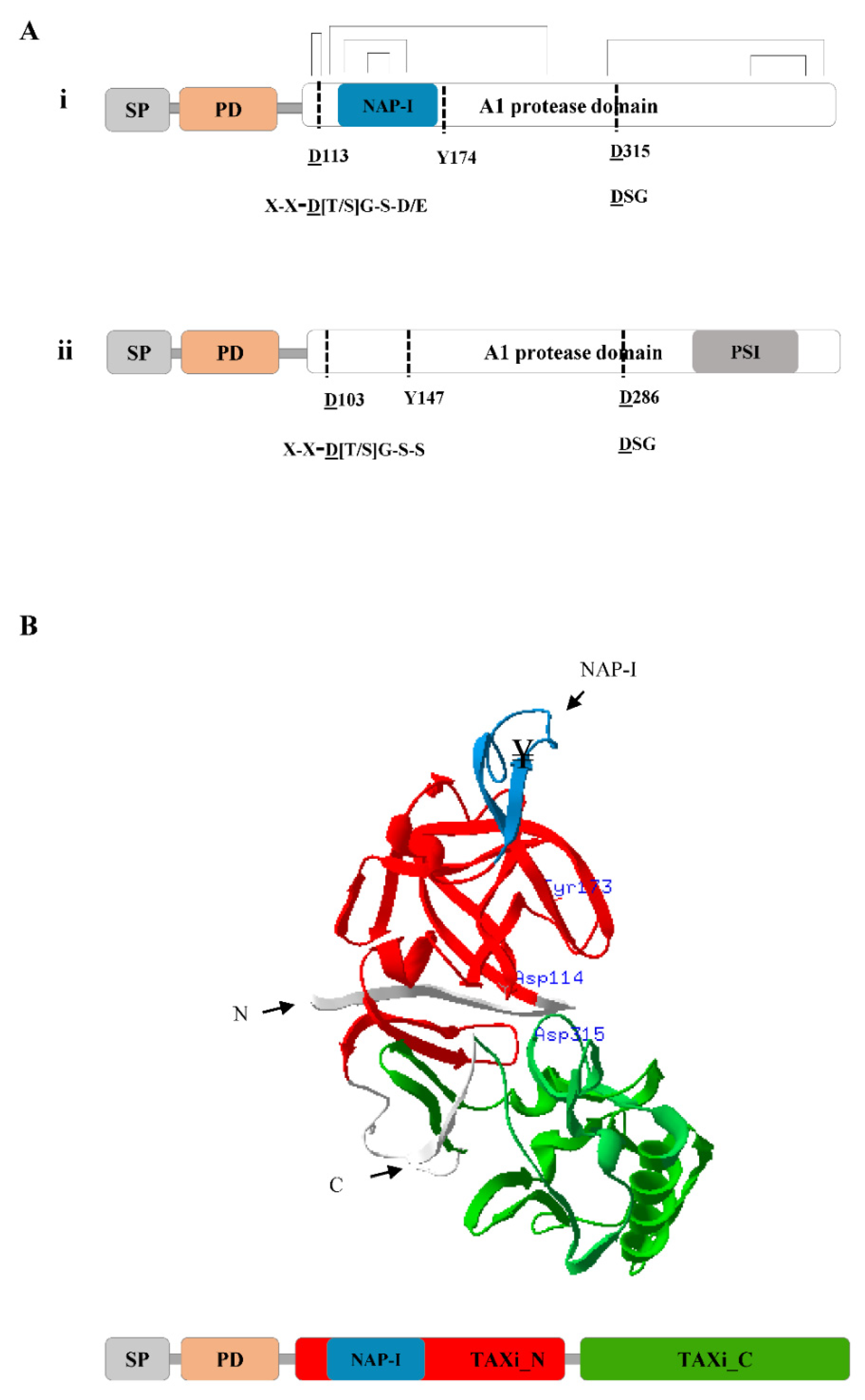
2. Molecular Structure of Plant Nepenthesins

The primary structure of nepenthesins is comparable to the structure of pepsin, but few characteristic differences have been identified (Figure 1A). The precursor of nepenthesins contain an N-terminal signal sequence for secretion, a prodomain sequence of about 40 amino acids, two conserved catalytic Asp residues and a conserved Tyr residue that forms the so-called “flap” [1]. The two catalytic Asp residues are conserved in the Asp-Thr/Ser-Gly (D[T/S]G) and Asp-Ser-Gly (DSG) sequence motifs. The Tyr (Y) residue in the “flap” controls substrate specificity and binding. Nepenthesins are also characterized by the presence of a 20–30-residues-long NAP-I sequence (Figure 1A,i). This is different from the 100 residues long plant specific insertion sequence (PSI) that is specific to the A1A subfamily (Figure 1A,ii). Both the NAP-I and PSI are Cys-rich segments inserted at the N-and C-terminus of the protein, respectively. The NAP-I is usually placed between Trp39 and Tyr75 in the polypeptide (according to pepsin numbering) [23] and is often part of the mature enzyme [1,31].
NAP-I is characterized by its formation of two more disulfide bridges in the mature protein than the corresponding PEPs (Figure 1A,ii). The additional disulfide bridges in NAP-I contribute to the unusual stability of nepenthesins for a wider range of pH and temperature [32]. This gives nepenthesins the exceptional biochemical property to digest insect structures over long time without losing proteolytic activity. However, there are significant variations in the NAP-I of nepenthesins among species [32]. This may be associated to the specific roles of NAP-I in intracellular targeting or functional regulation of the enzyme in each species. The distinctive sequence features of nepenthesins are conserved among other members of the A1B subfamily, as recently reviewed in Soares etal. [31].
Prediction of the domain composition and tertiary structure of nepenthesins reveals extended molecular and structural features. For instance, according to the profile Hidden Markov Models (HMM) scan of the N. gracilis nepenthesin-2 [33], its A1 protease domain is mapped to the two xylanase inhibitor domains, TAXi_N (xylanase inhibitor N-terminal) and TAXi_C (xylanase inhibitor C-terminal) (Figure 1B). The tertiary structure prediction discloses that the two xylanase inhibitor domains form a lobe and thereby creates the catalytic pocket [34]. The active site is located in the cleft between the two lobes containing two catalytic Asp residues, one from each lobe (Figure 1B). The “flap” structure made up of a β-hairpin structure completes the active site and participates in substrate binding. The N- and C-termini of xylanase inhibitors are equally necessary for creating the catalytic pocket for cleaving xylanases [35,36,37]. As mentioned above, the NAP-I sequence does not appear to be directly involved in substrate binding or catalysis. It may be indirectly involved in maintaining the active site structure or participate in other cellular activities.

3. Regulation of Plant Nepenthesins

Nepenthesins are mostly expressed as inactive zymogens [23,32]. The prodomain is covalently bound to the active site of the enzyme, and acts as auto-inhibitor. The prodomain is not often part of the mature enzyme and has to be processed during zymogen maturation [31]. In most cases, the inactive zymogens are activated in acidic environments after removal of the prodomain. Autocatalytic processing of the prodomain is sufficient to release the active enzyme, as the prodomain is no more bound to the active site. The prodomain probably serves as a molecular “switch” to keep the proteases inactive until they reach their final subcellular destination and prevent untimely activation and proteolysis. In addition to the complex pH dependent intramolecular and intermolecular steps in the maturation of nepenthesins, a different prodomain processing and enzyme activation mechanism has been described for other members of NEPs. For instance, the active form of AtCDR1 and OsCDR1 recombinant enzymes have been reported without the irreversible removal of the prodomain [11,38]. In addition, the activation of CND41 depends on the interaction of the leucine-rich N-terminal region and positive charged residues in the active site [39,40]. So far, no study has reported these types of enzyme activation mechanisms for nepenthesins. Likewise, an in silico analysis of the A. thaliana APs revealed that the majority of nepenthesins are predicted to be located in the secretory pathway [41]. Therefore, they are probably be either in the vacuoles or in the extracellular space. Future structural studies on the nepenthesins may provide in-depth molecular information about other modes of activation and subcellular locations.

4. Biochemistry of Plant Nepenthesins

Nepenthesins are active and stable over a wider pH and temperature range than porcine pepsin A and other PEPs [23,32]. Moreover, in addition to the number of disulfide bridges, the stability of nepenthesins has largely been correlated to the level of N-glycosylation [42]. The majority of nepenthesins function optimally at acidic conditions, but few exceptions with basic pH optima have been reported (Table 1). As described for other members of APs, nepenthesins are inhibited by pepstatin and diazoacetyl-D,L-norleucine methyl ester (DAN) (Table 1). DAN only inhibited the activity of nepenthesins in the presence of cupric ions [23,32]. The effect of metal ions on the activity of inhibitors and chelating agents needs to be studied further for the nepenthesins. In contrast to members of PEPs, nepenthesins are more sensitive to various chemicals, reducing and denaturing agents such as TCEP (tris (2-carboxyethyl)phosphine), urea, and guanidine hydrochloride [43]. The denaturing agents considerably improved the activity of DTT that acts on the disulfide bridges of nepenthesins. TCEP alone or in combination with other denaturing agents significantly reduces the activity of nepenthesins [42]. However, the level of susceptibility to these chemical agents significantly varies between nepenthesins. For example, recombinant nepenthesin II from N. gracilis was more resistant to denaturing and reducing agents than nepenthesin I [44]. Its level of resistance to these denaturants and reducing agents was as good as pepsin.
The overall mechanism of peptide bond cleavage is the same for both NEPs and PEPs [45]. They use water molecules that act as a nucleophile and activates the catalysis via electrostatic interactions formed between the two catalytic Asp residues. The catalytic Asp residues stabilize the oxyanion hole of aspartic proteases. Although nepenthesins seem to have similar cleavage specificity to members of PEPs such as pepsin A and cathepsin D, additional cleavage sites have been identified [32,46]. In both studies, nepenthesin cleaved the B chain of oxidized insulin at Leu6-Cya7, Glu13-Ala14, Leu15-Tyr16 and Tyr16-Leu17 and Phe24-Phe25 sites to a significant level (Table 1). Moreover, nepenthesins cleave at Leu6-Cya7 (Cya for cysteic acid), which has never been described for members of PEPs [32]. Although Leu residue at the P1 position appears to be the ideal site for nepenthesins, high rate of cleavage by nepenthesin has been observed when Phe, Leu, Met, Lys, Arg, and Pro are present at the P1 position [47]. In addition to the B chain of oxidized insulin, nepenthesins have also shown strong affinity for a wide range of substrates such as casein, haemoglobin, myoglobin, and fungal phytase protein (Table 1).

5. Biological Roles of Nepenthesins in Carnivorous Plants

5.1. Nepenthesins Contributing to Plant Nutrition

Nepenthesins have been regarded as the main players in hydrolyzing insect protein and availing the nitrogen for assimilation by the plant [24]. Due to significant proteolytic activity in the fluid, carnivorous plants can derive 10%–80% of their total nitrogen from insects [53]. The efficiency hydrolysis varies depending on the occurrence of proteolytic enzymes and the type of trap employed by the plant. In this regard, nepenthesins represent the major proteolytic activity in the pitcher fluid [50]. As a result, carnivorous plants thrive in extremely low nitrogen environments. The regulation of nepenthesin activity in the digestive fluid has been examined in response to chemical stimuli and live prey [54]. Ammonium, protein, and live prey strongly induced the transcription of nepenthesins. Although ammonium induced the highest transcription of nepenthesins, the total protease activity is higher in response to protein and live prey [54]. The result indicated that carnivorous plants monitor and digest insect prey with the aid of nepenthesins.

5.2. Nepenthesins Play a Role in Plant Defense

Despite characterization of nepenthesins from various carnivorous plants, not a single attempt has been made to study their role in crop plants. Recently, we have identified and characterized the nepenthesin-1 gene (HvNEP-1) from barley [52]. The recombinant HvNEP-1 (rHvNEP-1) reduces the activity of fungal phytases, enzymes that dephosphorylate the phytic acid substrate [55]. The rHvNEP-1 also suppresses biomass accumulation and 15-acetyldeoxynivalenol (15ADON) mycotoxin production from different strains of Fusarium graminearum [52]. The study reports that rHvNEP-1 significantly represses the expression of trichothecene (TRI) biosynthesis and regulatory genes in F. graminearum. Among the four TRI genes (TRI4, TRI5, TRI6, and TRI12) examined, rHvNEP-1 significantly affects the expression of TRI4, TRI5, and TRI6 genes. The repression of TRI6 expression directly influences biomass accumulation and mycotoxin production, as it regulates the transcription of TRI biosynthesis genes and genes related to nutrient assimilation in Fusarium species [56]. Likewise, the suppression of TRI4 and TRI5 genes reduces the synthesis and accumulation of the trichothecene precursors early in the TRI biosynthesis pathway [57,58].
The reliability of HvNEP-1 in plant defense has been further examined by overexpressing in the endosperm of barley grains and infection with the two DON-Chemotype F. graminearum and F. culmorum species. Surprisingly, the overexpression of HvNEP-1 in the endosperm of barley grains significantly reduces the infection and disease progression of Fusarium head blight disease (FHB), under both Fusarium species. Furthermore, the analysis of deoxynivalenol (DON), nivalenol (NIV) and zearalenone mycotoxins indicates that HvNEP-1 overexpression substantially suppress the accumulation of DON in the grains than in the untransformed controls [59]. The level of DON accumulation in the grains of some transgenic lines has been far below the optimal level considered for human consumption.

6. Potentials of Plant Nepenthesins

6.1. Industrial Applications: Tool for Digestion in Hydrogen/Deuterium Exchange Mass Spectrometry

Hydrogen/deuterium exchange mass spectrometry (HDX-MS) has become an important method to study the structure of proteins and their complexes [60,61]. The technique requires proteases that can perform fast digestion at lower pH. For this application, pepsin is used more often, but it offers moderately low efficiency under the HDX-MS optimal conditions. Nepenthesins can be used as a substitute to pepsin in the HDX-MS application as they exhibit optimal biochemical properties and slightly wider cleavage specificities than pepsin [44,47,49]. It has been shown that nepenthesin is at least 1400-fold more efficient than pepsin under HDX-MS competent conditions (Table 2). In addition to similar cleavage sites as pepsin, nepenthesin also effectively cleaves C-terminal to Lys, Arg, His, and Pro residues under natural conditions. In general, nepenthesin-based digestion gives more sequence coverage and shorter average peptide length than pepsin [47,62]. Despite a number of cleavage sites common for nepenthesins, additional cleavage sites have been identified for some nepenthesins [44]. For instance, both nepenthesin I and II isolated from N. gracilis share similar biochemical properties, including fast digestion at low temperature and pH, and cleavage specificity. However, nepenthesin II specifically cuts the C-terminal to Trp.
Nepenthesins are ideal proteases to study the structural transitions of intrinsically disordered proteins and their complexes (Table 2). This has been envisaged by using nepenthesin-enriched secretions to analyze protein complexes involved in the non-homologous end-joining DNA damage repair pathway [47]. The study demonstrated the molecular mechanisms behind the interaction of the BRCT domains of Ligase IV to the full length and truncated form of XRCC4. Based on the peptide cleavage results from XRCC4 digestion, the interaction between the disordered C-terminal tail of XRCC4 and remote regions of the BRCT domains is required for complex formation. This has been possible due to the cleavage of XRCC4 at Pro and charged residues, which is impossible using pepsin [47].

6.2. Treatment of Celiac Disease

Celiac disease is a chronic autoimmune disease of the small intestine caused by the ingestion of dietary gluten. The common grains like wheat, barley and rye are characterized by high content of gluten proteins. Upon consumption of gluten proteins, celiac patients often develop villous atrophy of the small intestine, crypt hyperplasia, and mucosal inflammation [63,64]. Most gastric proteases digest proteins into peptides as small as 30-40 amino acid residues, which are still resistant to further digestion by intestinal proteases [65]. The 33-mer peptide, LQLQPFPQPQLPYPQPQLPYPQPQLPYPQPQPF, has been identified as the chief initiator of the inflammatory response to gluten in celiac patients [66,67]. This toxic peptide from gluten proteins contains six repetitive T-cell epitopes that arouses a potent T-cell response in celiac patients. The peptide is highly resistant to digestion by gastric pepsin, even under prolonged incubation. Therefore, celiac patients consider the exclusion of gluten-containing diets as the only lifetime treatment for the disease. However, avoiding gluten proteins from our daily menu can be more expensive and unpleasant.
Numerous strategies have been devised to alleviate the problem related to gluten-intolerant individuals. Some of the approaches include enzymatic hydrolysis of gluten proteins and the creation of low α-gliadins using RNAi and CRISPR/Cas9 transgenic technologies [68,69,70,71]. Enzyme-based detoxification of gluten proteins has been described as one of the most promising therapeutic approach, as there is still growing concern regarding the use of transgenic technologies [72]. In this regard, plant nepenthesins have displayed an impressive ability to degrade gluten into non-toxic peptides more efficiently than stomach enzymes like pepsin [62,63]. More importantly, they degrade the 33-mer peptide into non-toxic peptides [62,63]. It has also been demonstrated that nepenthesin showed efficient antigen processing at total substrate to enzyme ratios higher than 12,000:1 (Table 2). This has a direct implication for therapeutic use, as it requires low amount of nepenthesins for efficient detoxification of gluten proteins. On the contrary, a barley cysteine endopeptidase-B2 has been reported to hydrolyze glutens into small peptides, but it requires the prolyl endoprotease from Sphingomonas capsulate to produce non-toxic peptides [68,73]. Overall, nepenthesins provide better platforms for gluten intolerance therapy as (1) they are isolated from plants, (2) they are easy to isolate or produce, (3) they have very high substrate catalytic efficiency, (4) they hydrolyze gluten proteins into small and harmless peptides, and (5) they can be prepared as tablets or other formulations to use as food additives in gluten intolerance therapy [63].

6.3. Trait Improvement of Crop Plants

Interestingly, nepenthesin homologues are distributed in a wide range of crop species like wheat, barley, and maize [16]. Based on the available molecular and biochemical information, nepenthesins can aid the improvement of important traits like disease resistance. The recent discovery of HvNEP-1 as a defense gene against the fungal disease FHB provides additional genetic resource for the improvement of disease resistance trait in crop plants [59]. In the study, the relative expression of HvNEP-1 in the overexpression lines positively correlates to FHB resistance, both in the greenhouse and semi-field conditions. The increase in FHB resistance is also accompanied by the reduction of mycotoxin accumulation in the grains.
Fusarium mycotoxins have been the center of global concern for decades, due to their significant health risks to human and animals [74]. Their level in agricultural crops has been regulated in most countries worldwide [75]. Despite the present various control methods, the use of non-chemical methods like resistance breeding has been the focus of researchers, as it minimizes the production costs and environmental pollution. In this regard, HvNEP-1 can assist FHB resistance breeding in the common crops like wheat and maize.

7. Conclusions and Future Perspectives

Despite their extensive occurrence in plants, research on nepenthesins lags behind compared to other members of the A1B subfamily. Previous studies were limited to the biochemical characterization of nepenthesins isolated exclusively from carnivorous plants. The studies have linked nepenthesins mainly to the degradation of insect structures and mobilization of insect-derived nitrogen for plant nutrition. In addition to their role in plant nutrition, the recently characterized barley nepenthesin HvNEP-1 adds an additional potential of nepenthesins in plants as a defense protein. Furthermore, a study has also implicated nepenthesins in the treatment of bacterial infections in the gastrointestinal tract [63], which directs another research front for improving human health.
To gain a better understanding of the biological roles of nepenthesins, more studies are needed on the identification and characterization of various nepenthesin homologues from plants. In this respect, the investigation of their molecular mechanisms, subcellular locations, biological substrates, mechanism of regulation, and nepenthesin-associated receptors and transcription factors unlocks more mysteries related to their biological function. The role of the TAXi_N and TAXi_C domains of the nepenthesins in the inhibition of microbial xylanases requires further study. As described for the PSI domain of A1A APs [76], the role of the NAP-I in enzyme regulation and other species-specific function should also be investigated. In this regard, the ever-increasing computational and analytical tools in the area of genomics, transcriptomics and proteomics, and the evolution of new genome editing systems like CRSPR/Cas provide additional platforms for studying plant nepenthesins. The obtained information can eventually be used to improve enzymatic properties for industrial applications and for crop improvement programs.

Author Contributions

Z.E.B. conceived and designed the study, collected the data, and drafted the manuscript. G.D. conceived the study and review and approved the submitted version. H.B.-P. conceived and led the study and review and approved the submitted version. All authors have read and agreed to the published version of the manuscript.

Funding

The study was supported by Innovation Fund Denmark, grant 8055-00038B, ReTraQue.

Conflicts of Interest

The authors declare no conflict of interests.

References

  1. Simões, I.; Faro, C. Structure and function of plant aspartic proteinases. Eur. J. Biochem. 2004, 271, 2067–2075. [Google Scholar] [CrossRef] [Green Version]
  2. Hiraiwa, N.; Kondo, M.; Nishimura, M.; HaraNishimura, I. An aspartic endopeptidase is involved in the breakdown of propeptides of storage proteins in protein-storage vacuoles of plants. Eur. J. Biochem. 1997, 246, 133–141. [Google Scholar] [CrossRef] [Green Version]
  3. Mutlu, A.; Gal, S. Plant aspartic proteinases: Enzymes on the way to a function. Physiol. Plant. 1999, 105, 569–576. [Google Scholar] [CrossRef]
  4. Garciamartinez, J.L.; Moreno, J. Proteolysis of ribulose-1,5-bisphosphate carboxylase oxygenase in citrus leaf extracts. Physiol. Plant. 1986, 66, 377–383. [Google Scholar] [CrossRef]
  5. An, C.I.; Fukusaki, E.; Kobayashi, A. Aspartic proteinases are expressed in pitchers of the carnivorous plant Nepenthes alata Blanco. Planta 2002, 214, 661–667. [Google Scholar] [CrossRef] [PubMed]
  6. Tokes, Z.A.; Woon, W.C.; Chambers, S.M. Digestive enzymes secreted by carnivorous plant Nepenthes macferlanei L. Planta 1974, 119, 39–46. [Google Scholar] [CrossRef] [PubMed]
  7. Ge, X.C.; Dietrich, C.; Matsuno, M.; Li, G.J.; Berg, H.; Xia, Y.J. An Arabidopsis aspartic protease functions as an anti-cell-death component in reproduction and embryogenesis. EMBO Rep. 2005, 6, 282–288. [Google Scholar] [CrossRef] [PubMed] [Green Version]
  8. Vieira, M.; Pissarra, J.; Verissimo, P.; Castanheira, P.; Costa, Y.; Pires, E.; Faro, C. Molecular cloning and characterization of cDNA encoding cardosin B, an aspartic proteinase accumulating extracellularly in the transmitting tissue of Cynara cardunculus L. Plant Mol. Biol. 2001, 45, 529–539. [Google Scholar] [CrossRef] [Green Version]
  9. Ramalho-Santos, M.; Pissarra, J.; Veríssimo, P.; Pereira, S.; Salema, R.; Pires, E.; Faro, C.J. Cardosin A, an abundant aspartic proteinase, accumulates in protein storage vacuoles in the stigmatic papillae of Cynara cardunculus L. Planta 1997, 203, 204–212. [Google Scholar] [CrossRef]
  10. de Carvalho, M.H.C.; d’Arcy-Lameta, A.; Roy-Macauley, H.; Gareil, M.; El Maarouf, H.; Pham-Thi, A.T.; Zuily-Fodil, Y. Aspartic protease in leaves of common bean (Phaseolus vulgaris L.) and cowpea (Vigna unguiculata L. Walp): Enzymatic activity, gene expression and relation to drought susceptibility. Febs Lett. 2001, 492, 242–246. [Google Scholar] [CrossRef] [Green Version]
  11. Simoes, I.; Faro, R.; Bur, D.; Faro, C. Characterization of recombinant CDR1, an Arabidopsis aspartic proteinase involved in disease resistance. J. Biol. Chem. 2007, 282, 31358–31365. [Google Scholar] [CrossRef] [PubMed] [Green Version]
  12. Schaller, A.; Ryan, C.A. Molecular cloning of a tomato leaf cDNA encoding an aspartic protease, a systemic wound response protein. Plant Mol. Biol. 1996, 31, 1073–1077. [Google Scholar] [CrossRef] [PubMed]
  13. Guevara, M.G.; Daleo, G.R.; Oliva, C.R. Purification and characterization of an aspartic protease from potato leaves. Physiol. Plant. 2001, 112, 321–326. [Google Scholar] [CrossRef] [PubMed]
  14. Cordeiro, M.C.; Pais, M.S.; Brodelius, P.E. Tissue-specific expression of multiple forms of cyprosin (aspartic proteinase) in flowers of Cynara cardunculus. Physiol. Plant. 1994, 92, 645–653. [Google Scholar] [CrossRef]
  15. Chen, F.Q.; Foolad, M.R. Molecular organization of a gene in barley which encodes a protein similar to aspartic protease and its specific expression in nucellar cells during degeneration. Plant Mol. Biol. 1997, 35, 821–831. [Google Scholar] [CrossRef]
  16. Rawlings, N.D.; Alan, J.; Thomas, P.D.; Huang, X.D.; Bateman, A.; Finn, R.D. The MEROPS database of proteolytic enzymes, their substrates and inhibitors in 2017 and a comparison with peptidases in the PANTHER database. Nucleic Acids Res. 2018, 46, D624–D632. [Google Scholar] [CrossRef]
  17. Vines, S.H. The Proteolytic Enzyme of Nepenthes. Ann. Bot. 1897, 11, 563–584. [Google Scholar] [CrossRef]
  18. Vines, S.H. The Proteolytic Enzyme of Nepenthes. Ann. Bot. 1901, 15, 563–573. [Google Scholar] [CrossRef] [Green Version]
  19. Owen, T.P.; Lennon, K.A. Structure and development of the pitchers from the carnivorous plant Nepenthes alata (Nepenthaceae). Am. J. Bot. 1999, 86, 1382–1390. [Google Scholar] [CrossRef]
  20. Nakayama, S.; Amagase, S. Acid protease in nepenthes—Partial purification and properties of enzyme. Proc. Jpn. Acad. 1968, 44, 358. [Google Scholar] [CrossRef]
  21. Amagase, S.; Nakayama, S.; Tsugita, A. Acid protease in Nepenthes. 2. Study on specificity of nepenthesin. J. Biochem. 1969, 66, 431. [Google Scholar] [CrossRef] [PubMed]
  22. Amagase, S. Digestive enzymes in insectivorous plants part 3 acid proteases in the genus Nepenthes and Drosera peltata. J. Biochem. 1972, 72, 73–81. [Google Scholar] [CrossRef] [PubMed]
  23. Athauda, S.B.P.; Matsumoto, K.; Rajapakshe, S.; Kuribayashi, M.; Kojima, M.; Kubomura-Yoshida, N.; Iwamatsu, A.; Shibata, C.; Inoue, H.; Takahashi, K. Enzymic and structural characterization of nepenthesin, a unique member of a novel subfamily of aspartic proteinases. Biochem. J. 2004, 381, 295–306. [Google Scholar] [CrossRef] [PubMed]
  24. Ravee, R.; Mohd Salleh, F. ‘Imadi; Goh, H.-H. Discovery of digestive enzymes in carnivorous plants with focus on proteases. PeerJ 2018, 6, e4914. [Google Scholar] [CrossRef]
  25. Takahashi, K.; Matsumoto, K.; Nishii, W.; Muramatsu, M.; Kubota, K.; Shibata, C.; Athauda, S.B.P.; Peradeniya; Lanka, S. Comparative Studies on the Acid Proteinase Activities in the Digestive Fluids of Nepenthes, Cephalotus, Dionaea and Drosera; Carnivorous Plant Newsletter: Pinole, CA, USA, 2009; Volume 38, pp. 75–82. [Google Scholar]
  26. Takahashi, K.; Nishii, W.; Shibata, C. The digestive fluid of Drosera indica contains a cysteine endopeptidase similar to dionain from Dionaea muscipula. Carniv. Plant Newsl. 2012, 41, 132–134. [Google Scholar]
  27. Fukushima, K.; Fang, X.; Alvarez-Ponce, D.; Cai, H.; Carretero-Paulet, L.; Chen, C.; Chang, T.-H.; Farr, K.M.; Fujita, T.; Hiwatashi, Y.; et al. Genome of the pitcher plant Cephalotus reveals genetic changes associated with carnivory. Nat. Ecol. Evol. 2017, 1, 0059. [Google Scholar] [CrossRef]
  28. Schulze, W.X.; Sanggaard, K.W.; Kreuzer, I.; Knudsen, A.D.; Bemm, F.; Thøgersen, I.B.; Bräutigam, A.; Thomsen, L.R.; Schliesky, S.; Dyrlund, T.F.; et al. The protein composition of the digestive fluid from the venus flytrap sheds light on prey digestion mechanisms. Mol. Cell. Proteomics 2012, 11, 1306–1319. [Google Scholar] [CrossRef] [Green Version]
  29. Takahashi, K.; Niwa, H.; Yokota, N.; Kubota, K.; Inoue, H. Widespread tissue expression of nepenthesin-like aspartic protease genes in Arabidopsis thaliana. Plant Physiol. Biochem. 2008, 46, 724–729. [Google Scholar] [CrossRef]
  30. Chen, J.; Ouyang, Y.; Wang, L.; Xie, W.; Zhang, Q. Aspartic proteases gene family in rice: Gene structure and expression, predicted protein features and phylogenetic relation. Gene 2009, 442, 108–118. [Google Scholar] [CrossRef]
  31. Soares, A.; Ribeiro Carlton, S.M.; Simões, I. Atypical and nucellin-like aspartic proteases: Emerging players in plant developmental processes and stress responses. J. Exp. Bot. 2019. [Google Scholar] [CrossRef] [PubMed]
  32. Takahashi, K.; Athauda, S.B.P.; Matsumoto, K.; Rajapakshe, S.; Kuribayashi, M.; Kojima, M.; Kubomura-Yoshida, N.; Iwamatsu, A.; Shibata, C.; Inoue, H. Nepenthesin, a unique member of a novel subfamily of aspartic proteinases: Enzymatic and structural characteristics. Curr. Protein Pept. Sci. 2005, 6, 513–525. [Google Scholar] [CrossRef] [PubMed]
  33. Potter, S.C.; Luciani, A.; Eddy, S.R.; Park, Y.; Lopez, R.; Finn, R.D. HMMER web server: 2018 update. Nucleic Acids Res. 2018, 46, W200–W204. [Google Scholar] [CrossRef] [PubMed] [Green Version]
  34. Guex, N.; Peitsch, M.C. SWISS-MODEL and the Swiss-Pdb Viewer: An environment for comparative protein modeling. Electrophoresis 1997, 18, 2714–2723. [Google Scholar] [CrossRef] [PubMed]
  35. Fierens, K.; Brijs, K.; Courtin, C.M.; Gebruers, K.; Goesaert, H.; Raedschelders, G.; Robben, J.; Van Campenhout, S.; Volckaert, G.; Delcour, J.A. Molecular identification of wheat endoxylanase inhibitor TAXI-I1, member of a new class of plant proteins. FEBS Lett. 2003, 540, 259–263. [Google Scholar] [CrossRef] [Green Version]
  36. Sansen, S.; De Ranter, C.J.; Gebruers, K.; Brijs, K.; Courtin, C.M.; Delcour, J.A.; Rabijns, A. Structural basis for inhibition of Aspergillus niger xylanase by Triticum aestivum xylanase inhibitor-I. J. Biol. Chem. 2004, 279, 36022–36028. [Google Scholar] [CrossRef] [Green Version]
  37. Pollet, A.; Sansen, S.; Raedschelders, G.; Gebruers, K.; Rabijns, A.; Delcour, J.A.; Courtin, C.M. Identification of structural determinants for inhibition strength and specificity of wheat xylanase inhibitors TAXI-IA and TAXI-IIA. FEBS J. 2009, 276, 3916–3927. [Google Scholar] [CrossRef]
  38. Prasad, B.D.; Creissen, G.; Lamb, C.; Chattoo, B.B. Heterologous expression and characterization of recombinant OsCDR1, a rice aspartic proteinase involved in disease resistance. Protein Expr. Purif. 2010, 72, 169–174. [Google Scholar] [CrossRef]
  39. Kato, Y.; Murakami, S.; Yamamoto, Y.; Chatani, H.; Kondo, Y.; Nakano, T.; Yokota, A.; Sato, F. The DNA-binding protease, CND41, and the degradation of ribulose-1,5-bisphosphate carboxylase/oxygenase in senescent leaves of tobacco. Planta 2004, 220, 97–104. [Google Scholar] [CrossRef]
  40. Kato, Y.; Yamamoto, Y.; Murakami, S.; Sato, F. Post-translational regulation of CND41 protease activity in senescent tobacco leaves. Planta 2005, 222, 643–651. [Google Scholar] [CrossRef]
  41. Faro, C.; Gal, S. Aspartic Proteinase Content of the Arabidopsis Genome. Curr. Protein Pept. Sci. 2005, 6, 493–500. [Google Scholar] [CrossRef]
  42. Kadek, A.; Tretyachenko, V.; Mrazek, H.; Ivanova, L.; Halada, P.; Rey, M.; Schriemer, D.C.; Man, P. Expression and characterization of plant aspartic protease nepenthesin-1 from Nepenthes gracilis. Protein Expr. Purif. 2014, 95, 121–128. [Google Scholar] [CrossRef] [PubMed]
  43. Kubota, K.; Metoki, Y.; P Athauda, S.B.; Shibata, C.; Takahashi, K. Stability Profiles of Nepenthesin in Urea and Guanidine Hydrochloride: Comparison with Porcine Pepsin A. Biotechnol. Biochem. 2010, 74, 2323–2326. [Google Scholar] [CrossRef] [PubMed] [Green Version]
  44. Yang, M.; Hoeppner, M.; Rey, M.; Kadek, A.; Man, P.; Schriemer, D.C. Recombinant Nepenthesin II for Hydrogen/Deuterium Exchange Mass Spectrometry. Anal. Chem. 2015, 87, 6681–6687. [Google Scholar] [CrossRef] [PubMed]
  45. van der Hoorn, R.A.L. Plant proteases: From phenotypes to molecular mechanisms. Annu. Rev. Plant Biol. 2008, 59, 191–223. [Google Scholar] [CrossRef] [Green Version]
  46. Athauda, S.B.P.; Inoue, H.; Iwamatsu, A.; Takahashi, K. Acid proteinase from Nepenthes distillatoria (Bandura). In Aspartic Proteinases: Retroviral and Cellular Enzymes; James, M.N.G., Ed.; Advances in Experimental Medicine and Biology; Plenum Press: New York, NY, USA, 1998; Volume 59, pp. 453–458. ISBN 0-306-45809-8. [Google Scholar]
  47. Rey, M.; Yang, M.; Burns, K.M.; Yu, Y.; Lees-Miller, S.P.; Schriemer, D.C. Nepenthesin from Monkey Cups for Hydrogen/Deuterium Exchange Mass Spectrometry. Mol. Cell. Proteomics 2013, 12, 464–472. [Google Scholar] [CrossRef] [Green Version]
  48. Jentsch, J. Enzymes from carnivorous plants (Nepenthes). Isolation of the protease nepenthacin. FEBS Lett. 1972, 21, 273–276. [Google Scholar] [CrossRef] [Green Version]
  49. Kadek, A.; Mrazek, H.; Halada, P.; Rey, M.; Schriemer, D.C.; Man, P. Aspartic Protease Nepenthesin-1 as a Tool for Digestion in Hydrogen/Deuterium Exchange Mass Spectrometry. Anal. Chem. 2014, 86, 4287–4294. [Google Scholar] [CrossRef]
  50. Buch, F.; Kaman, W.E.; Bikker, F.J.; Yilamujiang, A.; Mithöfer, A.; Mithoefer, A. Nepenthesin Protease Activity Indicates Digestive Fluid Dynamics in Carnivorous Nepenthes Plants. PLoS ONE 2015, 10, e118853. [Google Scholar] [CrossRef] [Green Version]
  51. Lee, L.; Zhang, Y.; Ozar, B.; Sensen, C.W.; Schriemer, D.C. Carnivorous Nutrition in Pitcher Plants (Nepenthes spp.) via an Unusual Complement of Endogenous Enzymes. J. Proteome Res. 2016, 15, 3108–3117. [Google Scholar] [CrossRef]
  52. Bekalu, Z.E.; Dionisio, G.; Madsen, C.K.; Holme, I.B.; Etzerodt, T.P.; Fomsgaard, I.; Jørgensen, L.N.; Brinch-Pedersen, H. Nepenthesin-1 Derived Resistance to Fungal Pathogens in Major Crop Plants; WO 2019/057845 A1, filed 20 September 2018, and issued 28 March 2019; World Intellectual Property Organization: Geneva, Switzerland, 2019. [Google Scholar]
  53. Ellison, A.M.; Gotelli, N.J. Evolutionary ecology of carnivorous plants. Trends Ecol. Evol. 2001, 16, 623–629. [Google Scholar] [CrossRef]
  54. Saganová, M.; Bokor, B.; Stolárik, T.; Pavlovič, A. Regulation of enzyme activities in carnivorous pitcher plants of the genus Nepenthes. Planta 2018, 248, 451–464. [Google Scholar] [CrossRef] [PubMed]
  55. Lei, X.; Porres, J.; Mullaney; Edward; Brinch-Pedersen, H. Phytase: Source, structure and application. In Industrial Enzymes: Structure, Function and Applications; Polina, J., Mac Cabe, A.P., Eds.; Springer: Dordrecht, The Netherlands, 2007; pp. 505–529. [Google Scholar]
  56. Nasmith, C.G.; Walkowiak, S.; Wang, L.; Leung, W.W.Y.; Gong, Y.; Johnston, A.; Harris, L.J.; Guttman, D.S.; Subramaniam, R. Tri6 Is a Global Transcription Regulator in the Phytopathogen Fusarium graminearum. PLoS Pathog. 2011, 7, e1002266. [Google Scholar] [CrossRef] [PubMed]
  57. Hohn, T.M.; Vanmiddlesworth, F. Purification and characterization of the sesquiterpene cyclase trichodiene synthetase from Fusarium sporotrichioides. Arch. Biochem. Biophys. 1986, 251, 756–761. [Google Scholar] [CrossRef]
  58. Tokai, T.; Koshino, H.; Takahashi-Ando, N.; Sato, M.; Fujimura, M.; Kimura, M. Fusarium Tri4 encodes a key multifunctional cytochrome P450 monooxygenase for four consecutive oxygenation steps in trichothecene biosynthesis. Biochem. Biophys. Res. Commun. 2007, 353, 412–417. [Google Scholar] [CrossRef]
  59. Bekalu, Z.E.; Krogh Madsen, C.; Dionisio, G.; Bæksted Holme, I.; Jørgensen, L.N.; Fomsgaard, I.S.; Brinch-Pedersen, H. Overexpression of Nepenthesin HvNEP-1 in Barley Endosperm Reduces Fusarium Head Blight and Mycotoxin Accumulation. Agronomy 2020, 10, 203. [Google Scholar] [CrossRef] [Green Version]
  60. Marcsisin, S.R.; Engen, J.R. Hydrogen exchange mass spectrometry: What is it and what can it tell us? Anal. Bioanal. Chem. 2010, 397, 967–972. [Google Scholar] [CrossRef] [Green Version]
  61. Konermann, L.; Pan, J.; Liu, Y.H. Hydrogen exchange mass spectrometry for studying protein structure and dynamics. Chem. Soc. Rev. 2011, 40, 1224–1234. [Google Scholar] [CrossRef]
  62. Rey, M.; Yang, M.; Lee, L.; Zhang, Y.; Sheff, J.G.; Sensen, C.W.; Mrazek, H.; Halada, P.; Man, P.; McCarville, J.L.; et al. Addressing proteolytic efficiency in enzymatic degradation therapy for celiac disease. Sci. Rep. 2016, 6, 30980. [Google Scholar] [CrossRef]
  63. Schriemer, D.; Man, P.; Mrazek, H.; Rey, M. Treatment of Gluten Intolerance and Related Conditions. US 2015/0265686 A1, filed 3 October 2014, and issued 24 September 2015. U.S. Patent 9005610, 15 March 2013. [Google Scholar]
  64. Abadie, V.; Sollid, L.M.; Barreiro, L.B.; Jabri, B. Integration of Genetic and Immunological Insights into a Model of Celiac Disease Pathogenesis. Annu. Rev. Immunol. 2011, 29, 493–525. [Google Scholar] [CrossRef] [Green Version]
  65. Bethune, M.T.; Khosla, C. Oral enzyme therapy for celiac sprue. Methods Enzymol. 2012, 502, 241–271. [Google Scholar]
  66. Shan, L.; Molberg, Ø.; Parrot, I.; Hausch, F.; Filiz, F.; Gray, G.M.; Sollid, L.M.; Khosla, C. Structural basis for gluten intolerance in Celiac Sprue. Science (80-.) 2002, 297, 2275–2279. [Google Scholar] [CrossRef] [PubMed] [Green Version]
  67. Shan, L.; Qiao, S.-W.; Arentz-Hansen, H.; Molberg, Ø.; Gray, G.M.; Sollid, L.M.; Khosla, C. Identification and Analysis of Multivalent Proteolytically Resistant Peptides from Gluten: Implications for Celiac Sprue. J. Proteome Res. 2005, 4, 1732–1741. [Google Scholar] [CrossRef] [PubMed] [Green Version]
  68. Bethune, M.T.; Strop, P.; Tang, Y.; Sollid, L.M.; Khosla, C. Heterologous Expression, Purification, Refolding, and Structural-Functional Characterization of EP-B2, a Self-Activating Barley Cysteine Endoprotease. Chem. Biol. 2006, 13, 637–647. [Google Scholar] [CrossRef] [PubMed] [Green Version]
  69. Gil-Humanes, J.; Pistón, F.; Altamirano-Fortoul, R.; Real, A.; Comino, I.; Sousa, C.; Rosell, C.M.; Barro, F. Reduced-Gliadin Wheat Bread: An Alternative to the Gluten-Free Diet for Consumers Suffering Gluten-Related Pathologies. PLoS ONE 2014, 9, e90898. [Google Scholar] [CrossRef] [PubMed] [Green Version]
  70. Jouanin, A.; Boyd, L.; Visser, R.G.F.; Smulders, M.J.M. Development of Wheat With Hypoimmunogenic Gluten Obstructed by the Gene Editing Policy in Europe. Front. Plant Sci. 2018, 9. [Google Scholar] [CrossRef] [Green Version]
  71. Sánchez-León, S.; Gil-Humanes, J.; Ozuna, C.V.; Giménez, M.J.; Sousa, C.; Voytas, D.F.; Barro, F. Low-gluten, nontransgenic wheat engineered with CRISPR/Cas9. Plant Biotechnol. J. 2018, 16, 902–910. [Google Scholar] [CrossRef]
  72. Martinez, M.; Gómez-Cabellos, S.; Giménez, M.J.; Barro, F.; Diaz, I.; Diaz-Mendoza, M. Plant Proteases: From Key Enzymes in Germination to Allies for Fighting Human Gluten-Related Disorders. Front. Plant Sci. 2019, 10. [Google Scholar] [CrossRef] [Green Version]
  73. Gass, J.; Bethune, M.T.; Siegel, M.; Spencer, A.; Khosla, C. Combination Enzyme Therapy for Gastric Digestion of Dietary Gluten in Patients with Celiac Sprue. Gastroenterology 2007, 133, 472–480. [Google Scholar] [CrossRef] [Green Version]
  74. Desjardins, A.E. Fusarium Mycotoxins: Chemistry, Genetics and Biology. Plant Pathol. 2007, 56, 337. [Google Scholar]
  75. European Commission. Commission regulation (EU) 1881/2006 of 19 December 2006 setting maximum levels for certain contaminants in foodstuffs. Off. J. Eur. Union L 2006, 364, 5–24. [Google Scholar]
  76. Muñoz, F.F.; Mendieta, J.R.; Pagano, M.R.; Paggi, R.A.; Daleo, G.R.; Guevara, M.G. The swaposin-like domain of potato aspartic protease (StAsp-PSI) exerts antimicrobial activity on plant and human pathogens. Peptides 2010, 31, 777–785. [Google Scholar] [CrossRef] [PubMed]
Figure 1. Schematic representation of the structure of nepenthesins (NEPs) and phytepsins (PEPs). (A) The primary structure organization of N. gracilis nepenthesin-2 (MER0031641) (i) and Cynara cardunculus phytepsin (MER0004937) (ii). Both proteins contain a signal peptide (SP), a prodomain (PD) and the A1 protease domain. The protease domain contains the two catalytic Asp residues (underlined) and the “flap” Tyr residue. The position of the PSI and NAP-I sequences are indicated for phytepsin and nepenthesin, respectively. The PEP and NEP signature motifs that contain the two catalytic Asp residues are indicated corresponding to the two Asp residues for both phytepsin and nepenthesin, (Ø- hydrophobic residue, D- Asp, T-Thr, G- Gly, Ser, E-Glu; [31]). (B) The tertiary structure of N. gracilis nepenthesin-2 protein predicted using Swiss PDB viewer [34]. The 3D annotation is based on the HMM based profile scan of the primary structure (shown below). In the primary structure, the position of the SP, PD, TAXi_N and TAXi_C, NAP-I sequence, and structurally important six disulfide bridges are shown. Two of the disulfide bridges are located in the NAP-I sequence. The TAXi_N and TAXi_C domains and the NAP-I sequence are colored in the 3D structure, consistent to the primary structure. The N- and C- termini and the active site residues (D113 Y174 D315) of the protein are also shown.
Plants 09 00570 g001
Table 1. Biochemical properties of nepenthesins from various carnivorous and crop plants.
Source (Species)ProteasesMolecular Weight (kDa)Specific ActivitySubstratesOptimumInhibitorsSubstrate SpecificityReferences
pHT (°C)SubstratesCleavage Specificity
Nepenthes sp.Nepenthesinn.d.0.55 U mL−1Casein2.840n.d.Peptides (8)Leu-Asp, Ser-Asp, Thr-Asp, Ala-Ala, Tyr-Asp[21]
N. maxima
N. rafflesiana
N. ampullaria
N. x dyeriana
N. x mixta
Drosera peltata
Nepenthesinn.d.n.d.Casein3.040n.d.Peptides (6)Asp-Glu, Asp-Ala, Ala-Ala, Lys-Arg[22]
Nepenthes sp.Nepenthesin n.d.n.d.Casein2.940n.d.n.d.n.d.[48]
N. macfarlaneiNepenthesin I
Nepenthesin II
59.00
21.00
n.d.
n.d.
Bovine fibrin
Bovine serum albumin
Horse-heart cytochrome c
n.d.
n.d.
2.2
37
37
37
Pepstatin Horse-heart cytochrome cLys-Ala, Glu-Asp, Glu-Thr, Lys-Thr, Gly-Gly, Leu-Phe[6]
N. distillatoriaNepenthesin I
Nepenthesin II
51.00
45.00
874 U mg−1
809 U mg−1
Acid-denatured haemoglobin2.6
2.6
55
45
Pepstatin, DANOxidized insulin B chainPhe-Phe, Glu-Ala, Leu-Cya, Leu-Tyr, Tyr-Leu[23]
N. alata,
Cephalotus follicularis
D. muscipula
D. capensis
Nepenthesin I
Nepenthesin II
n.d.
n.d.
n.d.
n.d.
Haemoglobin
Haemoglobin
Oxidized insulin B chain
2.5
3.0
3.5
47–57
60
47
Pepstatin Oxidized insulin B chain Leu-Tyr, Phe-Phe, Glu-Ala, Ala-Leu, Tyr-Leu, Tyr-Thr, Lys-Ala, Gly-Phe, [25]
N. alataNepenthesin I
Nepenthesin IIa
Nepenthesin IIb
n.d.
n.d.
n.d.
n.d.
n.d.
n.d.
Haemoglobinn.d.
n.d.
n.d.
n.d.
n.d.
n.d.
n.d.
n.d.
n.d.
n.d.
n.d.
n.d.
n.d.
n.d.
n.d.
[43]
N. gracilisNepenthesin 143.73 n.d.Haemoglobin
Myoglobin
2.5
n.d.
50–60
n.d.
n.d.n.d.n.d.[42,49]
N. mirabilisNepenthesin I
Nepenthesin II
n.d.
45.00
n.d.
n.d.
PFU-093 (FRET peptide substrate)8.0
8.0
42
42
Pepstatin An.d.n.d.[50]
N. alataNepenthesin I
Nepenthesin II
n.d.
n.d.
n.d.
n.d.
PFU-093 (FRET peptide substrate) 8.0
8.0
42
42
Pepstatin An.d.n.d.
N. reinwardtiana
N. distillatoria
N. eymae
N. wittei
N. hookeriana
N. boschiana
N. maxima
Nepenthesin I
Nepenthesin II
n.d.
n.d.
n.d.
n.d.
PFU-093 (FRET peptide substrate)8.0
8.0
42
42
Pepstatin An.d.n.d.
N. gracilis *Nepenthesin I
Nepenthesin II
n.d.
37.50
n.d.
n.d.
Haemoglobin 2.5
2.5
50
55
n.d.XRCC4, XLF, PNK, BRCT, myoglobinSer-Ilu/Pro/Thr//Phe, Glu-Glu, Leu-Tyr, Phe-Phe, Glu-Ala, Ala-Leu[44]
N. rafflesianaNepenthesin-1
Nepenthesin-2
Nepenthesin-3
Nepenthesin-4
Nepenthesin-5
47.11
46.63
49.09
48.99
48.81
n.d.
n.d.
n.d.
n.d.
n.d.
Nonen.d.
n.d.
n.d.
n.d.
n.d.
n.d.
n.d.
n.d.
n.d.
n.d.
n.d.n.d.n.d.[51]
H. vulgareNepenthesin-1 (HvNEP-1)48.90n.d.Aspergillus ficuum phytase 5.040Pepstatin An.d.n.d.[52]
n.d.: not determined; kDa: kilo Dalton; T: temperature; FRET (fluorescent resonance energy transfer). For N. gracilis * nepenthesin I and II, 43 potential cleavage sites were identified but a small list of them is presented in the table. Two columns of substrates are indicated in the table. The first column of substrates was used for the determination of optimum condition for enzymatic activity, whereas substrates in the second column were used for enzyme-specific activity analysis. Peptide (6)/peptide (8) is to denote the number of specific peptide fragments used in the studies.
Table 2. Comparison of the proteolytic efficiency of nepenthesin to other proteases used for industrial applications.
ApplicationsProteasesPurificationSubstratesMolar Ratio (Protease: Substrate)References
HDX-MSPorcine pepsinReagent grade (Sigma)XRCC4, XLF, BRCT, PNK,
myoglobin, cytochrome C
1:0.00192[47]
Nepenthesin IICrudeXRCC4, XLF, BRCT, PNK,
myoglobin, cytochrome C
1:2.63
Celiac treatmentCysProt EP-B2Recombinant (E. coli)α2-gliadin1:10[68]
Nepenthesin IICrudeα2-gliadin1:12000[62]

