Article

Nitro-Oleic Acid in Seeds and Differently Developed Seedlings of Brassica napus L.

1
Institute of Pharmacognosy, Faculty of Pharmacy, University of Szeged, H-6701 Szeged, Hungary
2
Department of Plant Biology, Faculty of Science and Informatics, University of Szeged, H-6726 Szeged, Hungary
*
Author to whom correspondence should be addressed.
Plants 2020, 9(3), 406; https://doi.org/10.3390/plants9030406
Submission received: 5 February 2020 / Revised: 19 March 2020 / Accepted: 23 March 2020 / Published: 24 March 2020
(This article belongs to the Collection Feature Papers in Plant Physiology and Metabolism)

Abstract

:
Similar to animals, it has recently been proven that nitro-fatty acids such as nitro-linolenic acid and nitro-oleic acid (NO2-OA) have relevant physiological roles as signalling molecules also in plants. Although NO2-OA is of great therapeutic importance, its presence in plants as a free fatty acid has not been observed so far. Since Brassica napus (oilseed rape) is a crop with high oleic acid content, the abundance of NO2-OA in its tissues can be assumed. Therefore, we quantified NO2-OA in B. napus seeds and differently developed seedlings. In all samples, NO2-OA was detectable at nanomolar concentrations. The seeds showed the highest NO2-OA content, which decreased during germination. In contrast, nitric oxide (•NO) levels increased in the early stages of germination and seedling growth. Exogenous NO2-OA treatment (100 µM, 24 h) of Brassica seeds resulted in significantly increased •NO level and induced germination capacity compared to untreated seeds. The results of in vitro approaches (4-Amino-5-methylamino-2′,7′-difluorofluorescein (DAF-FM) fluorescence, •NO-sensitive electrode) supported the •NO liberating capacity of NO2-OA. We observed for the first time that Brassica seeds and seedlings contain free NO2-OA which may be involved in germination as an •NO donor as suggested both by the results of exogenous NO2-OA treatment of seeds and in vitro approaches. Due to their high NO2-OA content, Brassica sprouts can be considered as a good source of dietary NO2-OA intake.

1. Introduction

Nitro-fatty acids (NO2-FA) as endogenous signal molecules in animals and humans have gained great attention, since these nitrated lipid derivatives exert relevant bioactivity in association with anti-thrombotic, cytoprotective, and anti-inflammatory processes (recently reviewed in Ref. [1]). The addition reaction of nitric oxide (•NO) and •NO-derived higher oxides of nitrogen (peroxynitrite, nitrogen dioxide) with conjugated double bond-containing, unsaturated fatty acids results in the formation of NO2-FA; although, the in vivo mechanism is still unknown [2]. During the first proposed mechanism, radical hydrogen abstraction from a bis-allylic carbon takes place resulting in the formation of an alkyl radical which is followed by the formation of a peroxyl radical via double bond rearrangement and molecular oxygen insertion. The insertion of nitrogen dioxide (•NO2) and the consequent formation of a non-electrophilic nitroalkane-alkene product is also possible [3]. The second mechanism includes the formation of a carbon-centred radical as the result of the direct addition of •NO2. This can be followed by further oxidation steps yielding the electrophilic nitro-alkene [4].
Although the occurrence and bioactivity of nitrated fatty acids is well-characterized in animals, these interesting molecules have recently been the focus of attention in plants. The presence of endogenous NO2-FAs such as nitro-conjugated linoleic acid (NO2-cLA) and nitro-oleic acid-cysteine (NO2-OA-Cys) adducts have been detected in extra-virgin olive oil (EVOO) and fresh olives [5]. By incubating EVOO with nitrating agents, Fazzari et al. [4] was able to detect nitro-linolenic acid (NO2-Ln) and nitro-linoleic acid (NO2-LA). Later, in Arabidopsis thaliana at different developmental stages [6], in pea roots and leaves as well as in rice leaves, NO2-Ln proved to be the major endogenous NO2-FA [7]. Recently, Di Palma et al. [8,9] quantified NO2-OA in tomato cell suspensions treated with exogenous NO2-OA supporting the internalization of this nitrated derivative in tomato cells.
Nitro-fatty acids show electrophilic reactivity towards cellular nucleophilic targets such as reduced glutathione or protein cysteine (Cys) and histidine (His) residues. This results in the reversible formation of protein-NO2-FA adducts, which serve as a reservoir and also control the size of the free NO2-FA pool. The fast adduction of NO2-OA to thiol containing proteins and to glutathione has been revealed in mice plasma [10], while recently NO2-OA-glutathione adducts have been identified in tomato cell suspension [8].
At the same time, the electrophilic reactivity causes post-translational modifications of Cys and His containing proteins affecting their distribution and/or function [11]. These nitroalkylation reactions are partly responsible for the biological role of NO2-FA [12]. Additionally, NO2-FA have been reported to release •NO in aqueous environments thus acting as endogenous •NO donors and transducing the •NO signal [13,14,15,16]. The signalling role of NO2-FA (especially NO2-Ln) was further supported by the fact that NO2-Ln content was modified during plant development and by abiotic stress [6]. Moreover, transcriptomic analysis revealed several genes, the expression of which was positively or negatively modulated by NO2-Ln, indicating the signalling role of NO2-Ln in plant cells [6]. Recently, the signalling role of NO2-OA has also been revealed since it induces the production of reactive oxygen species possibly through the reduction of glutathione pool or via the activation of NADPH oxidase and triggers plant defence responses in tomato cells [8,9].
Due to its role in animal signal transduction [17,18,19] NO2-OA is the most intensively studied NO2-FA with potent anti-inflammatory effects [20]. Studies in animal models pointed out the protective role of NO2-OA in cardiovascular, renal, and metabolic diseases [10,21,22], therefore its therapeutic application is promising. Identifying plant-derived dietary sources of NO2-FAs, specially the well-studied NO2-OA and its beneficial effects, would constitute a suitable way to access to the valuable properties described for these relevant molecules.
Brassica sprouts are rich sources of nutrients, phytochemicals, vitamins, minerals, enzymes, amino acids, and fatty acids, therefore they have high nutritional value [23]. The most abundant fatty acids in Brassica sprouts are oleic, linoleic, linolenic, erucic, palmitic, and stearic acids which account for 89–94% of the total fatty acid content [24]. Brassica napus (oilseed rape) seedlings are especially rich in oleic acid (61% of the total fatty acids, [24]), but the endogenous presence of NO2-OA in this species as well as in other plants has not been observed so far. Therefore, our aim was to examine the presence of NO2-OA in differentially developed Brassica plants. Furthermore, we aimed to examine the putative biological functions of NO2-OA in Brassica seeds and seedlings.

2. Results and Discussion

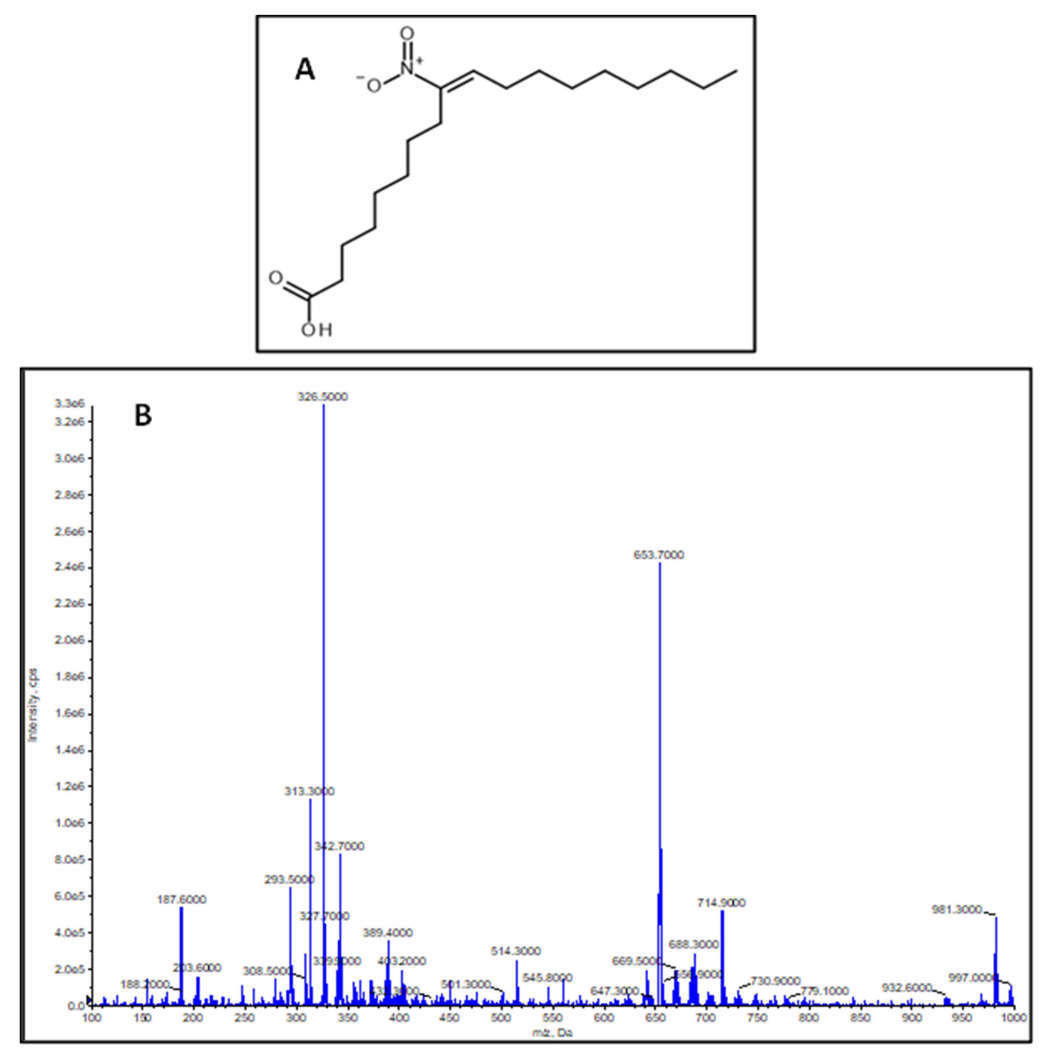
2.1. Characterization of the Synthesized NO2-OA Standard
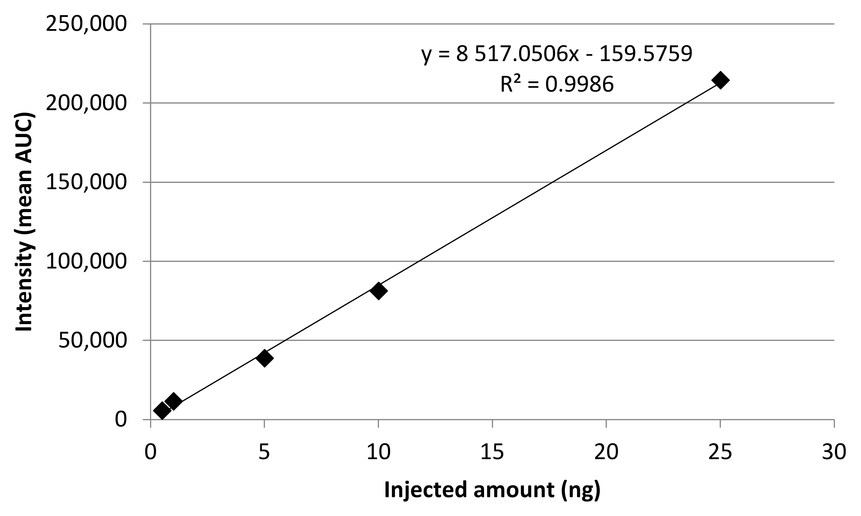
The synthesized compound was identified as (E)-9-nitrooctadec-9-enoic acid based on the identical 1H and 13C NMR data with those reported in the literature [25]. This was assured by mass spectrometric measurements where the measured molecule ion mass was shown to be m/z 326.5, calculated neutral molecule mass was 327.4589 Da, molecular formula C18H33NO4 (Figure 1A,B).

2.2. Calibration

From the synthesized standard a stock standard was prepared with methanol (HPLC grade) and working solutions were prepared (50–2500 ng/mL) by diluting the stock standard. Each solution was injected three times, the precision (Relative Standard Deviation, RSD, %) of the calibration measurements ranged between 1.2093–2.3633%. Calibration showed linear regression, the R2 value was 0.9986, the limit of detection (LOD, S/N = 3.3) was 0.1184 nmol/mL, and the limit of quantitation (LOD, S/N = 10) was 0.3588 nmol/mL (Figure 2).

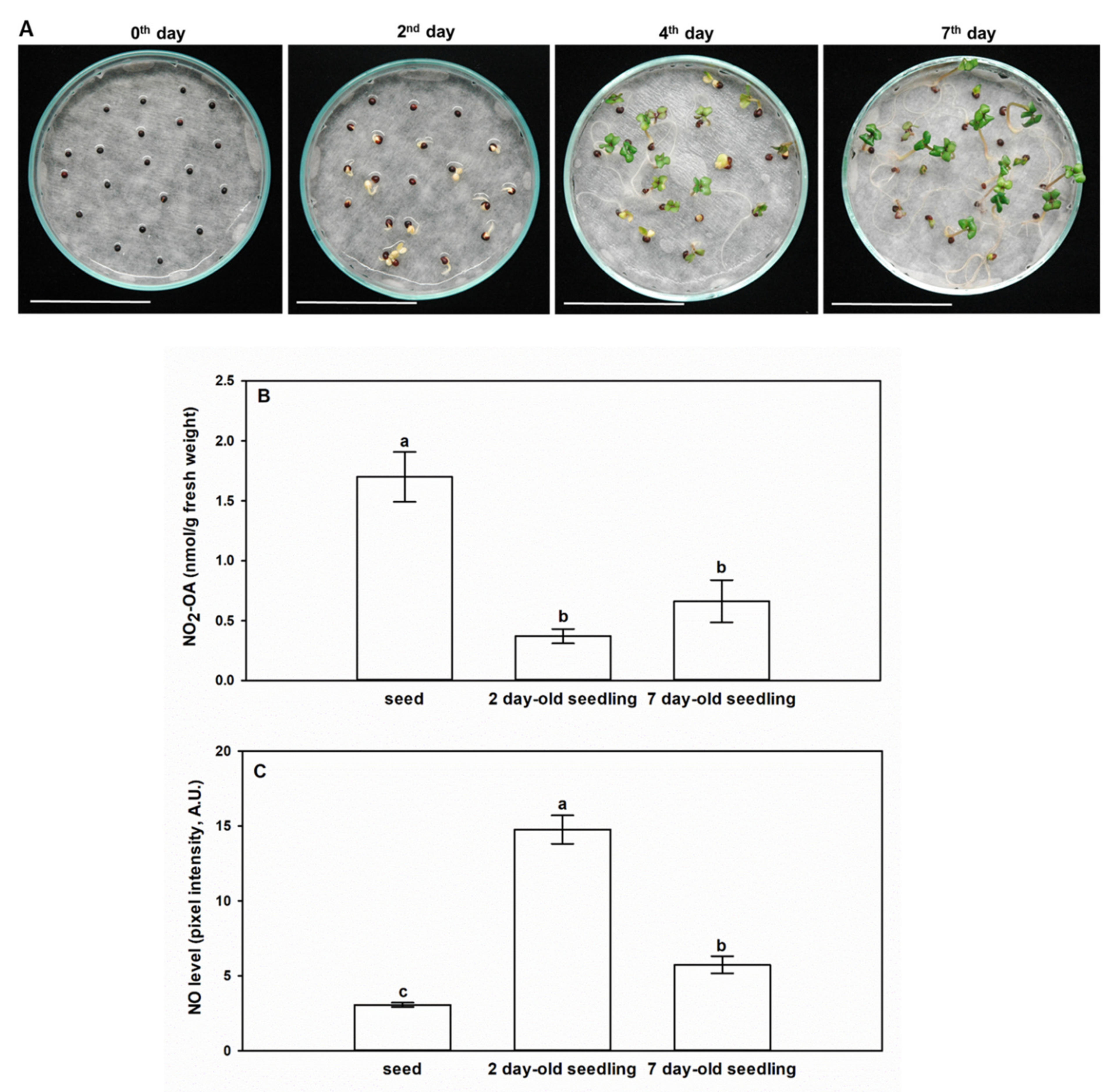
2.3. NO2-OA Content of Brassica napus at the Seed and Seedling Stages

Seeds and seedlings of Brassica napus are remarkably rich in the unsaturated fatty acid, oleic acid, which implies the possibility of the presence of NO2-OA in Brassica as well. Figure 3A represents Brassica napus seeds and seedlings at day 0, and 2nd, 4th, and 7th day after sowing. Therefore, we performed the analyses of NO2-OA concentrations and the quantitative data are presented in Table 1, while the mean NO2-OA concentrations with standard errors are shown in Figure 3. Ion chromatograms of the reference standard NO2-OA and of the 7-day-old Brassica napus seedlings are presented in Figure 4. Further chromatograms are presented as Figure S1. In samples, one further unidentified isomer of NO2-OA can be detected with higher retention time.
Brassica napus seeds contained a notably high amount of NO2-OA compared to seedlings (Table 1, Figure 3B). During the early phase of seedling growth (2nd day), the high NO2-OA content of Brassica seeds decreased by 78%, and it continued to decline in the following two days (4th day). At the 7th day of seedling growth, the seedlings showed an increased NO2-OA content compared to the 2- and 4-day-old seedlings, but it was only approximately 40% of the NO2-OA content of the seed. Separated analysis of the shoot and root material indicated that both organs of 7-day-old Brassica napus seedlings contained NO2-OA in similar quantities (Table 1). In Arabidopsis tissues, NO2-OA could not be detected, but NO2-Ln content was 2.9-fold higher in the seeds than in the 14-day-old seedlings ([6], Table 2). The relatively high NO2-Ln and NO2-OA contents of Arabidopsis and Brassica seeds (Table 1, Table 2) indicate the involvement of NO2-FA in seed germination possibly as endogenous •NO donors [16]. To examine this hypothesis, we detected •NO levels in Brassica seeds and seedlings (2nd, 7th days) (Figure 3C). Compared to seeds, on the 2nd day the •NO content increased by almost 5-fold. The opposite changes in NO2-OA and •NO levels suggest that following the induction of germination NO2-OA as endogenous donor in the seed may release NO resulting in the decrease in its own free level and concomitant •NO accumulation. Furthermore, it is important to note, that the measured NO2-OA concentrations in Brassica tissues are in the nanomolar range (Table 1, Figure 3B), that is an order of magnitude higher than the picomolar NO2-Ln concentrations of Arabidopsis seedlings, cell suspension, pea roots, leaves and rice leaves ([6,7], Table 2). In case of NO2-OA supplemented tomato cell suspension, the endogenous NO2-OA content was similar to that of the untreated Brassica napus seeds and seedlings ([8,9], Table 2). From the above comparisons, it can be concluded that the oleic acid content of Brassica napus seeds and seedlings is susceptible to physiological nitration making the plant a rich source of NO2-OA.

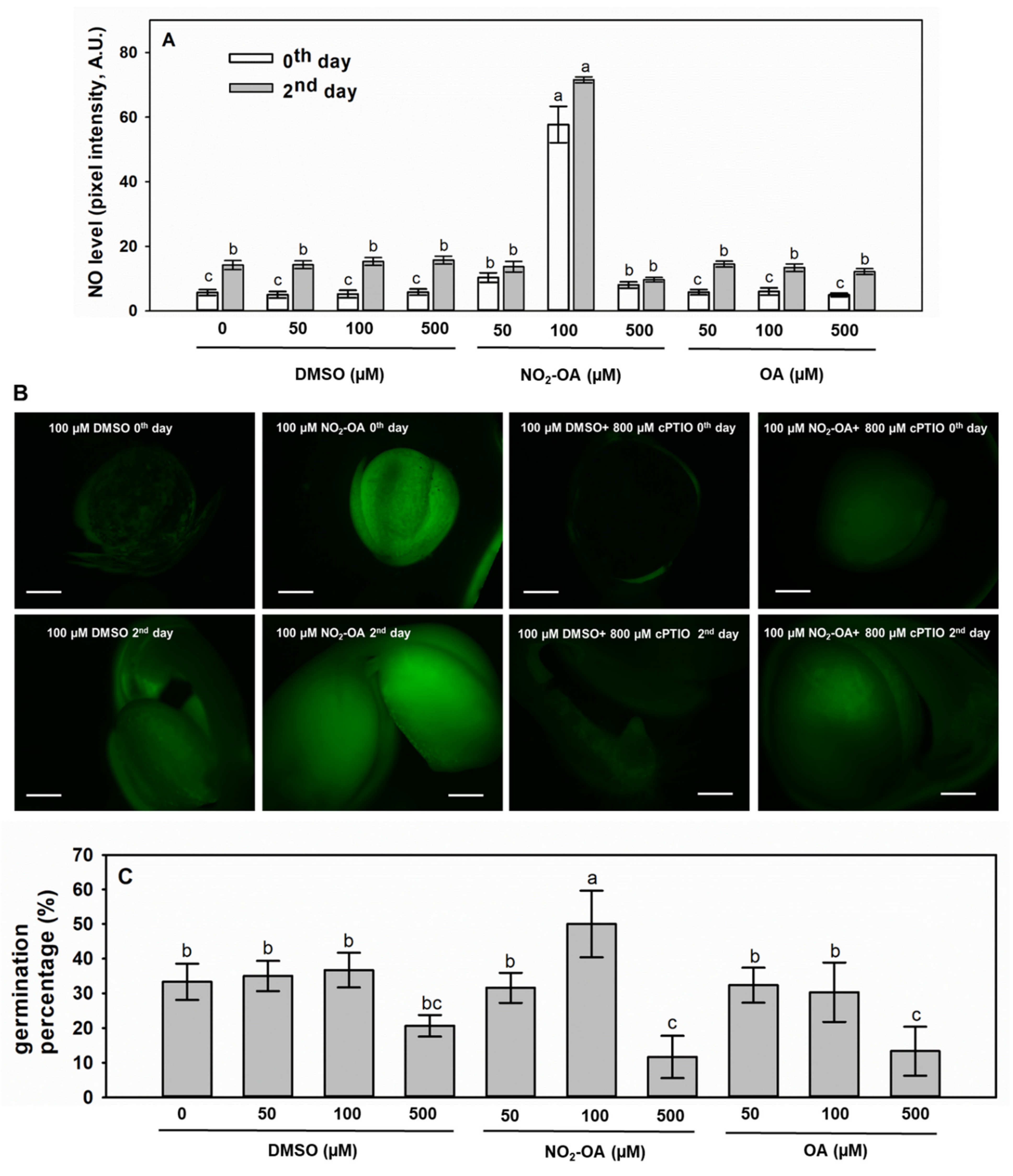
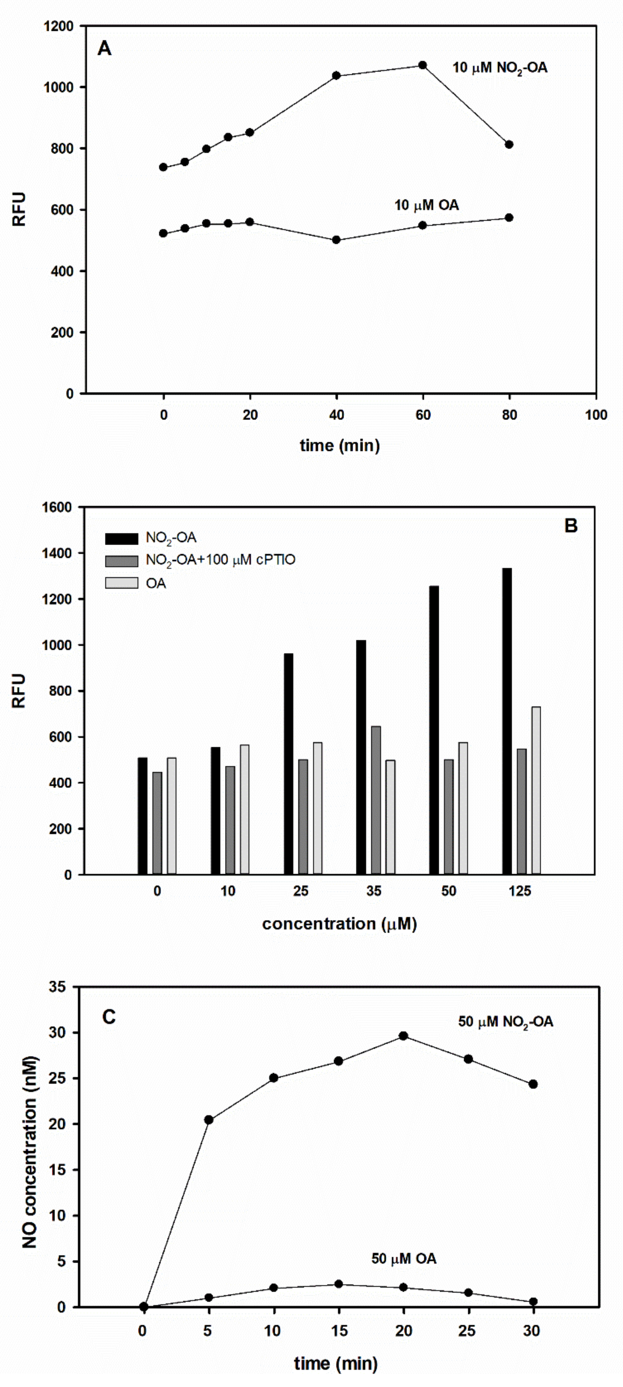
2.4. Exogenous NO2-OA Treatment of Brassica Seeds Positively Influences •NO Levels and Germination Capacity

The 24-hour-long treatment with 100 µM NO2-OA caused 10-fold increment in the endogenous •NO level of seeds (Figure 5A,B), while NO2-OA at 50 µM or 500 µM concentrations did not cause significant changes in •NO levels compared to controls (50 or 500 µM DMSO, respectively). The significant effect of 100 µM NO2-OA on •NO level prevailed also on the 2nd day after sowing, since the •NO level of NO2-OA-supplemented seedlings was 4.7 times that of the control (100 µM DMSO, Figure 5A,B). The NO2-OA-induced DAF-FM fluorescence was quenched by the •NO scavenger cPTIO (Figure 5B) indicating that the alterations in DAF fluorescence correspond to •NO level changes. Oleic acid (OA) treatments applied as controls did not influence •NO levels either in seeds or in 2-day-old seedlings (Figure 5A).
The germination percentages were altered in accordance with •NO levels, since 100 µM NO2-OA treatment doubled the percentage of germinated seeds compared to control (100 µM DMSO) (Figure 5C). DMSO alone at 50 µM or 100 µM concentrations had no effect on seed germination, although in the presence of 500 µM DMSO germination percentage of Brassica seeds slightly decreased compared to control (0 µM DMSO). Moreover, OA at 50 or 100 µM concentration exerted no effect on germination of B. napus seeds (Figure 5C). The effect of DMSO could dominate in case of 500 µM NO2-OA or 500 µM OA treatments, since in these seeds the germination ability was weaker than in control and •NO levels remained at control level (Figure 5A). These data clearly indicate that exogenous NO2-OA can result in the long-term increase of endogenous •NO level in Brassica napus seeds and seedlings, i.e., it may act as a •NO donor. The fact that nitro-fatty acids may act as •NO donors in plants has been previously raised [16]. It was revealed that NO2-Ln has the ability to release in vivo, in leaves and roots of old Arabidopsis plants [26], in Arabidopsis seedlings [16], and in Arabidopsis cell-suspension cultures [27]. Contrary to NO2-Ln and to NO2-OA in our study, exogenous application of NO2-OA (0.5, 5, 10, 12.5, 25, or 50 µM, 1 h or 6 h) did not increase •NO level in tomato cell suspension as was recently reported by Di Palma et al. [8,9]. Contradiction of the results can be explained by that here higher concentration of NO2-OA (100 µM) were applied for longer time period (24 h) than in the studies of Di Palma et al. [8,9].

2.5. NO2-OA Releases •NO in Vitro

Additionally, the •NO donor nature of NO2-Ln was proved by using several different in vitro approaches (DAF fluorescence, oxyhaemoglobin method, ozone chemiluminescence, [27]. In this work, we performed in vitro tests to support the hypothesis regarding the •NO donor role of NO2-OA (Figure 6). Spectrofluorometric measurement of DAF-FM-associated fluorescence revealed that the sample containing 10 µM NO2-OA liberated a relatively small amount of •NO during the 80 min period, while the same dosage of OA did not induce •NO level increase compared to the blind sample containing only buffer and the fluorophore (Figure 6A). Moreover, NO2-OA liberated •NO in a concentration-dependent manner and the fluorescence increase could be quenched by cPTIO. Elevating doses of OA did not increase •NO levels (Figure 6B).
Using the more sensitive •NO electrode, we could quantify •NO liberation in NO2-OA solution (pH 5.8) within 5 min reaching its maximum (~30 nM •NO) after 20 min incubation. The produced •NO concentrations (20–30 nM) quantified by ISO-NOP electrode in this study are similar to those measured by •NO autoanalyzer in the case of 100 µM NO2-Ln or 80 µM NO2-LA [27]. The same concentration of OA showed no relevant •NO releasing capacity in the solution (Figure 6C). These in vitro data support the •NO donor character of NO2-OA in solutions and the degree of •NO liberation is similar to other NO2-FAs. However, the same dosage of S-nitrosoglutathione (GSNO) or sodium nitroprusside (SNP) produced approx. 10-fold higher •NO concentration in solutions following 20 min incubation in light (data not shown) indicating that NO2-FAs (including NO2-OA) •NO donor capacity is much lower compared to “classical •NO donors”.
In our experimental system, NO2-OA (100 µM, 24 h) treatment of Brassica seeds also promoted germination, presumably through the induction of high •NO levels. In vitro tests revealed that the concentration of liberated •NO is relatively low (Figure 6), but in seeds NO2-OA treatment caused intense •NO formation (Figure 5). Therefore, we assume that beyond direct •NO emission, secondary signal processes may also be activated in the presence of NO2-OA leading to the activation of •NO metabolic routes in seeds and seedlings. It is well known, that •NO attenuates seed dormancy and promotes germination and we are beginning to recognize also the molecular mechanisms of •NO action [28].

3. Materials and Methods

3.1. Plant Material and Growing Conditions

Experiments were carried out on Brassica napus L. (cv. GK Gabriella) seedlings. The seeds were obtained from the Cereal Research Non-profit Ltd., Szeged, Hungary. Brassica seeds were surface sterilised in 70% (v/v) ethanol and 5% (v/v) sodium hypochlorite, then placed on moistened filter paper in Petri dishes (9 cm diameter, 30 seeds/Petri dish). Germination took place under controlled conditions (150 µmol m−2 s−1 photon flux density, 12 h/12 h light/dark cycle, relative humidity 55–60% and temperature 25 ± 2 °C). For the NO2-OA analysis, the samples were taken at the 2nd, 4th and 7th day after sowing. Additionally, seeds after imbibition, at the early stage of germination (day 0) were also sampled for NO2-OA quantification. Plant material (5 g) was collected, frozen in liquid nitrogen and stored at –80 °C until the analyses.

3.2. Synthesis and Structure Determination of 9-Nitro-Oleic Acid Standard

Using the slightly modified method of Woodcock et al. [29], 9-nitro-oleic acid was synthesized. Bromononanoic acid was used as starting material for allylization to gain 9-bromononanoic acid allyl ester. This compound was nitrated using silver nitrite. In the next step, 10-hydroxy-9-nitro-octadecanoic acid allyl ester was synthesized by the addition of nonyl aldehyde and 1,8-diazabicyclo[5.4.0]undec-7-ene. This compound was acylated with acetic anhydride in the presence of p-toluenesulfonic acid to gain 10-acetoxy-9-nitro-octadecanoic acid allyl ester. Deacylation in the presence of Na2CO3 led to the synthesis of 9-nitro-oleic acid allyl ester. The final product 9-nitro-oleic acid was gained by a catalytic hydrolysis using palladium tetrakis(triphenylphosphine). Purification was carried out by flash chromatography on silica gel (Merck, 40–63 µm) using a gradient of 0.5% acetic acid. Fractions with similar compositions were combined and the combined fraction containing a spot with remarkable absorption at 254 nm and with an Rf approximately 0.5 (silica gel plate, eluent CHCl3–MeOH 95:5) was subjected to NMR identification and purity check.
1H (500.1 MHz) and 13C (125.6 MHz) NMR spectra were recorded in CDCl3 on a Bruker Avance DRX-500 spectrometer. The peaks of the residual solvent were taken as reference points. The compound was identified by comparison of its chemical shifts with literature data [25,30]. Mass spectrometric identification was performed on an API 2000 triple quadrupole tandem mass spectrometer (MDS Sciex, Toronto, ON, Canada) equipped with electrospray ion source. Mass spectrometric measurement was carried out by direct infusion in Q1 MS scan type in negative mode. Flow rate was set to 40 µL/min, scan range was set from m/z 100 to 1000, ion source temperature was set to 100 °C. The nebulizer gas was set to 18 psi. Measured molecule ion mass shown to be m/z 326.5, calculated neutral molecule mass was 327.4589 Da.

3.3. LC-MS Quantification of NO2-OA in Brassica Seeds and Seedlings

For the quantification of NO2-OA, 5 g of fresh plant material (seeds, whole seedlings or separated root and shoot) was used and the analysis was conducted by LC-MS. Mass spectrometry measurement was performed using single ion monitoring (SIM) on an API 2000 triple quadrupole tandem mass spectrometer (MDS Sciex, Toronto, ON, Canada) equipped with electrospray ion source. The nebulizer and heater gas was nitrogen, generated from a Peak NM20Z nitrogen generator (Peak Scientific Instruments Ltd., Scotland, UK) coupled with an Atlas Copco SF 4FF compressor. The nebulizer gas was set to 30 psi, the heater gas was set to 80 psi. The ion source temperature was set to 300 °C. Measurement was in negative mode. The voltage volumes were adjusted to m/z 326.5, the collision energy was set to −5 V, focusing potential to −330 V, declustering potential to −61 V and entrance potential to −10 V. HPLC separation was performed with Shimadzu HPLC system (Kyoto, Kyoto Prefecture, Japan): DGU-20A3 degasser, CBM-20A controller, two LC-20AD pumps, SIL-20A HT autosampler, CTO-20AC column thermostat, SPD-20A UV-Vis detector, using Kinetex F5 (100 × 4.6 mm, 2.6 µm, 100Ä) (Phenomenex, Inc., Torrance, CA, USA). Elution was carried out with the gradient system of H2O–methanol (0 min: H2O–methanol 2:8, 0.6 min: 2:8, 1.5 min: 0:1, 3 min: 0:1, 3.2 min: 2:8, 8.2 min: 2:8). The oven was set to 40 °C and the flow rate was 600 µL/min. The retention time of NO2-OA was 4.32 min. Data acquisition was performed with Analyst software (ver. 1.6.3). Quantification of nitro-oleic acid was carried out by using the synthesized standard (see in Section 2.2).
Plant material was extracted with pure MeOH (HPLC grade) in a VWR Ultrasonic Cleaner USC 300D (capacity: 1 L, internal dimension: W × D × H 240 × 135 × 100 mm) ultrasonic bath for 10 min, with dual half-wave sound with sweep, frequency was set to 45 kHz, ultrasonic power to 80 W, temperature was set to 25 °C, and filtered through a syringe filter (PTFE, 0.45 µm pore size, Labex Ltd.), the first half ml of the filtrate was thrown into the waste. From each plant part, three parallel samples were prepared, and each sample was injected three times.

3.4. •NO Detection in Brassica napus Seeds and Seedlings

Nitric oxide levels were examined in seeds as well as in 2- and 7-day-old seedlings of Brassica napus using the 4-amino-5-methylamino- 2′,7′-difluorofluorescein diacetate (DAF-FM DA) according to Kolbert et al. [31]. Plant samples were incubated in 10 µM dye solution for 30 min (darkness, 25 ± 2 °C) and washed twice with Tris-HCl (10 mM, pH 7.4). Microscopic analysis was accomplished under Zeiss Axiovert 200 M inverted microscope (Carl Zeiss, Jena, Germany) equipped with a high-resolution digital camera (AxiocamHR, HQ CCD, Carl Zeiss, Jena, Germany) and filter set 10 (exc.: 450–490, em.: 515–565 nm). Pixel intensities were measured on digital photographs (at least 10 photographs per sample per experiment) using Axiovision Rel. 4.8 software (Carl Zeiss, Jena, Germany).

3.5. NO2-OA Treatment of Brassica napus Seeds

The synthesized NO2-OA or OA was dissolved in dimethyl sulfoxide (DMSO) in order to obtain a stock solution (10 mM). The NO2-OA or OA stock was diluted with distilled water to the final concentrations (50, 100, 500 µM). Control solutions were prepared by measuring the volume of DMSO corresponding to the stock solutions (indicated as 0, 50, 100, or 500 DMSO). Brassica napus seeds (30 seeds per treatment) were incubated in NO2-OA, in OA or in control solutions for 24 h on an orbital shaker and then were placed on moist filter paper in Petri dishes (20 seeds/Petri dish). Sets of seeds were treated with NO2-OA or control solutions in the presence of 800 µM 2-(4-carboxyphenyl)-4,4,5,5-tetramethylimidazoline-1-oxyl-3-oxide (cPTIO). Germination took place as indicated above. Nitric oxide levels were detected in seeds prior to germination (day 0), and in seedlings 2 days after sowing (2nd day). Germination percentages (%) were also calculated.

3.6. Spectrofluorometric Determination of •NO Levels

•NO liberation from NO2-OA solutions were detected by fluorescence spectrophotometry (Ref. [27] with modifications). Reaction mixtures (2 mL final volume) containing 0 (blind) 10, 25, 35, 50, 125 µM NO2-OA or the same concentrations of OA, plus 2 µM DAF-FM and Tris-HCl (pH 7.4) buffer were incubated at room temperature in the dark for several time periods (5–80 min) and the emitted fluorescence was recorded by a spectrofluorimeter (Hitachi F-4500, Hitachi Ltd., Tokyo, Japan). Excitation wavelength was set at 485 nm and emissions were measured at 515 nm. The NO2-OA-induced fluorescence was quenched by the addition of 100 µM cPTIO.

3.7. Measurement of •NO Concentration by •NO-Specific Electrode

The •NO -sensitive electrode (ISO-NOP, 2 mm, World Precision Instruments Inc., Sarasota, FL, USA) was calibrated using a method based on S-nitroso-N-acetylpenicillamine (SNAP) decomposition to •NO in the presence of copper [32]. Two mL of NO2-OA or OA (both at 50 µM concentration) solutions were prepared in a 5-mL glass bottle and were measured immediately after preparation. To ensure constant mixing of the solution a magnetic stirrer was applied during the measurement. •NO concentration (nM) was calculated from a standard curve.

3.8. Statistical Analysis

All results are shown as mean ± SE. Data were statistically evaluated by the Holm–Sidak method (One-way ANOVA, P ≤ 0.001) using SigmaPlot 12.

4. Conclusions

Following successful standard synthesis and method optimization, we have been the first to observe that Brassica seeds and seedlings contain free NO2-OA. Exogenous treatment of Brassica seeds with NO2-OA promoted germination and increased endogenous •NO level suggesting that NO2-OA may be involved in germination as an •NO donor. The •NO liberating capacity of NO2-OA was proved also by in vitro approaches (spectrofluorometric detection of DAF-FM fluorescence and •NO-sensitive electrode). Due to their relatively high NO2-OA concentrations, Brassica sprouts can be considered as a good source of dietary NO2-OA intake in addition to their nutrient, mineral and vitamin content. Therefore, future studies should quantify the NO2-OA content of additional Brassica species and food plants.

Supplementary Materials

The following are available online at https://www.mdpi.com/2223-7747/9/3/406/s1. Figure S1: Chromatograms of B. napus seeds or seedlings (A) Ion chromatogram of m/z 326.5 of a sample derived from Brassica napus seed. (B) Ion chromatogram of m/z 326.5 of a sample derived from 2-day-old Brassica napus seedling. (C) Ion chromatogram of m/z 326.5 of a sample derived from 4-day-old Brassica napus seedling. (D) Ion chromatogram of m/z 326.5 of a sample derived from 7-day-old Brassica napus shoot. (E) Ion chromatogram of m/z 326.5 of a sample derived from 7-day-old Brassica napus root.

Author Contributions

Conceptualization, D.C. and Z.K.; Investigation, M.V., G.F., D.O., A.H., Á.M., and N.K.; Writing—Original Draft Preparation, M.V., D.C., and Z.K.; Writing—Review and Editing, A.Ö., D.C., and Z.K. All authors have read and agreed to the published version of the manuscript.

Funding

This research was funded by the National Research, Development and Innovation Fund (Grant no. NKFI-6, K120383) and by the EU-funded Hungarian grant EFOP-3.6.1-16-2016-00008. Zs. K. was supported by the János Bolyai Research Scholarship of the Hungarian Academy of Sciences (Grant no. BO/00751/16/8) and by UNKP-18-4 New National Excellence Program of the Ministry of Human Capacities.

Acknowledgments

The Authors thank Éva Kapásné Török for her valuable assistance during the experiments.

Conflicts of Interest

The authors declare that there is no conflict of interest.

References

  1. Melo, T.; Montero-Bullón, J.F.; Domingues, P.; Domingues, M.R. Discovery of bioactive nitrated lipids and nitro-lipid-protein adducts using mass spectrometry-based approaches. Redox Biol. 2019, 23, 101–106. [Google Scholar] [CrossRef]
  2. Buchan, G.J.; Bonacci, G.; Fazzari, M.; Salvatore, S.R.; Gelhaus Wendell, S. Nitro-fatty acid formation and metabolism. Nitric Oxid. 2018, 79, 38–44. [Google Scholar] [CrossRef] [Green Version]
  3. Pryor, W.A.; Lightsey, J.W.; Church, D.F. Reaction of nitrogen dioxide with alkenes and polyunsaturated fatty acids: Addition and hydrogen abstraction mechanisms. J. Am. Chem. Soc. 1982, 104, 6685–6692. [Google Scholar] [CrossRef]
  4. d’Ischia, M.; Napolitano, A.; Manini, P.; Panzella, L. Secondary targets of nitrite-derived reactive nitrogen species: Nitrosation/nitration pathways, antioxidant defense mechanisms and toxicological implications. Chem. Res. Toxicol. 2011, 24, 2071–2092. [Google Scholar] [CrossRef]
  5. Fazzari, M.; Trostchansky, A.; Schopfer, F.J.; Salvatore, S.R.; Sánchez-Calvo, B.; Vitturi, D.; Valderrama, R.; Barroso, J.B.; Radi, R.; Freeman, J.B.; et al. Olives and olive oil are sources of electrophilic fatty acid nitroalkenes. PLoS ONE 2014, 9, e84884. [Google Scholar] [CrossRef] [PubMed] [Green Version]
  6. Mata-Pérez, C.; Sánchez-Calvo, B.; Padilla, M.N.; Begara-Morales, J.C.; Luque, F.; Melguizo, M.; Jiménez-Ruiz, J.; Fierro-Risco, J.; Peñas-Sanjuán, A.; Valderrama, R.; et al. Nitro-fatty acids in plant signaling: Nitro-linolenic acid induces the molecular chaperone network in Arabidopsis. Plant Physiol. 2016, 170, 686–701. [Google Scholar] [CrossRef] [PubMed]
  7. Mata-Pérez, C.; Sánchez-Calvo, B.; Padilla, M.N.; Begara-Morales, J.C.; Valderrama, R.; Corpas, F.J.; Barroso, J.B. Nitro-fatty acids in plant signaling: New key mediators of nitric oxide metabolism. Redox Biol. 2017, 11, 554–561. [Google Scholar] [CrossRef] [PubMed]
  8. Di Palma, A.A.; Di Fino, L.M.; Salvatore, S.R.; D’Ambrosio, J.M.; Grozeff, G.E.G.; García-Mata, C.; Schopfer, F.J.; Laxalt, A.M. Nitro-oleic acid induced reactive oxygen species formation and plant defense signaling in tomato cell suspensions. BioRxiv 2018, in press. [Google Scholar] [CrossRef] [Green Version]
  9. Di Palma, A.A.; Di Fino, L.M.; Salvatore, S.R.; D’Ambrosio, J.M.; García-Mata, C.; Schopfer, F.J.; Laxalt, A.M. Nitro-oleic acid triggers ROS production via NADPH oxidase activation in plants: A pharmacological approach. J. Plant Physiol. 2020, 246–247, 153128, in press. [Google Scholar] [CrossRef]
  10. Rudolph, V.; Rudolph, T.K.; Schopfer, F.J.; Bonacci, G.; Woodcock, S.R.; Cole, M.P.; Baker, P.R.S.; Ramani, R.; Freeman, B.A. Endogenous generation and protective effects of nitro-fatty acids in a murine model of focal cardiac ischaemia and reperfusion. Cardiovasc. Res. 2010, 85, 155–166. [Google Scholar] [CrossRef] [PubMed] [Green Version]
  11. Baker, L.M.; Baker, P.R.; Golin-Bisello, F.; Schopfer, F.J.; Fink, M.; Woodcock, S.R.; Branchaud, B.P.; Radi, R.; Freeman, B.A. Nitro-fatty acid reaction with glutathione and cysteine. Kinetic analysis of thiol alkylation by a Michael addition reaction. J. Biol. Chem. 2007, 282, 31085–31093. [Google Scholar] [CrossRef] [PubMed] [Green Version]
  12. Aranda-Caño, L.; Sánchez-Calvo, B.; Begara-Morales, J.C.; Chaki, M.; Mata-Pérez, C.; Padilla, M.N.; Valderrama, R.; Barroso, J.B. Post-translational modification of proteins mediated by nitro-fatty acids in plants: Nitroalkylation. Plants 2019, 8, 82. [Google Scholar] [CrossRef] [Green Version]
  13. Rubbo, H.; Parthasarathy, S.; Barnes, S.; Kirk, M.; Kalyanaraman, B.; Freeman, B.A. Nitric oxide inhibition of lipoxygenase-dependent liposome and low-density lipoprotein oxidation: Termination of radical chain propagation reactions and formation of nitrogen-containing oxidized lipid derivatives. Arch. Biochem. Biophys. 1995, 324, 15–25. [Google Scholar] [CrossRef] [PubMed]
  14. Lima, É.S.; Bonini, M.G.; Augusto, O.; Barbeiro, H.V.; Souza, H.P.; Abdalla, D.S.P. Nitrated lipids decompose to nitric oxide and lipid radicals and cause vasorelaxation. Free Radic. Biol. Med. 2005, 39, 532–539. [Google Scholar] [CrossRef] [PubMed]
  15. Schopfer, F.J.; Baker, P.R.S.; Giles, G.; Chumley, P.; Batthyany, C.; Crawford, J.; Patel, R.P.; Hogg, N.; Branchaud, B.P.; Lancaster, J.R.; et al. Fatty acid transduction of nitric oxide signaling: Nitrolinoleic acid is a hydrophobically stabilized nitric oxide donor. J. Biol. Chem. 2005, 280, 19289–19297. [Google Scholar] [CrossRef] [Green Version]
  16. Mata-Pérez, C.; Sánchez-Calvo, B.; Begara-Morales, J.C.; Padilla, M.N.; Valderrama, R.; Corpas, F.J.; Barroso, J.B. Nitric oxide release from nitro-fatty acids in Arabidopsis roots. Plant Signal. Behav. 2016, 11, e1154255. [Google Scholar] [CrossRef] [Green Version]
  17. Villacorta, L.; Zhang, J.; Garcia-Barrio, M.T.; Chen, X.; Freeman, B.A.; Chen, Y.E.; Cui, T. Nitro-linoleic acid inhibits vascular smooth muscle cell proliferation via the Keap1/Nrf2 signaling pathway. Am. J. Physiol. Heart Circ. Physiol. 2007, 293, 770–776. [Google Scholar] [CrossRef] [Green Version]
  18. Nie, H.; Xue, X.; Li, J.; Liu, X.; Lv, S.; Guan, G.; Liu, H.; Liu, G.; Liu, S.; Chen, Z. Nitro-oleic acid attenuates ogd/r-triggered apoptosis in renal tubular cells via inhibition of Bax mitochondrial translocation in a PPAR-γ-dependent manner. Cell. Physiol. Biochem. 2015, 35, 1201–1218. [Google Scholar] [CrossRef]
  19. Kansanen, E.; Kuosmanen, S.M.; Ruotsalainen, A.K.; Hynynen, H.; Levonen, A.L. Nitro-oleic acid regulates endothelin signaling in human endothelial cells. Mol. Pharmacol. 2017, 92, 481–490. [Google Scholar] [CrossRef]
  20. Yang, R.X.; Fan, J.G. Nitro-oleic acid as a new drug candidate for non-alcoholic steatohepatitis. EBioMedicine 2019, 42, 32–33. [Google Scholar] [CrossRef] [Green Version]
  21. Liu, H.; Jia, Z.; Soodvilai, S.; Guan, G.; Wang, M.H.; Dong, Z.; Symons, J.D.; Yang, T. Nitro-oleic acid protects the mouse kidney from ischemia and reperfusion injury. Am. J. Physiol. Renal Physiol. 2008, 295, 942–949. [Google Scholar] [CrossRef] [PubMed] [Green Version]
  22. Rom, O.; Xu, G.; Guo, Y.; Zhu, Y.; Wang, H.; Zhang, J.; Fan, Y.; Liang, W.; Lu, H.; Liu, Y.; et al. Nitro-fatty acids protect against steatosis and fibrosis during development of nonalcoholic fatty liver disease in mice. EBioMedicine 2019, 41, 62–72. [Google Scholar] [CrossRef] [PubMed] [Green Version]
  23. Márton, M.; Mándoki, Z.S.; Csapó-Kiss, Z.S.; Csapó, J. The role of sprouts in human nutrition. A review. Acta Univ. Sapientiae Aliment. 2010, 3, 81–117. [Google Scholar]
  24. Sharafi, Y.; Majidi, M.M.; Goli, S.A.H.; Rashidi, F. Oil content and fatty acids composition in Brassica species. Int. J. Food Proper. 2015, 18, 2145–2154. [Google Scholar] [CrossRef] [Green Version]
  25. Gorczynski, M.J.; Huang, J.; King, S.B. Regio- and stereospecific syntheses and nitric oxide donor properties of (E)-9- and (E)-10-nitrooctadec-9-enoic acids. Org. Lett. 2006, 8, 2305–2308. [Google Scholar] [CrossRef] [Green Version]
  26. Sánchez-Calvo, B.; Barroso, J.B.; Corpas, F.J. Hypothesis: Nitro-fatty acids play a role in plant metabolism. Plant Sci. 2013, 199, 1–6. [Google Scholar] [CrossRef]
  27. Mata-Pérez, C.; Sánchez-Calvo, B.; Begara-Morales, J.C.; Carreras, A.; Padilla, M.N.; Melguizo, M.; Valderrama, R.; Corpas, F.J.; Barroso, J.B. Nitro-linoleic acid is a nitric oxide donor. Nitric Oxid. 2016, 57, 57–63. [Google Scholar] [CrossRef]
  28. Signorelli, S.; Considine, M.J. Nitric oxide enables germination by a four-pronged attack on ABA-induced seed dormancy. Front. Plant Sci. 2018, 9, 296. [Google Scholar] [CrossRef]
  29. Woodcock, S.R.; Bonacci, G.; Gelhaus, S.L.; Schopfer, F.J. Nitrated fatty acids: Synthesis and measurement. Free Radic. Biol. Med. 2013, 59, 14–26. [Google Scholar] [CrossRef] [Green Version]
  30. Woodcock, S.R.; Marwitz, A.J.V.; Bruno, P.; Branchaud, B.P. Synthesis of nitrolipids. all four possible diastereomers of nitrooleic acids: (E)- and (Z)-, 9- and 10-nitro-octadec-9-enoic acids. Org. Lett. 2006, 8, 3931–3934. [Google Scholar] [CrossRef]
  31. Kolbert, Z.S.; Pető, A.; Lehotai, N.; Feigl, G.; Ördög, A.; Erdei, L. In Vivo and In Vitro studies on fluorophore-specificity. Acta Biol. Szeged. 2012, 65, 37–41. [Google Scholar]
  32. Zhang, X. Real time and In Vivo monitoring of nitric oxide by electrochemical sensors from dream to reality. Front. Biosci. 2004, 9, 3434–3446. [Google Scholar] [CrossRef] [PubMed] [Green Version]
Figure 1. (A) Structure of (E)-9-nitrooctadec-9-enoic acid. (B) Q1 MS scan of nitro-oleic acid (NO2-OA) standard. Scan range was set between m/z 100 and 1000 Da. Molecule ion peak [M − H] was m/z 326.5.
Plants 09 00406 g001
Figure 2. Calibration line of NO2-OA standard. Axis X shows the range of injected NO2-OA amount (ng) from working solutions, axis Y shows the mean area under curve (AUC) of the working solutions.
Plants 09 00406 g002
Figure 3. Levels of NO2-OA and •NO change in opposite ways in the early stage of germination (day 0) and during seedling growth (2nd, 4th and 7th days). (A) Representative photographs taken from Brassica napus seedlings on day 0, and 2nd, 4th, and 7th day after sowing. Bars = 4.5 cm. (B) Mean concentrations of NO2-OA (nmol/g fresh weight, with standard errors) in seeds and differently developed seedlings of Brassica napus. (C) •NO levels (pixel intensity, arbitrary unit) in Brassica napus seeds, 2 day-old and 7 day-old seedlings. Different letters indicate significant differences according to the Holm–Sidak test (P ≤ 0.001).
Plants 09 00406 g003
Figure 4. (A) Ion Chromatogram of m/z 326.5 of NO2-OA working solution (100 ng/mL). Peak retention time (RT) = 4.3173 min. (B) Ion Chromatogram of m/z 326.5 of a sample derived from 7- day- old Brassica napus seedlings. Peak retention time (RT) = 4.3420 min.
Plants 09 00406 g004
Figure 5. Exogenous NO2-OA increases endogenous •NO levels and triggers germination of Brassica napus seeds. (A) Nitric oxide level in seeds (day 0 of germination) and seedlings (2nd day after sowing) of Brassica napus treated for 24 h with 50, 100 or 500 µM NO2-OA or with equal concentrations of OA. As controls, seeds were treated with 0, 50, 100 or 500 µM DMSO. Different letters indicate significant differences according to Holm–Sidak test (P ≤ 0.001). (B) Representative fluorescent microscopic images of DAF FM-stained Brassica seeds and seedlings treated with 100 µM NO2-OA or 100 µM DMSO for 24 h in the presence or absence of 800 µM cPTIO. Bars = 800 µm. (C) Germination percentage (determined at the 2nd day after sowing) of Brassica seeds treated with 50, 100, or 500 µM NO2-OA or with equal concentrations of OA. As controls, seeds were treated with 0, 50, 100, or 500 µM DMSO. Different letters indicate significant differences according to the Holm–Sidak test (P ≤ 0.001).
Plants 09 00406 g005
Figure 6. NO2-OA liberates •NO in solutions (A) Relative fluorescence units (RFU) of DAF-FM fluorescence in solutions in the presence of 10 µM NO2-OA or 10 µM OA measured at different time points. (B) Relative fluorescence units (RFU) of DAF-FM fluorescence in solutions containing different concentrations of NO2-OA (with or without 100 µM cPTIO) or OA. Data were recorded after 20 min incubation. (C) •NO concentration (nM) measured by ISO-NOP (2 mm) electrode in solutions containing 50 µM NO2-OA or OA measured at different time points.
Plants 09 00406 g006
Table 1. Concentrations of nitro-oleic acid (NO2-OA) in B. napus seeds and differently developed seedlings. N/A (not available) indicates data under the detection limit. Samples were injected three times, the precision (RSD, %) of the sample measurements ranged between 0.1348–3.9822 RSD%. The precision between the parallel samples ranged between 9.4828–26.6930 RSD%.
SampleNO2-OA Concentration (nmol/g Fresh Weight)Mean NO2-OA Concentration (nmol/g Fresh Weight)RSD (%)
Seed 11.45821.698712.2753
Seed 21.8293
Seed 31.8086
Seedling 2nd day 10.43780.370116.1281
Seedling 2nd day 20.3475
Seedling 2nd day 30.3250
Seedling 4th day 1N/A
Seedling 4th day 2
Seedling 4th day 3
Seedling 7th day 10.55690.662226.6930
Seedling 7th day 20.8663
Seedling 7th day 30.5635
Shoot 7th day 10.31240.347012.2250
Shoot 7th day 20.3943
Shoot 7th day 30.3343
Root 7th day 10.46810.42609.4828
Root 7th day 20.3876
Root 7th day 30.4221
Table 2. Concentration values of free NO2-FA determined in different plant species and experimental systems (modified from Ref. [7]). Abbreviations: ACSC = Arabidopsis cell suspension culture.
Plant SpeciesOrgan or Xperimental SystemType of NO2-FA DetectedConcentration of NO2-FA Detected (pmol/g Fresh Weight)Refs.
Arabidopsis thalianaseedNO2-Ln11.18[6]
14-day-old seedlingNO2-Ln3.84
30-day-old leavesNO2-Ln0.36
45 day-old leavesNO2-Ln0.54
9-day-old ACSCNO2-Ln0.28
Pisum sativumrootNO2-Ln0.072[7]
leafNO2-Ln0.084
mitochondriaNO2-Ln0.282
peroxisomesNO2-Ln
Oryza sativaleafNO2-Ln0.748
Solanum lycopersicumcell suspension treated with NO2-OA (0.5, 5, 10, 12.5, 25, 50 µM, 1 h or 6 h)NO2-OA~2500[8]
[9]

