Review

Refractory Behavior in Plant Cells—Calcium Signaling Induced by Biotic Stress

1
PlasmidFactory GmbH, Meisenstr. 96, D-33607 Bielefeld, Germany
2
Center for Biotechnology—CeBiTec & Faculty of Biology, Bielefeld University, Universitätsstr. 25, D-33615 Bielefeld, Germany
3
Medical School OWL, Bielefeld University, Universitätsstr. 25, D-33615 Bielefeld, Germany
*
Author to whom correspondence should be addressed.
Plants 2026, 15(9), 1395; https://doi.org/10.3390/plants15091395

Submission received: 1 February 2026 / Revised: 14 March 2026 / Accepted: 27 April 2026 / Published: 2 May 2026
(This article belongs to the Section Plant Cell Biology)

Abstract

When in contact with microbes or other pathogens plants develop an induced defense response. This reaction is triggered by pathogen-derived molecules that provoke the so-called microbe-associated molecular pattern (MAMP)-triggered immunity (MTI) or pathogen-associated molecular pattern (PAMP)-triggered immunity (PTI). Recognition of a MAMP or PAMP by a pattern recognition receptor (PRR) activates rapid downstream signaling, manifested in, e.g., a rise in the cytosolic Ca2+ concentration. As a consequence, defense-related genes are expressed and antimicrobial substances are produced. There is also evidence that Ca2+-induced responses show a refractory behavior in plant cells, as the reaction to an identical stimulus applied shortly after the first one is strongly suppressed, if it can be observed at all. Subsequent elicitations over a longer period of time, on the other hand, can trigger stronger Ca2+ responses, which lead to so-called “defense priming”. Although refractory behavior has been documented in various plant cell types, its underlying function and causative mechanisms remain unclear. In this review article we give an overview of the refractory machinery, including elicitors, receptors, typical Ca2+ responses, and signal transduction pathways. We shed light on possible explanatory scenarios and address open questions.

1. Introduction

Plants lack a system of internal mobile cells and therefore did not evolve specialized mobile immune cells, as in the case of mammals. However, they still need to deal with biotic and abiotic stresses. Abiotic stress manifests itself through changes in temperature, salt concentration, lack of water, or mechanical disturbances. Plant cells also need to be capable of deploying various innate immune responses to ward off biotic stresses such as pathogens or toxins produced by pathogens [1]. Primarily, plants deploy non-host resistance, which describes a resistance mechanism against pathogens common to all plants of an entire species. The preformed barriers, such as waxy cuticles or cork cells, and antimicrobial secondary metabolites (e.g., phytoalexins) repel attempts by a pathogen to colonize the plant [2,3,4]. When in contact with microbes or other pathogens plants can develop an induced disease resistance: a microbe-associated molecular pattern (MAMP)-triggered immunity (MTI) or a pathogen-associated molecular pattern (PAMP)-triggered immunity (PTI). Recognition of MAMP or PAMP by pattern recognition receptors (PRRs) activates downstream signaling and leads to the expression of defense related genes and the production of antimicrobial substances [1,5,6,7]. Typical defense mechanisms are the production of reactive oxygen species (ROS), e.g., H2O2, or the biosynthesis of phytoalexins as well as the lignification of cell walls [8]. Defense gene expression is also activated by biotic stress. Part of the induced signal transduction involves mitogen-activated protein kinases (MAPK) which lead to the phosphorylation of WRKY transcription factors (TFs), which in turn bind to the DNA-binding domain containing the WRKYGQK motif, regulating defense gene expression [9]. An overview of the roles of WRKY TFs in the context of stress responses in plants and possible agricultural applications is given by Wani et al. [10]. The induction of an oxidative burst can be observed minutes after pathogen contact, while the induction of PAMP-induced gene- as protein-expressionand the synthesis of defense-related small molecules might take hours.
An exposure of living plant cells to an elicitor (stimulus) often influences the response to a later stimulus, which is known as defense priming or trained immunity or acquired immunity [1,11]. Long-term plant cell memory can be obtained through the modification of histones [12,13], demethylation of DNA in the promoter region of defense genes [14,15], the deposition of dormant cellular signaling enzymes, as, e.g., MAPK [16,17], or an increased level of PRRs [18,19]. In contrast to the trained immunity, plants also exhibit a short-term adaptation. The refractory behavior, acting on a far smaller timescale, is observed in many plants that are exposed to a stress situation. In this case, the reaction becomes progressively smaller upon repeated stimulation and a refractory period is required before a full response is observed again [20].
In this review, we provide an overview of typical elicitors as well as their receptors that are considered to trigger biotic stress responses in plants. We place particular emphasis on calcium signaling in response to an elicitor stimulus, which leads to the activation of defense mechanisms. As the refractory behavior of the calcium signature has not been thoroughly discussed in the literature yet, we provide a view on current and future research approaches.

2. Elicitors and Receptors

Elicitors (PAMPs) of a plant defense response are molecules mostly produced or released by pathogens. They mainly consist of oligosaccharides, peptides, lipids or proteins. These molecules can be recognized by cell surface-localized receptors and activate plant immunity [21]. Plant immunity is expressed in the rapidly induced production of reactive oxygen species (ROS), which have antimicrobial effects and crosslink cell wall compounds, as well as in programmed cell death and the activation of resistance and defense genes [22,23,24]. However, not only biotic molecules can induce plant immunity; abiotic factors, such as temperature and mechanical pressure, can elicit plant immunity as well [25,26,27,28].
In general, elicitors can be identified as conserved structural cell components that are part of the cell wall or cytoplasm of the pathogen like glucan, chitin, flagellin and lipopolysaccharides (LPSs), or even released by pathogen derived enzymes from the host cell, as, e.g., oligogalacturonides (OGs) [29]. Oligosaccharides released by plant enzymes from the fungal cell wall induce, e.g., multiple defense reactions and resistance responses like, e.g., oxidative burst through H2O2 production in grapevine cells [30,31]. Pep-13, an oligopeptide fragment of a 42 kD P. sojae cell wall glycoprotein, leads to an induced Ca2+ response in parsley cells and activates production of ROS and phytoalexins [32]. Cytosolic Ca2+ signals in N. tabacum cell cultures can also be induced by elicitors such as penta-N-acetylchitopentaose (Ch5) oligosaccharides [33], among others. Calcium signaling is not limited to pathogenic plant–microbe interactions. During the symbiosis between legumes and rhizobial bacteria, specific nodulation signals known as Nod factors are secreted. Nod factors consist of a chitin oligomer backbone with an attached fatty acid. These factors can induce a cytosolic Ca2+ signal, which has been measured in aequorin-expressing soybean cells [34,35].
The perception of extracellular elicitors involves surface-based receptors that will interact with patterns derived from the pathogen. The PRR that has bound an elicitor triggers a stimulus in the cytoplasm, which in turn triggers different defense mechanisms. However, only a few receptors that interact with elicitor molecules have been identified [29,36]. A prominent example is the Flagellin-sensitive 2 (FLS2) receptor, which was identified by Gomez-Gomez and Boller in 2000 [37]. It is a PRR with an extracellular leucine-rich repeat motif (LRR), a transmembrane domain, and a cytoplasmatic serine/threonine kinase domain. Plants employ PRRs for sensitive and rapid detection of the potential danger caused by microbes and other pathogens. In Arabidopsis, perception of flagellin occurs by recognition of the most conserved part represented by the peptide flg22. Perception of flg22 by FLS2 activates a downstream MAPK pathway [38]. For chitin-triggered immune responses, receptors with extracellular lysin motif (LysM) domains were found to be essential [39]. Johnson et al. introduced the novel elicitor cellotriose, a compound isolated from the cell wall extract of the fungus Piriformospora indica, that induces a mild defense-like response in host plant roots, which in turn leads to resistance to biotic and abiotic stress [40,41]. Degradation of the plant cell wall also leads to cellotriose, which is recognized as a damage-associated molecular pattern (DAMP) via the cellooligomer receptorkinase 1 (CORK 1) in A. thalina roots [42]. Bacterial lipopolysaccharides are of special interest, as these molecules are perceived by animal cells with outstanding sensitivity. In 2011 it was shown that the LPS of a plant pathogen is sensed by a non-host N. tabacum cell culture, resulting in a characteristic calcium signature and oxidative burst [43]. Further work showed that the lipid A subunit of the molecule is recognized by the plant [44] and that the LPS is internalized via receptor-mediated endocytosis [45]. As for other PAMPs, a specific receptor kinase, here the bulb-type lectin S-domain-1 receptor-like kinase LORE (SD1-29), seems to recognize the LPS [46]. Recent analysis in A. thaliana mutants indicates that also other receptor kinases are involved [47]. In crop plants such as Glycine max, pathogen recognition receptors (PRRs) constitute a large and diverse receptor repertoire at the plasma membrane. Although only a few PRRs have been experimentally characterized, genomic analyses indicate that soybean encodes more than a hundred receptor-like kinases and receptor-like proteins with potential PRR functions [48]. Further work will be needed to identify the ligands of these receptors and functionally group the connected signal transduction networks.
In Table 1 we provide an overview of the most common PAMPs with their known receptors. A more detailed summary of elicitors and receptors can be looked up in the existing literature [22,29,49].

3. Calcium Response to Elicitation and Refractory Behavior

Calcium (Ca2+) is an essential plant nutrient and intracellular second messenger [66], which enters the cytoplasm of the cell either through permeable ion channels in the plasma membrane or from internal stores. A high Ca2+ concentration in the cytosol is toxic, and Ca2+ homeostasis is important [67]. Therefore, Ca2+—ATPase and H+/Ca2+—antiporters remove cytosolic calcium and relocate it in the apoplast or intracellular organelles that act as Ca2+ stores, such as the vacuole, the endoplasmatic reticulum (ER), chloroplasts and mitochondria [68,69]. The removal of cytosolic Ca2+ is essential for plant growth; a change in cytosolic Ca2+ levels addresses the switch between plant growth and immunity [70].
Selective calcium channels are activated by cytosolic signal molecules, such as inositol trisphosphate (IP3), inositol hexakisphoshate (IP6) or cyclic guanosine monophosphate (cGMP) [71]. These second messengers are involved in calcium-mediated signal transduction and target ligand-gated Ca2+ channels within organelle membranes to release calcium into the cytosol [72]. There is evidence of cross-talk between Ca2+ and cyclic nucleotides to selectively regulate the activity of signaling pathways [73]. In their mini-review Park and Shin give a summary of various ion channels involved in Ca2+ influx (e.g., cation channels, voltage-dependent channels, two-pore channels) as well as membrane proteins to regulate Ca2+ efflux (e.g., Ca2+-ATPase and Ca2+/H+-exchangers) [74].
A diverse range of stresses can initiate a cellular response that is characterized by a rapid influx of Ca2+ from the organelles that serve as internal stores or from the apoplast. As a consequence, the Ca2+ concentration in the immediate environment is thoroughly increased [72]. Calcium dependent processes take place in the cytoplasm, mitochondria [75], chloroplasts [76] and nuclei [77,78].
A rise in the cytosolic Ca2+ concentration in plant cells, usually effected by an influx from the apoplast and organelles, is a typical response to biotic and abiotic stresses. These perturbations in cytosolic calcium levels are often referred to as Ca2+ signature. In addition to the influx and efflux of Ca2+ from different internal stores, which may shape the cytosolic Ca2+ variations, calcium influx through plasma membrane channels has been shown to be required [79]. The IP3 release is Ca2+ dependent [80]. As for intracellular Ca2+ release channels, inositol 1,4,5-trisphosphate receptors (IP3R) are the most widely expressed ones [71,81].
The Ca2+ signature is embedded in a fast and interconnected signaling network that also involves other ion fluxes (e.g., potassium), protein phosphorylation, and the generation of additional second messengers. These transient Ca2+ dynamics function as part of a molecular decision-making system that determines the cellular response to external stimuli [82]. The Ca2+ signature elicited by a given stimulus is characterized by its amplitude, duration, frequency, and subcellular location. This signature encodes specific information that, upon decoding by downstream components, contributes to the generation of defined physiological outputs [83]. As in the signal transduction of animal cells, it seems that also in plant cells the calcium signature observed upon elicitor perception is only part of a more complex signal transduction network. The calcium transient is a requirement, but is not sufficient on its own to induce the whole set of defense reactions. A sustained high cytosolic Ca2+ concentration, however, is implicated in apoptosis. In order to effect adaptive responses, the cytosolic Ca2+ perturbations must either be of a comparably low amplitude, if sustained over a longer time period, or take a transient form [20,84]. The specific kinetics of this calcium response are influenced by many factors, e.g., channel density and state, intracellular store replenishment, and other second messengers. Moreover, different signals will lead to calcium signatures of different shapes, and the induced kinetics are unique. Some cellular states will enable calcium oscillations [85]. Changes in calcium concentration not only occur in response to biotic or abiotic stress, but oscillations in the calcium concentration associated with the nucleus are at the core of the symbiosis signaling pathway. Nod factors, released from rhizobia, bind to their receptors on legumes. NFR1 and NFR5 together form a heterodimer and the transduced signal leads to calcium oscillations in the nucleus, where genes that are important for the establishment of symbiosis are activated [86,87].
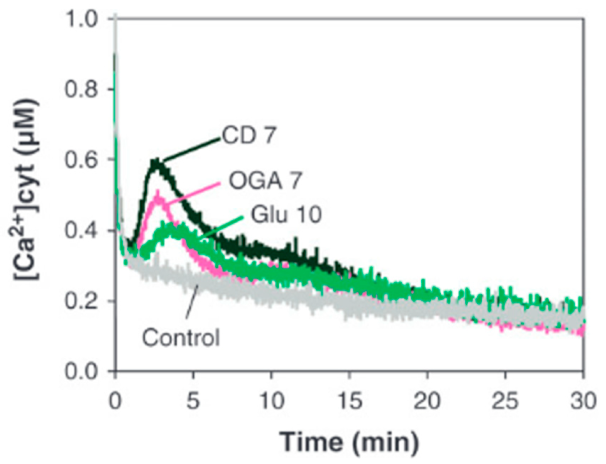
In contrast to symbiosis-associated calcium oscillations, a calcium response to biotic stress typically occurs in the cytoplasm. When grapevine cells were challenged with cellodextrins (CD), a rapid and transient increase in the cytosolic Ca2+ concentration was observed [31]. Figure 1 shows a typical Ca2+ signature, which is characterized by a steep rise in the Ca2+ concentration, followed by a comparably weaker decline, right after the peak has been reached.
Although the cytosolic Ca2+ concentration depends on the deployed elicitor concentration, the Ca2+ response becomes saturated once a specific elicitor concentration is reached. Elicitation of, e.g., apoaequorin-transformed parsley cells with Pep13, induces a Ca2+ response that already becomes saturated at a concentration >5 nM [32]. Obviously, the plant detection system for PAMPs is highly sensitive. Moreover, it shows a very short response time, which is typically below one minute (see also Figure 1; [32]).
After the treatment of transgenic soybean cells with OGs, the cytosolic Ca2+ response also appears to be a rapid and transient event. The degree of polymerization of the OGs has a strong influence on the kinetics of the Ca2+ responses, a higher degree of polymerization results in a higher cytosolic calcium concentration [64]. Dose-dependent treatments with cryptogein (Cry) and OGs were performed in aequorin-transformed N. tabacum cell suspension cultures, inducing a biphasic cytosolic Ca2+ rise, which increases with higher elicitor concentration. Compared to the Cry-induced Ca2+ elevation, N. tabacum cells treated with OGs showed a faster calcium reaction and a faster return to the basal calcium level [79].
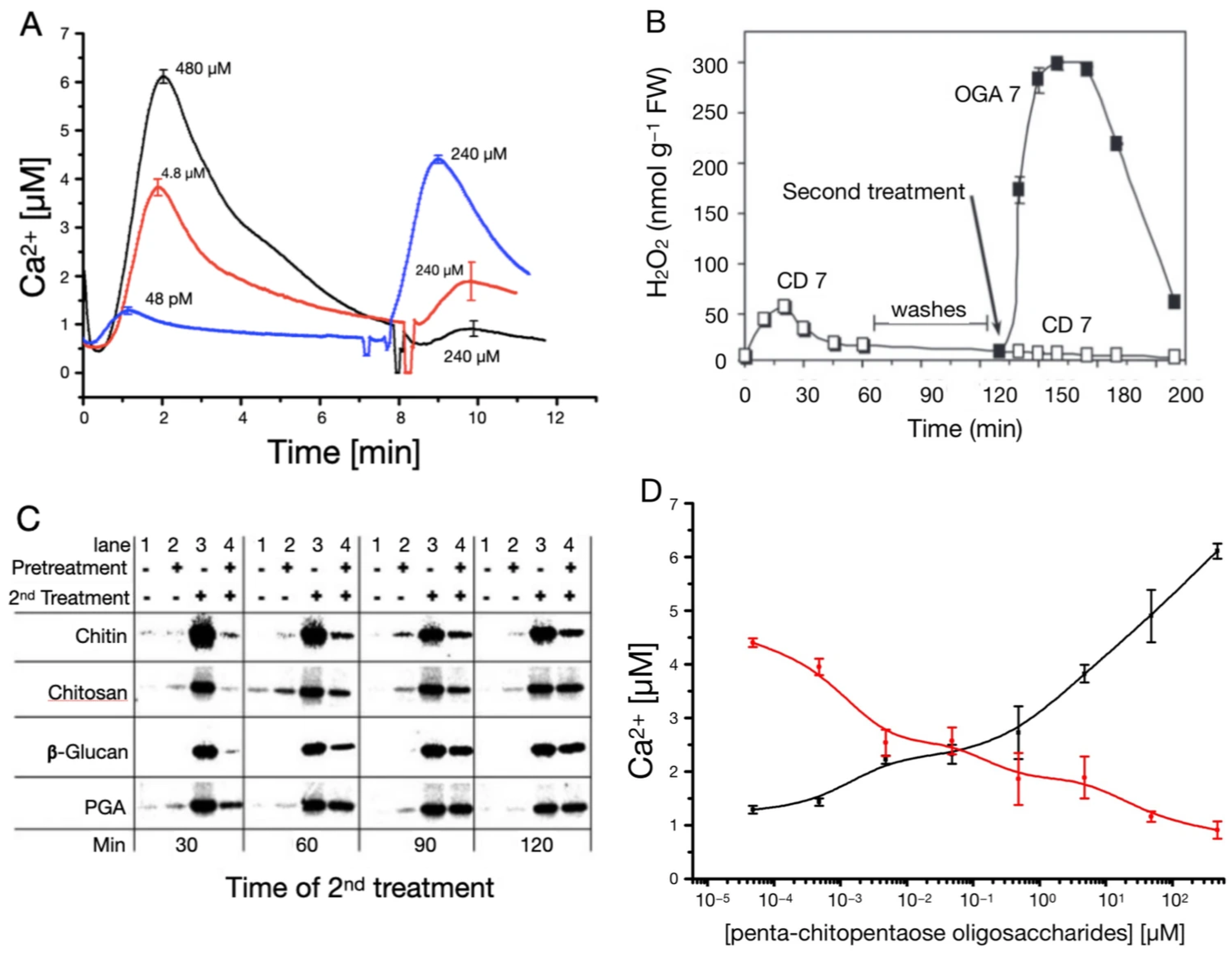
Already in the early 1990s there was evidence of refractory behavior regarding Ca2+ responses, as tomato cells elicited with chitin fragments did not respond to a second stimulus applied shortly after the first one [88]. Obviously, there is a period of time—the refractory period—after a stimulus in which the cells can only respond less or not at all to a stimulus of the same kind. However, throughout this refractory period the tomato cells remain responsive to a different stimulus [58,88]. Even if the elicitor is “washed out” between two elicitations, the cells remain less responsive to the second stimulation. As an example, grapevine cells pretreated with CD (CD 7 at 0.5 mg/mL) did not respond to a second treatment with CD 7 (Figure 2B). However, they responded to an application of a different elicitor type, e.g., OG (OGA 7 at 0.5 mg/mL) [31]. Other oligosaccharide elicitors (OEs: chitosan, chitotetraose, ß-glucan and PGA) were shown to induce a refractory state as well. Given that the receptor type is an essential element of stimulus reception, whereby different stimuli might in rare cases also be received by a common receptor [89], we can speculate that the receptor itself is involved in the refractory behavior of this system.
Refractory behavior can also be observed at the MAPK kinase activity level. MAP kinases can be activated by a Ca2+ response, and their activity reaches its maximum after ten minutes. Once active, further responses are also reduced within a specific time interval. MAPK activity levels obviously recover after 120 min, as can be seen in Figure 2C. After two hours the response to a second treatment is almost as strong as the one induced without pretreatment [90]. Downstream, MAPK cascades act as temporal integrators, preferentially responding to signals of defined duration or persistence and translating transient early events into stable transcriptional reprogramming.
Small GTPases—particularly plant Rho-of-plants (ROP/RAC) proteins—act as central molecular switches that couple stimulus perception with activation, spatial organization, and amplification of the Ca2+-ROS-MAPK network. Rather than functioning as linear intermediates, they provide regulatory control over signal initiation, localization, and intensity. In addition to ROS regulation, small GTPases also influence Ca2+ dynamics itself. By affecting membrane organization, cytoskeletal architecture, and possibly channel positioning, ROPs modulate the spatial patterning of Ca2+ influx and activation of the NADPH-oxidase, which is responsible for the production of ROS, which will influence pathogen as symbiont interactions [91]. This is particularly evident in processes such as tip growth, where localized ROP activity establishes Ca2+ gradients. In immune signaling, similar principles likely apply at the scale of plasma membrane nanodomains, enabling spatially confined ROS and Ca2+ microdomains that shape downstream decoding. More research is needed to integrate the role of plant GTPases and their respective regulatory proteins in the Ca2+-ROS-MAPK network.
On the other hand, it is possible to trigger an increased second response if the level of the first elicitation is sufficiently reduced. The corresponding Ca2+ responses of tobacco cell cultures treated with Ch5 show this behavior in more detail (Figure 2A, ref. [33]). A first elicitation with 48 pM, followed by a second elicitation with 240 µM, leads to a Ca2+ response even bigger than the response to a single stimulation with, e.g., 4.8 µM. When looking at the dose–response curves of the first and second elicitation separately, the first elicitation shows the typical distinct rise, while the second elicitation shows the expected decrease (Figure 2D, ref. [33]). Generally, Figure 2 gives an overview of the refractory behavior of Ca2+ responses, measured in terms of Ca2+ concentration, H2O2 (Figure 2B, ref. [31]) production and MAPK (Figure 2C, ref. [90]) activity. In this context it is noteworthy that all tested PAMP receptors induce MAPK activity (see Figure 2C) through Ca2+ signaling, but show a differential activation of the various MAPK pathways [92].
LPS and cyclic adenosine monophosphate (cAMP) induce a cytosolic Ca2+ elevation in intact leaves of wild type Arabidopsis thaliana, but are absent in leaves lacking cyclic nucleotide-gated cation channels [93]. The plasma membrane cation binding protein 1 (PCaP1) is required for late defense responses to elicitors and for the recovery of full responsiveness to OGs after an elicitor treatment [94].
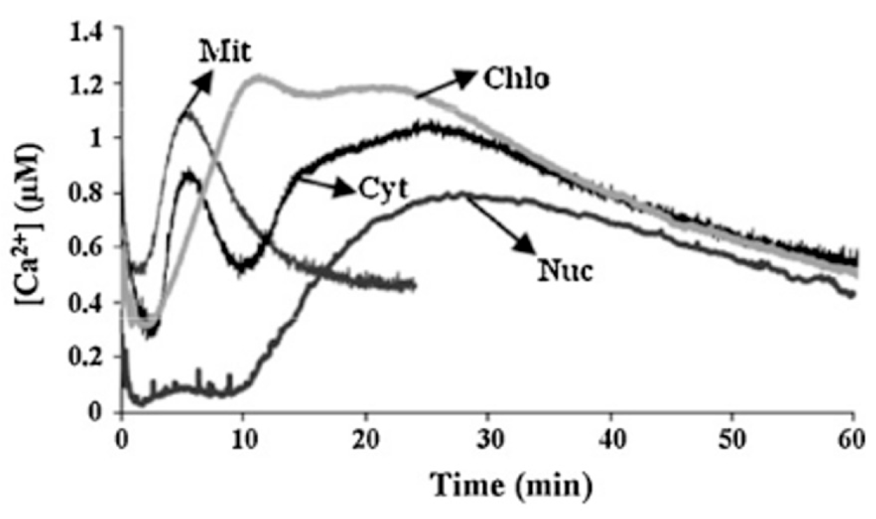
As already stated earlier, the Ca2+ increase in the cytosol is affected by the Ca2+ influx from the apoplast or from other intracellular organelles [20]. But Ca2+ signatures are not exclusively observed in the cytosol. Ca2+ is typically stored in the vacuoles, the ER, the mitochondria and the chloroplasts, and Ca2+ signatures are observed in the cytosol, the chloroplasts, the mitochondria, and the nucleus (Figure 3). Apart from the above-mentioned dose-dependent treatments, the two PAMPs (Cry and OGs) were used to study whether Ca2+ variations could affect important physiological processes in organelles, such as photosynthesis and mitochondrial respiration. Cry, through Ca2+ signaling, causes perturbations in two important organelle functions, namely mitochondrial respiration and chloroplastic energy dissipation. This strengthened the idea that Ca2+ in organelles contributes to plant defense signaling [79].

4. Possible Causes of Refractory Behavior in the Context of Calcium Signaling

A rise in the cytosolic Ca2+ concentration, the so called “Ca2+ answer”, in response to biotic or abiotic stresses, leads to the expression of defense-related genes, ROS production or apoptosis. However, little is known about what triggers the refractory behavior in the context of Ca2+ responses.
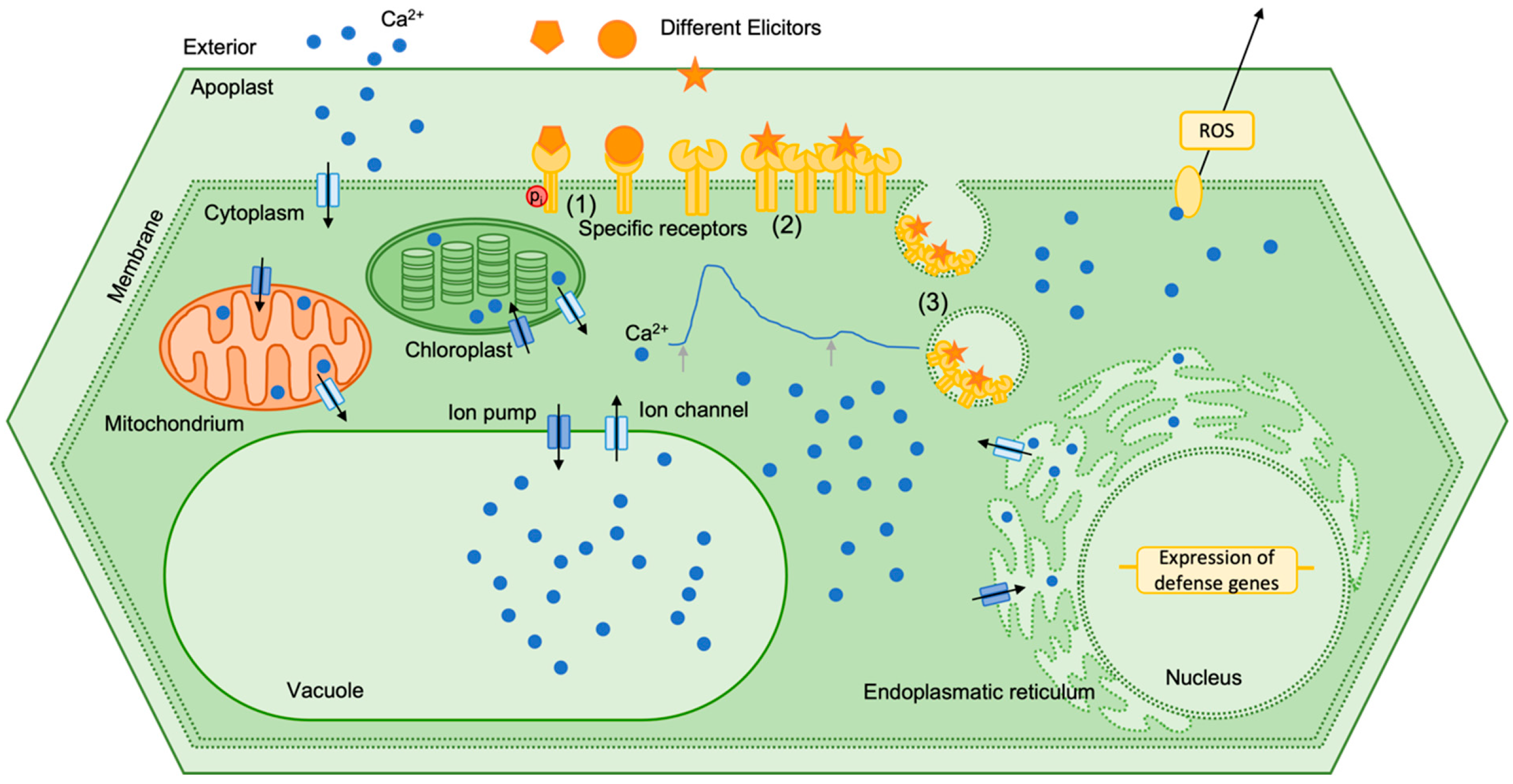
Cytosolic calcium signals are the consequence of a complex perception system that releases calcium from external as well as internal stores. And signal transduction of the cytosolic calcium transient is not less complex. Figure 4 illustrates the journey of the calcium ions to show possible causes of refractory behavior.
Phosphorylation of specific PRRs is one possible causes of refractory behavior [95]. The conformational change can prevent the connection to an elicitor. It could be shown that MAPK are also reversibly phosphorylated and may undergo a refractory state before the next activation [90]. Interestingly, in most studies phosphorylation leads to the activation of PRRs and precedes all responses and intracellular signaling events. Leucine-rich repeat receptor kinases (LRR-RK) located at the plasma membrane in particular play an important role in the perception of extracellular signals. For FLS2 (flagellin-sensitive 2) the interaction with BAK1 (BRI1-associated receptor kinase 1) was shown to be required for signal transduction. After stimulation with flg22 the phosphorylation of the FLS2-BAK1 complex stimulates signal transduction and leads to activated plant defense reactions [96]. Tyrosine phosphorylation in particular plays an important role in the activation of PRRs. The tyrosine kinase inhibitor A23 reduced BIK1 (botrytis-induced kinase 1) phosphorylation and the production of ROS, suggesting that tyrosine phosphorylation regulates immune signaling induced by PAMPs [97]. Not only can PRRs be phosphorylated, but also the phosphorylation of Ca2+ channels could be a possible cause for refractory behavior. Receptor-like cytoplasmatic kinases are able to phosphorylate ion channels to regulate ion transport and early signaling during plant-triggered immunity [98]. The loss of the cyclic nucleotide-gated calcium channels leads to a hypersensitive temperature-dependent Ca2+ influx and a corresponding hyper-thermoresponsive profile of heat shock response activation [99]. In addition to phosphorylation, other modifications of receptors, channels and downstream kinases are also involved. Ubiquitiniation, lipidation, acetylation and SUMOylation are reported protein modifications that can contribute to the refractory behavior of plant cells in response to stress [95].
Another cause for refractory behavior could be receptor clustering. The hypothesis is that receptors somehow interact, so that within a stimulated cluster there are also receptors that are not bound to an elicitor molecule, thereby reducing the number of free receptors for a second stimulus. As an example, the quorum-sensing molecule diffusible signal factor (DSF) impaired the FLS2 nanoclustering on the cell surface, which therefore desensitized Arabidopsis’ immune response to flg22 [100].
A most likely cause for refractory behavior is receptor endocytosis. After the internalization of the receptors an additional elicitation signal cannot be transmitted until new receptors are synthesized. Endocytosis is a well-known mechanism for cellular desensitization, where ligand-bound receptors from the cell surface are removed. In many plant defense studies endocytic processes could be observed. Shortly after elicitation with a labeled elicitor fluorescence in vesicle-like structures is found [45,101,102,103]. A possible role in endocytosis after elicitation is played by PCaP1. PCaP1 is organized in membrane microdomains and is internalized in endocytic vesicles in response to OGs. However, endocytosis is not altered in mutants lacking PCaP1 [94].

5. Refractory Behavior and Defense Priming

There is considerable evidence that cytosolic Ca2+ signatures can be modified by previous exposure to a stimulus. The magnitude of the Ca2+ elevation becomes progressively smaller upon repeated stimulation, and a refractory period of several minutes or even hours is required before a full response is observed again. There is also evidence that the Ca2+ signatures elicited by one environmental challenge can be modified by prior exposure to a contrasting one [84,104]. When primed, plants respond to very low stimulation with a faster and stronger defense than unprimed plants [105].
This suggests that plants can discriminate between a single stress and repeated stresses and can modify their expression of the stress-responsive genes [106]. In Table 2 an overview of responses to biotic and abiotic stresses is shown. Changes in enzyme activity, cytosolic Ca2+ concentration and in metabolite levels may imply defense priming and memory to pathogen contact. By becoming more resistant to harmful stimuli on the one hand and less sensitive to harmless stimuli on the other, plants develop a so-called learning memory [107]. Transcriptional regulation enables plants to adapt to different stress environments [10]. To gain a better understanding of stress memory and priming in plants, changes at the epigenetic, transcriptional, proteomic and physiological level have to be considered [108].
Pathogen-induced Ca2+ signaling is intimately linked to the generation of ROS, underscoring the evolutionary conservation of this defense module. In plants, the NADPH oxidase RbohD functions as a central ROS-producing enzyme, and its activation is at least partly mediated by Ca2+. Notably, both the structural organization of NADPH oxidases and their regulation by Ca2+ are conserved across plants and animals [109], highlighting the ancient origin of this signaling architecture. Consequently, mechanistic insights gained from model species such as Arabidopsis thaliana and Nicotiana tabacum are frequently transferable to agronomically important crop plants [110].
Comparative transcriptome analysis in Brassica napus has provided further support for this concept. In a study contrasting the low aphid-susceptible cultivar “APL01” with the highly susceptible cultivar “Holly” under aphid challenge, genes associated with Ca2+ signaling were identified as differentially regulated in relation to resistance phenotypes [111]. These findings emphasize the functional relevance of Ca2+-dependent pathways in crop defense responses. For the identification of robust marker genes applicable to breeding programs, defense-associated signaling must be understood as an integrated network. In particular, calcium-dependent protein kinases (CDPKs) and MAPKs represent critical nodes linking Ca2+ dynamics to transcriptional reprogramming [112,113]. In addition to the search for a gene-marker-based selection of traits more resistant to stress, small molecules could enable the development of novel agrochemicals. Small molecules such as salicylic acid, jasmonate and other secondary metabolites are involved in signal transduction in plant decision making [114]. A recent study on rice showed the induced plant resilience to stress through the involvement of secondary plant metabolites and calcium signaling [115].
Mechanistically, the refractory phase arises from multilayered negative feedback, including Ca2+-dependent inactivation of Ca2+-permeable channels, receptor desensitization and turnover, depletion and refilling kinetics of intracellular Ca2+ stores, and induction of inhibitory signaling components such as phosphatases and Ca2+-binding regulators. Functionally, refractoriness prevents signal saturation and cytotoxic Ca2+ overload, but more importantly, it provides temporal resolution and encoding capacity to the Ca2+ signaling network.
The systematic identification of central signal transduction hubs within this network will provide a rational basis for breeding crop varieties that are able to respond in a timely and effective manner to pathogen attack.

6. Conclusions and Outlook

In summary, plant immunity is still a very complex phenomenon, although many defense mechanisms are known. Different elicitors, typically made up of molecules such as oligosaccharides, peptides, lipids, and proteins can trigger different plant defense mechanisms. Induced Ca2+ signatures are typically observed in the cytosol, the chloroplasts, the mitochondria, and the nucleus [116]. The calcium responses to biotic stimuli can have different meanings. Oscillating calcium responses are associated with symbiotic signaling pathways while transient calcium responses trigger defense-associated pathways [117]. At the beginning of the defense mechanism cascade a PRR binds to an elicitor. To date only a few receptors are known that interact with elicitor molecules [29]. Work in cell cultures is an excellent tool for analyzing basic mechanisms of Ca2+ signatures, but may fail to understand the reaction of the entire plant. Recent work by Wang et al. [118] shows the complex interplay of receptors, channels, and Ca2+ signatures at the tissue level.
In many studies the calcium responses of different model plants to elicitors are recorded. However, the reactions to a second elicitation with the same elicitor can be of a different type. A refractory period can be observed, i.e., a period of time after a stimulus in which the cells can only respond less or not at all to a stimulus of the same kind. However, during this refractory period cells remain responsive to a different stimulus. This refractory behavior is observed in many plants that are exposed to stress. Calcium levels can be reduced or vanish; however, sometimes it is possible to trigger even an increased second response, which is observed in the context of defense priming or trained or acquired immunity. There is considerable evidence that cytosolic Ca2+ signatures can be modified by previous exposure to a stimulus.
In search of the causes of refractory behavior, phosphorylation of specific PRRs is one possible option. Receptor clustering, which reduces the number of free receptors for a second stimulus, is another plausible cause. Receptor endocytosis is yet another probable reason for a refractory time interval. In addition, experiments with repeated elicitation can help us to find out more about the refractory interval period. Interestingly, Yakushiji et al. studied the influence of bacterial DNA on Arabidopsis thaliana and found that the endocytosis inhibitors wortmannin and amantadine significantly inhibited the plant’s defense response that had been DNA-induced [119].
In the context of the plant immune system, it is important to obtain a clear definition of “stress”. Biotic and abiotic stimuli can act as stressors and trigger defense reactions. The calcium response at the cellular level could be utilized to set a standard measure and make different forms of stress more comparable. In their review on Ca2+ signatures in response to environmental stresses [120] the authors discuss strategies to identify potential Ca2+ channels as stress sensors. In view of the growing number of confirmed calcium channels, Jiang and Ding raise the question of their spatio-temporal function [121].
As priming is a part of the induced resistance that neither significantly alters growth nor fruit or seed set, its importance increases in terms of disease control [122]. The thorough study of the plant immune system, including at the omics level, will support crop development for food safety and security. Changes in the epigenome, transcriptome, proteome and metabolome in reaction to stress can inscribe themselves in the memory of the plants, which enables them to respond to future stress in a more pronounced way [123]. Recent findings by Jia et al. [124] show that deciphering the link between NLR (leucine-rich repeat receptor)-mediated Ca2+ signaling and ROS transport is crucial in plant immunity. The group identified a calcium-dependent protein kinase regulating ROS signaling during effector-triggered immunity. In plants, the relationship between diverse environmental inputs and specific physiological outputs is generated through the dynamic properties of the interconnected Ca2+-ROS-MAPK signaling network rather than through strictly linear signaling pathways. Distinct stimuli such as elicitors, cold shock, or mechanical perturbation activate stimulus-specific receptors or mechanosensitive channels, thereby producing characteristic cytosolic Ca2+ signatures that differ in amplitude, duration, frequency, and spatial distribution. Signal transduction, or—in other words—information processing, is a property of all living cells. Calcium signals are part of a complex and multidimensional network that integrates numerous information to generate an optimal output for the particular cell, organ or individuum. A systems biology approach, including modeling of this network, is needed to better understand this decision-making network [82].

Author Contributions

Conceptualization, M.K.K., V.F., K.N. and P.L.; methodology, M.K.K. and P.L.; investigation, M.K.K., V.F., K.N. and P.L.; resources, M.K.K., V.F., K.N. and P.L.; writing—original draft preparation, M.K.K. and P.L.; writing—review and editing, M.K.K., V.F., K.N. and P.L.; visualization, M.K.K.; supervision, K.N. and P.L.; project administration, P.L. All authors have read and agreed to the published version of the manuscript.

Funding

This research received no external funding.

Data Availability Statement

No new data were created or analyzed in this study.

Acknowledgments

We want to thank Aron Dransfeld, Leonie Lancelle and Andrea Wiegers for valuable additions and comments.

Conflicts of Interest

Author Mareike Kristin Keßler was employed by the company PlasmidFactory GmbH. The remaining authors declare that the research was conducted in the absence of any commercial or financial relationships that could be construed as a potential conflict of interest.

References

  1. Reimer-Michalski, E.-M.; Conrath, U. Innate immune memory in plants. Semin. Immunol. 2016, 28, 319–327. [Google Scholar] [CrossRef]
  2. Ahuja, I.; Kissen, R.; Bones, A.M. Phytoalexins in defense against pathogens. Trends Plant Sci. 2012, 17, 73–90. [Google Scholar] [CrossRef] [PubMed]
  3. Heath, M.C. Nonhost resistance and nonspecific plant defenses. Curr. Opin. Plant Biol. 2000, 3, 315–319. [Google Scholar] [CrossRef] [PubMed]
  4. Lipka, U.; Fuchs, R.; Kuhns, C.; Petutschnig, E.; Lipka, V. Live and let die—Arabidopsis nonhost resistance to powdery mildews. Eur. J. Cell Biol. 2010, 89, 194–199. [Google Scholar] [CrossRef]
  5. Jones, J.D.G.; Dangl, J.L. The plant immune system. Nature 2006, 444, 323–329. [Google Scholar] [CrossRef]
  6. Zipfel, C. Plant pattern-recognition receptors. Trends Immunol. 2014, 35, 345–351. [Google Scholar] [CrossRef]
  7. Boller, T.; He, S.Y. Innate Immunity in Plants: An Arms Race Between Pattern Recognition Receptors in Plants and Effectors in Microbial Pathogens. Science 2009, 324, 742–744. [Google Scholar] [CrossRef]
  8. Mahalingam, R.; Fedoroff, N. Stress response, cell death and signalling: The many faces of reactive oxygen species. Physiol. Plant. 2003, 119, 56–68. [Google Scholar] [CrossRef]
  9. Mao, G.; Meng, X.; Liu, Y.; Zheng, Z.; Chen, Z.; Zhang, S. Phosphorylation of a WRKY Transcription Factor by Two Pathogen-Responsive MAPKs Drives Phytoalexin Biosynthesis in Arabidopsis. Plant Cell 2011, 23, 1639–1653. [Google Scholar] [CrossRef]
  10. Wani, S.H.; Anand, S.; Singh, B.; Bohra, A.; Joshi, R. WRKY transcription factors and plant defense responses: Latest discoveries and future prospects. Plant Cell Rep. 2021, 40, 1071–1085. [Google Scholar] [CrossRef] [PubMed]
  11. van der Meer, J.W.M.; Joosten, L.A.B.; Riksen, N.; Netea, M.G. Trained immunity: A smart way to enhance innate immune defence. Mol. Immunol. 2015, 68, 40–44. [Google Scholar] [CrossRef] [PubMed]
  12. Henikoff, S.; Shilatifard, A. Histone modification: Cause or cog? Trends Genet. 2011, 27, 389–396. [Google Scholar] [CrossRef]
  13. Badeaux, A.I.; Shi, Y. Emerging roles for chromatin as a signal integration and storage platform. Nat. Rev. Mol. Cell Biol. 2013, 14, 211–224. [Google Scholar] [CrossRef] [PubMed]
  14. Roudier, F.; Ahmed, I.; Bérard, C.; Sarazin, A.; Mary-Huard, T.; Cortijo, S.; Bouyer, D.; Caillieux, E.; Duvernois-Berthet, E.; Al-Shikhley, L.; et al. Integrative epigenomic mapping defines four main chromatin states in Arabidopsis: Organization of the Arabidopsis epigenome. EMBO J. 2011, 30, 1928–1938. [Google Scholar] [CrossRef] [PubMed]
  15. Schwämmle, V.; Aspalter, C.-M.; Sidoli, S.; Jensen, O.N. Large Scale Analysis of Co-existing Post-translational Modifications in Histone Tails Reveals Global Fine Structure of Cross-talk. Mol. Cell. Proteom. 2014, 13, 1855–1865. [Google Scholar] [CrossRef]
  16. Conrath, U.; Beckers, G.J.M.; Langenbach, C.J.G.; Jaskiewicz, M.R. Priming for Enhanced Defense. Annu. Rev. Phytopathol. 2015, 53, 97–119. [Google Scholar] [CrossRef] [PubMed]
  17. Bruce, T.J.A.; Matthes, M.C.; Napier, J.A.; Pickett, J.A. Stressful “memories” of plants: Evidence and possible mechanisms. Plant Sci. 2007, 173, 603–608. [Google Scholar] [CrossRef]
  18. Tateda, C.; Zhang, Z.; Shrestha, J.; Jelenska, J.; Chinchilla, D.; Greenberg, J.T. Salicylic Acid Regulates Arabidopsis Microbial Pattern Receptor Kinase Levels and Signaling. Plant Cell 2014, 26, 4171–4187. [Google Scholar] [CrossRef]
  19. Robatzek, S.; Wirthmueller, L. Mapping FLS2 function to structure: LRRs, kinase and its working bits. Protoplasma 2013, 250, 671–681. [Google Scholar] [CrossRef]
  20. Tuteja, N.; Mahajan, S. Calcium Signaling Network in Plants: An Overview. Plant Signal. Behav. 2007, 2, 79–85. [Google Scholar] [CrossRef]
  21. Boller, T. Chemoperception of Microbial Signals in Plant Cells. Annu. Rev. Plant. Physiol. Plant. Mol. Biol. 1995, 46, 189–214. [Google Scholar] [CrossRef]
  22. Sanabria, N.M.; Huang, J.-C.; Dubery, I.A. Self/non-self perception in plants in innate immunity and defense. Self/Nonself 2010, 1, 40–54. [Google Scholar] [CrossRef]
  23. Nürnberger, T.; Scheel, D. Signal transmission in the plant immune response. Trends Plant Sci. 2001, 6, 372–379. [Google Scholar] [CrossRef]
  24. Ren, H.; Zhao, X.; Li, W.; Hussain, J.; Qi, G.; Liu, S. Calcium Signaling in Plant Programmed Cell Death. Cells 2021, 10, 1089. [Google Scholar] [CrossRef] [PubMed]
  25. Cramer, G.R.; Urano, K.; Delrot, S.; Pezzotti, M.; Shinozaki, K. Effects of abiotic stress on plants: A systems biology perspective. BMC Plant Biol. 2011, 11, 163. [Google Scholar] [CrossRef]
  26. Zhu, J.-K. Abiotic Stress Signaling and Responses in Plants. Cell 2016, 167, 313–324. [Google Scholar] [CrossRef] [PubMed]
  27. Zhang, Y.; Xu, J.; Li, R.; Ge, Y.; Li, Y.; Li, R. Plants’ Response to Abiotic Stress: Mechanisms and Strategies. Int. J. Mol. Sci. 2023, 24, 10915. [Google Scholar] [CrossRef]
  28. Ravi, B.; Foyer, C.H.; Pandey, G.K. The integration of reactive oxygen species (ROS) and calcium signalling in abiotic stress responses. Plant Cell Environ. 2023, 46, 1985–2006. [Google Scholar] [CrossRef] [PubMed]
  29. Malik, N.A.A.; Kumar, I.S.; Nadarajah, K. Elicitor and Receptor Molecules: Orchestrators of Plant Defense and Immunity. Int. J. Mol. Sci. 2020, 21, 963. [Google Scholar] [CrossRef]
  30. Ebel, J.; Ayers, A.R.; Albersheim, P. Host-Pathogen Interactions: XII. Response of Suspension-cultured Soybean Cells to the Elicitor Isolated from Phytophthora megasperma var. sojae, a Fungal Pathogen of Soybeans. Plant Physiol. 1976, 57, 775–779. [Google Scholar] [CrossRef] [PubMed]
  31. Aziz, A.; Gauthier, A.; Bezier, A.; Poinssot, B.; Joubert, J.-M.; Pugin, A.; Heyraud, A.; Baillieul, F. Elicitor and resistance-inducing activities of -1,4 cellodextrins in grapevine, comparison with -1,3 glucans and -1,4 oligogalacturonides. J. Exp. Bot. 2007, 58, 1463–1472. [Google Scholar] [CrossRef]
  32. Blume, B.; Nürnberger, T.; Nass, N.; Scheel, D. Receptor-Mediated Increase in Cytoplasmic Free Calcium Required for Activation of Pathogen Defense in Parsley. Plant Cell 2000, 12, 1425–1440. [Google Scholar] [CrossRef] [PubMed]
  33. Mrozek, K.; Niehaus, K.; Lutter, P. Experimental Measurements and Mathematical Modeling of Cytosolic Ca2+ Signatures upon Elicitation by Penta-N-acetylchitopentaose Oligosaccharides in Nicotiana tabacum Cell Cultures. Plants 2013, 2, 750–768. [Google Scholar] [CrossRef]
  34. Müller, J.; Staehelin, C.; Xie, Z.-P.; Neuhaus-Url, G.; Boller, T. Nod Factors and Chitooligomers Elicit an Increase in Cytosolic Calcium in Aequorin-Expressing Soybean Cells. Plant Physiol. 2000, 124, 733–740. [Google Scholar] [CrossRef] [PubMed][Green Version]
  35. Yuan, P.; Luo, F.; Gleason, C.; Poovaiah, B.W. Calcium/calmodulin-mediated microbial symbiotic interactions in plants. Front. Plant Sci. 2022, 13, 984909. [Google Scholar] [CrossRef]
  36. Erickson, F.L.; Dinesh-Kumar, S.P.; Holzberg, S.; Ustach, C.V.; Dutton, M.; Handley, V.; Corr, C.; Baker, B.J. Interactions between tobacco mosaic virus and the tobacco N gene. Philos. Trans. R. Soc. Lond. B 1999, 354, 653–658. [Google Scholar] [CrossRef] [PubMed]
  37. Gómez-Gómez, L.; Boller, T. FLS2. Mol. Cell 2000, 5, 1003–1011. [Google Scholar] [CrossRef]
  38. Zipfel, C.; Robatzek, S.; Navarro, L.; Oakeley, E.J.; Jones, J.D.G.; Felix, G.; Boller, T. Bacterial disease resistance in Arabidopsis through flagellin perception. Nature 2004, 428, 764–767. [Google Scholar] [CrossRef]
  39. Tanaka, K.; Nguyen, C.T.; Liang, Y.; Cao, Y.; Stacey, G. Role of LysM receptors in chitin-triggered plant innate immunity. Plant Signal. Behav. 2013, 8, e22598. [Google Scholar] [CrossRef]
  40. Johnson, J.M.; Thürich, J.; Petutschnig, E.K.; Altschmied, L.; Meichsner, D.; Sherameti, I.; Dindas, J.; Mrozinska, A.; Paetz, C.; Scholz, S.S.; et al. A Poly(A) Ribonuclease Controls the Cellotriose-Based Interaction between Piriformospora indica and Its Host Arabidopsis. Plant Physiol. 2018, 176, 2496–2514. [Google Scholar] [CrossRef]
  41. Priya Reddy, Y.N.; Johnson, J.M.; Oelmüller, R. A cell wall extract of a Fusarium incarnatum strain requires the mitochondrial poly(A)-specific ribonuclease AtPARN for inducing cytoplasmic calcium elevation in Arabidopsis roots. Physiol. Mol. Biol. Plants 2025, 31, 851–861. [Google Scholar] [CrossRef]
  42. Gandhi, A.; Reichelt, M.; Furch, A.; Mithöfer, A.; Oelmüller, R. Cellooligomer/Cellooligomer Receptor Kinase1 Signaling Exhibits Crosstalk with PAMP-Triggered Immune Responses and Sugar Metabolism in Arabidopsis Roots. Int. J. Mol. Sci. 2024, 25, 3472. [Google Scholar] [CrossRef] [PubMed]
  43. Meyer, A.; Pühler, A.; Niehaus, K. The lipopolysaccharides of the phytopathogen Xanthomonas campestris pv. campestris induce an oxidative burst reaction in cell cultures of Nicotiana tabacum. Planta 2001, 213, 214–222. [Google Scholar] [CrossRef] [PubMed]
  44. Braun, S.G.; Meyer, A.; Holst, O.; Pühler, A.; Niehaus, K. Characterization of the Xanthomonas campestris pv. campestris Lipopolysaccharide Substructures Essential for Elicitation of an Oxidative Burst in Tobacco Cells. Mol. Plant Microbe Interact. 2005, 18, 674–681. [Google Scholar] [CrossRef]
  45. Gross, A.; Kapp, D.; Nielsen, T.; Niehaus, K. Endocytosis of Xanthomonas campestris pathovar campestris lipopolysaccharides in non-host plant cells of Nicotiana tabacum. New Phytol. 2005, 165, 215–226. [Google Scholar] [CrossRef]
  46. Ranf, S.; Gisch, N.; Schäffer, M.; Illig, T.; Westphal, L.; Knirel, Y.A.; Sánchez-Carballo, P.M.; Zähringer, U.; Hückelhoven, R.; Lee, J.; et al. A lectin S-domain receptor kinase mediates lipopolysaccharide sensing in Arabidopsis thaliana. Nat. Immunol. 2015, 16, 426–433. [Google Scholar] [CrossRef] [PubMed]
  47. Offor, B.C.; Mhlongo, M.I.; Dubery, I.A.; Piater, L.A. Plasma Membrane-Associated Proteins Identified in Arabidopsis Wild Type, lbr2-2 and bak1-4 Mutants Treated with LPSs from Pseudomonas syringae and Xanthomonas campestris. Membranes 2022, 12, 606. [Google Scholar] [CrossRef]
  48. Pei, Y.; Zhao, Y.; Wang, H.; Guo, Y.; Gu, X.; Lv, J.; Guo, Z.; Chen, Y.; Ren, Y.; Ren, Y.; et al. Uncovering Convergent Pattern Recognition Receptors Recognising Phytophthora Across Plant Lineages. Plant Biotechnol. J. 2026, 24, 1098–1109. [Google Scholar] [CrossRef] [PubMed]
  49. Nürnberger, T.; Lipka, V. Non-host resistance in plants: New insights into an old phenomenon. Mol. Plant Pathol. 2005, 6, 335–345. [Google Scholar] [CrossRef]
  50. Koga, J.; Yamauchi, T.; Shimura, M.; Ogawa, N.; Oshima, K.; Umemura, K.; Kikuchi, M.; Ogasawara, N. Cerebrosides A and C, Sphingolipid Elicitors of Hypersensitive Cell Death and Phytoalexin Accumulation in Rice Plants. J. Biol. Chem. 1998, 273, 31985–31991. [Google Scholar] [CrossRef] [PubMed]
  51. Wan, J.; Tanaka, K.; Zhang, X.-C.; Son, G.H.; Brechenmacher, L.; Nguyen, T.H.N.; Stacey, G. LYK4, a Lysin Motif Receptor-Like Kinase, Is Important for Chitin Signaling and Plant Innate Immunity in Arabidopsis. Plant Physiol. 2012, 160, 396–406. [Google Scholar] [CrossRef]
  52. Kaku, H.; Nishizawa, Y.; Ishii-Minami, N.; Akimoto-Tomiyama, C.; Dohmae, N.; Takio, K.; Minami, E.; Shibuya, N. Plant cells recognize chitin fragments for defense signaling through a plasma membrane receptor. Proc. Natl. Acad. Sci. USA 2006, 103, 11086–11091. [Google Scholar] [CrossRef]
  53. Miya, A.; Albert, P.; Shinya, T.; Desaki, Y.; Ichimura, K.; Shirasu, K.; Narusaka, Y.; Kawakami, N.; Kaku, H.; Shibuya, N. CERK1, a LysM receptor kinase, is essential for chitin elicitor signaling in Arabidopsis. Proc. Natl. Acad. Sci. USA 2007, 104, 19613–19618. [Google Scholar] [CrossRef]
  54. Shinya, T.; Motoyama, N.; Ikeda, A.; Wada, M.; Kamiya, K.; Hayafune, M.; Kaku, H.; Shibuya, N. Functional Characterization of CEBiP and CERK1 Homologs in Arabidopsis and Rice Reveals the Presence of Different Chitin Receptor Systems in Plants. Plant Cell Physiol. 2012, 53, 1696–1706. [Google Scholar] [CrossRef]
  55. Wang, L.; Albert, M.; Einig, E.; Fürst, U.; Krust, D.; Felix, G. The pattern-recognition receptor CORE of Solanaceae detects bacterial cold-shock protein. Nat. Plants 2016, 2, 16185. [Google Scholar] [CrossRef]
  56. Ricci, P.; Bonnet, P.; Huet, J.-C.; Sallantin, M.; Beauvais-Cante, F.; Bruneteau, M.; Billard, V.; Michel, G.; Pernollet, J.-C. Structure and activity of proteins from pathogenic fungi Phytophthora eliciting necrosis and acquired resistance in tobacco. Eur. J. Biochem. 1989, 183, 555–563. [Google Scholar] [CrossRef]
  57. Zipfel, C.; Kunze, G.; Chinchilla, D.; Caniard, A.; Jones, J.D.G.; Boller, T.; Felix, G. Perception of the Bacterial PAMP EF-Tu by the Receptor EFR Restricts Agrobacterium-Mediated Transformation. Cell 2006, 125, 749–760. [Google Scholar] [CrossRef]
  58. Granado, J.; Felix, G.; Boller, T. Perception of Fungal Sterols in Plants (Subnanomolar Concentrations of Ergosterol Elicit Extracellular Alkalinization in Tomato Cells). Plant Physiol. 1995, 107, 485–490. [Google Scholar] [CrossRef]
  59. Lee, J.; Klessig, D.F.; Nürnberger, T. A Harpin Binding Site in Tobacco Plasma Membranes Mediates Activation of the Pathogenesis-Related Gene HIN1 Independent of Extracellular Calcium but Dependent on Mitogen-Activated Protein Kinase Activity. Plant Cell 2001, 13, 1079–1093. [Google Scholar] [CrossRef]
  60. Basse, C.W.; Fath, A.; Boller, T. High affinity binding of a glycopeptide elicitor to tomato cells and microsomal membranes and displacement by specific glycan suppressors. J. Biol. Chem. 1993, 268, 14724–14731. [Google Scholar] [CrossRef]
  61. Osman, H.; Vauthrin, S.; Mikes, V.; Milat, M.-L.; Panabières, F.; Marais, A.; Brunie, S.; Maume, B.; Ponchet, M.; Blein, J.-P. Mediation of Elicitin Activity on Tobacco Is Assumed by Elicitin-Sterol Complexes. Mol. Biol. Cell 2001, 12, 2825–2834. [Google Scholar] [CrossRef] [PubMed][Green Version]
  62. Bailey, B.A.; Dean, J.F.D.; Anderson, J.D. An Ethylene Biosynthesis-Inducing Endoxylanase Elicits Electrolyte Leakage and Necrosis in Nicotiana tabacum cv Xanthi Leaves. Plant Physiol. 1990, 94, 1849–1854. [Google Scholar] [CrossRef] [PubMed]
  63. Montesano, M.; Kõiv, V.; Mäe, A.; Palva, E.T. Novel receptor-like protein kinases induced by Erwinia carotovora and short oligogalacturonides in potato: E. carotovora induced receptor kinase in potato. Mol. Plant Pathol. 2001, 2, 339–346. [Google Scholar] [CrossRef] [PubMed]
  64. Navazio, L.; Moscatiello, R.; Bellincampi, D.; Baldan, B.; Meggio, F.; Brini, M.; Bowler, C.; Mariani, P. The role of calcium in oligogalacturonide-activated signalling in soybean cells. Planta 2002, 215, 596–605. [Google Scholar] [CrossRef]
  65. Gravino, M.; Locci, F.; Tundo, S.; Cervone, F.; Savatin, D.V.; De Lorenzo, G. Immune responses induced by oligogalacturonides are differentially affected by AvrPto and loss of BAK1/BKK1 and PEPR1/PEPR2: Differential RLK requirement for OG responses. Mol. Plant Pathol. 2017, 18, 582–595. [Google Scholar] [CrossRef]
  66. Marschner, H. Marschner’s Mineral Nutrition of Higher Plants; Elsevier: Amsterdam, The Netherlands, 1995. [Google Scholar]
  67. Wang, C.; Luan, S. Calcium homeostasis and signaling in plant immunity. Curr. Opin. Plant Biol. 2024, 77, 102485. [Google Scholar] [CrossRef]
  68. Hirschi, K. Vacuolar H+/Ca2+ transport: Who’s directing the traffic? Trends Plant Sci. 2001, 6, 100–104. [Google Scholar] [CrossRef]
  69. Sze, H.; Liang, F.; Hwang, I.; Curran, A.C.; Harper, J.F. Diversity and Regulation of Plant Ca2+ PUMPS: Insights from Expression in Yeast. Annu. Rev. Plant. Physiol. Plant. Mol. Biol. 2000, 51, 433–462. [Google Scholar] [CrossRef]
  70. Wang, C.; Tang, R.-J.; Kou, S.; Xu, X.; Lu, Y.; Rauscher, K.; Voelker, A.; Luan, S. Mechanisms of calcium homeostasis orchestrate plant growth and immunity. Nature 2024, 627, 382–388. [Google Scholar] [CrossRef]
  71. Laxalt, A.M.; Van Hooren, M.; Munnik, T. Plant PI-PLC signaling in stress and development. Plant Physiol. 2025, 197, kiae534. [Google Scholar] [CrossRef]
  72. White, P.J. Calcium channels in higher plants. Biochim. Biophys. Acta (BBA) Biomembr. 2000, 1465, 171–189. [Google Scholar] [CrossRef]
  73. Świeżawska-Boniecka, B.; Duszyn, M.; Kwiatkowski, M.; Szmidt-Jaworska, A.; Jaworski, K. Cross Talk Between Cyclic Nucleotides and Calcium Signaling Pathways in Plants–Achievements and Prospects. Front. Plant Sci. 2021, 12, 643560. [Google Scholar] [CrossRef]
  74. Park, C.-J.; Shin, R. Calcium channels and transporters: Roles in response to biotic and abiotic stresses. Front. Plant Sci. 2022, 13, 964059. [Google Scholar] [CrossRef]
  75. Logan, D.C.; Knight, M.R. Mitochondrial and Cytosolic Calcium Dynamics Are Differentially Regulated in Plants. Plant Physiol. 2003, 133, 21–24. [Google Scholar] [CrossRef]
  76. Johnson, C.H.; Knight, M.R.; Kondo, T.; Masson, P.; Sedbrook, J.; Haley, A.; Trewavas, A. Circadian Oscillations of Cytosolic and Chloroplastic Free Calcium in Plants. Science 1995, 269, 1863–1865. [Google Scholar] [CrossRef]
  77. Lecourieux, D.; Lamotte, O.; Bourque, S.; Wendehenne, D.; Mazars, C.; Ranjeva, R.; Pugin, A. Proteinaceous and oligosaccharidic elicitors induce different calcium signatures in the nucleus of tobacco cells. Cell Calcium 2005, 38, 527–538. [Google Scholar] [CrossRef]
  78. Mazars, C.; Thuleau, P.; Lamotte, O.; Bourque, S. Cross-Talk between ROS and Calcium in Regulation of Nuclear Activities. Mol. Plant 2010, 3, 706–718. [Google Scholar] [CrossRef]
  79. Manzoor, H.; Chiltz, A.; Madani, S.; Vatsa, P.; Schoefs, B.; Pugin, A.; Garcia-Brugger, A. Calcium signatures and signaling in cytosol and organelles of tobacco cells induced by plant defense elicitors. Cell Calcium 2012, 51, 434–444. [Google Scholar] [CrossRef]
  80. Marchant, J.S.; Taylor, C.W. Cooperative activation of IP3 receptors by sequential binding of IP3 and Ca2+ safeguards against spontaneous activity. Curr. Biol. 1997, 7, 510–518. [Google Scholar] [CrossRef]
  81. Thillaiappan, N.B.; Chakraborty, P.; Hasan, G.; Taylor, C.W. IP3 receptors and Ca2+ entry. Biochim. Biophys. Acta (BBA) Mol. Cell Res. 2019, 1866, 1092–1100. [Google Scholar] [CrossRef]
  82. Bray, D. Intracellular signalling as a parallel distributed process. J. Theor. Biol. 1990, 143, 215–231. [Google Scholar] [CrossRef]
  83. Vadassery, J.; Oelmüller, R. Calcium signaling in pathogenic and beneficial plant microbe interactions: What can we learn from the interaction between Piriformospora indica and Arabidopsis thaliana. Plant Signal. Behav. 2009, 4, 1024–1027. [Google Scholar] [CrossRef]
  84. White, P.J. Calcium in Plants. Ann. Bot. 2003, 92, 487–511. [Google Scholar] [CrossRef] [PubMed]
  85. Trewavas, A. Le Calcium, C’est la Vie: Calcium Makes Waves1. Plant Physiol. 1999, 120, 1–6. [Google Scholar] [CrossRef] [PubMed]
  86. Oldroyd, G.E.D. Speak, friend, and enter: Signalling systems that promote beneficial symbiotic associations in plants. Nat. Rev. Microbiol. 2013, 11, 252–263. [Google Scholar] [CrossRef] [PubMed]
  87. Granqvist, E.; Sun, J.; Op den Camp, R.; Pujic, P.; Hill, L.; Normand, P.; Morris, R.J.; Downie, J.A.; Geurts, R.; Oldroyd, G.E.D. Bacterial-induced calcium oscillations are common to nitrogen-fixing associations of nodulating legumes and non-legumes. New Phytol. 2015, 207, 551–558. [Google Scholar] [CrossRef] [PubMed]
  88. Felix, G.; Regenass, M.; Boller, T. Specific perception of subnanomolar concentrations of chitin fragments by tomato cells: Induction of extracellular alkalinization, changes in protein phosphorylation, and establishment of a refractory state. Plant J. 1993, 4, 307–316. [Google Scholar] [CrossRef]
  89. Héloir, M.-C.; Adrian, M.; Brulé, D.; Claverie, J.; Cordelier, S.; Daire, X.; Dorey, S.; Gauthier, A.; Lemaître-Guillier, C.; Negrel, J.; et al. Recognition of Elicitors in Grapevine: From MAMP and DAMP Perception to Induced Resistance. Front. Plant Sci. 2019, 10, 1117. [Google Scholar] [CrossRef] [PubMed]
  90. Holley, S.R.; Yalamanchili, R.D.; Moura, D.S.; Ryan, C.A.; Stratmann, J.W. Convergence of Signaling Pathways Induced by Systemin, Oligosaccharide Elicitors, and Ultraviolet-B Radiation at the Level of Mitogen-Activated Protein Kinases in Lycopersicon peruvianum Suspension-Cultured Cells. Plant Physiol. 2003, 132, 1728–1738. [Google Scholar] [CrossRef]
  91. Kiirika, L.M.; Bergmann, H.F.; Schikowsky, C.; Wimmer, D.; Korte, J.; Schmitz, U.; Niehaus, K.; Colditz, F. Silencing of the Rac1 GTPase MtROP9 in Medicago truncatula Stimulates Early Mycorrhizal and Oomycete Root Colonizations But Negatively Affects Rhizobial Infection. Plant Physiol. 2012, 159, 501–516. [Google Scholar] [CrossRef]
  92. Cardinale, F.; Jonak, C.; Ligterink, W.; Niehaus, K.; Boller, T.; Hirt, H. Differential activation of four specific MAPK pathways by distinct elicitors. J. Biol. Chem. 2000, 275, 36734–36740. [Google Scholar] [CrossRef] [PubMed]
  93. Ma, W.; Qi, Z.; Smigel, A.; Walker, R.K.; Verma, R.; Berkowitz, G.A. Ca2+, cAMP, and transduction of non-self perception during plant immune responses. Proc. Natl. Acad. Sci. USA 2009, 106, 20995–21000. [Google Scholar] [CrossRef] [PubMed]
  94. Giovannoni, M.; Marti, L.; Ferrari, S.; Tanaka-Takada, N.; Maeshima, M.; Ott, T.; De Lorenzo, G.; Mattei, B. The plasma membrane–associated Ca 2+-binding protein, PCAP1, is required for oligogalacturonide and flagellin-induced priming and immunity. Plant Cell Environ. 2021, 44, 3078–3093. [Google Scholar] [CrossRef] [PubMed]
  95. Zhong, C.; Wang, X.; Li, Y.; Dinesh-Kumar, S.P.; Zhang, Y. Regulation of plant NLRs by post-translational modifications. Trends Biochem. Sci. 2026, 51, 51–63. [Google Scholar] [CrossRef]
  96. Schulze, B.; Mentzel, T.; Jehle, A.K.; Mueller, K.; Beeler, S.; Boller, T.; Felix, G.; Chinchilla, D. Rapid Heteromerization and Phosphorylation of Ligand-activated Plant Transmembrane Receptors and Their Associated Kinase BAK1. J. Biol. Chem. 2010, 285, 9444–9451. [Google Scholar] [CrossRef]
  97. Macho, A.P.; Schwessinger, B.; Ntoukakis, V.; Brutus, A.; Segonzac, C.; Roy, S.; Kadota, Y.; Oh, M.-H.; Sklenar, J.; Derbyshire, P.; et al. A Bacterial Tyrosine Phosphatase Inhibits Plant Pattern Recognition Receptor Activation. Science 2014, 343, 1509–1512. [Google Scholar] [CrossRef]
  98. Sun, L.; Zhang, J. Regulatory role of receptor-like cytoplasmic kinases in early immune signaling events in plants. FEMS Microbiol. Rev. 2020, 44, 845–856. [Google Scholar] [CrossRef]
  99. Finka, A.; Cuendet, A.F.H.; Maathuis, F.J.M.; Saidi, Y.; Goloubinoff, P. Plasma Membrane Cyclic Nucleotide Gated Calcium Channels Control Land Plant Thermal Sensing and Acquired Thermotolerance. Plant Cell 2012, 24, 3333–3348. [Google Scholar] [CrossRef]
  100. Tran, T.M.; Ma, Z.; Triebl, A.; Nath, S.; Cheng, Y.; Gong, B.-Q.; Han, X.; Wang, J.; Li, J.-F.; Wenk, M.R.; et al. The bacterial quorum sensing signal DSF hijacks Arabidopsis thaliana sterol biosynthesis to suppress plant innate immunity. Life Sci. Alliance 2020, 3, e202000720. [Google Scholar] [CrossRef]
  101. Leborgne-Castel, N.; Lherminier, J.; Der, C.; Fromentin, J.; Houot, V.; Simon-Plas, F. The Plant Defense Elicitor Cryptogein Stimulates Clathrin-Mediated Endocytosis Correlated with Reactive Oxygen Species Production in Bright Yellow-2 Tobacco Cells. Plant Physiol. 2008, 146, 1255–1266. [Google Scholar] [CrossRef]
  102. Mbengue, M.; Bourdais, G.; Gervasi, F.; Beck, M.; Zhou, J.; Spallek, T.; Bartels, S.; Boller, T.; Ueda, T.; Kuhn, H.; et al. Clathrin-dependent endocytosis is required for immunity mediated by pattern recognition receptor kinases. Proc. Natl. Acad. Sci. USA 2016, 113, 11034–11039. [Google Scholar] [CrossRef] [PubMed]
  103. Erwig, J.; Ghareeb, H.; Kopischke, M.; Hacke, R.; Matei, A.; Petutschnig, E.; Lipka, V. Chitin-induced and chitin elicitor receptor kinase1 (CERK1) phosphorylation-dependent endocytosis of Arabidopsis thaliana lysin motif-containing receptor-like kinase5 (LYK5). New Phytol. 2017, 215, 382–396. [Google Scholar] [CrossRef] [PubMed]
  104. Knight, H. Calcium Signaling during Abiotic Stress in Plants. In International Review of Cytology; Elsevier: Amsterdam, The Netherlands, 1999; Volume 195, pp. 269–324. [Google Scholar]
  105. Levine, A.; Pennell, R.I.; Alvarez, M.E.; Palmer, R.; Lamb, C. Calcium-mediated apoptosis in a plant hypersensitive disease resistance response. Curr. Biol. 1996, 6, 427–437. [Google Scholar] [CrossRef] [PubMed]
  106. Hilker, M.; Schmülling, T. Stress priming, memory, and signalling in plants: Stress priming, memory, and signalling in plants. Plant Cell Environ. 2019, 42, 753–761. [Google Scholar] [CrossRef]
  107. Thellier, M.; Lüttge, U. Plant memory: A tentative model. Plant Biol. 2013, 15, 1–12. [Google Scholar] [CrossRef]
  108. Liu, X.; Quan, W.; Bartels, D. Stress memory responses and seed priming correlate with drought tolerance in plants: An overview. Planta 2022, 255, 45. [Google Scholar] [CrossRef]
  109. Canton, J.; Grinstein, S. Priming and activation of NADPH oxidases in plants and animals. Trends Immunol. 2014, 35, 405–407. [Google Scholar] [CrossRef]
  110. Zhang, C.; Song, Y.; Kudla, J. Calcium signaling in crops. New Phytol. 2026, 249, 1644–1658. [Google Scholar] [CrossRef]
  111. Li, Y.; Cai, L.; Ding, T.; Tian, E.; Yan, X.; Wang, X.; Zhang, J.; Yu, K.; Chen, Z. Comparative Transcriptome Analysis Reveals the Molecular Basis of Brassica napus in Response to Aphid Stress. Plants 2023, 12, 2855. [Google Scholar] [CrossRef]
  112. Deng, L.; Ji, S.; Wang, G.; Liu, X. Calcium-dependent protein kinases in plant immunity: From calcium signaling to network integration. Front. Plant Sci. 2026, 16, 1704615. [Google Scholar] [CrossRef]
  113. Azupio, S.; Wang, Y.; Obeng, J.; Issah, A.K.; Chu, J.; Zhang, H.; Xie, Q.; Jiang, X. Calcium-dependent protein kinases as central hubs in plant abiotic stress signaling: Mechanisms and prospects for crop improvement. Front. Plant Sci. 2026, 16, 1711405. [Google Scholar] [CrossRef]
  114. Tian, L.; Hossbach, B.M.; Feussner, I. Small size, big impact: Small molecules in plant systemic immune signaling. Curr. Opin. Plant Biol. 2024, 81, 102618. [Google Scholar] [CrossRef]
  115. Ikram, M.; Batool, M.; Ullah, M.; Khalid, B.; El-Badri, A.M.; Mohamed, I.A.A.; Zhang, L.; Kuai, J.; Xu, Z.; Zhao, J.; et al. Molecular Alchemy: Converting Stress into Resilience via Secondary Metabolites and Calcium Signaling in Rice. Rice 2025, 18, 32. [Google Scholar] [CrossRef]
  116. Steiner, P.; Zierler, S. Inter-Organellar Ca2+ Homeostasis in Plant and Animal Systems. Cells 2025, 14, 1204. [Google Scholar] [CrossRef]
  117. Jacott, C.N.; Del Cerro, P. CNGC15 and DMI1 ion channel gating in nuclear calcium signaling: Opening new questions and closing controversies. J. Exp. Bot. 2024, 75, 6998–7005. [Google Scholar] [CrossRef]
  118. Wang, H.; Li, X.; Zhai, K.; Rhodes, J.; Sang, T.; Zhao, J.; Gao, Y.; Ma, S.; Song, B.; Pan, Q.; et al. A regulatory network promotes apoplastic alkalinization to prime plant immunity in tissues distal to site of infection. Cell 2026, 189, 1389–1406.e19. [Google Scholar] [CrossRef]
  119. Yakushiji, S.; Ishiga, Y.; Inagaki, Y.; Toyoda, K.; Shiraishi, T.; Ichinose, Y. Bacterial DNA activates immunity in Arabidopsis thaliana. J. Gen. Plant Pathol. 2009, 75, 227–234. [Google Scholar] [CrossRef]
  120. Lee, H.-J.; Seo, P.J. Ca2+ talyzing Initial Responses to Environmental Stresses. Trends Plant Sci. 2021, 26, 849–870. [Google Scholar] [CrossRef]
  121. Jiang, Y.; Ding, P. Calcium signaling in plant immunity: A spatiotemporally controlled symphony. Trends Plant Sci. 2023, 28, 74–89. [Google Scholar] [CrossRef]
  122. Conrath, U. Chapter 9 Priming of Induced Plant Defense Responses. In Advances in Botanical Research; Elsevier: Amsterdam, The Netherlands, 2009; Volume 51, pp. 361–395. [Google Scholar]
  123. Kambona, C.M.; Koua, P.A.; Léon, J.; Ballvora, A. Stress memory and its regulation in plants experiencing recurrent drought conditions. Theor. Appl. Genet. 2023, 136, 26. [Google Scholar] [CrossRef]
  124. Jia, Z.; Yu, W.; Guo, X.; Li, J.; Qi, F.; Zhu, Y.; Cai, X.; Li, X.; Li, N.; Dong, H.; et al. CPK12 decodes effector-triggered calcium signaling and phosphorylates PIP2;1 to facilitate apoplastic ROS transport into the cytoplasm in Arabidopsis. Mol. Plant 2025, 18, 1724–1741. [Google Scholar] [CrossRef] [PubMed]
Figure 1. Change in cytosolic Ca2+ concentration in aequorin-transformed grapevine cells after treatment with β-1,4 cellodextrins (CD 7, 0.5 mg/mL), β-1,3 glucans (Glu 10, 0.5 mg/mL) or α-1,4 oligogalacturonides (OGA 7, 0.5 mg/mL). The same volume of water was used as a control. Figure modified from [31].
Plants 15 01395 g001
Figure 2. Refractory behavior of Ca2+ signaling measured in terms of cytosolic Ca2+ concentration, H2O2 production and MAPK activity. (A) Tobacco cell cultures were elicited with 480 μM, 4.8 μM and 48 pM of Ch5. Approximately 7 min later, a second elicitation with a Ch5 stimulus of 240 μM was carried out. (B) H2O2 production in grapevine cell suspensions after successive addition of CD 7 and OGA 7, demonstrating refractory behavior. Cells were first treated at time 0 with 0.5 mg/mL CD 7, washed three times with fresh medium within the second hour after first treatment, then treated a second time with 0.5 mg/mL CD 7 (open squares) or with 0.5 mg/mL OGA 7 (black squares), respectively. (C) MAPK activity in reaction to successive oligosaccharide elicitor (OE) treatments. L. peruvianum suspension-cultured cells were pretreated with the OEs chitin (100 µM chitotetraose), chitosan (1.7 µg/mL), ß-glucan (10 µg/mL), or polygalacturonic acid (PGA, 830 µg/mL), or were left untreated. At 30, 60, 90, and 120 min thereafter, treated and untreated cells were either left untreated or were exposed to the same OE a second time. (D) Dose–response diagram for first (black curve) and second (red curve) elicitations. Ch5 concentration of the first elicitation was increased, while the second elicitation was constantly held at 240 µM. Ca2+ responses were set in relation to stimulus level of first stimulation. Modified from [31] (B), ref. [33] (A,D), ref. [90] (C).
Plants 15 01395 g002
Figure 3. Ca2+ responses induced by Cry (100 nM) in the cytosol (Cyt), chloroplasts (Chlo), mitochondria (Mit) and nucleus (Nuc). Figure modified from [79].
Plants 15 01395 g003
Figure 4. Model for refractory behavior in calcium signaling. Different PAMPs acting as elicitors are detected by surface-located PRRs. Perception of the signal leads to an influx of calcium ions through different calcium channels, resulting in an induced cytosolic calcium concentration. Thus, different defense responses are triggered, like, e.g., ROS production or expression of defense-related genes. After the event calcium is removed from the cytosol via specific calcium pumps. Numbers indicate different possibilities that are responsible for refractory behavior: (1) phosphorylation of PRRs leads to conformational changes and disables interaction with elicitor molecules, (2) clustering of receptors reduces the possible binding sites and reduces interaction between PRR and PAMP molecules, and (3) receptor endocytosis removes the PPR with bound elicitor molecules from the cell surface; the plant cell cannot respond to a new stimulus until new PRRs are synthesized and brought to the cell surface.
Plants 15 01395 g004
Table 1. Overview of the most common pathogen-associated molecular patterns (PAMPs) with known receptors and biological responses. Modified from [22,49].
PAMP (and Pathogens)Known Receptor GenesBiological ResponseReference
Cellotriose (Fungi, Piriformospora indica)Not identified
(n.i)
production of ROS, changes in membrane potential, expression of genes involved in growth regulation and root development[40]
Cerebrosides A,C (Fungi, Magnaporthe spp.)n.i.phytoalexin production in rice[50]
Chitin/chitosan, Ch5 (Fungi)CEBIP, CERK1 (LYK1, RLK1), LYM2, LYK4perception of chitin oligosaccharide elicitor for defense responses[39,51,52,53,54]
Cold shock protein (Gram-negative bacteria, Gram-positive bacteria)Cold shock protein receptor (CORE)oxidative burst[55]
Cryptogein (Fungi, Phytophthora cryptogea)n.i.induces local and distal defense responses, elicits leaf necrosis[56]
Elongation factor (EF-Tu) (Gram-negative bacteria)EFRdetermines the specific perception of EF-Tu[57]
Ergosterol (Fungi)n.i.induces ion fluxes in tomato[58]
Flagellin (flg22) (Gram-negative bacteria)FLS2flagellin-binding initiates the innate immune MAP kinase signaling cascade, resulting in enhanced resistance against pathogens; activates a downstream MAPK pathway[37,38]
Harpin (HrpZ) (Gram-negative bacteria)n.i.HR-like cell death, induces defense responses in various plants[59]
Invertase (Yeast)n.i.activation of the phenylpropanoid pathway, ethylene production in tomato[60]
Lipid-transfer proteins (Elicitins) (Oomycetes Phytophthora spp., Pythium spp.)n.i.HR-like cell death, induces defense responses in tobacco, systemic acquired resistance to microbial infection[61]
LPS (Gram-negative bacteria)n.i.oxidative burst[43]
Necrosis-inducing proteins (Bacteria Bacillus spp., fungi Fusarium spp., oomycetes Phytophthora spp., Pythium spp.)n.i.HR-like cell death[62]
OGs (Bacteria E. carotovora)Potato receptor-like kinase (PRK1-4)induce a rise in cytosolic calcium concentration[63,64]
Pep-13 (Glycoprotein) (Oomycete Phytophthora sojae)PEPR1, PEPR2senses an endogenous elicitor that potentiates PAMP-inducible plant responses[32,65]
TMV replicase (Protein of the Tobacco Mosaic Virus)n.i.senses the TMV replicase and induces rapid cell death in tobacco[36]
Table 2. List of effects and outcomes in response to abiotic and biotic stresses. Modified from [106].
TriggerEffectOutcome
Abiotic stress (e.g., UV radiation, water,
temperature, salinity)
Changes in DNA methylation/chromatin patternImproved stress management/memory
Biotic stress (PAMPs, MAMPs)Accumulation of transcription factorsForgetting (e.g., autophagy)
 Changing phytohormone/
metabolite levels
 
 Post-translational modifications 
Disclaimer/Publisher’s Note: The statements, opinions and data contained in all publications are solely those of the individual author(s) and contributor(s) and not of MDPI and/or the editor(s). MDPI and/or the editor(s) disclaim responsibility for any injury to people or property resulting from any ideas, methods, instructions or products referred to in the content.